Submitted:
28 March 2026
Posted:
30 March 2026
You are already at the latest version
Abstract
Paper-based electrochemical diagnostics have emerged as promising point-of-care (POC) platforms for rapid, low-cost analysis of biofluids such as blood, urine, and saliva. Compared to colorimetric assays, electrochemical detection enables more objective and quantitative readouts while preserving portability and capillary-driven operation. This review summarizes recent advances in integrating electrochemical sensing with paper-based platforms, including microfluidic paper-based analytical devices and lateral flow systems, and their use in techniques such as voltammetry and impedance spectroscopy. Key challenges to reliable performance in real samples are identified, particularly biological matrix effects and miniaturization constraints. Variability in biofluid composition, including hematocrit, protein fouling, ionic strength, and viscosity, can disrupt mass transport, alter conductivity, and reduce signal stability. These effects are amplified in miniaturized systems, where small volumes and reduced electrode areas increase susceptibility to noise and drift. Engineering strategies to address these challenges include antifouling surface modifications, integrated sample conditioning, electrode optimization, redox-mediated signal enhancement, and matrix-aware calibration. Overall, future progress depends on integrated device designs that ensure reproducibility and reliability in real-world samples.
Keywords:
paper-based diagnostics
; electrochemical biosensors
; biofluid analysis
; point-of-care testing
; biosensing
; microfluidics
; clinical diagnostics
; sensor miniaturization
; signal stability
; low-cost diagnostics
Introduction
Rapid diagnostic testing is essential for timely clinical decision-making, yet conventional laboratory testing often requires centralized processing and 24–48-hour turnaround times, delaying treatment and triage [1]. Point-of-care (POC) diagnostics address this by enabling bedside testing and improving access, particularly in rural settings [1]. Blood, urine, and saliva are key diagnostic biofluids, reflecting systemic physiology and disease states, with urine and saliva offering non-invasive sampling and blood serving as the quantitative reference standard [2]. Paper-based analytical devices provide low-cost, portable platforms using capillary-driven flow, but traditional colorimetric formats are limited by visual subjectivity and poor quantitative accuracy. These limitations have driven interest in electrochemical detection methods, which offer more objective and quantitative analysis for complex biofluids [3].
This mini review focuses on paper-based diagnostics that use electrochemical readouts to quantify analytes in blood, urine, and saliva. These platforms support POC applications due to their accessibility, cost-effectiveness, ease of use, disposability, and capillary-driven transport [3]. However, most existing paper-based tests rely on colorimetric readouts, which are susceptible to user interpretation and lighting conditions. In contrast, electrochemical detection enables compact, objective, and quantitative analysis using low-cost potentiostats [3]. This review examines how paper-based microfluidics and electrode integration enable electrochemical assays, while highlighting two key constraints: (1) biological matrix effects from complex biofluids (e.g., pH, ionic strength, viscosity), and (2) miniaturization constraints associated with microliter-scale volumes, capillary-driven transport, evaporation, and surface interactions. The literature is organized by paper format (e.g., μPADs, strips, lateral flow) and electrochemical readout (amperometry, voltammetry, EIS), with emphasis on failure modes and mitigation strategies such as filtration, dilution, surface modification, and built-in controls. Recent advances in μPADs, screen-printed electrodes (SPEs), and lateral-flow electrochemical systems have improved scalability, reproducibility, and quantitative performance [4,5,6]. However, performance in real biofluids remains limited by matrix-related effects. Protein fouling, ionic variability, and hematocrit disrupt transport, conductivity, and calibration, while small channel dimensions further limit mass transport and amplify variability [7,8,9,10,11]. Miniaturization also reduces electrode area, increasing noise and signal drift if not properly engineered [9]. These combined factors increase the risk of inaccurate results and complicate the sensitivity–specificity balance required for clinical translation [14].
This mini review makes three primary contributions to paper-based electrochemical diagnostics for clinical applications: (1) synthesizing biological matrix effects across biofluids, (2) explaining how miniaturization amplifies interference and variability, and (3) consolidating engineering strategies that improve robustness and support clinical translation. Despite progress, the literature remains fragmented across materials engineering, electroanalytical optimization, and biofluid-specific interference mitigation, with limited integration of how these factors interact in real-world systems. While many studies demonstrate proof-of-concept performance under controlled conditions, few evaluate reliability in clinically relevant matrices where biological variability and miniaturization constraints directly affect signal stability [10]. As portable potentiostats and disposable platforms become more accessible, translation toward real-world deployment is accelerating. However, incomplete understanding of how biofluid composition and device scaling influence performance continues to limit reproducible clinical accuracy. Therefore, a structured review linking biological matrix effects with platform-level constraints is needed.
Paper-Based Diagnostic Platforms
Microfluidic Paper-Based Analytical Devices (µPADs)
Microfluidic paper-based analytical devices (μPADs) are widely studied paper-based platforms that provide cost-effective and portable architectures for POC diagnostics [11]. They operate through capillary-driven flow within porous paper, where hydrophobic patterning guides fluid transport without external power sources. When a liquid sample is introduced, it flows through the paper matrix via surface tension–driven capillary action, enabling analytes to reach defined detection zones. Control over pore size and surface treatment allows μPADs to perform complex assays within compact formats [12]. These devices typically include a sample inlet, microfluidic channels, and reagent-functionalized detection zones. Advanced designs incorporate 3D configurations for vertical flow and multiplexing, enabling simultaneous detection of multiple biomarkers [13]. Compared to traditional lab-on-a-chip systems, μPADs offer advantages in cost, fabrication simplicity, and accessibility [14]. Fabrication methods such as wax printing, screen printing, and inkjet deposition support rapid prototyping and scalable production without specialized facilities [12]. As a result, μPADs are well-suited for POC diagnostics in resource-limited settings, where affordability, portability, and minimal user training are essential [5]. They support multiple detection modalities, including colorimetric, fluorescence, and electrochemical readouts, making them versatile platforms for on-site clinical biofluid analysis [15].
Paper Strips and Lateral-Flow Formats
Paper strips and lateral-flow formats are widely used diagnostic platforms due to their ease of use, low cost, rapid analysis, and minimal invasiveness [11]. Lateral flow assays (LFAs) consist of porous components on a backing card, including the sample pad, conjugate pad, reaction membrane, and absorbent pad. The reaction membrane, typically nitrocellulose, supports immobilization of capture reagents at test and control lines. Upon sample introduction, fluid migrates through the strip via capillary action, transporting analytes to these regions where binding generates a detectable signal. LFAs require small sample volumes and provide near-instant results, making them attractive for POC applications. They are applicable to a wide range of biofluids, including blood, urine, and saliva, and typically provide qualitative or semi-quantitative readouts through visible test lines. This strip-based format is appealing due to its low cost, portability, and scalability. However, conventional LFAs are often limited to binary or semi-quantitative outputs and may exhibit reduced sensitivity for low-abundance biomarkers in complex samples. Recent efforts have focused on improving strip materials, capture chemistry, signal amplification, and reader integration to enable more sensitive and quantitative diagnostics [11].
Capillary-Driven Flow Principles
The primary transport mechanism in paper-based diagnostics is capillary flow, driven by the intrinsic porosity and surface energy of cellulose membranes [16]. When a sample is deposited, adhesive interactions between the liquid and cellulose fibers, combined with narrow pore structures, generate capillary pressure that drives fluid movement. Flow in paper is commonly described by the Lucas–Washburn model, where distance traveled is proportional to the square root of time, resulting in rapid initial flow that gradually slows. Capillary flow depends on material and design parameters, including pore size, paper thickness, fiber density, fluid viscosity, surface tension, contact angle, and channel geometry. More hydrophilic substrates or smaller pore sizes increase capillary pressure, while more viscous samples, such as whole blood, reduce flow rate. This behavior governs sample distribution, reagent rehydration, mixing, and reaction time. In lateral-flow strips, continuous wicking transports analytes across conjugate, test, and control regions [16]. In μPADs, hydrophobic barriers confine flow to defined pathways, enabling controlled transport to multiple reaction zones or multilayer structures. As a result, capillary flow is both a transport mechanism and a key design factor influencing assay speed, sensitivity, and reproducibility [13].
Integration of Electrodes into Paper
The integration of electrodes into paper based devices enables quantitative electrochemical readouts beyond qualitative detection [17]. Paper supports the electrochemical cell, allowing analyte detection within the paper network while enabling passive fluid handling in compact devices for point of care applications. A common approach is fabricating working, reference, and counter electrodes directly onto paper using techniques such as screen printing or conductive inks. Alternatively, electrodes can be prepared on separate substrates and integrated with the paper fluidic layer to form hybrid structures with improved stability. Screen printing is widely used due to its simplicity, scalability, and compatibility with carbon and metal inks. Paper can also be modified with conductive additives or nanostructured materials to enhance electron transfer and sensitivity. However, electrode integration introduces challenges, as the porous and heterogeneous structure of paper can lead to non uniform deposition, affecting reproducibility, conductivity, and signal stability. Capillary flow can further complicate control over reagent distribution and interfacial electrochemistry. Despite these limitations, electrode integration remains central to paper based electrochemical diagnostics and point of care biosensing [17].
Why Paper Is Ideal for POC Diagnostics
Paper has emerged as an ideal substrate for POC diagnostics due to its unique combination of physical, chemical, and practical advantages [18]. Its intrinsic capillary action enables passive fluid transport without external pumps, while its low cost and compatibility with fabrication techniques such as wax and screen printing support large-scale production in resource-limited settings. The porous cellulose structure provides high surface area for immobilization of biomolecules such as enzymes, antibodies, and aptamers, enabling pre-loaded, ready-to-use assays with minimal user steps. Paper also supports multiple detection modalities, including colorimetric, fluorescence, and electrochemical readouts, with electrochemical methods offering improved sensitivity and quantitative capability. Its lightweight, portable, and disposable nature aligns well with POC requirements. Although challenges such as variability in material properties and environmental sensitivity remain, ongoing advances in material engineering and device integration continue to improve performance and reliability [18].
Electrochemical Detection methods
Voltammetry
Voltammetry is an electroanalytical technique in which current is measured as a function of applied potential, enabling detection of electroactive species [19]. A controlled voltage is applied at the working electrode relative to a reference electrode, driving redox reactions at the electrode surface. The resulting current is proportional to analyte concentration and electrochemical behavior, producing a voltammogram that provides both qualitative and quantitative information. This response arises from electron transfer between the electrode and analyte during oxidation or reduction, with current magnitude reflecting electron transfer rate and analyte concentration under controlled conditions. Several voltammetric techniques exist, differing in how potential is applied. Cyclic voltammetry (CV) is widely used, where potential is swept forward and reversed to characterize redox behavior and reversibility. Other methods, such as linear sweep and square-wave voltammetry, offer improved sensitivity and signal resolution. When integrated with paper substrates and printed electrodes, these techniques enable low-cost POC devices for detecting clinically relevant biomarkers in complex biofluids such as blood, urine, and saliva [19].
Electrochemical Impedance Spectroscopy (EIS)
Electrochemical impedance spectroscopy (EIS) is an electroanalytical technique used to study electrode analyte interactions by measuring impedance over a range of frequencies [20]. Unlike voltammetry, which measures current as a function of applied potential, EIS evaluates frequency dependent impedance, enabling analysis of double layer effects, diffusion, and binding events. A small sinusoidal perturbation is applied, and the current response is used to determine impedance magnitude and phase, providing insight into interfacial electrical and physicochemical properties. When biomolecules such as antibodies or aptamers are immobilized on an electrode, target binding alters surface properties and changes charge transfer resistance, allowing label free detection of biomolecular interactions. EIS is particularly useful in paper based diagnostics and point of care systems due to its simplicity and low reagent use. Data are typically presented as Nyquist and Bode plots and interpreted using equivalent circuit models that link spectral features to interfacial processes. In paper based systems, EIS enables sensitive, quantitative, and low volume analysis and can be integrated with printed electrodes to detect biomarkers in complex biofluids such as blood, urine, and saliva. Despite more complex data interpretation, EIS remains a powerful tool for label free biosensing [20].
Electrochemical Sensing Principles and Readout
Electrochemical sensing converts biochemical interactions into measurable electrical signals. A typical system includes a recognition element, such as an antibody immobilized on a working electrode. Target binding alters interfacial properties, producing changes in current or impedance through electron transfer, either directly from the analyte or via redox active labels, with signal magnitude proportional to analyte concentration [21]. Compared to colorimetric methods, electrochemical sensing provides quantitative output, lower detection limits, and reduced sensitivity to sample colour or turbidity, making it suitable for complex biofluids such as blood and saliva. These systems can also be miniaturized and integrated with compact electronics for point of care use. Advances in portable potentiostats and smartphone based readers have improved practicality. Modern devices support techniques such as voltammetry and impedance spectroscopy while maintaining low power and small form factors. Integration with smartphones enables real time data acquisition, processing, and display, improving accessibility and usability. As a result, electrochemical sensing platforms are increasingly used for rapid and reliable diagnostics outside laboratory settings [21].
Figure 1.
Schematic of an electrochemical paper-based analytical device illustrating signal generation, localized detection, and portable readout integration. Adapted from [23].
Figure 1.
Schematic of an electrochemical paper-based analytical device illustrating signal generation, localized detection, and portable readout integration. Adapted from [23].


Table 1.
PAPER-BASED DIAGNOSTIC STRATEGIES AND THEIR APPLICATIONS. Adapted from [24].
Table 1.
PAPER-BASED DIAGNOSTIC STRATEGIES AND THEIR APPLICATIONS. Adapted from [24].
| Sensing Strategy | Application |
| Colorimetric detection | |
| Paper-based ELISA | Exosome detection and isolation |
| pH change-based colorimetric PADs | Chronic kidney disease biomarker detection |
| Electrochemical Detection | |
| Origami PAD, enzymatic mediated reaction | Wound infection monitoring |
| Paper-based electrochemical platform | Detection of copper ions |
| Fluorescence Detection | |
| Fluorescent PAD | Histidine detection |
| LFA fluorescent assay | Rheumatoid arthritis antibodies detection |
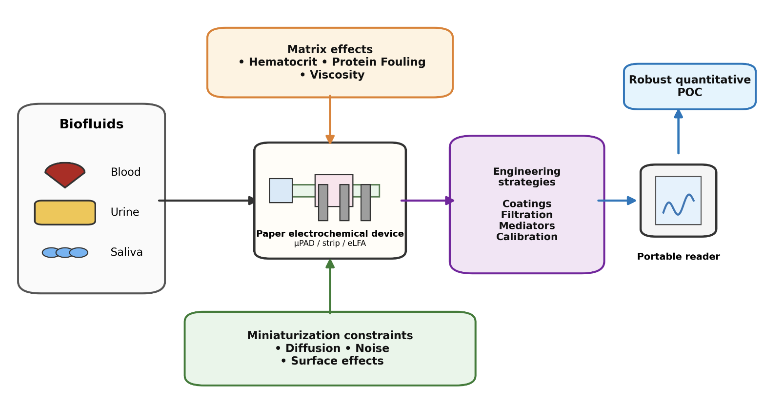
Biological Matrix Effects in Biofluids
Biological matrix effects are a critical consideration in paper-based electrochemical diagnostics, as real biofluids introduce complexities that influence signal behavior. Unlike controlled laboratory conditions, samples such as blood, urine, and saliva contain interfering components, including cells, proteins, electrolytes, and metabolites, which affect signal generation through mass transport limitations, nonspecific adsorption, and changes in conductivity. These factors reduce sensitivity, accuracy, and reproducibility, particularly in minimally processed POC samples [2,4]. Among biofluids, blood presents one of the most complex matrices. A key factor is hematocrit, the proportion of red blood cells in whole blood. Variations in hematocrit increase viscosity and restrict analyte transport to the electrode surface, reducing signal intensity and increasing variability. This effect is amplified in paper-based systems, where fluid transport is already limited, and diffusion is strongly hematocrit-dependent [25]. Additionally, blood proteins such as albumin and fibrinogen can adsorb onto electrode surfaces, causing fouling. This forms an insulating layer that blocks active sites, impedes electron transfer, reduces sensitivity, and contributes to signal drift over time, remaining a major limitation for electrochemical biosensors, especially in continuous or repeated measurements [10,26].
In contrast to blood, urine is a less complex matrix in terms of cellular content but still presents challenges due to variability in ionic strength. Its composition varies with hydration, diet, and metabolic state, leading to fluctuations in electrolyte concentrations such as sodium chloride (NaCl), which affect solution conductivity and electrochemical measurements. Changes in ionic strength alter impedance, current response, and signal stability, resulting in inconsistent sensor output if uncorrected [27]. In paper-based devices, where signal depends on solution resistance and ion transport, this variability must be addressed through calibration or normalization. Urine metabolites such as urea and creatinine may also contribute to background signals or interfere with sensing mechanisms. Saliva is attractive due to its non-invasive collection but introduces matrix-related challenges. Its viscosity, influenced by mucins and glycoproteins, can slow capillary flow and limit analyte delivery, affecting assay timing and reproducibility. Fluid transport in porous materials is strongly dependent on viscosity and wettability [8]. Additionally, saliva contains proteins and enzymes that can cause surface fouling and interfere with electrochemical reactions, impacting sensor stability and long-term performance. Thus, saliva-based biosensors must be designed to mitigate these effects while maintaining sensitivity and specificity. Across all biofluids, matrix effects influence electrochemical diagnostics through three primary mechanisms: mass transport limitations, surface interactions (fouling), and electrolyte-dependent conductivity changes. These effects are particularly pronounced in paper-based devices, which prioritize simplicity, portability, and low cost, but remain sensitive to variations in sample composition [6]. As a result, successful implementation requires consideration of biofluid-specific properties alongside engineering strategies such as surface modification, optimized device architecture, and sample preprocessing. Advances in electrochemical paper-based analytical devices and lateral flow systems continue to improve robustness and enable more reliable detection in complex biological environments [7].
Miniaturization Constraints in Paper-Based Electrochemical Devices
Despite advances in paper-based electrochemical platforms, their effectiveness in real biofluids remains limited by matrix interference and miniaturization constraints [28]. While electrochemical detection improves quantification compared to colorimetric assays by reducing visual subjectivity and colour interference, complex biofluids introduce variability that is amplified at small scales [28]. In paper-based systems, miniaturization confines fluid transport to microliter volumes within porous, capillary-driven networks, affecting mass transport, surface interactions, and signal stability [7,8,9]. These challenges cannot be addressed through simple scaling of conventional systems. A major limitation is restricted mass transport [8]. In paper-based microfluidics, analyte movement relies on capillary flow and diffusion rather than convection, resulting in slower and less uniform transport. At smaller length scales, increased liquid–solid interactions raise flow resistance and reduce analyte mobility [8]. Additionally, higher hematocrit decreases diffusion rates due to red blood cell obstruction, an effect amplified in small pore structures [25]. Miniaturization also reduces electrode surface area and current output, making signals more susceptible to electrical noise and baseline fluctuations, thereby impacting diagnostic accuracy [10]. Electrolyte composition further influences signal behavior; for example, increasing NaCl concentration decreases impedance and increases conductivity, altering electrochemical response [27]. In miniaturized systems, these effects introduce significant variability. Evaporation within porous substrates can further destabilize signals by altering local concentrations over time [11,14].
Another critical limitation is biofouling. As device dimensions decrease, surface effects dominate and high surface to volume ratios increase biomolecular adsorption [26]. Protein adsorption blocks active electrode sites and reduces electrochemical signals. Saxena et al. showed that protein rich samples decreased current, while zwitterionic coatings reduced adsorption by about 67 percent and improved signal retention [26]. In miniaturized systems, fouling is intensified by limited electrode surface area and porous paper structures that trap proteins, creating localized fouling. Miniaturization also reduces current output and increases susceptibility to noise and drift, lowering reliability [9]. Engineering strategies such as high surface area electrodes and electrochemical lateral flow assays can improve signal response and quantification, but performance remains sensitive to matrix and transport limitations [7,9]. Overall, miniaturization amplifies challenges including restricted mass transport, signal instability, and surface interactions. Variability in protein content, ionic strength, and hematocrit further complicates calibration and increases error near detection limits [14]. Therefore, integrated engineering strategies are required to maintain sensitivity, specificity, and reliability in real world applications.
Engineering Strategies for Robust Diagnostics
Engineering strategies are essential for improving the reliability of paper based electrochemical diagnostics in real biofluids. Although these devices provide quantitative readouts, performance is limited by protein fouling, ionic variability, hematocrit related transport effects, and signal instability in miniaturized systems [7,22]. Accordingly, recent work has focused on practical design strategies that enable robust operation in clinically relevant samples [5,23]. A key approach is surface modification to reduce biofouling. Proteins and macromolecules adsorb onto electrode surfaces, blocking active sites and reducing electron transfer, sensitivity, and reproducibility. These effects are amplified in paper based systems due to high surface area to volume ratios. Zwitterionic antifouling coatings have been shown to reduce protein adsorption and preserve performance in complex fluids, supporting sensing in unprocessed samples [26]. Another strategy is sample conditioning through dilution and on chip filtration. Variations in viscosity, ionic composition, and cellular content can distort transport and electrochemical response. Controlled dilution reduces matrix effects and stabilizes calibration, while filtration removes particulates or separates plasma prior to detection. Microfluidic paper based devices enable integrated preprocessing through passive flow and fluid handling. Together, these approaches improve assay reliability and reproducibility in complex samples [4,29]. Another strategy involves redox mediators and electrode engineering to enhance signal output. Miniaturization reduces electrode area and current, increasing susceptibility to noise and drift, but improving interfacial electron transfer can compensate for these losses. High surface area electrodes with superwetting and aptamer functionalization increase active area and signal response, while electrochemical lateral flow assays improve quantification through signal amplification [7,9]. Calibration adapted to matrix variability is also critical. Changes in salt concentration, conductivity, and sample composition can shift baseline current and impedance, making buffer based calibration unreliable in clinical samples. This is especially relevant in urine and saliva, where ionic strength varies widely, and in miniaturized systems where small shifts produce large relative errors. NaCl concentration has been shown to significantly alter impedance, reinforcing the need for matrix aware calibration [27]. Robust devices should incorporate internal references, built in controls, or adaptive calibration rather than relying solely on buffer based methods [4,27]. Overall, integrating antifouling, filtration, dilution, signal enhancement, and calibration strategies is essential to maintain sensitivity, specificity, and reproducibility in complex samples and support reliable clinical translation [5,8,23].
Conclusion and Future Outlook
Although paper-based electrochemical diagnostics have advanced rapidly, several barriers still limit clinical translation. While many devices perform well in simplified samples, accuracy often declines in real biofluids due to proteins, ionic variability, viscosity differences, and cellular content that affect transport and electrochemical behavior [4,29]. Future progress must address matrix variability, improve reproducibility, and overcome regulatory challenges while integrating portable technologies such as smartphone-based potentiostats. A key priority is achieving robust performance in minimally processed clinical samples, requiring antifouling interfaces, improved fluid control, and matrix-aware calibration that accounts for variations in hematocrit, ionic strength, and protein content [4,30]. Integrated sample preparation is also critical, including on-chip filtration, plasma separation, and controlled dilution, which can reduce user error and improve reproducibility while maintaining low-cost, portable designs [29]. The field is also advancing toward more versatile and connected systems, with smartphone-integrated platforms and electrochemical lateral flow assays enabling quantitative, decentralized testing [7]. Paper-based electrochemical diagnostics offer a low-cost, portable approach for point-of-care testing in blood, urine, and saliva. Their strength lies in combining capillary-driven simplicity with quantitative electrochemical readouts. However, translation depends on more than miniaturization, as performance in real biofluids is strongly influenced by matrix-dependent factors such as hematocrit, protein fouling, ionic variability, and viscosity. To address these challenges, the field has developed strategies including antifouling coatings, integrated sample conditioning, optimized electrode design, and matrix-aware calibration, improving reliability in complex samples. Ultimately, progress will depend on integrated system-level design and standardized validation in real samples to bridge the gap between laboratory prototypes and clinical use.
Acknowledgments
This work was completed at the University of Calgary. The authors thank Dr. Pandey for insightful discussions. No external funding was received for this work.
Authorship Credits: All authors contributed to the conception, literature review, writing, and editing of this manuscript. Each author reviewed and approved the final version of the paper.
References
- Drain, P. K. Point-of-Care Diagnostics (POCD) in Resource-Limited Settings. Diagnostics 2024, vol. 14(no. 17). [Google Scholar] [CrossRef]
- Lee, Y.-H.; Wong, D. T. Saliva: An emerging biofluid for early detection of diseases. Am. J. Dent. 2009, vol. 22(no. 4), 241–248. [Google Scholar]
- Yetisen, K.; Akram, M. S.; Lowe, C. R. Paper-based microfluidic point-of-care diagnostic devices. Lab. Chip 2013, vol. 13(no. 12), 2210–2251. [Google Scholar] [CrossRef] [PubMed]
- Shahid, Z.; Veenuttranon, K.; Lu, X.; Chen, J. Recent Advances in the Fabrication and Application of Electrochemical Paper-Based Analytical Devices. Biosensors 2024, vol. 14(no. 11). [Google Scholar] [CrossRef] [PubMed]
- Boobphahom, S. Recent Advances in Microfluidic Paper-Based Analytical Devices toward High-Throughput Screening. Molecules 2020, vol. 25(no. 13). [Google Scholar] [CrossRef]
- Malik, S. Paper-based sensors: affordable, versatile, and emerging analyte detection platforms. Anal. Methods 2024, vol. 16(no. 18), 2777–2809. [Google Scholar] [CrossRef]
- Abarintos, V.; Piper, A.; Merkoci, A. Electrochemical lateral flow assays: A new frontier for rapid and quantitative biosensing. Curr. Opin. Electrochem. 2025, vol. 54, 101750. [Google Scholar] [CrossRef]
- Chang, Y.; Zhang, Y.; Niu, Z.; Chen, X.; Du, M.; Yang, Z. A Heterogeneous Viscosity Flow Model for Liquid Transport through Nanopores Considering Pore Size and Wettability. Molecules 2024, vol. 29(no. 13). [Google Scholar] [CrossRef]
- Hauke. Superwetting and aptamer functionalized shrink-induced high surface area electrochemical sensors. Biosens. Bioelectron. 2017, vol. 94, 438–442. [Google Scholar] [CrossRef]
- Monošík, R.; Stred’anský, M.; Šturdík, E. Application of Electrochemical Biosensors in Clinical Diagnosis. J. Clin. Lab. Anal. 2012, vol. 26(no. 1), 22–34. [Google Scholar] [CrossRef]
- Kakkar, S. Lateral flow assays: Progress and evolution of recent trends in point-of-care applications. Mater. Today Bio 2024, vol. 28, 101188. [Google Scholar] [CrossRef] [PubMed]
- Lim, H.; Jafry, A. T.; Lee, J. Fabrication, Flow Control, and Applications of Microfluidic Paper-Based Analytical Devices. Molecules 2019, vol. 24(no. 16). [Google Scholar] [CrossRef] [PubMed]
- Pradela-Filho, L. A.; Veloso, W. B.; Rocha, D. S.; Silva Junior, G. J.; Ferreira, B.; Paixão, T. R. L. C. Electrochemical paper-based microfluidic devices: A review of separation, flow analysis, and origami-inspired architectures. Anal. Chim. Acta 2026, 345414. [Google Scholar] [CrossRef]
- Cai, H.; Wang, D.; Zhao, Y.; Yang, C. Recent Advances in Microfluidic Chip Technology for Laboratory Medicine: Innovations and Artificial Intelligence Integration. Biosensors 2026, vol. 16(no. 2). [Google Scholar] [CrossRef]
- Zhang, T. Research Progress and Future Trends of Microfluidic Paper-Based Analytical Devices in In-Vitro Diagnosis. Biosensors 2022, vol. 12(no. 7). [Google Scholar] [CrossRef]
- Anushka; Bandopadhyay, A.; Das, P. K. Paper based microfluidic devices: a review of fabrication techniques and applications. Eur. Phys. J. Spec. Top. 2023, vol. 232(no. 6), 781–815. [Google Scholar] [CrossRef]
- Benjamin, S. R.; de Lima, F.; do Nascimento, V. A.; de Andrade, G. M.; Oriá, R. B. Advancement in Paper-Based Electrochemical Biosensing and Emerging Diagnostic Methods. Biosensors 2023, vol. 13(no. 7), 689. [Google Scholar] [CrossRef]
- Isa; Gharibi, M.; Cetinkaya, A.; Ozkan, S. A. Sustainable and scalable detection: Paper-based analytical devices and miniaturized detection systems for modern diagnostics. Microchem. J. 2025, vol. 212, 113210. [Google Scholar] [CrossRef]
- Alyamni, N.; Abot, J. L.; Zestos, A. G. Perspective—Advances in Voltammetric Methods for the Measurement of Biomolecules. Ecs Sens. Plus 2024, vol. 3(no. 2), 027001. [Google Scholar] [CrossRef]
- Lazanas, Ch.; Prodromidis, M. I. Electrochemical Impedance Spectroscopy─A Tutorial. ACS Meas. Sci. Au 2023, vol. 3(no. 3), 162–193. [Google Scholar] [CrossRef]
- Grieshaber, D.; MacKenzie, R.; Vörös, J.; Reimhult, E. Electrochemical Biosensors - Sensor Principles and Architectures. Sensors 2008, vol. 8(no. 3), 1400–1458. [Google Scholar] [CrossRef] [PubMed]
- Kalligosfyri, P. M. Paper Matters: Technical Evaluation of Paper-Based Substrates for Enhanced Preconcentration of Biomolecules in Liquid Biopsy Diagnostics. Anal. Chem. vol. 97(no. 45), 24936–24945. [CrossRef] [PubMed]
- Bezinge, L.; Shih, C.-J.; Richards, D. A.; deMello, A. J. Electrochemical Paper-Based Microfluidics: Harnessing Capillary Flow for Advanced Diagnostics. Small 2024, vol. 20(no. 38), 2401148. [Google Scholar] [CrossRef] [PubMed]
- Kalligosfyri, P. M.; Cinti, S. Paper-Based Materials for Diagnostics. ACS Mater. Lett. 2024, vol. 6(no. 4), 1184–1198. [Google Scholar] [CrossRef]
- Malin, M. J.; Mihalik, M. C.; Sclafani, L. Determination of hematocrit based on diffusion of an inert molecular probe from agarose gels into whole blood. Anal. Biochem. 1983, vol. 129(no. 2), 434–445. [Google Scholar] [CrossRef]
- Saxena, S.; Lu, Y.; Zhang, Z.; Li, Y.; Soleymani, L.; Hoare, T. Zwitter-repel: An anti-fouling coating promoting electrochemical biosensing in biological fluids. Chem. Eng. J. 2024, vol. 495, 153522. [Google Scholar] [CrossRef]
- Widodo, C. S.; Sela, H.; Santosa, D. R. The effect of NaCl concentration on the ionic NaCl solutions electrical impedance value using electrochemical impedance spectroscopy methods. AIP Conf. Proc. 2018, vol. 2021(no. 1), 050003. [Google Scholar] [CrossRef]
- Das; Borthakur, P. P.; Das, D.; Sahariah, J. J.; Kalita, P.; Pathak, K. Emerging Trends in Paper-Based Electrochemical Biosensors for Healthcare Applications. Eng. Proc. vol. 106(no. 1), 8, 2025. [CrossRef]
- Noviana, E. Microfluidic Paper-Based Analytical Devices: From Design to Applications. Chem. Rev. 2021, vol. 121(no. 19), 11835–11885. [Google Scholar] [CrossRef]
- Colozza, N.; Mazzaracchio, V.; Arduini, F. Paper-Based Electrochemical (Bio)Sensors for the Detection of Target Analytes in Liquid, Aerosol, and Solid Samples. Annu. Rev. Anal. Chem. 2024, vol. 17(no. Volume 17, 2024), 127–147. [Google Scholar] [CrossRef]
Disclaimer/Publisher’s Note: The statements, opinions and data contained in all publications are solely those of the individual author(s) and contributor(s) and not of MDPI and/or the editor(s). MDPI and/or the editor(s) disclaim responsibility for any injury to people or property resulting from any ideas, methods, instructions or products referred to in the content. |
© 2026 by the authors. Licensee MDPI, Basel, Switzerland. This article is an open access article distributed under the terms and conditions of the Creative Commons Attribution (CC BY) license.
Copyright: This open access article is published under a Creative Commons CC BY 4.0 license, which permit the free download, distribution, and reuse, provided that the author and preprint are cited in any reuse.


