

Submitted:
27 March 2026
Posted:
28 March 2026
You are already at the latest version
Abstract
The integration of electronics with biological systems reached a critical inflection point in 2026. The traditional model of rigid, flat, silicon devices has mostly been replaced by a new class of "tissue-equivalent" electronics—systems that match the mechanical modu-lus, ionic conductivity, and dynamic geometry of living matter. This review provides an extensive analysis of the materials (conductive hydrogels, MXenes, bioresorbable metals) and architectures (filamentary meshes, kirigami structures, fractal coatings) that define this era. This review evaluates the transition from surface electronic sensing to volumetric ionic transduction, highlighting recent discoveries in the field of injectables and transient implants.
Keywords:
bio-interfacing
; soft electronics
; neuro-engineering
1. Introduction: The Bio-Abiotic Interface
The integration of high-performance microelectronics with living systems is fundamentally limited by profound impedance mismatch—both mechanical and electrochemical—at the biotic-abiotic interface. Although semiconductor technologies have achieved nanoscale precision, the disparity between rigid, dry hardware and soft, wet biological tissues represents the primary obstacle to achieving chronic device stability and maintaining high-fidelity signal transduction.
1.1. The Mechanical Mismatch (Stiffness)
The mechanical divergence between abiotic and biotic materials is vast, spanning over six orders of magnitude. Neural tissue is viscoelastic and ultra-soft. For instance, the human brain possesses a Young’s modulus (E) of approximately 0.5–1 kPa, comparable to soft gelatin [1]. Traditional electrode materials are rigid. Silicon (E ≈ 130–170 GPa), Gold (E ≈79 GPa), and even Polyimide (E ≈2.5 GPa) act as rigid spikes within this soft matrix. This mismatch results in "micromotion" friction. As the brain pulsates with respiration and blood pressure (≈30 µm displacement), the rigid probe remains stationary, shearing adjacent cells. This triggers the Foreign Body Response (FBR), where astrocytes encapsulate the device in a glial scar (>50 µm thick), insulating it from the target neurons and causing signal loss over weeks [2].
In Figure 1 one can see the material stiffness spectrum where a logarithmic scale illustrates the vast difference in Young's modulus between soft biological tissues and traditional rigid electronic materials. Modern soft electronics aim to bridge this gap by developing materials that mechanically match the host tissue.
1.2. The Electrochemical Mismatch (Transduction)
While the mechanical mismatch is physical, the electrochemical mismatch is communicative. Conventional electronics utilize electrons/holes for charge transport, whereas biological systems rely on ionic fluxes (K+, Na+, Ca2+). Communication happens at the electrode-electrolyte interface. In capacitive metals like gold, this interaction is limited to the Helmholtz double layer (≈0.5 nm thick), resulting in high impedance (Z) for small electrodes. High impedance creates thermal noise (Johnson–Nyquist noise) that obscures weak neural signals [3]. To overcome high impedance during stimulation, higher voltages are required. If this voltage exceeds the "water window" (≈0.82V for Platinum), it triggers irreversible Faradaic reactions (water hydrolysis), producing toxic pH changes and gas bubbles that produce damage to the tissue [4].
Figure 2 depicts the two fundamental incompatibilities that arise when conventional rigid electronic devices are implanted in soft biological tissues: mechanical (physical) incompatibility and electrochemical (communicative) incompatibility.
2. The Concept of Bio-Integration
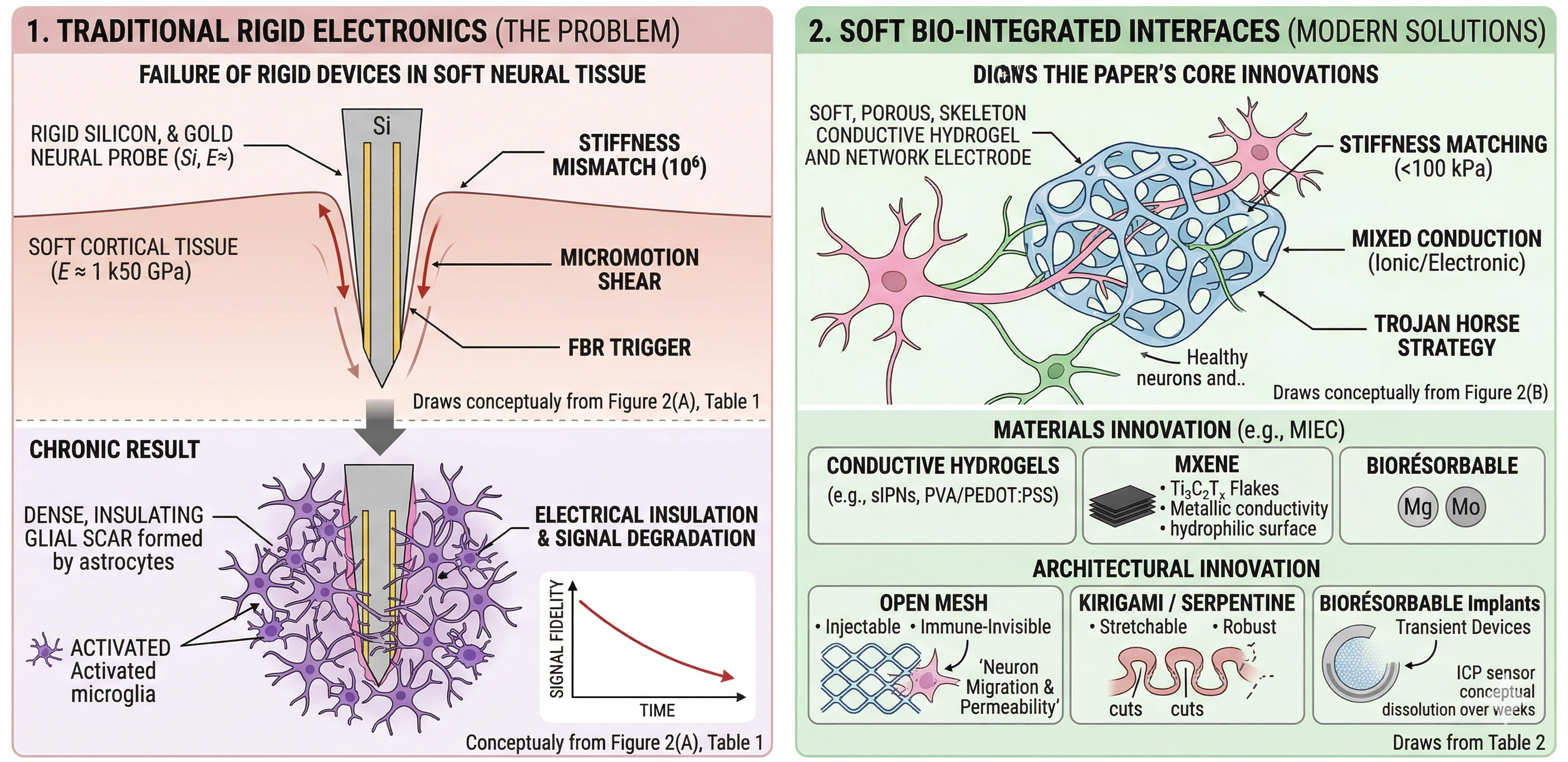
In recent years, research has focused on "bio-integration"—an approach that goes beyond simple biocompatibility (inertia) and aims to design materials that effectively disappear into the host tissue. This "invisibility" is achieved when the device mimics the host so perfectly that the body ceases to recognize it as a foreign object, creating a perfect abiotic-biotic continuum. This concept is illustrated in Figure 3.
Figure 3 (A) illustrates how traditional rigid bioelectronic devices (such as silicon or metal rods) fail when implanted in soft neural tissue. The rigid probe has a significantly higher stiffness (150 GPa) than the surrounding brain tissue (1 kPa). This discrepancy causes constant friction through micromovements between the device and the tissue, leading to chronic mechanical irritation. The trauma of insertion and the subsequent presence of the rigid object trigger an immediate immune response. Microglia (purple, active cells) activate and invade the implant site. Over time, astrocytes (star-shaped cells) and activated microglia form a dense protective barrier known as glial scar around the electrode. This scar tissue acts as an electrical insulator. It physically pushes healthy neurons (pink/beige cells) away from the electrode surface, degrading the signal-to-noise ratio and ultimately causing the device to fail to record neural activity.
Figure 3 (B) illustrates a soft, biocompatible electrode (often made of conductive hydrogels or electronic networks) designed to mimic the extracellular matrix (ECM) of the brain. The soft material of the electrode (10-100 kPa) matches the mechanical compliance of brain tissue, eliminating shear forces during movement and reducing chronic irritation. Because the properties of the material mimic the body's own tissue, the immune system does not aggressively attack it. The "furious" microglia (purple cells) are largely absent or remain in a resting state. The porous, skeleton-like structure allows healthy neurons and neurites (green arrows) to grow into and through the electrode itself. As shown in the inset magnification, the neurites squeeze through the pores of the blue hydrogel matrix. This proximity ensures a stable, high-fidelity electrical connection that can last for months or years without being isolated by scar tissue.
The transition from A to B represents the shift from treating the electrode as a sensor inserted into tissue to a structure that becomes an integral part of the tissue. By masking the "foreignness" of the device through mechanical softness and porosity (the "Trojan horse" strategy), the body accepts the interface instead of rejecting it.
2.1. Modulus Matching
Traditional implants are mechanically distinct from tissue. A silicon probe (E≈150 GPa) in the brain (E≈1–5 kPa) creates a stiffness mismatch of >106. Under physiological micromotion (respiration, blood flow), the rigid device shears adjacent cells, triggering glial scarring that insulates the electrode [7]. Therefore, researchers are synthesizing "Tissue-Equivalent" Hydrogels that match the shear modulus of the target organ.
Example: PEDOT:PSS/PVA Semi-Interpenetrating Networks (sIPNs). By adjusting the crosslinking density of the PVA matrix, the modulus can be tuned from 1 kPa (brain-like) to 100 kPa (muscle-like) while maintaining high ionic conductivity (>2000 S/m) [8]. The result is an interface that deforms synchronously with the tissue, reducing interfacial stress to near zero.
Figure 4.
Bioelectronic interfaces: traditional implant (left) vs tissue-equivalent hydrogel (right).
Figure 4.
Bioelectronic interfaces: traditional implant (left) vs tissue-equivalent hydrogel (right).
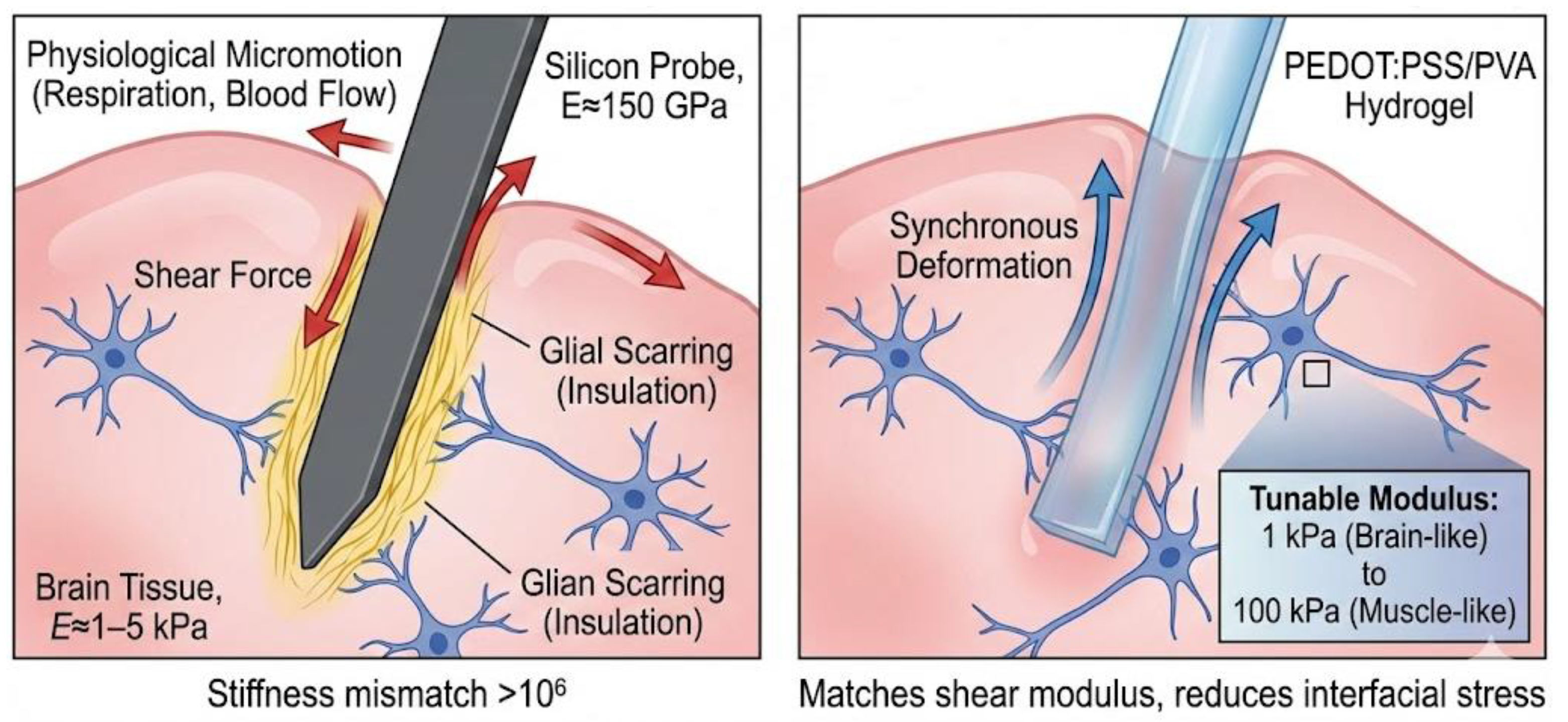

Table 1.
Mechanical Mismatch vs. Bio-Mimicry: A Comparative Analysis of Silicon and Hydrogel Neural Interfaces.
Table 1.
Mechanical Mismatch vs. Bio-Mimicry: A Comparative Analysis of Silicon and Hydrogel Neural Interfaces.
| Feature | Traditional (Silicon) | Tissue-Equivalent (Hydrogel) |
|---|---|---|
| Stiffness (E) | app 150 GPa (Rigid) | app 1 kPa (Soft) |
| Interface Dynamics | Shear / Friction | Synchronous Movement |
| Biological Response | Inflammation & Scarring | Seamless Integration |
| Signal Stability | Degrades over time (scarring) | Stable long-term |
2.2. Geometric Permeability
Even soft solid devices replace native cells, creating a barrier to nutrient transport and network connectivity. A new design is introduced: Macro-porous "mesh" electronics. These architectures act more like scaffolding than walls.
Example: Syringe-injectable meshes. Recent work by Lieber et al. (2024) uses gold/polymer meshes with >90% porosity and strut widths comparable to single axons (<10μm). Upon injection, neurons and glial cells migrate through the mesh holes, effectively integrating the electronic components into the neural circuitry [9]. Since the immune system cannot "isolate" a structure without a continuous surface, the foreign body response (FBR) is virtually eliminated.
Table 2.
Comparison of Solid Devices vs. Mesh Electronics.
| Feature | Solid Device (The Wall) | Mesh Electronics (The Scaffolding) |
|---|---|---|
| Structure | Continuous, solid block. | Open net with large holes. |
| Effect on Cells | Pushes cells away or kills them. | Allows cells to grow through and around it. |
| Nutrient Flow | Blocks flow (cells near the device may starve). | Allows free flow of fluids and nutrients. |
| Result | The body treats it as a foreign object. | The device becomes part of the tissue. |
This distinction is essential for biocompatibility. If you implant a "wall" in the brain, the body often attacks it (scarring), causing the device to malfunction over time. The "scaffold" electronic components are so open and flexible that the body's immune system often does not even realize they are there, allowing the device to function for years without causing damage.
2.3. Transient Electronics
Postoperative monitoring usually requires a second surgical procedure to remove the device, which introduces new risks. The solution could be a bioresorbable (temporary) implant designed to function for a specific therapeutic period and then hydrolyze into secondary metabolic products.
Example: molybdenum-PLGA structures. Devices constructed with molybdenum (Mo) nanomembrane interconnects and PLGA substrates can monitor intracranial pressure (ICP) for 30 days before dissolving into harmless molybdates and lactic acid [10]. The device physically disappears, leaving no trace in the patient's body.
Figure 5.
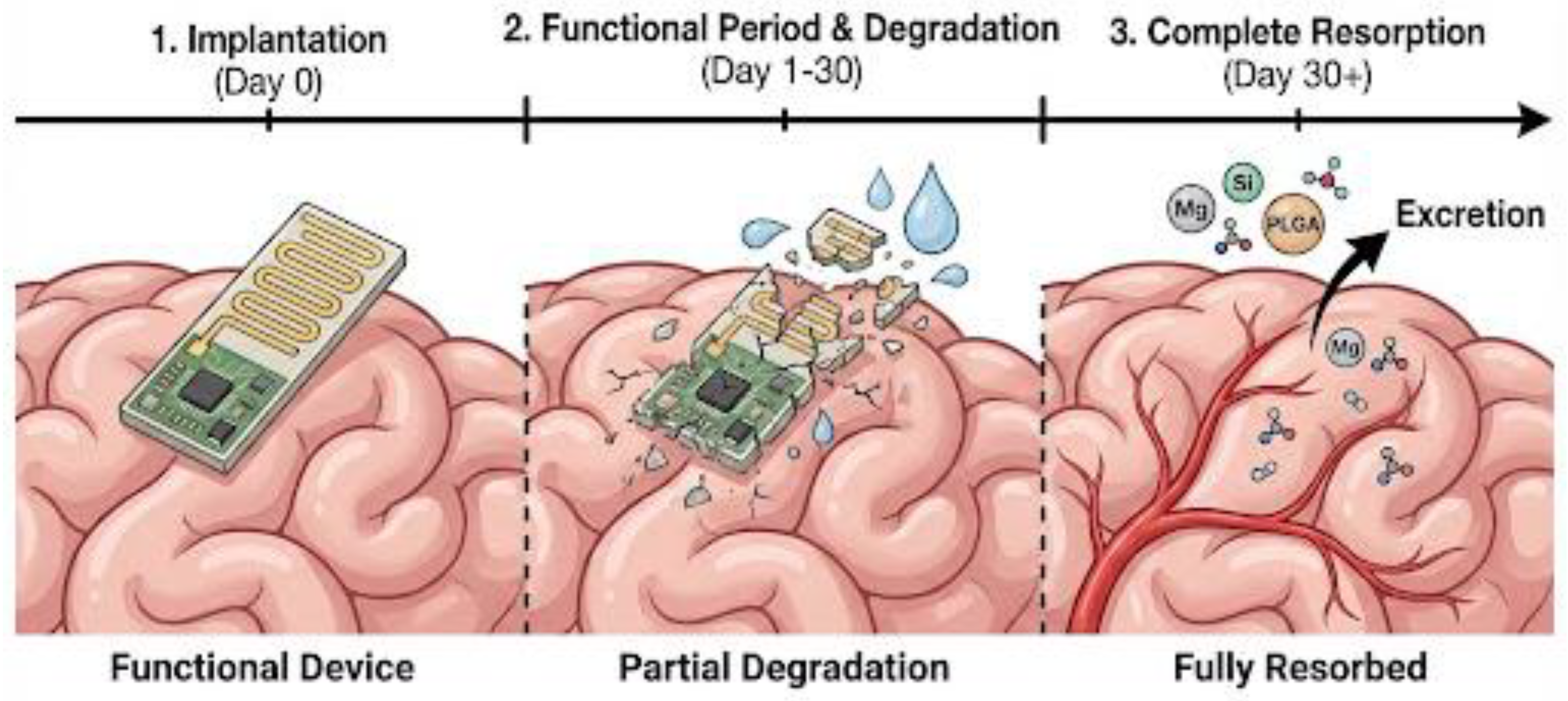
The life cycle of a bioresorbable molybdenum-PLGA implant, showing its transition from active intracranial pressure monitoring to programmed hydrolysis into harmless metabolic products, effectively eliminating the need for surgical removal [10].
Figure 5.
The life cycle of a bioresorbable molybdenum-PLGA implant, showing its transition from active intracranial pressure monitoring to programmed hydrolysis into harmless metabolic products, effectively eliminating the need for surgical removal [10].
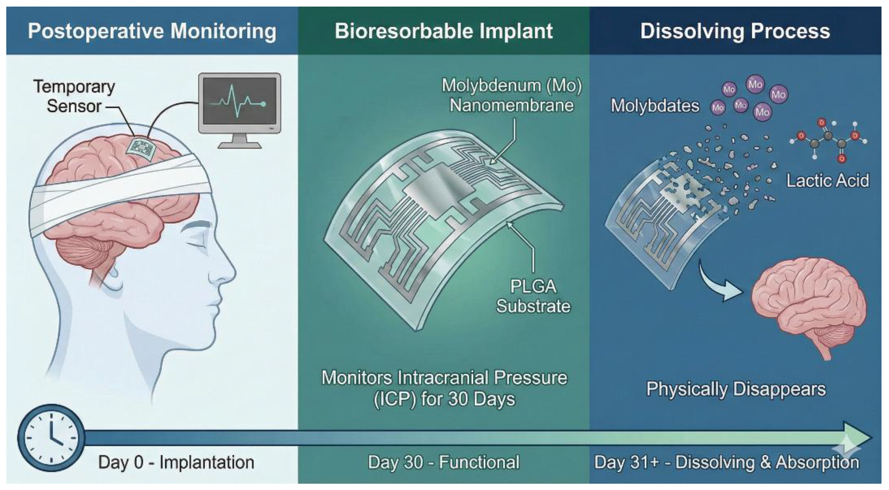
The dissolution rate of PLGA is highly adjustable. In fact, this "programmability" is the main reason why PLGA is the gold standard for bioresorbable electronics and drug delivery. PLGA is a copolymer formed from two different monomers: lactic acid (hydrophobic/slow) and glycolic acid (hydrophilic/fast). By changing the ratio between them, the speed at which water can penetrate the material is changed. The "Goldilocks" ratio (50:50) is unique because it is amorphous (non-crystalline). The lack of crystalline structure allows water to penetrate the matrix quickly, resulting in the fastest degradation (usually 1-2 months). This is probably the formula used for the ICP sensor in your image. Lactide-rich ratio (75:25 or 85:15): as you add more lactide, the material becomes more hydrophobic (repels water). These ratios are used for implants that need to last 4-6 months. Pure PLA (100:0) is semi-crystalline and highly hydrophobic, often taking years to degrade (used in bone screws).
Leaving the ends of polymer chains in the form of carboxylic acids makes the material hydrophilic and acidic. This triggers "autocatalysis," in which the polymer's own acidity accelerates its decomposition. "Coating" the ends with an ester group makes the polymer more hydrophobic, extending its life.
It is important to note that PLGA does not dissolve from the outside in (like a bar of soap). It undergoes bulk erosion. Water immediately penetrates the entire sensor. The polymer chains are randomly cut inside the device, but the device appears solid. Once the chains become short enough (the critical threshold), the material collapses and dissolves in body fluids.
This behavior is perfect for the ICP sensor: it maintains complete structural strength to protect the electronic components for 4 weeks, then rapidly loses its integrity and is eliminated as the patient recovers.
3. Advanced Materials: The Chemistry of Soft Interfaces
3.1. Conductive Hydrogels (CHs): The Ion-Electron Bridge
Hydrogels have become the de facto standard for chronic interfaces due to their high-water content (>70%) and tissue-like softness, effectively bridging the gap between electron-conducting hardware and ion-conducting biology. The "gold standard" material architecture is the semi-interpenetrating network (sIPN) based on PEDOT:PSS. The innovation of sIPN lies in its ability to resolve the "conductivity-resistance" trade-off. Pure conductive polymers (such as PEDOT:PSS) are brittle when dry and prone to excessive swelling when wet. In an sIPN, two distinct polymer networks function in parallel:
- The Conductive Network in which PEDOT:PSS chains form a percolation path for electron transport.
- The Structural Network: A secondary, tough hydrogel matrix (e.g., Polyacrylamide, PVA, or Polyurethane) provides mechanical elasticity and prevents the conductive chains from dissolving into the body [11].
Figure 6.
Hydrogels & sIPN PEDOT: PSS- The gold standard for chronic bio-electronic interfaces.
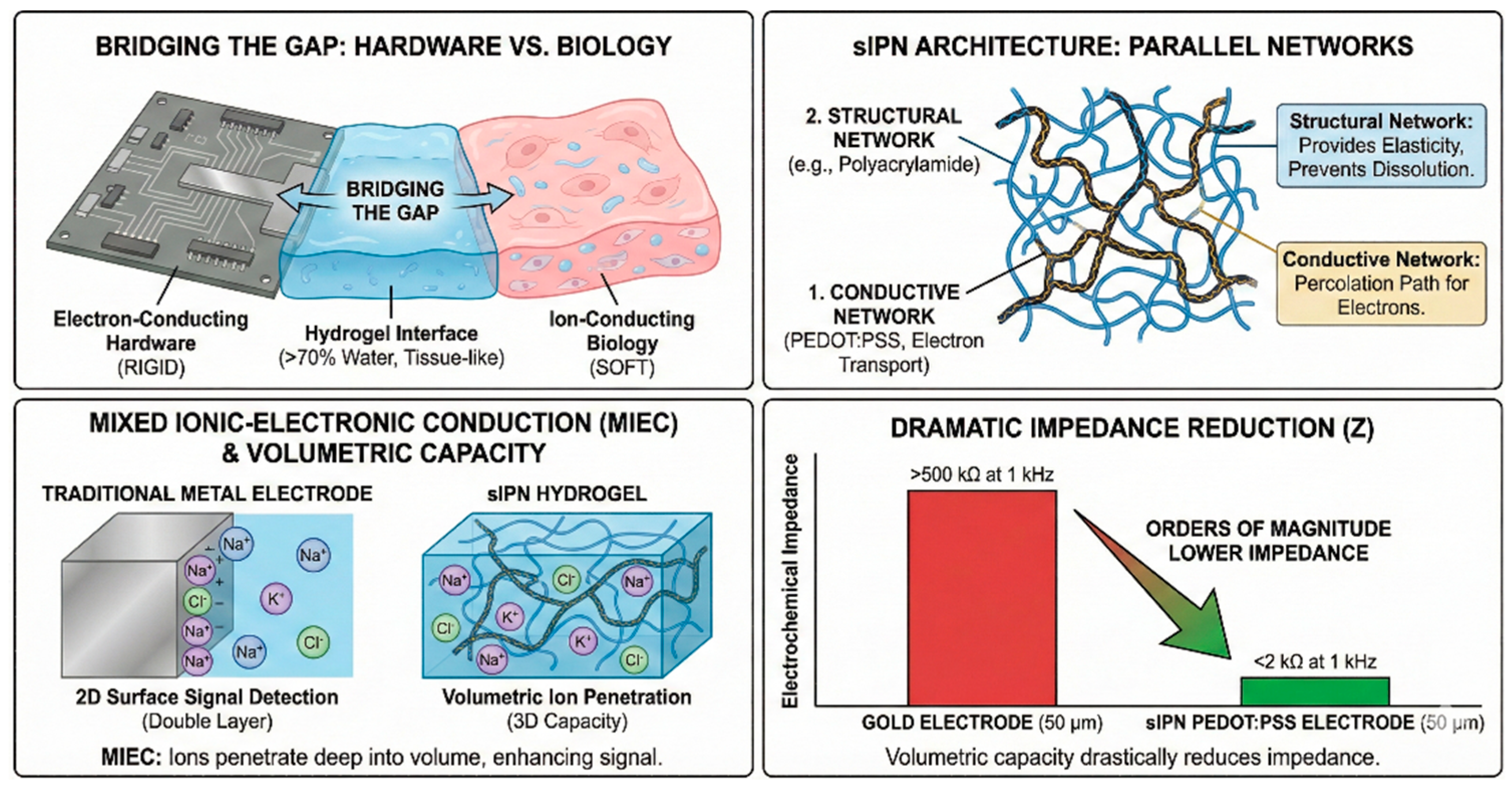
The unique advantage of this material is its mixed ionic-electronic conduction (MIEC). Despite traditional metal electrodes which can detect signals only at their 2D surface (double layer), in hydrogel, sIPN allows physiological ions (Na+, K+) to penetrate deep into the volume of the material. This creates a "volumetric capacity" that reduces electrochemical impedance (Z) by orders of magnitude compared to metals [10]. Note: A typical 50 μm sIPN PEDOT:PSS electrode has an impedance of <2 kΩ at 1 kHz, while a gold electrode of the same size has >500 kΩ. Recent formulations have pushed the boundaries of what these materials can achieve:
• High conductivity injectables: A study conducted in 2025 by Goestenkors et al. demonstrated that a PEDOT:PSS/polyacrylamide sIPN achieves a conductivity of >2000 S/m (comparable to that of ionic fluids) while maintaining a Young's modulus of ≈10 kPa (corresponding to brain tissue). This material can be injected using a syringe and then physically cross-linked in situ using triggers at physiological temperatures [12].
• Volumetric capacitance: Because ions can penetrate the sIPN mass, these electrodes have a charge injection capacitance (CIC) of >30 mC/cm2, which is nearly two orders of magnitude higher than flat platinum. This allows the electrodes to be miniaturized to the cellular scale (<20μm) without losing their ability to effectively stimulate neurons [13].
• Stretchable "E-Skin" sensors: Using a polyurethane matrix, researchers have created sIPN hydrogels capable of stretching up to 300% without losing their conductivity. This is essential for wearable sensors that monitor joint movement or heartbeat [14].
3.2. MXenes: High-Performance 2D Materials
Transition metal carbides/nitrides, particularly MXenes Ti3C2Tx, have emerged as superior alternatives to graphene for biosensors and energy storage. Although graphene offers exceptional intrinsic conductivity, its practical application in biofluids is hampered by hydrophobicity and chemical inertness (lack of binding sites). MXenes solve this problem by combining metallic conductivity with a hydrophilic, functionalized surface. Unlike graphene, which consists of inert sp2 carbon bonds, Ti3C2Tx MXene nanosheets possess a unique "ceramic-metallic" duality: The core (Ti3C2) provides metallic conductivity (>10,000 S/cm), ensuring rapid electron transport [15], and the surface (Tx) is naturally terminated with hydrophilic functional groups (-OH, -O, and -F). This allows MXenes to disperse perfectly in aqueous biological solutions without toxic surfactants (which graphene requires) and provides abundant anchorage points for enzymes and antibodies [16].
Figure 7 shows the advantages of MXene (Ti3C2Tx) over graphene. Unlike graphene, which is hydrophobic and repels water, MXene has a hydrophilic surface that allows water molecules to integrate into the material, rather than forming droplets on its surface. The diagram highlights that MXene is not chemically inert, unlike graphene. Instead, it possesses "hydrophilic surface groups (Tx)" (containing atoms such as fluorine and oxygen) that cover its metallic core. Due to its surface chemistry, MXene achieves "perfect dispersion in aqueous biofluids." In contrast, graphene clumps together and requires surfactants to dissolve in biofluids. MXene's functionalized surface allows it to interact effectively with biological agents such as antibodies, whereas these agents tend to bounce off the chemically inert surface of graphene. Despite its active surface, MXene retains a stable "metallic core (Ti3C2)" composed of titanium and carbon, which supports its function in electronics. These chemical characteristics make MXene superior for use in biosensors and energy storage, while graphene has limited biological applications due to its inertness and low solubility.
Table 3.
Graphene vs. MXene: Conductivity Comparison.
| Feature | Graphene (The Standard) | MXene (Ti3C2Tx) (The Challenger) |
|---|---|---|
| Conductivity Type | Semi-metal (Zero-bandgap) | Metallic |
| Theoretical Limit | Extremely High (~60,000+ S/cm) | High (~10,000 – 20,000 S/cm) |
| Real-World Bio-Films | Lower. Requires surfactants to dissolve in water, which insulate the material and drop conductivity. | Retained. Dissolves naturally in water; forms highly conductive films without insulating surfactants. |
| Electron Mobility | Exceptional (>200,000 cm2/V⋅s) | Good, but lower than Graphene due to surface functional groups (Tx). |
Graphene has higher bulk conductivity, but MXene often performs better in practical biological applications because it does not require conductivity-reducing chemicals to stick to wet tissue.
In biosensors, graphene often suffers from high noise or requires complex processing (CVD) to maintain conductivity. MXene: conductivity allows for an incredibly high signal-to-noise ratio. Because it is hydrophilic, it creates a tighter "electrical bond" with enzymes and antibodies, detecting lower concentrations of biomarkers (e.g., dopamine, glucose).
For energy storage (supercapacitors/batteries), graphene stores energy primarily through double-layer electric capacitance (EDLC) (physical adsorption of ions), and MXene combines EDLC with pseudocapacitance. Surface atoms (titanium) undergo rapid redox reactions, allowing it to store more charge per unit volume than graphene, making it ideal for microbatteries in medical implants.
In biosensing, the signal-to-noise ratio is defined by how closely the sensor can interact with the biological target. The hydrophilic surface of MXene acts like "molecular Velcro." In a 2025 study on glucose detection, researchers demonstrated that the enzyme glucose oxidase (GOx) spontaneously adsorbs into the space between MXene layers, retaining 95% of its bioactivity. In contrast, graphene often denatures enzymes due to hydrophobic interactions. Example (neurotransmitters): Ma et al. (2025) developed a Ti3C2Tx microelectrode capable of detecting dopamine and glutamate with a detection limit of 10 fM (femtomolar). The metallic edge areas of MXene flakes catalyzed the oxidation of dopamine at lower potentials than carbon fiber, significantly reducing ascorbic acid interference [17].
For implantable electronic devices, space is the main constraint. Batteries must be microscopic but powerful. MXenes excel in pseudocapacitance. Unlike graphene, which stores charge only through physical adsorption (EDLC), MXenes allow ions (H+, Na+, K+) to intercalate between layers, undergoing rapid redox reactions at the titanium levels. Recent micro-supercapacitors based on vertical MXene arrays have achieved a volumetric capacity of >1500 F/cm3, about 3-5 times higher than porous graphene electrodes. This density enables "dust-sized" batteries that can power a neural transmitter for days on a single charge [18].
3.3. Transient (Bioresorbable) Materials
"Transient electronic devices" represent a transition from permanent implants to devices with a programmable lifespan. They are designed to perform a specific therapeutic or diagnostic function for a determined period, after which they dissolve completely into biocompatible by-products, eliminating the need for a second surgical procedure for removal.
The main engineering challenge is selecting materials whose hydrolysis products are biologically harmless and can be eliminated by the kidneys. Conductors and interconnections: Molybdenum (Mo) and tungsten (W) are the preferred metals. Molybdenum dissolves slowly and predictably into molybdates (MoO42−), making it ideal for stable electrical pathways, while magnesium (Mg) dissolves more rapidly and is often used for power sources or trigger mechanisms [19]. Substrates and encapsulation: Biopolymers such as PLGA (polylactic-co-glycolic acid) and silk fibroin act as a structural platform. Their degradation rate can be precisely adjusted from days to months by modifying the molecular weight or crystallinity. For example, a higher glycolic ratio in PLGA accelerates its decomposition [20]. Semiconductors: Extremely thin films of monocrystalline silicon (nanomembranes, <300 nm thick) are used for logic gates. Although silicon is stable in its raw state, at the nanoscale it dissolves in biofluids at a rate of approximately 2-4 nm/day, forming harmless silicic acid (Si(OH)4) [21].
Application Example: Post-Operative Monitoring
One primary application is temporary monitoring after traumatic brain injury or neurosurgery. A study conducted in 2024 by Bae et al. demonstrated the effectiveness of a "cortical electrode" designed to be placed on the cerebral cortex. The device, constructed of molybdenum interconnects on a PLGA substrate, monitored intracranial pressure and temperature for 14 days. After this critical period, the device began to dissolve, eliminating the need for high-risk surgical extraction and reducing the risk of advanced infection [22].
Figure 8.
The Lifecycle of Transient Bioelectronics. A timeline showing a bioresorbable device from implantation on the brain (Day 0), through a functional period where it begins to degrade (Day 1-30), to complete resorption and excretion of its biocompatible byproducts (Day 30+).
Figure 8.
The Lifecycle of Transient Bioelectronics. A timeline showing a bioresorbable device from implantation on the brain (Day 0), through a functional period where it begins to degrade (Day 1-30), to complete resorption and excretion of its biocompatible byproducts (Day 30+).
3.4. Liquid Metals: The "Infinite" Conductor
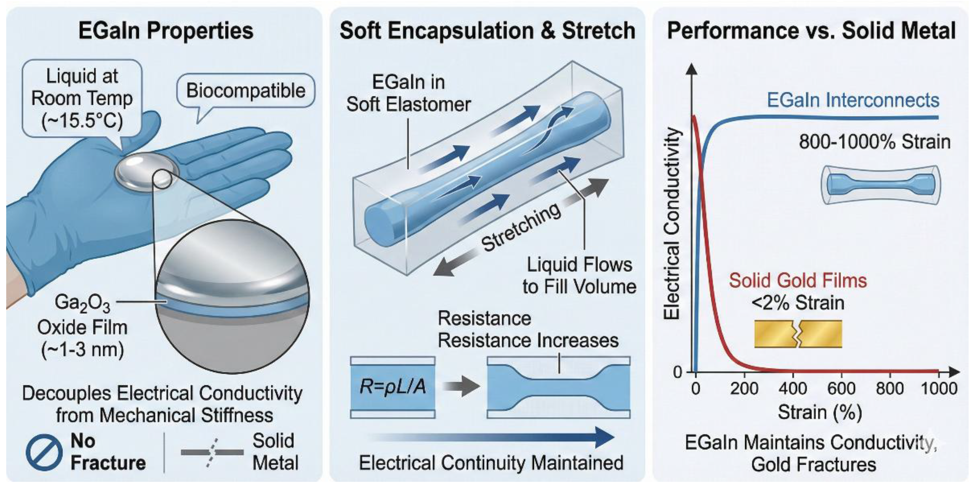
Gallium-Indium eutectic (EGaIn) has established itself as the primary material for high-demand bioelectronics, particularly where solid metals would fracture. Unlike mercury (which is toxic), EGaIn is biocompatible and remains liquid at room temperature (melting point ≈15.50 °C). The unique capability of EGaIn lies in its ability to decouple electrical conductivity from mechanical stiffness. EGaIn is a low-viscosity fluid (2×10⁻³ Pa·s), but spontaneously forms a thin passivation oxide film (Ga2O3, ≈1–3 nm thick) upon exposure to air. This film acts as a surfactant, stabilizing the liquid in specific shapes (such as wires or droplets) within microfluidic channels [23]. When encapsulated in a soft elastomer (such as silicone or EcoFlex), the liquid metal channel can stretch. As the channel elongates, the liquid simply flows to fill the new volume. Although resistance increases due to geometric narrowing (R=ρL/A), electrical continuity is never interrupted. EGaIn interconnects maintain conductivity up to 800–1000% strain, while solid gold films fracture at <2% strain [24].
Figure 9.
EGaIn's liquid properties enable extreme stretchability and sustained electrical continuity, contrasting its performance against brittle solid metals that fracture under strain.
Figure 9.
EGaIn's liquid properties enable extreme stretchability and sustained electrical continuity, contrasting its performance against brittle solid metals that fracture under strain.
Application Examples
Wearable joint monitoring: Monitoring large movements such as knee bending (>900) or elbow flexion. A study conducted in 2023 by Roy et al. demonstrated the effectiveness of a "smart knee brace" that uses EGaIn microchannels printed on a textile substrate. The sensor accurately tracked the rehabilitation progress of ACL patients, correlating changes in resistance with joint angle without impeding natural movement [25].
Soft robotics and artificial skin: Providing tactile feedback for soft robotic grippers. EGaIn grids embedded in a robotic finger allow it to sense curvature and pressure when grasping a delicate object (such as a strawberry). The liquid metal deforms with the gripper, providing real-time proprioceptive feedback [23].
4. Architectural Engineering: Geometry as Function
Currently, material compliance alone will not be sufficient to solve the challenge of bio integration. Geometric architecture determines how the device distributes stress and integrates into the 3D biological environment. By designing the geometry, researchers can transform rigid materials (such as gold, silicon, or graphene) into stretchable, tissue-compatible systems.
4.1. Syringe-Injectable Mesh Electronics
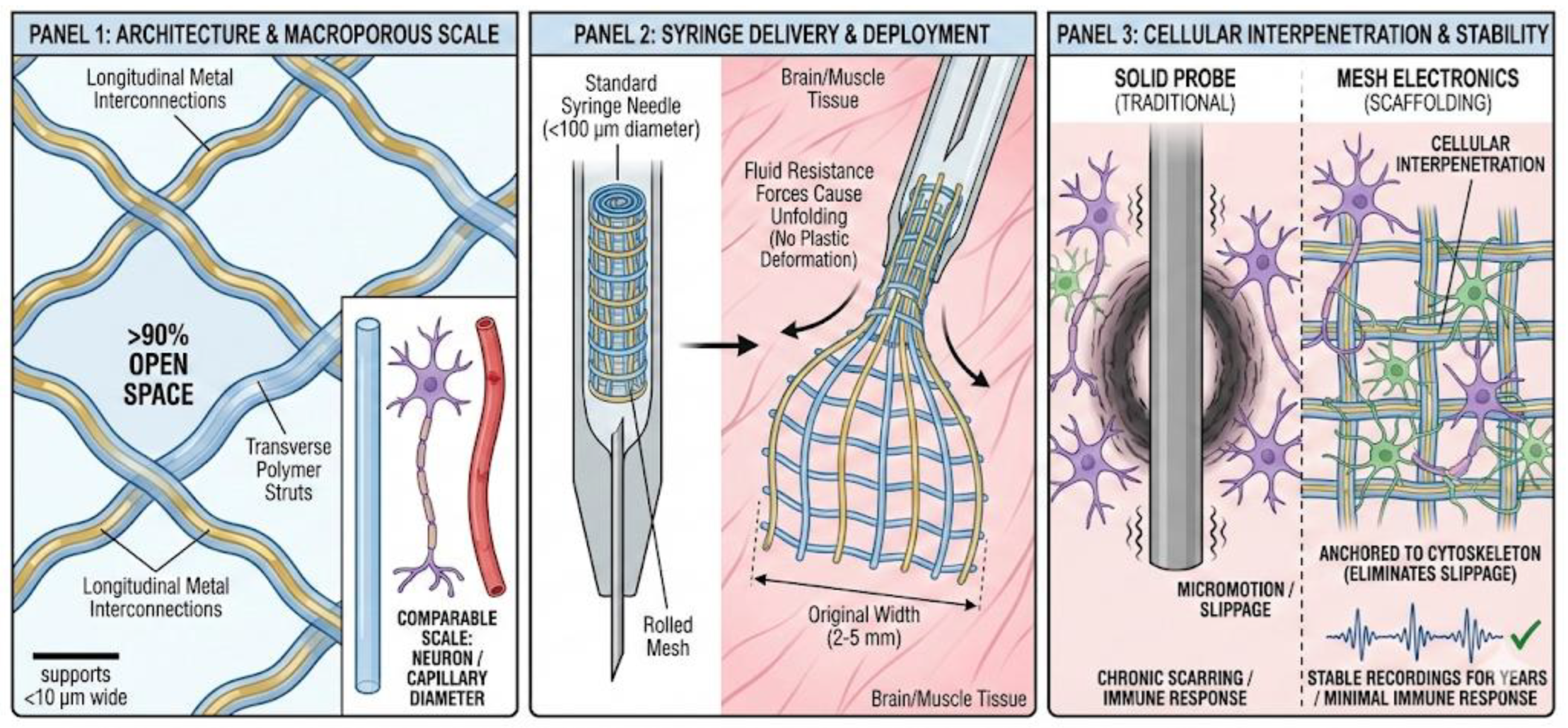
Conceived by the Lieber group and significantly refined in 2024-2025, this architecture decouples the device footprint from the trauma of implantation. The device consists of longitudinal metal interconnections and transverse polymer struts, forming a macroporous network with >90% open space. The characteristic dimensions (supports <10 μm wide) are comparable to the sum of a single neuron or the diameter of a capillary [26]. The high porosity allows the entire mesh to be tightly wrapped inside a standard syringe needle (diameter <100 μm). When injected into the brain or muscle, fluid resistance forces cause the mesh to unfold to its original width (often 2-5 mm) without plastic deformation. Unlike solid probes that push cells aside, the open mesh allows neurons and glial cells to grow through the network. This cellular interpenetration effectively anchors the electronic components to the cytoskeleton of the tissue, eliminating relative slippage (micromotion) that causes chronic scarring. Histological studies show that mesh electrodes provide stable, deviation-free recordings for years with minimal immune response [27].
Figure 10.
Ultra-flexible mesh electronics: decoupling footprint from trauma [27].
Figure 10.
Ultra-flexible mesh electronics: decoupling footprint from trauma [27].
4.2. Kirigami and Serpentine Interconnects
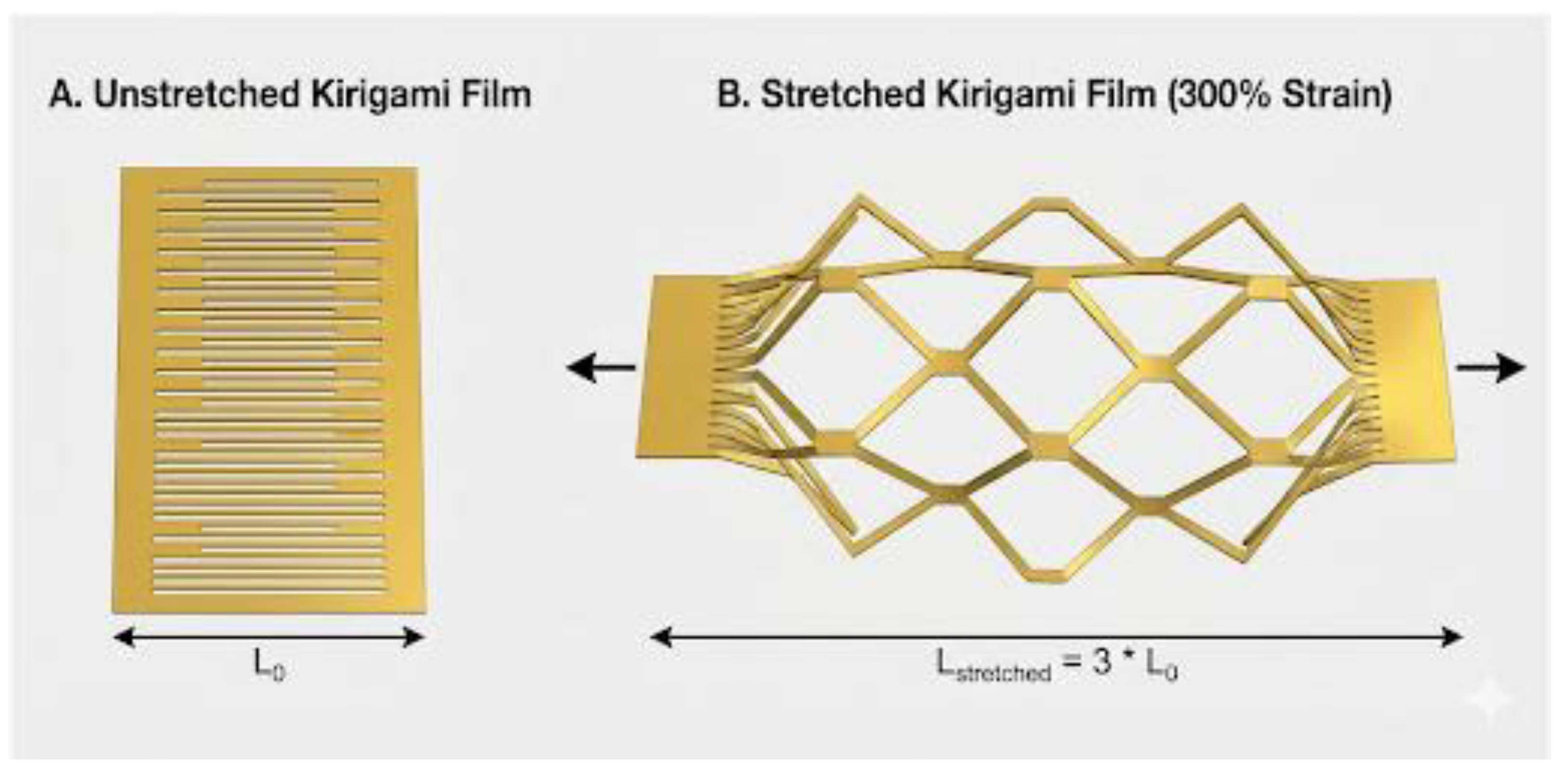
To adapt to massive volume changes in dynamic organs (e.g., heartbeats, lung expansion, or joint movement), engineers use Kirigami (Japanese paper cutting) and Serpentine geometries to create "structural elasticity." By introducing specific cutting patterns (e.g., alternating horizontal slits) into a 2D conductive film, tensile stress is delocalized. When the tissue stretches, the 2D film deforms out of plane into a 3D mesh, rather than intrinsically stretching the material. A 2024 study by Xu et al. demonstrated that graphene films with kirigami patterns can withstand over 300% deformation while maintaining less than 5% resistance variation. This allows high-strain surfaces such as the knee or elbow to be monitored without electrical failure [28]. Microlithography is used to pattern S-shaped or horseshoe-shaped interconnects. These act as springs in the plane. When the substrate stretches, the loops unfold, transforming dangerous tensile stress into safer bending stress. This is the fundamental architecture for "electronic skin" (E-Skin) [29].
Figure 11.
Kirigami Strain Relief for Stretchable Electronics. (A) An unstretched conductive film with a kirigami cut pattern. (B) The same film stretched to 300% strain. The cuts open into a diamond lattice, allowing the overall structure to elongate without fracturing the conductive material.
Figure 11.
Kirigami Strain Relief for Stretchable Electronics. (A) An unstretched conductive film with a kirigami cut pattern. (B) The same film stretched to 300% strain. The cuts open into a diamond lattice, allowing the overall structure to elongate without fracturing the conductive material.
4.3. Fractal and "Fuzzy" Surface Coatings
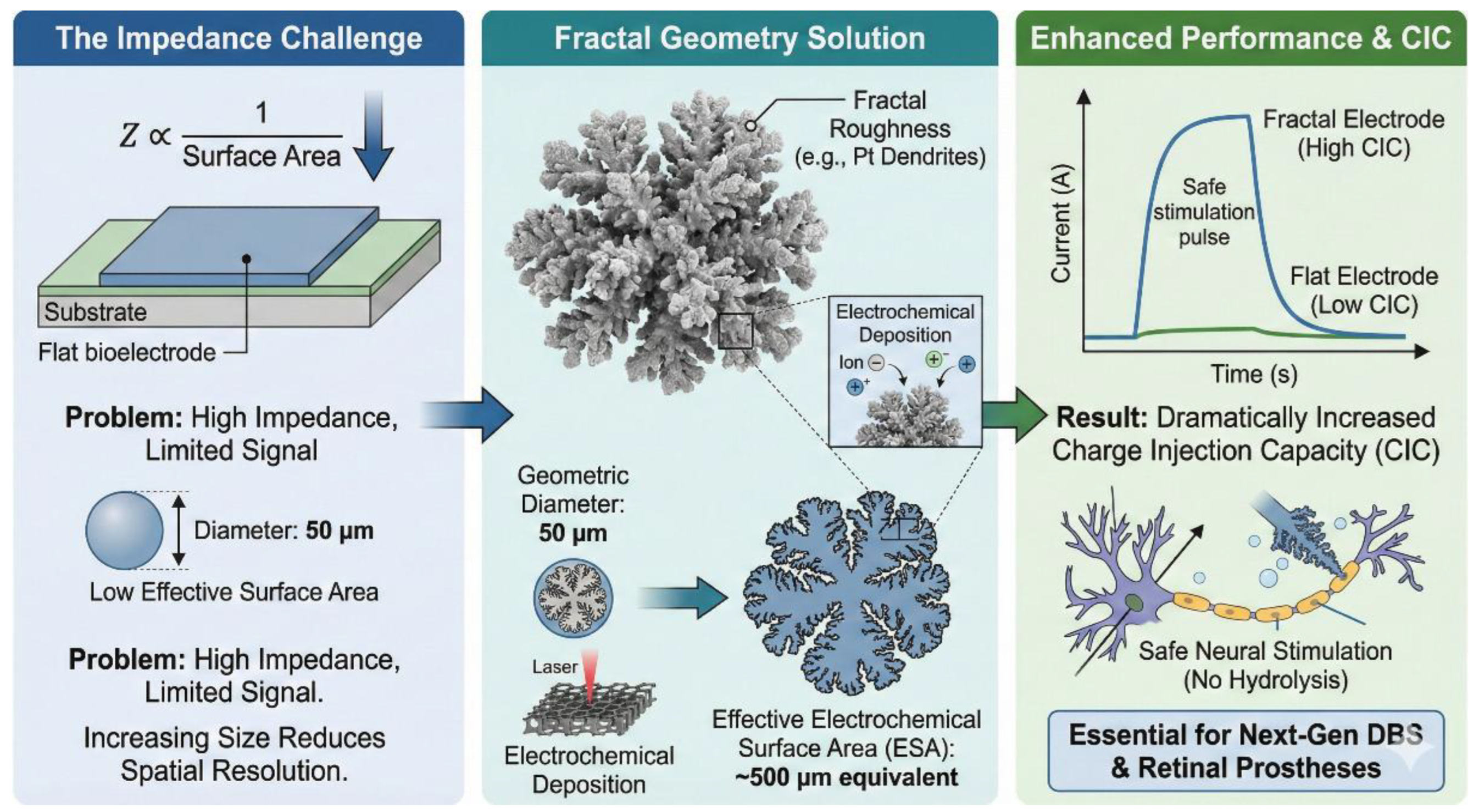
The performance of a bioelectrode is determined by its impedance (Z), which is inversely proportional to its surface area (Z∝1/Surface area). To reduce impedance without increasing the geometric footprint (which would reduce spatial resolution), surfaces are designed with fractal roughness. Fractal geometries (e.g., cauliflower-like structures) maximize the effective surface area (ESA). A geometric point with a diameter of 50 μm can have the electrochemical surface area of a 500 μm pad if the roughness factor is sufficiently high. Electrochemical deposition creates platinum structures with a high degree of dendritization. Using lasers to etch patterns into polyimide creates a porous, foam-like graphene structure. This dramatically increases the charge injection capacity (CIC)—the amount of current that can be safely injected to stimulate neurons without causing harmful hydrolysis of water. Fractal coatings with high CIC are essential for next-generation deep brain stimulation (DBS) and retinal prostheses [30].
Figure 12 illustrates how engineering fractal surface roughness maximizes the effective surface area of bioelectrodes without increasing their physical footprint, thereby lowering impedance, and significantly boosting Charge Injection Capacity (CIC) for safer neural stimulation.
5. Comparative Analysis: Material Performance Standards
The selection of electrode materials is a trade-off between electrical performance (conductivity, charge injection), mechanical compliance (modulus), and biological stability. The table and analysis below summarize how modern materials stack up against traditional standards.
Table 4.
Comparison of Key Bioelectronic Materials.
| Material Class | Electrical Conductivity (σ) | Young's Modulus (E) | Charge Injection Capacity (CIC) | Primary Advantage | Major Disadvantage | Best Application |
|---|---|---|---|---|---|---|
| Noble Metals (Au, Pt) | High (4.1 x 10⁷S/m) | ≈79 GPa (Rigid) | ≈0.1 mC/cm² | Excellent stability, chemical inert, high conductivity. | Massive mechanical mismatch; surface-only sensing. | Standard clinical DBS probes, pacemakers. |
| Conductive Hydrogels (PEDOT:PSS sIPN) | Moderate (2 x 10³ S/m) | 10 kPa – 1 MPa (Soft) | >30 mC/cm² | Tissue-like softness (kPa range), volumetric capacitance, low impedance. | Swelling in water; lower tensile strength. | High-fidelity neural recording, E-skin. |
| MXenes (Ti3C2Tx) | High (106 S/m) | ≈330 GPa (Flake stiffness) | 3–8 mC/cm² | Hydrophilic; high surface area; easy functionalization. | Oxidizes in water over time (requires encapsulation). | Supercapacitors, enzymatic biosensors. |
| Liquid Metal (EGaIn) | High (3.4 x 10⁶ S/m) | ≈0 Pa (Fluid) | N/A (Flows) | Infinite stretchability (>800%); self-healing. | Leakage risk; difficult to package reliably. | Wearable joint sensors, soft robotics. |
| Carbon Nanotubes (CNTs) | High (105 S/m) | ≈1 TPa (Very Stiff) | 1–3 mC/cm² | High strength; high aspect ratio. | Fabrication variability; potential cytotoxicity. | Fiber electrodes, micro-thread probes. |
Gold and silicon are much stiffer than the brain, but conductive hydrogels (sIPN) have effectively solved this problem. By adapting to the brain's modulus (≈1-10 kPa), they reduce the triggering of the immune response. The trade-off is conductivity; hydrogels are significantly less conductive than metals, requiring larger cross-sections to prevent signal attenuation [31,32].
Metals rely on "double-layer capacitance" (only at the surface). This limits the amount of current that can be injected before causing dangerous hydrolysis of water (≈0.1 mC/cm² for Pt). Hydrogels and MXenes use "volumetric capacitance" (ions penetrate the material). This allows for massive charge injection capacities (>30 mC/cm² for PEDOT:PSS), enabling the miniaturization of stimulation electrodes to the scale of individual neurons without losing effectiveness [33,34].
Platinum is chemically inert and very durable, MXenes providing incredible performance but are susceptible to oxidation. A challenge remains to encapsulate MXenes in antioxidant polymers (such as vitamin C-doped hydrogels) to ensure they survive for years in the body [35].
Table 5.
Design Trade-offs in Modern Neural Probes: Planar, Mesh, and Kirigami Architectures.
| Architecture | Pros | Cons |
|---|---|---|
| Planar Thin Film | Standard lithography fabrication (cheap, scalable). | 2D geometry does not match 3D biology; delamination risk. |
| Open Mesh | "Invisible" to immune system, drift-free. | Complex deployment (injection), fragile interconnects. |
| Kirigami/Serpentine | High stretchability, robust. | Lower effective pixel density (due to cuts/gaps). |
6. Future Work and Improvements (2026–2030)
Looking beyond 2026, the focus is shifting from simply "softening" electronic components to creating autonomous, living, intelligent systems. The current standard of care—permanent implants powered by bulky batteries—will disappear as devices are designed to harvest their own energy and then bio-resorb after therapy, eliminating the need for surgical extraction. There are several pillars that define the research and development roadmap for the next five years.
6.1. The "Bio-Cyborg" Fusion: Living Electrodes
The definitive solution for foreign body response (FBR) is not to hide the foreign object, but to eliminate its "foreignness" by integrating living biological components into the abiotic device. The concept of "living electrodes" involves covering the conductive skeletons with the patient's own (autologous) stem cells or ex vivo neural progenitors prior to implantation. Once implanted, these cells act as a "biological bridge," synapsing with host neurons on one side and electrochemically coupling with the electrode on the other. Researchers are developing "camouflaged" interfaces in which electrode surfaces are functionalized with zwitterionic polymers that mimic the cell membrane or seeded with induced pluripotent stem cells (iPSCs). Ratner points out that such hybrid systems can theoretically maintain perfect integration for decades, as the living coating renews itself and releases anti-inflammatory cytokines to pacify the immune system [36].
6.2. 4D Printing: Shape-Morphing Scaffolds
4D printing adds the dimension of time to 3D printing. It uses "smart materials" (shape memory polymers) that are printed into one shape but transform into another shape when exposed to a specific stimulus (e.g., body heat or pH change). This technology solves the "Implementation vs. Integration" paradox. A device must be small and rigid to be inserted (like a needle), but large and soft to integrate (like a mesh). Scheideler & Im (2025) describe "thermomorphable probes." These are printed as straight, rigid needles (E≈1 GPa) to facilitate surgical insertion. However, once inserted into the body and heated to 37 °C, the polymer matrix undergoes a glass transition (Tg), softening dramatically (E≈10 kPa) and expanding into a complex basket shape that wraps around a nerve bundle or conforms to the cortical surface [37,38].
6.3. Closed-Loop AI on Chip: Neuromorphic Edge Computing
Current biological interfaces face a "data bottleneck": wireless transmission of raw neural data, with high bandwidth (GB/hour), to external computers requires enormous power, which heats up the tissue. The solution is Edge Computing. Integrating organic electrochemical transistors (OECT) and neuromorphic hardware directly at the recording site to process signals locally. This shifts the paradigm from "record and transmit" to "analyze and act." By 2026, prototypes of "pulse sorting sensors" have emerged. These chips use internal analog circuits to detect certain patterns of neural activity (e.g., the onset of an epileptic seizure) in real time (<1 ms). They transmit only a simple "trigger" signal or automatically provide stimulation to stop the event. This local processing reduces energy consumption by >90%, enabling fully autonomous, battery-free implants powered exclusively by glucose-based biofuel cells [39].
6.4. Transient Electronics: The "Disappearing" Implant
The "unidirectional" surgical model is becoming a reality. Instead of requiring a second high-risk surgery to remove a temporary implant (e.g., a postoperative intracranial pressure monitor), devices are constructed from materials that hydrolyze into metabolic byproducts. The silicon nanomembrane (less than 300 nm thick) dissolves in biological fluids at a rate of ≈2 nm/day, forming harmless silicic acid (Si(OH)4) [40]. Metals such as molybdenum (Mo) and tungsten (W) are used for stable conductivity, slowly degrading into benign oxides, while magnesium (Mg) is used for rapidly dissolving components. Liu et al. (2024) demonstrated a "cortex-type electrode" for the cerebral cortex. It actively monitored neural signals for 14 days after surgery and then dissolved completely, leaving no traces of the device or immune scars [41].
6.5. Energy Autonomy: Breaking the Battery Bottleneck
Dependence on external batteries or transcutaneous cables is the main factor limiting the miniaturization of devices. The generation of implants from 2026 to 2030 will be self-powered. Enzymatic biofuel cells (BFCs) use the body's own chemical energy. Electrodes coated with enzymes (e.g., glucose oxidase) catalyze the oxidation of glucose in the blood, generating a continuous flow of electrons. Recent MXene-based BFCs have achieved power densities of >500μW/cm2, sufficient to power a low-power Bluetooth transmitter or pacemaker without the need for recharging [40,42]. Triboelectric nanogenerators (TENG) harvest biomechanical energy from organ movement. By exploiting the separation effect of contact between layers of biocompatible polymers (such as PLGA and silk) during heartbeats or breathing, TENGs generate high-voltage alternating current pulses. Jae-Young Bae et al. (2024) highlighted degradable TENGs that can stimulate the heart during temporary arrhythmia and then dissolve once the heart rhythm stabilizes [43].
7. Conclusion
The field of bioelectronics is currently navigating a historic transition from the "Silicon Age" to the "Bio-Integration Age." As detailed in this review, the fundamental barrier of the mechanical and biological mismatch is being systematically dismantled through three converging pillars of innovation:
Material Softness: The adoption of Conductive Hydrogels (sIPNs) and MXene nanocomposites has lowered the elastic modulus of interfaces from gigapascals (metals) to kilopascals (tissues), bridging the gap between abiotic and biotic worlds.
Geometric Intelligence: Architectures like injectable meshes and kirigami lattices allow electronics to mechanically disappear into the body, moving and growing with the host rather than fighting against it.
Active Biology: The next generation of "Living Electrodes"—coated with patient-specific cells or immune-camouflaging proteins—promises to eliminate the foreign body response entirely.
Conclusion for the Decade
By 2030, the "Bio-Cyborg" fusion will likely be perfect. We will move from inserting hardware into the body to integrating functional tissues—soft, wet, self-powered and temporary hybrid systems. Ultimately, the goal is no longer just to measure biology but to fuse with it perfectly, treating the electrode not as an instrument but as a new organ.
Funding
“This research received no external funding”
Acknowledgments
Generative AI tools were employed during the production of this work solely to enhance the visual quality and layout of the technical figures. The authors have reviewed and edited the output and take full responsibility for the content of this publication.
Conflicts of Interest
The authors declare no conflicts of interest.
Abbreviations
The following abbreviations are used in this manuscript:
| Ag | Silver |
| AgCl | Silver Chloride |
| Al₂O₃ | Alumina (used as a substrate) |
| Au | Gold |
| CE | Counter Electrode |
| CHs | Conductive Hydrogels |
| CIC | Charge Injection Capacitance |
| Cr | Chromium |
| CVD | Chemical Vapor Deposition |
| E | Young’s modulus (a measure of stiffness) |
| ECM | Extracellular Matrix |
| EDLC | Double-layer Electric Capacitance |
| EGaIn | Gallium-Indium eutectic (a liquid metal) |
| FBR | Foreign Body Response |
| fM | Femtomolar |
| GPa | Gigapascal (unit of pressure/stiffness) |
| ICP | Intracranial Pressure |
| kHz | Kilohertz |
| kΩ | kKilohm |
| kPa | Kilopascal (unit of pressure/stiffness) |
| Mg | Magnesium |
| MIEC | Mixed Ionic-Electronic Conduction |
| Mo | Molybdenum |
| PEDOT:PSS: | Poly(3,4-ethylenedioxythiophene) polystyrene sulfonate (a conductive polymer) |
| PLA | Polylactic Acid |
| PLGA | Polylactic-co-glycolic Acid (a bioresorbable polymer) |
| PR | Photoresist |
| Pt | Platinum |
| PVA | Polyvinyl Alcohol |
| PVD | Physical Vapor Deposition |
| RE | Reference Electrode |
| sIPN | Semi-Interpenetrating Network |
| TPa | Terapascal (unit of stiffness) |
| WE | Working Electrode |
| Z | Electrochemical Impedance |
References
- Kim, S., Kim, K.S. & Koo, J., "Soft, conformal tissue–electrode interfaces for bioelectronic devices: material, fabrication strategies, and applications.," Biomed. Eng. Lett. 15, pp. 963-994, 2025. [CrossRef]
- Qiao, Y., Luo, J., Cui, T. et al., "Soft Electronics for Health Monitoring Assisted by Machine Learning.," Nano-Micro Lett, vol. 15, no. 66, 20023. [CrossRef]
- Muralee Gopi CVV, Alzahmi S, Narayanaswamy V, Raghavendra KVG, Issa B, Obaidat IM., "A review on electrode materials of supercapacitors used in wearable bioelectronics and implantable biomedical applications," Mater Horiz., no. 12, pp. 4092-4132, 2025.
- SF., Cogan, "Neural stimulation and recording electrodes. Annu Rev Biomed Eng," vol. 10, pp. 275-309., 2008. [CrossRef]
- Liu, J., Fu, T. M., Cheng, Z., Hong, G., Zhou, T., Jin, L., ... & Lieber, C. M., "Syringe-injectable electronics," Nature Nanotechnology, vol. 10(7), pp. 629-636., 2015. [CrossRef]
- Feig, V. R., Tran, H., & Bao, Z., "Guiding the design of next-generation soft electronics based on properties of biological tissues. ACS Central Science, 4(3), 337-348.," 2018.
- Reis Carneiro, Mahmoud Tavakoli, Carmel Majidi,, "Soft skin-interfacing electrodes for wearable bioelectronics," Matter, vol. 8, no. 10, pp. ISSN 2590-2385, 2025. [CrossRef]
- R. Malečková et al., "Novel conductive PEDOT:DBSA hydrogels with tuneable properties for bioelectronics††Electronic supplementary information (ESI) available. , Volume 6, Issue 4,2025,Pages," Materials Advances, vol. 6, no. 4, pp. 1278-1287, 2025.
- Guosong Hong, Xiao Yang, Tao Zhou, Charles M Lieber, "Mesh electronics: a new paradigm for tissue-like brain probes," Current Opinion in Neurobiology,, vol. 50, pp. 33-41, 2018. [CrossRef]
- Bae, JY., Hwang, GS., Kim, YS. et al., "A biodegradable and self-deployable electronic tent electrode for brain cortex interfacing.," Nat Electron, vol. 7, pp. 815-828, 2024. [CrossRef]
- Yang, M., Wang, L., Liu, W. et al., "Highly-stable, injectable, conductive hydrogel for chronic neuromodulation.," Nat Commun, vol. 15, 2024. [CrossRef]
- Goestenkors AP, Yu JS, Park J, Wu Y, Vargas Espinoza CJ, Friedman LC, Okafor SS, Liu T, Chatterjee S, Debnath A, Semar BA, O'Hare CP, Alvarez RM, Singamaneni S, Raman B, Rutz AL., "PEDOT:PSS Microparticles for Extrudable and Bioencapsulating Conducting Granular Hydrogel Biolelectronics," Small, vol. 21, no. 47, 2025. [CrossRef]
- Tumová, Š., Malečková, R., Kubáč, L. et al., "Novel highly stable conductive polymer composite PEDOT:DBSA for bioelectronic applications. 55, 983–995 (2023).," Polym J, vol. 55, pp. 983-995, 2023. [CrossRef]
- Boufidis, D., Garg, R., Angelopoulos, E. et al., "Bio-inspired electronics: Soft, biohybrid, and “living” neural interfaces. 16, 1861 (2025).," Nat Commun, vol. 16, p. 1861, 2025. [CrossRef]
- Zaheer Ud Din Babar, Vincenzo Iannotti, Giulio Rosati, Ayesha Zaheer, Raffaele Velotta, Bartolomeo Della Ventura, Ruslan Álvarez-DidukArben M, "MXenes in healthcare: synthesis, fundamentals and applications," Chem. Soc. Rev., vol. 54, pp. 3387-3440, 20205. [CrossRef]
- A. Amani et. Al., "MXenes in biosensing: Enhancing sensitivity and flexibility – A review of properties, applications, and future directions," Sensing and Bio-Sensing Research, vol. 47, no. ISSN 2214-1804,, p. 100732, 2025.
- Ma, J.; Yang, S.; Yang, Z.; He, Z.; Du, Z. , "Functional Nanomaterials for Advanced Bioelectrode Interfaces: Recent Advances in Disease Detection and Metabolic Monitoring.," Sensors, vol. 25, p. 4412, 2025. [CrossRef]
- Lorencova, L., Kasak, P., Kosutova, N. et al. , "MXene-based electrochemical devices applied for healthcare applications.," Microchim Acta, vol. 88, p. 191, 2024.
- Jae-Young Bae, Myung-Kyun Choi, Seung-Kyun Kang,, "Transient electronics for sustainability: Emerging technologies and future directions," Beilstein Journal of Nanotechnology,, vol. 16, pp. 1545-1556, 20025.
- Y. S.Choi, J.Koo, Y. J.Lee, G.Lee, R.Avila, H.Ying, J.Reeder, L.Hambitzer, K.Im, J.Kim, K.-M.Lee, J.Cheng, Y.Huang, S.-K.Kang, J. A.Rogers,, "Biodegradable Polyanhydrides as Encapsulation Layers for Transient Electronics," Adv. Funct. Mater, vol. 30, 2020.
- Suk-Won Hwang et al., "A Physically Transient Form of Silicon Electronics," Science, vol. 337, pp. 1640-1644, 2012.
- Bae, JY., Hwang, GS., Kim, YS. et al., "A biodegradable and self-deployable electronic tent electrode for brain cortex interfacing," Nat Electron, vol. 7, p. 815–828, 2024. [CrossRef]
- Dickey M. D, "Stretchable and Soft Electronics using Liquid Metals.," Advanced materials (Deerfield Beach, Fla.),, Vols. 29(27),, no. 10.1002/adma.201606425., 2017. [CrossRef]
- Wang, Yuwen, Wu, Chong, Zhang, Yaokang. Du, Shiwei, Zhou, Xuechang, "Wearable and implantable transient bioelectronics," Journal of Materials Chemistry C, vol. 13, no. 29, pp. 14682-14696, 2025. [CrossRef]
- Roy, A., Adhikari, A., " Soft Materials for Wearable Electronics," In: Kumar, V., Mishra, Y.K. (eds) Soft Materials for Functional Applications. Advanced Structured Materials, vol. 225, no. Springer, Singapore, 2024.
- Shumao Xu, Xiao Xiao, Farid Manshaii, and Jun Chen, , "Injectable Fluorescent Neural Interfaces for Cell-Specific Stimulating and Imaging," Nano Letters, vol. 16, pp. 4703-4716,, 2024. [CrossRef]
- Dai X, Hong G, Gao T, Lieber CM., "Mesh Nanoelectronics: Seamless Integration of Electronics with Tissues. 2018 Feb 20;51(2):309-318.," Acc Chem Res., vol. 12, no. PMID: 29381054; PMCID: PMC5820158., pp. 309-318, 2018. [CrossRef]
- Yang C, Zhang H, Liu Y, Yu Z, Wei X, Hu Y. , "Kirigami-Inspired Deformable 3D Structures Conformable to Curved Biological Surface.," Adv Sci (Weinh)., vol. 5, no. PMID: 30581706; PMCID: PMC6299731., 2018. [CrossRef]
- Zhao, C., Park, J., Root, S.E. et al. , "Skin-inspired soft bioelectronic materials, devices and systems.," Nat Rev Bioeng 2, vol. 2, p. 671–690 , 2024. [CrossRef]
- Scheideler WJ, Im J., " Recent Advances in 3D Printed Electrodes - Bridging the Nano to Mesoscale.," Adv Sci (Weinh). , vol. 11, no. PMID: 39930958; PMCID: PMC11884523, 2025.
- Manuel Reis Carneiro, Mahmoud Tavakoli, Carmel Majidi,, " Soft skin-interfacing electrodes for wearable bioelectronics,," Matter, , vol. 8, no. 10, 2025.
- Sun H, Wang S, Wang P, Bai L, Tan M, Yang F, Wang R, He X. , "Highly Stretchable, Adhesive, and Conductive PEDOT Nanocomposite Hydrogels for High-Performance Flexible Bioelectronics.," Adv Sci (Weinh)., vol. 12, no. 48, 2025.
- Sengupta, J.; Hussain, C.M. , "MXene-Based Electrochemical Biosensors: Advancing Detection Strategies for Biosensing (2020–2024).," Biosensors , vol. 127, 2025.
- Warren M. Grill, Sharon E. Norman, Ravi V. Bellamkonda., "Implanted Neural Interfaces: Biochallenges and Engineered Solutions.," Annual Review Biomedical Engineering., vol. 11, pp. 1-24, 2009.
- Garg R, Vitale F. , "Latest advances on MXenes in biomedical research and health care.," MRS Bull., vol. 3, no. PMID: 36846314; PMCID: PMC9943034., pp. 283-290, 2023.
- Buddy D. Ratner., "Biomaterials: Been There, Done That, and Evolving into the Future.," Annual Review Biomedical Engineering., vol. 21, pp. 171-191., 2019.
- Munir, S., Ali, B. & Gul, S. , "The advancements of 3D-printed electrodes in electrochemistry.," Discov. Electrochem., vol. 1, no. 14, 2024.
- Yanbin Li, Jie Yin, , "Metamorphosis of three-dimensional kirigami-inspired reconfigurable and reprogrammable architected matter,," Materials Today Physics, vol. 21, no. ISSN 2542-5293,, 2021.
- Song E, Serdijn WA. , "Bioelectronic medicine: wearable and implantable electronics.," Bioelectron Med. , vol. 1, no. PMID: 40968385; PMCID: PMC12447594., 2025.
- Beyer, C. , "A biodegradable tent electrode.," Nat Rev Bioeng , vol. 2, p. 719, 2024.
- Liu S, Wang Y, Zhao Y, Liu L, Sun S, Zhang S, Liu H, Liu S, Li Y, Yang F, Jiao M, Sun X, Zhang Y, Liu R, Mu X, Wang H, Zhang S, Yang J, Xie X, Duan X, Zhang J, Hong G, Zhang XD, Ming D. A, "Nanozyme-Based Electrode for High-Performance Neural Recording.," Adv Mater, vol. 6, no. 36, 2024.
- Aibobek Seitak, Shaohong Luo, Ning Cai, Kin Liao, Anna-Maria Pappa, Sungmun Lee, Vincent Chan,, "Emergence of MXene-based electrochemical biosensors for biomolecule and pathogen detection,," Sensors and Actuators Reports, vol. 6, no. ISSN 2666-0539, 2023.
- Jae-Young Bae, Myung-Kyun Choi, Seung-Kyun Kang, , "Transient electronics for sustainability: Emerging technologies and future directions,," Beilstein Journal of Nanotechnology,, vol. 16, no. ISSN 2190-4286,, pp. 1545-1556,, 2025. [CrossRef]
Figure 1.
Mechanical difference between soft biological tissues (kPa range) and traditional rigid materials (GPa/TPa range), highlighting the need for developing "soft electronics" that match the stiffness of the human body.
Figure 1.
Mechanical difference between soft biological tissues (kPa range) and traditional rigid materials (GPa/TPa range), highlighting the need for developing "soft electronics" that match the stiffness of the human body.
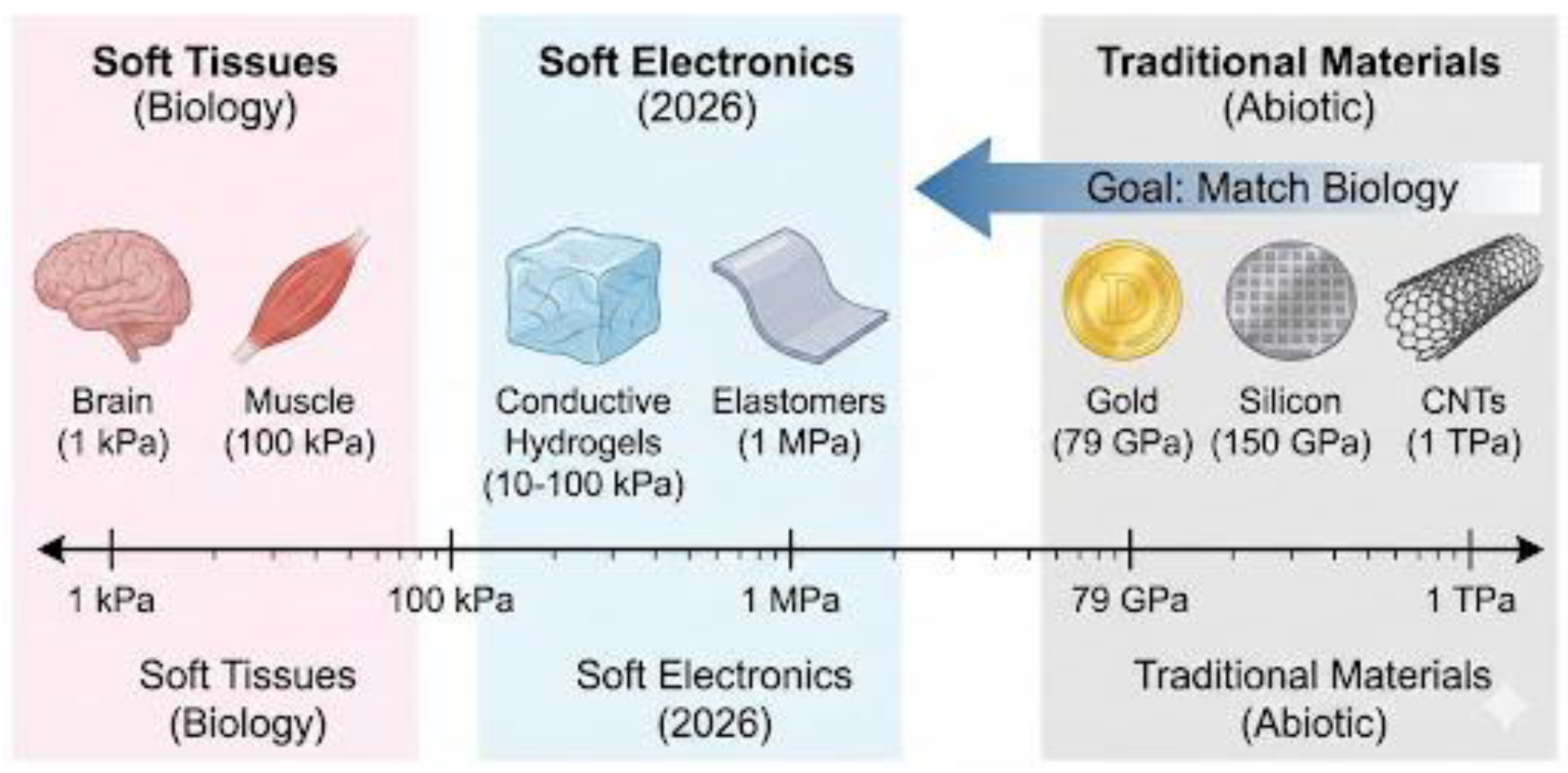
Figure 2.
Bioelectronic interface: physical and communicative barriers.
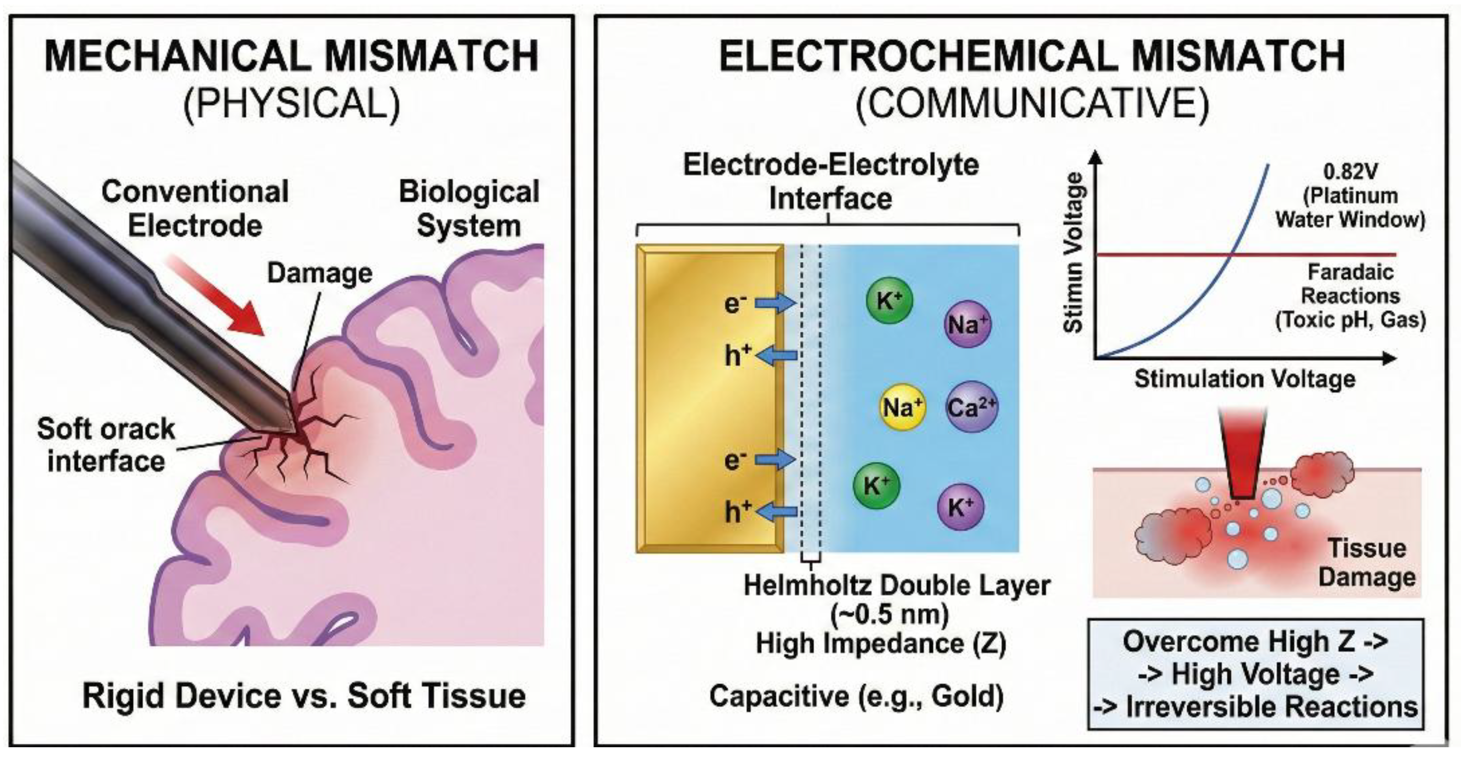
Figure 3.
The "Trojan Horse" Electrode: Bio-Integration vs. Foreign Body Response. (A) A traditional rigid probe triggers a severe immune response, leading to a glial scar that insulates the electrode from neurons. (B) A soft, bio-integrated electrode mimics the tissue's properties, allowing neurons to grow directly onto its surface with minimal immune reaction. [5] [6].
Figure 3.
The "Trojan Horse" Electrode: Bio-Integration vs. Foreign Body Response. (A) A traditional rigid probe triggers a severe immune response, leading to a glial scar that insulates the electrode from neurons. (B) A soft, bio-integrated electrode mimics the tissue's properties, allowing neurons to grow directly onto its surface with minimal immune reaction. [5] [6].
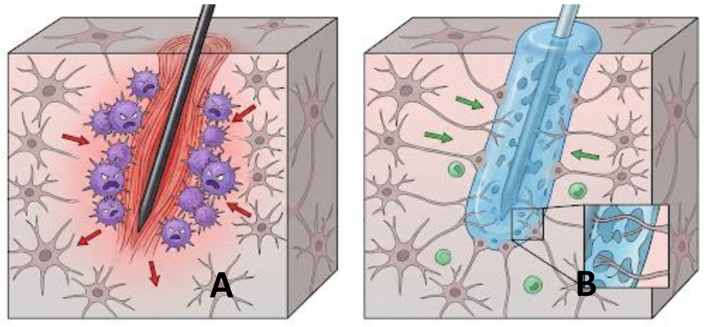
Figure 7.
MXenes VS. Graphene: emerging superiority for bio-applications.
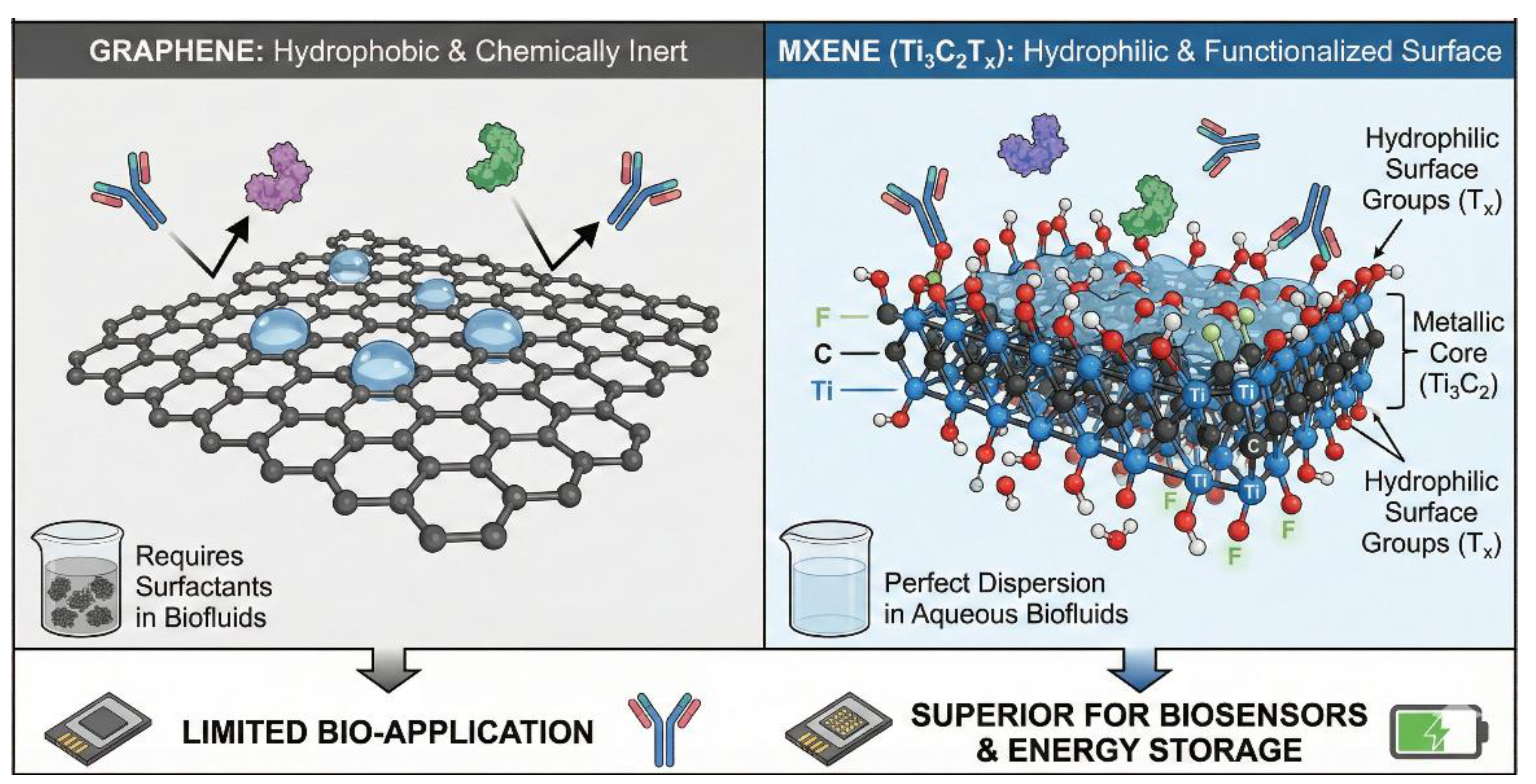
Figure 12.
Fractal geometries increase effective surface area to lower impedance and boost charge injection capacity.
Figure 12.
Fractal geometries increase effective surface area to lower impedance and boost charge injection capacity.
Disclaimer/Publisher’s Note: The statements, opinions and data contained in all publications are solely those of the individual author(s) and contributor(s) and not of MDPI and/or the editor(s). MDPI and/or the editor(s) disclaim responsibility for any injury to people or property resulting from any ideas, methods, instructions or products referred to in the content. |
© 2026 by the authors. Licensee MDPI, Basel, Switzerland. This article is an open access article distributed under the terms and conditions of the Creative Commons Attribution (CC BY) license (http://creativecommons.org/licenses/by/4.0/).
Copyright: This open access article is published under a Creative Commons CC BY 4.0 license, which permit the free download, distribution, and reuse, provided that the author and preprint are cited in any reuse.


