Preprint
Article

This version is not peer-reviewed.

Treadmill Exercise Enhances the Effects of Zoledronate on Bone Microarchitecture and Mechanical Strength in Ovariectomized Rat Model of Osteoporosis

A peer-reviewed version of this preprint was published in:
Journal of Functional Morphology and Kinesiology 2026, 11(2), 159. https://doi.org/10.3390/jfmk11020159

Submitted:

24 March 2026

Posted:

25 March 2026

You are already at the latest version

Abstract

Background: The combination of pharmacological therapy and exercise is frequently recommended for osteoporosis management; however, whether antiresorptive agents may interfere with exercise-induced bone adaptation remains unclear. This study aimed to investigate the independent and combined effects of zoledronate and treadmill exercise on bone microarchitecture and mechanical strength in an ovariectomized rat model. Methods: Twenty-four female Sprague–Dawley rats underwent ovariectomy and were assigned to four groups: Control, zoledronate (ZA), treadmill exercise (T), and combined zoledronate and exercise (ZA + T). An additional sham-operated group was included. Zoledronate was administered as a single subcutaneous injection, and a 6-week treadmill exercise was performed. Bone microarchitecture was assessed using micro-computed tomography, and a three-point bending test was employed for evaluating mechanical properties. Results: The combined ZA + T group demonstrated significant improvements in trabecular bone parameters, including bone volume/tissue volume and trabecular number, compared with the Control group. Mechanical strength parameters, including maximum load and stiffness, were also significantly enhanced in the ZA + T group. Cortical bone parameters exhibited no significant changes. Conclusion: Treadmill exercise did not attenuate the effects of zoledronate and may offer additive benefits in enhancing trabecular bone microarchitecture and mechanical strength. These findings suggest that exercise therapy can complement bisphosphonate treatment and contribute to optimizing therapeutic strategies for osteoporosis.

Keywords: 
;  ;  ;  ;  ;  ;  ;  

1. Introduction

Osteoporosis is characterized by increased bone fragility due to age-related loss of bone mass and bone microarchitecture deterioration, resulting in an elevated risk of fractures even after minor trauma [1]. Osteoporotic fractures in older adults are associated with a decline in activities of daily living, prolonged bedridden status, and worsened prognosis, thereby posing significant challenges for both healthcare and long-term care systems [2]. In Japan, the number of patients with osteoporosis aged ≥40 years has been reported to be approximately 6.4 and 10.7 million based on lumbar spine and proximal femur measurements, respectively [3]. With the rapid progression of a super-aging society, the prevalence of osteoporosis and the incidence of related fractures are anticipated to further increase. These fractures are associated with high recurrence and mortality rates, imposing a considerable socioeconomic burden through increased medical and long-term care costs [4,5]. Osteoporosis has been recently recognized not merely as a condition of reduced bone mass but as a complex disorder involving bone quality deterioration. As understanding of bone microarchitecture and bone remodeling dynamics advances, the significance of nonpharmacological interventions, including exercise therapy, in addition to pharmacological treatment has garnered increasing attention. Therefore, developing effective therapeutic strategies for maintaining bone metabolism and preventing fractures has become an urgent issue.
Pharmacological therapy remains the cornerstone of osteoporosis treatment, among which bisphosphonates (BPs) are broadly used as first-line agents with well-established fracture-prevention efficacy. BPs demonstrate a high affinity for hydroxyapatite and accumulate on bone surfaces. During the process of bone resorption, they are taken up by osteoclasts, where they inhibit the mevalonate pathway, particularly the activity of farnesyl pyrophosphate synthase, thereby inducing osteoclast dysfunction and apoptosis [6]. Through this mechanism, BPs suppress bone resorption and decrease bone turnover. Evidence from animal studies has indicated that zoledronate administration in ovariectomized rat models preserves trabecular bone mass and microarchitecture and prevents reductions in the mechanical strength of the tibia and femur [7]. These findings suggest that BPs can contribute to maintaining bone mass and preserving bone quality. Similarly, clinical studies have revealed that BP agents, including alendronate, risedronate, and zoledronate, significantly mitigate the risk of both vertebral and nonvertebral fractures, and their anti-fracture efficacy is believed to contribute to improved long-term outcomes in patients with osteoporosis [8,9,10]. Although rare but life-threatening adverse events, including BP-related osteonecrosis of the jaw and atypical femoral fractures, have been reported with long-term use, BPs continue to be broadly prescribed worldwide owing to their favorable risk–benefit profile in osteoporosis management [11].
In addition to pharmacological treatment, exercise therapy plays a crucial role in osteoporosis management. Although pharmacological interventions are frequently initiated following the occurrence of fragility fractures, exercise therapy can be implemented at earlier stages of bone loss or as a preventive strategy before fractures occur [12,13]. Exercise contributes to improvements in bone strength and offers added benefits, including fall prevention and physical function maintenance. Evidence from animal studies has demonstrated that treadmill exercise in ovariectomized rats significantly improves trabecular microarchitecture in the proximal femur and increases mechanical strength, including maximal load capacity. Similarly, in experimental models, jump exercise loading has been shown to stimulate bone formation [14,15]. Furthermore, clinical studies have supported the beneficial effects of exercise therapy. In postmenopausal females (mean age, 65.8 years), exercise has been reported to heighten trabecular bone mineral density (BMD) at the distal tibia and cortical BMD at the tibial shaft, as measured using peripheral quantitative computed tomography (CT), by 0.87% and 0.89%, respectively [16]. The main mechanism underlying these osteogenic effects is believed to involve the suppression of sclerostin expression derived from osteocytes. Mechanical loading activates the Wnt/β-catenin signaling pathway, which promotes bone modeling and ultimately improves bone microarchitecture and mechanical strength [17].
In clinical practice, BP therapy combined with exercise is frequently recommended to maximize fracture prevention in patients with osteoporosis. However, the antiresorptive effects of BPs and the mechanical loading-induced osteogenic effects may involve distinct and potentially opposing mechanisms of bone remodeling. Previous studies have mainly focused on bone mass or bone mineral content (BMC), and comprehensive evaluations of trabecular microarchitecture, cortical bone, and mechanical strength under combined therapy remain limited [18,19]. Therefore, whether the suppression of bone resorption by BPs could attenuate the adaptive bone-forming responses induced by exercise remains unclear. Accordingly, this study aimed to investigate the independent and combined effects of zoledronate administration and treadmill exercise using an ovariectomized rat model. Bone microarchitecture was assessed using μCT analysis, and mechanical properties were evaluated using biomechanical testing. Through these analyses, we sought to determine whether combined pharmacological and mechanical interventions could improve bone morphological and mechanical properties.

2. Materials and Methods

2.1. Surgical Techniques Used for Ovariectomy

The Oita University Institutional Animal Ethics Committee approved all animal experiments (approval number: 1624001). Ovariectomy was performed as previously described [20]. Fourteen female Sprague–Dawley rats (12 weeks old; CLEA Japan, Inc., Tokyo, Japan) were anesthetized by intraperitoneal injection of a mixed anesthetic comprising medetomidine (0.15 mg/kg), midazolam (2 mg/kg), and butorphanol (2.5 mg/kg) at total volumes of 0.3–0.4 mL. All surgical procedures were performed under standard sterile conditions. The rats were placed in the supine position, and the abdominal area was disinfected. A midline abdominal skin incision (approximately 2-cm long) was created using a scalpel. To expose the right subperitoneal ovary, the peritoneum was carefully incised and gently spread using forceps. The surrounding adipose tissue was grasped using atraumatic forceps and gently exteriorized to facilitate the elevation of the ovary, oviduct, and a portion of the uterus. The oviduct and the associated blood vessels beneath the ovary were ligated using silk sutures, and the ovaries were excised. Following ovariectomy, the proximal end of the oviduct was returned to the peritoneal cavity. Subsequently, the left ovary was excised using the same procedure. The peritoneum and skin were closed using 3-0 nylon sutures.
In the sham group, the ovaries were exteriorized similarly but were returned to the peritoneal cavity without excision. The peritoneum and skin were closed using identical suturing techniques. Ovariectomized and sham rats were maintained for 8 weeks postoperatively; during this period, body weight was measured to confirm postoperative weight gain.

2.2. Experimental Design

Overall, this study used 24 female Sprague–Dawley rats aged 24 weeks. All animals underwent bilateral ovariectomy to induce osteoporosis. At 8 weeks post-ovariectomy, the rats were randomly assigned to four intervention groups using a randomization procedure (n = 5 per group): Control (Control group; saline administration), zoledronate (ZA group; zoledronate 0.1 mg/kg, single subcutaneous injection; Novartis Pharma K.K., Tokyo, Japan), treadmill exercise (T group), and zoledronate administration combined with treadmill exercise (ZA + T group) (Figure 1). Moreover, four age-matched rats without ovariectomy were included as a sham-operated group (Sham group).
A formal a priori sample-size calculation was not performed. The sample size was determined on the basis of previous animal studies using similar osteoporosis models and following ethical considerations to minimize the number of animals used.
Zoledronate was administered as a single subcutaneous injection to the ZA and ZA + T groups at 8 weeks post-ovariectomy. Zoledronate dose and administration regimen were determined on the basis of our previous studies [21]. At the same time point, treadmill exercise was initiated in the T and ZA + T groups. Before the intervention period, the rats in the T and ZA + T groups underwent a 1-week acclimation period on a rodent treadmill (MK-680; Muromachi Kikai, Tokyo, Japan). During acclimation, the animals ran for 10–15 min/day at a 5–10-m/min speed with a 0° incline. Following the acclimation period, treadmill exercise was continued for an additional 5 weeks, resulting in a 6-week total exercise period. The exercise protocol comprised running at a maximum speed of 20 m/min on a 0° incline for 60 min/days, 5 days/week. The exercise protocol was determined on the basis of previously published studies using treadmill exercise in rat models [18].
All animals were euthanized at 14 weeks post-ovariectomy, corresponding to 6 weeks following the initiation of zoledronate administration and treadmill exercise. Euthanasia was performed under deep anesthesia using the same anesthetic protocol as that used for the ovariectomy procedure. Blood samples were collected by cardiac puncture under anesthesia, and the right femur and gastrocnemius muscle were harvested for subsequent analyses.

2.3. High-Resolution Micro-CT Analysis

Bone micro-CT imaging was performed according to established guidelines [22]. The explanted specimens were scanned using a SkyScan 1172 (Bruker, Kontich, Belgium) with a 20-µm voxel size. Data acquisition was conducted at 100 kV and 100 mA, and the images were reconstructed using a cone-beam algorithm.
Each specimen was positioned on the object stage, and scanning was performed over a 180° rotation with a 105-ms exposure time. Data analysis was performed using a CT Analyzer (Bruker, Kontich, Belgium). The region of interest for femur bone morphometry was defined as 0.8–3.8 mm proximal to the growth-cartilage plate for trabecular bone analysis and 3.0–3.8 mm for cortical bone analysis, and the tissue area in each slice was manually specified. The following parameters were measured: tissue volume (TV), bone volume (BV), BV fraction (BV/TV), trabecular bone thickness (Tb.Th), trabecular bone number (Tb.N), trabecular bone separation (Tb.Sp), cortical bone area (Cr.Ar), and cortical bone thickness (Cr.Th).
Cortical and trabecular bone BMDs were calculated using calibration with a reference phantom containing hydroxyapatite embedded in water-equivalent plastic, which was scanned together with the samples. The CT values obtained from the phantom were used for converting the measured CT values of the bone samples into BMD.

2.4. Biomechanical Analysis

The biomechanical properties of the tibia were assessed using a three-point bending test with a universal material testing system (Instron 5865; Instron, Kanagawa, Japan). Before mechanical testing, the longitudinal and transverse diameters of the mid-diaphysis of each tibia were measured using digital calipers.
For the three-point bending test, each tibia was positioned on the sample supports with the anterior surface facing upward and the midpoint of the tibial shaft aligned with the center of the supports. A load was applied at a constant displacement rate of 1 mm/s until a fracture occurred. The yield load was determined from the load–displacement curve that was obtained in the elastic deformation region.
Mechanical parameters, including stiffness and Young’s modulus, were calculated using the load–displacement data and from the previously measured longitudinal and transverse diameters. All biomechanical parameters were derived using the Instron software.

2.5. Body Weight and Gastrocnemius Muscle Measurement

Body weight was measured for each rat immediately before euthanasia. The gastrocnemius muscle was carefully excised from the surrounding tissues, and connective tissues were removed. The gastrocnemius muscle wet weight was subsequently measured. The relative gastrocnemius muscle weight was calculated by normalizing the muscle wet weight to the body weight to account for inter-individual differences in body size.

2.6. Serum Marker Level Measurement

Serum samples collected by cardiac puncture during euthanasia were used for biomarker analyses. Blood samples were centrifuged to obtain serum, which was stored at −80 °C until analysis. A commercial laboratory (SRL Inc., Tokyo, Japan) measured the serum adiponectin and type I collagen C-terminal telopeptide (1CTP) levels. Adiponectin and 1CTP levels were measured using latex immunoturbidimetry and the RIA-2 antibody method, respectively. Measurements were performed in three rats per group owing to a limited serum volume available for outsourced laboratory analysis.

2.7. Statistical Analysis

All animals were included in the analyses, and no animals or samples were excluded from this study. Statistical analyses were performed using GraphPad Prism (version 11.0; GraphPad Software, San Diego, CA, USA). Group comparisons, including sham-operated animals, were performed for μCT-derived bone morphometric parameters, biomechanical testing outcomes, body weight, and gastrocnemius muscle wet weight; serum biomarkers were measured using enzyme-linked immunosorbent assay (ELISA). Owing to the limited serum volume, ELISA measurements were performed in three rats per group (n = 3).
Data were presented as means ± standard deviations. Differences among groups were analyzed using one-way analysis of variance, followed by the Tukey–Kramer post-hoc test for multiple comparisons when appropriate. A p-value of <0.05 was considered statistically significant.
As an a priori sample-size calculation was not performed, a post-hoc power analysis was conducted using G*Power software (version 3.1). BV/TV obtained from μCT analysis was the primary outcome measure. Based on the observed effect size (f = 1.17), the statistical power for detecting differences among groups was 0.99 at an alpha level of 0.05, indicating sufficient statistical power for the primary outcome.

3. Results

3.1. BMD

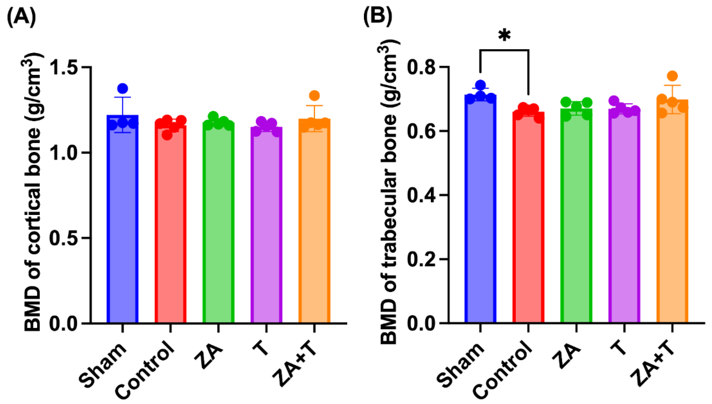
The cortical and trabecular bone BMDs measured using μCT are depicted in Figure 2. No significant differences in cortical bone BMD were observed among the groups (Figure 2A). By contrast, the Control group exhibited a significantly lower trabecular bone BMD than the Sham group (p < 0.05). However, no significant differences were observed among the Control, ZA, T, and ZA + T groups (Figure 2B).

3.2. Trabecular Bone Microarchitecture

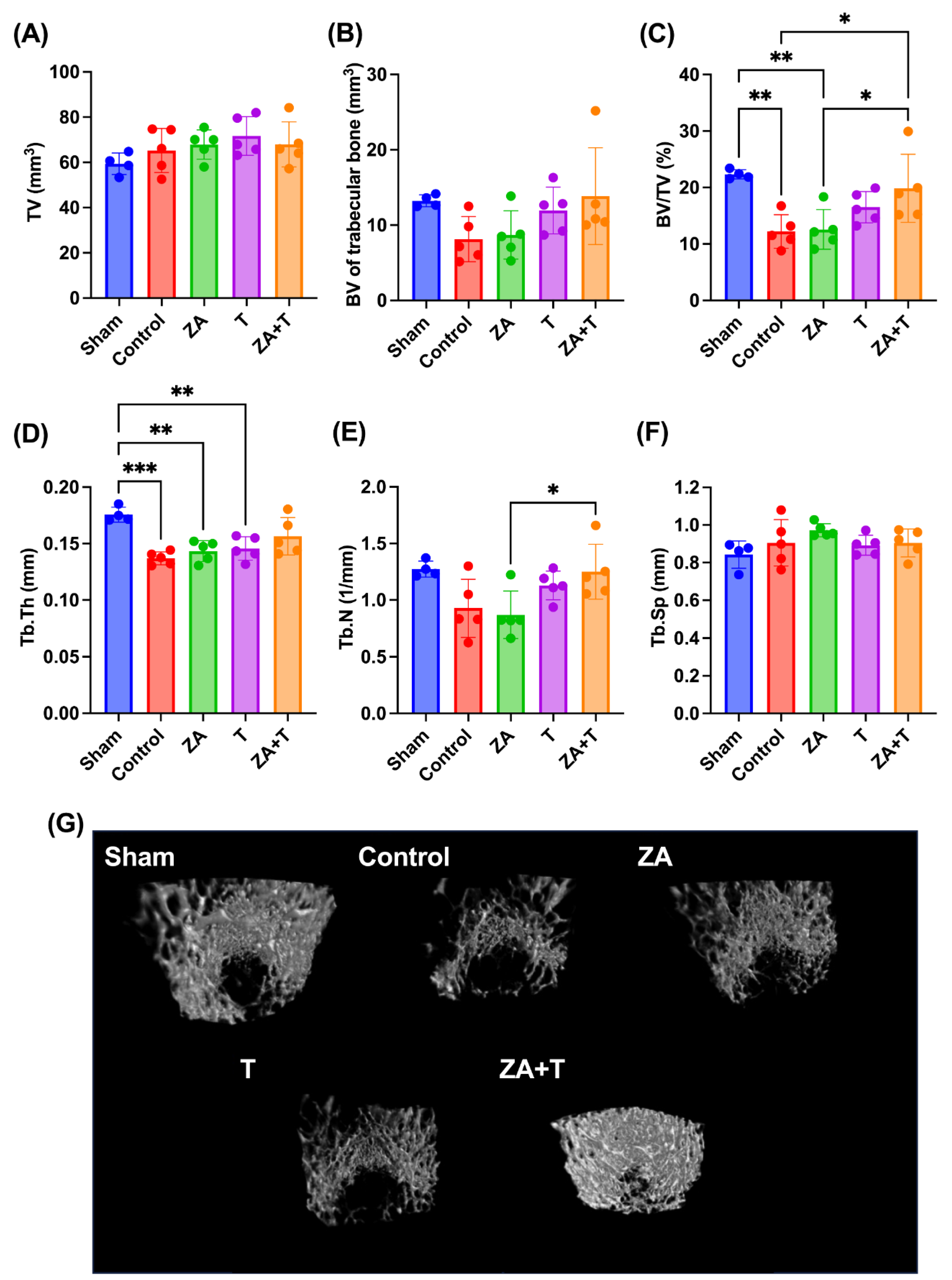
The μCT-derived trabecular bone morphometric parameters are presented in Figure 3. The Control and ZA groups demonstrated a significantly lower BV/TV than the Sham group (p < 0.01). By contrast, the ZA + T group showed a significantly higher BV/TV than the Control and ZA groups (p < 0.05) (Figure 3C). The Control (p < 0.001), ZA (p < 0.01), and T groups (p < 0.01) showed a significantly lower Tb.Th than the Sham group (Figure 3D). The ZA + T group exhibited a significantly higher Tb.N than the ZA group (p < 0.05) (Figure 3E). No significant differences were observed among the groups in terms of TV, BV, and Tb.Sp (Figure 3A, B, F). The representative three-dimensional μCT images of trabecular bone are shown in Figure 3G.

3.3. Cortical Bone Morphometry

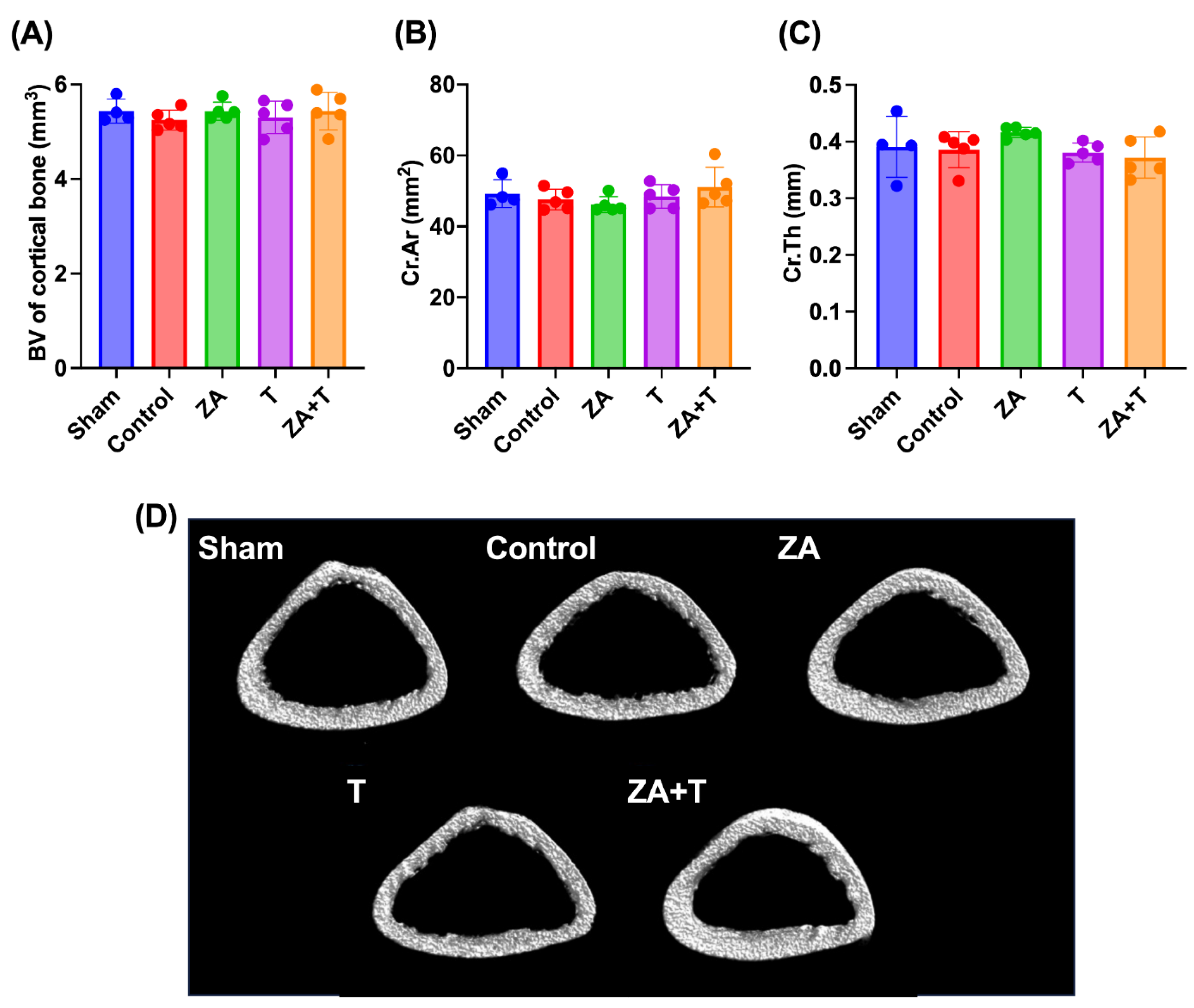
The cortical bone morphometric parameters are depicted in Figure 4. No significant differences were observed among the groups in terms of cortical BV, Cr.Ar, and Cr.Th (Figure 4A–C). Representative cross-sectional μCT images of cortical bone are presented in Figure 4D.

3.4. Mechanical Strength

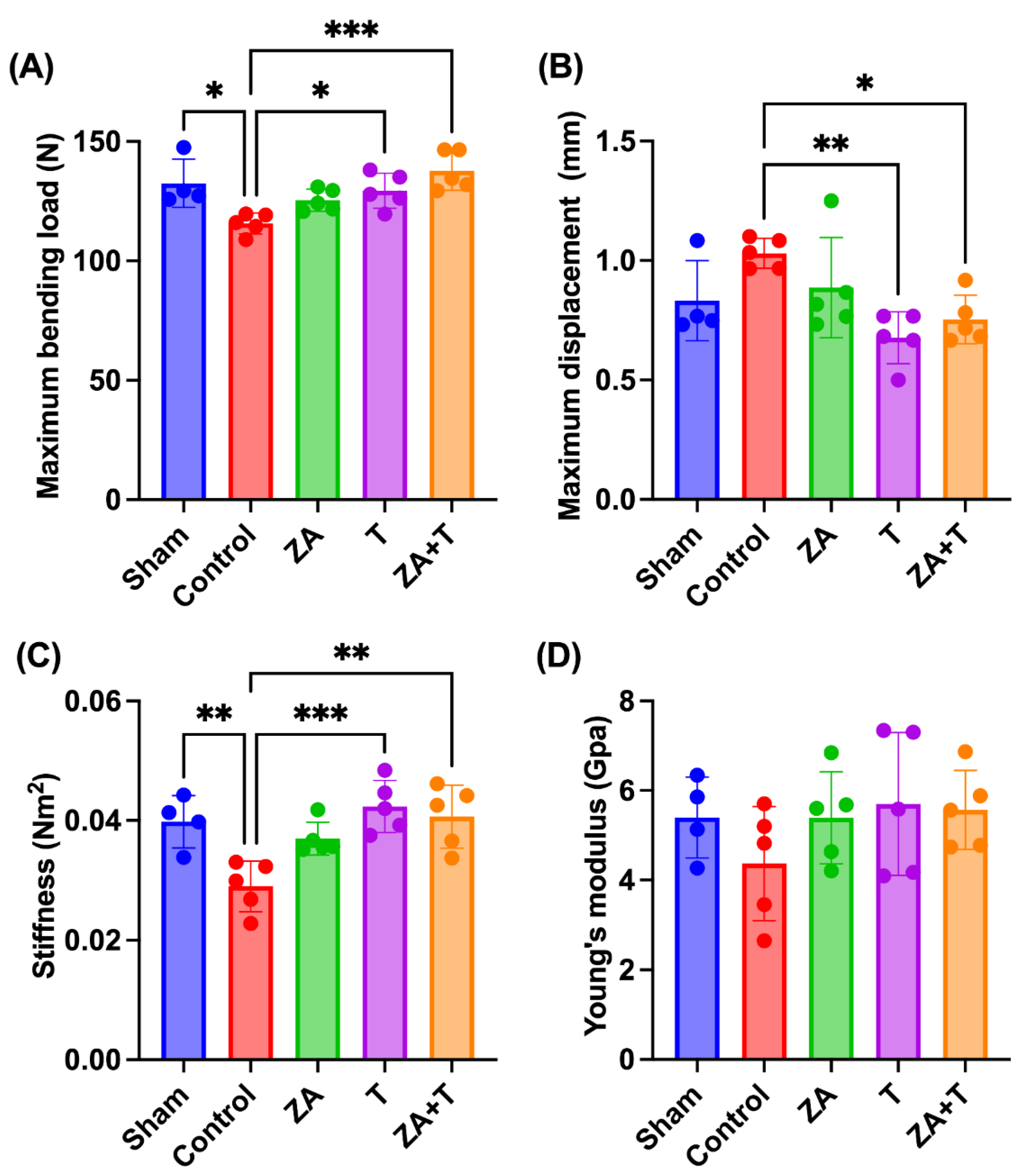
The three-point bending test results are shown in Figure 5. The Sham (p < 0.05), T (p < 0.05), and ZA + T groups (p < 0.001) demonstrated a significantly higher maximum bending load than the Control group (Figure 5A). The T (p < 0.01) and ZA + T groups (p < 0.05) exhibited a significantly lower maximum displacement than the Control group (Figure 5B). The Sham (p < 0.01), T (p < 0.001), and ZA + T groups (p < 0.01) had a significantly higher stiffness than the Control group (Figure 5C). No significant differences in Young’s modulus were noted among the groups (Figure 5D).

3.5. Body Weight and Gastrocnemius Muscle Measurements

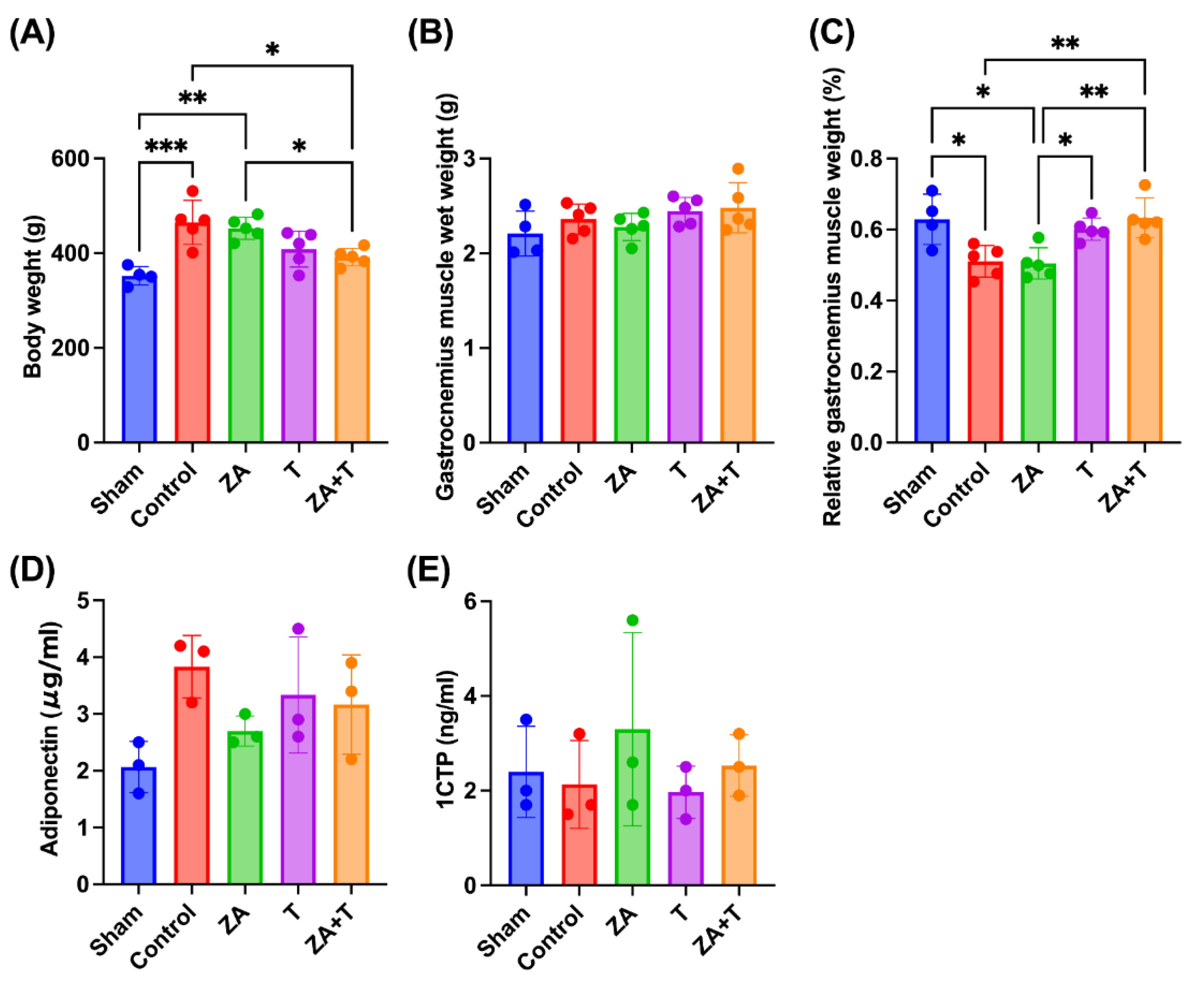
The body weight, gastrocnemius muscle wet weight, and relative gastrocnemius muscle weight are shown in Figure 6A–C. The Control (p < 0.001) and ZA groups (p < 0.01) showed a significantly higher body weight than the Sham group. Moreover, the Control (p < 0.05) and ZA groups (p < 0.05) exhibited a significantly higher body weight than the ZA + T group (Figure 6A).
No significant differences were observed in gastrocnemius muscle wet weight among the groups (Figure 6B). The Control (p < 0.05) and ZA groups (p < 0.05) demonstrated a significantly lower relative gastrocnemius muscle weight than the Sham group. Furthermore, the ZA group showed a significantly higher relative gastrocnemius muscle weight than the T group (p < 0.05). Additionally, the Control and ZA groups exhibited significantly higher values than the ZA + T group (p < 0.01) (Figure 6C).

3.6. Serum Biomarkers

The serum adiponectin and 1CTP levels measured using ELISA are shown in Figure 6D, E. No significant differences in adiponectin or 1CTP levels were noted among the groups.

4. Discussion

This study investigated the independent and combined effects of zoledronate administration and treadmill exercise on bone microarchitecture and mechanical strength in an ovariectomized rat model of osteoporosis. The principal finding of this study was that treadmill exercise did not attenuate the antiresorptive effects of zoledronate and could instead offer additive benefits for bone structural and mechanical properties. In particular, compared with pharmacological treatment alone, zoledronate administration combined with treadmill exercise significantly improved trabecular bone microarchitecture and mechanical strength. These findings suggest that mechanical loading through exercise can complement pharmacological therapy without compromising the therapeutic effects of BPs.
This study revealed that zoledronate administration combined with treadmill exercise yielded additive improvements in trabecular bone microarchitecture and mechanical strength compared with either intervention alone. This finding is noteworthy because previous studies investigating combined pharmacological and exercise interventions have predominantly focused on changes in BMC, whereas comprehensive analyses of trabecular microarchitectural parameters remain relatively scarce [18].
BPs accumulate on bone surfaces and are taken up by osteoclasts during bone resorption, where they inhibit the mevalonate pathway and induce osteoclast apoptosis [6]. Through this mechanism, BPs suppress bone resorption and enhance bone structure and mechanical strength. This process is frequently described as remodeling-based bone formation, wherein bone turnover suppression reduces bone formation and resorption. However, the period during which bone formation exceeds bone resorption, referred to as the anabolic window, may be prolonged, thereby contributing to improvements in bone morphological and mechanical properties [23]. By contrast, the major mechanism underlying exercise-induced bone adaptation is believed to involve osteocyte-derived sclerostin expression suppression in response to mechanical loading [24,25]. This suppression activates the Wnt/β-catenin signaling pathway, which promotes bone formation while simultaneously inhibiting bone resorption [17]. Consequently, exercise induces modeling-based bone formation and enhances bone structural integrity [14,26]. As BP therapy mainly suppresses bone resorption through remodeling-dependent mechanisms within the endosteal region, whereas exercise encourages bone formation through modeling-dependent mechanisms at the periosteal surface, the sites and mechanisms of action of these two interventions may be complementary [18]. Consequently, their combination may exert additive effects on trabecular microarchitecture and mechanical strength while preserving each other’s biological actions. Nevertheless, this study was restricted to morphological assessments using μCT and biomechanical testing. Serum bone metabolic markers and histological analyses were not comprehensively investigated. Therefore, the previously proposed mechanistic interpretations remain speculative. To clarify the biological interactions between BP therapy and exercise-induced mechanical loading, further studies incorporating molecular and histological analyses will be crucial. Accordingly, the present findings should be interpreted as preliminary experimental evidence suggesting the potential benefits of combined pharmacological and mechanical interventions for improving bone structural and mechanical properties.
In this study, zoledronate monotherapy did not generate significant improvements in bone microarchitecture or mechanical strength following ovariectomy. This finding contrasts with previous studies reporting marked bone-protective effects of zoledronate in similar animal models. For example, using in vivo micro-CT, Brouwers et al. demonstrated that early administration of zoledronate at 2 weeks following ovariectomy restored trabecular microarchitecture to near baseline levels, whereas delayed administration at 8 weeks caused only partial recovery [27]. Differences in dosing regimen, administration timing, and observation duration may explain these discrepancies. Furthermore, the relatively low dose and single-administration protocol used in the present study may have contributed to the limited response. In this study, zoledronate was administered at a relatively late stage following ovariectomy, and the observation period was limited to 6 weeks. Therefore, the limited efficacy observed in the ZA group may reflect insufficient time for the full pharmacological effects to manifest. To elucidate the time-dependent effects of zoledronate on bone structural and mechanical properties, longer-term studies are warranted.
Notably, the improvements in trabecular bone microarchitecture observed in this study were accompanied by enhanced mechanical strength, suggesting that the combined intervention of zoledronate administration and treadmill exercise translated into functionally meaningful skeletal benefits. Remarkably, these beneficial effects were predominantly observed in the trabecular compartment, whereas cortical bone parameters were not significantly altered. This preferential response of the trabecular bone may be attributed to its higher metabolic activity and greater mechanosensitivity than the cortical bone [28,29]. The trabecular bone undergoes more rapid remodeling and is therefore more responsive to pharmacological and mechanical stimuli. By contrast, the cortical bone exhibits a slower turnover rate and may necessitate a longer intervention period or higher mechanical loading to demonstrate detectable structural changes. Moreover, the loading intensity applied in this study could be inadequate to induce periosteal adaptation in the cortical bone. Despite the limited effects observed in the cortical bone, the improvements in trabecular microarchitecture may have substantially contributed to the observed increases in mechanical strength. These findings align with previous studies indicating that mechanical loading-based interventions preferentially influence the trabecular bone, particularly under conditions of estrogen deficiency [30,31].
In this study, gastrocnemius muscle wet weight and serum biomarkers were evaluated as secondary outcomes. Although the groups demonstrated no statistically significant differences in terms of absolute muscle wet weight, the combined intervention group tended to exhibit a higher relative muscle weight than the single-treatment groups, suggesting a potential beneficial effect of zoledronate administration combined with exercise on skeletal muscle mass. The effects of BPs on skeletal muscle remain debated. In vitro studies have suggested that alendronate can impair cytoskeletal organization and inhibit differentiation in undifferentiated human skeletal muscle cells, while exerting minimal effects on mature myotubes [32]. By contrast, clinical studies have suggested an association between BP therapy and increases in skeletal muscle mass indices, potentially through indirect effects mediated by improved bone metabolism and physical activity levels [33]. In the present study, the observed tendency toward increased muscle mass in the combined group may reflect the additive influence of mechanical loading and systemic effects of pharmacological treatment; however, the underlying mechanisms remain unclear. Increases in skeletal muscle mass may affect bone metabolism through muscle–bone crosstalk mediated by myokines, including insulin-like growth factor 1 and myostatin, potentially contributing to improved bone structure and strength [34,35,36].
Furthermore, serum adiponectin and 1CTP levels were assessed in a limited number of samples (n = 3 per group) as exploratory outcomes to investigate potential changes in lipid metabolism and bone turnover associated with ovariectomy and exercise intervention. However, the groups demonstrated no significant differences, and the small sample size limited the interpretability of these findings. These analyses were exploratory in nature and were not intended as primary outcome measures. Therefore, the skeletal muscle- and serum biomarker-related findings should be considered hypothesis-generating. To elucidate the interactions among muscles, bones, and adipose tissues, further studies with larger sample sizes and comprehensive analyses, including myokines and adipokines, are warranted.
The findings of this study have potential clinical implications for managing osteoporosis. The present results suggest that pharmacological therapy combined with mechanical loading can more effectively improve bone structural integrity and mechanical strength than either intervention alone, without compromising the antiresorptive effects of BPs. These findings support the clinical recommendation that exercise therapy should be encouraged even during BP therapy to optimize fracture prevention strategies.
However, this study had several limitations. First, this was an experimental study using an ovariectomized rat model, and the results may not be directly generalizable to human clinical populations. Therefore, the findings may not fully reflect clinical conditions owing to differences between animal models and human osteoporosis. Second, the intervention period was relatively short, and longer-term effects of combined therapy on bone remodeling remained unclear. Lastly, mechanistic insights were limited, as molecular and histological analyses were not performed. Moreover, serum biomarker analyses were conducted in a small sample size, which may have limited the statistical power for detecting subtle changes in bone metabolism. Despite these limitations, the present findings offer preliminary evidence supporting the potential benefits of combining BP therapy with exercise. To elucidate the underlying mechanisms and establish optimal therapeutic strategies for improving bone health, further studies incorporating larger sample sizes, longer intervention periods, and comprehensive molecular and histological evaluations are required.

5. Conclusion

This study demonstrated that zoledronate administration combined with treadmill exercise caused additive improvements in trabecular bone microarchitecture and mechanical strength in an ovariectomized rat model of osteoporosis. Notably, treadmill exercise did not attenuate the antiresorptive effects of zoledronate, suggesting that pharmacological and mechanical interventions can be effectively combined without compromising their respective benefits.
These findings suggest that exercise therapy can complement BP therapy, contributing to optimized bone quality and strength. Although the underlying mechanisms require further investigation, the present results offer preliminary evidence supporting the potential utility of combined therapeutic strategies for improving bone health and preventing fractures.

Author Contributions

Conceptualization, Y.T.; Methodology, Y.T. and M.K.; Investigation (Operative Procedure), Y.T., T.K., R.T., and M.K.; Investigation (Micro-CT analysis), Y.T.; Investigation (Biomechanical Analysis), Y.T., T.K., R.T., T.O, and M.K.; Investigation (ELISA), Y.T., R.H., and M.K.; Formal analysis, Y.T.; Writing—Original Draft Preparation, Y.T.; Writing—Review and Editing, M.K. and N.K.; Supervision, M.K.; Project Administration, N.K. All authors have read and agreed to the published version of the manuscript.

Funding

This study was supported by JSPS KAKENHI (Grant Number JP19K19870).

Institutional Review Board Statement

This study was approved by the Oita University Animal Research Committee before animal experimentation (Oita University Institutional Animal Ethics Committee no. 1624001).

Data Availability Statement

The datasets generated and analyzed during the current study are available from the corresponding author upon reasonable request.

Acknowledgments

The authors gratefully acknowledge Ms. Minori Oshima for her valuable technical assistance during the experiments and her support in laboratory procedures. During the preparation of this manuscript, the authors used ChatGPT (OpenAI) for language refinement and structural editing. After using this tool, the authors reviewed and edited the content as needed and take full responsibility for the content of the published article.

Conflicts of Interest

The authors declare that there are no conflicts of interest related to this study.:

Abbreviations

The following abbreviations are used in this manuscript:
ADL Activities of daily living.
BMC Bone mineral content.
BMD Bone mineral density.
BP Bisphosphonate.
BV Bone volume.
BV/TV Bone volume fraction.
Cr.Ar Cortical bone area.
Cr.Th Cortical bone thickness.
ELISA Enzyme-linked immunosorbent assay.
FPPS Farnesyl pyrophosphate synthase.
IGF-1 Insulin-like growth factor 1.
ROI Region of interest.
Tb.N Trabecular bone number.
Tb.Sp Trabecular bone separation.
Tb.Th Trabecular bone thickness.
TV Tissue volume.
1CTP Type I collagen C-terminal telopeptide.

References

  1. Rachner, T.D.; Khosla, S.; Hofbauer, L.C. Osteoporosis: Now and the future. Lancet 2011, 377, 1276–1287. [Google Scholar] [CrossRef]
  2. Cauley, J.A.; Thompson, D.E.; Ensrud, K.C.; Scott, J.C.; Black, D.M. Risk of mortality following clinical fractures. Osteopor. Int. 2000, 11, 556–561. [Google Scholar] [CrossRef]
  3. Yoshimura, N.; Muraki, S.; Oka, H.; Mabuchi, A.; En-Yo, Y.; Yoshida, M.; Saika, A.; Yoshida, H.; Suzuki, T.; Yamamoto, S.; Nakamura, K.; Kawaguchi, H.; Yoshimura, N. Prevalence of knee osteoarthritis, lumbar spondylosis, and osteoporosis in Japanese men and women: The research on osteoarthritis/osteoporosis against disability study. J. Bone Miner. Metab. 2009, 27, 620–628. [Google Scholar] [CrossRef]
  4. Mori, T.; Komiyama, J.; Fujii, T.; Sanuki, M.; Kume, K.; Kato, G.; Mori, Y.; Ueshima, H.; Matsui, H.; Tamiya, N. Medical expenditures for fragility hip fracture in Japan: A study using the nationwide health insurance claims database. Arch. Osteoporos. 2022, 17, 61. [Google Scholar] [CrossRef]
  5. Hagino, H. Fragility fracture prevention: Review from a Japanese perspective. Yonago Acta Med. 2012, 55, 21–28. [Google Scholar] [CrossRef] [PubMed]
  6. Reszka, A.A.; Rodan, G.A. Bisphosphonate mechanism of action. Curr. Rheumatol. Rep. 2003, 5, 65–74. [Google Scholar] [CrossRef] [PubMed]
  7. Perilli, E.; Le, V.; Ma, B.; Salmon, P.; Reynolds, K.; Fazzalari, N.L. Detecting early bone changes using in vivo micro-CT in ovariectomized, zoledronic acid-treated, and sham-operated rats. Osteopor. Int. 2010, 21, 1371–1382. [Google Scholar] [CrossRef]
  8. Black, D.M.; Reid, I.R.; Boonen, S.; Bucci-Rechtweg, C.; Cauley, J.A.; Cosman, F.; Cummings, S.R.; Hue, T.F.; Lippuner, K.; Lakatos, P.; Leung, P.C.; Man, Z.; Mautalen, C.; Mesenbrink, P.; Hu, H.; Caminis, J.; Tong, K.; Rosario-Jansen, T.; Krasnow, J.; Eriksen, E.F. The effect of 3 versus 6 years of zoledronic acid treatment of osteoporosis: A randomized extension to the HORIZON-Pivotal Fracture Trial (PFT). J. Bone Miner. Res. 2012, 27, 243–254. [Google Scholar] [CrossRef]
  9. Harrington, J.T.; Ste-Marie, L.G.; Brandi, M.L.; Civitelli, R.; Fardellone, P.; Grauer, A.; Barton, I.; Boonen, S. Risedronate rapidly reduces the risk for nonvertebral fractures in women with postmenopausal osteoporosis. Calcif. Tissue Int. 2004, 74, 129–135. [Google Scholar] [CrossRef]
  10. Cummings, S.R.; Black, D.M.; Thompson, D.E.; Applegate, W.B.; Barrett-Connor, E.; Musliner, T.A.; Palermo, L.; Prineas, R.; Rubin, S.M.; Scott, J.C.; Vogt, T.; Wallace, R.; Yates, A.J.; LaCroix, A.Z. Effect of alendronate on risk of fracture in women with low bone density but without vertebral fractures: Results from the Fracture Intervention Trial. JAMA 1998, 280, 2077–2082. [Google Scholar] [CrossRef] [PubMed]
  11. Reid, I.R.; Billington, E.O. Drug therapy for osteoporosis in older adults. Lancet 2022, 399, 1080–1092. [Google Scholar] [CrossRef] [PubMed]
  12. Sakuma, M.; Endo, N.; Oinuma, T.; Endo, E.; Yazawa, T.; Watanabe, K.; Watanabe, S. Incidence and outcome of osteoporotic fractures in 2004 in Sado City, Niigata Prefecture, Japan. J. Bone Miner. Metab. 2008, 26, 373–378. [Google Scholar] [CrossRef] [PubMed]
  13. Solomon, D.H.; Avorn, J.; Katz, J.N.; Finkelstein, J.S.; Arnold, M.; Polinski, J.M.; Brookhart, M.A. Compliance with osteoporosis medications. Arch. Intern. Med. 2005, 165, 2414–2419. [Google Scholar] [CrossRef]
  14. Kang, Y.S.; Kim, C.H.; Kim, J.S. The effects of downhill and uphill exercise training on osteogenesis-related factors in ovariectomy-induced bone loss. J. Exerc. Nutr. Biochem. 2017, 21, 1–10. [Google Scholar] [CrossRef]
  15. Okubo, R.; Sanada, L.S.; Castania, V.A.; Louzada, M.J.; de Paula, F.J.A.; Maffulli, N.; Shimano, A.C. Jumping exercise preserves bone mineral density and mechanical properties in osteopenic ovariectomized rats even following established osteopenia. Osteopor. Int. 2017, 28, 1461–1471. [Google Scholar] [CrossRef]
  16. Polidoulis, I.; Beyene, J.; Cheung, A.M. The effect of exercise on pQCT parameters of bone structure and strength in postmenopausal women—A systematic review and meta-analysis of randomized controlled trials. Osteopor. Int. 2012, 23, 39–51. [Google Scholar] [CrossRef]
  17. Robling, A.G.; Niziolek, P.J.; Baldridge, L.A.; Condon, K.W.; Allen, M.R.; Alam, I.; Mantila, S.M.; Gluhak-Heinrich, J.; Bellido, T.M.; Harris, S.E.; Turner, C.H. Mechanical stimulation of bone in vivo reduces osteocyte expression of Sost/Sclerostin. J. Biol. Chem. 2008, 283, 5866–5875. [Google Scholar] [CrossRef]
  18. Fuchs, R.K.; Shea, M.; Durski, S.L.; Winters-Stone, K.M.; Widrick, J.; Snow, C.M. Individual and combined effects of exercise and alendronate on bone mass and strength in ovariectomized rats. Bone 2007, 41, 290–296. [Google Scholar] [CrossRef]
  19. Lespessailles, E.; Jaffre, C.; Beaupied, H.; Nanyan, P.; Dolleans, E.; Benhamou, C.L.; Courteix, D. Does exercise modify the effects of zoledronic acid on bone mass, microarchitecture, biomechanics, and turnover in ovariectomized rats? Calcif. Tissue Int. 2009, 85, 146–157. [Google Scholar] [CrossRef]
  20. Takase, R.; Tsubouchi, Y.; Otsu, T.; Kataoka, T.; Iwasaki, T.; Kataoka, M.; Tsumura, H. The effects of romosozumab combined with active vitamin D3 on fracture healing in ovariectomized rats. J. Orthop. Surg. Res. 2022, 17, 376. [Google Scholar] [CrossRef] [PubMed]
  21. Tsubouchi, Y.; Ikeda, S.; Kataoka, M.; Tsumura, H. Combination therapy with low-dose teriparatide and zoledronate contributes to fracture healing on rat femoral fracture model. J. Orthop. Surg. Res. 2018, 13, 267. [Google Scholar] [CrossRef]
  22. Bouxsein, M.L.; Boyd, S.K.; Christiansen, B.A.; Guldberg, R.E.; Jepsen, K.J.; Müller, R. Guidelines for assessment of bone microstructure in rodents using micro-computed tomography. J. Bone Miner. Res. 2010, 25, 1468–1486. [Google Scholar] [CrossRef]
  23. Jensen, P.R.; Andersen, T.L.; Chavassieux, P.; Roux, J.-P.; Delaisse, J.-M. Bisphosphonates impair the onset of bone formation at remodeling sites. Bone 2021, 145, 115850. [Google Scholar] [CrossRef]
  24. Liao, H.W.; Huang, T.H.; Chang, Y.H.; Liou, H.H.; Chou, Y.H.; Sue, Y.M.; Hung, P.H.; Chang, Y.T.; Ho, P.C.; Tsai, K.J. Exercise alleviates osteoporosis in rats with mild chronic kidney disease by decreasing sclerostin production. Int. J. Mol. Sci. 2019, 20, 3869. [Google Scholar] [CrossRef]
  25. Ominsky, M.S.; Boyce, R.W.; Li, X.; Ke, H.Z. Effects of sclerostin antibodies in animal models of osteoporosis. Bone 2017, 96, 63–75. [Google Scholar] [CrossRef] [PubMed]
  26. Eriksen, E.F.; Chapurlat, R.; Boyce, R.W.; Shi, Y.; Brown, J.P.; Horlait, S.; Betah, D.; Libanati, C.; Chavassieux, P. Modeling-based bone formation after 2 months of romosozumab treatment: Results from the FRAME clinical trial. J. Bone Miner. Res. 2022, 37, 36–40. [Google Scholar] [CrossRef] [PubMed]
  27. Brouwers, J.E.M.; Lambers, F.M.; Gasser, J.A.; van Rietbergen, B.; Huiskes, R. Bone degeneration and recovery after early and late bisphosphonate treatment of ovariectomized Wistar rats assessed by in vivo micro-computed tomography. Calcif. Tissue Int. 2008, 82, 202–211. [Google Scholar] [CrossRef]
  28. Oftadeh, R.; Perez-Viloria, M.; Villa-Camacho, J.C.; Vaziri, A.; Nazarian, A. Biomechanics and mechanobiology of trabecular bone: A review. J. Biomech. Eng. 2015, 137, 0108021–01080215. [Google Scholar] [CrossRef] [PubMed]
  29. Gong, H.; Zhu, D.; Gao, J.; Lv, L.; Zhang, X. An adaptation model for trabecular bone at different mechanical levels. Biomed. Eng. Online 2010, 9, 32. [Google Scholar] [CrossRef]
  30. Takagi, S.; Yamashita, T.; Miura, T. Does a treadmill running exercise contribute to preventing deterioration of bone mineral density and bone quality of the femur in KK-Ay mice, a type 2 diabetic animal model? Calcif. Tissue Int. 2017, 101, 631–640. [Google Scholar] [CrossRef] [PubMed]
  31. Lespessailles, E.; Jaffré, C.; Beaupied, H.; Nanyan, P.; Dolléans, E.; Benhamou, C.L.; Courteix, D. Does exercise modify the effects of zoledronic acid on bone mass, microarchitecture, biomechanics, and turnover in ovariectomized rats? Calcif. Tissue Int. 2009, 85, 146–157. [Google Scholar] [CrossRef]
  32. Shiomi, K.; Nagata, Y.; Kiyono, T.; Harada, A.; Hashimoto, N. Differential impact of the bisphosphonate alendronate on undifferentiated and terminally differentiated human myogenic cells. J. Pharm. Pharmacol. 2014, 66, 418–427. [Google Scholar] [CrossRef] [PubMed]
  33. Harada, A.; Ito, S.; Matsui, Y.; Sakai, Y.; Takemura, M.; Tokuda, H.; Hida, T.; Shimokata, H. Effect of alendronate on muscle mass: Investigation in patients with osteoporosis. Osteoporos. Sarcopenia 2015, 1, 53–58. [Google Scholar] [CrossRef]
  34. Shao, M.; Wang, Q.; Lv, Q.; Zhang, Y.; Gao, G.; Lu, S. Advances in the research on myokine-driven regulation of bone metabolism. Heliyon 2024, 10, e22547. [Google Scholar] [CrossRef]
  35. Kim, B.J. Effects of muscles on bone metabolism—With a focus on myokines. Ann. Geriatr. Med. Res. 2022, 26, 63–71. [Google Scholar] [CrossRef] [PubMed]
  36. Bettis, T.; Kim, B.J.; Hamrick, M.W. Impact of muscle atrophy on bone metabolism and bone strength: Implications for muscle–bone crosstalk with aging and disuse. Osteopor. Int. 2018, 29, 1713–1720. [Google Scholar] [CrossRef] [PubMed]
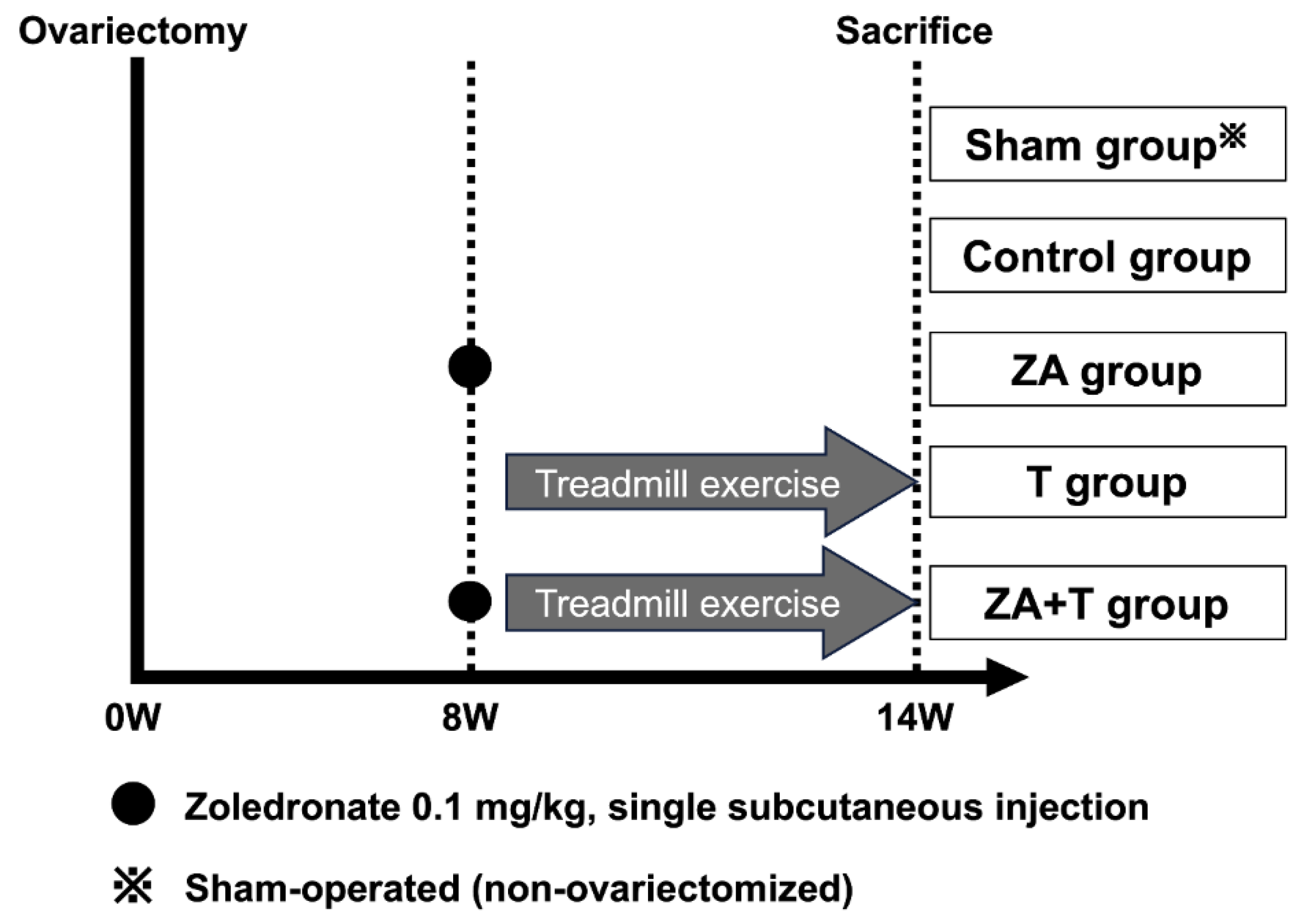
Figure 1. Experimental design of the study. Schematic overview of the experimental protocol. All rats underwent bilateral ovariectomy at week 0 to induce osteoporosis. At 8 weeks post-ovariectomy, rats were randomly assigned to four intervention groups (n = 5 per group): Control group (saline administration), zoledronate group (ZA; single subcutaneous injection of zoledronate, 0.1 mg/kg), treadmill exercise group (T), and zoledronate combined with treadmill exercise group (ZA + T). Treadmill exercise was performed for 6 weeks, including a 1-week acclimation period (60 min/day, 5 days/week, maximum speed of 20 m/min). Zoledronate was administered as a single subcutaneous injection at week 8. All rats were euthanized at week 14 post-ovariectomy. An additional sham-operated group without ovariectomy (Sham group) was included.
Preprints 204760 g001
Figure 2. Bone mineral density (BMD) measured using μCT. (A) Cortical bone BMD and (B) trabecular bone BMD. Data are presented as means ± standard deviations (SDs). Statistical analysis was performed using one-way analysis of variance (ANOVA), followed by the Tukey–Kramer post-hoc test. *p < 0.05. n = 4 for the Sham group; n = 5 for the other groups.
Preprints 204760 g002
Figure 3. Trabecular bone microarchitecture assessed using μCT. (A) Total volume (TV), (B) bone volume (BV), (C) bone volume fraction (BV/TV), (D) trabecular bone thickness (Tb.Th), (E) trabecular bone number (Tb.N), and (F) trabecular bone separation (Tb.Sp). (G) Representative three-dimensional μCT images of the trabecular bone. Data are presented as means ± SDs. Statistical analysis was performed using one-way ANOVA, followed by the Tukey–Kramer post-hoc test. *p < 0.05, **p < 0.01, ***p < 0.001. n = 4 for the Sham group; n = 5 for the other groups.
Preprints 204760 g003
Figure 4. Cortical bone morphometric parameters assessed using μCT. (A) Cortical BV, (B) cortical bone area (Cr.Ar), and (C) cortical bone thickness (Cr.Th). (D) Representative cross-sectional μCT images of the cortical bone. Data are presented as means ± SDs. n = 4 for the Sham group; n = 5 for the other groups.
Preprints 204760 g004
Figure 5. Mechanical properties of the femur assessed using the three-point bending test. (A) Maximum bending load, (B) maximum displacement, (C) stiffness, and (D) Young’s modulus. Data are presented as means ± SDs. Statistical significance was determined using one-way ANOVA, followed by the Tukey–Kramer post-hoc test. *p < 0.05, **p < 0.01, ***p < 0.001. n = 4 for the Sham group; n = 5 for the other groups.
Preprints 204760 g005
Figure 6. Body weight, gastrocnemius muscle measurements, and serum biomarkers. (A) Body weight, (B) gastrocnemius muscle wet weight, and (C) relative gastrocnemius muscle weight normalized to body weight are shown. (D) Serum adiponectin and (E) type I collagen C-terminal telopeptide levels measured using enzyme-linked immunosorbent assay (ELISA) are also presented. Data are presented as means ± SDs. Statistical analysis was performed using one-way ANOVA, followed by the Tukey–Kramer post-hoc test. *p < 0.05, **p < 0.01, ***p < 0.001. n = 4 for the Sham group; n = 5 for the other groups. ELISA measurements were performed in three rats per group (n = 3).
Preprints 204760 g006
Disclaimer/Publisher’s Note: The statements, opinions and data contained in all publications are solely those of the individual author(s) and contributor(s) and not of MDPI and/or the editor(s). MDPI and/or the editor(s) disclaim responsibility for any injury to people or property resulting from any ideas, methods, instructions or products referred to in the content.
Copyright: This open access article is published under a Creative Commons CC BY 4.0 license, which permit the free download, distribution, and reuse, provided that the author and preprint are cited in any reuse.
Prerpints.org logo

Preprints.org is a free preprint server supported by MDPI in Basel, Switzerland.

Subscribe

Disclaimer

Terms of Use

Privacy Policy

Privacy Settings

© 2026 MDPI (Basel, Switzerland) unless otherwise stated