

Submitted:
20 March 2026
Posted:
23 March 2026
You are already at the latest version
Abstract
Soil is a fundamental resource for humankind’s sustenance; however, excessive or inadequate use contributes to its degradation and erosion, therefore limiting its health and capacity to sustain biological activity. An option for restoration is the use of biocrusts—formations of photosynthetic organisms that protect and stimulate soil. The present research aimed to determine whether changes in soil conditions occur when treated with cyanobacterial biocrusts, specifically examining differences in microbial counts. Our results show significant changes in soil humidity, indicating an improvement in water retention. We also found that all microbial counts increased in the treatment group, but this increase was not statistically significant; additionally, there were no changes in the total organic carbon content. These results could be linked to low production of exopolysaccharides by the used species, low photosynthetic activity, and the need for a longer evaluation period for biocrust treatment.
Keywords:
biocrusts
; cyanobacteria
; cell count
; soil microbiology
; soil restoration
1. Introduction
Soil is a critical resource for humankind: approximately 38% of the earth’s area is dedicated to agricultural activities [1], which form the foundation for most of the food and textile industries. However, its extensive and unsustainable use drives its degradation, which manifests in different interconnected phenomena, including erosion, diminished nutrient availability, and loss of its biological diversity [2,3]. According to Colombia’s Institute of Hydrology, Meteorology and Environmental Studies [4], 40% of the country’s soil presents some degree of erosion. This degradation compromises ecosystem’s health and integrity and reduces the productivity of soil-dependent systems.
Identifying methods to mitigate or counteract soil degradation is therefore important for ecosystem preservation, food security and sustainable development [5,6]. One promising strategy involves the use of biological crusts (or biocrusts), formations of photoautotrophic organisms like bryophytes, algae, lichen or cyanobacteria in association with microorganisms which cover soil surface [7].
Research indicates that biocrusts’ activity enhances key soil properties, impacting in water availability, improving soil structure and fertility and elevating levels of organic carbon and nitrogen, which aid plant germination and [8]. The formation of biocrusts can be driven by several cyanobacteria groups —mostly filamentous cyanobacteria, like those belonging to the orders Oscillatoriales, Nostocales or Leptolyngbyales—, many of which are also capable of nitrogen fixation [9,10,11]. Furthermore, most of these biocrust forming cyanobacteria also secrete polysaccharides, which have in turn been reported to be used as nitrogen and carbon sources for heterotrophic soil microbes [9,12].
This means that the conditions which favor the increase in soil microbial density —namely water, nitrogen and carbon availability— [13,14] are precisely those conditions derived from the presence of cyanobacterial biocrusts [8,12,15]. Indeed, there is work showing that biocrusts such as those formed by bryophytes or the cyanobacteria Microcoleus vaginatus enhance the diversity and development of the edaphic microbiome [16,17]. Despite these links and evidence, most studies on the use of cyanobacterial biocrusts for soil restoration focus on quantifying changes in soil’s structure or physicochemical properties [9,15], but there remains a gap in the understanding of their effects on soil microbiology.
Consequently, a clear understanding of how biocrusts influence microbial diversity and abundance remains elusive. Elucidating these relationships might be important for a better understanding of the ecological effects of biocrusts, and to contribute into developing strategies for soil restoration, or the creation of new biotechnological approaches to soil management or improvement.
This research aimed to evaluate the effects of biocrusts formed by a cyanobacteria isolated from colonies of Nostoc cf. commune on some general edaphological characteristics (humidity, organic matter and pH) and the density of the soil microbe populations, including some groups of microorganisms with plant growth promoting activity.
2. Materials and Methods
2.1. Type of Study
This was an experimental, exploratory, and applied investigation, with quantitative data and longitudinal monitoring.
2.2. Sampling and Soil Microcosms
2.2.1. Identification and Selection of Biocrusts forming Colonies.
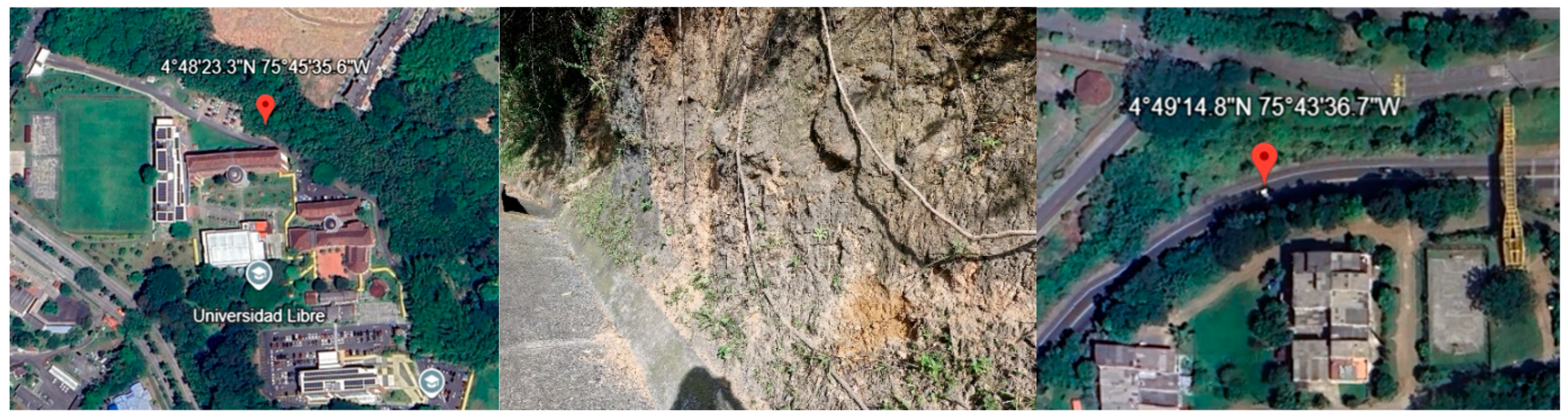
Samples from colonies of N. cf. commune were taken from the soil surrounding a parking lot in the Universidad Libre campus in Belmonte, in Pereira city, Colombia (4°48'23.3"N 75°45'35.6"W) (Figure 2). Dry colonies were taken and rehydrated with distilled water for an hour, selecting those colonies that were green and had a diameter between 4 and 6 cm. Colonies were identified according to their macroscopic characteristics: being moist, green, with a mucous yet solid appearance, having a smooth surface when extended (i.e. not being hair like or spherical) and gelatinous texture. Once colonies with these traits had been selected, they were examined using an optical microscope: looking for curved but not coiled trichomes with heterocysts in the extremes and straight hormogonia with no heterocysts (Figure 1); checking for characteristics of the Nostoc genus according to Bergey’s manual of systematic bacteriology [18].
2.2.2. Isolation of Cyanobacteria from N. cf. Commune Colonies
Colonies were cleaned with distilled water to remove rocks and plant residues and successively washed with 70% ethanol and distilled water three times. Washed colonies were macerated with a mortar and pestle, using saline solution (in a proportion 1:1 to the mass of the sample), then Tween 20 was added to a final concentration of 10% and the mixture was homogenized with a vigorous vortexing for 10 minutes. The resulting solution was irradiated with UV-C light for 8 minutes to reduce contamination from accompanying microbes. Then, 100 µL of the inoculum was streaked in BG11 agar (Phytotech labs) containing 200 µg/mL of cycloheximide. Plates were maintained with an irradiance of 25 µmol photons m-2 s-1 in a photoperiod of 16:8. When colonies that matched the morphology of cyanobacteria grew, they were successively cultured in BG11 agar adjusted to pH 9 and BG11 with cycloheximide until only cyanobacteria colonies grew.
2.2.3. Biomass production
The obtained isolates were cultured for approximately 30 days in liquid BG11 medium under the same conditions of irradiance and photoperiod and using air from a fish tank water pump filtered through a syringe filter with pores of 0,45 µm. The isolate with the fastest growth was then selected and cultured to obtain as much biomass as needed for soil inoculation.
2.2.4. Identification of Cyanobacteria Isolate
2.2.5. Soil Sampling
For the evaluation of the effects of the biocrusts in soil, a sample was taken from a slope near the Romelia-El Pollo Avenue in Pereira, Colombia (4°49'14.8"N 75°43'36.7"W) —as a slope it is characterized by having low vegetal coverture and being highly exposed to erosion (although some parts had presence of biocrusts)— (Figure 2). Soil was taken from the first 5 cm of the slope in parts that had no plants or biocrusts.
Figure 2.
Sampling place for cyanobacteria colonies, parking lot of the Universidad Libre in Pereira (4°48'23.3"N 75°45'35.6"W) (left) Sampling place for soil, slope near the Romelia-El Pollo Avenue (4°49'14.8"N 75°43'36.7"W) (center and left). Source: Google Maps v.25.22.0 (https://www.google.com/maps).
Figure 2.
Sampling place for cyanobacteria colonies, parking lot of the Universidad Libre in Pereira (4°48'23.3"N 75°45'35.6"W) (left) Sampling place for soil, slope near the Romelia-El Pollo Avenue (4°49'14.8"N 75°43'36.7"W) (center and left). Source: Google Maps v.25.22.0 (https://www.google.com/maps).
2.2.6. Microcosm Experiment
Microcosms were made in 90 mm Petri dishes, using 120g of soil for each. Two groups of microcosms were used: a negative control with no treatment and one with the addition of 4 g of biomass from the cyanobacteria isolate, added so that it would cover most of the soil surface; each group had 12 replicates. Water was added to fully get each microcosm wet every 48 h.
2.3. Monitoring the Conditions of the Microcosms
2.3.1. Sampling Moments
An initial determination of the characteristics of the soil was made before the microcosm experiment, pH, humidity and organic matter content, as well as counts for aerobic mesophilic bacteria, molds and yeasts and microbes with activity as phosphate and potassium solubilizers and as nitrogen fixers were all evaluated. After the microcosm experiment started, all the determinations were made again every two weeks for three months. For this monitoring, biocrusts were retired from the microcosm (in the treated group), the soil was mixed and needed amounts for each determination were taken, destroying the used microcosm.
2.3.2. Determination of Physicochemical Characteristics.
pH was determined in an aqueous solution prepared using 10 mL of distilled water and 10 g of soil, which were mixed for 5 minutes and then allowed to sit for an hour before reading pH with a potentiometer [21]. For humidity determination, 20 g of soil was taken and dried at 105°C for 24 hours. Humidity was determined as the difference in mass before and after drying [22]. Organic matter content was determined using the method of loss on ignition. The dried sample resulting from the humidity determination was taken (having previously registered its weight) and heated to 550°C for 5 hours. Finally, the mass was measured again, with the soil’s organic content being the difference between the dried mass before and after being heated [23].
2.3.3. Determination of Microbial Counts
All the microbiological evaluations were made starting from a series of dilutions of a 25 g soil sample in buffered peptone water [24]. This process used two series of dilutions for each treatment (from two different microcosms) every time it was evaluated. Counts were made for aerobic mesophilic bacteria (using plate count agar) and mold and yeast (Sabouraud agar with chloramphenicol) as indicators of the general microbial density, and phosphate solubilizing microbes (NIBRIP agar) [25], potassium solubilizing microbes (modified Pikovskaya agar) [26] and nitrogen fixing microbes (Ashby agar) [27]. Plates for aerobic mesophilic bacteria were incubated at 37°C for 48 hours, with the rest of the plates being incubated at 27°C (for 48 hours for potassium solubilizers and 5 days for the rest).
2.4. Data Analysis
The evolution of the monitored characteristics was represented graphically to better see their changes (Microsoft Excel). The Statgraphics 19 pack was used for statistical analysis of data; applying a normality test (Royston’s H), a two-way variance analysis (to check whether there were significant changes between treatments and across time) and creating a matrix with the Pearson’s correlation between each of the variables (presenting the table generated by the statistical software).
3. Results
3.1. Isolation of Cyanobacteria from N. cf. Commune Colonies
Six isolates of cyanobacteria were obtained, of which 3 were successfully cultured without other microbes impeding or covering its growth. All three of the isolates grew grouping themselves in the liquid media, forming a sort of film and adhering to the flasks or air tubes. Of the three isolates, the most productive one was selected for biomass production, according to the measure of their wet mass (the selected isolate produced 34.5 g in a 250 mL culture after 3 weeks).
The isolate showed growth in BG11 medium without an organic nitrogen source. The growth was characterized according to the identification guides mentioned in the methodology, having cells that were longer than they were wide and being organized in long sheathed trichomes, which were curved but not coiled, nor did they show ramifications (Figure 3). The construction between cells was thin. Despite showing diazotrophic growth, there were no heterocysts. This description is compatible with that of the genus Leptolyngbya.3.2.
3.2. Monitoring of the Microcosms
During most of the experiment, the conditions of humidity in the microcosms were higher in the treated group than in the control (Figure 4) showing continuous growth up until the 8th week. The other variables showed no tendency for continued growth or decrease, and they were rather changing between weeks. For pH, the soils were acid and remained that way all the time, moving between 4.7 and 6.3, showing that both groups changed in the same way between weeks. In the case of organic matter, there was little variation, and both groups were always between 13 and 15%.
Regarding microbial counts, there was no clear increase or decrease during time either but instead oscillating changes in the evaluated microbial densities (Figure 5 and Figure 6). However, in most cases there was an increase in the count at the end when compared with the beginning of the experiment (except in mold and yeasts). The highest levels for each evaluated group were reached a couple weeks before the end of the experiment, being at the 10th week for aerobic mesophiles and potassium solubilizers and 6th for nitrogen fixers; then all of them showed reductions in the following weeks.
Results for Royston’s H show that the multivariate data is normal (p>0.25). For the counts of aerobic mesophiles, potassium solubilizers, phosphate solubilizers and nitrogen fixers, the final results were higher, and the results were also higher in the group treated with biocrusts. Nonetheless, only the variables of pH (p=0.02) and humidity (p=0.005) showed significant differences (p<0.05) between the groups, and only pH (p=0.002) and mold and yeasts (p=0.01) showed significant difference regarding time.
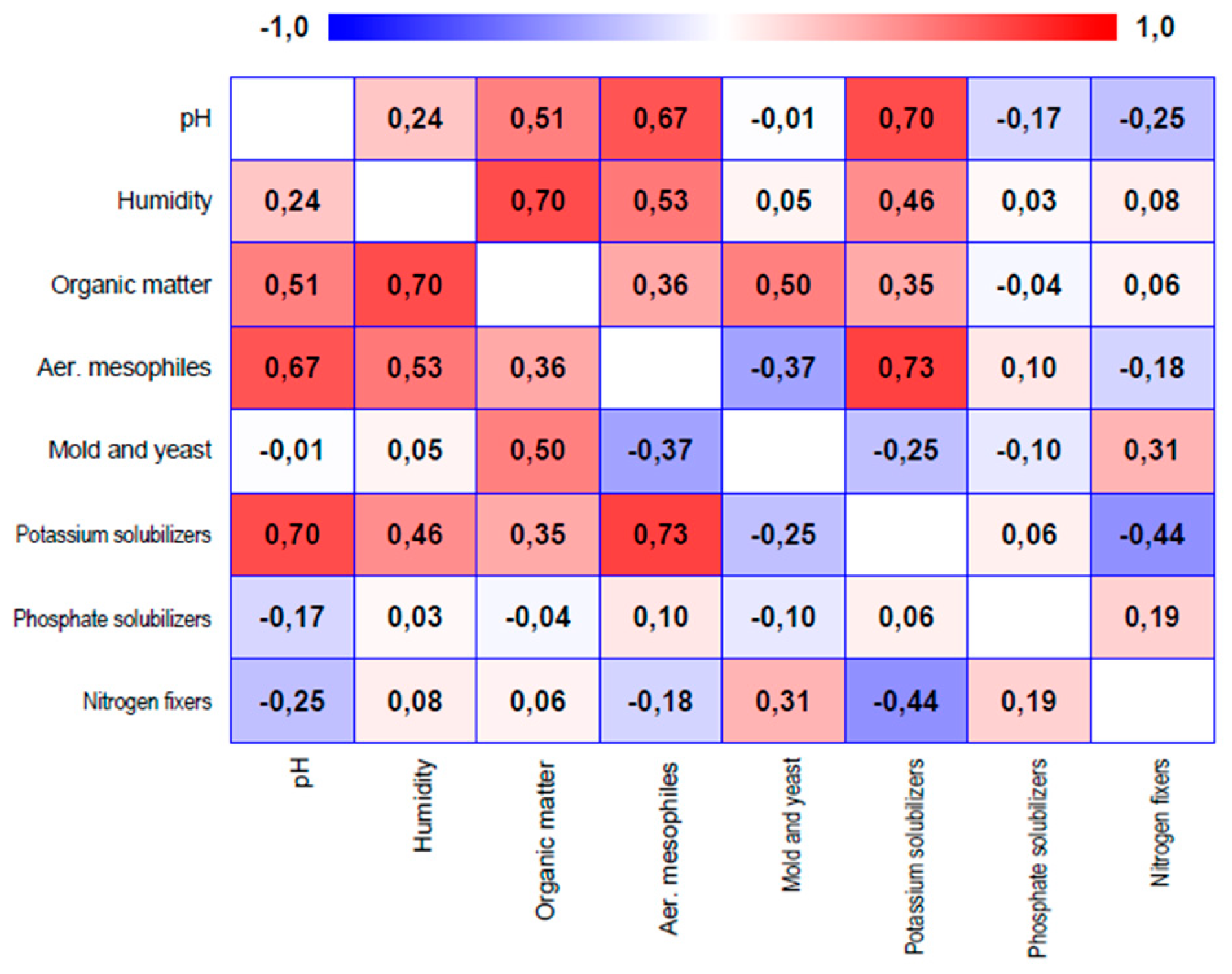
On the other hand, there were slightly strong correlations (more than 0.6) (Figure 7) between the counts for aerobic mesophiles and pH (r = 0.67), between aerobic mesophiles and potassium solubilizers (r = 0.73) and between humidity and organic matter (r = 0.7); with also important positive correlations —albeit, not so strong— between the physicochemical properties (pH, humidity and organic matter) and the counts of aerobic mesophiles and potassium solubilizers, with the stronger ones being pH and humidity.
4. Discussion
4.1. Isolation of Cyanobacteria from N. cf. Commune Colonies
The isolation protocol successfully yielded cyanobacterial colonies. Contrary to initial expectations, the isolates were identified as belonging to the genus Leptolyngbya instead of Nostoc. This finding is still coherent with the existent referents on biocrust formation in the environment and their complexity. While species like N. commune form the basic structure of the crust with their exopolysaccharide matrix, constitute a consortium with other cyanobacteria as well —that are in fact part of the microbial succession process in colonized soils [28,29]. The isolation of Leptolyngbya spp. described here is also congruent with antecedents mentioning its ability to form biocrusts [30].
While the morphological characterization led to the isolates being classified as Leptolyngbya spp., it should be confirmed with molecular biology methods. Morphological identification of cyanobacteria is useful in guiding initial screening but is prone to not actually reflect on the actual genetic similitude of the species [31,32]. We consider the use of amplification and sequencing of the 16S rRNA gene as the next step in confirmation.
Accurate taxonomical identification of the isolates is key for the further application of this kind of restoration research as the use of native microorganisms would be promising for addressing local problems in soil degradation. Additionally, its growth morphology—forming cohesive films instead of existing in a planktonic state—offers advantages in the inoculation process. This trait could make it easier to apply in soil and promote faster establishment of the biocrusts, compared to the conventional approaches relying on cyanobacterial suspensions and a wait time until the formation of the crust [10].
4.2. Effects of Biocrusts on Microcosms’ Soil
During the experiment, it was observed that the cyanobacteria films adhered to the soil surface, suggesting their adaptation and the formation of biological crusts. These crusts showed resistance to desiccation, becoming hardened when the soil was not humid, but rehydrating quickly after water was added —a property noted to be key for the structure and function of biological soil crusts [7,33].
Among the evaluated variables, soil humidity showed the most difference (being statistically significant) between the treated and control groups, presenting an increase of about 10% after the first two weeks and maintaining it for most of the time. Humidity was important for this study as an indicator of soil’s ability to retain water — as both groups received the same amount of water at the same time. Increased water retention ability is an important function of biocrusts and an important indicator of soil health [34]. Similarly, increases in soil humidity and its water retention are directly related to long-term improvements in its microbial richness and diversity [35,36].
Nevertheless, there was a decrease in the humidity for both groups near the end of the experiment, with the treatment microcosms getting to levels like that of the control group. These variations could be related to changes in environmental conditions in the region (either a temperature increased or decreased air humidity) [37]. Further address this by including strategies to minimize the effect of environmental conditions on the experiment, as well as better controlling the watering of microcosms.
On the other hand, the conditions of organic matter were stable for both groups, showing no clear variation or tendencies. These results are not consistent with what was expected based on the literature [15,38]. There are, however, some plausible explanations for this, including the possibility that the isolate has a low carbon sequestration ability or low polysaccharide liberation (which are generally the way in which cyanobacteria contribute to increasing soil’s organic content), or that the assay’s conditions (which was not made in the open, but in a laboratory near a window as natural light source) were not adequate for promoting enough photosynthetic activity to produce significant carbon fixation.
Regarding microbial counts, all the evaluated groups showed increases by the end of the experiment, and were higher in the treated microcosms, but none of these differences was statistically significant. It is important to note that, despite this increase, the initial conditions of the soil were in the range of what is considered as normal soil conditions [39,40,41], meaning that despite the low vegetation, low humidity and medium levels of organic matter, the used soil could have not been intensely degraded to begin with.
It is also possible that the lack of significant results is related to time rather than to a lack of biological effect, being that there were changes between groups and across time, but it could be that the time used for this assay was not enough to produce strong changes. Knowing this, future experiments could use longer treatment times in order to allow for a more decisive determination of the effects of biocrust and to bring clarity as to how much time might be needed for adequate treatment.
Still, one of the hypotheses guiding this study was that, provided biocrusts were known to increase soil humidity and organic matter, these changes should be associated with improvements in soil microbial density —as both water and carbon sources are two basic needs for the proliferation and development of microbial communities [13,14]—. Despite this, these results could not clarify that relation, as there was not an increase in soil organic matter —and it could be considered that these limitations on the increase of soil organic content could in turn be related to the limitations at increasing microbial counts.
Finally, the analysis of the variables shows there is a correlation between humidity and organic content and (although weaker) correlations between soil’s physicochemical properties (pH, humidity and organic matter) and microbial counts, mainly aerobic mesophiles and potassium solubilizers (both being some of the most abundant, and therefore more relevant, evaluated groups). These correlations then suggest the existence of a correspondence between the direct effects of biocrusts in soil and the conditions in the edaphic microbiome, thereby reinforcing the need for further study into those dynamics.
5. Conclusions
This study allowed for the successful establishment of a protocol for cyanobacteria isolation from biocrusts, leading to isolates identified as Leptolyngbya spp., one of which was used for evaluation of its effects on slope soil after being inoculated as biocrusts. Results showed significant changes in soil humidity, indicating improved water retention capabilities, and in soil pH, possibly due to the activity of the biocrusts’ cyanobacteria. However, there were no significant changes in soil organic matter.
There were increments in microbial counts for aerobic mesophiles, potassium solubilizers, phosphate solubilizers and nitrogen fixers, both when comparing between treated and not treated groups and when considering the experiment’s time, but these changes were not statistically significant. Our results seem to support the idea that the improved soil physicochemical properties caused by biocrusts (mainly due to better water retention) are related to changes in soil microbial communities. However, any further elucidation of these interactions was limited by the lack of changes on soil organic matter (either because the experiment’s design is to be improved, or because the used isolate lacks the ability to make any such changes) and the lack of further statistical support on microbial count changes (which could be related to the limitations on organic matter increases, or the need for a longer treatment time).
Author Contributions
Conceptualization, A.F.H.T, S.C.R.R and D.G.A; Methodology A.F.H.T, S.C.R.R and D.G.A; Validation, A.F.H.T, E.A.R, J.J.F.C; Formal analysis A.F.H.T, S.C.R.R and D.G.A; Investigation, A.F.H.T, E.A.R and J.J.F.C; Resources, S.C.R.R and D.G.A; Data curation, A.F.H.T; Writing – Original Draft Preparation, A.F.H.T; Writing – Review & Editing, A.F.H.T, S.C.R.R and D.G.A; Visualization, A.F.H.T; Supervision, S.C.R.R and D.G.A; Project administration, A.F.H.T, S.C.R.R and D.G.A; Funding acquisition, A.F.H.T, S.C.R.R and D.G.A.
Funding
This research was funded by Universidad Libre de Colombia as a part of project 161202326 “Conociendo las microalgas como aliadas para la seguridad alimentaria, la restauración de suelos y la biorremediación” for the grant “Convocatoria de investigación multicampus para la conformación de un banco de proyectos elegibles 2024”
Acknowledgments
To the Universidad Libre of Colombia, sectional Pereira, for the development of the phycology laboratory and the funding provided for this research. To professor Fernando Siller López, for engaging with the initial discussion over the conception of this project and for his support for the acquisition of funds. To my colleague Tomás Vélez Londoño for his contributions towards this research and the standardization of the cyanobacteria isolation protocol; and to Luisa Maria Aguilar Jiménez and Angee Natalia Sánchez Montoya for all their support as laboratory colleagues.
Conflicts of Interest
The authors declare no conflict of interest.
References
- FAO, “Land use in agriculture by the numbers.” [Online]. Available: https://www.fao.org/sustainability/news/detail/en/c/1274219/#:~:text=Globally agricultural land area is,and pastures) for grazing livestock.
- K. Lorenz, R. Lal, and K. Ehlers, “Soil organic carbon stock as an indicator for monitoring land and soil degradation in relation to United Nations’ Sustainable Development Goals,” Land Degrad. Dev., vol. 30, no. 7, pp. 824–838, Apr. 2019. [CrossRef]
- FAO, “Proceedings of the Global Symposium on Soil Erosion 2019,” Rome, 2019.
- Instituto de Hidrología Meteorología y Estudios Ambientales, “Monitoreo y seguimiento del estado de calidad de los suelos.” [Online]. Available: https://www.ideam.gov.co/web/ecosistemas/monitoreo-seguimiento-estado-calidad-suelos/-/document_library_display/9mDFEmPPfxAk/view/95764895#:~:text=(Ideam%2C 2019).,grados severos y muy severos.
- R. Bommarco, G. Vico, and S. Hallin, “Exploiting ecosystem services in agriculture for increased food security,” Glob. Food Sec., vol. 17, pp. 57–63, Jun. 2018. [CrossRef]
- P. Pereira, I. Bogunovic, M. Muñoz-Rojas, and E. C. Brevik, “Soil ecosystem services, sustainability, valuation and management,” Curr. Opin. Environ. Sci. Health, vol. 5, pp. 7–13, Oct. 2018. [CrossRef]
- B. Weber et al., “What is a biocrust? A refined, contemporary definition for a broadening research community,” Biological Reviews, May 2022. [CrossRef]
- S. Chamizo, E. Rodríguez-Caballero, J. R. Román, and Y. Cantón, “Effects of biocrust on soil erosion and organic carbon losses under natural rainfall,” Catena (Amst)., vol. 148, pp. 117–125, 2017.
- B. Roncero-Ramos, J. R. Román, C. Gómez-Serrano, Y. Cantón, and F. G. Acién, “Production of a biocrust-cyanobacteria strain (Nostoc commune) for large-scale restoration of dryland soils,” J. Appl. Phycol., vol. 31, no. 4, pp. 2217–2230, Aug. 2019. [CrossRef]
- F. Rossi, G. Mugnai, and R. De Philippis, “Cyanobacterial biocrust induction: A comprehensive review on a soil rehabilitation-effective biotechnology,” Geoderma, vol. 415, p. 115766, Jun. 2022. [CrossRef]
- M. Á. Muñoz-Martín, I. Becerra-Absalón, E. Perona, L. Fernández-Valbuena, F. Garcia-Pichel, and P. Mateo, “Cyanobacterial biocrust diversity in Mediterranean ecosystems along a latitudinal and climatic gradient,” New Phytologist, vol. 221, no. 1, pp. 123–141, Jan. 2019. [CrossRef]
- S. Obana, K. Miyamoto, S. Morita, M. Ohmori, and K. Inubushi, “Effect of Nostoc sp. on soil characteristics, plant growth and nutrient uptake,” J. Appl. Phycol., vol. 19, no. 6, pp. 641–646, Dec. 2007. [CrossRef]
- Y. Cui et al., “Diversity patterns of the rhizosphere and bulk soil microbial communities along an altitudinal gradient in an alpine ecosystem of the eastern Tibetan Plateau,” Geoderma, vol. 338, pp. 118–127, 2019.
- W. Li et al., “Structure and driving factors of the soil microbial community associated with Alhagi sparsifolia in an arid desert,” PLoS One, vol. 16, no. 7, p. e0254065, Jul. 2021. [CrossRef]
- M. Muñoz-Rojas et al., “Cyanobacteria inoculation enhances carbon sequestration in soil substrates used in dryland restoration,” Science of The Total Environment, vol. 636, pp. 1149–1154, Sep. 2018. [CrossRef]
- M. García-Carmona et al., “Moss biocrust accelerates the recovery and resilience of soil microbial communities in fire-affected semi-arid Mediterranean soils,” Science of The Total Environment, p. 157467, 2022.
- J. Belnap and O. L. Lange, Biological soil crusts: structure, function, and management, vol. 150. Springer, 2001.
- R. W. Castenholz et al., “Phylum BX. Cyanobacteria,” in Bergey’s Manual® of Systematic Bacteriology, New York, NY: Springer New York, 2001, pp. 473–599. [CrossRef]
- 19. Culture Collection of Algae and Protozoa, “BG11 (Blue-Green Medium),” Scottish Marine Institute. Accessed: Sep. 29, 2024. [Online]. Available: https://www.ccap.ac.uk/wp-content/uploads/MR_BG11.pdf.
- M. M. Allen, “Simple Conditions for Growth of Unicellular Blue-green Algae on Plates 1, 2,” J. Phycol., vol. 4, no. 1, pp. 1–4, Mar. 1968. [CrossRef]
- G. O. Dayo-Olagbende, K. O. Sanni, S. A. Adejoro, R. O. Afingba, and B. S. Ewulo, “Fate and Form of Nitrogen under Different Soil Redox Status,” International Journal of Environment, Agriculture and Biotechnology, vol. 7, no. 2, pp. 143–149, 2022. [CrossRef]
- P. Robertson and S. VanderWulp, “Soil Moisture ‒ Gravimetric,” Kerllog Biological Station | Long-term Ecological Research. Protocols. [Online]. Available: https://lter.kbs.msu.edu/protocols/24#:~:text=To measure soil moisture content,per mass of dry soil.
- M. J. J. Hoogsteen, E. A. Lantinga, E. J. Bakker, J. C. J. Groot, and P. A. Tittonell, “Estimating soil organic carbon through loss on ignition: effects of ignition conditions and structural water loss,” Eur. J. Soil Sci., vol. 66, no. 2, pp. 320–328, Mar. 2015. [CrossRef]
- J. Foght and J. Aislabie, “Enumeration of Soil Microorganisms,” in Manual for Soil Analysis - Monitoring and Assessing Soil Biorremediation, 1st editio., R. Margesen and F. Schinner, Eds., Springer, 2005, pp. 261–279.
- C. S. Nautiyal, “An efficient microbiological growth medium for screening phosphate solubilizing microorganisms,” FEMS Microbiol. Lett., vol. 170, no. 1, pp. 265–270, Jan. 1999. [CrossRef]
- M. F. Guevara Granja, “Aislamiento e Identificación de Microorganismos Solubilizadores de Potasio a partir de Muestras de Suelo y Raíces de Cultivos de Alcachofa de la Localidad de la Remonta, Cantón de Cayambe,” Escuela Politénica del Ejército, 2010.
- L. Lin et al., “Plant Growth-Promoting Nitrogen-Fixing Enterobacteria Are in Association with Sugarcane Plants Growing in Guangxi, China,” Microbes Environ., vol. 27, no. 4, pp. 391–398, 2012. [CrossRef]
- S. Lan, A. D. Thomas, J. B. Rakes, F. Garcia-Pichel, L. Wu, and C. Hu, “Cyanobacterial community composition and their functional shifts associated with biocrust succession in the Gurbantunggut Desert,” Environ. Microbiol. Rep., vol. 13, no. 6, pp. 884–898, Dec. 2021. [CrossRef]
- Y. Zhang, P. Duan, P. Zhang, and M. Li, “Variations in cyanobacterial and algal communities and soil characteristics under biocrust development under similar environmental conditions,” Plant Soil, vol. 429, no. 1–2, pp. 241–251, Aug. 2018. [CrossRef]
- G. Mugnai, F. Rossi, S. Chamizo, A. Adessi, and R. De Philippis, “The role of grain size and inoculum amount on biocrust formation by Leptolyngbya ohadii,” Catena (Amst)., vol. 184, p. 104248, 2020.
- J. Komárek, J. Kaštovský, J. Mareš, and J. R. Johansen, “Taxonomic classification of cyanoprokaryotes (cyanobacterial genera) 2014, using a polyphasic approach,” Preslia, vol. 86, no. 4, pp. 295–335, 2014.
- J. M. Walter, F. H. Coutinho, B. E. Dutilh, J. Swings, F. L. Thompson, and C. C. Thompson, “Ecogenomics and Taxonomy of Cyanobacteria Phylum,” Front. Microbiol., vol. 8, Nov. 2017. [CrossRef]
- C. Baubin, N. Ran, H. Siebner, and O. Gillor, “Divergence of biocrust active bacterial communities in the Negev Desert during a hydration-desiccation cycle,” May 09, 2022. [CrossRef]
- S. C. Reed, M. Delgado-Baquerizo, and S. Ferrenberg, “Biocrust science and global change,” New Phytol., vol. 223, no. 3, pp. 1047–1051, 2019.
- M. Hartmann et al., “A decade of irrigation transforms the soil microbiome of a semi-arid pine forest,” Mol. Ecol., vol. 26, no. 4, pp. 1190–1206, Feb. 2017. [CrossRef]
- S. E. Rothenberg, M. Anders, N. J. Ajami, J. F. Petrosino, and E. Balogh, “Water management impacts rice methylmercury and the soil microbiome,” Science of The Total Environment, vol. 572, pp. 608–617, Dec. 2016. [CrossRef]
- K. Furtak and A. Wolińska, “The impact of extreme weather events as a consequence of climate change on the soil moisture and on the quality of the soil environment and agriculture – A review,” Catena (Amst)., vol. 231, p. 107378, Oct. 2023. [CrossRef]
- H. Kheirfam, “Increasing soil potential for carbon sequestration using microbes from biological soil crusts,” J. Arid Environ., vol. 172, p. 104022, 2020.
- I. A. F. Djuuna, S. Prabawardani, and M. Massora, “Population Distribution of Phosphate-solubilizing Microorganisms in Agricultural Soil,” Microbes Environ., vol. 37, no. 1, p. ME21041, 2022. [CrossRef]
- I. L. Pepper and T. J. Gentry, “Earth Environments,” in Environmental Microbiology, Elsevier, 2015, pp. 59–88. [CrossRef]
- A. Ratnakar and Shikha, “Assessment of Co-Contamination in Soil Samples from Agricultural Areas in and Around Lucknow City, Uttar Pradesh, India,” Curr. Sci., vol. 115, no. 12, p. 2267, Dec. 2018. [CrossRef]
Figure 1.
Nostoc cf. commune samples: trichomes with heterocysts (left) and hormogonia (right).
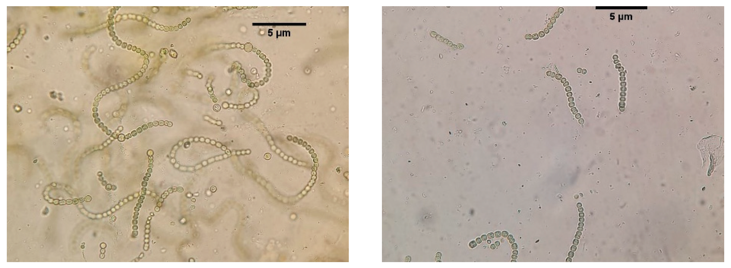
Figure 3.
Micrograph of one of the isolates, identified Leptolyngbya sp.
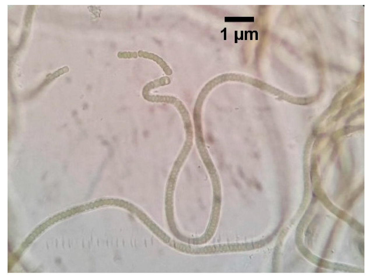
Figure 4.
Physicochemical characteristics of soil: humidity (left up), organic matter (right up), and pH (bottom center).
Figure 4.
Physicochemical characteristics of soil: humidity (left up), organic matter (right up), and pH (bottom center).
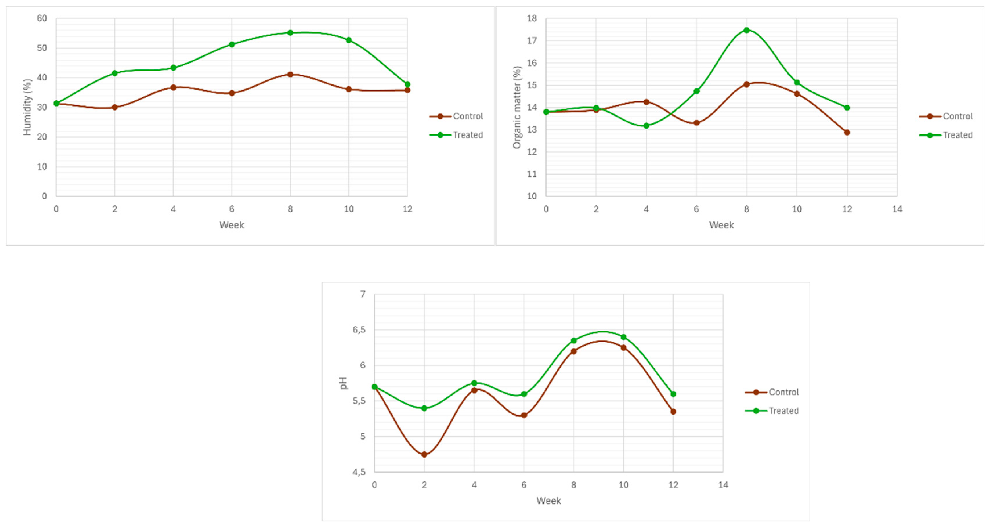
Figure 5.
Transformed recounts (log10) for aerobic mesophiles (left) and mold and yeasts (right) of the microcosms.
Figure 5.
Transformed recounts (log10) for aerobic mesophiles (left) and mold and yeasts (right) of the microcosms.
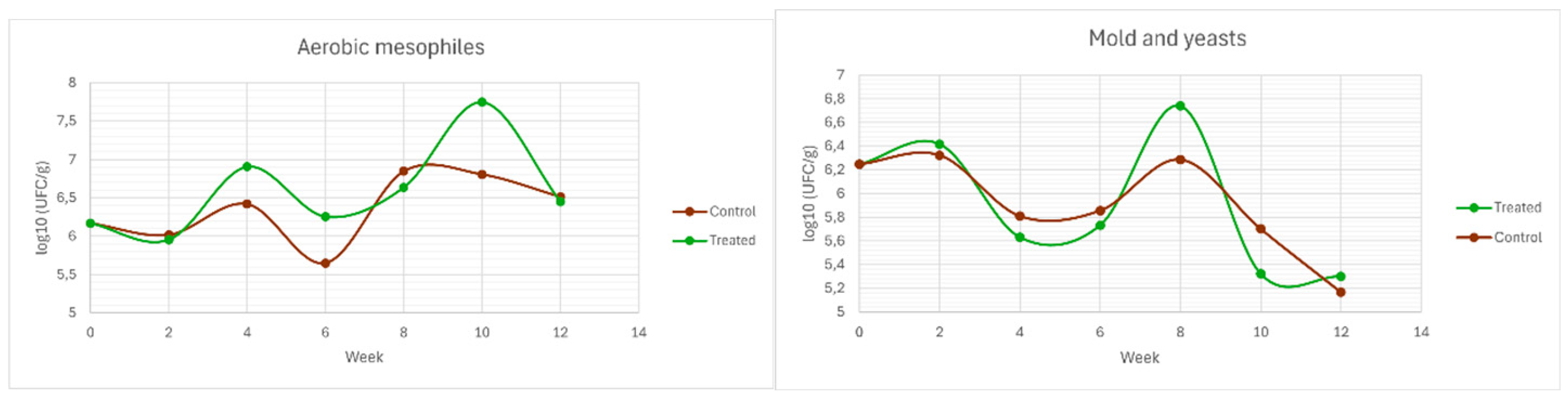
Figure 6.
Transformed recounts (log10) for potassium solubilizers (left up), phosphate solubilizers (right up), and nitrogen fixers (bottom center) of the microcosms.
Figure 6.
Transformed recounts (log10) for potassium solubilizers (left up), phosphate solubilizers (right up), and nitrogen fixers (bottom center) of the microcosms.
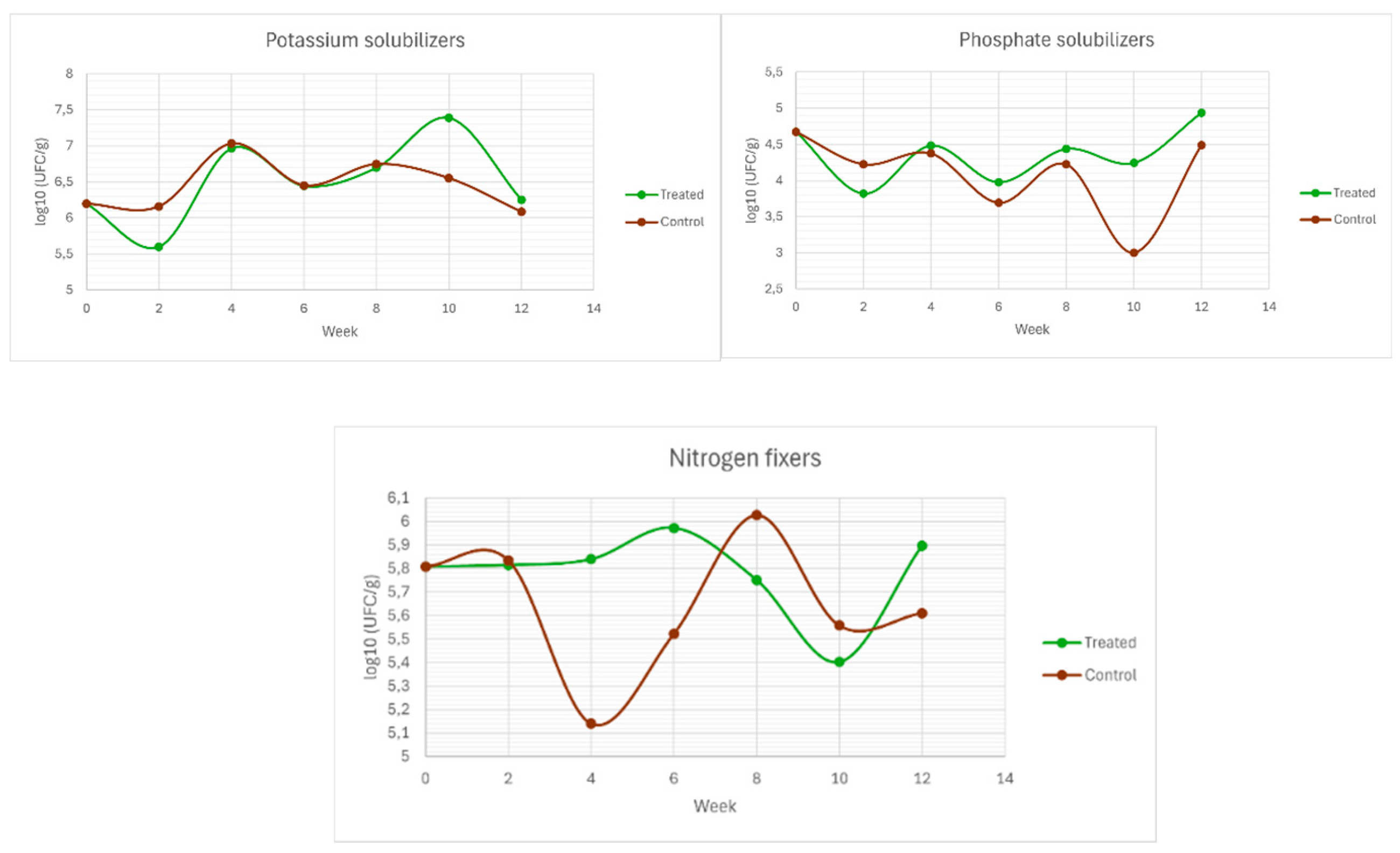
Figure 7.
Pearson correlation matrix between the evaluated variables.
Disclaimer/Publisher’s Note: The statements, opinions and data contained in all publications are solely those of the individual author(s) and contributor(s) and not of MDPI and/or the editor(s). MDPI and/or the editor(s) disclaim responsibility for any injury to people or property resulting from any ideas, methods, instructions or products referred to in the content. |
© 2026 by the authors. Licensee MDPI, Basel, Switzerland. This article is an open access article distributed under the terms and conditions of the Creative Commons Attribution (CC BY) license.
Copyright: This open access article is published under a Creative Commons CC BY 4.0 license, which permit the free download, distribution, and reuse, provided that the author and preprint are cited in any reuse.


