

Submitted:
19 March 2026
Posted:
20 March 2026
Read the latest preprint version here
Abstract
DNA/cell mass homeostasis is a universal feature of living organisms. As the cell grows in response to nutrient availability, it must duplicate each chromosome once and only once each division cycle. Across the Tree of Life, cells differ in their sizes in a manner that depends directly on the amount of DNA they harbor, what has been termed the “nucleotypic effect”: cell size expands or contracts as DNA content increases or decreases. In eukaryotes, any deviation from DNA/mass homeostasis results in the dis-regulation of the developmental program and the initiation of carcinogenesis and other genetic pathologies. In bacteria, deviation from or perturbation of DNA/mass homeostasis alters important physiological features such as the cell cycle timing of DNA replication initiation and the co-ordination of initiation with replication termination and cell division. In prokaryotes, the timing of initiation occurs at a relatively constant and growth rate invariant mass, termed the initiation mass (Mi), and depends strictly on DNA replication fork rates and membrane biogenesis. Complex “machines”, frequently referred to as hyperstructures or factories, mediate the phase transitions that define the different periods of the bacterial cell cycle. The following will examine how the machines gate the phase transitions that organize the cell cycle in time and space.
Keywords:
DNA replication
; DnaA
; ribonucleotide reductase
; nucleoid complexity
; nucleotypic effect
; transertion
1. Introduction
In 1961, Peter Mitchell proposed the chemiosmotic theory: the coupling of phosphorylation to hydrogen transfer generates the proton motive force that drives the synthesis of ATP from ADP. According to the theory, the cell membrane plays a central role in a “type of mechanism that is based directly on the group translocation conception,” which directly involves the membrane in a functional, regulatory role [1].
Mitchell states: “This type of mechanism … depends absolutely on a supra-molecular organization of the enzymes concerned … that can be due to the spatially directed channeling of a chemical component or group along a pathway specified in space by the physical organization of the system.” He added: “the chemiosmotic coupling between the so-called ATPases and the electron and hydrogen transfer chain is mediated by the translocation of electrons and elements of water across the membrane of mitochondria, chloroplast grana and chromatophores.”
The theory met with immediate and stiff resistance, gradually finding support ten years later. Opponents of the theory held the more orthodox view that “water is expelled spontaneously between two chemical groups by the formation of a strong [chemical] bond,” which when weakened by redox reactions allows the synthesis of ATP by the opening and closing of the high-energy [phosphophate] bond. Today the theory, in its fundamentals, is generally accepted but with some proposed modifications [2].
It is now well established in bacteria that the rate of DNA synthesis is coupled to cell growth rate, and in particular to the increase in cell volume [3]. This coupling occurs at the level of DNA replication initiation rather than at the level of the replication fork rate: the C-period, duration of chromosome duplication, is growth rate invariant at fast growth rates and lasts approximately 60 minutes. Importantly, as noted above, the initiation process depends on the ATP bound form of DnaA, which is inactivated by hydrolysis of ATP to ADP [4]. ATP plays an allosteric regulatory, as opposed to a bio-energetic role, in replication initiation, since DnaA unbound by ATP remains competent to initiate DNA synthesis.
Meselson and Stahl first observed the constancy of the C-period, stating “the generation time is very nearly the same for all DNA molecules in the population. This raises the question of whether in any one nucleus this clock regulates nuclear and cellular division as well.” [5], Cooper and Helmstetter later confirmed Meselson-Stahl hypothesis and established the classical paradigm of the bacterial cell cycle [6]. Based on these observations, Cooper proposed the “Continuum Model” [7]. which held that macromolecular synthesis in bacteria proceeds at an invariant rate until a critical mass is reached at which the cell divides. Lacking a detailed mechanistic explanation, the model remains a hypothesis.
The increase in cell volume depends strictly on the growth rate of the cell membrane [8], while membrane growth rate (cell volume and length) depends itself directly on the rate of ATP synthesis and the availability of ATP to drive macromolecular synthesis and the accumulation of biomass. The timing of replication initiation is thus necessarily coupled to cell growth rate, and consequently to a given cell mass that is approximately invariant at all growth rates: the so-called initiation mass (Mi). How the cell couples initiation timing to Mi has remained enigmatic since it was first proposed in 1963 [9]. To this day the concept remains controversial, but widely accepted.
Recent studies have revealed a link between Central Carbon Metabolism (CCM) and DNA replication [10,11,12,15,16,17,1314]. The metabolic genes involved are suppressors of themo-sensitive DNA replication initiation and elongation genes, referred to as dna(ts). The effect on DNA replication was found to be direct and independent of perturbations in cell growth rate [18]. Presumably, but as yet experimentally undemonstrated, the effect of CCM mutants on DNA synthesis might be mediated by cell membrane growth and possibly cell size [19]. How these suppressors operate to restore DNA synthesis in dna(ts) mutants remains a question of continuing interest, but most if not all of them appear to converge on nucleotide synthesis (see below).
The latter feature, an effect of cell size, is supported by the observation that perturbing replication forks (experimentally slowing them) results in increased nucleoid complexity and a nucleotypic affect, according to which DNA content (number of origins) regulates cell size [20]. Conversely, experimentally increasing fork rates results in a shortened C-period without affecting cell division [21]. Hence, replication fork rate corresponds to a major parameter governing the rate of initiation and cell size: fork rate and initiation frequency during a single cell cycle form what has been termed a “homeostatic pair” that maintains DNA/cell mass equilibrium [22]. The latter proposal is consistent with Cooper’s Continuum Model [23], and has gained increasing experimental support.
ATP synthesis and availability thus link membrane growth to initiation of DNA replication at oriC, which occurs at a frequency depending on replication fork rates [24,25]. Cell envelope synthesis has long been of interest in regulating DNA replication initiation, but its exact role in coupling DNA synthesis to cell growth rate still remains unclear. It is known, however, that the nucleoid is spatially and non-randomly associated with the cell membrane via factors involved in initiation, elongation, chromosome segregation and cell division.
The relationship between these cell cycle transitions is currently the focus of intense investigations. Cell envelope synthesis, for example, is closely associated with ribosome assembly and phospholipid synthesis [26], suggesting both direct and indirect interactions between ppGTPpp, CTP, GTP and ATP synthesis. As noted above for DnaA and ATP, these co-factors might play an intrinsic allosteric role that regulates the activities of the respective proteins that bind them. Functioning as regulatory co-factors they mediate and coordinate cell metabolism, cell growth, replication initiation timing, DNA replication fork rates and cell division. These diverse functions might in turn be integrated through a “supra-molecular” membrane-bound structure that senses metabolic status, cell envelope synthesis as well as increases in cell volume [27,28]. How the cell integrates these diverse cell functions, which depend on cell growth rate, to form a coherent and reproducible cell cycle remains an outstanding question involving multiple areas of research.
2. Is the Nucleotypic Effect Related to Nucleoid Complexity?
The bacterial cell cycle in slow growing cells (> 60 min doubling time) is divided into three phases: a variable B-period during which the cell recharges and redistributes ATP and undergoes macromolecular synthesis in preparation for the initiation of DNA replication and duplication of the chromosome—the C-period—and a D-period during which the cell prepares for cell division following chromosome segregation. In fast growing cells (< 60 min), the B-period is eliminated in daughter cells, indicating that DNA replication has been initiated in the mother cell prior to cell division [29].
Consequently, in fast growing cells multiple replication forks per chromosome are present, and cell size/volume is correspondingly larger. The number of replication forks in fast growing cells has been termed “nucleoid complexity” [30]. It has been recently established under a variety of experimental conditions that growth rate-dependent incremental cell length as well as cell width are correlated with nucleoid complexity, thus generating cell-shape homeostasis [31,32]. The growth rate invariance of cell-shape homeostasis, over a three-fold range of doubling times, suggests that nucleoid complexity affects cell width. In eukaryotes, it is well known that cell size varies directly in proportion to DNA content: cells with more DNA are correspondingly larger than cells with less DNA, an observation made across the eukaryote Tree of Life, and referred to as the “nucleotypic effect” [33,34,35].
Why and how cell size varies with DNA content remains an unresolved question of both evolutionary and physiological interest. This universal DNA/cell mass homeostasis can explain the long-standing observation that in bacterial cells initiation of DNA replication occurs at a growth rate invariant mass termed the initiation mass, Mi [36]: the number of replication origins (oriC) and cell mass at initiation of DNA replication is independent of cell growth rate. Cell size in bacteria therefore depends on the number of active replication forks per chromosome (nucleoid complexity: Figure 1). Surprisingly, DNA/cell mass homeostasis is maintained not only under conditions of differing nutrient availability (growth rate) but also under conditions that perturbed DNA synthesis occurring either environmentally (DNA replication fork collapse) or experimentally (inhibiting, or slowing, replication fork speed)[37,38].
3. Is a Negative Correlation Between DNA Replication Fork Rate and Initiation Frequency a Universal Feature of Cellular Life?
In both prokaryotes and eukaryotes a negative correlation between replication fork rate and initiation frequency has been consistently observed. Slower DNA replication forks result in increased nucleoid complexity in bacteria (more initiations per chromosome), and in eukaryotes slower DNA replication forks result in correspondingly shorter inter-origin distances [39,40]. The negative correlation therefore accounts for how the cell maintains DNA/cell mass homeostasis, and suggests that the Mi is simply a consequence of the homeostatic mechanisms underlying the equilibrium between the number of forks per chromosome and the corresponding fork rates.
Two interpretations of the inverse correlation between fork rate and initiation frequency have so far been proposed: 1) In eukaryotes, so-called dormant replication origins fire passively before being replicated (and inactivated) when replication forks slow or stall. In bacteria, cell mass continues to accumulate at a constant rate until the Mi is reached, which then triggers replication initiation; 2) stalled or perturbed replication forks in both prokaryotes and eukaryotes induce the gene coding for the enzyme ribonucleotide reductase, which supplies replication forks and DNA repair systems with essential dNTPs. The latter scenario suggests that the forks themselves control initiation via dNTP synthesis as a necessary, but not sufficient, condition for initiation. Both scenarios, however, are likely to interact synergistically in maintaining DNA/cell mass homeostasis.
In bacteria, increased nucleoid complexity in response to perturbed replication forks was first observed in thyA mutants experiencing thymine-less death [41,42]. The cells not only accumulated more replication forks per chromosome (increased nucleoid complexity) but also the cells increased in mass and size (nucleotypic effect). Later it was found that a mutant defective in CTP synthetase (cmk) exhibited similar phenotypes [43]: DNA replication forks slowed but the frequency DNA replication initiation increased to compensate exactly for the slower fork rate (a reduction of two fold in fork rate resulted in an increase of two fold in replication initiation). The inverse correlation between dNTP pool turnover and initiation frequency has been confirmed experimentally by titrating RNR to slow replication fork movement, increase nucleoid complexity and increase cell size [44]. Together, these findings suggest that dNTP pool levels exert a central regulatory role over both cellular and DNA metabolism.
4. Do DnaA and RNR Form a Homeostatic Pair?
The DnaA protein is the central regulator of DNA replication initiation. When bound to oriC in the ATP form, DnaA-ATP acts to melt the DNA double helix, which permits the loading of the enzymes essential for DNA synthesis and genome duplication (initiation). The enzyme ribonucleotide reductase is the central regulator of DNA replication elongation. As the central regulator, its activity is rate limiting for replication fork movement and for DNA repair [45].
How these two proteins interact to control DNA metabolism has to date attracted little attention, presumably because the interaction is indirect and mediated via dNTP metabolism. Overexpressing RNR, for example, does not directly affect the frequency of replication initiation, while overexpressing DnaA has little effect on replication fork rates (C-period), presumably because overexpressing DnaA has a limited effect on either the frequency of initiation or the initiation mass [46,47]. When the two enzymes, however, are overexpressed together overnight DNA chromosome equivalents increase dramatically and cell size increases accordingly (unpublished)—similar to the eel phenotype [48,49]—thus maintaining DNA/cell mass homeostasis in cells that are highly filamented.
DnaA protein synthesis is required for initiation each division cycle, while RNR synthesis is required for elongation and chromosome duplication. Expression from the dnaA gene, however, can be inhibited without affecting initiation [53, 54. Both dnaA and nrdAB are expressed in coordination with initiation, but expression of the two genes apparently oscillates out of phase at slow growth. Although the genes and their products have not been shown to interact directly, low levels of DnaA-ATP have been shown to stimulate nrdAB expression while high levels act to repress nrdAB gene expression [55].
The cell cycle regulation of the two gene products, moreover, appears to be coordinated. While evidence has been found that the DnaA-ATP/Dna-ADP ratio oscillates during the cell cycle [56], the DnaA protein, itself, does not oscillate and remains constant, whereas RNR enzyme activity and dNTP pool sizes oscillate in response to the demand for dNTPs during replication. Consequently, dNTP pool levels are low prior to initiation, increase during the C-period and then decrease dramatically when replication terminates as the cell enters the D-period [57].
This suggests that RNR activity, rather than bulk dNTP pool sizes alone, is rate limiting for replication forks. RNR is allosterically regulated by dATP and depends on the reducing agent NADPH to supply the electrons needed to reduce NTPs to dNTPs. Consequently, high dATP pool levels at the end of the C-period allosterically inactivate RNR [58], indicating that high dNTP pool levels in the absence of active replication forks would inhibit replication initiation and DNA synthesis once the chromosome has been fully duplicated. The expected inhibitory effect of dNTP pool size on replication initation has not been reported in bacteria, but evidence for an effect of high dNTP pool sizes inhibiting the transition to S-phase in eukaryotes has been observed [59].
5. Does the Bacterial Cell Coordinate dnaA and nrdAB Gene Expression with Cell Growth?
Cell size and growth rate depend directly on metabolic rate. Over the past ten years substantial evidence has accumulated revealing a role in Central Carbon Metabolism (CCM) in the regulation of both replication initiation and elongation. Suppressors of DNA replication mutants fall into four principle categories: ATP supply and redox balance (aceE, lpd, ackA, pta, relA, spoT), ribose-5 phosphate and energy supply (pgi, tktA/B), one carbon/thymidine supply (thyA, folA, glyA and gvc) and finally dNTP supply (ndk, nrdAB/EF, ctpS). Importantly the suppressor mutants do not interact directly with replication factors (DnaA, DnaB, DnaC, DnaE, DnaQ), but rather indirectly via the metabolites they produce [60]. Hence, directly (via the folate cycle) and indirectly (via the pentose phosphate pathway and the stringent response) the suppressor effect is due to, and converges on, the synthesis of dNTPs (Figure 2).
The integration of nucleotide, carbon and energy metabolism guarantees that DNA replication can proceed under a variety of normal and compromising conditions. The homeostatic relationship between replication initiation and elongation also applies to cells that aberrantly initiate DNA replication. In bacteria, for example, the hda mutant is defective in hydrolysing the oriC-bound form of DnaA-ATP to the inactive form of DnaA-ADP [61], which is associated with access of SeqA to hemimethylated oriC and transient inhibition of initiation (1/3 rd of the cell cycle). Consequently, this mutant over-initiates replication initiation. Over-expressing RNR, however, rescues the cells and restores replication by over-supplying dNTPs [62]. Likewise, over-expressing nrdAB rescues the cold sensitive dnaAcos mutant that over-initiates DNA replication at non-permissive temperature [63]. In eukaryotes a yeast strain that over-initiates DNA replication is viable if and only if RNR and dNTPs are overproduced [64].
6. Does Dna Synthesis Direct Membrane Biogenesis?
Finally, in rapidly growing E. coli cells DNA replication and membrane growth are are tightly coupled. A direct biochemical link between DNA synthesis and phopholipid synthesis is mediated by the nucleotide CTP [65]. Phospholipid synthesis consumes CTP and therefore affects dCTP pools and replication fork rates (see above). CTP is used in producing CDP-diacylglycerol, a phospolipid precursor. The folate cycle provides the precursors for dNTP synthesis, and E. coli synthesizes methionine from folate (Figure 3), but unlike mammals E. coli does not methylate the phospholipid phosphatidylenthanolamine.
Instead, folate coordinates nucleotide supply with methylation reactions that are critical for rapidly growing cells. Methonine synthesized from 5-methyl tetrahydrofolate is used to produce S-adenosylmethione (SAM) for methylating RNA, DNA and proteins. SAM oscillates with the cell cycle, and cells lacking SAM fail to divide, resulting in large filaments (the eel phenotype) while exhibiting an otherwise normal cell cycle. Hence, SAM and methylation establish a link between DNA synthesis and cell division (Figure 4), and converts the DnaA-RNR replication loop from a biochemical circuit into a cell cycle system, capable not only of duplicating chromosomes but also of dividing to produce two daughter cells.
Notably, the CTP mutant cmk is dysfunctional in membrane biogenesis and transertion [69], an important process in spatio-temporarily organizing the E. coli nucleoid. Recently, for example, membrane associated transertion (transcription-translation-membrane insertion) has been shown to integrate nucleoid organization, membrane biogenesis and DNA replication [70]. Inhibiting cell wall synthesis, however, does not affect nucleoid organization [71]. Transcription and translation are therefore essential for nucleoid attachment to the membrane, and by implication DNA replication. This close association is mediated by MreB, which controls cell wall synthesis and shape [72]. This might suggest that transertion forms a transcription condensate with the membrane during membrane biogenesis that directly couples DNA replication to cell growth [73,74,75].
7. How Might Dna Synthesis Drive Cell Growth?
A nucleotypic effect (NE) suggests cells will divide at a size that depends on the number of replication forks, which reflects the C-value (picograms of haploid genome) in eukaryotes and chromosomal equivalents (number of replication origins) in prokaryotes. Nucleoid complexity (NC) and the nucleotypic effect are therefore physiologically related, and reflect the homeostasis between replication initiation (number of new forks) and elongation (fork rate). In cells with high NC, and consequently slower fork rates, the correspondingly larger daughter cells at birth will therefore initiate DNA replication earlier in the cell cycle but at an initiation mass that correlates with the initiation mass in the mother cell [76].
NC also affects cell width (W) such that the ratio W/NC is independent of growth rate, a phenomenon referred to as “cell-shape homeostasis” [77]. Since NC reflects the homeostatic relationship between the number of forks per chromosome and replication fork rate, cell-shape homeostasis and initiation-elongation homeostasis are biologically commensurate, and both are therefore related and coupled to nucleotide metabolism. Evidence for such a relationship is provided by the observation that CCM suppressors of DNA replication mutants all converge directly or indirectly on dNTP synthesis and nrdAB up-regulation during perturbed DNA synthesis: both cell size (W) and nucleoid complexity (NC) correlate with replication fork rate, and are therefore linked, or coupled together, through the activity of ribonucleotide reductase (RNR).
In accordance with the transertion hypothesis [78], it seems reasonable then to consider—although it would be entirely hypothetical itself—that the cell membrane is growing around and in proportion to the growing amount of DNA being synthesized, and continues to do so until chromosomal duplication is completed and the demand for dNTPs ceases, at which point the cell divides. Phospholipid synthesis, for example, increases stepwise at or shortly after the initiation of DNA replication [79,80,81].
In E. coli both chromosomal synthesis and cell envelope phospholipid synthesis undergo sudden increases at replication initiation and termination [82]. Simply stated, cell size and cell cycle control are coupled via initiation-elongation homeostasis, or the number of active forks and the rate at which they synthesize new DNA. Membrane biogenesis, and consequently cell growth, can therefore be viewed as physiologically parallel to and perhaps physically tethered to chromosome duplication [83], with the dynamics of one mutually driving the dynamics of the other in what could be characterized as a symbiotic relationship. But that remains to be determined.
8. Discussion
Mitchell emphasized the association between ATP synthetase activity and the cell membrane. The membrane appears to play an active and integrating role in the major spatio-temporal transitions that govern the bacterial cell cycle. The replication origin, oriC, is transiently attached to the membrane via SeqA and other proteins active in transertion [84]; and the nucleoid intself spatially correlates with MreB, which binds to and forms membrane associated patches that direct peptidoglycan synthesis during cell growth [85,86].
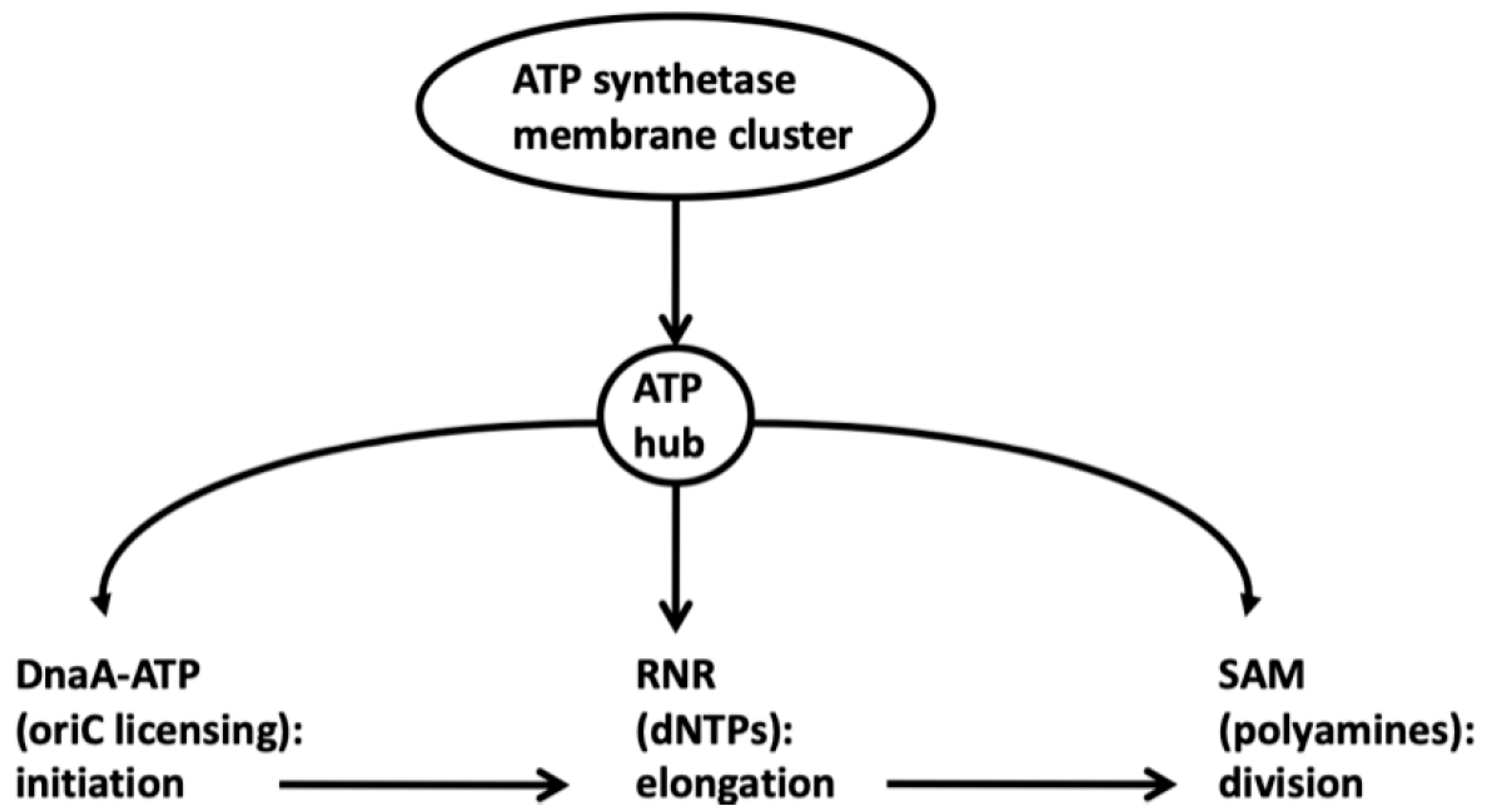
Duplication of the nucleoid is therefore itself closely associated with membrane biogenesis. ATP synthetase, likewise, is not uniformly distributed across the membrane but forms clusters (membrane microdomains) that produce high localized levels of ATP [87,88]. Such localized energy hubs might support replication initiation (Dna-ATP), elongation (RNR and CTP) and cell division (SAM, polyamines, FtsZ), and form membrane associated microdomains or condensates. ATP availability is the central organizing principle that unifies these three branches of cell physiology, providing the basis for their metabolic interactions and respective cell cycle functions (Figure 5).
Moreover, the spatial distribution of ATP synthetase is dynamic, which allows for rapid adaptation to changes in internal (perturbed replication forks, DNA damage, etc.) and external (nutrient supply, temperature, etc.) environments. It remains to be demonstrated, however, how energy metabolism, membrane biogenesis, DNA replication and cell division spatio-temporarily interact to form higher order molecular machines from the lower order hyperstructures. The organizing principle nonetheless is the fluid-mosaic structure of the membrane and the associated dynamics of ATP synthetase, the replication initiation and elongation hyperstructures, nuceloid organization and the divisome hyperstructure.
This review has attempted to provide a tentative overview of the genome duplication and cell growth processes, which, although coupled, are in several ways independent and parallel processes. One question raised here addressed the role of DNA synthesis in driving not only membrane biogenesis but also cell growth and therefore cell division. Duplication of the chromosome, for example, can occur in the absence of cell division, whereas cell division depends on completed DNA replication and daughter chromosome segregation (via nucleoid occlusion). The nucleotypic effect and nucleoid complexity, generated by initiation/elongation homeostasis, suggests that DNA replication forks and fork rates significantly influence cell size when the DnaA-RNR homeostasis loop is perturbed: fork rates remain strictly inversely correlated with the frequency of replication initiation in all cells examined so far, prokaryote and eukaryote alike, and under all growth conditions.
9. Conculsion
Nucleotide and phospholipid synthesis are clearly linked through a number of metabolic pathways that guarantee DNA/cell mass homeostasis, in particular via high energy metabolites such as ATP, GTP, CTP, Acetyl-CoA, SAM and NAD(P)H [89,90]. These cellular functions appear to be organized in what Mitchell termed membrane dependent “supra-molecular” machines, presumably built up from physico-chemically interacting hyperstructures that drive the dynamic continuum of the cell cycle [91]. Assuming such machines are not pure speculation [92], how the hyperstructures dynamically interact to provide the essential functions for survival and reproduction is not only of physiological importance, but also of interest to evolutionary biologists who study open questions concerning cell size, genome size and body mass evolution, and why and how mutation rates and the mutation distribution in the genome vary so dramatically across the Tree of Life and within prokaryote and eukaryote lineages [93,94].
Conflicts of Interest
The author declares no conflicts of interest.
References
- Mitchell, P. Coupling of phosphorylation to electron and hydrogen transfer by a chemi-osmotic type of mechanism. Nature 1961, 191(4784), 144–148. [Google Scholar] [CrossRef]
- Nath, S. Two-ion theory of energy coupling in ATP synthesis rectifies a fundamental flaw in the governing equations of the chemiosmotic theory. Biophysical Chemistry 2017, 230, 45–52. [Google Scholar] [CrossRef]
- Hallgren, Joel; Jonas, Kristina. Nutritional control of bacterial DNA replication. Current Opinion in Microbiology 2024, 77, 102403. [Google Scholar] [CrossRef] [PubMed]
- Katayama, T.; Ozaki, S.; Keyamura, K.; et al. Regulation of the replication cycle: conserved and diverse regulatory systems for DnaA and oriC. Nat Rev Microbiol 2010, 8, 163–170. [Google Scholar] [CrossRef]
- Meselson, M.; Stahl, F. W. The replication of DNA. In Cold Spring Harbor symposia on quantitative biology; Cold Spring Harbor Laboratory Press., January 1958; Vol. 23, pp. 9–12. [Google Scholar] [CrossRef]
- Cooper, S.; Helmstetter, C. E. Chromosome replication and the division cycle of Escherichia coli Br. Journal of molecular biology 1968, 31(3), 519–540. [Google Scholar] [CrossRef]
- Cooper, S. The continuum model: statistical implications. Journal of theoretical biology 1982, 94(4), 783–800. [Google Scholar] [CrossRef] [PubMed]
- Si, F.; Le Treut, G.; Sauls, J. T.; Vadia, S.; Levin, P. A.; Jun, S. Mechanistic origin of cell-size control and homeostasis in bacteria. Current Biology 2019, 29(11), 1760–1770. [Google Scholar] [CrossRef] [PubMed]
- Donachie, W. D. Relationship between cell size and time of initiation of DNA replication. Nature 1968, 219(5158), 1077–1079. [Google Scholar] [CrossRef]
- Jannière, L; Canceill, D; Suski, C; Kanga, S; Dalmais, B; Lestini, R; et al. Genetic Evidence for a Link Between Glycolysis and DNA Replication. PLoS ONE 2007, 2(5), e447. [Google Scholar] [CrossRef]
- Barańska, S.; Glinkowska, M.; Herman-Antosiewicz, A.; et al. Replicating DNA by cell factories: roles of central carbon metabolism and transcription in the control of DNA replication in microbes, and implications for understanding this process in human cells. Microb Cell Fact 2013, 12, 55. [Google Scholar] [CrossRef]
- Nouri, H.; Monnier, A. F.; Fossum-Raunehaug, S.; Maciąg-Dorszyńska, M.; Cabin-Flaman, A.; Kepes, F.; Janniere, L. Multiple links connect central carbon metabolism to DNA replication initiation and elongation in Bacillus subtilis. DNA Research 2018, 25(6), 641–653. [Google Scholar] [CrossRef]
- Tymecka-Mulik, J; Boss, L; Maciąg-Dorszyńska, M; Matias Rodrigues, JF; Gaffke, L; Wosinski, A; et al. Suppression of the Escherichia coli dnaA46 mutation by changes in the activities of the pyruvate-acetate node links DNA replication regulation to central carbon metabolism. PLoS ONE 2017, 12(4), e0176050. [Google Scholar] [CrossRef]
- Maciąg-Dorszyńska, M.; Ignatowska, M.; Jannière, L.; Węgrzyn, G.; Szalewska-Pałasz, A. Mutations in central carbon metabolism genes suppress defects in nucleoid position and cell division of replication mutants in Escherichia coli. Gene 2012, 503(1), 31–35. [Google Scholar] [CrossRef]
- Maciąg, M.; Nowicki, D.; Szalewska-Pałasz, A.; Węgrzyn, G. Central carbon metabolism influences fidelity of DNA replication in Escherichia coli. Mutation Research/Fundamental and Molecular Mechanisms of Mutagenesis 2012, 731(1-2), 99–106. [Google Scholar] [CrossRef] [PubMed]
- Maciąg, M.; Nowicki, D.; Janniere, L.; Szalewska-Pałasz, A.; Węgrzyn, G. Genetic response to metabolic fluctuations: correlation between central carbon metabolism and DNA replication in Escherichia coli. Microbial Cell Factories 2011, 10(1), 19. [Google Scholar] [CrossRef]
- Soultanas, P.; Janniere, L. The metabolic control of DNA replication: mechanism and function. Open biology 2023, 13(8). [Google Scholar] [CrossRef] [PubMed]
- Krause, K.; Maciąg-Dorszyńska, M.; Wosinski, A.; Gaffke, L.; Morcinek-Orłowska, J.; Rintz, E.; Węgrzyn, G. The role of metabolites in the link between DNA replication and central carbon metabolism in Escherichia coli. Genes 2020, 11(4), 447. [Google Scholar] [CrossRef]
- Fingland, N.; Flåtten, I.; Downey, C. D.; Fossum-Raunehaug, S.; Skarstad, K.; Crooke, E. Depletion of acidic phospholipids influences chromosomal replication in E scherichia coli. Microbiologyopen 2012, 1(4), 450–466. [Google Scholar] [CrossRef]
- Zaritsky, A.; Vollmer, W.; Männik, J.; Liu, C. Does the nucleoid determine cell dimensions in Escherichia coli? Frontiers in Microbiology 2019, 10, 1717. [Google Scholar] [CrossRef]
- Zhu, M.; Dai, X.; Guo, W.; Ge, Z.; Yang, M.; Wang, H.; Wang, Y. P. Manipulating the bacterial cell cycle and cell size by titrating the expression of ribonucleotide reductase. MBio 2017, 8(6), 10–1128. [Google Scholar] [CrossRef] [PubMed]
- Herrick, J.; Norris, V.; Kohiyama, M. 60 Years of Studies into the Initiation of Chromosome Replication in Bacteria. Biomolecules 2025, 15(2), 203. [Google Scholar] [CrossRef]
- Cooper, S. The central dogma of cell biology. Cell biology international reports 1981, 5(6), 539–549. [Google Scholar] [CrossRef]
- Gonzalez Moreno, S.; Mata Martin, C.; Ferrera Guillen, E.; Guzmán, E. C. Tuning the replication fork progression by the initiation frequency. Environmental microbiology 2013, 15(12), 3240–3251. [Google Scholar] [CrossRef]
- Salguero, I.; Lopez Acedo, E.; Guzman, E. C. Overlap of replication rounds disturbs the progression of replicating forks in a ribonucleotide reductase mutant of Escherichia coli. Microbiology 2011, 157(7), 1955–1967. [Google Scholar] [CrossRef]
- Dewachter, L.; Deckers, B.; Mares-Mejía, I.; Louwagie, E.; Vercauteren, S.; Matthay, P.; Michiels, J. The role of the essential GTPase ObgE in regulating lipopolysaccharide synthesis in Escherichia coli. Nature communications 2024, 15(1), 9684. [Google Scholar] [CrossRef] [PubMed]
- Murray, H.; Koh, A. Multiple regulatory systems coordinate DNA replication with cell growth in Bacillus subtilis. PLoS genetics 2014, 10(10), e1004731. [Google Scholar] [CrossRef] [PubMed]
- Pierucci, O. Phospholipid synthesis during the cell division cycle of Escherichia coli. Journal of bacteriology 1979, 138(2), 453–460. [Google Scholar] [CrossRef]
- Cooper, S.; Helmstetter, C. E. Chromosome replication and the division cycle of Escherichia coli Br. Journal of molecular biology 1968, 31(3), 519–540. [Google Scholar] [CrossRef]
- Arieh, Zaritsky; Woldringh Conrad, L. Chromosome replication, cell growth, division and shape: a personal perspective. Frontiers in Microbiology 2015, Volume 6. [Google Scholar] [CrossRef]
- Taheri-Araghi, S.; Bradde, S.; Sauls, J. T.; Hill, N. S.; Levin, P. A.; Paulsson, J.; Jun, S. Cell-size control and homeostasis in bacteria. Current biology 2015, 25(3), 385–391. [Google Scholar] [CrossRef] [PubMed]
- Howard, C. B.; Rabinovitch, A.; Yehezkel, G.; Zaritsky, A. Tight coupling of cell width to nucleoid structure in Escherichia coli. Biophysical Journal 2024, 123(4), 502–508. [Google Scholar] [CrossRef]
- Szarski, H. Cell size and nuclear DNA content in vertebrates. International Review of Cytology 1976, 44, 93–111. [Google Scholar] [CrossRef] [PubMed]
- Cavalier-Smith, Thomas. Nuclear volume control by nucleoskeletal DNA, selection for cell volume and cell growth rate, and the solution of the DNA C-value paradox. Journal of cell science 1978, 34.1, 247–278. [Google Scholar] [CrossRef] [PubMed]
- Gregory, T. R. The bigger the C-value, the larger the cell: genome size and red blood cell size in vertebrates. Blood Cells, Molecules, and Diseases 2001, 27(5), 830–843. [Google Scholar] [CrossRef]
- Donachie, W. D.; Begg, K. J.; Vicente, M. Cell length, cell growth and cell division. Nature 1976, 264(5584), 328–333. [Google Scholar] [CrossRef]
- Zaritsky, A.; Pritchard, E. H. Replication time of the chromosome in thymineless mutants of Escherichia coli. Journal of molecular biology 1971, 60(1), 65–74. [Google Scholar] [CrossRef]
- Zaritsky, A.; Pritchard, R. H. Changes in cell size and shape associated with changes in the replication time of the chromosome of Escherichia coli. Journal of bacteriology 1973, 114(2), 824–837. [Google Scholar] [CrossRef]
- Hand, R. Regulation of DNA replication on subchromosomal units of mammalian cells. The Journal of cell biology 1975, 64(1), 89–97. [Google Scholar] [CrossRef] [PubMed]
- Conti, C.; Sacca, B.; Herrick, J.; Lalou, C.; Pommier, Y.; Bensimon, A. Replication fork velocities at adjacent replication origins are coordinately modified during DNA replication in human cells. Molecular biology of the cell 2007, 18(8), 3059–3067. [Google Scholar] [CrossRef]
- Pritchard, R. H.; Zaritsky, A. Effect of thymine concentration on the replication velocity of DNA in a thymineless mutant of Escherichia coli. Nature 1970, 226(5241), 126–131. [Google Scholar] [CrossRef]
- Meacock, P. A.; Pritchard, R. H. Relationship between chromosome replication and cell division in a thymineless mutant of Escherichia coli B/r. Journal of Bacteriology 1975, 122(3), 931–942. [Google Scholar] [CrossRef]
- Fricke, J.; Neuhard, J.; Kelln, R. A.; Pedersen, S. The cmk gene encoding cytidine monophosphate kinase is located in the rpsA operon and is required for normal replication rate in Escherichia coli. Journal of Bacteriology 1995, 177(3), 517–523. [Google Scholar] [CrossRef] [PubMed]
- Ho, P. Y.; Amir, A. Simultaneous regulation of cell size and chromosome replication in bacteria. Frontiers in microbiology 2015, 6, 662. [Google Scholar] [CrossRef] [PubMed]
- Guzmán, E. C.; Caballero, J. L.; Jiménez-Sánchez, A. Ribonucleoside diphosphate reductase is a component of the replication hyperstructure in Escherichia coli. Molecular microbiology 2002, 43(2), 487–495. [Google Scholar] [CrossRef] [PubMed]
- Flåtten, I.; Fossum-Raunehaug, S.; Taipale, R.; Martinsen, S.; Skarstad, K. The DnaA protein is not the limiting factor for initiation of replication in Escherichia coli. PLoS genetics 2015, 11(6), e1005276. [Google Scholar] [CrossRef]
- Hansen, F. G.; Atlung, T. The DnaA tale. Frontiers in microbiology 2018, 9, 319. [Google Scholar] [CrossRef]
- El-Hajj, Z. W.; Newman, E. B. An Escherichia coli mutant that makes exceptionally long cells. Journal of bacteriology 2015, 197(8), 1507–1514. [Google Scholar] [CrossRef]
- Newman, E. B.; Budman, L. I.; Chan, E. C.; Greene, R. C.; Lin, R. T.; Woldringh, C. L.; D’ari, R. Lack of S-adenosylmethionine results in a cell division defect in Escherichia coli. Journal of bacteriology 1998, 180(14), 3614–3619. [Google Scholar] [CrossRef]
- Pountain, A. W.; Jiang, P.; Yao, T.; Homaee, E.; Guan, Y.; McDonald, K. J.; Yanai, I. Transcription–replication interactions reveal bacterial genome regulation. Nature 2024, 626(7999), 661–669. [Google Scholar] [CrossRef]
- Gon, S.; Camara, J. E.; Klungsøyr, H. K.; Crooke, E.; Skarstad, K.; Beckwith, J. A novel regulatory mechanism couples deoxyribonucleotide synthesis and DNA replication in Escherichia coli. The EMBO journal 2006, 25(5), 1137. [Google Scholar] [CrossRef]
- Sun, L.; Fuchs, J. A. Escherichia coli ribonucleotide reductase expression is cell cycle regulated. Molecular biology of the cell 1992, 3(10), 1095–1105. [Google Scholar] [CrossRef]
- Knöppel, A.; Broström, O.; Gras, K.; Elf, J.; Fange, D. Regulatory elements coordinating initiation of chromosome replication to the Escherichia coli cell cycle. Proceedings of the National Academy of Sciences 2023, 120(22), e2213795120. e2213795120. [Google Scholar] [CrossRef] [PubMed]
- Li, D.; Zheng, H.; Bai, Y.; Zhang, Z.; Cheng, H.; Huang, X.; Liu, C. Extrusion-modulated DnaA activity oscillations coordinate DNA replication with biomass growth. Elife 2025, 14, RP107214. [Google Scholar] [CrossRef]
- Olliver, A.; Saggioro, C.; Herrick, J.; Sclavi, B. DnaA-ATP acts as a molecular switch to control levels of ribonucleotide reductase expression in Escherichia coli. Molecular microbiology 2010, 76(6), 1555–1571. [Google Scholar] [CrossRef]
- Berger, M.; Wolde, P. R. T. Robust replication initiation from coupled homeostatic mechanisms. Nature communications 2022, 13(1), 6556. [Google Scholar] [CrossRef]
- Sánchez-Romero, M. A.; Molina, F.; Jiménez-Sánchez, A. Correlation between ribonucleoside-diphosphate reductase and three replication proteins in Escherichia coli. BMC Molecular Biology 2010, 11(1), 11. [Google Scholar] [CrossRef]
- Funk, M. A.; Zimanyi, C. M.; Andree, G. A.; Hamilos, A. E.; Drennan, C. L. How ATP and dATP act as molecular switches to regulate enzymatic activity in the prototypical bacterial class Ia ribonucleotide reductase. Biochemistry 2024, 63(19), 2517–2531. [Google Scholar] [CrossRef] [PubMed]
- Chabes, Andrei; Stillman, Bruce. Constitutively high dNTP concentration inhibits cell cycle progression and the DNA damage checkpoint in yeast Saccharomyces cerevisiae. Proceedings of the National Academy of Sciences 2007, 104(no. 4), 1183–1188. [Google Scholar] [CrossRef] [PubMed]
- Wieczorek, A.; Fornalewicz, K.; Mocarski, Ł.; Łyżeń, R.; Węgrzyn, G. Double silencing of relevant genes suggests the existence of the direct link between DNA replication/repair and central carbon metabolism in human fibroblasts. Gene 2018, 650, 1–6. [Google Scholar] [CrossRef]
- Babu, V. M.; Itsko, M.; Baxter, J. C.; Schaaper, R. M.; Sutton, M. D. Insufficient levels of the nrdAB-encoded ribonucleotide reductase underlie the severe growth defect of the Δhda E. coli strain. Molecular microbiology 2017, 104(3), 377–399. [Google Scholar] [CrossRef]
- Babu, V. M.; Homiski, C.; Scotland, M. K.; Chodavarapu, S.; Kaguni, J. M.; Sutton, M. D. Elevated Levels of the Escherichia coli nrdAB-Encoded Ribonucleotide Reductase Counteract the Toxicity Caused by an Increased Abundance of the β Clamp. Journal of Bacteriology 2021, 203(23), 10–1128. [Google Scholar] [CrossRef]
- Fujimitsu, K.; Su'etsugu, M.; Yamaguchi, Y.; Mazda, K.; Fu, N.; Kawakami, H.; Katayama, T. Modes of overinitiation, dnaA gene expression, and inhibition of cell division in a novel cold-sensitive hda mutant of Escherichia coli. Journal of bacteriology 2008, 190(15), 5368–5381. [Google Scholar] [CrossRef]
- Santos, M. M.; Johnson, M. C.; Fiedler, L.; Zegerman, P. Global early replication disrupts gene expression and chromatin conformation in a single cell cycle. Genome Biology 2022, 23(1), 217. [Google Scholar] [CrossRef]
- Chiaramello, A. E.; Zyskind, J. W. Coupling of DNA replication to growth rate in Escherichia coli: a possible role for guanosine tetraphosphate. Journal of bacteriology 1990, 172(4), 2013–2019. [Google Scholar] [CrossRef]
- Souza, W. D. Prokaryotic cells: structural organisation of the cytoskeleton and organelles. Memórias do Instituto Oswaldo Cruz 2012, 107, 283–293. [Google Scholar] [CrossRef] [PubMed]
- Igarashi, K.; Kashiwagi, K. Effects of polyamines on protein synthesis and growth of Escherichia coli. Journal of Biological Chemistry 2018, 293(48), 18702–18709. [Google Scholar] [CrossRef]
- Newman, E. B.; Budman, L. I.; Chan, E. C.; Greene, R. C.; Lin, R. T.; Woldringh, C. L.; D’ari, R. Lack of S-adenosylmethionine results in a cell division defect in Escherichia coli. Journal of bacteriology 1998, 180(14), 3614–3619. [Google Scholar] [CrossRef] [PubMed]
- Yamanaka, K. U. N. I. T. O. S. H. I.; Ogura, T. E. R. U.; Niki, H. I. R. O. N. O. R. I.; Hiraga, S. O. T. A. Identification and characterization of the smbA gene, a suppressor of the mukB null mutant of Escherichia coli. Journal of bacteriology 1992, 174(23), 7517–7526. [Google Scholar] [CrossRef]
- Bloch, S.; Sinden, R. R.; Wien, F.; Węgrzyn, G.; Arluison, V. DNA Transactions in bacteria and membranes: A place for the Hfq protein? Membranes 2025, 15(4), 103. [Google Scholar] [CrossRef]
- Spahn, C.; Middlemiss, S.; Gómez-de-Mariscal, E.; Henriques, R.; Bode, H. B.; Holden, S.; Heilemann, M. The nucleoid of rapidly growing Escherichia coli localizes close to the inner membrane and is organized by transcription, translation, and cell geometry. Nature Communications 2025, 16(1), 3732. [Google Scholar] [CrossRef]
- Wang, Y.; Jiang, Y.; Song, Z.; Zhu, C.; Tang, Y.; Peng, J.; Liu, P. MreB: unraveling the molecular mechanisms of bacterial shape, division, and environmental adaptation. Cell Communication and Signaling 2025, 23(1), 377. [Google Scholar] [CrossRef]
- Ladouceur, A. M.; Parmar, B. S.; Biedzinski, S.; Wall, J.; Tope, S. G.; Cohn, D.; Weber, S. C. Clusters of bacterial RNA polymerase are biomolecular condensates that assemble through liquid–liquid phase separation. Proceedings of the National Academy of Sciences 2020, 117(31), 18540–18549. [Google Scholar] [CrossRef] [PubMed]
- Nandana, V.; Schrader, J. M. Roles of liquid–liquid phase separation in bacterial RNA metabolism. Current opinion in microbiology 2021, 61, 91–98. [Google Scholar] [CrossRef]
- Shen, B. A.; Landick, R. Transcription of bacterial chromatin. Journal of molecular biology 2019, 431(20), 4040–4066. [Google Scholar] [CrossRef]
- Odsbu, I.; Morigen; Skarstad, K. A reduction in ribonucleotide reductase activity slows down the chromosome replication fork but does not change its localization. PloS one 2009, 4(10), e7617. [Google Scholar] [CrossRef] [PubMed]
- Zaritsky, A. Cell-shape homeostasis in Escherichia coli is driven by growth, division, and nucleoid complexity. Biophysical journal 2015, 109(2), 178–181. [Google Scholar] [CrossRef] [PubMed]
- Woldringh, C. L. The role of co-transcriptional translation and protein translocation (transertion) in bacterial chromosome segregation. Molecular microbiology 2002, 45(1), 17–29. [Google Scholar] [CrossRef]
- Pierucci, O. Phospholipid synthesis during the cell division cycle of Escherichia coli. Journal of bacteriology 1979, 138(2), 453–460. [Google Scholar] [CrossRef]
- Hakenbeck, R. E. G. I. N. E.; Messer, W. A. L. T. E. R. Oscillations in the synthesis of cell wall components in synchronized cultures of Escherichia coli. Journal of Bacteriology 1977, 129(3), 1234–1238. [Google Scholar] [CrossRef]
- Pierucci, O. Dimensions of Escherichia coli at various growth rates: model for envelope growth. Journal of bacteriology 1978, 135(2), 559–574. [Google Scholar] [CrossRef]
- Joseleau-Petit, D. A. N. I. J. L. E.; Kepes, F.; Peutat, L.; D'Ari, R. I. C. H. A. R. D.; Képès, A. D. A. M. DNA replication initiation, doubling of rate of phospholipid synthesis, and cell division in Escherichia coli. Journal of bacteriology 1987, 169(8), 3701–3706. [Google Scholar] [CrossRef]
- Wu, F.; Swain, P.; Kuijpers, L.; Zheng, X.; Felter, K.; Guurink, M.; Dekker, C. Cell boundary confinement sets the size and position of the E. coli chromosome. Current Biology 2019, 29(13), 2131–2144. [Google Scholar] [CrossRef] [PubMed]
- Woldringh, C. L. The bacterial nucleoid: from electron microscopy to polymer physics—a personal recollection. Life 2023, 13(4), 895. [Google Scholar] [CrossRef] [PubMed]
- Dame, R. T.; Rashid, F. Z. M.; Grainger, D. C. Chromosome organization in bacteria: mechanistic insights into genome structure and function. Nature Reviews Genetics 2020, 21(4), 227–242. [Google Scholar] [CrossRef]
- Chastanet, A.; y Lopez, R. C. The actin-like MreB proteins in Bacillus subtilis: a new turn. Frontiers in bioscience (Scholar edition) 2012, 4(2), 1582–1606. [Google Scholar]
- Erhardt, H.; Dempwolff, F.; Pfreundschuh, M.; Riehle, M.; Schäfer, C.; Pohl, T.; Friedrich, T. Organization of the E scherichia coli aerobic enzyme complexes of oxidative phosphorylation in dynamic domains within the cytoplasmic membrane. Microbiologyopen 2014, 3(3), 316–326. [Google Scholar] [CrossRef]
- Magalon, A.; Alberge, F. Distribution and dynamics of OXPHOS complexes in the bacterial cytoplasmic membrane. Biochimica et Biophysica Acta (BBA)-Bioenergetics 2016, 1857(3), 198–213. [Google Scholar] [CrossRef]
- Kint, C.; Verstraeten, N.; Hofkens, J.; Fauvart, M.; Michiels, J. Bacterial Obg proteins: GTPases at the nexus of protein and DNA synthesis. Critical reviews in microbiology 2014, 40(3), 207–224. [Google Scholar] [CrossRef]
- Qiu, H.; Ye, C. Phospholipid biosynthesis: an unforeseen modulator of nuclear metabolism. Biology of the Cell 2025, 117(3), e70002. [Google Scholar] [CrossRef]
- Cooper, S. The continuum model: application to G1-arrest and G (0). In Cell Growth; Springer New York: Boston, MA, 1982; pp. 315–336. [Google Scholar]
- Tlusty, T.; Libchaber, A. Life sets off a cascade of machines. Proceedings of the National Academy of Sciences 2025, 122(4), e2418000122. [Google Scholar] [CrossRef]
- Nishimura, I.; Kurokawa, M.; Liu, L.; Ying, B. W. Coordinated changes in mutation and growth rates induced by genome reduction. MBio 2017, 8(4), 10–1128. [Google Scholar] [CrossRef] [PubMed]
- Banfalvi, G. Ribose selected as precursor to life. DNA and Cell Biology 2020, 39(2), 177–186. [Google Scholar] [CrossRef]
- Grucela, PK; Fuhrer, T; Sauer, U; Chao, Y; Zhang, YE. Ribose 5-phosphate (R5B): the key metabolite bridging the metabolisms of nucleotides and amino acids during stringent response in Escherichia coli? Microb Cell. 2023, 10(7), 141–144. [Google Scholar] [CrossRef] [PubMed]
- Seregina, TA; Shakulov, RS; Petrushanko, IY; Lobanov, KV; Mironov, AS. Biosynthesis of Ribose-5-Phosphate-Metabolic Regulator of Escherichia coli Viability. Cells 2025, 14(22), 1775. [Google Scholar] [CrossRef] [PubMed]
Figure 1.
Nucleoid Complexity (NC) and the Nucleotypic Effect Mediate DNA/cell mass Homeostasis Under Normal and Perturbed Cell Growth Conditions. The open circles in the stick figures represent DnaA-ATP licensed origins of replication (oriC). The y-shaped (branched) lines represent the number of origins per chromosome (NC). The width of the cylinder represents the size of the cell relative to chromosomal content (NC). The top arrow (x-axis) represents cellular productivity (biomass accumulation). The left arrow (y-axis) represents individual cell size (cell-shape homeostasis). A: Nutrient limited (slow) growth. The successive arrows reflect the transitions between the B-period (Left); C-period (Middle); and the D-period (Right). B: Nutrient unlimited (fast) growth. During fast growth, DnaA-ATP licenses origins in the mother cell presumably following the eclipse period (SeqA sequestration of oriC: 1/3 of cell cycle). Consequently, the B-period is absent from a normal cell cycle (the left arrow corresponds to the C-period; the right arrow corresponds to the transition to the D-period). According to a recent hypothesis: “(p)ppGpp coordinates the dynamic metabolic conversions of nucleotides to amino acids.” (95, 96). It is proposed here that a B-period emerges from a metabolic switch mediated by Ribose 5-Phosphate (potentially involving (p)ppGpp levels) between nucleotide metabolism and ammino acid metabolism. Under nutrient/amino acid scarcity, limiting nucleotide pools and low rates of one-carbon and amino acid metabolism restrict membrane growth while limiting the frequency of replication initiation. Hence, low levels of nucleotides during the B-period delay initiation of DNA replication following cell division until sufficient biomass (cell membrane growth) has accumulated to shift metabolic equilibrium in favor of nucleotide synthesis.
Figure 1.
Nucleoid Complexity (NC) and the Nucleotypic Effect Mediate DNA/cell mass Homeostasis Under Normal and Perturbed Cell Growth Conditions. The open circles in the stick figures represent DnaA-ATP licensed origins of replication (oriC). The y-shaped (branched) lines represent the number of origins per chromosome (NC). The width of the cylinder represents the size of the cell relative to chromosomal content (NC). The top arrow (x-axis) represents cellular productivity (biomass accumulation). The left arrow (y-axis) represents individual cell size (cell-shape homeostasis). A: Nutrient limited (slow) growth. The successive arrows reflect the transitions between the B-period (Left); C-period (Middle); and the D-period (Right). B: Nutrient unlimited (fast) growth. During fast growth, DnaA-ATP licenses origins in the mother cell presumably following the eclipse period (SeqA sequestration of oriC: 1/3 of cell cycle). Consequently, the B-period is absent from a normal cell cycle (the left arrow corresponds to the C-period; the right arrow corresponds to the transition to the D-period). According to a recent hypothesis: “(p)ppGpp coordinates the dynamic metabolic conversions of nucleotides to amino acids.” (95, 96). It is proposed here that a B-period emerges from a metabolic switch mediated by Ribose 5-Phosphate (potentially involving (p)ppGpp levels) between nucleotide metabolism and ammino acid metabolism. Under nutrient/amino acid scarcity, limiting nucleotide pools and low rates of one-carbon and amino acid metabolism restrict membrane growth while limiting the frequency of replication initiation. Hence, low levels of nucleotides during the B-period delay initiation of DNA replication following cell division until sufficient biomass (cell membrane growth) has accumulated to shift metabolic equilibrium in favor of nucleotide synthesis.
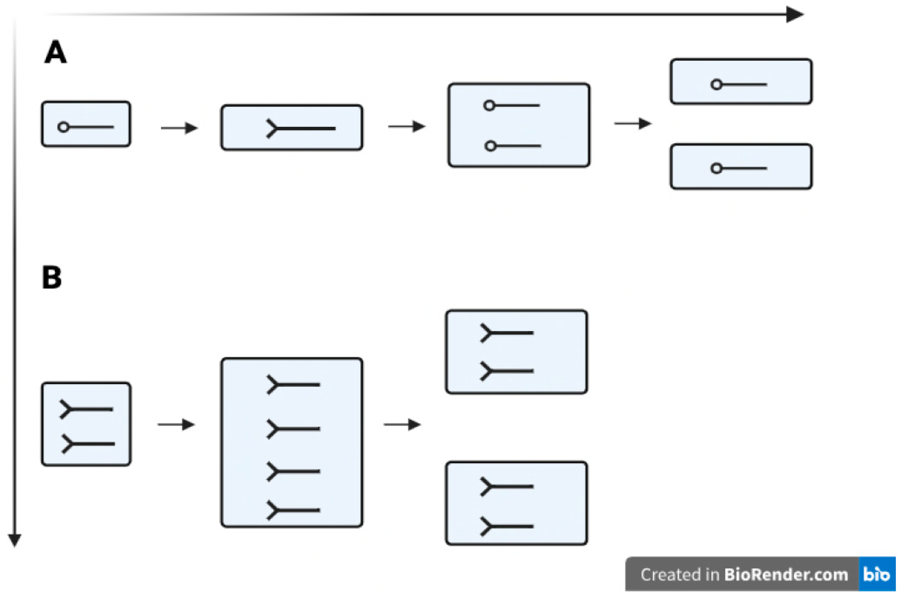
Figure 2.
Central Carbon Metabolism (CCM) Suppressors of DNA Replication Mutants Converge on Nucleotide Metabolism to Couple the Rate of DNA Replication (Initiation Frequency and/or Replication Fork Rate) to the Rate of Phospholipid/Carbon Metabolism. It is proposed here that the coupling suggests a plausible interpretation of how cells maintain DNA/cell mass homeostasis under such a broad range of advantageous and adverse growth and replication conditions: the cell membrane (mass) simultaneously grows around, and in proportion to, the growing amount of DNA.
Figure 2.
Central Carbon Metabolism (CCM) Suppressors of DNA Replication Mutants Converge on Nucleotide Metabolism to Couple the Rate of DNA Replication (Initiation Frequency and/or Replication Fork Rate) to the Rate of Phospholipid/Carbon Metabolism. It is proposed here that the coupling suggests a plausible interpretation of how cells maintain DNA/cell mass homeostasis under such a broad range of advantageous and adverse growth and replication conditions: the cell membrane (mass) simultaneously grows around, and in proportion to, the growing amount of DNA.
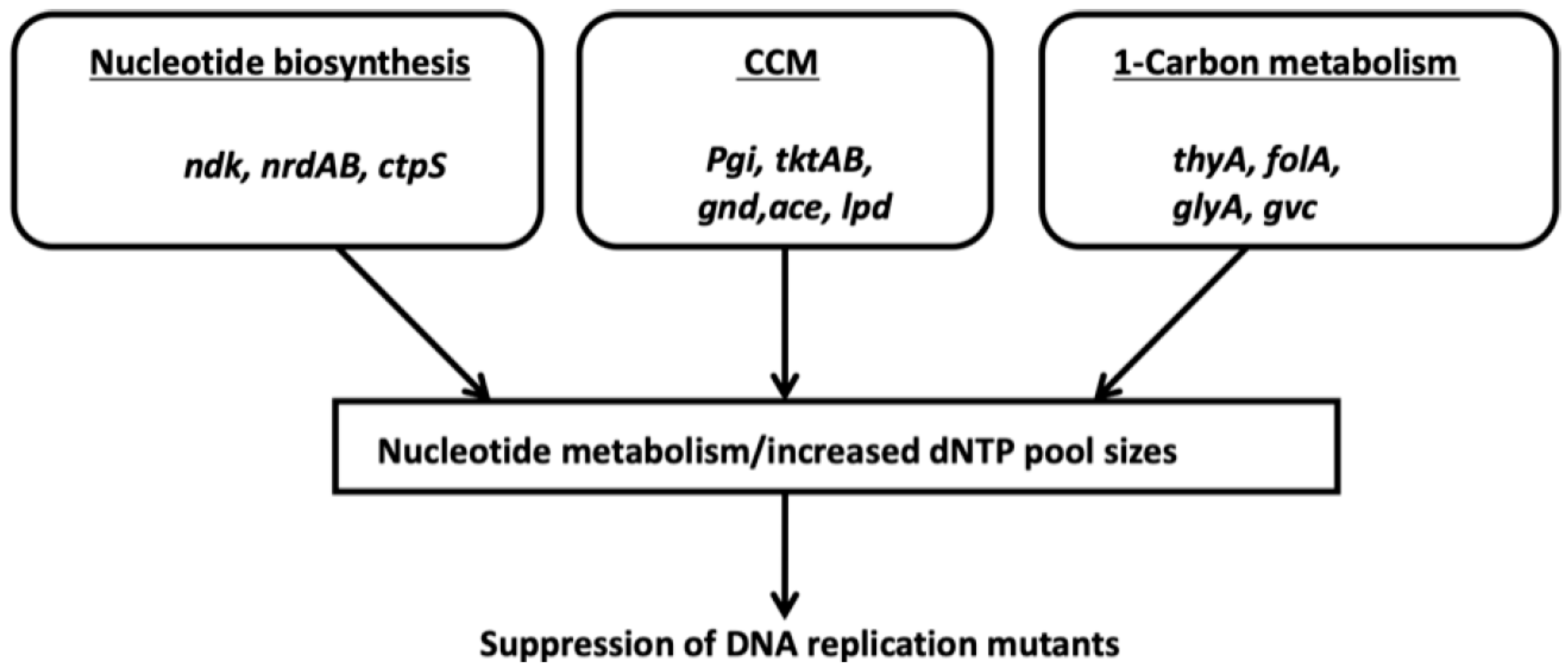
Figure 3.
CTP Synthesis Pathway Couples DNA synthesis to Phospholipid Synthesis thus Contributing to DNA/cell mass Homeostasis.
Figure 3.
CTP Synthesis Pathway Couples DNA synthesis to Phospholipid Synthesis thus Contributing to DNA/cell mass Homeostasis.
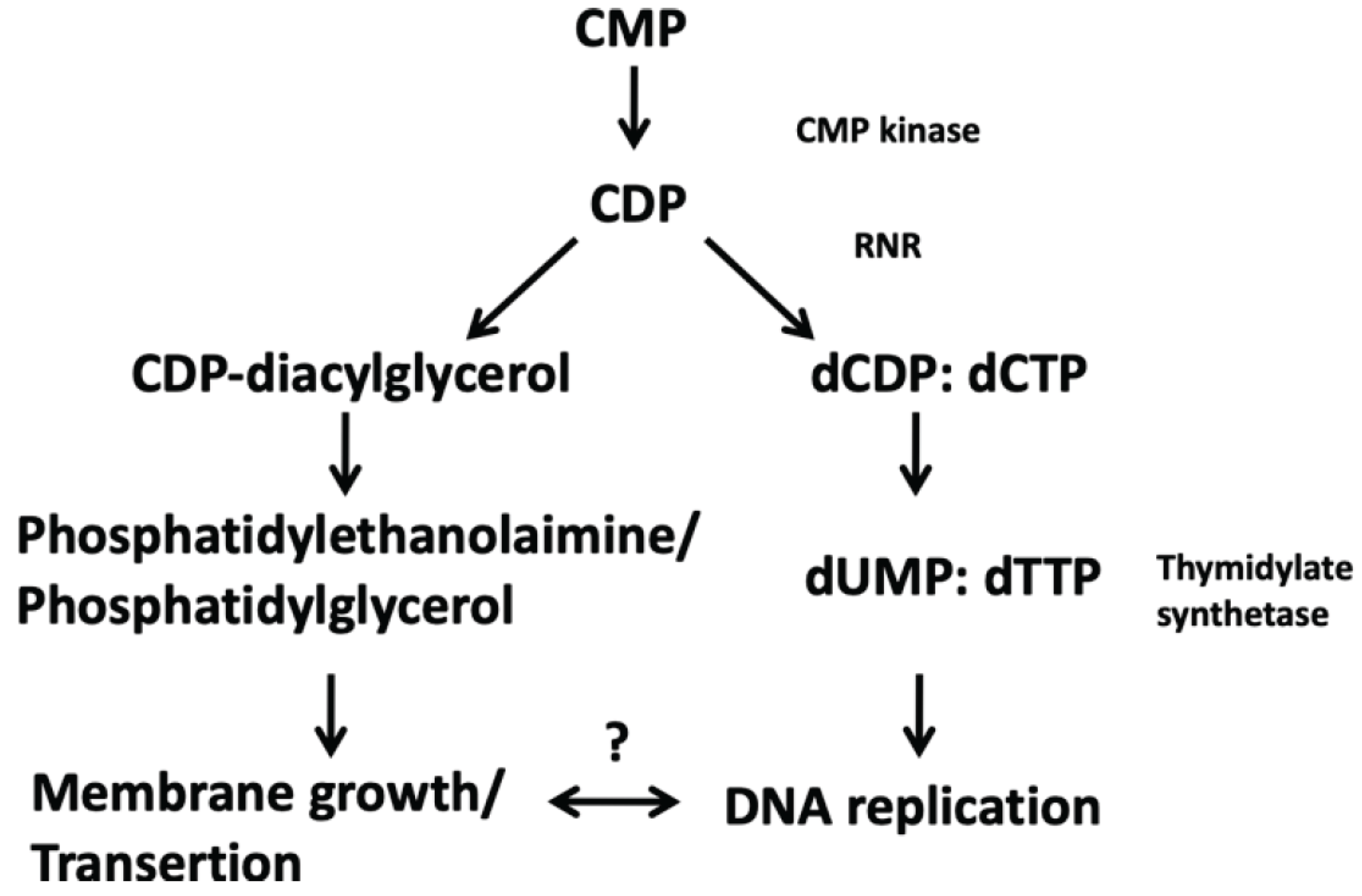
Figure 4.
The Folate Cycle (Purine Biosynthesis)/Pentose Phosphate Pathway (NADPH; R5B) Coordinate dNTP Biosynthesis and the Production of S-adenosylmethionine. The coordination between the two pathways suggests that nucleotide synthesis and SAM driven polyamine synthesis couple DNA replication initiation to cell division. Mutations in the Dam methyltransferase disrupt initiation-elongation homeostasis until the cell adapts to maintain DNA/cell mass homeostasis: cells with perturbed replication forks upregulate the nrdAB gene increasing the rates of dNTP synthesis that are necessary for cell survival and division. Experimentally overproduced RNR, for example, can rescue cells having a lethal cold sensitive hyper initiation phenotype (such as the dnaAcos and hda mutants). The E. coli origin of replication is enriched in GATC sites that, when oriC is hemimethylated, are sequestered by SeqA from the Dam methyltransferase and aberrant replication re-initiation. SAM therefore acts to coordinate replication initiation with cell division. Why cells without SAM cannot couple DNA replication to cell division remains unclear, but one possibility is through a membrane defect and/or polyamine depletion that might disrupt the FtsZ ring required for mother cells to divide [66,67,68].
Figure 4.
The Folate Cycle (Purine Biosynthesis)/Pentose Phosphate Pathway (NADPH; R5B) Coordinate dNTP Biosynthesis and the Production of S-adenosylmethionine. The coordination between the two pathways suggests that nucleotide synthesis and SAM driven polyamine synthesis couple DNA replication initiation to cell division. Mutations in the Dam methyltransferase disrupt initiation-elongation homeostasis until the cell adapts to maintain DNA/cell mass homeostasis: cells with perturbed replication forks upregulate the nrdAB gene increasing the rates of dNTP synthesis that are necessary for cell survival and division. Experimentally overproduced RNR, for example, can rescue cells having a lethal cold sensitive hyper initiation phenotype (such as the dnaAcos and hda mutants). The E. coli origin of replication is enriched in GATC sites that, when oriC is hemimethylated, are sequestered by SeqA from the Dam methyltransferase and aberrant replication re-initiation. SAM therefore acts to coordinate replication initiation with cell division. Why cells without SAM cannot couple DNA replication to cell division remains unclear, but one possibility is through a membrane defect and/or polyamine depletion that might disrupt the FtsZ ring required for mother cells to divide [66,67,68].
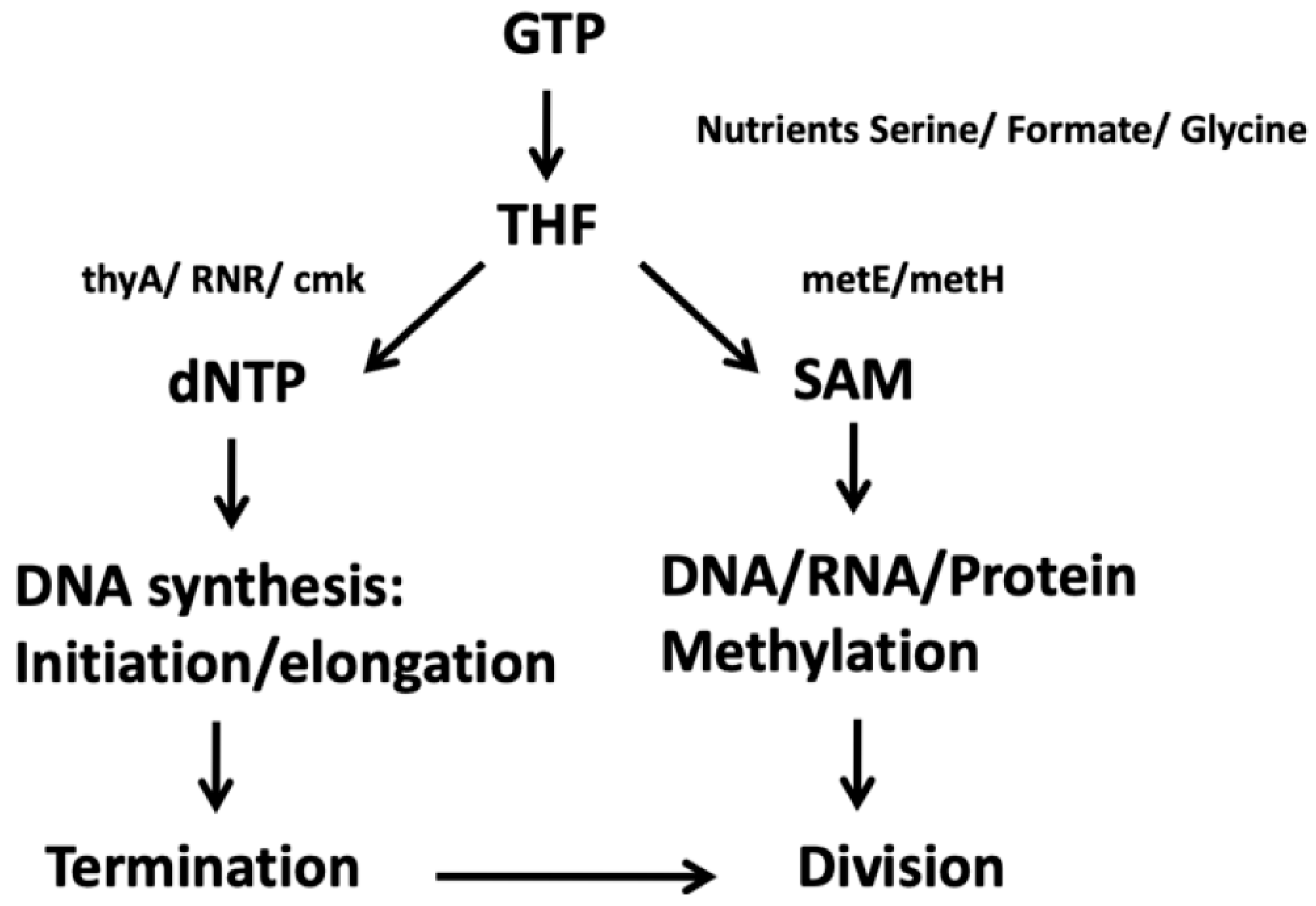
Figure 5.
Membrane Bound ATP Synthetase and Oxidative Phosphorylation Integrate Membrane Growth with Initiation-Elongation Hyperstructures into “Cascading von Neumann Machines.” The proposed “machines” couple energy production to DNA doubling and cell mass doubling, consistent with the Continuum Model of the cell cycle, with a relatively invariant cell mass at initiation (under normal and perturbed growth conditions) and with the Transertion Hypothesis.
Figure 5.
Membrane Bound ATP Synthetase and Oxidative Phosphorylation Integrate Membrane Growth with Initiation-Elongation Hyperstructures into “Cascading von Neumann Machines.” The proposed “machines” couple energy production to DNA doubling and cell mass doubling, consistent with the Continuum Model of the cell cycle, with a relatively invariant cell mass at initiation (under normal and perturbed growth conditions) and with the Transertion Hypothesis.
Disclaimer/Publisher’s Note: The statements, opinions and data contained in all publications are solely those of the individual author(s) and contributor(s) and not of MDPI and/or the editor(s). MDPI and/or the editor(s) disclaim responsibility for any injury to people or property resulting from any ideas, methods, instructions or products referred to in the content. |
© 2026 by the authors. Licensee MDPI, Basel, Switzerland. This article is an open access article distributed under the terms and conditions of the Creative Commons Attribution (CC BY) license (http://creativecommons.org/licenses/by/4.0/).
Copyright: This open access article is published under a Creative Commons CC BY 4.0 license, which permit the free download, distribution, and reuse, provided that the author and preprint are cited in any reuse.


