Submitted:
17 March 2026
Posted:
18 March 2026
You are already at the latest version
Abstract
This study aimed to identify angiotensin I-converting enzyme (ACE) inhibitory peptides from royal jelly (RJ) proteins and elucidate their inhibition patterns and mechanisms. RJ proteins were analyzed for ACE inhibition potential using in silico tools, and suitable enzymes were selected for peptide release. Hydrolysis conditions were optimized using response surface methodology (RSM), and the resulting peptides were fractionated and purified. Mass spectrometry identified 57 peptides, with seven selected for synthesis based on scoring. IDFDF, DVNFR, and SFHRL showed the highest ACE inhibition, with IC50 values of 16.9 μM, 42.5 μM, and 242.6 μM, respectively. Lineweaver-Burk plots revealed IDFDF as a competitive inhibitor, DVNFR as a non-competitive inhibitor, and SFHRL as a mixed inhibitor. Molecular docking indicated that peptide-ACE interactions were primarily mediated through hydrogen bonds and Zn(II) coordination. These findings support the high-value utilization of RJ and the development of food-derived ACE inhibitory peptides.
Keywords:
royal jelly
; ACE inhibitory peptide
; in silico
; purification
; molecular docking
1. Introduction
Royal jelly (RJ) is a natural substance secreted by the hypopharyngeal and mandibular glands of nurse honeybees (Apis mellifera) [1]. It plays a crucial role in the development of queen bees and larvae within honeybee colonies and has been widely used in human health and beauty products [2]. The caste system in honeybees is nutritionally mediated, with larvae fed exclusively on RJ developing into queens [3,4]. Notably, queen honeybees exhibit a body size approximately double and a lifespan tenfold greater than that of worker bees of the same genotype [2].
RJ consists of approximately 60–70% water, 9–18% proteins, 11–23% carbohydrates, and 4–8% lipids [5]. It also contains trace amounts of mineral salts, phenols, flavonoids, free amino acids, vitamins, and other components. The protein content of RJ is predominantly composed of Major Royal Jelly Proteins (MRJPs), which account for 83–90% of the total proteins [5]. Among these, MRJP1 is the most abundant, constituting 31–66% of the total RJ proteins, followed by MRJP3 (26%), MRJP2 (16%), and MRJP5 (9%)[6].
RJ has been shown to possess numerous pharmacological activities, including antioxidant, anti-inflammatory, anti-aging, neuroprotective, antimicrobial, anti-allergic, and antitumoral properties [7,8]. However, several limitations hinder its commercial potential, including poor stability, limited water solubility, an undesirable spicy and sour tastes, and the potential to cause allergic reactions in susceptible individuals [9]. China is the world’s largest producer and exporter of RJ, with an annual production exceeding 4,000 tons and a market value surpassing $2.5 billion, accounting for approximately 90% of global RJ production [10,11]. However, a significant portion of RJ is exported in raw form, resulting in relatively low profitability. Therefore, extensive processing of RJ is essential to address these issues, particularly through the development of bioactive peptides with enhanced functionality, stability, and added value.
Bioactive peptides, typically consisting of 2–20 amino acids, are inactive within the parent protein but exhibit beneficial health effects upon release [12]. The preparation and characterization of bioactive peptides from RJ is a relatively new field. To date, only a limited number of studies have reported the production of peptides with antioxidant properties [13], as well as ACE inhibitory [14,15] and renin inhibitory activities [16]. ACE inhibitory peptides are of particular interest due to their ability to lower blood pressure, thereby reducing the risk of hypertension-related conditions such as myocardial infarction, stroke, and coronary artery disease [17]. Although synthetic ACE inhibitors like captopril, enalapril, and lisinopril are widely used, they are often associated with adverse side effects after prolonged use [18]. Thus, ACE inhibitory peptides represent a promising alternative to synthetic drugs.
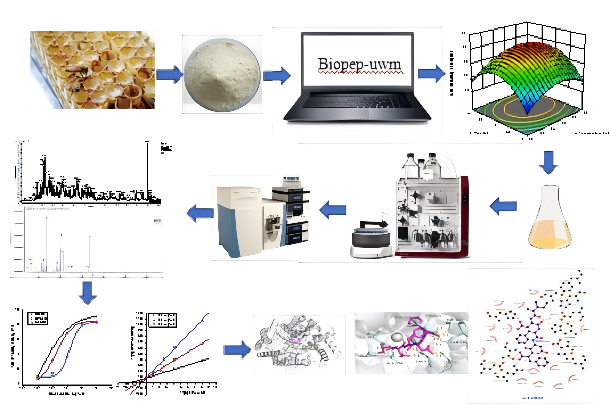
Despite the identification of some ACE inhibitory peptides from RJ proteins, such as Ile-Tyr, Val-Tyr, and Lys-Phe [14,15], there remains a pressing need to identify additional peptides and elucidate their mechanisms of action. Therefore, the objectives of this study were to: (a) use in silico methods to select suitable enzymes for generating ACE inhibitory peptides from RJ proteins; (b) optimize hydrolysis conditions using response surface methodology (RSM); (c) purify ACE inhibitory peptides using Sephadex G-25 chromatography, anion exchange chromatography, and reverse-phase high-performance liquid chromatography (RP-HPLC); (d) identify peptides in the most active fractions using LC-MS/MS; (e) chemically synthesize and determine the IC50 values of the most active peptides; and (f) explore the ACE inhibition patterns and mechanisms of the selected peptides.
2. Materials and Methods
2.1. Materials and Reagents
RJ proteins were purchased from Beijing Jinwang Health Technology Co., Ltd. (Beijing, China). Proteinase K (EC 3.4.21.64, 40 U/mg), ACE (from rabbit lung) and its substrate N-[3-(2-Furyl)acryloyl]-L-phenylalanyl-glycyl-glycine (FAPGG) were purchased from Solarbio Science & Technology Co., Ltd. (Beijing, China). Sephadex G-25 was supplied by Shanghai Yuanye BioTechnology Co., Ltd. (Shanghai, China). HPLC-grade acetonitrile, methanol, and trifluoroacetic acid (TFA) were obtained from Fisher Scientific Company (Waltham, MA, USA). All other chemicals used were of analytical reagent grade.
2.2. Evaluation of RJ Proteins as a Source of Bioactive Peptides
An in silico analysis was conducted to assess the potential of RJ proteins as a source of bioactive peptides. Nine RJ protein sequences were selected, including MRJP1 (AAM73637.1), MRJP2 (ACS66837.1), MRJP3 (ADC55524.1), MRJP4 (ADB82660.1), MRJP5 (NP_001011599.1), MRJP6 (AAQ82184.1), MRJP7 (DAA01512.1), MRJP8 (AAR83734.1), and MRJP9 (AAY21180.1), which collectively account for over 83% of RJ proteins. The analysis of bioactive peptides within these proteins was performed using the BIOPEP-UWM platform (http://www.uwm.edu.pl/biochemia), following the methodology described by Minkiewicz et al. [19].
The frequency of occurrence (A) of peptides with specific biological activities within each MRJP sequence was evaluated using the “calculations” tab. Parameter A was calculated as the ratio of the number of specific bioactive peptides to the total amino acid count in each MRJP sequence, as outlined by Minkiewicz et al. [19]. Additionally, the aggregate occurrence frequency (ΣA) of all bioactive peptides within each protein sequence was assessed.
2.3. In Silico Analysis of ACE Inhibitory Peptides Released from MRJPs
The MRJP sequences were subjected to enzymatic hydrolysis within the BIOPEP-UWM database using the “batch processing” feature [19]. The analysis included the theoretical degree of hydrolysis (DHt) and the incidence of ACE inhibitory peptides released by proteolytic enzymes. DHt represents the proportion of cleaved peptide bonds to the total number of peptide bonds in a protein sequence [19]. Seven distinct enzymes were used: pepsin (pH 1.3) (EC 3.4.23.1), chymotrypsin(A) (EC 3.4.21.1), trypsin (EC 3.4.21.4), papain (EC 3.4.22.2), stem bromelain (EC 3.4.22.32), subtilisin (EC 3.4.21.62), and proteinase K (EC 3.4.21.64).
2.4. Measurement of ACE Inhibitory Activity
The ACE inhibitory activity was determined following the methodology outlined by Cao et al. with slight modifications [20]. Briefly, 10 µL of 0.1 U/mL ACE solution was added to 40 µL of the sample in a 96-well microtiter plate and incubated for 10 min at 37 °C. The reaction was initiated by adding 50 µL of FAPGG buffer (1.0 mmol/L, containing 80 mM HEPES and 0.3 M NaCl, pH 8.3), and the plate was immediately transferred to a multi-wavelength microplate reader set at 37 °C for 30 min. The absorbance difference of the sample before and after the reaction was measured at 340 nm and recorded as and . A blank was prepared similarly, replacing the sample with the same volume of 80 mM HEPES buffer. The absorbance difference of the blank was recorded as and . Samples were assayed in triplicate. The ACE inhibitory activity (%) was calculated using the following equation.
The IC50 value, defined as the inhibitor concentration required to reduce ACE activity by 50%, was determined by plotting the percentage inhibition against peptide concentration.
2.5. Optimization of RJ Protease Hydrolysis Process Using Response Surface Methodology
RSM was employed to optimize the enzymatic hydrolysis conditions. The enzyme-to-substrate (E/S) mass ratio was maintained at 1.0%. The independent variables were enzymatic hydrolysis temperature (A), pH (B), and time (C), with ACE inhibitory activity (Y) as the dependent variable. The Box-Behnken design was used to optimize the processing parameters, with each variable encoded at three levels: −1, 0, and +1 (Table 1).
Experimental design and regression analysis were conducted using Design-Expert software (version 8.0.6). After each hydrolysis step, the hydrolysates were heated at 100 °C for 15 min to inactivate enzymes, cooled to room temperature, and centrifuged at 9,600 g for 10 min. The resulting supernatants were collected and analyzed for ACE inhibitory activity. The predictive model for enzymatic hydrolysis conditions was validated by assessing the ACE inhibitory activity of the hydrolysate produced under optimized conditions.
2.6. Isolation and Purification of ACE Inhibitory peptides
The enzymatic hydrolysate was filtered using a 0.45 µm aqueous polyethersulfone ultrafiltration membrane (Jinlong Co., Ltd., Tianjin, China). The filtrate was then purified on a Sephadex G-25 gel column, eluted with distilled water at a flow rate of 0.5 mL/min. The elution was monitored at 214 nm, and fractions were collected every 5 min. The fractions were lyophilized, and their ACE inhibitory activity was assessed. Protein content in each fraction was quantified using the Folin-phenol method [21]. The most active fraction was further purified on a HiTrap DEAE FF column (16 mm × 100 mm, GE Healthcare, Sweden), pre-equilibrated with 50 mM Tris/HCl buffer (pH 7.0). The column was eluted with a NaCl gradient (0–1 mol/L) in the same buffer. Fractions were collected, desalted, and subjected to ACE activity assays. Fraction C4 was further purified by semi-preparative reverse-phase HPLC (RP-HPLC) using a preparative C18 column (20 × 250 mm, 15 µm, Waters, Milford, MA, USA) coupled with a 2998 photodiode array (PDA) detector on a Waters 600 system. Samples (150 µL, 20 mg/mL) were eluted with a linear gradient (flow rate: 1.5 mL/min) of water (solvent A, containing 0.1% TFA) and acetonitrile (solvent B, containing 0.1% TFA). The elution program was as follows: 0–5 min, 100% solvent B; 5–25 min, 50% solvent A and 50% solvent B; 25–30 min, 100% solvent A. The elution peak was monitored at 214 nm. Subfractions were freeze-dried and used for ACE inhibitory activity determination. The most active subfraction was selected for peptide sequence identification.
2.7. Peptide Sequence Analysis by LC-MS/MS
ACE inhibitory peptides were identified using liquid chromatography-tandem mass spectrometry (LC-MS/MS) by Bio-Tech Pack Technology Company Ltd. (Beijing, China). The HPLC system used was an easy nLC 1200 (Thermo Fisher Scientific, MA, USA). Chromatographic separation was achieved using a custom-packed column with Acclaim PepMap RPLC C18 (1.9 µm, 100 Å, 150 µm × 15 cm) media (Dr. Maisch GmbH, Germany). The mobile phase consisted of solvent A (0.1% formic acid in water) and solvent B (0.1% formic acid and 80% acetonitrile in water). The flow rate was set at 600 nL/min. The gradient elution profile was as follows: 4% B-phase at 0 min, increasing to 8% at 2 min, 28% at 43 min, 40% at 10 min, 95% at 1 min, and maintained at 95% for 10 min. Mass spectrometry analysis was performed using an Orbitrap Fusion Lumos instrument (Thermo Fisher Scientific, MA, USA). The analytical conditions included a spray voltage of 2.2 kV, a capillary temperature of 270 °C, and a resolution of 70,000 at 400 m/z for the primary mass analyzer and 17,500 at m/z 200 for the secondary. The precursor ion scan range was m/z 100–1500. The automatic gain control (AGC) target for MS1 was 3e6 with an ion injection time of 100 ms, while for MS2, the AGC target was 1e5 with an ion injection time of 50 ms. The ion selection window was 1.6 m/z, the fragmentation mode was higher-energy collisional dissociation (HCD), the normalized collision energy (NCE) was 32, the data-dependent MS/MS was set to Top 20, and the dynamic exclusion time was 60 s. The LC-MS/MS data were processed and analyzed using MaxQuant version 1.6.10.
2.8. Scoring Method for de Novo Results
Each selected peptide sequence was assigned three subscores. The first subscore, score A, was derived from the peptide’s abundance in the mass spectrometry (MS) spectrum. The abundance values were normalized using the min-max scaling technique to compute score A. The second subscore, score B, was determined by the confidence level of de novo sequencing, with linear normalization applied to the pNovel confidence score values. The third subscore, score C, was associated with the binding energy between the peptide and ACE. The three subscores were weighted as follows: 40% for score A, 30% for score B, and 30% for score C. The aggregate score for each peptide was calculated by summing the weighted values of the three subscores.
2.9. Synthesis of Peptides
Selected peptides were synthesized using solid-phase synthesis by Sangon (Shanghai, China). The purity of the synthesized peptides exceeded 95%, as determined by HPLC, and their sequences were verified by analytical HPLC-MS/MS.
2.10. ACE inhibition Kinetics
The ACE inhibitory activity of the most potent peptides at varying concentrations was assessed using different concentrations of the substrate, FAPGG. The inhibition pattern was elucidated using Lineweaver-Burk plots. The Y- and X-axis intercepts of the primary plots represent the inhibition parameters and , respectively. was calculated as the X-axis intercept from a secondary plot of Lineweaver-Burk line slopes versus peptide concentrations.
2.11. Molecular Docking
The 3D structure of the human ACE-lisinopril complex (PDB code: 1O86) was obtained from the Protein Data Bank (https://www.rcsb.org/). After removing lisinopril and crystallographic water, hydrogen atoms were added to the ACE receptor molecule. Ligand structures for IDFDF, DVNFR, and SFHRL were constructed and energy-minimized before docking simulations. Molecular docking was performed using AutoDock version 4.2.6. The docking simulations were conducted with the coordinates set at x: 36.99, y: 41.25, and z: 43.45. The grid box dimensions were 50 × 50 × 50, with a grid spacing of 0.375 Å. The best-ranked docking poses of the peptides in the ACE active site were selected based on binding energy values.
2.12. Statistical Analysis
Statistical analysis was performed using SPSS Version 16.0 software (Chicago, IL, USA). Data were expressed as mean ± standard errors (n ≥ 3). Statistical significance among mean values was determined using one-way ANOVA, followed by Duncan’s multiple range test, with a significance threshold of P < 0.05.
3. Results
3.1. In Silico Analysis of Bioactive Peptides Encrypted in MRJPs
Analysis using BIOPEP-UWM revealed that the nine MRJP sequences contained a total of 31 unique bioactive peptide types, as illustrated in Figure 1. Notably, all MRJP sequences shared 14 common bioactive peptide functionalities, including ACE inhibitory activity, activation of ubiquitin-mediated protein hydrolysis, inhibition of alpha-glucosidase, anti-inflammatory properties, antioxidant activity, inhibition of calcium/calmodulin-dependent protein kinase II (CaMPDE), inhibition of dipeptidyl peptidase III (DPP-III), inhibition of DPP-IV, inhibition of lactocepin, neuropeptide regulatory effects, inhibition of renin, stimulatory effects, and inhibition of xaa-pro dipeptidyl peptidase. The cumulative occurrence frequency (∑A) of all peptides within the MRJPs ranged between 1.1738 and 1.4806, with Mrjp5 displaying the lowest and Mrjp8 the highest values.
Among the 31 identified bioactive peptides, dipeptidyl peptidase-IV (DPP-IV) inhibitory peptides and ACE inhibitory peptides exhibited the highest occurrence frequency within each MRJP. The parameter A values for these peptides ranged from 0.6250 to 0.6803 and from 0.3190 to 0.4399, respectively. Specifically, Mrjp8 and Mrjp9 showed the highest parameter A values for ACE inhibitory peptides at 0.4399 and 0.4279, respectively, while Mrjp4 exhibited the lowest A value at 0.3190. These findings suggest that Mrjp8 and Mrjp9 are promising sources for the development of ACE inhibitory peptides.
3.2. In Silico Proteolysis of MRJPs for the Production of ACE Inhibitory Peptides
Peptides with specific structures and functionalities are predominantly encoded within protein sequences. Upon release, these fragments can exhibit biological activities. Seven distinct proteases were selected to catalyze the hydrolysis of MRJPs for peptide generation within the BIOPEP-UWM database.
The degree of hydrolysis (DHt) of MRJPs achieved using stem bromelain ranged between 43.5780% and 51.3253%, representing the highest observed values (Figure 2a). In contrast, the use of animal enzymes such as trypsin, which yielded a DHt from undetectable levels up to 13.4003%, and pepsin at pH 1.3, which ranged from 7.8727% to 15.1807%, resulted in significantly lower DHt values (Figure 2a). Among the enzymes employed, proteinase K exhibited the highest frequency of release for ACE inhibitory peptides, with a value of 0.4047, followed by subtilisin at 0.3922 (Figure 2b). Trypsin demonstrated the lowest frequency of release for ACE inhibitory peptides, with a value of merely 0.0169 (Figure 2b).
3.3. In Vitro Proteolysis of RJ Proteins for the Production of ACE Inhibitory Peptides by RSM
Proteinase K was theoretically identified as the most potent protease for inducing the greatest ACE inhibition activity. However, enzyme-substrate interactions and the extent of hydrolysis observed in silico may not accurately reflect experimental conditions. Therefore, actual proteolysis of RJ proteins was conducted, incorporating insights from in silico hydrolysis results.
In this study, RSM was employed to optimize hydrolysis parameters for producing ACE inhibitory peptides from RJ proteins. Table 2 presents the effects of enzymatic hydrolysis temperature (50-60 °C), pH (8.0-9.0), and hydrolysis time (2-4 h) on the ACE inhibitory activity of the resulting hydrolysates. The outcomes of variance analysis and the fitness of the model are summarized in Table 3.
The determination coefficient (R2) of the model was 0.9868, indicating a strong fit with the actual data and validating its use for predicting variations in ACE inhibitory activity. ANOVA analysis revealed that temperature, pH, and time are statistically significant factors. Both the linear and quadratic components of these factors significantly influenced ACE inhibitory activity, with all P-values below 0.01. Additionally, the statistical assessment of the models indicated that the “lack of fit” was not statistically significant (P = 0.1072 > 0.05), further substantiating the model’s appropriateness and accuracy.
Figure 3 presents the response surface plots to depict the impact of independent variables on the dependent variable. The optimal conditions for achieving maximum ACE inhibitory activity were determined to be a pH of 8.70, a temperature of 56.03 °C, and an enzymolysis duration of 3.09 h. Under these conditions, the predicted ACE inhibitory activity of the protein hydrolysates was 66.31%, while the experimental value was 65.58%. This close correlation underscores the reliability of the RSM optimization parameters, making their practical application feasible.
3.4. Isolation and Purification of ACE Inhibitory Peptides
As depicted in Figure 4a, Sephadex G-25 gel column chromatography segregated the hydrolysates into four distinct fractions (A, B, C, and D). Fraction C exhibited significantly higher ACE inhibitory activity compared to the other subfractions (p < 0.05), prompting further refinement through anion exchange chromatography. Subsequent purification of fraction C yielded six prominent peaks (C1, C2, C3, C4, C5, and C6) (Figure 4b). Fraction C4, identified as having the highest ACE inhibitory activity, was further purified using RP-HPLC with a semi-preparative C18 column, revealing three major peaks (C4-1, C4-2, and C4-3) (Figure 4c). Fraction C4-1, which exhibited the highest ACE inhibitory activity, was selected for amino acid sequencing using LC-MS/MS.
3.5. Identification, Screening and Activities of the ACE Inhibitory Peptides
A total of 57 peptides were identified in C4-1 via mass spectrometry. Based on the screening method described in Section 2.8, the top seven peptides by total score were chosen (Table 4). These peptides were KNYPF (Lys-Asn-Tyr-Pro-Phe), VEIPH (Val-Glu-Ile-Pro-His), KPYPDWS (Lys-Pro-Tyr-Pro-Asp-Trp-Ser), IDFDF (Ile-Asp-Phe-Asp-Phe), FDYDFG (Phe-Asp-Tyr-Asp-Phe-Gly), SFHRL (Ser-Phe-His-Arg-Leu), and DVNFR (Asp-Val-Asn-Phe-Arg). The MS/MS spectra of these peptides are shown in Figure S1.
The seven screened peptides were synthesized using the solid-phase procedure, purified via HPLC, and their structures verified by LC-MS/MS. The ACE inhibitory activities of each peptide at a concentration of 1 mg/mL were measured in vitro. The results (Table 4) showed that the ACE inhibitory activities of the synthesized peptides ranged from 61.15±1.89% for FDYDFG to 87.04±1.12% for IDFDF. Among the seven synthesized peptides, IDFDF, DVNFR, and SFHRL exhibited the highest ACE inhibitory activities at 87.04%, 83.26%, and 81.43%, respectively, and were selected for IC50 determination. The IC50 values for these peptides were 16.9 μM, 42.5 μM, and 242.6 μM, respectively (Figure 5). IDFDF had the lowest IC50 value, making it the most effective ACE inhibitor among the synthesized peptides.
3.6. Determination of the ACE Inhibition Pattern of the Purified Peptides
The ACE inhibition patterns of the three purified peptides with the highest IC50 values were assessed using Lineweaver-Burk plots (Figure 6). IDFDF exhibited a competitive inhibition pattern, maintaining a constant but varying with increasing peptide concentration. DVNFR showed a non-competitive inhibition pattern, with constant and varying values. SFHRL exhibited a mixed inhibition pattern, as the plots crossed to the left of the axis but above the axis.
3.7. Molecular Docking
Previous studies have reported that ACE contains a zinc ion (Zn(II)) and three active pockets. The zinc-binding domain includes residues His383, His387, and Glu411. The S1 pocket consists of residues Ala354, Glu384, and Tyr523, while the S2 pocket includes Gln281, His353, Lys511, His513, and Tyr520. The S′ pocket contains only residue Glu162 [17]. Peptides and ACE residues are primarily linked through hydrogen bonds, hydrophobic interactions, and polar, Van der Waals, and electrostatic forces. Hydrogen bond interactions play a crucial role in stabilizing the complex structure and ACE reaction [17].
The binding energies of IDFDF, DVNFR, and SFHRL were determined as -9.5 kcal/mol, -8.9 kcal/mol, and -8.8 kcal/mol, respectively (Table 2). Lower binding energy indicates higher affinity with ACE. The interactions between the three peptides and ACE were evaluated. As shown in Figure 7, IDFDF formed nine hydrogen bonds with Tyr523 (S1 pocket), Gln281 (S2 pocket), Lys511 (S2 pocket), His513 (S2 pocket), His383 (zinc ligand), Ala356, Glu376, and Thr282. Additionally, it coordinated with the Zn(II) of ACE. DVNFR formed the largest number of hydrogen bonds (fourteen) with ACE, but these residues were neither in the ACE active pocket nor in the zinc ion binding domain, suggesting non-competitive inhibition. SFHRL formed eight hydrogen bonds with Tyr523 (S1 pocket), Glu411 (zinc ligand), Asn70, Arg402, Val399, and Asn66, and also coordinated with the Zn(II) of ACE, consistent with its mixed inhibition pattern.
4. Discussion
Although ACE inhibitory peptides have been identified from MRJP1 through in silico analysis [22] or from RJ proteins via enzymatic hydrolysis [14], this study is the first to generate peptides from nine MRJPs using both in silico and in vitro strategies. While computer-aided simulations can optimize the production of ACE inhibitory peptides, certain constraints of in silico analyses must be acknowledged. In silico proteolysis does not guarantee that computationally generated peptides can be replicated experimentally, as proteins may not be entirely susceptible to protease hydrolysis [23]. Additionally, in silico proteolysis relies on documented cleavage preferences of proteases, which may not always conform to described patterns [24]. The actual proteolytic activity of enzymes is more complex than represented in in silico models. Furthermore, RJ contains other proteins such as aspirin, jelly, and royalsin, as well as peptides that can affect protein hydrolysis results [25]. Therefore, further validation studies on protein hydrolysis in vitro are necessary.
The in silico analysis indicates that RJ proteins are a rich source of bioactive peptides, with ∑A values ranging between 1.1738 and 1.4806, exceeding those reported for quinoa protein (1.0139-1.2508) and soy protein (1.1628-1.2100) [6]. The A values for ACE inhibition in RJ proteins range from 0.3190 to 0.4399, compared to 0.3451-0.4208 for quinoa protein and 0.3833-0.3934 for soy protein [6], suggesting that RJ proteins are a viable source of ACE inhibitory peptides.
In silico enzymatic hydrolysis significantly reduces the workload of enzyme screening. This study compared the hydrolysis effects of seven proteases from different sources. Plant- and microbial-derived proteases generally achieved higher DHt than animal-derived proteases, likely due to their broader cleavage sites. For example, stem bromelain and papain have at least seven cleavage sites, while proteinase K lacks specific cleavage sites. In contrast, pepsin (pH 1.3) specifically cleaves peptide bonds after Phe and Leu residues, and trypsin acts only on bonds after Lys and Arg residues, resulting in lower DHt [26]. Although stem bromelain exhibited the highest degree of hydrolysis, proteinase K demonstrated the greatest propensity for generating ACE inhibitory peptides, suggesting that the efficacy of enzymatic hydrolysis and the frequency of ACE inhibitory peptide release are not entirely congruent. Insufficient hydrolysis may hinder the release of active peptides, while excessive hydrolysis may damage peptide activity.
The ACE inhibitory activity of RJ protein hydrolysates produced using proteinase K was 65.58%, compared to 20.65% for whole RJ protein concentrates. This difference may be attributed to the liberation of latent ACE inhibitory peptides through enzyme-catalyzed hydrolysis [12]. Hydrolysis of RJ proteins followed by Sephadex G-25 gel chromatography, ion exchange chromatography, and reverse C18 chromatography further enhanced ACE inhibitory activity. However, the study did not isolate a single active peptide component, possibly due to the semi-preparative C18 chromatographic column’s limited separation efficiency. Higher separation efficiency columns are needed to isolate individual peptide components, though this would reduce separation flux and increase costs.
Identifying peptides with potent ACE inhibitory effects from a vast array of candidates is challenging. Synthesizing and evaluating each peptide’s ACE inhibitory activity is impractical, and predictions based solely on peptide-ACE affinity are highly inaccurate. In RJ protein hydrolysates, the peptide pivotal to ACE inhibitory activity may not be abundant but could have a potent inhibitory effect, while some peptides with moderate inhibitory effects may occur in substantial quantities. This study employed a holistic scoring approach to identify potential ACE inhibitory peptides, as previously used by Sheng et al. [27]. for selecting antioxidative peptides from defatted walnut meal hydrolysate and by Zhou et al. [28]. for selecting α-amylase inhibitory peptides from quinoa protein hydrolysate. The top seven peptides with the highest comprehensive scores were synthesized and assessed for ACE inhibitory activity. The IC50 values for the three most active peptides ranged from 16.9 μM to 242.6 μM. Panyayai et al. [29]. reported that peptides with IC50 values below 1000 µM have potential blood pressure-lowering effects, suggesting that the three new ACE inhibitory peptides discovered in this study have potential applications.
IDFDF functions as a competitive inhibitor by binding to ACE’s substrate-binding site, directly impeding substrate access and altering enzymatic activity. DVNFR acts as a non-competitive inhibitor, attaching to a distinct binding site on the enzyme molecule, suppressing ACE activity. SFHRL is a mixed inhibitor, interacting with either the active site or allosteric sites on the enzyme to diminish catalytic efficiency. Numerous ACE inhibitory peptides with varying lengths and primary structures have been identified, but no identical peptides were found in the database, suggesting that the ACE inhibitory peptides discovered in this study are novel.
5. Conclusions
In summary, this study revealed that RJ proteins have significant potential for producing various bioactive peptides, particularly DPP-IV and ACE inhibitory peptides. In silico methods for selecting proteases and using RSM to optimize the enzymatic hydrolysis process can efficiently produce high ACE inhibitory hydrolysates. Novel ACE inhibitory peptides, including IDFDF, DVNFR, and SFHRL, were identified through a series of chromatographic separations, screening, and in vitro tests. These peptides exhibited strong ACE inhibitory activity, with IC50 values of 16.9 μM, 42.5 μM, and 242.6 μM, respectively. The structure-activity relationships of the peptides were explored using Lineweaver-Burk plots and molecular docking. IDFDF is a competitive inhibitor, DVNFR is a non-competitive inhibitor, and SFHRL is a mixed-type inhibitor. Molecular docking revealed that steric hindrance significantly affects the inhibition mechanism. This study provides new insights into the utilization of RJ proteins and identifies three promising ACE inhibitory peptides. Future work will focus on the antihypertensive effects and in vivo mechanisms of these peptides.
Supplementary Materials
The following supporting information can be downloaded at the website of this paper posted on Preprints.org, Figure S1: Seven screened peptides from RJ proteins identified by LC-MS/MS. Note: MS/MS spectra of (a)KNYPF, (b)VEIPH, (c)KPYPDWS,(d) IDFDF,(e) FDYDFG, (f)SFHRL, and (g)DVNFR.
Author Contributions
Conceptualization, J.Z.; methodology, S.G. and Y.G.; software, Y.G.; validation, H.M.; formal analysis, Y.Z. and H.M.; investigation, Y.Z.; resources, J.Z.; data curation, S.G.; writing—original draft preparation, Y.Z.; writing—review and editing, J.Z.; visualization, Y.Z.; supervision, J.Z.; project administration, Y.Z.; funding acquisition, J.Z. All authors have read and agreed to the published version of the manuscript.
Institutional Review Board Statement
Not applicable.
Informed Consent Statement
Not applicable.
Data Availability Statement
The original contributions presented in this study are included in the article/Supplementary Materials. Further inquiries can be directed to the corresponding authors.
Conflicts of Interest
The authors declare no conflicts of interest.
References
- Simuth, J. Some properties of the main protein of honeybee (Apis mellifera) royal jelly. Apidologie 2001, 32, 69–80. [CrossRef]
- Tian, W.L.; Li, M.; Guo, H.Y.; Peng, W.J.; Xue, X.F.; Hu, Y.F.; Liu, Y.; Zhao, Y.Z.; Fang, X.M.; Wang, K.; Li, X.T.; Tong, Y.F.; Conlon, M.A.; Wu, W.; Ren, F.Z.; Chen, Z.Z. Architecture of the native major royal jelly protein 1 oligomer. Nat. Commun. 2018, 9, 3373. [CrossRef]
- Buttstedt, A.; Ihling, C. H.; Pietzsch, M.; Moritz, R. F. Royalactin is not a royal making of a queen. Nature 2016, 537, E10–E12. [CrossRef]
- Kucharski, R.; Maleszka, J.; Foret, S.; Maleszka, R. Nutritional control of reproductive status in honeybees via DNA methylation. Science 2008, 319, 1827–1830. [CrossRef]
- Fratini, F.; Cilia, G.; Mancini,S.; Felicioli, A. Royal Jelly: An Ancient remedy with remarkable antibacterial properties. Microbiol. Res. 2016, 192, 130–141. [CrossRef]
- Guo, H. M.; Richel, A.; Hao, Y. Q.; Fan, X.; Everaert, N.; Yang, X. S.; Ren, G. X. Novel dipeptidyl peptidase-IV and angiotensin-I-converting enzyme inhibitory peptides released from quinoa protein by in silico proteolysis. Food Sci. Nutr. 2020, 8, 1415–1422. [CrossRef]
- Ahmad, S.; Campos, M.G.; Fratini, F.; Altaye, S.Z.; Li, J. New insights into the biological and pharmaceutical properties of royal jelly. Int. J. Mol. Sci. 2020, 21, 382. [CrossRef]
- Lv, G. D.; Kang, W.P.; Wei, Q.H.; Gao, H.N.; Hu, H. Effect of royal jelly on kidney of D-galactose induced aging mice. J. Food Sci. Technol. 2023, 5, 45-57.
- Rosmilah, M.; Shahnaz, M.; Patel, G.; Lock, J.; Rahman, D.; Masita, A.; Noormalin, A. Characterization of major allergens of royal jelly Apis mellifera. Trop. Biomed. 2008, 25, 243–251.
- Altaye, S.Z.; Meng, L.F.; Lu, Y.; Li, J.K. The Emerging Proteomic Research Facilitates in-Depth Understanding of the Biology of Honeybees. Int. J. Mol. Sci. 2019, 20, 4252. [CrossRef]
- Feng, M.; Fang,Y.; Han, B.; Xu, X.; Fan, P.; Hao, Y.; Qi, Y.; Hu, H.; Huo, X.; Meng, L. In-depth N-glycosylation reveals species-specific modifications and functions of the royal jelly protein from Western (Apis mellifera) and Eastern Honeybees (Apis cerana). J. Proteome Res. 2015,14, 5327–5340. [CrossRef]
- Abdelhedi, O.; Nasri, R.; Mora, L.; Jridi, M.; Toldra, F.; Nasri, M. In silico analysis and molecular docking study of angiotensin I-converting enzyme inhibitory peptides from smooth-hound viscera protein hydrolysates fractionated by ultrafiltration. Food Chem. 2018, 239, 453–463. [CrossRef]
- Guo, H.; Kouzuma, Y.; Yonekura, M. Structures and properties of antioxidative peptides derived from royal jelly protein. Food Chem. 2009, 113, 238–245. [CrossRef]
- Matsui, T.; Yukiyoshi, A.; Doi, S.; Sugimoto, H.; Yamada, H.; Matsumoto, K. Gastrointestinal enzyme production of bioactivepeptides from royal jelly protein and their antihypertensive abilityin SHR. J Nutr. Biochem. 2002, 13, 80–86.
- Tokunaga, K.; Yoshida, C.; Suzuki, K.; Maruyama, H.; Futamura, Y.; Araki, Y.; Mishima, S. Antihypertensive effect of peptide from royal jelly in spontaneously hypertensive rats. Biol. Pharm. Bull. 2004, 27, 189–192. [CrossRef]
- Sultana, A.; Nabi, A. H.; Nasir, U. M.; Maruyama, H.; Suzuki, K. M.; Mishima, S.; Suzuki, F. A dipeptide YY derived from royaljelly proteins inhibits renin activity. Int. J. Mol. Med. 2008, 21, 677–681. [CrossRef]
- Chen, J.; Yu, X.; Chen, Q.; Wu, Q.; He, Q. Screening and mechanisms of novel angiotensin-I-converting enzyme inhibitory peptides from rabbit meat proteins: A combined in silico and in vitro study. Food Chem. 2022, 370, 131070. [CrossRef]
- Liu, C.; Fang, L.; Min, W.; Liu, J.; Li, H. Exploration of the molecular interactions between angiotensin-I-converting enzyme (ACE) and the inhibitory peptides derived from hazelnut (Corylus heterophylla Fisch.). Food Chem. 2018, 245, 471−480. [CrossRef]
- Minkiewicz, P.; Iwaniak, A.; Darewicz, M. BIOPEP-UWM database of bioactive peptides: Current opportunities. Int. J. Mol. Sci. 2019, 20, 5978. [CrossRef]
- Cao, S.; Wang, Y.; Hao, Y.; Zhang, W.; Zhou, G. Antihypertensive effects in vitro and in vivo of novel angiotensin-converting enzyme inhibitory peptides from bovine bone gelatin hydrolysate. J. Agric. Food Chem. 2020, 68, 759–768. [CrossRef]
- Lowry, O. H.; Rosebrough, N. J.; Farr, A. L.; Randall, R. J. Protein measurement with the folin phenol reagent. J. Biol. Chem. 1951, 193, 265–275. [CrossRef]
- Tahir, R.A.; Bashir, A.; Yousaf, M.N.; Ahmed, A.; Dali, Y.; Khan, S.; Sehgal, S.A. In Silico identification of angiotensin-converting enzyme inhibitory peptides from MRJP1. PLoS ONE 2020, 15, e0228265. [CrossRef]
- Udenigwe, C. C. Bioinformatics approaches, prospects and challenges of food bioactive peptide research. Trends. Food Sci. Tech. 2014, 36, 137–143. [CrossRef]
- Ligné, T.; Pauthe, E.; Monti, J.P.; Gacel, G.; Larreta-Garde, V. Additional data about thermolysin specificity in buffer-and glycerol-containing media. Biochim. Biophys. Acta (BBA)-Protein Structure and Molecular Enzymology, 1997, 1337, 143–148. [CrossRef]
- Giampieri, F.; Quiles, J. L.; Cianciosi, D.; Forbes-Hernández, T. Y.; Orantes-Bermejo, F. J.; Alvarez-Suarez, J. M.; Battino, M. Bee Products: An Emblematic Example of Underutilized Sources of Bioactive Compounds. J. Agric. Food Chem. 2022, 70, 6833–6848. [CrossRef]
- Duan, X. J.; Dong, Y. F.; Zhang, Min.; Li, Z. H.; Bu, G. H.; Chen, F. S. Identification and molecular interactions of novel ACE inhibitory peptides from rapeseed protein. Food Chem. 2023, 422, 136085. [CrossRef]
- Sheng, J. Y.; Yang, X.Y.; Chen, J. T.; Peng, T. H.; Yin, X. Q.; Liu, W.; Liang, M.; Wan, J. L.; Yang, X. L. Antioxidative effects and mechanism study of bioactive peptides from defatted walnut (Juglans regia L.) meal hydrolysate. J. Agric. Food Chem. 2019, 67, 3305–3312.
- Zhou, H.; Safdar, B.; Li, H.; Yang, L.; Ying, Z.; Liu, X. Identification of a novel α-amylase inhibitory activity peptide from quinoa protein hydrolysate. Food Chem. 2023, 403, 134434. [CrossRef]
- Panyayai, T.; Sangsawad, P.; Pacharawongsakda, E.; Sawatdichaikul, O.; Tongsima, S.; Choowongkomon, K. The potential peptides against angiotensin-I converting enzyme through a virtual tripeptide-constructing library. Comput. Biol. Chem. 2018, 77, 207–213. [CrossRef]
Figure 1.
The occurrence frequency of bioactive peptides in MRJPs by analysis in the BIOPEP-UWM database.
Figure 1.
The occurrence frequency of bioactive peptides in MRJPs by analysis in the BIOPEP-UWM database.

Figure 2.
The theoretical degree of hydrolysis (A) and the frequency of ACE inhibitory peptides released (B) by in silico hydrolysis of MRJPs using different enzymes.
Figure 2.
The theoretical degree of hydrolysis (A) and the frequency of ACE inhibitory peptides released (B) by in silico hydrolysis of MRJPs using different enzymes.

Figure 3.
Response surface plots for the effects of independent factors on ACE inhibitory activity of the hydrolysates. (a) time and temperature; (b) pH and temperature; (c) time and pH. Data are presented as mean ± standard deviation (n = 3).
Figure 3.
Response surface plots for the effects of independent factors on ACE inhibitory activity of the hydrolysates. (a) time and temperature; (b) pH and temperature; (c) time and pH. Data are presented as mean ± standard deviation (n = 3).

Figure 4.
Purification of ACE inhibitory peptides. (a) gel filtration chromatography on Sephadex G-25 of the RJ proteins hydrolysate; (b) Ion exchange elution chromatography of the C fraction; (c) RP-HPLC chromatogram of the C4 fraction. The inserted table shows the ACE inhibitory activity of each fraction. ACE inhibitory assessment of fractions was carried out at a concentration of 1.0 mg/mL.

Figure 5.
ACE inhibitory activity of chemically synthesized peptides. The IC50 value is defined as the inhibitor concentration that is able to decrease ACE activity by 50%. Data were fitted using a four parameter logistic model.
Figure 5.
ACE inhibitory activity of chemically synthesized peptides. The IC50 value is defined as the inhibitor concentration that is able to decrease ACE activity by 50%. Data were fitted using a four parameter logistic model.

Figure 6.
Lineweaver−Burke plot of ACE activity in the presence of the peptides: (a) IDFDF (b) DVNFR (c) SFHRL.
Figure 6.
Lineweaver−Burke plot of ACE activity in the presence of the peptides: (a) IDFDF (b) DVNFR (c) SFHRL.

Figure 7.
Molecular docking analysis and ligand interaction of (a) IDFDF, (b) DVNFR, and (c) SFHRL with ACE. The pink ball in ACE is zinc ion.
Figure 7.
Molecular docking analysis and ligand interaction of (a) IDFDF, (b) DVNFR, and (c) SFHRL with ACE. The pink ball in ACE is zinc ion.


Table 1.
Coded values and independent variables of the response surface experiment.
| Coded Level | Independent Variable | ||
|---|---|---|---|
| A:temperature (℃) | B: pH | C: time (h) | |
| -1 | 45 | 8 | 2 |
| 0 | 50 | 8.5 | 3 |
| 1 | 55 | 9 | 4 |
Table 2.
Design and results of ACE inhibitory activity (%) for the optimization of the hydrolysis conditions by RSM. Data are presented as mean ± standard deviation (n = 3).
Table 2.
Design and results of ACE inhibitory activity (%) for the optimization of the hydrolysis conditions by RSM. Data are presented as mean ± standard deviation (n = 3).
| number | A:Temperature (℃) | B: pH | C:Time (h) | ACE inhibitory activity (%) |
|---|---|---|---|---|
| 1 | 50 | 8 | 3 | 55.62±0.88 |
| 2 | 60 | 8 | 3 | 57.54±0.79 |
| 3 | 50 | 9 | 3 | 56.93±1.20 |
| 4 | 60 | 9 | 3 | 63.89±0.95 |
| 5 | 50 | 8.5 | 2 | 50.80±1.10 |
| 6 | 60 | 8.5 | 2 | 52.78±0.87 |
| 7 | 50 | 8.5 | 4 | 54.60±0.89 |
| 8 | 60 | 8.5 | 4 | 57.51±0.69 |
| 9 | 55 | 8 | 2 | 53.71±0.93 |
| 10 | 55 | 9 | 2 | 55.10±0.73 |
| 11 | 55 | 8 | 4 | 54.45±0.88 |
| 12 | 55 | 9 | 4 | 58.82±1.13 |
| 13 | 55 | 8.5 | 3 | 64.89±1.04 |
| 14 | 55 | 8.5 | 3 | 66.30±0.85 |
| 15 | 55 | 8.5 | 3 | 65.41±0.96 |
| 16 | 55 | 8.5 | 3 | 66.20±1.22 |
| 17 | 55 | 8.5 | 3 | 65.89±1.51 |
Table 3.
Analysis of variance of the regression coefficients of the fitted equation for ACE inhibitory activity.
Table 3.
Analysis of variance of the regression coefficients of the fitted equation for ACE inhibitory activity.
| source | Sum of squares | df | mean square | F-value | P-value Prob>F |
|---|---|---|---|---|---|
| model | 453.42 | 9 | 50.38 | 58.08 | <0.0001a |
| A-Temp | 23.81 | 1 | 23.81 | 27.44 | 0.0012a |
| B-pH | 22.78 | 1 | 22.78 | 26.26 | 0.0014a |
| C-TM | 20.8 | 1 | 20.80 | 23.98 | 0.0018a |
| AB | 6.5 | 1 | 6.5 | 7.5 | 0.0290a |
| AC | 0.2025 | 1 | 0.2025 | 0.2334 | 0.6437 |
| BC | 2.25 | 1 | 2.25 | 2.59 | 0.1513 |
| A2 | 81.52 | 1 | 81.52 | 93.97 | <0.0001a |
| B2 | 33.6 | 1 | 33.6 | 38.74 | 0.0004a |
| C2 | 229.01 | 1 | 229.01 | 263.99 | <0.0001a |
| residual | 6.07 | 7 | 0.8675 | ||
| Lack of Fit | 4.55 | 3 | 1.52 | 3.99 | 0.1072 |
| Pure error | 1.52 | 4 | 0.38 | ||
| Cor Totol | 459.50 | 16 | |||
| Std.Dev. | 0.93 | R-Squared | 0.9868 | ||
| Mean | 73.83 | Adj R-Squared | 0.9698 | ||
| C.V.% | 1.26 | Pred R-Squared | 0.8363 | ||
| PRESS | 75.22 | Adeq Precision | 20.841 |
a Significant within a 99% confidence interval.
Table 4.
The scoring and ACE inhibitory activity of the selected ACE inhibitory peptides.
| Peptide sequence | Abundance | Score A | Confidence | Score B | Binding energy | Score C | Total score | ACE inhibitory activity (%) |
|---|---|---|---|---|---|---|---|---|
| KNYPF | 1.58×10¹⁰ | 95.05 | 418.0 | 99.09 | -8.4 | 88.42 | 94.27 | 78.56±1.24 |
| VEIPH | 1.41×10¹⁰ | 84.53 | 420.6 | 99.72 | -8.6 | 90.53 | 90.89 | 75.78±1.02 |
| KPYPDWS | 1.61×10¹⁰ | 96.26 | 420.2 | 99.62 | -7.7 | 81.05 | 92.71 | 71.55±1.91 |
| IDFDF | 1.50×10¹⁰ | 90.02 | 416.2 | 98.67 | -9.5 | 100.00 | 95.61 | 87.04±1.12 |
| FDYDFG | 1.60×10¹⁰ | 95.82 | 412.5 | 97.80 | -6.8 | 71.58 | 89.14 | 61.15±1.89 |
| SFHRL | 1.67×10¹⁰ | 100.00 | 421.8 | 100.0 | -8.8 | 92.63 | 97.79 | 81.43±1.25 |
| DVNFR | 1.56×10¹⁰ | 93.41 | 419.8 | 99.53 | -8.9 | 93.68 | 95.32 | 83.26±1.37 |
Disclaimer/Publisher’s Note: The statements, opinions and data contained in all publications are solely those of the individual author(s) and contributor(s) and not of MDPI and/or the editor(s). MDPI and/or the editor(s) disclaim responsibility for any injury to people or property resulting from any ideas, methods, instructions or products referred to in the content. |
© 2026 by the authors. Licensee MDPI, Basel, Switzerland. This article is an open access article distributed under the terms and conditions of the Creative Commons Attribution (CC BY) license (http://creativecommons.org/licenses/by/4.0/).
Copyright: This open access article is published under a Creative Commons CC BY 4.0 license, which permit the free download, distribution, and reuse, provided that the author and preprint are cited in any reuse.


