Submitted:
24 February 2026
Posted:
26 February 2026
You are already at the latest version
Abstract
Objective: Primary extranodal lymphomas of the head and neck region relatively rare and represent a biologically distinct subset. The diagnosis and differential diagnosis of head and neck lymphomas are important and deserve special attention. The aim of the present study was to retrospectively evaluate patients diagnosed with primary head and neck lymphomas at the Department of Pathology between January 2020 and January 2026. Histopathological subtypes, localization, relative frequencies, overall survival were analyzed. Material and Method: This retrospective study included 31 cases diagnosed with lymphoma involving the head and neck region. Medical records were reviewed. Histopathological slides re-evaluated under light microscopy by experienced pathologists. All cases were classified according to the current World Health Organization (WHO) classification of tumors of haematopoietic and lymphoid tissues. An extensive immunohistochemical panel was applied. Statistical analyses were performed using SPSS statistical software. Results: The study group included 31 patients with head-and-neck lymphoma. The most common histological type was diffuse large B-cell lymphoma (DLBCL) (54.8%). Other histological subtypes included follicular lymphoma (FL), mantle cell lymphoma (MCL), extranodal natural killer/T-cell lymphoma (NKTCL), anaplastic large cell lymphoma (ALCL), and Hodgkin lymphoma (HL). Most common location was tonsil (38.7%). Other locations were nasopharynx, oral cavity, nasal cavity, salivary gland and thyroid. Epstein–Barr virus (EBV) positivity was detected in two patients (6.5%), and human immunodeficiency virus (HIV) infection was identified in two patients (6.5%). At the time of last follow-up, 27 patients (87.1%) were alive, whereas four patients (12,9%) had died. The mortality rate of 6.5%. The median overall survival was 28 months (95% CI: 10–45). Conclusions: Malignant lymphoma should be kept in mind when evaluating head and neck masses, and histopathological assessment of the affected tissue remains the cornerstone of diagnosis.
Keywords:
extranodal lymphoma
; head and neck neoplasm
; lymphoma
; pathology
1. Introduction
Malignant lymphomas of lymphoid cell origin constitute approximately 5% of all malignant neoplasms arising in the head and neck region [1,2]. Among these, extranodal head and neck lymphomas represent a distinct clinical entity, as they predominantly originate in extranodal tissues rather than lymph nodes. Although the gastrointestinal tract represents the most common site of extranodal lymphoma involvement, the head and neck region is the second most frequently affected anatomical location [1,2,3]. These lymphomas are initially diagnosed in the head and neck region and may present with or without involvement of contiguous regional lymph nodes at the time of diagnosis. A wide variety of anatomical sites can be affected, including the Waldeyer ring, salivary glands, nasal cavity, paranasal sinuses, orbit, oral cavity, and larynx. Due to their diverse anatomical distribution and histopathological subtypes, extranodal head and neck lymphomas often demonstrate heterogeneous clinical manifestations, radiological findings, and biological behavior, which may complicate early diagnosis and management [1,2,3,4].
According to the World Health Organization (WHO) classification, lymphomas are broadly categorized into Hodgkin lymphoma (HL) and non-Hodgkin lymphoma (NHL) [2,5]. The majority of lymphomas occurring in the head and neck region are non-Hodgkin lymphomas of B-cell origin, with diffuse large B-cell lymphoma (DLBCL) being the most common histopathological subtype [2,4,5]. In contrast, HL rarely presents as a primary extranodal disease in the head and neck region. HL typically presents as a localized nodal disease, most commonly involving the mediastinum and cervical lymph nodes, while primary extranodal presentation is uncommon [6].
Waldeyer’s ring constitutes the most commonly involved site, accounting for more than half of all reported cases of extranodal head and neck lymphomas [1,2,7]. Other frequently affected extranodal sites include the nasal cavity and paranasal sinuses, oral cavity, ocular adnexa, nasopharynx, and thyroid gland [2,8,9,10,11,12,13,14,15].
Primary extranodal lymphomas of the head and neck represent a heterogeneous group of malignancies, reflecting substantial geographic variations in epidemiology, clinical behavior, and etiological factors worldwide [2,9,14,16,17]. Furthermore, epidemiological studies have demonstrated a steady increase in the global incidence of lymphoma, with an estimated annual rise of approximately 3%–4% [2,3].
Chemotherapy is the most frequently employed treatment approach, with radiotherapy and immunotherapy used as adjunctive modalities [7,14]. The presence of complex pathological features frequently complicates early clinical recognition, leading to delays in diagnosis and treatment. As a result, disease progression may occur, and patients with advanced-stage head and neck lymphomas tend to experience poor clinical outcomes, including decreased overall survival [7].
The aim of the present study was to retrospectively evaluate patients diagnosed with primary head and neck lymphomas at the Department of Pathology between 2020 and 2026. Histopathological subtypes, relative frequencies, overall survival were systematically analyzed.
2. Materials and Methods
Clinical data from 31 patients diagnosed with head and neck lymphoma who were hospitalized at Aydın Adnan Menderes University, Medical Faculty between January 2020 and January 2026 were retrospectively collected and analyzed. Only patients who received their initial diagnosis and first-line treatment at our institution were included in the study. The diagnosis of lymphoma was confirmed based on histopathological examination and immunohistochemical analysis of tissue samples. Patients with incomplete or insufficient medical records, those with a history of other malignant tumors, and cases with suboptimal or poor-quality paraffin-embedded tissue sections were excluded from the analysis.
Medical records were retrospectively reviewed to collect data on demographic characteristics, initial clinical presentation, and potential risk factors. When required, patients or their relatives were contacted to obtain follow-up information regarding clinical status and survival outcomes. All patients were evaluated for known risk factors, including human immunodeficiency virus (HIV), hepatitis C virus (HCV), hepatitis B virus (HBV), and Epstein Barr virus (EBV). Imaging studies—such as Doppler ultrasonography, magnetic resonance imaging (MRI), and/or contrast-enhanced computed tomography (CT) were performed to assess lesion extent and evaluate possible bone involvement.
Histopathological slides corresponding to each case were retrieved from the pathology archives and independently re-evaluated under light microscopy by experienced pathologists. All cases were classified according to the World Health Organization (WHO, 5th ed) classification of tumors of hematopoietic and lymphoid tissues [5] as follows: diffuse large B cell lymphoma (DLBCL), Follicular lymphoma (FL), mantle cell lymphoma (MCL), anaplastic large cell lymphoma (ALCL), NK T-cell lymphoma: (NKTCL), Hodgkin lymphoma (HL).
In the 5th edition of the WHO Classification of Tumors of Hematopoietic and Lymphoid Tissues, DLBCL is divided into germinal center B-cell–like (GCB) and non-germinal center B-cell –like (non-GCB) subtypes according to immunohistochemical (IHC) findings. FL grading is determined by counting the number of centroblasts per high-power field in 10 representative neoplastic follicles: grade 1 includes 0–5 centroblasts, grade 2 includes 6–15 centroblasts, and grade 3 is defined by more than 15 centroblasts per field. Tissue specimens were initially assessed by light microscopy following hematoxylin and eosin (H&E) staining and subsequently analyzed using immunohistochemical technique (5).
IHC analyses were performed using a fully automated immunostaining system on formalin-fixed, paraffin-embedded tissue sections. An extensive immunohistochemical panel was applied, including EMA, CD2, CD3, CD5, CD10, CD20, CD23, CD45, MUM-1, CD56, CD79a, BCL-2, BCL-6, TIA-1, granzyme B, CD30, CD15. Additional IHC stains were performed in selected cases when required to. The complete immunohistochemical panel is summarized in Table 1.
IHC positivity for bcl-2 was defined using a cutoff value of ≥70%, while positive expression of CD10, bcl-6, and MUM-1 was determined by a threshold of ≥30% [18,19]. Based on Han’s classification system, patients were stratified into GCB and non-GCB subtypes [20]. Disease staging was performed for all patients in accordance with the Ann Arbor staging system, which was initially developed in 1971 for HL and later adopted for the staging of NHL [21]. Furthermore, patients were categorized into two groups (A and B) according to the absence or presence of systemic B symptoms. Systemic B symptoms were defined as unexplained weight loss exceeding 10% of body weight within a six-month period, unexplained fever, and/or the occurrence of night sweats.
Each patient was treated specifically with chemotherapy, and/or immunotherapy, and/or radiotherapy (RT).
2.1. Statistical Analyses
Statistical analyses were performed using SPSS statistical software (version 27.0; IBM Corp., Armonk, NY, USA). Continuous variables were summarized using appropriate descriptive statistics, including the mean and standard deviation (SD) for normally distributed data, and the median with interquartile range (IQR) for non-normally distributed data. Categorical variables were expressed as frequencies and percentages. Suitable parametric or non-parametric statistical methods were applied for comparative analyses. All statistical tests were two-tailed, and a p-value of less than 0.05 was considered indicative of statistical significance. The Survival study was determined by performing Kaplan_Meier analysis with longrank test.
2.2. Ethics Statement
This retrospective study was reviewed and approved by the Institutional Ethics Committee of Aydın Adnan Menderes University (Approval No: E-53043469-050.04-794560; date: January 23, 2026). The study was conducted using archived clinical and pathological materials. Due to the retrospective nature of the study and the use of existing data, the requirement for informed consent was waived by the ethics committee. All patient data were fully anonymized prior to analysis and were evaluated in strict accordance with the ethical principles outlined in the Declaration of Helsinki.
The authors declare that this study did not receive any financial support from public, commercial, or non-profit funding agencies, and that there are no conflicts of interest related to this work.
3. Results
A total of 31 patients were included in the study. Of these, 19 patients (61.3%) were male and 12 (38.7%) were female. Male to female ratio was 1.6:1. The mean age at diagnosis was 60.9 ± 18.4 years (IGR: 13–87 years), with a median age of 69.7 years.
In addition, 27 cases (87.1%) were at stage I or II, and 4 cases (12.9%) were at stage III or IV according to the Ann Arbor stage.
The clinical manifestations of head and neck lymphoma were diverse, reflecting differences in primary sites, pathological types, and symptom severity. Statistical analysis showed that 10 patients (32.3%) presented with B symptoms, including unexplained weight loss, fever, and/or night sweats. Primary site swelling was observed in 5 cases (16.1%). Pharyngeal pain with dysphagia occurred in 4 patients (12.9%). Dyspnea (1 case, 3.2%) and hoarseness (2 cases, 6.5%) were less common. Nasal symptoms included obstruction (3 cases, 9.7%), rhinorrhea (2 cases, 6.5%), nasal odor (2 cases, 6.5%), and epistaxis (1 case, 3.2%).
In all 31 patients diagnosed with head and neck lymphoma, pathological specimens were collected either through surgical procedures or via endoscopic biopsy using electronic nasolaryngoscopy.
Physical examination and endoscopic findings showed that the most common lesion morphology was a mass (5 cases, 16.1%), followed by localized ulceration (4 cases, 12.9%), a tumor with associated ulceration (3 cases, 9.7%), and isolated mucosal swelling (2 cases, 6.5%). Chi-square analysis was performed to compare morphological features among different pathological types, and no statistically significant differences were found (p > 0.05).
EBV positivity was detected in two patients (6.5%), and HIV infection was identified in two patients (6.5%). Among the EBV-positive cases, one was diagnosed as DLBCL localized to the tonsil, whereas the other was NKTCL involving the nasal cavity. Of the HIV-positive cases, one consisted of oral mucosa–localized NKTCL, while the other was DLBCL involving the oral cavity.
Cases were classified according to histopathological subtype. The most common subtype was DLBCL (Figure 1A, B), accounting for 17 cases (54.8%). Other subtypes included FL in three cases (9.7%), MCL in two cases (6.5%) (Figure 1C, D), NKTCL in five cases (16.1%), ALCL in one case (3.2%), and HL in three cases. The distribution of histopathological subtypes is shown in Figure 2.
Twelve lymphomas were located in the tonsils (38.7%), eight in the nasopharynx (25.8%), five in the oral mucosa (16.1%), three in the nasal cavity (9.7%), two in the salivary glands (6.5%), and one in the thyroid gland (3.2%). The specific relationship between pathological types and primary sites is shown in Table 2.
According to the immunohistochemical results, the 31 cases were divided into 23 (74.2%) with Ki-67 < 50% and 8 (25.8%) with Ki-67 ≥ 50. There was no statistically significant difference in survival between patients with high versus low Ki-67 expression (p > 0.05).
Bcl-2 positive cases were 14 (54.8%) (Figure 3 A-B), 3B, Bcl-2 negative cases were 14 (.45.2%).
Among the non-GCB subtype according to Han’s classification (the GCB subtype include CD10+ or CD10 -, Bcl6+ and MUM1-; non-GCB subtype include CD10-, Bcl-6- or Bcl-6+. All laboratory and imaging examinations were completed and analyzed for all the 31 patients before treatment.
All cases of Hodgkin lymphoma identified in this study were histopathologically classified as the mixed cellularity subtype. In these cases, Reed–Sternberg cells or Reed–Sternberg–like cells were observed in a background rich in lymphocytes, eosinophils, plasma cells, and histiocytes, and demonstrated positive staining for CD30 on immunohistochemical analysis (Figure 4A-B)
all the 31 patients, 17 had the pathological DLBCL type, which were divided into 14 (80.2%) with the GCB subtype and 3 (19.8%) with
All patients were referred to oncologists to receive appropriate therapies.
At the time of last follow-up, 27 patients (87.1%) were alive, whereas four patients (%12,9) had died. The mortality rate of 6.5%.
The median overall survival was 28 months (95% CI: 10–45). A log-rank test was performed to compare survival distributions across the diagnosis groups, and no statistically significant difference was observed between them (p > 0.05) (Figure 5). Similarly, when survival was analyzed according to tumor location (tonsil, nasopharynx, and oral mucosa), no significant impact on survival was found (p > 0.05) (Figure 6). Overall, these findings suggest that neither diagnosis group nor tumor location had a statistically significant effect on survival in this sample.
4. Discussion
In the present study, a retrospective evaluation of 31 patients with extranodal lymphoma of the head and neck region was performed. Consistent with previous reports, lymphomas occurring in this anatomical area demonstrated highly heterogeneous and nonspecific clinical presentations, which often mimic benign, inflammatory, or infectious conditions [2,3,9,13]. Moreover, the complex anatomy of the head and neck region, together with the diverse histopathological spectrum of extranodal lymphomas, substantially complicates early and accurate diagnosis. These diagnostic challenges may result in delayed recognition of the disease and postponement of appropriate therapeutic interventions. Such delays can adversely affect treatment outcomes and may contribute to more advanced disease at presentation, ultimately impacting prognosis [2,3].
In studies reported in the literature, it has been found that lymphomas of the head and neck region are more common in males than in females [7,23,24]. On the proposed study, a slight prevalence of male was also found compared to female M/F:1.16/1). No significant difference in gender distribution was observed when compared with the existing literature [1,2,3,4].
Regarding prognostic factors, age, stage, and state of health are important. Some studies show that patients over the age of 60, with higher stage and/or poor health, have a higher mortality rate [3,4]. In this study, the mean age of the patients was 60.9 ± 18.4 years (IGR: 13–87 years), with a median age of 69.7 years. 21patients (67.7%) were older than 60 years, while 10 patients (32.3%) were younger than 60 years. There is significant difference in death of two groups (p= 0,002). Consistent with previous reports and our findings, age at the time of diagnosis appears to be a significant prognostic factor.
In line with previous studies [2,3,4,8,9,14], head and neck lymphomas in our cohort demonstrated highly heterogeneous and nonspecific clinical features, making early diagnosis challenging. The most frequent morphological presentation was a mass lesion, followed by localized ulceration, masses with ulceration, and isolated mucosal swelling. On clinical and endoscopic evaluation, swelling at the primary site was commonly observed, either alone or in association with a mass. Notably, these findings are not specific to lymphoma and can closely mimic benign inflammatory disorders such as pharyngitis, sinusitis, and tonsillitis, as well as other head and neck malignancies and otorhinolaryngological conditions.
Given the rarity of head and neck lymphomas and the absence of distinctive clinical features, these entities are prone to delayed recognition or misdiagnosis, which may contribute to advanced disease at presentation and adversely affect clinical outcomes [9,14].
Treatment of malignant lymphoma is very diverse and treatment is only possible with an accurate diagnosis [7,8,21,22].
In our cohort, 28 cases were non-Hodgkin lymphoma (NHL) and 3 were Hodgkin lymphoma (HL). Among NHL cases, diffuse large B-cell lymphoma (DLBCL) was the most common subtype, accounting for 54.8% of cases, which is consistent with published data [1,2,3,4,9,10,11,12,14,17].
Approximately half of extranodal lymphomas in the head and neck region are located in Waldeyer’s ring, with the tonsils being the most frequently involved site [1,2,3,4]. In our study, the most common location was the tonsil (38.7%), followed by the nasopharynx (25.8%). Chi-square analysis was performed to compare location of the tumor with pathological types (diagnoses), and no statistically significant differences were found (p > 0.05).
Nasal cavity lymphomas are reported to be more prevalent among individuals of Asian and Latin American descent than among those of European ancestry, suggesting a potential role of genetic or ethnic susceptibility factors in disease development. This predisposition appears to be further enhanced by EBV infection, which is strongly associated with the pathogenesis of certain lymphoma subtypes, particularly NKTCL, nasal type [13,16,23]. In our series, three cases originated in the sinonasal region. Of these, two cases were diagnosed as NKTCL, nasal type, and one case was classified as DLBCL. Notably, EBV positivity was detected in one of the cases, supporting the established association between EBV infection and sinonasal lymphomas. In addition, one patient in this subgroup was found to be HIV-positive, a condition known to contribute to immune dysregulation and increased susceptibility to aggressive lymphoid malignancies. These findings are consistent with the existing literature and underscore the importance of considering underlying viral infections and immunosuppressive states in the diagnostic evaluation and risk stratification of patients presenting with sinonasal lymphomas [22,23]. Early histopathological assessment, together with immunohistochemical and virological testing, may facilitate prompt diagnosis and the initiation of appropriate therapeutic strategies.
Primary lymphomas of the salivary glands are rare and pose a considerable diagnostic challenge in clinical practice. Although various lymphoma subtypes can involve the salivary glands, the parotid is the most frequently affected site, and mucosa-associated lymphoid tissue (MALT) lymphoma is generally reported as the most common subtype, often arising from the glandular parenchyma in the context of chronic inflammatory or autoimmune conditions [1,2,24]. In our study, however, no cases of MALT lymphoma were identified; instead, two lymphomas were detected in the parotid gland, both classified as DLBCL. This absence of MALT lymphoma may reflect the small sample size or suggest possible histological transformation from an indolent lymphoma to a more aggressive subtype such as DLBCL. Accurate differentiation between malignant lymphoma and benign inflammatory conditions, particularly extensive chronic sialadenitis, remains essential, though challenging due to overlapping clinical and radiological features. Definitive diagnosis therefore relies on careful histopathological examination supported by immunohistochemical analysis, ensuring precise diagnosis and appropriate therapeutic management.
Primary oral lymphomas are not common. They may involve the palate, gingiva, tongue, buccal mucosa, floor of the mouth, or lips. NHL is the most frequent type in the oral cavity. Oral NHL has been reported with increased frequency in patients with AIDS. However, distinguishing infection-related lymphoproliferative disorders from lymphoma can be difficult [16,23,25]. EBV-positive mucocutaneous ulcers, particularly in immunocompromised patients such as those with HIV infection, pose a diagnostic challenge, as they may mimic DLBCL, NKCL, or even Hodgkin lymphoma [16,21,25]. Notably, WHO 5th edition of the Head and Neck tumor classification (2024) introduced significant updates, including the inclusion of non-neoplastic conditions such as reactive lymphoid proliferations, as well as distinct entities like Epstein-Barr virus–positive mucocutaneous ulcers (EBV-MCU), IgG4-related disorders, pediatric-type follicular lymphoma (PFL), large B-cell lymphoma with IRF4 rearrangement (LBCL-IRF4), and various histiocytic neoplasms now recognized as independent entities [5]. Accurate diagnosis requires integration of histopathological, immunohistochemical, and clinical findings [21,22]. In our study, five cases involved the oral mucosa. One patient was HIV-positive and presented with an irregular gingival mass; this case was diagnosed as NKTCL. Due to its immunohistochemical profile, the differential diagnosis was relatively straightforward.
Primary thyroid lymphoma (PTL) is an extremely rare malignancy of the thyroid gland. It usually manifests as a rapidly growing mass in the neck, causing compression symptoms. It is seen predominantly in women. This gender predominance reflects the strong association between thyroid lymphoma and Hashimoto’s thyroiditis, which is more common in females. The relative risk of developing PTL in patients with Hashimoto’s thyroiditis is estimated to be 67–80 times higher than in the general population [12,25,26]. The relationship between PTL and autoimmune disease with chronic antigenic stimulation and accumulation of lymphoid tissue is well known [28,29]. Large cell lymphoma is an aggressive disease and usually is not a significant diagnostic challenge from the pathological point of view. Small cell lymphoma, however, can sometimes be difficult to distinguish from chronic lymphocytic thyroiditis. Histologically, the great majority of thyroid lymphomas are B-cell non-Hodgkin lymphomas, and the most frequent types are DLBCL and Extranodal MALT lymphoma. In our series, one patient was diagnosed as DLBCL. The patient was female and had a long-standing history of Hashimoto’s thyroiditis. She had dyspnea.
The predominance of DLBCL in our study is consistent with the literature [1,2,9,10,14,15,16,17]. Reports of oral FL in the head and neck region are limited [25,26]. In our series, three cases (9.7%) were diagnosed as FL; all occurred in adults and were classified as low-grade.
MCL involving the tonsil is rare, with only a limited number of studies reported in the literature [15,32]. In our study, two cases (6.5%) were diagnosed as MCL. Microscopically, mantle cell lymphoma (MCL) is characterized by a proliferation of monomorphic small- to medium-sized lymphoid cells with irregular nuclear contours and inconspicuous nucleoli. The infiltrative pattern may be diffuse, nodular, or show a mantle zone or follicular growth pattern [31,32]. The blastoid and pleomorphic variants are biologically more aggressive. Both of our cases showed the classical microscopic appearance. Immunohistochemically, the tumor cells were positive for nuclear cyclin D1.
ALCL occurring in the head and neck region is uncommon. ALCL is a distinct subtype of T-cell NHL. Therefore, an accurate diagnosis requires meticulous morphological and immunohistochemical assessment in conjunction with clinical and systemic findings. Histopathological examination revealed a relatively uniform proliferation of medium- to large-sized atypical cells with abundant amphophilic to eosinophilic cytoplasm. Tumor cells frequently exhibited eccentric, horseshoe-shaped, or kidney-shaped nuclei, along with increased mitotic activity. Based on these microscopic features, the differential diagnosis included lymphoma, poorly differentiated carcinoma, amelanotic melanoma, and rhabdomyosarcoma [27]. In our case, immunohistochemical analysis demonstrated strong CD30 expression in more than 70% of the neoplastic cell. A high proliferative index was observed, with Ki-67. In our serie, one ALCL case was localized to the tonsil and was positive for anaplastic lymphoma kinase (ALK).
T cell lymphoma (TCL) is a group of rare and aggressive diseases. TCL primary to head and neck organs often present as extranodal NKTCL, nasal type. Systemic TCL with initial head and neck presentation is extremely rare [33,34]. NKTCLs are aggressive NHL subtypes, with the highest frequency reported in Asian populations and a much lower incidence in Western countries [1,2,3,14,32,33]. We identified five cases (16.1%), which is higher than typically reported in western countries. According to the literature, follicular lymphoma is the second most common lymphoma of the head and neck region following diffuse large B-cell lymphoma in western countries. In contrast, NKTCL lymphoma represented the second most frequent subtype in our series [16.1%], whereas follicular lymphoma ranked third (9.7%). This variation may reflect the limited size and specific characteristics of our cohort, as well as the increasing prevalence of immunodeficiency in recent years.
Classical HL is characterized by the proliferation of malignant cells of the lymphoreticular system and most commonly involves the lymph nodes, spleen, liver, and bone marrow. Involvement of the head and neck region is rare. The majority of published data consist of isolated case reports rather than large case series [6,34,35]. HL typically presents as a nodal disease, while extranodal involvement is observed in only a small proportion of cases (approximately 5%). In the present study, three cases of HL (9.7%) were identified. This relatively higher frequency may be attributed to regional factors and/or the limited size of the study cohort. The predominant histopathological subtypes were mixed cellularity and nodular sclerosis, which are relatively uncommon in this location [34,35]. All of three cases were of the mixed cellularity subtype. HL involving extranodal sites in the head and neck region represents a distinct clinical entity. All patients in this study demonstrated a favorable prognosis and remained alive and symptom-free following therapy. These findings confirm the excellent outcome of HL—predominantly diagnosed at an early stage. All of three cases were alive with early stage.
Patients diagnosed at Ann Arbor stage III or IV demonstrated a poor prognosis, indicating that advanced-stage disease is associated with significantly reduced survival [7]. These findings underscore the critical importance of early diagnosis and prompt treatment in improving outcomes for patients with head and neck lymphomas. In this study, Twenty-seven cases (87.1%) were at stage I or II, and 4 cases (12.9%) were at stage III or IV according to the Ann Arbor stage. A statistically significant difference in mortality rates was observed between Ann Arbor stage I-II and stage III-IV patients (p=0.018), although the number of events was limited. Accordingly, comprehensive diagnostic evaluation is essential in suspicious cases, including imaging studies, pathological assessment, and immunohistochemical analysis. Among these, histopathological and immunohistochemical examinations remain the cornerstone of accurate diagnosis for head and neck lymphomas. Given that treatment response and prognosis are influenced by multiple factors, it is imperative to differentiate lymphomas from other malignant tumors of the head and neck in routine clinical practice to prevent delays in diagnosis and management [1,2,4,7,8,9,10].
The global incidence of lymphoma has been increasing steadily, with an estimated annual rise of 3%–4%. Although the exact reasons for this increase remain unclear, potential contributing factors include immunosuppression such as AIDS and the use of immunosuppressive therapies, infectious agents, autoimmune disorders, and environmental influences [15,17,21,23].
In the present study, only four patient deaths were recorded, while the remaining patients are currently alive. The median overall survival was 28 months (95% CI:10-45) according to Kaplan–Meier analysis.
The lack of a statistically significant difference in survival between tumor locations and diagnosis may be related to the limited sample size and the duration of follow-up. A small number of cases reduces the statistical power of the analysis, making it more difficult to detect potential differences between groups even if such differences truly exist. In addition, a relatively short follow-up period may not allow sufficient time for survival differences to become apparent. Therefore, the absence of a significant association in this study should be interpreted with caution, and studies including larger patient populations with longer follow-up periods are needed to better evaluate the potential impact of tumor location on survival outcomes.
Study Limitations
The main limitations of this study include its retrospective design and incomplete hospital records in some cases. Furthermore, the relatively small sample size and short follow-up duration may limit the generalizability of the findings. Another limitation of this study is the lack of molecular analyses, neverthless, comprehensive immunohistochemical evaluation enabled accurate clinicopathological chaacterization of the cases
5. Conclusions
Lymphomas are the most common non-epithelial tumors of the head and neck area. For this reason the diagnosis and differential diagnosis of head and neck lymphomas are important and deserve special attention.
Author Contributions
Füruzan Kacar Döger; investigation, supervision, methodology, writing—original draft preparation. Büşra Ekinci.; formal analysis, data curation. Yeşim Başal: investigation, writing—review and editing, data curation. All authors have read and agreed to the published version of the manuscript.
Funding
This research received no external funding.
Institutional Review Board Statement
This retrospective study was approved by the Institutional Ethics Committee of Aydın Adnan Menderes Universty (Approval No: E-53043469-050.04-794560, 2026.01.23) The study was conducted using archival materials. Informed consent was not required due to the retrospective design of the study. All data were anonymized and evaluated in accordence with the principles of the Declaration of Helsinki.
Informed Consent Statement
Informed consent was not required due to the retrospective design of the study.
Acknowledgments
During the preparation of this manuscript, the authors used ChatGPT for the purposes of English grammar correction in complex sentences. The authors have reviewed and edited the output and take full responsibility for the content of this publication.
Conflicts of Interest
The authors declare no conflicts of interest.
References
- Vega, F; Lin, Pİ; Medeiros, J. Extra nodal lymhomas of the head and neck. Ann Diagn Pathol. 2005, 9, 340–350. [Google Scholar] [CrossRef] [PubMed]
- Kamiński, B. Lymphomas of the head-and-neck region. J Cancer Res Ther. 2021, 17, 1347–1350. [Google Scholar] [CrossRef] [PubMed]
- Picard, A; Cardinne, C; Denoux, Y; Wagner, I; Chabolle, F; Bach, CA. Extranodal lymphoma of the head and neck: A 67-case series. Eur Ann Otorhinolaryngol Head Neck Dis. 2015, 132, 71–75. [Google Scholar] [CrossRef] [PubMed]
- Mishra, P; Prashar, M; Rehman, N. Primary extranodal lymphomas: Five-year experience from a tertiary care center of North. Indian J Cancer 2024, 61, 16–21. [Google Scholar] [CrossRef] [PubMed]
- Akkari, Y; Alaggio, R; Chan, J. K. C; et al. Who classification of hematolymphoid tumors, Part B, 5th edition; International Agency for Research on Cancer (IARC): Lyon, France, 2024; pp. 295–795. [Google Scholar]
- Akhtar, S; Khafaga, Y; Edesa, W; Al Mubarak, M. Hodgkin lymphoma involving extranodal sites in head and neck: report of twenty-nine cases and review of three-hundred and fifty-seven cases. Hematology 2021, 26, 103–110. [Google Scholar] [CrossRef]
- Bello-Castro, A; Mosquera-Orgueira, A; Gude-Smpedro, F; Varela-Aneiros, I; Seoane-Romero, J; Martin-Biedma, B; Castelo-Baz. Prognostic and survival factors in head and neck extra-nodal non-Hodgkin's lymphoma. Oral Surg Oral Med Oral Pathol Oral Radiol. 2025, 139, 201–210. [Google Scholar] [CrossRef]
- Etemad-Moghadam, S; Tirgary, F; Keshavarz, S; Alaeddini, M. Head and neck non-Hodgkin's lymphoma: A 20-year demographic study of 381 cases Int J Oral Maxillofac Surg. 2010, 39, 869–872. [Google Scholar]
- Wei, MG; Tian, B; Xiong, JK; Feng, J; Wu, ZT; Zhang, X; Zheng, YH. Primary head and neck lymphoid neoplasms in adolescents and young adults: demographics, distribution and survival outcomes. Ann Hematol. 2024, 103, 5871–5880. [Google Scholar] [CrossRef]
- Cabeçadas, J; Martinez, D; Andreasen, S; et al. Lymphomas of the head and neck region: an update. Virchows Arch. 2019, 474, 649–665. [Google Scholar] [CrossRef] [PubMed]
- Brown, NA; Elenitoba-Johnson, KS. Update from the 4th Edition of the World Health Organization Classification of Head and Neck Tumours: Hematolymphoid Tumours. Head Neck Pathol. 2017, 11, 96–109. [Google Scholar] [CrossRef]
- Singh, R; Shaik, S; Negi, BS; Rajguru, JP; Patil, PB; Parihar, AS; Sharma, U. Non-Hodgkin's lymphoma: A review. J Family Med Prim Care 2020, 30, 1834–1840. [Google Scholar]
- Etemad-Moghadam, S; Tirgary, F; Keshavarz, S; Alaeddini, M. Head and neck non-Hodgkin's lymphoma: A 20-year demographic study of 381 cases Int J Oral Maxillofac Surg. 2010, 39, 869–872. [Google Scholar] [PubMed]
- Salplahta, D; Comănescu, MV; Anghelina, F; Ioniţă, E; Mogoantă, CA; Anghelina, L. Non-Hodgkin lymphoma of the Waldeyer’s Ring. Rom J Morphol Embryol. 2012, 53, 1057–1060. [Google Scholar]
- Alli, N; Meer, S. Head and neck lymphomas: A 20-year review in an Oral Pathology Unit, Johannesburg, South Africa, a country with the highest global incidence of HIV/AIDS. Oral Oncol. 2017, 67, 17–23. [Google Scholar] [CrossRef] [PubMed]
- Vega, F; Lin, P; Medeiros, LJ. Extranodal lymphomas of the head and neck. Ann Diagn Pathol. 2005, 9, 340–350. [Google Scholar] [CrossRef]
- Auerbach, A; Aguilera, N. Epstein-Barr virus (EBV)-associated lymphoid lesions of the head and neck. Semin Diagn Pathol. 2015, 32, 12–22. [Google Scholar] [CrossRef] [PubMed]
- Ma, JJ; Zhang, H; Wang, CC; Ji, WL; Zhao, Y; Li, XX. Clinical characteristics and prognostic analysis of three hundred and nineteen cases of primary gastrointestinal diffuse large B-cell lymphoma. World J Gastrointest Oncol. 2025, 17, 113661. [Google Scholar] [CrossRef]
- Meyer, PN; Fu, K; Greiner, TC. Immunohistochemical methods for predicting cell of origin and survival in patients with diffuse large B-cell lymphoma treated with rituximab. Clin Oncol. 2011, 29, 200–207. [Google Scholar] [CrossRef] [PubMed]
- Hans, C P; Weisenburger, D D; Greiner, T. C.; et al. Confirmation of the molecular classification of diffuse large B-cell lym-phoma by immunohistochemistry using a tissue microarray. Blood 2004, 103, 275–282. [Google Scholar] [CrossRef]
- Di Santo, D; Bramati, C; Festa, BM; Pace, GM; Comini, LV; Luparello, P; Cascardi, E; Galizia, D; Galli, A; De Virgilio, A; Giordano, L; Bondi, S. Current evidence on diagnosis and treatment of parotid gland lymphomas: a systematic review. Eur Arch Otorhinolaryngol. 2023, 280, 5219–5227. [Google Scholar] [CrossRef]
- Costes, V. Lymphoid lesions of the head and neck. Ann Pathol. 2009, 29, 323–334. [Google Scholar] [CrossRef]
- Fei, F; Reddy, V; Peker, D; Patel, C; Al Diffalha, S. EBV Positive Mucocutaneous Ulcer (EBVMCU): Single Center Series of Three Cases and Review of Literature. Ann Clin Lab Sci. 2021, 51, 124–130. [Google Scholar]
- Quintanilla-Martinez, L; Swerdlow, SH; Tousseyn, T; Barrionuevo, C; Nakamura, S; Jaffe, ES. New concepts in EBV-associated B, T, and NK cell lymphoproliferative disorders. Virchows Arch. 2023, 482, 227–244. [Google Scholar] [CrossRef]
- Rezkallah, E; Hanna, RS; Elsaify, WM. Thyroid Lymphoma: Case Series Review. Am Surg. 2023, 89, 4811–4816. [Google Scholar] [CrossRef] [PubMed]
- Tzioni, MM; Watanabe, N; Chen, Z; Wu, F; Madej, E; Makker, J; Guo, S; Attygalle, AD; Wotherspoon, A; Sugino, K; Ito, K; Du, MQ. Primary thyroid B-cell lymphoma: molecular insights into its clonal evolution and relapse. J Pathol. 2025, 265, 23–131. [Google Scholar] [CrossRef] [PubMed]
- Shastri, A; Janakiram, M; Mantzaris, I; Yu, Y; Londono, JS; Verma, AK; Barta, SK. Sites of extranodal involvement are prognostic in patients with stage 1 follicular lymphoma. Oncotarget 2017, 8, 78410–78418. [Google Scholar] [CrossRef] [PubMed]
- Jacobsen, E. Lymphoma: 2023 update on diagnosis and management. Am J Hematol. 2022, 97, 1638–1651. [Google Scholar] [CrossRef]
- Tashakori, M; Kim, DH; Kanagal-Shamanna, R; Vega, F; Miranda, RN; Jain, P; Wang, M; Medeiros, LJ; Ok, CY. Mantle cell lymphoma involving tonsils: a clinicopathologic study of 83 cases. Hum Pathol. 2021, 118, 60–68. [Google Scholar] [CrossRef]
- Georgaki, M; Theofilou, VI; Pettas, E; Piperi, E; Stoufi, E; Panayiotidis, P; Nikitakis, NG. Blastoid Mantle Cell Lymphoma of the Palate: Report of a Rare Aggressive Entity and Review of the Literature. Head Neck Pathol. 2022, 16, 631–642. [Google Scholar] [CrossRef]
- de Andrade, BAB; Fontes, MD; Roza, ALOC; Vargas, PA; Agostini, M; Canedo, NHS; Ramos, DD; Morais, JC; Milito, CB; Romañach, MJ. Anaplastic Large Cell Lymphoma with Oral Manifestation: A Series of Four Cases and Literature Review. Head Neck Pathol. 2020, 14, 991–1000. [Google Scholar] [CrossRef]
- de Leval, L; Feldman, AL; Pileri, S; Nakamura, S; Gaulard, P. Extranodal T- and NK-cell lymphomas. Virchows Arch. 2023, 482, 245–264. [Google Scholar] [CrossRef] [PubMed]
- Danielson, DT; Aguilera, NS; Auerbach, A. Head and Neck Classic Hodgkin, T and NK Lymphomas with Eosinophilia. Head Neck Pathol. 2025, 19, 10. [Google Scholar] [CrossRef] [PubMed]
- Iyengar, P; Mazloom, A; Shihadeh, F; et al. Hodgkin lymphoma involving extranodal and nodal head and neck sites: characteristics and outcomes. Cancer 2010, 116, 3825–3829. [Google Scholar] [CrossRef] [PubMed]
- Pereira, MAS; Santos, GR; Legarrea, JMA. A rare development of classical Hodgkin lymphoma in the head and neck region: Case report and review of the literature. J Am Dent Assoc. 2024, 155, 781–786. [Google Scholar] [CrossRef]
Figure 1.
(A) Diffuse large B cell lymphoma (DLBCL) in tonsil, H&Ex100. (B); CD20 positivty in DLBCL , CD20x40. (C) Mantle cell lymphoma (MCL) in tonsil H&Ex100. (D) Cylin D1 positity in MCL, Cylin D1x100.
Figure 1.
(A) Diffuse large B cell lymphoma (DLBCL) in tonsil, H&Ex100. (B); CD20 positivty in DLBCL , CD20x40. (C) Mantle cell lymphoma (MCL) in tonsil H&Ex100. (D) Cylin D1 positity in MCL, Cylin D1x100.
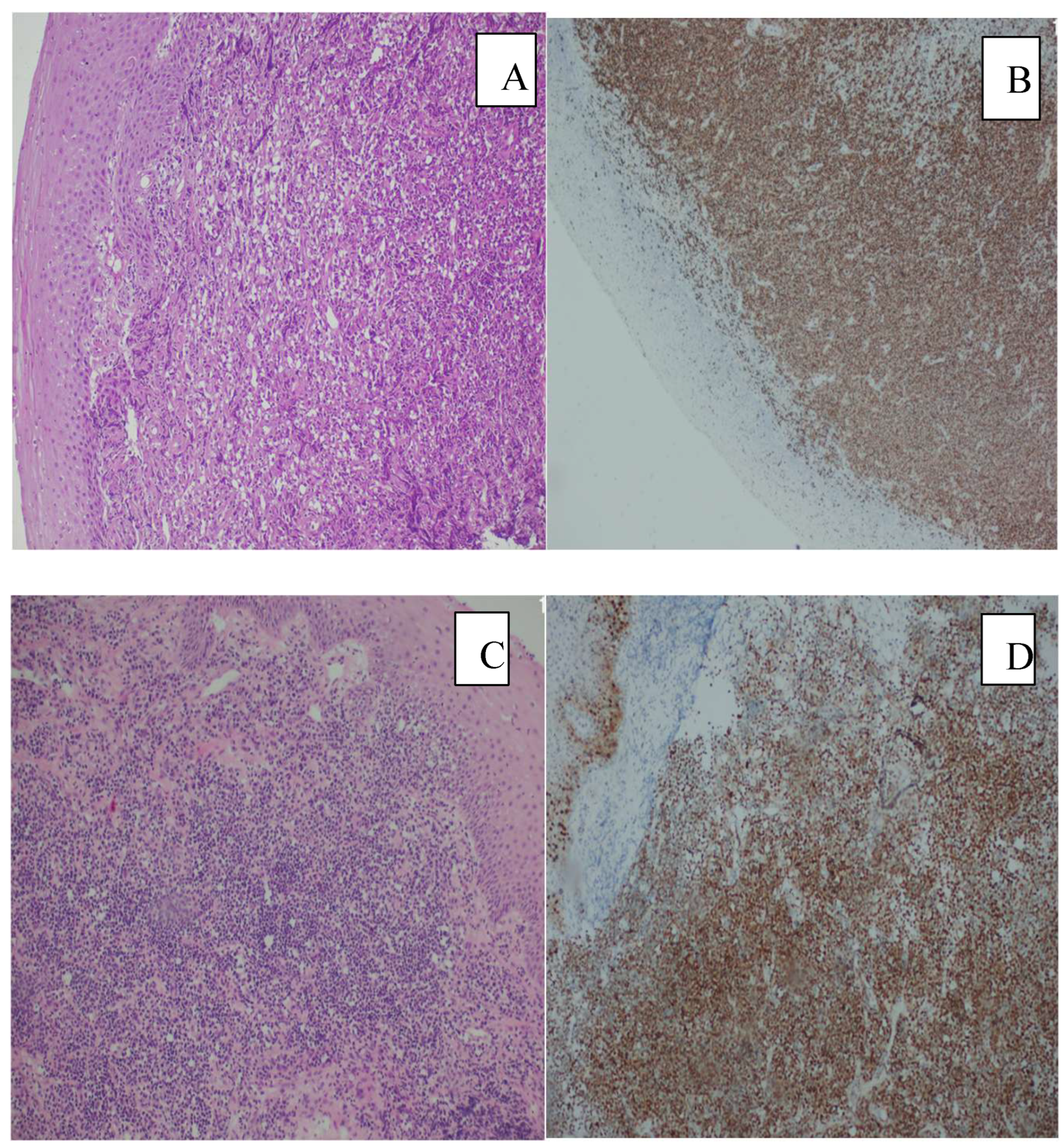
Figure 2.
Histopathological distribution of lymphoma subtypes. Diffuse Large B cell lymphoma (DLBCL), Follicular Lymphoma (FL), Anaplastic large cel lymphoma (ALCL), N/K T cell lymphoma (NKTCL), Mantle cell lymphoma (MCL), Hodgkin Lenfoma (HL).
Figure 2.
Histopathological distribution of lymphoma subtypes. Diffuse Large B cell lymphoma (DLBCL), Follicular Lymphoma (FL), Anaplastic large cel lymphoma (ALCL), N/K T cell lymphoma (NKTCL), Mantle cell lymphoma (MCL), Hodgkin Lenfoma (HL).
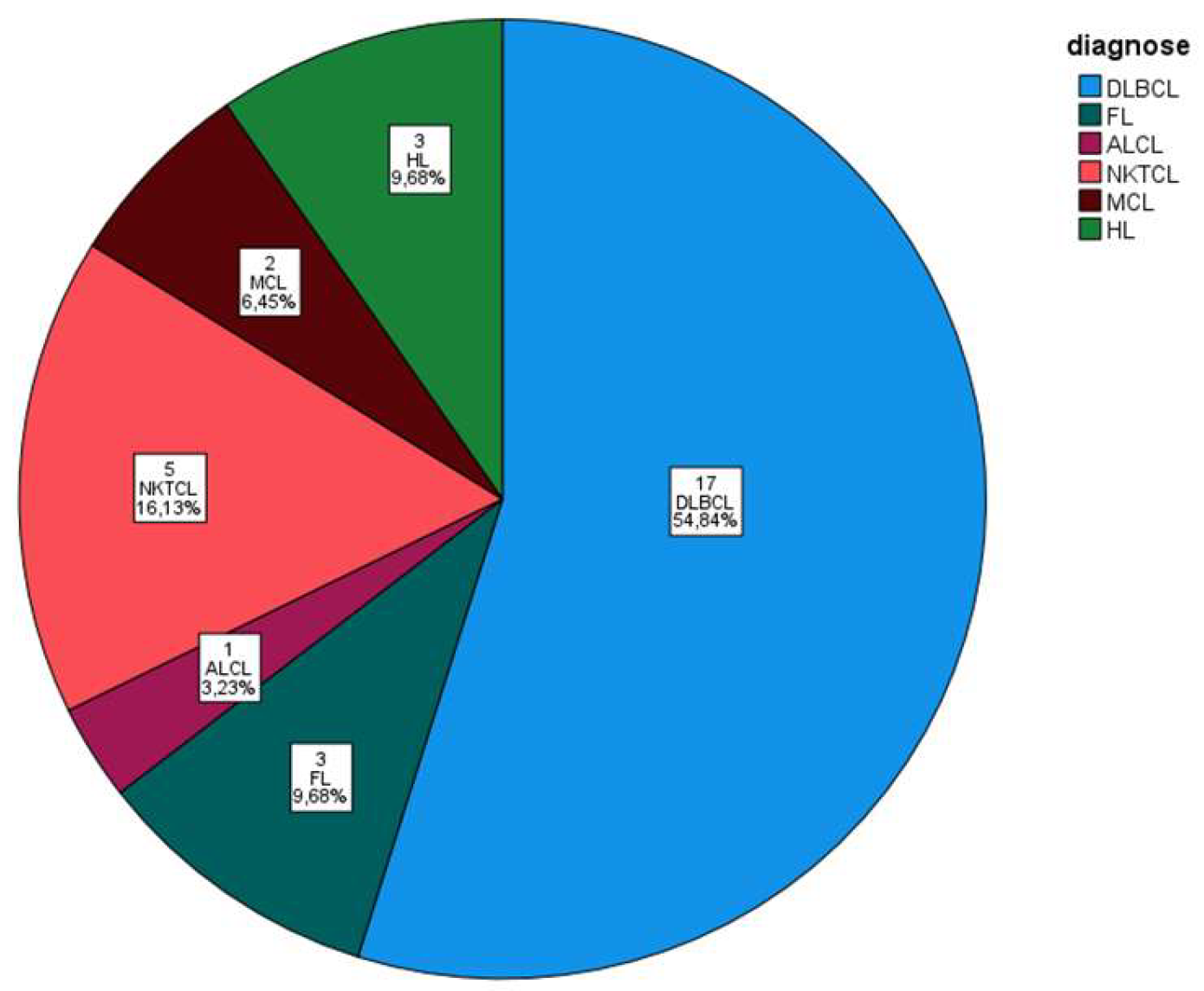
Figure 3.
(A). Follicular Lymphoma (FL)H&Ex40. (B) BCL2 positivity in FL.
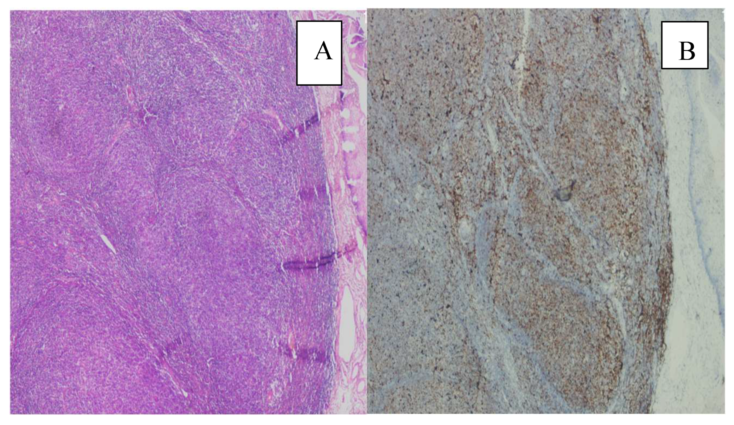
Figure 4.
(A) Hodgkin lymphoma H&Ex200. (B) Hodgkin-Reed-Sternberg cells showing positive staining for CD30, CD30X200.
Figure 4.
(A) Hodgkin lymphoma H&Ex200. (B) Hodgkin-Reed-Sternberg cells showing positive staining for CD30, CD30X200.
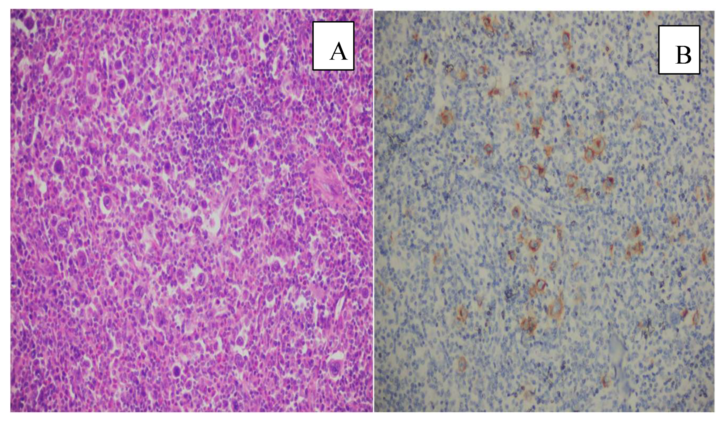
Figure 5.
Kaplan–Meier survival curves showing cumulative survival probabilities according to diagnosis group DLBCL, ALCL, and NKTCL. No statistically significant difference in survival was observed between groups (log-rank test, p>0.05).
Figure 5.
Kaplan–Meier survival curves showing cumulative survival probabilities according to diagnosis group DLBCL, ALCL, and NKTCL. No statistically significant difference in survival was observed between groups (log-rank test, p>0.05).
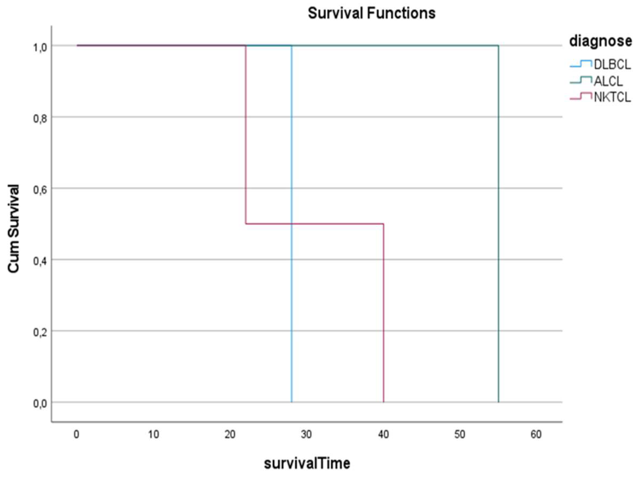
Figure 6.
Comparison of survival distributions according to tumor location using the Log-Rank test. No statistically significant difference was observed between the groups ( p >0.05).
Figure 6.
Comparison of survival distributions according to tumor location using the Log-Rank test. No statistically significant difference was observed between the groups ( p >0.05).
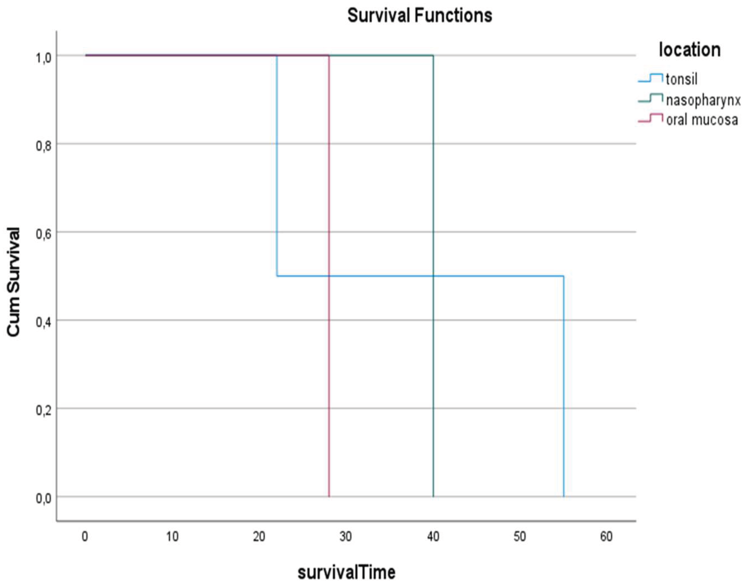

Table 1.
Immunohistochemical (IHC) panel used in the study.
| ANTİJEN | CLONE | SOURCE | DİLUTİON |
|---|---|---|---|
| ALK | ALK1 | DAKO | RTU |
| BCL-2 | 124 | DAKO | RTU |
| BCL-6 | PG-B6p | DAKO | RTU |
| CD3 | M7254 | DAKO | RTU |
| CD5 | 4C7 | DAKO | RTU |
| CD8 | C8/144B | DAKO | RTU |
| CD10 | 56C6 | DAKO | RTU |
| CD20 | L26 | DAKO | 1/200 |
| CD30 | Ber-H2 | DAKO | RTU |
| CD43 | DF-T1 | DAKO | RTU |
| CD45 | 2B11+PD7/26 | DAKO | RTU |
| CD56 | 123C3 | DAKO | RTU |
| CD79a | JCB117 | DAKO | RTU |
| CYCLİN D1 | EP12 | DAKO | RTU |
| EMA | E29 | DAKO | 1/200 |
| TİA1 | TİA1 | DAKO | RTU |
| Granzym B | GrB-7 | DAKO | RTU |
| CD30 | M0751 | DAKO | 1/100 |
| CD15 | M3631 | DAKO | 1/100 |
| MUM1 | MUM1p | DAKO | RTU |
Table 2.
The relationship between pathological types and primary sites.
| Tonsil | Naso-pharynx | Oral cavite | Nasal cavite | Salivary gland | Thyroid | Total | |
|---|---|---|---|---|---|---|---|
| DLBCL | 6 | 3 | 4 | 1 | 2 | 1 | 17 |
| FL | 2 | 1 | 0 | 0 | 0 | 0 | 3 |
| ALCL | 1 | 0 | 0 | 0 | 0 | 0 | 1 |
| NKTCL | 1 | 1 | 1 | 2 | 0 | 0 | 5 |
| MCL | 2 | 0 | 0 | 0 | 0 | 0 | 2 |
| HL | 0 | 3 | 0 | 0 | 0 | 0 | 3 |
| Total | 12 | 8 | 5 | 3 | 2 | 1 | 31 |
Abbreviations: diffuse large B cell lymphoma (DLBCL), Follicular lymphoma (FL), mantle cell lymphoma (MCL), anaplastic large cell lymphoma (ALCL), NK T-cell lymphoma: (NKTCL), Hodgkin lymphoma (HL).
Disclaimer/Publisher’s Note: The statements, opinions and data contained in all publications are solely those of the individual author(s) and contributor(s) and not of MDPI and/or the editor(s). MDPI and/or the editor(s) disclaim responsibility for any injury to people or property resulting from any ideas, methods, instructions or products referred to in the content. |
© 2026 by the authors. Licensee MDPI, Basel, Switzerland. This article is an open access article distributed under the terms and conditions of the Creative Commons Attribution (CC BY) license (http://creativecommons.org/licenses/by/4.0/).
Copyright: This open access article is published under a Creative Commons CC BY 4.0 license, which permit the free download, distribution, and reuse, provided that the author and preprint are cited in any reuse.


