Submitted:
14 February 2026
Posted:
26 February 2026
You are already at the latest version
Abstract
Cerebral toxoplasmosis is a rare but potentially fatal opportunistic infection in renal transplant recipients receiving long-term immunosuppressive therapy. It may result from donor-derived transmission or reactivation of latent infection. We report the case of a 70-year-old female who underwent cadaveric kidney transplantation in 2004 for end-stage renal disease due to glomerulonephritis. She was maintained on cyclosporine, mycophenolate mofetil, and prednisone. In September 2024, she presented with headache, mood changes, and right-sided hemiparesis. Brain multislice computed tomography revealed a large temporoparietal lesion initially suspected to be glioblastoma. Craniotomy and histopathological analysis demonstrated encysted Toxoplasma gondii bradyzoites within gliotic tissue. Polymerase chain reaction testing confirmed the presence of T. gondii DNA, while human immunodeficiency virus testing was negative. The patient reported frequent contact with domestic cats. Treatment with pyrimethamine, sulfadiazine, and leucovorin, alongside adjustment of immunosuppressive therapy, led to marked neurological improvement and radiological regression of the lesion. However, nine months later, she succumbed to multidrug-resistant urosepsis. This case highlights the diagnostic challenges of cerebral toxoplasmosis in transplant recipients, as radiological findings are often nonspecific and can mimic neoplastic or lymphoproliferative lesions. Polymerase chain reaction and histopathological analysis remain essential for definitive diagnosis. Awareness of this rare complication is critical for early recognition and prompt initiation of anti-toxoplasma therapy, which can significantly improve outcomes. Although cerebral toxoplasmosis is uncommon after kidney transplantation, it should be considered in immunosuppressed patients presenting with neurological symptoms. Early detection and targeted therapy are key to reducing morbidity and mortality in this population.
Keywords:
cerebral toxoplasmosis
; immunosupresive therapy
; renal transplant
; Toxoplasma gondii
1. Introduction
Infectious complications remain a leading cause of morbidity and mortality following kidney transplantation and are closely associated with adverse graft outcomes, including allograft dysfunction, rejection, and graft loss. Both opportunistic pathogens and community-acquired microorganisms contribute substantially to the post-transplant infectious burden. The immunosuppressed state frequently results in atypical or attenuated clinical presentations, complicating timely diagnosis. Given the requirement for lifelong immunosuppression, kidney transplant recipients are predisposed to severe manifestations of infections, including those caused by organisms typically associated with mild disease. Early and accurate diagnostic assessment is therefore essential to optimize patient and graft outcomes [1].
Up to 70% of recipients experiencing at least one infectious episode within the first three years. The timing and spectrum of infections are closely related to the intensity of immunosuppression and may be further modified by treatment for acute rejection and concomitant viral infections, including cytomegalovirus and Epstein–Barr virus. In the first month following transplantation, infections are predominantly associated with surgical complications, healthcare-related exposures, and donor-derived pathogens. During this period, multidrug-resistant organisms such as methicillin-resistant Staphylococcus aureus, vancomycin-resistant Enterococcus, and carbapenem-resistant Enterobacteriaceae, as well as Clostridioides difficile, represent important causes of infection. Reactivation of latent pathogens, including BK polyomavirus, hepatitis C virus, and Mycobacterium tuberculosis, may also occur, although routine antimicrobial prophylaxis has reduced the incidence of infections caused by Pneumocystis jirovecii, herpesviruses, and hepatitis B virus. Beyond six months post-transplantation, immunosuppressive burden is typically reduced; however, recipients remain susceptible to community-acquired and environmental infections, recurrent infections, and late-onset viral disease, particularly cytomegalovirus after discontinuation of prophylaxis. Pre-transplant donor and recipient screening remains essential for infection prevention, while parasitic infections are notably absent from discussion in this otherwise comprehensive review [2].
Organ transplant recipients may acquire parasitic infections through three different mechanisms: transmission via the graft, de novo infection, or reactivation of a latent infection as a result of immunosuppression [3]. The prevalence of symptomatic parasitosis infections in renal transplant patients is 2.4%. Most of the infections are caused by Strongyloids stercoralis, followed by Giardia lamblia and very rare Toxoplasma gondii and Trypanosoma cruzi [4]. Toxoplasmosis is a globally distributed parasitic disease caused by Toxoplasma gondii, an obligate intracellular protozoan. The parasite can infect all nucleated cells of warm-blooded species, including humans. Transmission dynamics are complex and involve environmental exposure to oocysts shed by felids (cats), as well as dietary acquisition through consumption of raw or undercooked meat containing tissue cysts. Serological evidence suggests that approximately one third of the world’s population has been exposed to the parasite. Infection is usually clinically silent, but there is potentially fatal central nervous system involvement in immunocompromised patients, adverse pregnancy outcomes such as spontaneous abortion and congenital malformations following primary maternal infection, as well as sight-threatening ocular disease in otherwise healthy individuals [5].
Cerebral infection with Toxoplasma gondii is a life-threatening opportunistic disease usually described in patients with advanced HIV infection, but can also occur in solid organ transplant recipients receiving chronic immunosuppressive therapy. In these patients, toxoplasmosis can be transmitted by seropositive organ donors or represent a reactivation of a latent disease due to immunosuppressive therapy [6]. Phenotypic switching may enable unchecked tachyzoite proliferation, overwhelming the already compromised host immune response and leading to severe, potentially fatal disease. Whether immunosuppression directly initiates parasite reactivation or instead permits uncontrolled replication of tachyzoites that would otherwise be contained by the immune system remains unclear [7]. Because of the intensive use of immunosuppression in the early post-transplant period, most cases of toxoplasmosis occur within the first three months after transplantation, while late reactivation is rare [8]. Neurological manifestations of toxoplasmosis typically present with a subacute course and may include headache, focal motor deficits such as hemiparesis, cranial nerve involvement, ataxia, seizures, or altered level of consciousness. Serological testing for anti-Toxoplasma IgG and IgM antibodies remains the diagnostic gold standard; however, additional diagnostic modalities, including cerebrospinal fluid examination, histopathological analysis, and neuroimaging, may provide supportive evidence.
Complete eradication of toxoplasmosis is not currently achievable, as available antimicrobial agents are unable to eliminate T. gondii tissue cysts. In patients with neuroimaging findings of multiple cerebral lesions, empiric antitoxoplasma therapy is recommended regardless of serological status. When there is a high risk of cerebral herniation, surgical biopsy with decompression should be considered. First-line treatment consists of pyrimethamine-based regimens or trimethoprim–sulfamethoxazole, with adjunctive corticosteroid therapy reserved for cases complicated by increased intracranial pressure. The recommended duration of treatment is a minimum of six weeks [9].
Across Europe, screening for Toxoplasma gondii infection in the pretransplant setting is not standardized and is largely determined by individual transplant center policies. In certain countries, such as France, serological testing for Toxoplasma is mandated by law for all organ donors and is strongly recommended for transplant candidates prior to transplantation. Within the Eurotransplant network, which comprises 29 participating countries, routine Toxoplasma serological testing is performed for solid organ donors in only a subset of countries, while others apply a selective testing strategy based on donor-specific characteristics [10].
2. Case Report
We report the case of a 70-year-old female, a retired store worker and mother of two, who lived in a rural area and had lifelong contact with domestic animals, including cats. There was no family history of renal disease. At the age of 16, she underwent tonsillectomy. In young age a renal biopsy was also performed due to proteinuria and hematuria. Histopathological analysis revealed chronic glomerulonephritis. Over the next 20 years, she was regularly followed by a nephrologist for chronic kidney disease. At age 42, she progressed to end-stage renal disease and started chronic hemodialysis. She underwent pre-transplant evaluation and received a cadaveric kidney transplant in 2004 at the age of 50. Her comorbidities included arterial hypertension, osteoporosis, and atrial fibrillation. She experienced several hospitalizations for acute deterioration of renal function secondary to enterocolitis but generally maintained stable graft function on follow-up. Post-transplant, she received standard triple immunosuppressive therapy with cyclosporine, mycophenolate mofetil and prednisone.
In September 2024, she presented with general weakness and was admitted to the Department of Nephrology at the University Hospital Center Osijek, Croatia. The family reported behavioral changes, describing her as withdrawn, apathetic and excessively demanding. At the time of admission, she was not receiving corticosteroids. The patient complained of severe headache, a sensation of “pressure in the head,” and mood disturbances. During hospitalization, she developed right-sided hemiparesis. Brain multislice computed tomography (MSCT) revealed an extensive lesion in the left temporoparietal region measuring 68 × 33 × 45 mm, with surrounding edema and midline shift, initially suspected to be glioblastoma. Magnetic resonance imaging (MRI) confirmed a left temporoparietal expansile lesion measuring 55 × 32 × 42 mm with ring-enhancing post-contrast signal intensity, consistent with glioblastoma and features of intratumoral hemorrhage (Figure 1).
A craniotomy was performed with maximal resection of the lesion. Histopathological examination revealed numerous reactive astrocytes (GFAP+), a connective pseudocapsule, and moderate to dense mononuclear infiltration predominantly composed of CD68+ macrophages and CD3+ T lymphocytes. Within the gliotic areas, nodular, granular, basophilic structures consistent with encysted bradyzoites of Toxoplasma gondii were identified. Laboratory testing showed stable blood counts without systemic infection, while biochemical results reflected chronic kidney disease. Polymerase chain reaction (PCR) confirmed the presence of T. gondii DNA. Serology revealed negative anti-Toxoplasma IgM and positive IgG, while citomegalovirus (CMV) PCR was <100 IU/mL and HIV testing was negative. A diagnosis of cerebral toxoplasmosis was established. The patient was treated with pyrimethamine, sulfadiazine and leucovorin. Immunosuppressive therapy (mycophenolate mofetil and cyclosporine) was temporarily discontinued and prednisone was introduced at a dose of 10 mg daily.
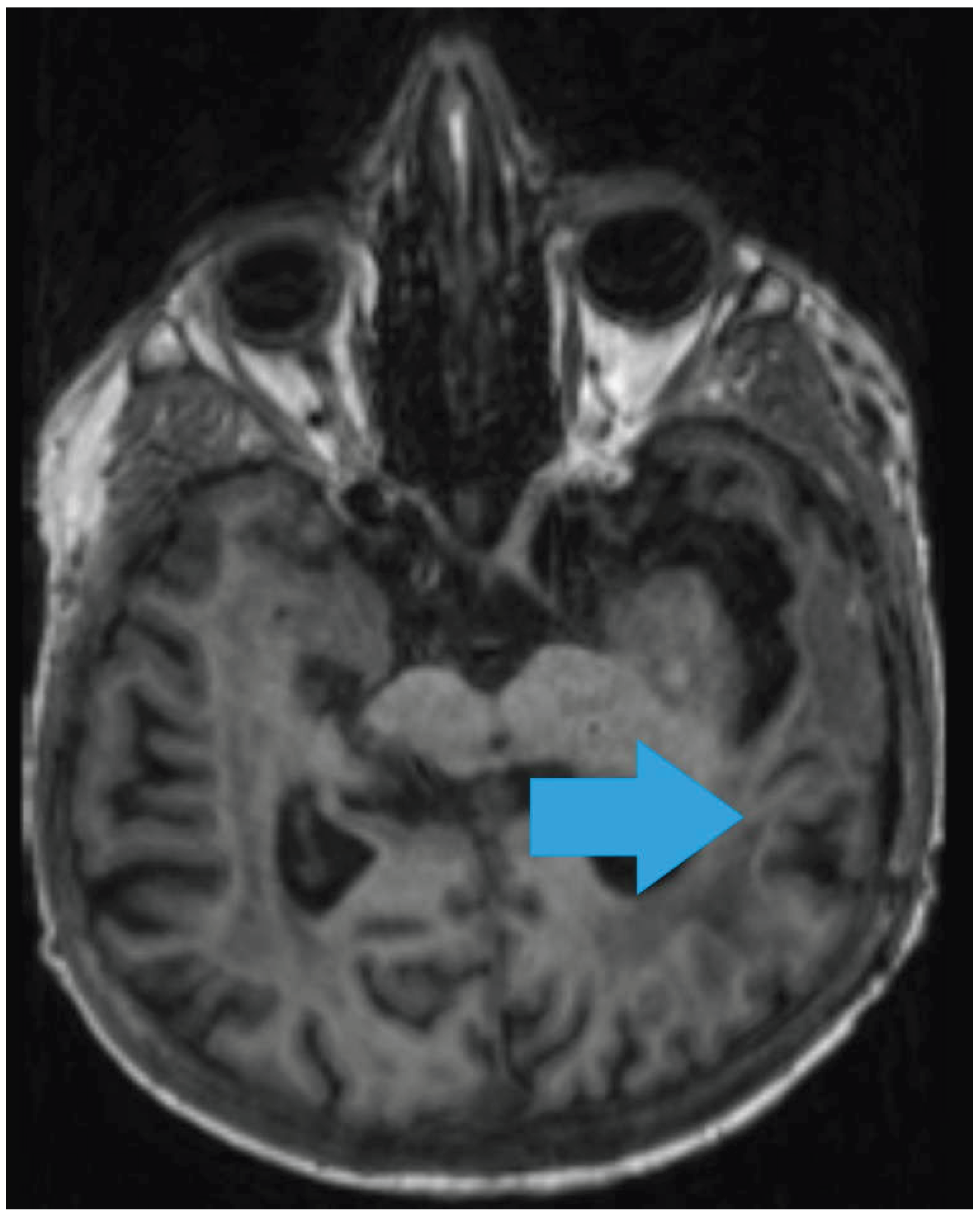
One month after initiation of therapy, brain MRI demonstrated a malacic lesion in the left temporal lobe measuring 28 × 22 mm (post-surgical site) and two newly appearing ring-enhancing foci along the caudal border of the right frontal horn of the lateral ventricle measuring up to 15 × 12 mm, indicating progressive inflammatory lesions consistent with cerebral toxoplasmosis. Clinical improvement was observed within two months and subsequent neuroimaging showed partial regression of the lesions (Figure 2 and Figure 3).
After ten weeks of antimicrobial therapy, the dose of sulfadiazine was reduced. At fifteen weeks, serology revealed anti-Toxoplasma IgM <3 AU/mL (negative), IgG >650 AU/mL (positive), and CMV DNA PCR 11,391.92 IU/mL (positive). Immunosuppressive therapy was reintroduced. The infectious disease team recommended prophylactic trimethoprim-sulfamethoxazole 480 mg once daily for at least six months. Following regression of cerebral lesions, the patient remained in good condition and continued regular nephrological follow-up. In June 2025, she was hospitalized with urosepsis caused by multidrug-resistant Klebsiella pneumoniae and Escherichia coli. Despite intensive antibiotic therapy, the patient deceased due to infection.
3. Discussion
Cerebral toxoplasmosis represents a rare but severe opportunistic infection in solid organ transplant recipients, associated with significant morbidity and mortality. Although most commonly described in patients with advanced HIV infection, cerebral toxoplasmosis is increasingly recognized in other immunocompromised populations, including recipients of kidney transplants. Diagnosis remains challenging due to nonspecific clinical presentation, overlapping radiological features with neoplastic and inflammatory conditions, and limited sensitivity of serological testing in immunosuppressed hosts [11,12]. This case highlights the importance of considering cerebral toxoplasmosis in the differential diagnosis of focal neurological symptoms in kidney transplant recipients, even decades after transplantation.
In transplant recipients, infection with Toxoplasma gondii may occur either as a primary infection transmitted via a seropositive donor organ or, more frequently, as reactivation of latent infection in a seropositive recipient during periods of immunosuppression [13]. The majority of reported cases occur within the first three months following transplantation, coinciding with the period of maximal immunosuppressive therapy [14]. Nevertheless, late-onset toxoplasmosis has been described several years after transplantation, emphasizing that the risk persists lifelong in seropositive individuals [15,16]. In the present case, the exceptionally long interval of 20 years between kidney transplantation and disease onset strongly suggests reactivation of latent infection rather than donor-derived transmission.
Immunosuppressive regimens play a central role in the pathogenesis of reactivated toxoplasmosis. Agents such as calcineurin inhibitors, mycophenolate mofetil, and corticosteroids impair cell-mediated immunity, particularly T-lymphocyte function, which is essential for controlling latent T. gondii infection [13,17]. Advanced age, multiple comorbidities, and cumulative immunosuppressive burden may further increase susceptibility. Importantly, reactivation may occur even in patients receiving relatively stable long-term immunosuppression, as illustrated by this case, highlighting that apparent clinical stability does not exclude the risk of opportunistic infections.
Clinical manifestations of cerebral toxoplasmosis are heterogeneous and frequently nonspecific. Common symptoms include headache, altered mental status, personality changes, focal neurological deficits, seizures, and gait disturbances [12,18]. In transplant recipients, such symptoms often raise suspicion of primary or metastatic brain tumors, abscesses, vascular lesions, or post-transplant lymphoproliferative disorder. In our patient, progressive neuropsychiatric changes and focal deficits, together with radiological findings, initially suggested glioblastoma, illustrating the diagnostic complexity and frequent delay associated with this condition.
Neuroimaging findings in cerebral toxoplasmosis typically include multiple ring-enhancing lesions with surrounding vasogenic edema, commonly involving the basal ganglia or corticomedullary junction [18,19]. However, solitary lesions are not uncommon and may closely resemble high-grade gliomas or metastatic disease. Magnetic resonance imaging with contrast is the preferred diagnostic modality, but imaging characteristics alone lack sufficient specificity to establish a definitive diagnosis in immunocompromised patients [19]. Advanced imaging techniques, including diffusion-weighted imaging and perfusion studies, may aid differentiation but are not universally available and remain inconclusive in many cases.
Serological testing has limited diagnostic utility in transplant recipients. While IgM antibodies may indicate recent infection, they are frequently absent in immunosuppressed patients, and IgG positivity merely reflects prior exposure rather than active disease [20]. In our patient, negative IgM and strongly positive IgG supported the hypothesis of reactivation. Molecular diagnostics, such as polymerase chain reaction (PCR) for T. gondii DNA in cerebrospinal fluid or blood, may increase diagnostic confidence but demonstrate variable sensitivity and are not universally available [16]. Consequently, histopathological examination remains the diagnostic gold standard in cases with atypical presentation or inconclusive noninvasive testing. In this case, brain biopsy revealed characteristic bradyzoites within inflammatory lesions, providing definitive confirmation.
Early initiation of appropriate therapy is crucial, as delayed treatment is associated with poor outcomes. Standard first-line therapy consists of pyrimethamine, sulfadiazine, and folinic acid, which has demonstrated high efficacy in both HIV-infected and transplant populations [21]. Alternative regimens, including trimethoprim–sulfamethoxazole, may be used in patients with renal dysfunction, hematologic toxicity, or intolerance to first-line agents [22]. Our patient demonstrated gradual clinical and radiological improvement following initiation of standard therapy, despite initial progression of lesions, consistent with previous observations that radiological resolution may lag behind clinical recovery [18].
Management of cerebral toxoplasmosis in transplant recipients also requires careful adjustment of immunosuppressive therapy. Reduction of immunosuppression is often necessary to enhance immune-mediated parasite control, but must be balanced against the risk of graft rejection [15]. This delicate balance underscores the importance of multidisciplinary collaboration among nephrologists, neurologists, neurosurgeons, and infectious disease specialists. In our case, temporary modification of immunosuppressive therapy allowed infection control without immediate graft failure.
Prophylaxis with trimethoprim–sulfamethoxazole has been shown to significantly reduce the incidence of toxoplasmosis in solid organ transplant recipients and is widely recommended [23]. Nevertheless, breakthrough infections may occur, particularly after discontinuation of prophylaxis, subtherapeutic dosing, or prolonged immunosuppression. Notably, routine screening for T. gondii serostatus in kidney transplant donors and recipients is not universally implemented, potentially contributing to delayed recognition and diagnosis [16].
4. Conclusions
Cerebral toxoplasmosis remains a rare but potentially life-threatening complication in kidney transplant recipients and may present many years after transplantation. This case illustrates that reactivation of latent Toxoplasma gondii infection can occur even under long-term, apparently stable immunosuppressive therapy, posing a significant diagnostic challenge. Clinical presentation and neuroimaging findings are often nonspecific and may closely mimic malignant or inflammatory brain lesions, frequently leading to delayed diagnosis.
Definitive diagnosis requires a high index of suspicion and, in selected cases, histopathological confirmation, particularly when noninvasive diagnostic methods are inconclusive. Prompt initiation of appropriate antiparasitic therapy, together with careful adjustment of immunosuppressive treatment, can result in substantial neurological and radiological improvement. However, overall prognosis remains guarded due to the high vulnerability of this patient population to subsequent infectious complications.
This case underscores the importance of including cerebral toxoplasmosis in the differential diagnosis of focal neurological symptoms in renal transplant recipients, regardless of the time elapsed since transplantation. Increased awareness, multidisciplinary collaboration, and consideration of preventive strategies may contribute to earlier diagnosis and improved outcomes in this vulnerable population.
Author Contributions
Conceptualization, D.M..; software, J.H.; validation, D.M. and Z.S.; investigation, J.H., P.V. Z.S.; resources, J.M., Z.S., P.V. and J.H.; writing—original draft preparation, D.M., J.H. and P.V.; writing—review and editing, D.M., Z.S., J.H., P.V. and J. M.; visualization, J.H. and J. M.; supervision, D.M. All authors have read and agreed to the published version of the manuscript.
Funding
This research received no external funding.
Institutional Review Board Statement
The study was conducted in accordance with the Declaration of Helsinki, and approved by the Ethics Committee of University Hospital Center Osijek (R1-910/2026, 23.1.2026.
Informed Consent Statement
Informed consent was obtained from all subjects involved in the study.
Data Availability Statement
The data presented in this paper are available on request from the corresponding author.
Conflicts of Interest
The authors declare no conflicts of interest.
References
- Sawinski, D.; Blumberg, E.A. Infection in Renal Transplant Recipients. In Chronic Kidney Disease, Dialysis, and Transplantation; Gilbert, S.J., Weiner, D.E., Eds.; Elsevier: Philadelphia, PA, USA, 2019; pp. 621–638.e6. [Google Scholar]
- Nambiar, P.; Silibovsky, R.; Belden, K.A. Infection in Kidney Transplantation. In Contemporary Kidney Transplantation; Gabardi, S., Ed.; Springer: Cham, Switzerland, 2018; pp. 307–327. [Google Scholar]
- Yadav, P.; Khalil, S.; Mirdha, B.R. Molecular appraisal of intestinal parasitic infection in transplant recipients. Indian J. Med. Res. 2016, 144, 258–263. [Google Scholar] [CrossRef]
- Valar, C.; Keitel, E.; Dal Prá, R.L.; Gnatta, D.; Santos, A.F.; Bianco, P.D.; Sukiennik, T.C.; Pegas, K.L.; Bittar, A.E.; Oliveira, K.T.; Garcia, V.D. Parasitic infection in renal transplant recipients. Transplant Proc. 2007, 39, 460–462. [Google Scholar] [CrossRef] [PubMed]
- Smith, N.C.; Goulart, C.; Hayward, J.A.; Kupz, A.; Miller, C.M.; van Dooren, G.G. Control of human toxoplasmosis. Int. J. Parasitol. 2021, 51, 95–121. [Google Scholar] [CrossRef] [PubMed]
- Robert-Gangneux, F.; Dardé, M.L. Epidemiology of and diagnostic strategies for toxoplasmosis. Clin. Microbiol. Rev. 2012, 25, 264–296. [Google Scholar] [CrossRef] [PubMed]
- Elsheikha, H.M.; Khan, N.A. Protozoa traversal of the blood-brain barrier to invade the central nervous system. FEMS Microbiol. Rev. 2010, 34, 532–553. [Google Scholar] [CrossRef]
- Martino, R.; Bretagne, S. Toxoplasmosis in hematopoietic stem cell transplant recipients. Clin. Infect. Dis. 2000, 31, 1188–1195. [Google Scholar] [CrossRef]
- Elsheikha, H.M.; Marra, C.M.; Zhu, X.Q. Epidemiology, Pathophysiology, Diagnosis, and Management of Cerebral Toxoplasmosis. Clin. Microbiol. Rev. 2021, 34, e00115-19. [Google Scholar] [CrossRef]
- Human organ transplantation in Europe; an overview. Report from the General Health and Consumer Protection Public Health and Risk Assessment Directorate; European Commission: Brussels, Belgium, 2003; p. 120. Available online: http://ec.europa.eu/health/ph_threats/human_substance/documents/organ_survey.pdf (accessed on 25 January 2026).
- Luft, B.J.; Remington, J.S. Toxoplasmic encephalitis in AIDS. Clin. Infect. Dis. 1992, 15, 211–222. [Google Scholar] [CrossRef]
- Porter, S.B.; Sande, M.A. Toxoplasmosis of the central nervous system in the acquired immunodeficiency syndrome. N. Engl. J. Med. 1992, 327, 1643–1648. [Google Scholar] [CrossRef]
- Montoya, J.G.; Liesenfeld, O. Toxoplasmosis. Lancet 2004, 363, 1965–1976. [Google Scholar] [CrossRef]
- Derouin, F.; Pelloux, H.; et al. Toxoplasmosis in organ transplant recipients. Clin. Infect. Dis. 1997, 24, 625–634. [Google Scholar]
- Martina, M.N.; Cervera, C.; et al. Toxoplasmosis after solid organ transplantation. Transpl. Int. 2010, 23, 845–851. [Google Scholar]
- Robert-Gangneux, F.; Belaz, S. Toxoplasmosis in transplant recipients, Europe, 2010–2014. Euro Surveill. 2015, 20, 210–218. [Google Scholar] [CrossRef] [PubMed]
- Luft, B.J.; Hafner, R.; et al. Toxoplasmic encephalitis in patients with the acquired immunodeficiency syndrome. N. Engl. J. Med. 1993, 329, 995–1000. [Google Scholar] [CrossRef]
- Maschke, M.; Kastrup, O.; et al. Opportunistic infections of the central nervous system. Lancet Neurol. 2004, 3, 485–498. [Google Scholar]
- Post, M.J.; Chan, J.C.; et al. Central nervous system toxoplasmosis in the era of highly active antiretroviral therapy. AJNR Am. J. Neuroradiol. 2001, 22, 1349–1357. [Google Scholar]
- Kotton, C.N.; Fishman, J.A. Infectious complications of transplantation. Am. J. Transplant. 2013, 13 (Suppl. 4), 280–303. [Google Scholar]
- Luft, B.J.; Chua, A.; et al. Treatment of toxoplasmic encephalitis with pyrimethamine and sulfadiazine. N. Engl. J. Med. 1993, 329, 995–1000. [Google Scholar] [CrossRef]
- Hughes, W.T.; Leoung, G.S.; et al. Comparison of pyrimethamine–sulfadiazine and trimethoprim–sulfamethoxazole for treatment of toxoplasmic encephalitis. N. Engl. J. Med. 1990, 323, 776–782. [Google Scholar]
- Derouin, F.; Pelloux, H.; et al. Prevention of toxoplasmosis in transplant recipients. Clin. Infect. Dis. 2008, 46, 167–175. [Google Scholar]
- Martínez, J.; Torre-Cisneros, J.; et al. Toxoplasmosis after kidney transplantation: clinical features and outcomes. Transpl. Infect. Dis. 2011, 13, 1–7. [Google Scholar]
Figure 1.
Magnetic resonance imaging (MRI) - a left temporoparietal expansile lesion (55 × 32 × 42 mm) with ring-enhancing post-contrast signal intensity - glioblastoma and features of intratumoral hemorrhage.
Figure 1.
Magnetic resonance imaging (MRI) - a left temporoparietal expansile lesion (55 × 32 × 42 mm) with ring-enhancing post-contrast signal intensity - glioblastoma and features of intratumoral hemorrhage.
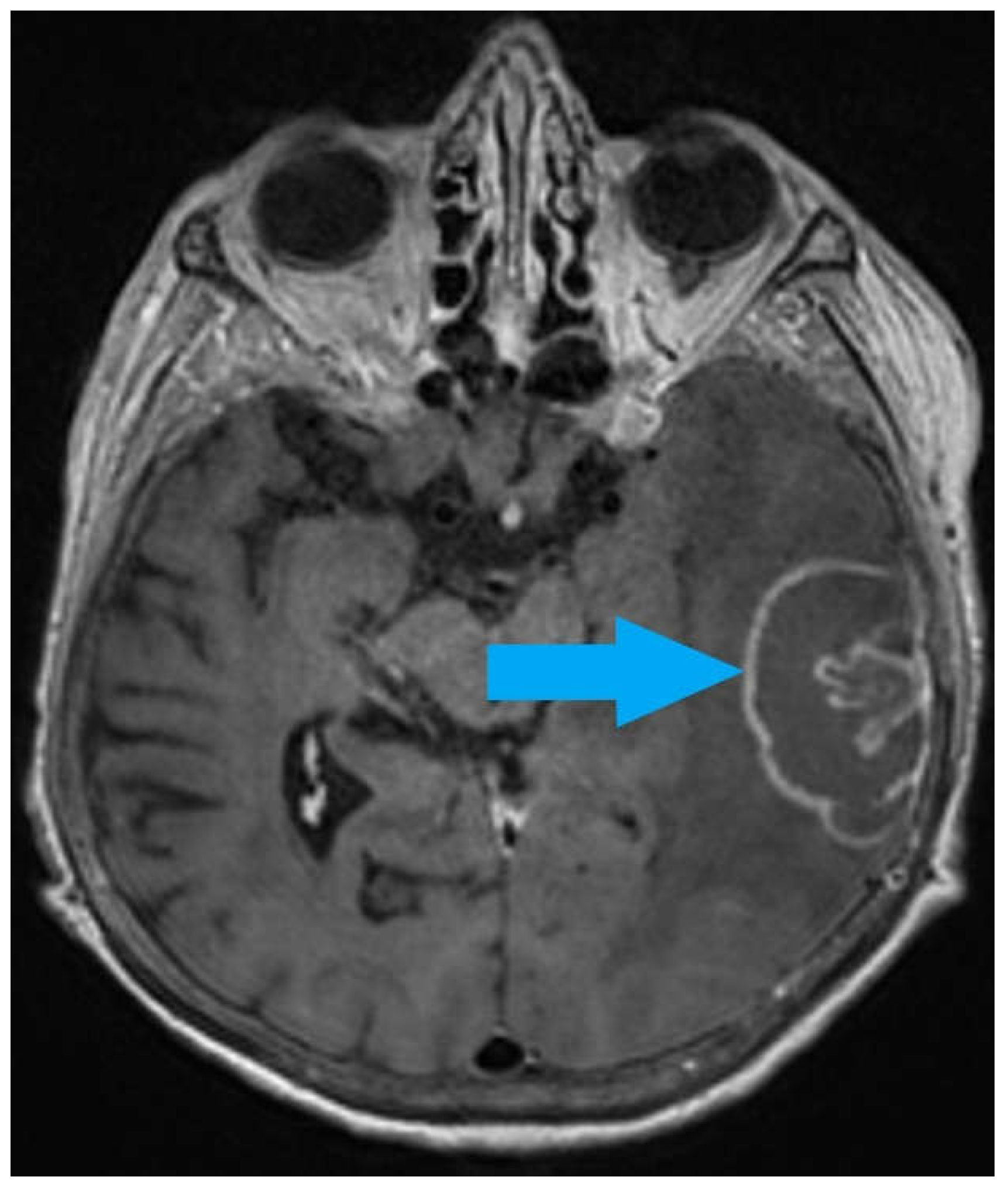
Figure 2.
Post surgical brain MRI - progressive inflammatory lesions consistent with cerebral toxoplasmosis.
Figure 2.
Post surgical brain MRI - progressive inflammatory lesions consistent with cerebral toxoplasmosis.
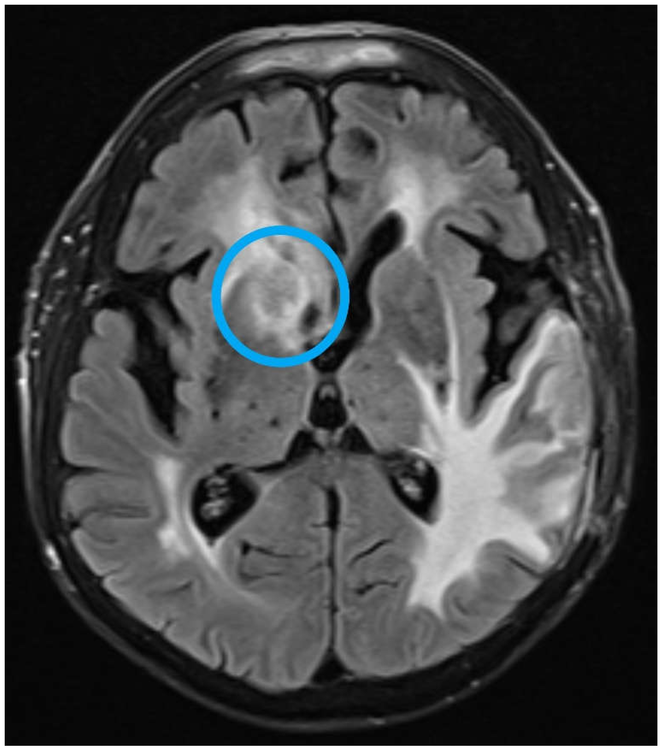
Figure 3.
Brain MRI - partial regression of the lesions.
Disclaimer/Publisher’s Note: The statements, opinions and data contained in all publications are solely those of the individual author(s) and contributor(s) and not of MDPI and/or the editor(s). MDPI and/or the editor(s) disclaim responsibility for any injury to people or property resulting from any ideas, methods, instructions or products referred to in the content. |
© 2026 by the authors. Licensee MDPI, Basel, Switzerland. This article is an open access article distributed under the terms and conditions of the Creative Commons Attribution (CC BY) license (http://creativecommons.org/licenses/by/4.0/).
Copyright: This open access article is published under a Creative Commons CC BY 4.0 license, which permit the free download, distribution, and reuse, provided that the author and preprint are cited in any reuse.


