Submitted:
15 February 2026
Posted:
16 February 2026
You are already at the latest version
Abstract
Background: Kawasaki disease (KD) is a systemic vasculitis, of unknown etiology, that usually occurs in children between the ages of six months and five years. Patients at the extremes of ages rarely meet all the clinical criteria required for the diagnosis of KD. Atypical or incomplete presentation can lead to delayed diagnosis and treatment, resulting in a higher incidence of cardiac complications. Case Presentation: We describe the case of a 2-month-old female infant who was admitted to our clinic with persistent fever, generalized maculopapular rash and bilateral conjunctivitis. During hospitalization, she developed oral mucosa and extremity changes. On the 7th day from the onset of fever, the diagnosis of KD was established, and she received intravenous immunoglobulin therapy. The patient responded well to the treatment, presenting no cardiac complications. Conclusions: The presented case underscores that even very young infants can develop complete Kawasaki disease. It also highlights the importance of early identification and appropriate treatment in preventing coronary artery lesions.
Keywords:
Kawasaki disease
; infant
; intravenous immunoglobulin
; vasculitis
1. Introduction
Kawasaki disease (KD) is an acute, self-limited, systemic vasculitis that primarily affects children between the ages of six months and five years. Also known as mucocutaneous lymph node syndrome, it was first described by Tomisaku Kawasaki in 1967 [1,2]. KD is the second most common vasculitis in childhood (after Henoch-Schönlein purpura), and is the leading cause of acquired heart disease in children in developed countries. It has been reported in children of any race or ethnic group, but occurs more often in the Asian population [3,4].
KD affects small and medium-sized arteries, especially coronary arteries. This condition can lead to coronary artery aneurysms, arrhythmia, valvular and aortic abnormalities, myocardial ischemia, myocardial infarction and sudden cardiac death [5,6,7,8]. The etiology of KD is still unknown, but it is believed to involve an abnormal immunologic response to an infectious trigger in genetically predisposed children [9,10,11,12,13].
The diagnosis of classic KD is based on the presence of fever persisting more than 5 days (generally high and spiking), associated with at least four of the following five signs and symptoms: bilateral non-exudative bulbar conjunctival injection, with limbus sparing; oral changes (erythema, dryness, cracking and bleeding of the lips, strawberry tongue, erythema of the oropharyngeal mucosa); polymorphous exanthema (can be morbilliform or scarlatiniform, but non-vesicular); extremity changes (hyperemia, swelling and induration of the hands and feet that progresses to desquamation of fingers and toes, Beau’s lines); cervical lymphadenopathy (most commonly unilateral, >1.5 cm) [7,11,14,15,16].
Infants younger than six months often have incomplete KD, with transient or subtle signs and symptoms, and they are at higher risk for cardiac complications [6,17]. According to the American Heart Association (AHA) guidelines for the diagnosis of KD, patients who do not perfectly meet the clinical criteria, are diagnosed as incomplete KD, if they have prolonged unexplained fever (≥ 5 days), associated with two or three of the five principle clinical features, and compatible laboratory findings (C-reactive protein ≥ 3 mg/dL or erythrocyte sedimentation rate ≥ 40 mm/h, or both), plus three or more additional laboratory findings (anemia for age, elevated platelet count ≥ 450,000/ mm3, albumin ≤ 3 g/dL, elevated ALT level, elevated white blood cell count ≥ 15,000/ mm3, and leukocyturia ≥ 10 WBC/hpf) or compatible echocardiographic findings (Z score of left anterior descending coronary artery or right coronary artery ≥ 2.5, or ≥ 3 other suggestive features, including decreased left ventricular function, mitral regurgitation, pericardial effusion or Z scores in left anterior descending coronary artery or right coronary artery of 2 to 2.5) [7].
The prognosis of KD depends on cardiovascular complications. Early diagnosis of KD allows prompt and effective treatment to prevent cardiac complications and improve long-term outcomes. Intravenous immunoglobulin (IVIG) and oral acetylsalicylic acid (ASA) are the standard treatment for patients with KD in the acute phase, as multiple studies have demonstrated their effectiveness in reducing the incidence of coronary artery aneurysms [4,18].
2. Case Report
A 2-month-old female infant, previously in good health, was admitted to our clinic with high fever (39-40 °C) persisting for 4 days, unresponsive to antipyretics, irritability, nasal congestion, occasional cough, bilateral conjunctivitis and generalized maculopapular rash (Figure 1).
The initial blood test results revealed mildly elevated white blood cell (WBC) count, microcytic hypochromic anemia and high C-reactive protein (CRP) level. Antigen testing for adenovirus, respiratory syncytial virus, SARS-CoV-2, influenza A and influenza B yielded negative results. The urine examination showed high leucocyte count.
The physical examination at the time of admission revealed influenced general status, high fever (39 °C), irritability, bilateral non-exudative conjunctival injection, pale skin, generalized maculopapular exanthema, dry and cracked lips, diffuse pharyngeal hyperemia, nasal congestion, bilateral cervical micro-adenopathy, with balanced cardiac and respiratory functions, liver and spleen within normal limits, without meningeal signs.
On the day of admission, due to the suspicion of a possible respiratory or urinary tract infection, empiric antibiotic therapy was initiated before the results of culture were available. On the 7th day of illness, which was the 3rd day of hospitalization, after 72 hours of intravenous Cefuroxime treatment and antipyretics, the patient continued having high fever and developed severe labial fissures (Figure 2).
On laboratory tests, hemoglobin (Hb) decreased from 10 g/dL to 7.8 g/dL, hematocrit (Ht) was 24%, red blood cell (RBC) count 2,570,000/mm3, mean corpuscular volume (MCV) 93.5 fL, and platelet (PLT) count 784,000/mm3. Additionally, we observed elevated immunoglobulin A (IgA) and immunoglobulin M (IgM) levels, hypoalbuminemia: 28.8 g/L, high value of erythrocyte sedimentation rate (ESR): 130 mm/h, and the CRP level increased from 28 mg/L to 124.5 mg/L. Urine and blood cultures were without bacterial growth. Abdominal ultrasound and chest X-ray showed no pathological modifications.
With the above clinical and paraclinical findings, on the 7th day from the onset of fever, the diagnosis of Kawasaki disease was made according to the American Heart Association (AHA) criteria, and our patient was referred to cardiologic consultation. Electrocardiogram was normal. Echocardiogram detected minimal pericardial effusion without hemodynamic compromise, and showed no coronary artery lesions. The infant received intravenous immunoglobulin (IVIG) 2 g/kg in a single infusion and oral acetylsalicylic acid in anti-inflammatory dose (45 mg/kg/day).
After administration of IVIG and aspirin, our patient became afebrile on the 8th day of illness. After 48 hours of afebrile period, the anti-inflammatory dose of aspirin was changed to antiaggregant dose (5 mg/kg/day). In the following days, laboratory tests showed thrombocytosis, persistence of anemia with reticulocytosis, and decreasing CRP level. D-dimers were positive both before and after the IVIG therapy. Two weeks after the onset of fever, inflammatory markers turned into normal values (CRP=3.1 mg/L), and NT-pro-BNP decreased from 2597 pg/mL to 155 pg/mL. On the 14th day of disease, our patient started to have periungual desquamation (Figure 3).
We decided to extend her hospitalization until the normalizing of laboratory parameters, because of the risk of thrombosis and other possible cardiovascular involvements, due to the significantly high level of platelet count (985,000/mm3). Serial echocardiography detected no coronary artery abnormalities. On the 3rd week of disease, blood tests showed a decreasing level of thrombocytes (619,000/mm3), therefore our patient was discharged in stable condition with a maintenance dose of aspirin.
Two weeks following her discharge, the patient was re-evaluated in our clinic, presenting good response to the antiaggregant treatment, with normalized laboratory parameters. Red blood cell indices were within normal limits (RBC count: 3,920,000/mm3, Hb: 11.3 g/dL, Ht: 34.3%, MCV: 87.6 fL), platelet count was 413,000/mm3, inflammatory markers became negative (CRP: <1.0 mg/L), AST was mildly elevated (AST: 50 U/L), serum albumin level was normalized (albumin: 43 g/L) (Table 1).
Periodic echocardiographic follow-up was recommended, in order to recognize in time eventual cardiovascular involvement, and to indicate the optimal duration of antiplatelet therapy.
3. Discussion
Diagnosing Kawasaki disease in children at the extremes of ages is challenging due to atypical and incomplete presentation [14,19,20]. Several studies have underscored, that infants under six months are at a higher risk for misdiagnosis or delayed diagnosis and treatment, which can lead to severe cardiovascular complications, particularly coronary artery aneurysms [1,4,17,21,22,23]. In the presented case, the infant was initially admitted to our clinic with fever, minimal respiratory symptoms, rash and conjunctivitis, with elevated inflammatory markers, microcytic anemia and leukocyturia. Despite of the administration of antipyretics and empiric antibiotics for the suspicion of a possible respiratory or urinary tract infection, the patient had persistent fever. During hospitalization, besides high fever which lasted a total of 8 days, she presented all the five clinical features of KD, including rash, conjunctival injection, oral changes, extremity changes and cervical lymphadenopathy. Serial echocardiograms showed no cardiac complications. This case illustrates that even a 2-month-old infant can develop complete Kawasaki disease, with all the clinical manifestations which are regularly observed in children within the typical age range. The diagnosis was also supported by several laboratory findings, such as anemia, leukocytosis, thrombocytosis, elevated inflammatory markers, hypoalbuminemia, and sterile pyuria.
Because of the absence of pathognomonic clinical features and specific diagnostic tests, the diagnosis of KD is based on specific clinical criteria, supported by the laboratory and echocardiography findings [7]. Consequently, the overlap of this condition with other pediatric diseases can make early diagnosis difficult, patients being frequently misdiagnosed. The differential diagnosis of KD includes numerous infectious and non-infectious illnesses, with fever, rash and mucosa changes, such as viral exanthems, bacterial infections, scarlet fever, Stevens-Johnson syndrome, toxic shock syndrome, pediatric inflammatory multisystem syndrome (PIMS) or drug reactions [2,7,10]. Due to the non-specific presentation at the day of admission, our patient initially was treated with empiric antibiotics for the suspicion of a possible respiratory or urinary tract infection. This treatment was ineffective, indicating the absence of an infection. During hospitalization, in order to exclude other illnesses, we used clinical and laboratory findings, serology and culture tests, echocardiography, abdominal ultrasound and chest X-ray. In this case, on the seventh day from the onset of fever, the definitive diagnosis of complete KD was made, based on the clinical criteria.
Timely diagnosis is crucial, as coronary artery involvement begins early in the course of the disease. According to studies, approximately 25% of untreated patients develop coronary artery lesions, including coronary artery dilatation and aneurysm formation. The prevalence of coronary artery aneurysms is increased in patients between 6 and 12 months of age (40%) and is the highest in infants under 6 months (68%) [16]. They have also found that appropriate therapy within 10 days of fever onset is strongly associated with a lower risk of coronary artery complications [24]. Despite our patient’s young age, she had no cardiac involvement due to early recognition and appropriate treatment which consisted of intravenous immunoglobulin therapy and aspirin.
The standard therapy for acute KD is IVIG 2 g/kg infused over 8 to 12 hours, which is generally well tolerated. The majority of children show a good clinical response after the administration of IVIG treatment, while approximately 10-20% of all patients with KD do not respond well or develop IVIG resistance. IVIG resistance consists of persistent or recrudescent fever at least 36 hours after the completion of the initial IVIG infusion [7,15,16]. Studies have indicated that infants younger than six months are more likely to develop resistance to IVIG therapy, compared to older children [1,2,18]. However, in our case, the patient responded well to the IVIG therapy, becoming afebrile, with gradual remission of the other signs and symptoms.
The oral administration of acetylsalicylic acid is also a crucial part of the treatment. Until the patient is afebrile for 48 to 72 hours, aspirin is administered in moderate (30-50 mg/kg/day) or high (80-100 mg/kg/day) dose for its anti-inflammatory and antipyretic effects. After defervescence, the aspirin dose is reduced to 3-5 mg/kg/day for its antiaggregant effect and is continued for 6 to 8 weeks after the onset of illness. If the patient presents cardiovascular complications, this antiplatelet dose is maintained for a longer period, in accordance with the cardiologist’s recommendations [15,16,25,26]. In the presented case, our patient had a good clinical and laboratory response to the early treatment with IVIG and ASA, as serial echocardiography and laboratory tests during her hospital stay showed no cardiovascular or other complications.
4. Conclusions
This case demonstrates that even in a 2-month-old infant all the principal clinical features of Kawasaki disease can be present. In the majority of KD cases, not all diagnostic criteria are present simultaneously. As a result, in the first days of fever onset, KD may be confused with other common pediatric illnesses, such as viral exanthema, bacterial infections, sepsis, toxic shock syndrome, pediatric inflammatory multisystem syndrome or hypersensitivity reactions.
This case report also highlights that early identification and appropriate treatment are essential to prevent the development of coronary artery lesions and other cardiovascular complications. Despite the high prevalence of coronary artery aneurysms in infants under 6 months of age, in the presented case, serial echocardiography showed no cardiac complications.
Author Contributions
Conceptualization, R.S., H.D. and Z.E.P.; methodology, R.S., H.D., L.E.M. and Z.E.P.; validation, R.S., L.E.M. and Z.E.P.; formal analysis, R.S., L.E.M. and D.T.; investigation, R.S., D.T., Z.E.P., L.E.M. and Z.D.; resources, R.S., L.E.M., D.T. and Z.D.; data curation, R.S., D.T. and H.D.; writing—original draft preparation, R.S. and H.D.; writing—review and editing, R.S., H.D., Z.E.P., L.E.M., D.T. and Z.D.; visualization, H.D.; supervision, R.S., Z.E.P., L.E.M. and D.T.; project administration, R.S., H.D. and L.E.M.; funding acquisition, R.S. All authors have read and agreed to the published version of the manuscript.
Funding
This research received no external funding.
Institutional Review Board Statement
The study was conducted in accordance with the Declaration of Helsinki, and approved by the Ethics Committee of Mureș County Clinical Hospital, Targu Mures, Romania (Nr. 18665/ January 26, 2026).
Informed Consent Statement
Written informed consent was obtained from the mother of the minor patient for the publication of the article.
Conflicts of Interest
The authors declare no conflicts of interest.
References
- Tsujioka, Y.; Handa, A.; Nishimura, G.; Miura, M.; Yokoyama, K.; Sato, K.; et al. Multisystem Imaging Manifestations of Kawasaki Disease. RadioGraphics 2022, 42(1), 268–288. [Google Scholar] [CrossRef]
- Kuo, H.C. Diagnosis, Progress, and Treatment Update of Kawasaki Disease. International Journal of Molecular Sciences 2023, 24(18), 13948. [Google Scholar] [CrossRef]
- Weiss, P.F. Pediatric Vasculitis. Pediatr Clin North Am. 2012, 59(2), 407–423. [Google Scholar] [CrossRef]
- Amorim-Figueiredo, R.; Pereira Lemos, A.; Rito, T.; Conde, M.; Brito, M.J.; Pinto, F. Multiresistant Kawasaki Disease in a Young Infant with Giant Aneurysms Growing Fast. J Cardiovasc Dev Dis. 2024, 11(5), 149. [Google Scholar] [CrossRef]
- Zhang, N.; Li, B.; Yan, Y.; Shao, S.; Hua, Y.; Duan, H.; et al. Infant Kawasaki disease complicated with supraventricular tachycardia: a case report and literature review. Pediatric Rheumatology 2025, 23(1), 16. [Google Scholar] [CrossRef]
- Yajima, D.; Shimizu, K.; Oka, K.; Asari, M.; Maseda, C.; Okuda, K.; et al. A Case of Sudden Infant Death Due to Incomplete Kawasaki Disease. J Forensic Sci. 2016, 61(S1), S259–64. [Google Scholar] [CrossRef] [PubMed]
- Jone, P.N.; Tremoulet, A.; Choueiter, N.; Dominguez, S.R.; Harahsheh, A.S.; Mitani, Y.; et al. Update on Diagnosis and Management of Kawasaki Disease: A Scientific Statement From the American Heart Association. Circulation 2024, 150(23), e481–e500. [Google Scholar] [CrossRef]
- Robinson, C.; Chanchlani, R.; Gayowsky, A.; Brar, S.; Darling, E.; Demers, C.; et al. Cardiovascular outcomes in children with Kawasaki disease: a population-based cohort study. Pediatr Res. 2023, 93(5), 1267–75. [Google Scholar] [CrossRef] [PubMed]
- Butters, C.; Curtis, N.; Burgner, D.P. Kawasaki disease fact check: Myths, misconceptions and mysteries. J Paediatr Child Health 2020, 56(9), 1343–1345. [Google Scholar] [CrossRef] [PubMed]
- Fukazawa, R.; Kobayashi, J.; Ayusawa, M.; Hamada, H.; Miura, M.; Mitani, Y.; et al. JCS/JSCS 2020 Guideline on Diagnosis and Management of Cardiovascular Sequelae in Kawasaki Disease. Circulation Journal 2020, 84(8), 1348–1407. [Google Scholar] [CrossRef]
- Tahir, M.; Mehmood, R.; Ahmad, M.S. Complete Kawasaki disease after COVID-19 infection in an infant. Pak J Med Sci. 2023, 39(2), 616–618. [Google Scholar] [CrossRef]
- Onouchi, Y. The genetics of Kawasaki disease. Int J Rheum Dis. 2018, 21(1), 26−30. [Google Scholar] [CrossRef]
- Noval Rivas, M.; Arditi, M. Kawasaki disease: pathophysiology and insights from mouse models. Nat Rev Rheumatol. 2020, 16(7), 391−405. [Google Scholar] [CrossRef] [PubMed]
- Li, T.; Feng, J.; Li, N.; Liu, T. Correct identification of incomplete Kawasaki disease. J Int Med Res. 2021, 49(3), 3000605211001712. [Google Scholar] [CrossRef] [PubMed]
- McCrindle, B.W.; Rowley, A.H.; Newburger, J.W.; Burns, J.C.; Bolger, A.F.; Gewitz, M.; et al. Diagnosis, treatment, and long-term management of Kawasaki disease: A scientific statement for health professionals from the American Heart Association. Circulation 2017, 135(17), e927–e999. [Google Scholar] [CrossRef]
- Rădulescu, C.R.; Drăgănescu, A.C.; Băncilă, D.M.; Bilaşco, A.; Bădescu, M.R.; Pleşca, D.A. Contemporary Diagnosis, Management, and Early Outcomes in Children with Kawasaki Disease in Romania: A Single-Center Experience. Diagnostics 2025, 15(6), 656. [Google Scholar] [CrossRef]
- Yeom, J.S.; Woo, H.O.; Park, J.S.; Park, E.S.; Seo, J.H.; Youn, H.S. Kawasaki disease in infants. Korean J Pediatr. 2013, 56(9), 377. [Google Scholar] [CrossRef] [PubMed]
- Lam, J.Y.; Song, M.S.; Kim, G.B.; Shimizu, C.; Bainto, E.; Tremoulet, A.H.; et al. Intravenous immunoglobulin resistance in Kawasaki disease patients: prediction using clinical data. Pediatr Res. 2024, 95(3), 692–697. [Google Scholar] [CrossRef]
- Idris, I.; Awadelkarim, A.M.; Saad, E.; Dayco, J.; Beker, S. Incomplete Kawasaki Disease in an Infant: A Case Report and Literature Review. Cureus 2022, 14(2), e22122. [Google Scholar] [CrossRef]
- Shen, M.; Liu, D.; Ye, F.; Zhang, J.; Wang, J. Kawasaki disease in neonates: a case report and literature review. Pediatric Rheumatology 2024, 22(1), 23. [Google Scholar] [CrossRef]
- Raza, A.A.; Khan, W.; Khan, A.A.; Mahrukh, S.K.; Balasubramanian, K. Atypical Kawasaki Disease Presentation in a Previously Healthy Infant: A Diagnostic Challenge. Cureus 2023, 15(10), e46748. [Google Scholar] [CrossRef]
- Ganeva, M.; Vasilev, T.; Temelkova, K.; Hristova, D.; Dimitrova, A.; Stefanov, S. Kawasaki disease with giant coronary aneurysms formation in a 3-month old infant: a case report and review of the literature. Biotechnology & Biotechnological Equipment 2024, 38(1), 2386071. [Google Scholar] [CrossRef]
- Marchesi, A.; Rigante, D.; Cimaz, R.; Ravelli, A.; Tarissi de Jacobis, I.; Rimini, A.; et al. Revised recommendations of the Italian Society of Pediatrics about the general management of Kawasaki disease. Ital J Pediatr. 2021, 47(1), 16. [Google Scholar] [CrossRef]
- Ren, Y. An Infant With Kawasaki Disease: A Case Report and Literature Review. Cureus 2024, 16(7), e65221. [Google Scholar] [CrossRef] [PubMed]
- Rife, E.; Gedalia, A. Kawasaki Disease: an Update. Curr Rheumatol Rep. 2020, 22(10), 75. [Google Scholar] [CrossRef] [PubMed]
- Lee, G.; Lee, S.E.; Hong, Y.M.; Sohn, S. Is High-Dose Aspirin Necessary in the Acute Phase of Kawasaki Disease? Korean Circ J. 2013, 43(3), 182. [Google Scholar] [CrossRef] [PubMed]
Figure 1.
First day of admission - generalized maculopapular exanthema.
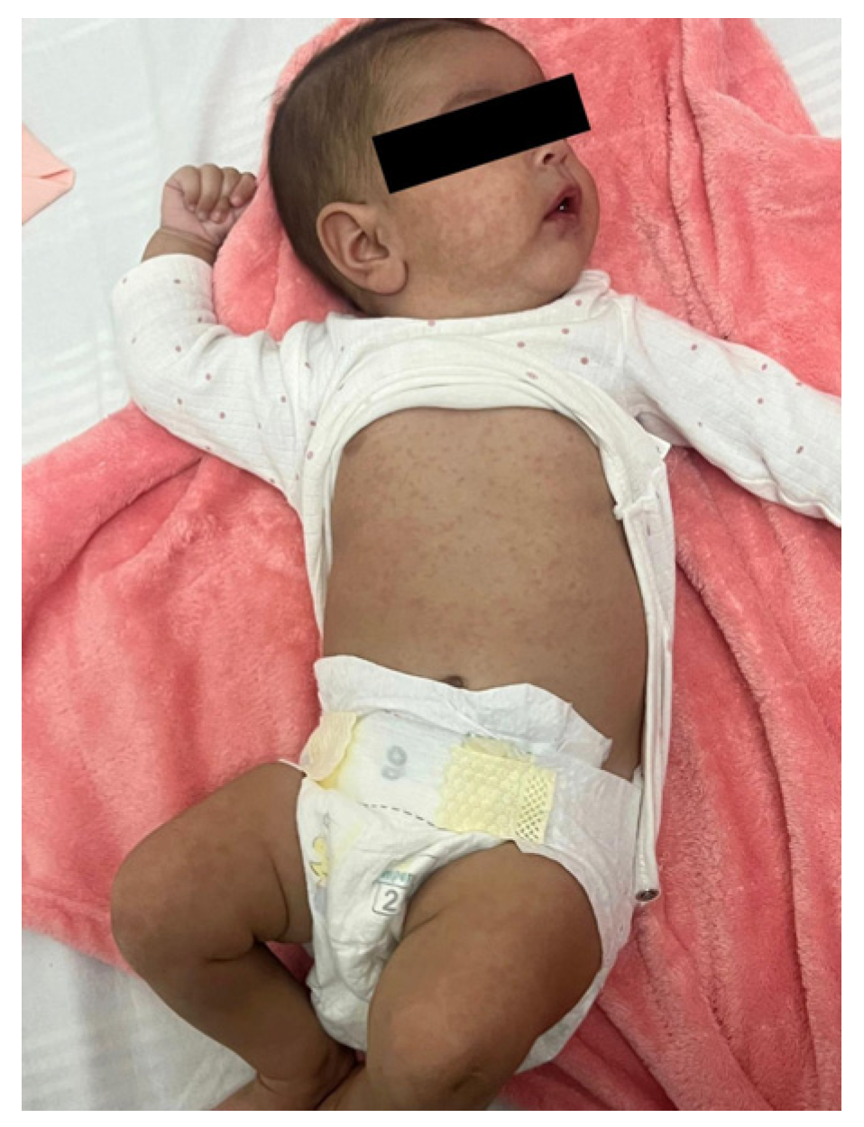
Figure 2.
Third day of admission – dry, cracked lips, and perioral rash.
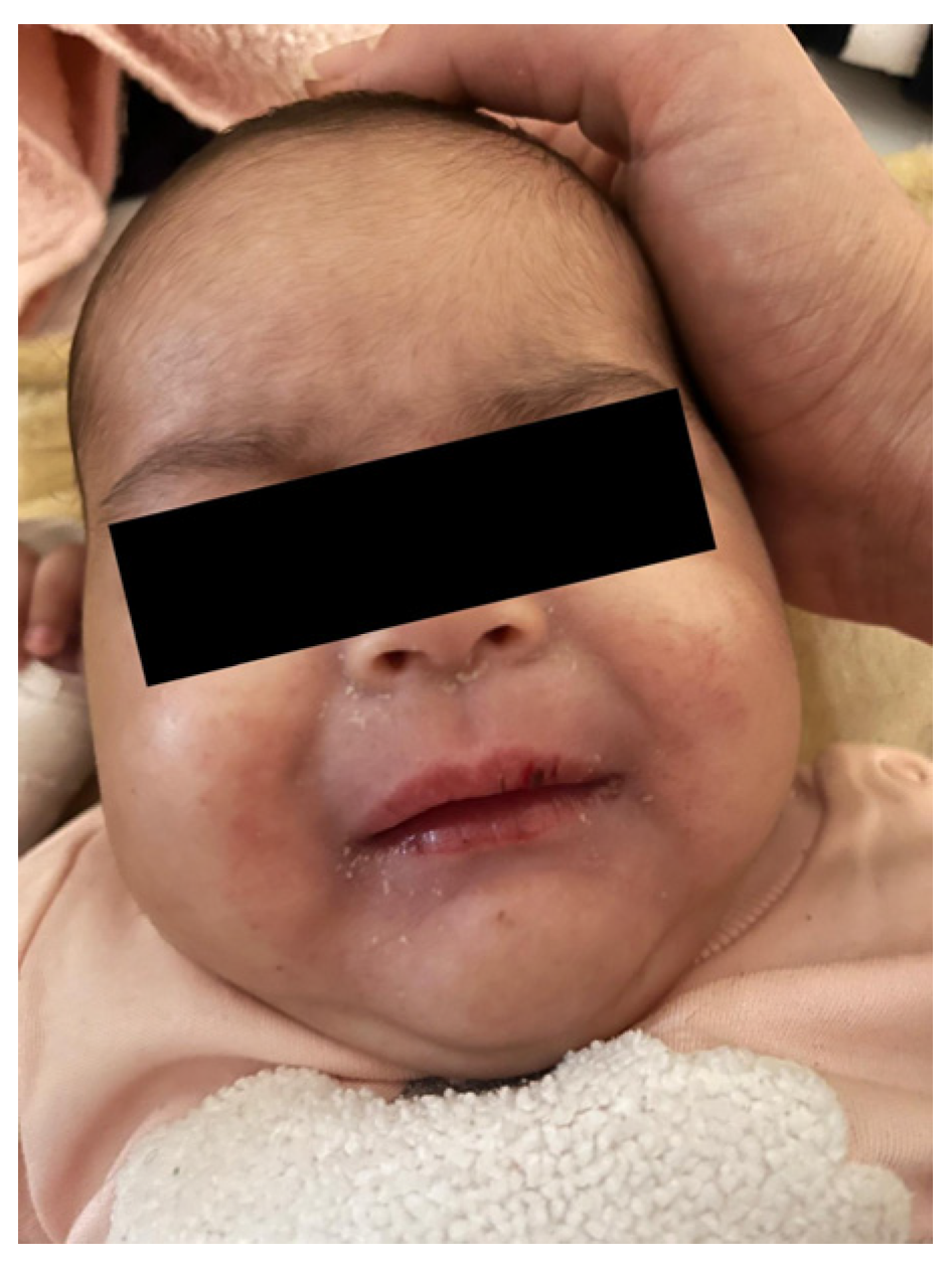
Figure 3.
Periungual desquamation in the second week after the onset of fever.
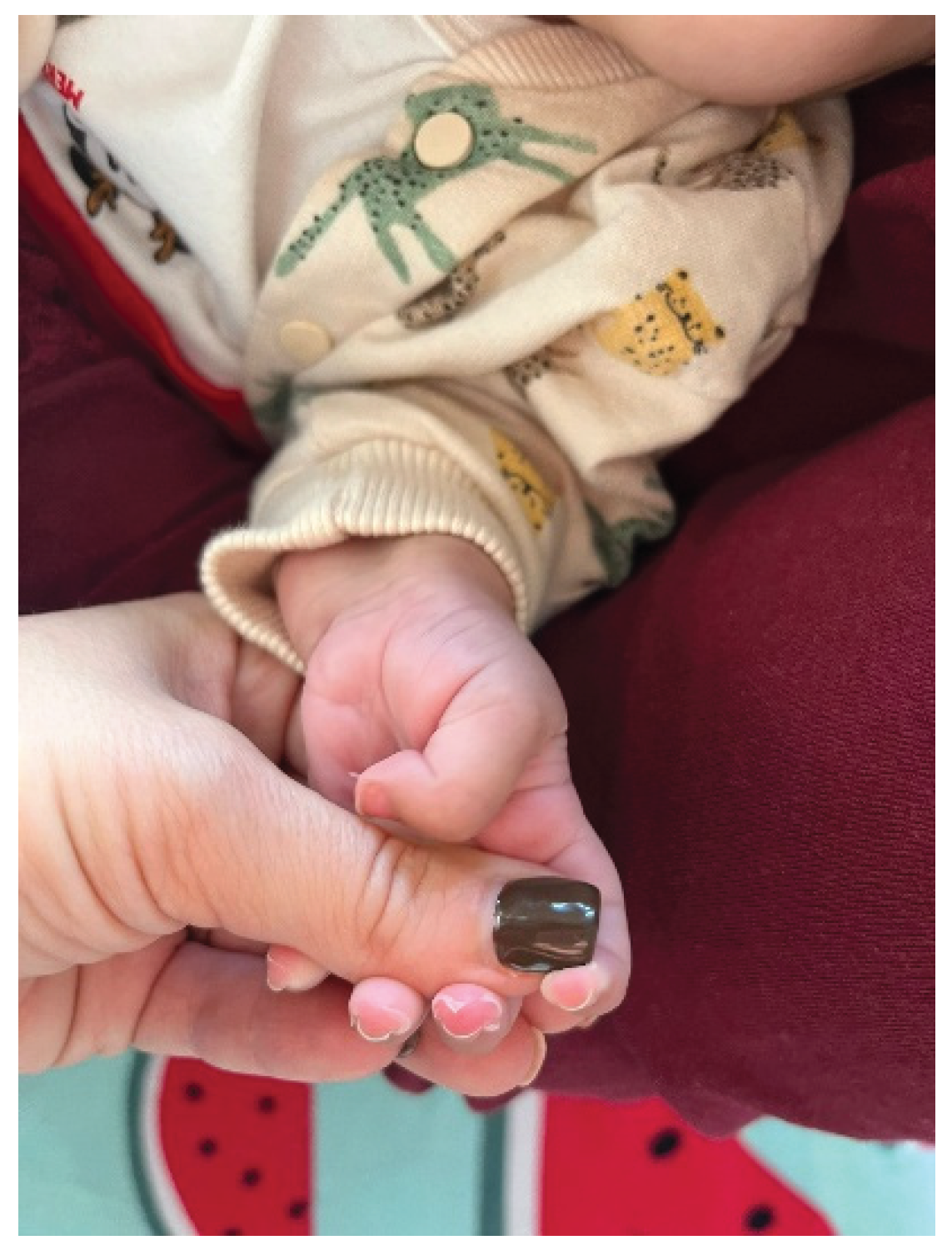

Table 1.
Laboratory findings during hospital stay and after discharge.
| At admission | Day 7 | Day 9 | Day 11 | Day 14 | Day 16 | Day 18 | At discharge | 2 weeks after discharge | |
| Hb (g/dL) | 10 | 9.2 | 7.8 | 9.1 | 9.4 | 9.9 | 10 | 9.7 | 11.3 |
| PLT (/mm3) | 320,000 | 454,000 | 517,000 | 789,000 | 910,000 | 985,000 | 712,000 | 619,000 | 413,000 |
| CRP (mg/L) | 28 | 116.6 | 124.5 | 22.9 | 3.1 | - | - | - | < 1.0 |
| ESR (mm/h) | - | 130 | - | 110 | - | - | - | - | - |
| Albumin (g/L) | - | 28.8 | - | 29.7 | - | - | - | - | 43.8 |
| D-dimer | - | - | positive | - | positive | - | - | - | - |
| NT-proBNP (pg/mL) | - | - | 2597 | - | - | 155 | - | - | - |
1 Hg = hemoglobin; PLT = platelet count; CRP = C-reactive protein (reference value < 5); ESR = erythrocyte sedimentation rate; NT-proBNP = N-terminal pro B-type natriuretic peptide (reference value < 125).
Disclaimer/Publisher’s Note: The statements, opinions and data contained in all publications are solely those of the individual author(s) and contributor(s) and not of MDPI and/or the editor(s). MDPI and/or the editor(s) disclaim responsibility for any injury to people or property resulting from any ideas, methods, instructions or products referred to in the content. |
© 2026 by the authors. Licensee MDPI, Basel, Switzerland. This article is an open access article distributed under the terms and conditions of the Creative Commons Attribution (CC BY) license (http://creativecommons.org/licenses/by/4.0/).
Copyright: This open access article is published under a Creative Commons CC BY 4.0 license, which permit the free download, distribution, and reuse, provided that the author and preprint are cited in any reuse.


