Submitted:
10 February 2026
Posted:
11 February 2026
You are already at the latest version
Abstract
Alpha-synuclein (αSyn) is one of the most abundant proteins in the nervous system and is currently associated with devastating synucleinopathies, yet its biology extends far beyond this. In this review, we outline a unified model suggesting that αSyn‑driven disease emerges within specific neural circuits through the combined effects of cell‑type‑specific roles, subcellular environments, and post‑translational modifications. These interacting and additive dimensions generate strain diversity within regions of co-pathology and, collectively, rather than αSyn alone, shape whether pathology manifests as Parkinson’s disease (PD), Parkinson’s disease dementia (PDD), dementia with Lewy bodies (DLB), multiple system atrophy (MSA), or mixed dementia phenotypes. We integrate recent advances on the physiological roles of αSyn in neurons and glia, its compartment-dependent functions, and the molecular transitions that convert functional assemblies into pathogenic conformers. Building on this foundation, we outline mechanisms through which these factors contribute to disease-specific vulnerability, progression, and clinical heterogeneity. Finally, we highlight how this multidimensional perspective can inform the development of next-generation biomarkers and precision therapies tailored to αSyn biology across distinct disorders.
Keywords:
alpha-synuclein
; neuron
; glia
; Parkinson’s disease
; Parkinson’s disease dementia
; dementia with Lewy bodies
; multiple system atrophy
1. Introduction
Alpha-synuclein (αSyn) biology is now understood as a multidimensional process that extends well beyond the traditional framework of “synucleinopathies,” which include Parkinson’s disease (PD), Parkinson’s disease dementia (PDD), dementia with Lewy bodies (DLB), and multiple system atrophy (MSA) [1,2]. Recent advances in biomarker technologies, most notably seed amplification assays (SAA) [3,4,5], together with emerging integrative staging and classification systems such as the neuronal αSyn disease integrated staging system (NSD-ISS) [6,7], SynNeurGe [8], and the NIA-AA [9], are shifting the field from symptom-based diagnosis toward a biology-driven understanding of disease (Figure 1).
Beyond classic synucleinopathies
Emerging conceptual frameworks that extend beyond traditional region-based presentation of neuronal pathology systems [10,11,12,13,14] position pathological αSyn as a central biological anchor while integrating genetic risk, co-pathology, and neuronal loss to better capture disease heterogeneity (Figure 1). This broader perspective is clinically meaningful because it situates αSyn within an interconnected biological network, allowing interpretation across conditions previously under-recognized in this context, including Alzheimer’s disease (AD) and even so called “normal” aging [15].
Lewy pathology, for instance, is observed in 30–50% of AD cases [16] and in up to 30% of neurologically normal elderly individuals, termed incidental Lewy body disease (iLBD) [17,18]. Likewise, positive αSyn detection by SAA is implicated in prodromal states such as incidental REM sleep behavior disorder (iRBD), a precursor to multiple synucleinopathies [19]. Establishing this biology-first framework is essential for understanding αSyn pathogenesis in its earliest stages, thereby enabling more definitive diagnoses and informing the development of targeted preventive interventions.
Central question
Despite these conceptual advances, a critical question remains: Why does αSyn pathology manifest as distinct clinicopathological entities—PD, PDD, DLB, MSA, or mixed dementia? An accompanying question is, what mechanisms determine disease-specific vulnerability and progression? Current staging and category frameworks do not fully account for cell-type-specific αSyn pathology, differentiate overlapping phenotypes such as PDD and DLB [2], or adequately address MSA. How do subcellular context and post-translational modifications (PTMs) contribute to strain diversity and regional co-pathology? Can a multidimensional framework integrating these factors explain clinical heterogeneity and guide precision diagnostics and therapies? This review addresses these questions by summarizing recent insights into αSyn biology and proposes a more comprehensive framework for disease-specific clinicopathology.
2. Physiological and Pathological Alpha-Synuclein
αSyn is a 140-amino acid protein in the synuclein family, which also includes β- and γ-synuclein [20]. As its name suggests, α-synuclein is highly conserved and abundantly expressed in the brain, with strong enrichment at presynaptic terminals [21,22,23], where most of its biological functions have been characterized as a purely synaptic vesicle protein. The protein is also detected in the nucleus [22,24], with recent studies highlighting its broader roles across multiple cellular compartments.
2.1. Physiological role of α-Synuclein in Neurons
At presynaptic sites, αSyn interacts with components of the synaptic vesicle (SV) system, including the SNARE complex protein VAMP2/synaptobrevin-2, synapsins, and SV membranes [25,26]. Through these interactions, αSyn regulates key steps in SV trafficking, such as vesicle clustering [27,28,29,30,31], docking [32], and recycling pool homeostasis [33,34,35], thereby modulating neurotransmitter release. Structural studies suggest that αSyn can bridge SVs to the presynaptic plasma membrane (PM) via a broken-helical conformation, supporting its role in vesicular organization [36,37]. In addition to exocytosis, triple synuclein knockout mice exhibit impaired clathrin-mediated endocytosis, suggesting its role in sustaining vesicle retrieval during high synaptic activity [38].
Beyond vesicle dynamics, αSyn impacts neurotransmitter synthesis. In dopaminergic neurons, it negatively regulates dopamine production by modulating tyrosine hydroxylase (TH) expression and activity [39], where αSyn downregulation enhances TH activity and dopamine synthesis [40]. Furthermore, αSyn contributes to synaptic plasticity. In hippocampal neurons, it facilitates long-term enhancement of neurotransmitter release via nitric oxide (NO)-cGMP signaling, which is essential for learning and memory [41]. Recent in vivo studies reveal that αSyn fine-tunes dopamine release by promoting release during short burst firing while attenuating it during prolonged activity, thereby adapting presynaptic output to firing patterns [42].
Collectively, these physiological roles in presynaptic organization, vesicle cycling, neurotransmitter synthesis, and plasticity provide a mechanistic basis for understanding how αSyn dysfunction leads to synaptic impairment and the pathogenesis of Lewy body diseases.
2.2. Endogenous α-Synuclein in Glial Cells Under Physiological Conditions
While αSyn is predominantly neuronal, its presence in glial cells under pathological conditions is well established, accumulating in astrocytes and microglia in PD and in oligodendrocytes in MSA, where it forms distinct disease-specific inclusions [43,44]. However, whether αSyn is expressed in glia under normal physiological conditions remains inconclusive.
Early studies reported only trace amounts of αSyn in cultured astrocytic cell lines detectable at both mRNA and protein levels [45]. Recent single-cell transcriptomic analyses confirm low SNCA expression in astrocytes and microglia in the healthy brain [46]. Immunohistochemistry (IHC) with proteinase K and formic acid pretreatment reveals low levels of αSyn in white matter astrocytes [47], while immuno-electron microscopy shows its distribution within astrocytic somata and processes, associated with subcellular organelles [47]. Functionally, astrocytes and microglia actively internalize and degrade αSyn under normal conditions, supporting protein clearance and homeostasis. Glial αSyn may also influence inflammatory signaling and synaptic support pathways [48]. Overall, its low basal expression suggests that most αSyn detected in these cells during disease likely originates from uptake rather than endogenous synthesis.
By contrast, oligodendrocytes exhibit low but consistent endogenous αSyn expression throughout their lineage under physiological conditions. This expression is developmentally regulated, diffuse, and non-aggregated, with enrichment in precursor and immature oligodendrocytes, implying roles in membrane dynamics [49]. This physiological pattern stands in sharp contrast to the glial cytoplasmic inclusions (GCIs) that define MSA [50,51]. Notably, GCIs emerge before overt neuronal loss, supporting the view that primary oligodendroglial dysfunction is a key driver of MSA pathogenesis [52]. Endogenous αSyn within oligodendrocytes may therefore represent the source of misfolded αSyn that accumulates in MSA [50,51].
Together, these observations underscore the cell-type–specific physiological roles and vulnerabilities to αSyn, shaping how pathology is initiated and propagated, and determining whether disease manifests as neuronal αSyn pathology in Lewy body disorders or oligodendroglial αSyn pathology in MSA.
2.3. Subcellular Localization and Organellar Biology
αSyn consists of three structurally and functionally distinct regions: an N-terminal domain (residues 1–60), a central hydrophobic non-amyloid-β component (NAC, residues 61–95), and a highly acidic, intrinsically disordered C-terminal domain (residues 96–140) [53,54,55,56,57]. The first 1-100 amino acids of αSyn contain seven imperfect 11-residue amphipathic repeats with the KTKGEV consensus motif. Upon membrane binding, these repeats adopt an α-helical conformation, enabling αSyn to function as an amphipathic lipid-binding protein [58,59,60].
2.3.1. Endogenous α-Synuclein at Presynaptic Membranes
This structural feature promotes preferential binding of αSyn to negatively charged phospholipids enriched in presynaptic membranes [59,61], mediated through both electrostatic and hydrophobic forces [62,63]. αSyn preferentially associates with the inner leaflet of the presynaptic PM, particularly within cholesterol-rich lipid raft microdomains [36]. Disease-associated mutations that disrupt αSyn–lipid raft interactions lead to its mislocalisation away from synaptic terminals, highlighting the importance of membrane binding for proper subcellular distribution [64]. αSyn is also recognised as a “curvature sensing” protein, exhibiting a strong preference for highly curved, negatively charged membranes [37,65,66]. SVs possess these biophysical properties and are optimal binding substrates [37,67]. At the molecular level, αSyn engages both SV membranes and SV-associated proteins through distinct domains: the N-terminus mediates membrane binding [34,58], whereas the C-terminus interacts with proteins such as VAMP2 and CSPα [68]. Through these interactions, αSyn is proposed to facilitate SV docking and promote SNARE complex assembly at the presynaptic PM [69]. Structural studies suggest that this function is enabled by a “broken” α-helical conformation [37], composed of helix-1 (residues 3–38) and helix-2 (residues 46–93), connected by a flexible linker (residues 39–45) [70]. This conformation allows αSyn to simultaneously bridge SVs and the PM, positioning it at sites of vesicle docking and fusion. Consistent with this model, biochemical fractionation studies show that αSyn is enriched on PM-associated docked vesicles relative to undocked vesicles in synaptosomal preparations [26].
2.3.2. Endogenous α-Synuclein in the Nucleus
The name of “α-synuclein” reflects its initial identification at both synapses and the nuclear envelope in Torpedo californica [22]. Subsequent work has provided substantial evidence for nuclear αSyn and its functional relevance in vitro and in vivo. In animal models, nuclear localisation has been observed primarily in the rat brain [71,72]. In transgenic mice, phosphorylation at serine 129 (pS129) promotes nuclear localization, suggesting that PTMs may regulate its subcellular distribution and nuclear functions [73]. In human brain tissue, αSyn has been detected in neuronal nuclei by IHC after formic acid treatment in both control and DLB cases, and in the isolated nuclear fraction by Western blotting [74]. Within the nucleus, αSyn interacts with DNA [75,76] and histones [77], influencing gene transcription [77,78] and participating in DNA damage response and repair pathways [24,76].
Glial nuclear inclusions (GNIs) [79,80,81], although less frequent than GCIs, are a distinguishing feature of MSA from Lewy pathology. GNI is composed of filamentous aggregations of αSyn [82,83]. Nuclear neuronal inclusions (NNIs) in MSA are characterized by intranuclear accumulation of αSyn, typically detected with antibodies against the C-terminal region (residues 98–115) and pS129 [84,85]. Experimental evidence indicates that cytoplasmic αSyn fibrils can penetrate the nuclear envelope and enter the nucleus, a process linked to compromised nuclear architecture, including disruption of lamin integrity [86]. Nuclear vulnerability may represent a shared pathogenic route across affected cell types [49,82,86]. Furthermore, analyses of preclinical MSA cases have revealed αSyn accumulations within and adjacent to the nuclear membrane, suggesting that nuclear αSyn pathology may play a significant role in the early stages of MSA [82].
2.3.3. Endogenous α-Synuclein at Other Organellar Membranes
Increasing evidence indicates that αSyn engages a broad array of cellular membranes, and these interactions likely contribute to both its physiological roles and pathological behavior. Beyond SVs and the nucleus, αSyn associates with multiple intracellular organelles, including mitochondria, the endoplasmic reticulum (ER), the Golgi apparatus (GA), and the endolysosomal system.
αSyn binds the inner mitochondrial membrane [87,88,89], via its N-terminal region, with residues 1–32 [90] or 1–25 [57] implicated in this interaction. This binding is driven by the high cardiolipin content of mitochondrial membranes, which provides a favourable negatively charged environment [91,92]. Pathological accumulation or aggregation of αSyn disrupts mitochondrial homeostasis, affecting mitochondrial dynamics [93], promoting fragmentation [93,94,95], and impairing degradation pathways [95].
αSyn localizes to mitochondria-associated ER membranes (MAMs), specialized contact sites that mediate Ca²⁺ transfer and lipid exchange between the ER and mitochondria [96,97]. Accumulation of αSyn at MAMs enhances ER–mitochondria coupling and perturbs Ca²⁺ homeostasis, linking mitochondrial dysfunction to ER stress [98,99]. Additionally, αSyn regulates the early secretory pathway by modulating SNARE–dependent ER–Golgi vesicle fusion. Disruption of this function impairs ER-to-Golgi trafficking and contributes to Golgi dysfunction [99,100,101]. Accumulated αSyn may further interfere with hydrolase trafficking at the cis-Golgi by aberrantly binding the scaffold protein GM130, thereby promoting lysosomal dysfunction [102].
αSyn also engages the endolysosomal system [103]. Intracellular αSyn aggregation has been observed within LAMP1-positive lysosomes, implicating lysosomal compartments in αSyn turnover and degradation [100,104]. Extracellular αSyn can be internalized via clathrin-mediated endocytosis and subsequently trafficked through multiple endosomal compartments. Following uptake, αSyn is sorted into Rab4A-positive fast recycling endosomes, Rab5A-positive early endosomes, Rab7-positive late endosomes, and Rab11-positive slow recycling endosomes, illustrating the complexity of its intracellular trafficking routes [105,106,107,108].
2.3.4. Endogenous α-Synuclein in Membraneless Condensates
Intracellularly, αSyn doesn’t restrict itself to classical membrane-bound organelles. It also engages with membraneless condensates formed through phase separation. It can partition into cytoplasmic stress granules [109] and processing bodies (P-bodies) [110], as well as nuclear nucleoli [24], where the crowded, dynamic environment can shift αSyn from its physiological state toward early condensate-like assemblies. These interactions highlight how αSyn operates at the crossroads of membrane biology and biomolecular condensation, a duality that may be crucial in the earliest steps of synucleinopathy.
Collectively, these observations show that αSyn occupies a remarkably broad and dynamic subcellular landscape, engaging both membrane-bound organelles and phase-separated condensates to support essential aspects of cellular homeostasis. While its interactions with classical membranes are well established, its involvement in phase-separated compartments remains comparatively new and less studied, yet may hold important clues to early pathogenic mechanisms. These diverse interactions are increasingly viewed as central not only to the physiological roles of αSyn in membrane trafficking and cellular homeostasis but also to the molecular events that drive its misfolding and aggregation in synucleinopathies.
2.4. Alpha-Synuclein in RNA Biology
The roles of αSyn in RNA biology have become one of the most exciting shifts in the field. It has been identified as an RNA-binding protein [110,111] and interacts with RNA, other RNA-binding proteins, and RNA granules in ways that are relevant to both biological function and neurodegeneration.
In the cytoplasm, RNA and proteins can undergo liquid–liquid phase separation (LLPS) via multivalent macromolecular interactions, thereby forming dynamic, membraneless compartments [111,112]. LLPS drives the assembly of ribonucleoprotein organelles enriched in RNA and RNA-binding proteins, including nucleoli, Cajal bodies, nuclear speckles, stress granules, P-bodies, and RNA granules [113,114]. The N-terminal region of αSyn mediates its interaction with P-body components, particularly the decapping protein EDC4 [110]. In PD, pathological accumulation of αSyn disrupts P-body homeostasis, leading to impaired mRNA decay and widespread alterations in neuronal gene expression [110]. Under cellular stress, αSyn can be recruited to stress granules, supporting a role in translational control and adaptive stress responses [109,114]. Beyond canonical ribonucleoprotein granules, emerging evidence indicates that specific RNA secondary structures, such as G-quadruplexes, can directly scaffold αSyn aggregation via its N-terminal region in neurons, further reinforcing the mechanistic interplay between RNA architecture and αSyn pathology [115]. Additionally, RNA accelerates αSyn fibrillization and becomes increasingly sequestered within aggregates, especially C-terminally truncated variants with higher nucleic acid affinity, suggesting that αSyn–RNA contacts facilitate amyloid assembly despite an unresolved structural mechanism [116].
2.5. Physiological Strains and Post-Translational Modifications
Extensive research has established the remarkable conformational plasticity of αSyn, a property central to its physiological functions. Under basal conditions, αSyn exists predominantly as a highly disordered, soluble monomer in the cell. Upon binding to lipid membranes, particularly SVs and other intracellular membranes, the first ~100 residues adopt ordered α-helical conformations [117]. These membrane-induced structural transitions, governed by lipid composition, curvature, and local protein–lipid interactions, enable αSyn to reversibly cycle between cytosolic and membrane-bound states while maintaining cellular homeostasis [118].
On highly curved membranes, αSyn can adopt two distinct α-helical conformations that do not necessarily engage the same membrane surface [119,120]. These helical states support functional models of SV–SV clustering and SV–PM interactions, thereby facilitating vesicle organisation and exocytosis. Physiologically, αSyn exists in dynamic equilibrium between largely unstructured cytosolic monomers (~14 kDa) and membrane-associated α-helical tetramers (~50–60 kDa) and higher order multimers (~80–100 kDa) [121,122,123].
Pathological accumulation of αSyn may also shift membraneless condensates from dynamic, functional states toward aberrant solid assemblies [124,125]. Growing evidence indicates that liquid-to-solid phase transitions arising from LLPS represent key intermediate steps in the aggregation of neurodegeneration-associated intrinsically disordered proteins, including αSyn [126]. An in vitro study demonstrated that αSyn undergoes LLPS to form dynamic liquid droplets that progressively mature into pathogenic aggregates, highlighting early phase-separation events as potential therapeutic targets for mitigating αSyn pathology [126]. αSyn remains largely monomeric (~90%) during early LLPS, with weak interactions supporting droplet formation. As droplets mature, monomers decrease, and fibrils accumulate, while oligomeric intermediates may reach a steady state during aggregation [126].
2.5.1. Physiological Multimers vs Pathological Oligomers
Recent liposome studies show that αSyn membrane binding involves a self-limiting multimerisation process that typically traps ~six monomers per membrane-associated assembly [127]. These multimers undergo dynamic monomer exchange, which becomes spatially restricted under physiological conditions. The number and distribution of membrane-binding sites are dictated by lipid-packing defects shaped by membrane curvature and composition [128,129,130].
Importantly, physiological multimers are distinct from pathological oligomers. Physiological multimers, including tetramers, are reversible, membrane-associated, and resistant to aggregation [127]. In contrast, pathological oligomers are β-sheet-rich, membrane-disruptive, and neurotoxic [131,132]. Consistent with this distinction, membrane-bound αSyn is generally protective against aggregation, whereas the soluble cytosolic pool is more vulnerable to misfolding [121,122].
2.5.2. The Physiological Post-Translational Modifications Landscape of α-Synuclein
Although often overlooked, PTMs play a pivotal role in regulating αSyn structural and functional plasticity. A wide range of PTMs, including acetylation, phosphorylation, nitration, ubiquitination, O-GlcNAcylation, oxidation, glycation, SUMOylation, and truncation, occur across the N-terminal, NAC, and C-terminal regions [43,133,134,135,136]. Under physiological conditions, αSyn exhibits a limited and tightly regulated PTM profile.
N-terminally acetylated (NTA) is one of the most abundant endogenous PTMs in human brain tissue [137] and critically modulates membrane interactions [57,138,139,140]. NTA-αSyn displays enhanced affinity for membranes, particularly neutral lipids [129,138]. C-terminal truncation also occurs physiologically and is not exclusively pathological [141,142]. Basal phosphorylation levels are low, with ~4% of αSyn phosphorylated at S129 in normal rat brain, although this site is highly sensitive to post-mortem dephosphorylation [143]. Additional phosphorylation sites, including Y39, S87, and Y125 (pY39, pS87, and pY125), are detectable in the soluble fractions of normal brain tissue [144,145].
2.5.3. The Post-Translational Modifications and Aggregation-Prone Conformers
The N-terminal domain is essential for membrane binding and α-helical formation, and notably, 35 of 52 identified PTMs localise to this region [137]. A recent study using a phosphomimetic mutation (Y39E) mouse model demonstrated reduced membrane interaction and increased soluble αSyn oligomers in midbrain fractions [146]. Phosphorylation at Y39 appears to impair membrane affinity by disrupting helix 2 and introducing electrostatic repulsion away from negatively charged lipids, thereby accelerating αSyn aggregation [147,148,149,150]. In contrast, nitration at the same residue alters membrane binding by steric and structural perturbation of the N-terminal helical architecture, disrupting helix packing [151]. Nitration additionally facilitates dityrosine cross-link formation, which can stabilise oligomeric assemblies and produce fibrils [151,152]. Within the NAC region, pS87 disrupts membrane binding and alters the aggregation propensity [153,154]. By contrast, the C-terminal phosphorylation at S129 does not directly affect membrane interactions but is preferentially added once αSyn has dissociated from membranes and adopted misfolded conformations [154,155,156].
More than 90% of αSyn within Lewy bodies is pS129, and 10–30% is C-terminally truncated [141,142]. C-terminal truncation removes the acidic tail that normally restrains aggregation, producing β-sheet-rich species with high seeding efficiency and further modifying fibril structure after formation [157,158]. C-terminally truncated αSyn is also present in MSA. However, current evidence indicates that its abundance is both region- and case-dependent, with some MSA cases showing a markedly higher proportion of C-terminally truncated species [158].
Systematic LC–MS/MS analyses reveal both shared and disease-specific PTM signatures across synucleinopathies [137], supporting the concept that pathological PTMs actively shift αSyn from functional assemblies toward aggregation-prone conformers.
2.5.4. Ubiquitination and SUMOylation
Ubiquitination provides a reversible regulatory mechanism that governs αSyn turnover and aggregation dynamics. Modification of lysine residues, such as K45, K58, and K60, may act as a quality-control checkpoint, sequestering membrane-damaging species and targeting cytosolic αSyn for degradation [159]. SUMOylation has similarly been proposed to stabilise soluble conformers and reduce toxic oligomer formation, although its physiological relevance remains under investigation [160].
Collectively, PTMs exert a profound influence on αSyn sites along the continuum from physiological, membrane- and membraneless-associated assemblies to pathogenic, aggregation-prone species. Rather than functioning solely as disease markers, PTMs actively sculpt conformational landscape and are central drivers of synucleinopathy pathogenesis.
3. Unified and Divergent Pathways of Alpha-Synuclein-Driven Neurodegeneration
PD, PDD, and DLB form a clinicopathological spectrum unified by the accumulation of misfolded αSyn within selectively vulnerable neuronal network systems. Increasing evidence indicates that Lewy pathology is best conceptualized as a network-level disease that reflects this clinical phenotypic variance, rather than as a process confined to discrete anatomical regions. In contrast, MSA represents a distinct synucleinopathy in which misfolded αSyn accumulates predominantly within oligodendrocytes, but also with degeneration of vulnerable neuronal populations within specific circuits [86]. This results in a characteristic pattern of network vulnerability and system-level failure that is mechanistically distinct from that in Lewy body disorders (Figure 2).
3.1. Lewy Pathology as a Network-Embedded Synucleinopathy
Two complementary trajectories, “body-first” and “brain-first” Lewy body diseases, have been proposed [161,162,163,164], yet both converge on the principle that αSyn pathology spreads non-randomly, following defined anatomical and functional connections. Neurons acting as network hubs or long-range projection nodes are disproportionately affected. Experimental studies highlight a cortical propagation route that begins with layer V projection neurons and subsequently engages layer II/III neurons, revealing layer-specific vulnerabilities in the cortex [165,166]. Site-specific αSyn seeding further demonstrates that the anatomical origin of pathology dictates distinct dendritic, synaptic, and circuit-level susceptibilities, underscoring that synucleinopathies are shaped by network context rather than aggregate load alone [167].
3.1.1. Selective Neuronal Vulnerability: Intrinsic Properties That Amplify Risk
Across Lewy body disorders, several neuronal populations exhibit distinct vulnerability to pathological αSyn. These include early-affected autonomic neurons of the enteric and peripheral nervous systems and brainstem autonomic nuclei [168], dopaminergic neurons in the substantia nigra pars compacta [169,170], noradrenergic neurons of the locus coeruleus [171], cholinergic neurons of the basal forebrain [172,173], and glutamatergic neurons in the basal ganglia, cortex, and hippocampus [174,175]. Despite their anatomical diversity, these neurons share intrinsic features that impose substantial energetic and proteostatic stress to the cell machinery in high metabolic demand [176], extensive axonal arborisation [177,178], autonomous pacemaking activity, and sustained intracellular Ca²⁺ load [179]. When challenged by pathological αSyn, these properties heighten susceptibility to mitochondrial dysfunction, oxidative stress, impaired trafficking, and proteasomal overload, predisposing these neurons to early functional decline and degeneration [177,180,181].
3.1.2. Propagation Dynamics: Prion-Like Spread Across Connected Circuits
The trans-neuronal propagation of misfolded αSyn aligns with a prion-like mechanism, prompting the development of computational models to formalize disease spread. These models differ in their biological specificity but collectively reinforce the concept that pathology progression is constrained by network architecture [182]. The Network Diffusion Model (NDM) conceptualizes passive diffusion driven by gradient pathology concentration along the structural connectivity [183]. Although NDM successfully recapitulates macroscopic disease patterns, it lacks biological specificity. The Epidemic Spreading Model (ESM) was introduced to address these limitations by incorporating protein production, clearance, and host responses, thereby capturing immune-mediated reactions to abnormal protein deposition and linking these processes to disease progression [182,184]. The more recent agent-based epidemic spreading model explicitly simulates prion-like seeding and removal dynamics but remains computationally intensive and challenging to parameterize in human studies [185]. Together, these models demonstrate that selective neuronal vulnerability emerges from the interplay between intrinsic cellular susceptibility and network embedding, while clinical heterogeneity reflects differences in seeding location, propagation routes, and connectome topology [182,186].
3.1.3. Divergent Clinical Phenotypes Shaped by Network Context
Phenotypic divergence among these Lewy body disorders reflects differences in the spatiotemporal distribution of αSyn pathology, the burden of co-pathologies, and network-specific vulnerability [2,167,187]. In PD, pathology predominantly affects the brainstem and nigrostriatal circuits, with limited or late cortical involvement [188]. In PDD, prolonged disease duration permits progressive invasion of limbic and associative cortical networks, superimposed on ongoing nigrostriatal degeneration, resulting in delayed cognitive decline [2]. By contrast, DLB is characterized by early and widespread cortical and cholinergic involvement, often accompanied by co-pathologies of amyloid-β (Aβ)and tau that accelerate cognitive impairment [2,189].
These patterns support a model in which PD, PDD, and DLB represent overlapping yet distinct manifestations of a shared network-embedded synucleinopathy. Clinical phenotype is determined not simply by aggregate burden but by the timing, anatomical origin, and network context of αSyn engagement within selectively vulnerable neuronal populations [187,190].
3.1.4. Preclinical Network Context
ILBD is widely regarded as a preclinical stage of PD or DLB [191,192]. Restricted and brainstem-predominant iLBD cases correspond to Braak Lewy pathology stages 1–3, a distribution that aligns with what is considered a preclinical PD-type form of Lewy body disease. However, a subset displays more widespread limbic or neocortical involvement, suggesting that some individuals with iLBD may be in a preclinical stage of DLB rather than exclusively preclinical PD [191,193]. In brainstem-predominant iLBD, early nigrostriatal abnormalities are already present, including reduced dopamine levels, deficits in vesicular monoamine transport, and lower striatal tyrosine hydroxylase expression, changes that fall between those seen in healthy individuals and those in patients with clinically diagnosed PD [194,195]. Electrophysiological studies show that iLBD also exhibits disrupted neural network activity even in the absence of clinical symptoms, reinforcing its status as a functional preclinical stage of Lewy body disorders [196]. Transcriptomic studies indicate that PD-vulnerable brain regions possess intrinsic gene-expression patterns that are already altered in iLBD, supporting an early molecular phase of preclinical Lewy body disorders [197].
3.2. Co-Pathology in Lewy Body Disorders
Lewy body disorders frequently coexist with other age-related neurodegenerative pathologies, most notably Aβ and tau. This convergence reflects a complex biological interplay in which αSyn, Aβ, and tau influence one another’s aggregation, propagation, and network-level effects. In DLB, αSyn pathology is primary and widespread, typically involving neocortical, limbic, and brainstem regions early, and although Aβ and tau burdens are generally lower than in AD, they are higher than in PDD, and their frequent co-occurrence strongly modulates clinical severity, cognitive decline, and disease heterogeneity [198]. Clinically, DLB remains challenging to diagnose, with misdiagnosis rates approaching 50% in early-onset cases, most often mistaken for AD [199], with definitive diagnosis relying on post-mortem neuropathological evaluation [200]. Survival interval in DLB is shorter than in AD, underscoring the aggressive nature of αSyn-driven copathologies [201].
3.2.1. Molecular Crosstalk Between α-Synuclein, Aβ, and Tau in Lewy Body Disorders
A defining feature of PDD and DLB is that αSyn pathology does not act in isolation. Instead, αSyn engages in dynamic, reciprocal interactions with Aβ and tau, forming a pathogenic network that amplifies neurodegeneration. Experimental and neuropathological studies show that Aβ and tau can enhance αSyn misfolding and seeding, while αSyn can accelerate Aβ and tau aggregation [202,203,204]. This tri-protein synergy disrupts proteostasis, promotes neuronal dysfunction, and helps explain why mixed pathology in DLB is associated with faster decline and more severe cognitive and neuropsychiatric symptoms. Notably, αSyn strains in the amygdala exhibit distinct immunohistochemical and biochemical signatures in DLB with AD, pointing to disease-specific conformational heterogeneity [205].
Evidence from AD further reinforces this molecular crosstalk. Cerebrospinal fluid (CSF) αSyn SAA detects aggregation-competent αSyn in 20–40% of patients with clinically diagnosed AD, confirming that αSyn co-pathology is both common and biologically active [202]. Neuropathological studies show that 40–60% of AD cases harbour αSyn pathology, typically concentrated in the amygdala and other limbic regions [202,206,207]. These limbic-predominant deposits suggested that Aβ- and tau-laden environments may act as permissive sites for initiating or amplifying αSyn aggregation.
3.2.2. Network-Level and Clinical Consequences of αSyn–Aβ–tau Synergy
From a network-level perspective, synergistic interactions among αSyn, Aβ, and tau accelerate neurodegeneration more than any single pathology alone [208]. At the molecular level, these proteins exhibit prion-like seeding across anatomically connected brain regions and engage in cross-seeding, whereby misfolded assemblies of one protein promote the misfolding of others, amplifying toxicity and hastening disease progression [209,210]. Immunohistochemical studies show that αSyn deposition in AD preferentially overlaps with Aβ-rich cortical territories and, to a lesser extent, with tau-affected regions. This pattern suggests that Aβ-vulnerable networks provide a permissive substrate for secondary αSyn pathology, contributing to mixed AD–Lewy body phenotypes.
Clinically, the presence of Lewy pathology in AD is associated with worse cognition, faster decline, and a higher frequency of neuropsychiatric symptoms such as depression and hallucinations compared with “pure” AD [207,211,212]. In DLB, coexisting Aβ and tau pathology shifts the clinical presentation toward earlier memory impairment and more rapid cognitive deterioration, whereas individuals with low AD biomarker burden tend to show more prominent core Lewy body features. These observations indicate that co-pathology not only accelerates disease progression but also reshapes the clinical phenotype of DLB [213].
3.3. Oligodendroglial α-Synuclein Pathology in Network-Defined Neuronal Systems
Although oligodendroglial inclusions are the pathological hallmark of MSA, neuronal degeneration is more extensive than the neuronal loss observed in Lewy body disorders, even though Lewy body disorders have more obvious neuronal pathologies. In contrast to the neuronal loss in MSA, there is little loss of oligodendroglial cell bodies [214,215,216]. There is widespread voxel-wise atrophy that disproportionately affects both white matter and grey matter in the cerebellum and brainstem [217].
The distinctive biology of oligodendrocytes—their specialised cellular environment, their support of long-range myelinated axons, and their integration into large-scale white matter networks—has led to the recent view of MSA as a network-embedded oligodendroglial synucleinopathy. However, this framework does not fully explain why the longest corticospinal tracts are not the earliest or most severely affected, whereas cerebellar peduncles show the earliest and selective vulnerability [217]. Emerging evidence suggests that NNIs in MSA may be particularly toxic [86], and that the pronounced cerebellar and brainstem atrophy reflects combined neuronal and fibre-tract degeneration. These observations raise the possibility that rapid neuronal loss associated with nuclear αSyn pathology impacts oligodendrocyte dysfunction or that oligodendrocyte dysfunction influences neuronal survival in these regions. Overall, dysfunctional myelination alone seems insufficient to account for the pattern of degeneration.
The following subsections outline the hypothesised mechanisms of αSyn pathology in MSA and highlight unresolved questions arising from these network-level and region-specific vulnerabilities.
3.3.1. Mechanistic Questions Surrounding Oligodendrocyte α-Synuclein Pathology
In MSA, the neuronal inclusions appear less regionally widespread than GCIs, a pattern that may reflect the rapid loss of the vulnerable neurons with impaired axonal transport, which limits the synaptic transmission route of αSyn, in contrast to Lewy pathology. In addition, oligodendrocytes lack the robust proteostatic and phagocytic machinery of microglia and astrocytes, which may contribute to the broader distribution of glial inclusions.
It remains unclear whether oligodendrocytes initiate αSyn pathology at multiple sites due to systemic deficiencies in cellular machinery, whether neuron-oligo mechanisms drive αSyn spread, or whether other pathways enable glial-to-glial transmission. The coexistence of neuronal and glial inclusions therefore raises a central unresolved question: what is the cellular origin of αSyn in MSA, and can the αSyn species present in neurons and oligodendrocytes be distinguished using pathological or biochemical markers? Addressing this question is essential for determining whether MSA is fundamentally a glial-initiated synucleinopathy or whether neuronal and glial αSyn pathologies arise through distinct yet convergent mechanisms.
3.3.2. Hypotheses on the Origins of α-Synuclein Pathology in MSA
- Neuron-to-glia transfer hypothesis
One major hypothesis proposes that pathological αSyn originates in neurons and is subsequently transferred to oligodendrocytes. Under this model, neurons release misfolded αSyn species via exocytosis, exosomes, or synaptic leakage, which are then internalized by oligodendrocytes [86,218]. Because oligodendrocytes possess limited proteostatic capacity to handle αSyn, internalized aggregates accumulate and seed GCI formation. This hypothesis is supported by the fact that neurons express αSyn abundantly, whereas oligodendrocytes express it at low physiological levels, suggesting that neurons are a plausible source of pathogenic αSyn [50]. Unlike familial PD, MSA is not known to be associated with SNCA mutations or multiplications. Although SNCA polymorphisms may modulate αSyn expression in oligodendrocytes, the absence of classical PD-linked mutations suggests a distinct pathogenic mechanism in MSA, rather than a neuron-initiated synucleinopathy manifesting in glia [219,220].
- The oligodendrogliopathy first hypothesis
A recent view argues that primary oligodendroglial dysfunction precedes and drives neuronal degeneration [221]. Several lines of evidence support this hypothesis. αSyn has been detected in isolated oligodendrocytes from neonatal wild-type mice and from MSA patient tissue, suggesting that oligodendrocytes may be intrinsically capable of accumulating αSyn [50,222]. Immunohistochemical studies show widespread GCI distribution across the MSA brain, whereas neuronal loss is more anatomically restricted (e.g., striatonigral degeneration and olivopontocerebellar atrophy) [84]. A key mechanistic insight involves the oligodendrocyte-specific phosphoprotein TPPP/p25α, which becomes mislocalised from myelin to the oligodendrocyte soma in MSA [83]. In this ectopic location, p25α strongly promotes αSyn misfolding and aggregation, indicating that the intracellular environment of diseased oligodendrocytes is highly permissive for pathogenic αSyn conformers [82,223,224]. A central mechanistic theme may still be the proteostastic failure in oligodendrocytes, driven by impaired autophagy–lysosome function and ubiquitin–proteasome stress [225]. Oligodendrocytes, specialized for high-throughput myelin production, are uniquely vulnerable to disruptions in protein and lipid balance [226]. Accumulation of αSyn within oligodendrocytes disrupts myelin integrity, in part through altered lipid composition, including increased monounsaturated fatty acids, thereby compromising white matter function [227,228].
- A membraneless organelle hypothesis of MSA
An alternative possibility is that MSA reflects a fundamental disturbance in αSyn-related membraneless organelles rather than a process confined primarily to oligodendrocytes. This hypothesis could account for the presence of neuronal nuclear αSyn pathology as well as the prominent oligodendroglial involvement. Oligodendrocytes rely heavily on RNA transport and local translation to sustain remote myelin synthesis [229]. MBP, CAII, Tau, and MOBP are all produced locally after their untranslated mRNAs are trafficked to distal myelin sheaths, associated with RNA transport granules. Disruption of αSyn-dependent phase-separation processes could therefore impair RNA handling, local translation, or the assembly of ribonucleoprotein granules in both neurons and oligodendrocytes. Such a mechanism would unify the emergence of neuronal nuclear inclusions with widespread oligodendroglial pathology and may help explain why MSA exhibits features that cannot be fully accounted for by demyelination or oligodendrocyte dysfunction alone.
3.3.3. Molecular Specificity: MSA-Associated α-Synuclein Strains
Structural and biophysical analyses demonstrate that MSA-derived αSyn aggregates are enriched in β-sheet content and exhibit greater toxicity and protease resistance than PD-derived aggregates [230]. Cryo-EM studies further reveal that αSyn filaments isolated from MSA brains adopt unique asymmetric protofilament architectures [231]. Additional work highlights the molecular heterogeneity of αSyn aggregates in MSA, including less pS129 [232] but abundant PTMs within the NAC domain [137] and C-terminal truncations [158].
4. Targeting Alpha-Synuclein: Biomarkers
αSyn biomarkers have become central to efforts to improve diagnostic accuracy, patient stratification, and therapeutic monitoring across synucleinopathies. However, the field remains challenged by the biological complexity of αSyn itself, its diverse conformations, cell-type-specific roles, and strain-dependent pathogenicity. Traditional assays that quantify total αSyn provide limited insight into this molecular heterogeneity, while next-generation approaches increasingly focus on detecting the diverse pathogenic assemblies, seeding activity, and strain-specific signatures. Together, these evolving biomarker strategies reflect a broader shift toward mechanistic, biology-driven diagnostics capable of capturing the multidimensional nature of αSyn pathology.
4.1. Current Biomarker Modalities
4.1.1. Diagnostic Challenges Across Synucleinopathies
The substantial overlap in clinical features and neuropathology among synucleinopathies complicates accurate diagnosis. Because disease progression involves intercellular transfer of misfolded αSyn among brain cells, αSyn is released into the extracellular space and becomes detectable in CSF, blood, saliva, and tears [233]. Among these, CSF remains the most reliable matrix, given its proximity to the CNS and reduced influence from peripheral αSyn sources [234,235].
4.1.2. αSyn Species: Total, Oligomeric, and Phosphorylated Forms
The conventional approach to quantifying peripheral αSyn has relied on ELISA-based detection, which is easy to implement but is limited by its sensitivity and dynamic range. High-throughput proteomic platforms such as aptamer-based assays (SomaLogic’s SomaScan) [236] now enable far more sensitive, multiplexed measurement of αSyn and related biomarkers from peripheral samples. Although these technologies are advancing biomarker discovery and improving resolution of disease-associated protein signatures, their application to αSyn detection remains relatively limited.
Altered CSF αSyn levels have been reported in PD, but total αSyn is highly susceptible to blood contamination, leading to variable results across studies [237,238,239]. In contrast, oligomeric αSyn is more consistently elevated, and the oligomeric-to-total αSyn ratio improves discrimination between PD and controls [240,241]. Meanwhile, the pathological relevance of pS129 αSyn and its detectability in CSF support its potential as a biomarker [242]. However, CSF pS129 does not consistently differ between PD and controls, and longitudinal studies show that neither absolute pS129 nor the pS129-to-total αSyn ratio reflects disease presence or progression [243]. Moreover, pS129 levels do not correlate with αSyn seeding activity, limiting its diagnostic value [244]. Generally, measurements of total, oligomeric, and pS129 αSyn fail to reliably differentiate PD, DLB, MSA, and other neurodegenerative disorders [235,238,245], and diagnostic accuracy remains suboptimal for both oligomeric and pS129 αSyn detection in CSF [245].
4.1.3. Seed Amplification Assays (SAAs)
SAAs, including real-time quaking-induced conversion (RT-QuIC), have emerged as promising tools for detecting misfolded αSyn with high accuracy in distinguishing synucleinopathies from tauopathies [246]. Interestingly, a recent study revealed that 5-34% cases are SAA-negative when the cohort comprises both sporadic and genetic PD cases [247]. While SAA-positive patients exhibit greater dopaminergic deficits, SAA-negative patients have more subcortical atrophy [247]. The SAA results in MSA remain inconsistent, with some studies reporting minimal or no seeding activity, even with ultra-sensitive protocols [248]. While these discrepancies highlight strain-specific and disease-specific limitations of current SAA platforms, when MSA αSyn aggregates are used as seeds, the amplified products faithfully preserve the biological and structural characteristics of the original brain-derived aggregates [249].
4.1.4. Other Assays and Combined Application
Recognition that αSyn pathology extends beyond body fluids to peripheral tissues has driven the development of alternative biomarker strategies targeting salivary glands, olfactory mucosa, the gastrointestinal tract, and skin [233,250,251,252]. Among these, skin biopsy has emerged as a highly accurate, minimally invasive, and readily accessible approach, enabling both single- and longitudinal-sampling for the diagnosis of PD and related synucleinopathies [253,254]. Skin biopsies enable direct detection of pathological αSyn within autonomic and somatosensory dermal nerve fibres, most commonly through immunostaining for pS129 αSyn, as well as conformation-selective antibodies targeting oligomeric αSyn (e.g., ASyO5) and aggregated αSyn species (e.g., 5G4), providing complementary information on αSyn burden and aggregation state [255,256,257]. In parallel, proximity ligation assay (PLA) has been applied to skin tissue to selectively amplify oligomeric and conformationally altered αSyn, improving pathological enrichment over conventional immunostaining while preserving cellular localisation [258,259]. More recently, RT-QuIC performed on skin biopsies has demonstrated high sensitivity and specificity for PD and DLB, with pathological αSyn detectable at prodromal stages such as iRBD [258,260,261,262]. Immunostaining, PLA, and SAA provide complementary readouts of peripheral αSyn pathology. In MSA, increased intraneural PLA signal supports distinct, oligodendroglial-dominant αSyn strain biology [258,263].
Growing evidence indicates that αSyn exists as distinct molecular strains with cell type- and disease-specificity. This paradigm shift underscores the need for biomarkers that capture biological features from conformation, strain, seeding, and cellular context, rather than relying solely on total protein abundance.
4.2. Strain-, Cell Type-, and Conformation-Specific Assays
αSyn SAA has shown some capacity to discriminate disease-specific αSyn strains in PD and MSA [230,248,264]. Distinct amplification profiles were identified: type 1 patterns in samples containing Lewy bodies and type 2 patterns in samples enriched for GCIs, which mirror the underlying cellular environments. Thus, a negative SAA result does not exclude αSyn pathology, particularly in MSA. Alternatively, the reduced sensitivity of CSF SAA in MSA may reflect the presence of non-neuronal, oligodendroglial αSyn conformers that are inefficiently amplified by assays optimised for Lewy body–typed strains [230,248,265]. Given that some αSyn seeds are more toxic in MSA, the lack of seeding in SAA may result from certain oligomeric αSyn species.
In addition, samples with mixed pathologies show greater variability in SAA readouts and may even inhibit amplification, indicating the need for rigorous pre-analytical handling and quantitative assay standardization [266]. This also implicates the importance of understanding the fluid chemical interactions among αSyn, Aβ, and tau/pTau.
Accumulating evidence indicates that αSyn exists as a spectrum of conformational strains with distinct biochemical properties, cellular tropisms, and pathogenic potentials. One of the major challenges in biomarker development is therefore discriminating physiological αSyn multimers from disease-associated oligomeric species. Increasing emphasis is placed on assays targeting oligomeric and conformationally distinct αSyn species, which are considered the primary neurotoxic forms [267]. Oligomer-selective immunoassays and PLA have been explored [268], but PLA detects molecular proximity rather than pathogenic structure, meaning it cannot inherently differentiate physiological from pathological assemblies [269,270]. Therefore, the ideal antibody in PLA is one that is highly specific pathologically and abundant in the oligomeric vs. the physiological state.
5. Summary and Future Directions
Recent research increasingly indicates that the clinical heterogeneity of αSyn–driven disorders cannot be explained solely by anatomical spread. Instead, disease arises from the intersection of multiple biological dimensions, including cell type, subcellular milieu, PTMs, strain diversity, and co-pathologies such as Aβ and tau, which collectively shape how αSyn behaves within specific cellular networks. These interacting factors determine not only where pathology develops, but how toxic it becomes, how it propagates, and why clinical trajectories diverge across PD, PDD, DLB, MSA, and mixed phenotypes. This understanding calls for a shift toward an αSyn biology–based nosology grounded in both cell-type specificity and network context, reflected in biomarker profiles that capture such pathogenic activity. Diagnostic tools should therefore be viewed as sampling distinct nodes within a shared pathological network rather than as isolated readouts. Integrating complementary biomarkers [271,272] within a network-aligned framework enables more accurate assessment, refined patient stratification, and the development of αSyn-targeted trials that account for underlying heterogeneity. Ultimately, embracing this multidimensional perspective is essential for advancing precision therapies that align with the diverse molecular landscapes of αSyn-related diseases.
Author Contributions
Writing—Feifei Su & YuHong Fu; Structure composing—YuHong Fu & Glenda Halliday; Revision—Woojin Kim & Glenda Halliday; Proofreading—Woojin Kim & Glenda Halliday.
Funding
This research was supported by funding from the Infectious Disease Society of America (ALZ-ID-0000000016) to Y.F. GMH is an NHMRC Senior Investigator (grant #1176607).
Conflicts of Interest
The authors declare no conflicts of interest.
Abbreviations
The following abbreviations are used in this manuscript:
| αSyn | Alpha-synuclein |
| AD | Alzheimer’s disease |
| Aβ | Amyloid-β |
| BBB | Blood–brain barrier |
| CSF | Cerebrospinal fluid |
| DLB | Dementia with Lewy bodies |
| ER | Endoplasmic reticulum |
| ESM | Epidemic Spreading Model |
| GCIs | Glial cytoplasmic inclusions |
| GA | Golgi apparatus |
| GNIs | Glial nuclear inclusions |
| iLBD | Incidental Lewy body disease |
| iRBD | Incidental REM sleep behavior disorder |
| IHC | Immunohistochemistry |
| LLPS | liquid–liquid phase separation |
| MSA | Multiple system atrophy |
| MAMs | Mitochondria-associated ER membranes |
| NAC | Non-amyloid-β component |
| NDM | Network Diffusion Model |
| NTA | N-terminally acetylated |
| NNIs | Neuronal nuclear inclusions |
| NSD-ISS | Neuronal αSyn disease integrated staging system |
| pS129 | Phosphorylation at serine 129 |
| P-bodies | Processing bodies |
| PD | Parkinson’s disease |
| PDD | Parkinson’s disease dementia |
| PTM | Post-translational modification |
| PLA | Proximity ligation assay |
| PM | Plasma membrane |
| RT-QuIC | Real-time quaking-induced conversion |
| SAA | seed amplification assay |
| SV | Synaptic vesicle |
| TH | Tyrosine hydroxylase |
References
- Park, H.; Kam, T.I.; Dawson, V.L.; Dawson, T.M. α-Synuclein pathology as a target in neurodegenerative diseases. Nat Rev Neurol 2025, 21, 32–47. [Google Scholar] [CrossRef]
- Fu, Y.; Halliday, G.M. Dementia with Lewy bodies and Parkinson disease dementia - the same or different and is it important? Nat Rev Neurol 2025, 21, 394–403. [Google Scholar] [CrossRef]
- Bellomo, G.; De Luca, C.M.G.; Paoletti, F.P.; Gaetani, L.; Moda, F.; Parnetti, L. α-Synuclein Seed Amplification Assays for Diagnosing Synucleinopathies. Neurology 2022, 99, 195–205. [Google Scholar] [CrossRef] [PubMed]
- Ma, Y.; Farris, C.M.; Weber, S.; Schade, S.; Nguyen, H.; Pérez-Soriano, A.; Giraldo, D.M.; Fernández, M.; Soto, M.; Cámara, A.; et al. Sensitivity and specificity of a seed amplification assay for diagnosis of multiple system atrophy: a multicentre cohort study. Lancet Neurol 2024, 23, 1225–1237. [Google Scholar] [CrossRef]
- Orrú, C.D.; Vaughan, D.P.; Vijiaratnam, N.; Real, R.; Martinez-Carrasco, A.; Fumi, R.; Jensen, M.T.; Hodgson, M.; Girges, C.; Gil-Martinez, A.-L. Diagnostic and prognostic value of α-synuclein seed amplification assay kinetic measures in Parkinson’s disease: a longitudinal cohort study. The Lancet Neurology 2025, 24, 580–590. [Google Scholar] [CrossRef] [PubMed]
- Simuni, T.; Chahine, L.M.; Poston, K.; Brumm, M.; Buracchio, T.; Campbell, M.; Chowdhury, S.; Coffey, C.; Concha-Marambio, L.; Dam, T.; et al. A biological definition of neuronal α-synuclein disease: towards an integrated staging system for research. Lancet Neurol 2024, 23, 178–190. [Google Scholar] [CrossRef] [PubMed]
- Dam, T.; Pagano, G.; Brumm, M.C.; Gochanour, C.; Poston, K.L.; Weintraub, D.; Chahine, L.M.; Coffey, C.; Tanner, C.M.; Kopil, C.M.; et al. Neuronal alpha-Synuclein Disease integrated staging system performance in PPMI, PASADENA, and SPARK baseline cohorts. npj Parkinson’s Disease 2024, 10, 178. [Google Scholar] [CrossRef]
- Höglinger, G.U.; Adler, C.H.; Berg, D.; Klein, C.; Outeiro, T.F.; Poewe, W.; Postuma, R.; Stoessl, A.J.; Lang, A.E. A biological classification of Parkinson’s disease: the SynNeurGe research diagnostic criteria. Lancet Neurol 2024, 23, 191–204. [Google Scholar] [CrossRef]
- Jack, C.R., Jr.; Bennett, D.A.; Blennow, K.; Carrillo, M.C.; Dunn, B.; Haeberlein, S.B.; Holtzman, D.M.; Jagust, W.; Jessen, F.; Karlawish, J.; et al. NIA-AA Research Framework: Toward a biological definition of Alzheimer’s disease. Alzheimers Dement 2018, 14, 535–562. [Google Scholar] [CrossRef]
- Braak, H.; Del Tredici, K.; Rüb, U.; De Vos, R.A.; Steur, E.N.J.; Braak, E. Staging of brain pathology related to sporadic Parkinson’s disease. Neurobiology of aging 2003, 24, 197–211. [Google Scholar] [CrossRef]
- Beach, T.G.; Adler, C.H.; Lue, L.; Sue, L.I.; Bachalakuri, J.; Henry-Watson, J.; Sasse, J.; Boyer, S.; Shirohi, S.; Brooks, R.; et al. Unified staging system for Lewy body disorders: correlation with nigrostriatal degeneration, cognitive impairment and motor dysfunction. Acta Neuropathol 2009, 117, 613–634. [Google Scholar] [CrossRef]
- McKeith, I.G.; Dickson, D.W.; Lowe, J.; Emre, M.; O’brien, J.; Feldman, H.; Cummings, J.; Duda, J.; Lippa, C.; Perry, E. Diagnosis and management of dementia with Lewy bodies: third report of the DLB Consortium. Neurology 2005, 65, 1863–1872. [Google Scholar] [CrossRef]
- Leverenz, J.B.; Hamilton, R.; Tsuang, D.W.; Schantz, A.; Vavrek, D.; Larson, E.B.; Kukull, W.A.; Lopez, O.; Galasko, D.; Masliah, E.; et al. Empiric refinement of the pathologic assessment of Lewy-related pathology in the dementia patient. Brain Pathol 2008, 18, 220–224. [Google Scholar] [CrossRef] [PubMed]
- Attems, J.; Toledo, J.B.; Walker, L.; Gelpi, E.; Gentleman, S.; Halliday, G.; Hortobagyi, T.; Jellinger, K.; Kovacs, G.G.; Lee, E.B.; et al. Neuropathological consensus criteria for the evaluation of Lewy pathology in post-mortem brains: a multi-centre study. Acta Neuropathol 2021, 141, 159–172. [Google Scholar] [CrossRef] [PubMed]
- Fu, Y.; Tanglay, O.; Li, H.; Halliday, G.M. The Role of Alpha-Synuclein Pathology. In Translational Methods for Parkinson’s Disease and Atypical Parkinsonism Research; Groppa, S., Schneider, S.A., Eds.; Springer US: New York, NY, 2025; pp. 21–48. [Google Scholar]
- Barnes, L.L.; Leurgans, S.; Aggarwal, N.T.; Shah, R.C.; Arvanitakis, Z.; James, B.D.; Buchman, A.S.; Bennett, D.A.; Schneider, J.A. Mixed pathology is more likely in black than white decedents with Alzheimer dementia. Neurology 2015, 85, 528–534. [Google Scholar] [CrossRef]
- Frigerio, R.; Fujishiro, H.; Ahn, T.B.; Josephs, K.A.; Maraganore, D.M.; DelleDonne, A.; Parisi, J.E.; Klos, K.J.; Boeve, B.F.; Dickson, D.W.; et al. Incidental Lewy body disease: do some cases represent a preclinical stage of dementia with Lewy bodies? Neurobiol Aging 2011, 32, 857–863. [Google Scholar] [CrossRef] [PubMed]
- Jellinger, K.A. Lewy body-related alpha-synucleinopathy in the aged human brain. J Neural Transm (Vienna) 2004, 111, 1219–1235. [Google Scholar] [CrossRef]
- Iranzo, A.; Tolosa, E.; Gelpi, E.; Molinuevo, J.L.; Valldeoriola, F.; Serradell, M.; Sanchez-Valle, R.; Vilaseca, I.; Lomeña, F.; Vilas, D.; et al. Neurodegenerative disease status and post-mortem pathology in idiopathic rapid-eye-movement sleep behaviour disorder: an observational cohort study. Lancet Neurol 2013, 12, 443–453. [Google Scholar] [CrossRef]
- George, J.M. The synucleins. Genome biology 2001, 3, 1–6. [Google Scholar] [CrossRef]
- Sharma, M.; Burré, J. α-Synuclein in synaptic function and dysfunction. Trends Neurosci 2023, 46, 153–166. [Google Scholar] [CrossRef]
- Maroteaux, L.; Campanelli, J.T.; Scheller, R.H. Synuclein: a neuron-specific protein localized to the nucleus and presynaptic nerve terminal. J Neurosci 1988, 8, 2804–2815. [Google Scholar] [CrossRef]
- Iwai, A.; Masliah, E.; Yoshimoto, M.; Ge, N.; Flanagan, L.; de Silva, H.A.; Kittel, A.; Saitoh, T. The precursor protein of non-A beta component of Alzheimer’s disease amyloid is a presynaptic protein of the central nervous system. Neuron 1995, 14, 467–475. [Google Scholar] [CrossRef]
- Lourenco, G.F.; Torres-Pacheco, M.E.; Fu, Y.; Li, H.; McCann, H.; Shepherd, C.E.; Kril, J.J.; Halliday, G.M. Nucleolar aggregation of key neuropathological proteins in the postmortem neurodegenerative brain. Acta Neuropathologica 2025, 150, 60. [Google Scholar] [CrossRef]
- Gao, V.; Briano, J.A.; Komer, L.E.; Burré, J. Functional and Pathological Effects of α-Synuclein on Synaptic SNARE Complexes. J Mol Biol 2023, 435, 167714. [Google Scholar] [CrossRef]
- Burré, J.; Sharma, M.; Südhof, T.C. α-Synuclein assembles into higher-order multimers upon membrane binding to promote SNARE complex formation. Proceedings of the National Academy of Sciences 2014, 111, E4274–E4283. [Google Scholar] [CrossRef]
- Wang, L.; Das, U.; Scott, D.A.; Tang, Y.; McLean, P.J.; Roy, S. α-synuclein multimers cluster synaptic vesicles and attenuate recycling. Current Biology 2014, 24, 2319–2326. [Google Scholar] [CrossRef]
- Diao, J.; Burré, J.; Vivona, S.; Cipriano, D.J.; Sharma, M.; Kyoung, M.; Südhof, T.C.; Brunger, A.T. Native α-synuclein induces clustering of synaptic-vesicle mimics via binding to phospholipids and synaptobrevin-2/VAMP2. elife 2013, 2, e00592. [Google Scholar] [CrossRef]
- Murphy, DD; Rueter, R.S.; Trojanowski, JQ; Lee, VM. Synucleins Are Developmentally Expressed, and a-Synuclein Regulates the Size of the Presynaptic Vesicular Pool in Primary Hippocampal Neurons.pdf. J Neurosci 2000. [Google Scholar] [CrossRef] [PubMed]
- Chandra, S.; Gallardo, G.; Fernandez-Chacon, R.; Schluter, O.M.; Sudhof, T.C. Alpha-synuclein cooperates with CSPalpha in preventing neurodegeneration. Cell 2005, 123, 383–396. [Google Scholar] [CrossRef] [PubMed]
- Lieknina, I.; Reimer, L.; Pantelejevs, T.; Lends, A.; Jaudzems, K.; El-Turabi, A.; Gram, H.; Hammi, A.; Jensen, P.H.; Tars, K. Structural basis of epitope recognition by anti-alpha-synuclein antibodies MJFR14-6-4-2. NPJ Parkinsons Dis 2024, 10, 206. [Google Scholar] [CrossRef] [PubMed]
- Man, W.K.; Tahirbegi, B.; Vrettas, M.D.; Preet, S.; Ying, L.; Vendruscolo, M.; De Simone, A.; Fusco, G. The docking of synaptic vesicles on the presynaptic membrane induced by α-synuclein is modulated by lipid composition. Nature Communications 2021, 12, 927. [Google Scholar] [CrossRef] [PubMed]
- Murphy, D.D.; Rueter, S.M.; Trojanowski, J.Q.; Lee, V.M. Synucleins are developmentally expressed, and alpha-synuclein regulates the size of the presynaptic vesicular pool in primary hippocampal neurons. J Neurosci 2000, 20, 3214–3220. [Google Scholar] [CrossRef]
- Sun, J.; Wang, L.; Bao, H.; Premi, S.; Das, U.; Chapman, E.R.; Roy, S. Functional cooperation of α-synuclein and VAMP2 in synaptic vesicle recycling. Proceedings of the National Academy of Sciences 2019, 116, 11113–11115. [Google Scholar] [CrossRef] [PubMed]
- Li, D.; Liu, K.; Li, D.; Brunger, A.; Li, C.; Burré, J.; Diao, J. α-Synuclein condensation in synaptic vesicle function and synucleinopathies. Trends in Cell Biology 2025. [Google Scholar] [CrossRef] [PubMed]
- Man, W.K.; Tahirbegi, B.; Vrettas, M.D.; Preet, S.; Ying, L.; Vendruscolo, M.; De Simone, A.; Fusco, G. The docking of synaptic vesicles on the presynaptic membrane induced by α-synuclein is modulated by lipid composition. Nat Commun 2021, 12, 927. [Google Scholar] [CrossRef]
- Snead, D.; Eliezer, D. Alpha-synuclein function and dysfunction on cellular membranes. Exp Neurobiol 2014, 23, 292–313. [Google Scholar] [CrossRef]
- Vargas, K.J.; Makani, S.; Davis, T.; Westphal, C.H.; Castillo, P.E.; Chandra, S.S. Synucleins regulate the kinetics of synaptic vesicle endocytosis. Journal of Neuroscience 2014, 34, 9364–9376. [Google Scholar] [CrossRef]
- Perez, R.G.; Waymire, J.C.; Lin, E.; Liu, J.J.; Guo, F.; Zigmond, M.J. A role for α-synuclein in the regulation of dopamine biosynthesis. Journal of Neuroscience 2002, 22, 3090–3099. [Google Scholar] [CrossRef]
- Liu, D.; Jin, L.; Wang, H.; Zhao, H.; Zhao, C.; Duan, C.; Lu, L.; Wu, B.; Yu, S.; Chan, P. Silencing α-synuclein gene expression enhances tyrosine hydroxylase activity in MN9D cells. Neurochemical research 2008, 33, 1401–1409. [Google Scholar] [CrossRef]
- Liu, S.; Ninan, I.; Antonova, I.; Battaglia, F.; Trinchese, F.; Narasanna, A.; Kolodilov, N.; Dauer, W.; Hawkins, R.D.; Arancio, O. α-Synuclein produces a long-lasting increase in neurotransmitter release. The EMBO journal 2004, 23, 4506–4516. [Google Scholar] [CrossRef]
- Somayaji, M.; Cataldi, S.; Choi, S.J.; Edwards, R.H.; Mosharov, E.V.; Sulzer, D. A dual role for α-synuclein in facilitation and depression of dopamine release from substantia nigra neurons in vivo. Proceedings of the National Academy of Sciences 2020, 117, 32701–32710. [Google Scholar] [CrossRef]
- Sagredo, G.T.; Tanglay, O.; Shahdadpuri, S.; Fu, Y.; Halliday, G.M. ⍺-Synuclein levels in Parkinson’s disease–Cell types and forms that contribute to pathogenesis. Experimental Neurology 2024, 379, 114887. [Google Scholar] [CrossRef] [PubMed]
- Wang, L.; Tanglay, O.; Su, F.; Li, H.; Liu, J.; Kim, W.S.; Halliday, G.M.; Fu, Y. Distinct AQP4 Alterations in Movement Disorders with Primary Synucleinopathy. Movement Disorders 2025, 40, 2804–2810. [Google Scholar] [CrossRef]
- Tanji, K.; Imaizumi, T.; Yoshida, H.; Mori, F.; Yoshimoto, M.; Satoh, K.; Wakabayashi, K. Expression of α-synuclein in a human glioma cell line and its up-regulation by interleukin-1β. Neuroreport 2001, 12, 1909–1912. [Google Scholar] [CrossRef]
- Batiuk, M.Y.; Martirosyan, A.; Wahis, J.; de Vin, F.; Marneffe, C.; Kusserow, C.; Koeppen, J.; Viana, J.F.; Oliveira, J.F.; Voet, T. Identification of region-specific astrocyte subtypes at single cell resolution. Nature communications 2020, 11, 1220. [Google Scholar] [CrossRef]
- Mori, F.; Tanji, K.; Yoshimoto, M.; Takahashi, H.; Wakabayashi, K. Demonstration of α-synuclein immunoreactivity in neuronal and glial cytoplasm in normal human brain tissue using proteinase K and formic acid pretreatment. Experimental neurology 2002, 176, 98–104. [Google Scholar] [CrossRef]
- Rostami, J.; Mothes, T.; Kolahdouzan, M.; Eriksson, O.; Moslem, M.; Bergström, J.; Ingelsson, M.; O’Callaghan, P.; Healy, L.M.; Falk, A. Crosstalk between astrocytes and microglia results in increased degradation of α-synuclein and amyloid-β aggregates. Journal of neuroinflammation 2021, 18, 124. [Google Scholar] [CrossRef]
- Hsiao, J.-H.T.; Tanglay, O.; Li, A.A.; Strobbe, A.Y.; Kim, W.S.; Halliday, G.M.; Fu, Y. Role of oligodendrocyte lineage cells in multiple system atrophy. Cells 2023, 12, 739. [Google Scholar] [CrossRef] [PubMed]
- Djelloul, M.; Holmqvist, S.; Boza-Serrano, A.; Azevedo, C.; Yeung, M.S.; Goldwurm, S.; Frisén, J.; Deierborg, T.; Roybon, L. Alpha-synuclein expression in the oligodendrocyte lineage: an in vitro and in vivo study using rodent and human models. Stem cell reports 2015, 5, 174–184. [Google Scholar] [CrossRef]
- Kaji, S.; Maki, T.; Kinoshita, H.; Uemura, N.; Ayaki, T.; Kawamoto, Y.; Furuta, T.; Urushitani, M.; Hasegawa, M.; Kinoshita, Y. Pathological endogenous α-synuclein accumulation in oligodendrocyte precursor cells potentially induces inclusions in multiple system atrophy. Stem cell reports 2018, 10, 356–365. [Google Scholar] [CrossRef] [PubMed]
- Wenning, G.K.; Stefanova, N.; Jellinger, K.A.; Poewe, W.; Schlossmacher, M.G. Multiple system atrophy: a primary oligodendrogliopathy. Annals of Neurology: Official Journal of the American Neurological Association and the Child Neurology Society 2008, 64, 239–246. [Google Scholar] [CrossRef]
- Cho, M.K.; Nodet, G.; Kim, H.Y.; Jensen, M.R.; Bernado, P.; Fernandez, C.O.; Becker, S.; Blackledge, M.; Zweckstetter, M. Structural characterization of α-synuclein in an aggregation prone state. Protein Sci 2009, 18, 1840–1846. [Google Scholar] [CrossRef] [PubMed]
- Fernandez, C.O.; Hoyer, W.; Zweckstetter, M.; Jares-Erijman, E.A.; Subramaniam, V.; Griesinger, C.; Jovin, T. NMR of alpha-synuclein-polyamine complexes elucidates the mechanism and kinetics of induced aggregation. EMBO J 2004, 23, 2039–2046. [Google Scholar] [CrossRef] [PubMed]
- Rodriguez, J.A.; Ivanova, M.I.; Sawaya, M.R.; Cascio, D.; Reyes, F.E.; Shi, D.; Sangwan, S.; Guenther, E.L.; Johnson, L.M.; Zhang, M. Structure of the toxic core of α-synuclein from invisible crystals. Nature 2015, 525, 486–490. [Google Scholar] [CrossRef]
- Zhang, C.; Pei, Y.; Zhang, Z.; Xu, L.; Liu, X.; Jiang, L.; Pielak, G.J.; Zhou, X.; Liu, M.; Li, C. C-terminal truncation modulates α-Synuclein’s cytotoxicity and aggregation by promoting the interactions with membrane and chaperone. Communications biology 2022, 5, 798. [Google Scholar] [CrossRef] [PubMed]
- Bartels, T.; Ahlstrom, L.S.; Leftin, A.; Kamp, F.; Haass, C.; Brown, M.F.; Beyer, K. The N-terminus of the intrinsically disordered protein α-synuclein triggers membrane binding and helix folding. Biophysical journal 2010, 99, 2116–2124. [Google Scholar] [CrossRef]
- Bussell, R., Jr.; Eliezer, D. A structural and functional role for 11-mer repeats in α-synuclein and other exchangeable lipid binding proteins. J Mol Biol 2003, 329, 763–778. [Google Scholar] [CrossRef]
- Davidson, W.S.; Jonas, A.; Clayton, D.F.; George, J.M. Stabilization of alpha-synuclein secondary structure upon binding to synthetic membranes. J Biol Chem 1998, 273, 9443–9449. [Google Scholar] [CrossRef]
- Eliezer, D.; Kutluay, E.; Bussell, R., Jr.; Browne, G. Conformational properties of α-synuclein in its free and lipid-associated states. Journal of molecular biology 2001, 307, 1061–1073. [Google Scholar] [CrossRef]
- Jo, E.; McLaurin, J.; Yip, C.M.; George-Hyslop, P.S.; Fraser, P.E. alpha-Synuclein membrane interactions and lipid specificity. J Biol Chem 2000, 275, 34328–34334. [Google Scholar] [CrossRef]
- Bussell, R.; Eliezer, D. Effects of Parkinson’s disease-linked mutations on the structure of lipid-associated α-synuclein. Biochemistry 2004, 43, 4810–4818. [Google Scholar] [CrossRef]
- Pfefferkorn, C.M.; Jiang, Z.; Lee, J. Biophysics of α-synuclein membrane interactions. Biochim Biophys Acta 2012, 1818, 162–171. [Google Scholar] [CrossRef]
- Fortin, D.L.; Troyer, M.D.; Nakamura, K.; Kubo, S.; Anthony, M.D.; Edwards, R.H. Lipid rafts mediate the synaptic localization of alpha-synuclein. J Neurosci 2004, 24, 6715–6723. [Google Scholar] [CrossRef]
- Pranke, I.M.; Morello, V.; Bigay, J.; Gibson, K.; Verbavatz, J.-M.; Antonny, B.; Jackson, C.L. α-Synuclein and ALPS motifs are membrane curvature sensors whose contrasting chemistry mediates selective vesicle binding. Journal of Cell Biology 2011, 194, 89–103. [Google Scholar] [CrossRef] [PubMed]
- Drin, G.; Antonny, B. Amphipathic helices and membrane curvature. FEBS letters 2010, 584, 1840–1847. [Google Scholar] [CrossRef] [PubMed]
- Takamori, S.; Holt, M.; Stenius, K.; Lemke, E.A.; Grønborg, M.; Riedel, D.; Urlaub, H.; Schenck, S.; Brügger, B.; Ringler, P. Molecular anatomy of a trafficking organelle. Cell 2006, 127, 831–846. [Google Scholar] [CrossRef] [PubMed]
- Chandra, S.; Gallardo, G.; Fernández-Chacón, R.; Schlüter, O.M.; Südhof, T.C. α-Synuclein cooperates with CSPα in preventing neurodegeneration. Cell 2005, 123, 383–396. [Google Scholar] [CrossRef]
- Wang, C.; Tu, J.; Zhang, S.; Cai, B.; Liu, Z.; Hou, S.; Zhong, Q.; Hu, X.; Liu, W.; Li, G.; et al. Different regions of synaptic vesicle membrane regulate VAMP2 conformation for the SNARE assembly. Nature Communications 2020, 11, 1531. [Google Scholar] [CrossRef]
- Ulmer, T.S.; Bax, A.; Cole, N.B.; Nussbaum, R.L. Structure and dynamics of micelle-bound human α-synuclein. Journal of Biological Chemistry 2005, 280, 9595–9603. [Google Scholar] [CrossRef]
- Yu, S.; Li, X.; Liu, G.; Han, J.; Zhang, C.; Li, Y.; Xu, S.; Liu, C.; Gao, Y.; Yang, H. Extensive nuclear localization of α-synuclein in normal rat brain neurons revealed by a novel monoclonal antibody. Neuroscience 2007, 145, 539–555. [Google Scholar] [CrossRef]
- Mori, F.; Tanji, K.; Yoshimoto, M.; Takahashi, H.; Wakabayashi, K. Immunohistochemical comparison of α-and β-synuclein in adult rat central nervous system. Brain research 2002, 941, 118–126. [Google Scholar] [CrossRef] [PubMed]
- Schell, H.; Hasegawa, T.; Neumann, M.; Kahle, P. Nuclear and neuritic distribution of serine-129 phosphorylated α-synuclein in transgenic mice. Neuroscience 2009, 160, 796–804. [Google Scholar] [CrossRef]
- Koss, D.J.; Erskine, D.; Porter, A.; Palmoski, P.; Menon, H.; Todd, O.G.; Leite, M.; Attems, J.; Outeiro, T.F. Nuclear alpha-synuclein is present in the human brain and is modified in dementia with Lewy bodies. Acta Neuropathologica Communications 2022, 10, 98. [Google Scholar] [CrossRef]
- Cherny, D.; Hoyer, W.; Subramaniam, V.; Jovin, T.M. Double-stranded DNA stimulates the fibrillation of α-synuclein in vitro and is associated with the mature fibrils: an electron microscopy study. Journal of molecular biology 2004, 344, 929–938. [Google Scholar] [CrossRef] [PubMed]
- Schaser, A.J.; Osterberg, V.R.; Dent, S.E.; Stackhouse, T.L.; Wakeham, C.M.; Boutros, S.W.; Weston, L.J.; Owen, N.; Weissman, T.A.; Luna, E. Alpha-synuclein is a DNA binding protein that modulates DNA repair with implications for Lewy body disorders. Scientific reports 2019, 9, 10919. [Google Scholar] [CrossRef]
- Goers, J.; Manning-Bog, A.B.; McCormack, A.L.; Millett, I.S.; Doniach, S.; Di Monte, D.A.; Uversky, V.N.; Fink, A.L. Nuclear localization of α-synuclein and its interaction with histones. Biochemistry 2003, 42, 8465–8471. [Google Scholar] [CrossRef]
- Paiva, I.; Pinho, R.; Pavlou, M.A.; Hennion, M.; Wales, P.; Schütz, A.-L.; Rajput, A.; Szegő, É.M.; Kerimoglu, C.; Gerhardt, E. Sodium butyrate rescues dopaminergic cells from alpha-synuclein-induced transcriptional deregulation and DNA damage. Human molecular genetics 2017, 26, 2231–2246. [Google Scholar] [CrossRef] [PubMed]
- Poewe, W.; Stankovic, I.; Halliday, G.; Meissner, W.G.; Wenning, G.K.; Pellecchia, M.T.; Seppi, K.; Palma, J.A.; Kaufmann, H. Multiple system atrophy. Nat Rev Dis Primers 2022, 8, 56. [Google Scholar] [CrossRef]
- Spillantini, M.G.; Schmidt, M.L.; Lee, V.M.Y.; Trojanowski, J.Q.; Jakes, R.; Goedert, M. α-Synuclein in Lewy bodies. Nature 1997, 388, 839–840. [Google Scholar] [CrossRef]
- Spillantini, M.G.; Crowther, R.A.; Jakes, R.; Hasegawa, M.; Goedert, M. alpha-Synuclein in filamentous inclusions of Lewy bodies from Parkinson’s disease and dementia with lewy bodies. Proc Natl Acad Sci U S A 1998, 95, 6469–6473. [Google Scholar] [CrossRef]
- Tanaka, M.T.; Miki, Y.; Kon, T.; Mori, F.; Wakabayashi, K. Pathological and Molecular Insights into the Early Stage of Multiple System Atrophy. Cells 2025, 14, 1966. [Google Scholar] [CrossRef]
- Song, Y.J.; Lundvig, D.M.; Huang, Y.; Gai, W.P.; Blumbergs, P.C.; Hojrup, P.; Otzen, D.; Halliday, G.M.; Jensen, P.H. p25alpha relocalizes in oligodendroglia from myelin to cytoplasmic inclusions in multiple system atrophy. Am J Pathol 2007, 171, 1291–1303. [Google Scholar] [CrossRef]
- Kon, T.; Mori, F.; Tanji, K.; Miki, Y.; Wakabayashi, K. An autopsy case of preclinical multiple system atrophy (MSA-C). Neuropathology 2013, 33, 667–672. [Google Scholar] [CrossRef]
- Lin, W.-L.; DeLucia, M.W.; Dickson, D.W. α-Synuclein immunoreactivity in neuronal nuclear inclusions and neurites in multiple system atrophy. Neuroscience letters 2004, 354, 99–102. [Google Scholar] [CrossRef]
- Wiseman, J.A.; Halliday, G.M.; Dieriks, B.V. Neuronal α-synuclein toxicity is the key driver of neurodegeneration in multiple system atrophy. Brain 2025, awaf030. [Google Scholar] [CrossRef]
- Li, W.W.; Yang, R.; Guo, J.C.; Ren, H.M.; Zha, X.L.; Cheng, J.S.; Cai, D.F. Localization of alpha-synuclein to mitochondria within midbrain of mice. Neuroreport 2007, 18, 1543–1546. [Google Scholar] [CrossRef]
- Ludtmann, M.H.; Angelova, P.R.; Ninkina, N.N.; Gandhi, S.; Buchman, V.L.; Abramov, A.Y. Monomeric alpha-synuclein exerts a physiological role on brain ATP synthase. Journal of Neuroscience 2016, 36, 10510–10521. [Google Scholar] [CrossRef]
- Robotta, M.; Gerding, H.R.; Vogel, A.; Hauser, K.; Schildknecht, S.; Karreman, C.; Leist, M.; Subramaniam, V.; Drescher, M. Alpha-Synuclein binds to the inner membrane of mitochondria in an α-helical conformation. Chembiochem 2014, 15, 2499–2502. [Google Scholar] [CrossRef] [PubMed]
- Devi, L.; Raghavendran, V.; Prabhu, B.M.; Avadhani, N.G.; Anandatheerthavarada, H.K. Mitochondrial import and accumulation of α-synuclein impair complex I in human dopaminergic neuronal cultures and Parkinson disease brain. Journal of Biological Chemistry 2008, 283, 9089–9100. [Google Scholar] [CrossRef] [PubMed]
- Ghio, S.; Kamp, F.; Cauchi, R.; Giese, A.; Vassallo, N. Interaction of α-synuclein with biomembranes in Parkinson’s disease—role of cardiolipin. Progress in lipid research 2016, 61, 73–82. [Google Scholar] [CrossRef] [PubMed]
- Nakamura, K.; Nemani, V.M.; Wallender, E.K.; Kaehlcke, K.; Ott, M.; Edwards, R.H. Optical reporters for the conformation of α-synuclein reveal a specific interaction with mitochondria. Journal of Neuroscience 2008, 28, 12305–12317. [Google Scholar] [CrossRef] [PubMed]
- Brown, H.J.; Fan, R.Z.; Bell, R.; Salehe, S.S.; Martínez, C.M.; Lai, Y.; Tieu, K. Imbalanced mitochondrial dynamics in human PD and α-synuclein mouse brains. Neurobiology of Disease 2025, 106976. [CrossRef]
- Vicario, M.; Cieri, D.; Brini, M.; Calì, T. The close encounter between alpha-synuclein and mitochondria. Frontiers in Neuroscience 2018, 12, 388. [Google Scholar] [CrossRef] [PubMed]
- Lurette, O.; Martín-Jiménez, R.; Khan, M.; Sheta, R.; Jean, S.; Schofield, M.; Teixeira, M.; Rodriguez-Aller, R.; Perron, I.; Oueslati, A. Aggregation of alpha-synuclein disrupts mitochondrial metabolism and induce mitophagy via cardiolipin externalization. Cell death & disease 2023, 14, 729. [Google Scholar]
- Ramezani, M.; Wagenknecht-Wiesner, A.; Wang, T.; Holowka, D.A.; Eliezer, D.; Baird, B.A. Alpha synuclein modulates mitochondrial Ca2+ uptake from ER during cell stimulation and under stress conditions. npj Parkinson’s Disease 2023, 9, 137. [Google Scholar] [CrossRef]
- Guardia-Laguarta, C.; Area-Gomez, E.; Rüb, C.; Liu, Y.; Magrané, J.; Becker, D.; Voos, W.; Schon, E.A.; Przedborski, S. α-Synuclein is localized to mitochondria-associated ER membranes. Journal of Neuroscience 2014, 34, 249–259. [Google Scholar] [CrossRef]
- Zeng, H.; Liu, Y.; Liu, X.; Li, J.; Lu, L.; Xue, C.; Wu, X.; Zhang, X.; Zheng, Z.; Lu, G. Interplay of α-Synuclein Oligomers and Endoplasmic Reticulum Stress in Parkinson’S Disease: Insights into Cellular Dysfunctions. Inflammation 2025, 48, 1590–1606. [Google Scholar] [CrossRef]
- Cooper, A.A.; Gitler, A.D.; Cashikar, A.; Haynes, C.M.; Hill, K.J.; Bhullar, B.; Liu, K.; Xu, K.; Strathearn, K.E.; Liu, F. α-Synuclein blocks ER-Golgi traffic and Rab1 rescues neuron loss in Parkinson’s models. Science 2006, 313, 324–328. [Google Scholar] [CrossRef]
- Thayanidhi, N.; Helm, J.R.; Nycz, D.C.; Bentley, M.; Liang, Y.; Hay, J.C. α-Synuclein delays endoplasmic reticulum (ER)-to-Golgi transport in mammalian cells by antagonizing ER/Golgi SNAREs. Molecular biology of the cell 2010, 21, 1850–1863. [Google Scholar] [CrossRef]
- Gitler, A.D.; Bevis, B.J.; Shorter, J.; Strathearn, K.E.; Hamamichi, S.; Su, L.J.; Caldwell, K.A.; Caldwell, G.A.; Rochet, J.-C.; McCaffery, J.M. The Parkinson’s disease protein α-synuclein disrupts cellular Rab homeostasis. Proceedings of the National Academy of Sciences 2008, 105, 145–150. [Google Scholar] [CrossRef]
- Mazzulli, J.R.; Zunke, F.; Tsunemi, T.; Toker, N.J.; Jeon, S.; Burbulla, L.F.; Patnaik, S.; Sidransky, E.; Marugan, J.J.; Sue, C.M.; et al. Activation of β-Glucocerebrosidase Reduces Pathological α-Synuclein and Restores Lysosomal Function in Parkinson’s Patient Midbrain Neurons. J Neurosci 2016, 36, 7693–7706. [Google Scholar] [CrossRef] [PubMed]
- Lee, H.-J.; Khoshaghideh, F.; Patel, S.; Lee, S.-J. Clearance of α-synuclein oligomeric intermediates via the lysosomal degradation pathway. Journal of Neuroscience 2004, 24, 1888–1896. [Google Scholar] [CrossRef]
- Sanyal, A.; Scanavachi, G.; Somerville, E.; Saminathan, A.; Nair, A.; Bango Da Cunha Correia, R.F.; Aylan, B.; Sitarska, E.; Oikonomou, A.; Hatzakis, N.S. Neuronal constitutive endolysosomal perforations enable α-synuclein aggregation by internalized PFFs. Journal of Cell Biology 2024, 224, e202401136. [Google Scholar] [CrossRef]
- Masaracchia, C.; Hnida, M.; Gerhardt, E.; Lopes da Fonseca, T.; Villar-Pique, A.; Branco, T.; Stahlberg, M.A.; Dean, C.; Fernández, C.O.; Milosevic, I. Membrane binding, internalization, and sorting of alpha-synuclein in the cell. Acta neuropathologica communications 2018, 6, 79. [Google Scholar] [CrossRef]
- Breda, C.; Nugent, M.L.; Estranero, J.G.; Kyriacou, C.P.; Outeiro, T.F.; Steinert, J.R.; Giorgini, F. Rab11 modulates α-synuclein-mediated defects in synaptic transmission and behaviour. Human molecular genetics 2015, 24, 1077–1091. [Google Scholar] [CrossRef]
- Keable, R.; Hu, S.; Pfundstein, G.; Kozlova, I.; Su, F.; Du, X.; Yang, H.; Gunnersen, J.; Schachner, M.; Leshchyns’ ka, I. The BACE1-generated C-terminal fragment of the neural cell adhesion molecule 2 (NCAM2) promotes BACE1 targeting to Rab11-positive endosomes. Cellular and Molecular Life Sciences 2022, 79, 555. [Google Scholar] [CrossRef] [PubMed]
- Su, F.; Pfundstein, G.; Sah, S.; Zhang, S.; Keable, R.; Hagan, D.W.; Sharpe, L.J.; Clemens, K.J.; Begg, D.; Phelps, E.A. Neuronal growth regulator 1 (NEGR1) promotes the synaptic targeting of glutamic acid decarboxylase 65 (GAD65). Journal of Neurochemistry 2025, 169, e16279. [Google Scholar] [CrossRef]
- Yuan, L.; Mao, L.-H.; Huang, Y.-Y.; Outeiro, T.F.; Li, W.; Vieira, T.C.; Li, J.-Y. Stress granules: emerging players in neurodegenerative diseases. Translational neurodegeneration 2025, 14, 22. [Google Scholar] [CrossRef]
- Hallacli, E.; Kayatekin, C.; Nazeen, S.; Wang, X.H.; Sheinkopf, Z.; Sathyakumar, S.; Sarkar, S.; Jiang, X.; Dong, X.; Di Maio, R. The Parkinson’s disease protein alpha-synuclein is a modulator of processing bodies and mRNA stability. Cell 2022, 185, 2035–2056. e2033. [Google Scholar] [CrossRef]
- Asamitsu, S.; Yabuki, Y.; Matsuo, K.; Kawasaki, M.; Hirose, Y.; Kashiwazaki, G.; Chandran, A.; Bando, T.; Wang, D.O.; Sugiyama, H. RNA G-quadruplex organizes stress granule assembly through DNAPTP6 in neurons. Science Advances 2023, 9, eade2035. [Google Scholar] [CrossRef] [PubMed]
- Brangwynne, C.P.; Eckmann, C.R.; Courson, D.S.; Rybarska, A.; Hoege, C.; Gharakhani, J.; Jülicher, F.; Hyman, A.A. Germline P granules are liquid droplets that localize by controlled dissolution/condensation. Science 2009, 324, 1729–1732. [Google Scholar] [CrossRef] [PubMed]
- Shin, Y.; Brangwynne, C.P. Liquid phase condensation in cell physiology and disease. Science 2017, 357, eaaf4382. [Google Scholar] [CrossRef] [PubMed]
- Wolozin, B.; Ivanov, P. Stress granules and neurodegeneration. Nature Reviews Neuroscience 2019, 20, 649–666. [Google Scholar] [CrossRef]
- Matsuo, K.; Asamitsu, S.; Maeda, K.; Suzuki, H.; Kawakubo, K.; Komiya, G.; Kudo, K.; Sakai, Y.; Hori, K.; Ikenoshita, S. RNA G-quadruplexes form scaffolds that promote neuropathological α-synuclein aggregation. Cell 2024, 187, 6835–6848. e6820. [Google Scholar] [CrossRef]
- Rupert, J.; Monti, M.; Zacco, E.; Tartaglia, G.G. RNA sequestration driven by amyloid formation: the alpha synuclein case. Nucleic Acids Research 2023, 51, 11466–11478. [Google Scholar] [CrossRef] [PubMed]
- Chandra, S.; Chen, X.; Rizo, J.; Jahn, R.; Sudhof, T.C. A broken α-helix in folded α-synuclein. Journal of Biological Chemistry 2003, 278, 15313–15318. [Google Scholar] [CrossRef]
- Gao, V.; Briano, J.A.; Komer, L.E.; Burré, J. Functional and pathological effects of α-synuclein on synaptic SNARE complexes. Journal of molecular biology 2023, 435, 167714. [Google Scholar] [CrossRef]
- Ferreon, A.C.M.; Gambin, Y.; Lemke, E.A.; Deniz, A.A. Interplay of α-synuclein binding and conformational switching probed by single-molecule fluorescence. Proceedings of the National Academy of Sciences 2009, 106, 5645–5650. [Google Scholar] [CrossRef]
- Fusco, G.; Pape, T.; Stephens, A.D.; Mahou, P.; Costa, A.R.; Kaminski, C.F.; Kaminski Schierle, G.S.; Vendruscolo, M.; Veglia, G.; Dobson, C.M. Structural basis of synaptic vesicle assembly promoted by α-synuclein. Nature communications 2016, 7, 12563. [Google Scholar] [CrossRef]
- Bartels, T.; Choi, J.G.; Selkoe, D.J. α-Synuclein occurs physiologically as a helically folded tetramer that resists aggregation. Nature 2011, 477, 107–110. [Google Scholar] [CrossRef]
- De Boni, L.; Watson, A.H.; Zaccagnini, L.; Wallis, A.; Zhelcheska, K.; Kim, N.; Sanderson, J.; Jiang, H.; Martin, E.; Cantlon, A. Brain region-specific susceptibility of Lewy body pathology in synucleinopathies is governed by α-synuclein conformations. Acta Neuropathologica 2022, 143, 453–469. [Google Scholar] [CrossRef]
- Román-Vendrell, C.; Medeiros, A.T.; Sanderson, J.B.; Jiang, H.; Bartels, T.; Morgan, J.R. Effects of excess brain-derived human α-synuclein on synaptic vesicle trafficking. Frontiers in neuroscience 2021, 15, 639414. [Google Scholar] [CrossRef]
- Hardenberg, M.C.; Sinnige, T.; Casford, S.; Dada, S.T.; Poudel, C.; Robinson, E.A.; Fuxreiter, M.; Kaminksi, C.F.; Kaminski Schierle, G.S.; Nollen, E.A. Observation of an α-synuclein liquid droplet state and its maturation into Lewy body-like assemblies. Journal of molecular cell biology 2021, 13, 282–294. [Google Scholar] [CrossRef]
- Piroska, L.; Fenyi, A.; Thomas, S.; Plamont, M.-A.; Redeker, V.; Melki, R.; Gueroui, Z. α-Synuclein liquid condensates fuel fibrillar α-synuclein growth. Science Advances 2023, 9, eadg5663. [Google Scholar] [CrossRef] [PubMed]
- Ray, S.; Singh, N.; Kumar, R.; Patel, K.; Pandey, S.; Datta, D.; Mahato, J.; Panigrahi, R.; Navalkar, A.; Mehra, S. α-Synuclein aggregation nucleates through liquid–liquid phase separation. Nature chemistry 2020, 12, 705–716. [Google Scholar] [CrossRef]
- Ma, D.-F.; Zhang, S.; Xu, S.-Y.; Huang, Z.; Tao, Y.; Chen, F.; Zhang, S.; Li, D.; Chen, T.; Liu, C. Self-limiting multimerization of α-synuclein on membrane and its implication in Parkinson’s diseases. Science Advances 2024, 10, eado4893. [Google Scholar] [CrossRef] [PubMed]
- Gautier, R.; Bacle, A.; Tiberti, M.L.; Fuchs, P.F.; Vanni, S.; Antonny, B. PackMem: a versatile tool to compute and visualize interfacial packing defects in lipid bilayers. Biophysical journal 2018, 115, 436–444. [Google Scholar] [CrossRef] [PubMed]
- Johnson, D.H.; Kou, O.H.; White, J.M.; Ramirez, S.Y.; Margaritakis, A.; Chung, P.J.; Jaeger, V.W.; Zeno, W.F. Lipid Packing Defects are Necessary and Sufficient for Membrane Binding of α-Synuclein. bioRxiv 2014, 2025, 2024. [Google Scholar] [CrossRef]
- Vanni, S.; Hirose, H.; Barelli, H.; Antonny, B.; Gautier, R. A sub-nanometre view of how membrane curvature and composition modulate lipid packing and protein recruitment. Nature communications 2014, 5, 4916. [Google Scholar] [CrossRef]
- Cremades, N.; Cohen, S.I.; Deas, E.; Abramov, A.Y.; Chen, A.Y.; Orte, A.; Sandal, M.; Clarke, R.W.; Dunne, P.; Aprile, F.A. Direct observation of the interconversion of normal and toxic forms of α-synuclein. Cell 2012, 149, 1048–1059. [Google Scholar] [CrossRef]
- Forloni, G. Alpha synuclein: neurodegeneration and inflammation. International journal of molecular sciences 2023, 24, 5914. [Google Scholar] [CrossRef]
- Sonustun, B.; Altay, M.F.; Strand, C.; Ebanks, K.; Hondhamuni, G.; Warner, T.T.; Lashuel, H.A.; Bandopadhyay, R. Pathological Relevance of Post-Translationally Modified Alpha-Synuclein (pSer87, pSer129, nTyr39) in Idiopathic Parkinson’s Disease and Multiple System Atrophy. Cells 2022, 11. [Google Scholar] [CrossRef] [PubMed]
- Altay, M.F.; Kumar, S.T.; Burtscher, J.; Jagannath, S.; Strand, C.; Miki, Y.; Parkkinen, L.; Holton, J.L.; Lashuel, H.A. Development and validation of an expanded antibody toolset that captures alpha-synuclein pathological diversity in Lewy body diseases. npj Parkinson’s Disease 2023, 9, 161. [Google Scholar] [CrossRef] [PubMed]
- Paleologou, K.E.; Oueslati, A.; Shakked, G.; Rospigliosi, C.C.; Kim, H.-Y.; Lamberto, G.R.; Fernandez, C.O.; Schmid, A.; Chegini, F.; Gai, W.P. Phosphorylation at S87 is enhanced in synucleinopathies, inhibits α-synuclein oligomerization, and influences synuclein-membrane interactions. Journal of Neuroscience 2010, 30, 3184–3198. [Google Scholar] [CrossRef] [PubMed]
- Hassanzadeh, K.; Liu, J.; Maddila, S.; Mouradian, M.M. Posttranslational modifications of α-Synuclein, their therapeutic potential, and crosstalk in health and neurodegenerative diseases. Pharmacological Reviews 2024, 76, 1254–1290. [Google Scholar] [CrossRef]
- Zhang, S.; Zhu, R.; Pan, B.; Xu, H.; Olufemi, M.F.; Gathagan, R.J.; Li, Y.; Zhang, L.; Zhang, J.; Xiang, W. Post-translational modifications of soluble α-synuclein regulate the amplification of pathological α-synuclein. Nature neuroscience 2023, 26, 213–225. [Google Scholar] [CrossRef]
- Dikiy, I.; Eliezer, D. N-terminal acetylation stabilizes N-terminal helicity in lipid-and micelle-bound α-synuclein and increases its affinity for physiological membranes. Journal of Biological Chemistry 2014, 289, 3652–3665. [Google Scholar] [CrossRef]
- Theillet, F.-X.; Binolfi, A.; Bekei, B.; Martorana, A.; Rose, H.M.; Stuiver, M.; Verzini, S.; Lorenz, D.; Van Rossum, M.; Goldfarb, D. Structural disorder of monomeric α-synuclein persists in mammalian cells. Nature 2016, 530, 45–50. [Google Scholar] [CrossRef]
- Bartels, T.; Choi, J.G.; Selkoe, D.J. α-Synuclein occurs physiologically as a helically folded tetramer that resists aggregation. Nature 2011, 477, 107–110. [Google Scholar] [CrossRef]
- Huang, F.; Yan, J.; Xu, H.; Wang, Y.; Zhang, X.; Zou, Y.; Lian, J.; Ding, F.; Sun, Y. Exploring the Impact of Physiological C-Terminal Truncation on α-Synuclein Conformations to Unveil Mechanisms Regulating Pathological Aggregation. J Chem Inf Model 2024, 64, 8616–8627. [Google Scholar] [CrossRef]
- Sorrentino, Z.A.; Giasson, B.I. The emerging role of α-synuclein truncation in aggregation and disease. Journal of Biological Chemistry 2020, 295, 10224–10244. [Google Scholar] [CrossRef]
- Anderson, J.P.; Walker, D.E.; Goldstein, J.M.; de Laat, R.; Banducci, K.; Caccavello, R.J.; Barbour, R.; Huang, J.; Kling, K.; Lee, M.; et al. Phosphorylation of Ser-129 is the dominant pathological modification of alpha-synuclein in familial and sporadic Lewy body disease. J Biol Chem 2006, 281, 29739–29752. [Google Scholar] [CrossRef]
- Zhang, S.; Zhu, R.; Pan, B.; Xu, H.; Olufemi, M.F.; Gathagan, R.J.; Li, Y.; Zhang, L.; Zhang, J.; Xiang, W.; et al. Post-translational modifications of soluble α-synuclein regulate the amplification of pathological α-synuclein. Nat Neurosci 2023, 26, 213–225. [Google Scholar] [CrossRef]
- Mahul-Mellier, A.-L.; Fauvet, B.; Gysbers, A.; Dikiy, I.; Oueslati, A.; Georgeon, S.; Lamontanara, A.J.; Bisquertt, A.; Eliezer, D.; Masliah, E. c-Abl phosphorylates α-synuclein and regulates its degradation: implication for α-synuclein clearance and contribution to the pathogenesis of Parkinson’s disease. Human molecular genetics 2014, 23, 2858–2879. [Google Scholar] [CrossRef] [PubMed]
- Kim, Y.; Vaidya, B.; McInnes, J.; Zoghbi, H.Y. Alpha-Synuclein Phosphomimetic Y39E and S129D Knock-In Mice Show Cytosolic Alpha-Synuclein Localization without Developing Neurodegeneration or Motor Deficits. eneuro 2025, 12. [Google Scholar] [CrossRef] [PubMed]
- Zhao, K.; Lim, Y.-J.; Liu, Z.; Long, H.; Sun, Y.; Hu, J.-J.; Zhao, C.; Tao, Y.; Zhang, X.; Li, D. Parkinson’s disease-related phosphorylation at Tyr39 rearranges α-synuclein amyloid fibril structure revealed by cryo-EM. Proceedings of the National Academy of Sciences 2020, 117, 20305–20315. [Google Scholar] [CrossRef]
- Dikiy, I.; Fauvet, B.; Jovičić, A.; Mahul-Mellier, A.L.; Desobry, C.; El-Turk, F.; Gitler, A.D.; Lashuel, H.A.; Eliezer, D. Semisynthetic and in Vitro Phosphorylation of Alpha-Synuclein at Y39 Promotes Functional Partly Helical Membrane-Bound States Resembling Those Induced by PD Mutations. ACS Chem Biol 2016, 11, 2428–2437. [Google Scholar] [CrossRef]
- Das, D.; Mattaparthi, V.S.K. Conformational Dynamics of Post-Translational-Modified α-Synuclein (pY39 and pS87) and its Interaction with Lipid Membrane. Current Biotechnology 2024, 13, 119–130. [Google Scholar] [CrossRef]
- Sevcsik, E.; Trexler, A.J.; Dunn, J.M.; Rhoades, E. Allostery in a disordered protein: oxidative modifications to α-synuclein act distally to regulate membrane binding. Journal of the American Chemical Society 2011, 133, 7152–7158. [Google Scholar] [CrossRef] [PubMed]
- Burai, R.; Ait-Bouziad, N.; Chiki, A.; Lashuel, H.A. Elucidating the role of site-specific nitration of α-synuclein in the pathogenesis of Parkinson’s disease via protein semisynthesis and mutagenesis. Journal of the American Chemical Society 2015, 137, 5041–5052. [Google Scholar] [CrossRef]
- Otzen, D.E.; Gamon, L.F.; Hägglund, P.; Nielsen, J.; Pedersen, J.N.; Nybo, T.; Nowak, J.S.; Amstrup, S.K.; Pirhaghi, M.; Davies, M.J. Nitrated products formed on α-synuclein are preferentially incorporated into oligomers but excluded from fibrils: A mechanism for accumulation of neurotoxic species. Biochimica et Biophysica Acta (BBA)-Proteins and Proteomics 2025, 141118. [CrossRef]
- Paleologou, K.E.; Oueslati, A.; Shakked, G.; Rospigliosi, C.C.; Kim, H.Y.; Lamberto, G.R.; Fernandez, C.O.; Schmid, A.; Chegini, F.; Gai, W.P.; et al. Phosphorylation at S87 is enhanced in synucleinopathies, inhibits alpha-synuclein oligomerization, and influences synuclein-membrane interactions. J Neurosci 2010, 30, 3184–3198. [Google Scholar] [CrossRef] [PubMed]
- Kumar, P.; Schilderink, N.; Subramaniam, V.; Huber, M. Membrane Binding of Parkinson’s Protein α-Synuclein: Effect of Phosphorylation at Positions 87 and 129 by the S to D Mutation Approach. Isr J Chem 2017, 57, 762–770. [Google Scholar] [CrossRef]
- Ramalingam, N.; Jin, S.-X.; Moors, T.E.; Fonseca-Ornelas, L.; Shimanaka, K.; Lei, S.; Cam, H.P.; Watson, A.H.; Brontesi, L.; Ding, L. Dynamic physiological α-synuclein S129 phosphorylation is driven by neuronal activity. npj Parkinson’s Disease 2023, 9, 4. [Google Scholar] [CrossRef]
- Paleologou, K.E.; Schmid, A.W.; Rospigliosi, C.C.; Kim, H.Y.; Lamberto, G.R.; Fredenburg, R.A.; Lansbury, P.T., Jr.; Fernandez, C.O.; Eliezer, D.; Zweckstetter, M.; et al. Phosphorylation at Ser-129 but not the phosphomimics S129E/D inhibits the fibrillation of alpha-synuclein. J Biol Chem 2008, 283, 16895–16905. [Google Scholar] [CrossRef]
- Sorrentino, Z.A.; Vijayaraghavan, N.; Gorion, K.M.; Riffe, C.J.; Strang, K.H.; Caldwell, J.; Giasson, B.I. Physiological C-terminal truncation of α-synuclein potentiates the prion-like formation of pathological inclusions. J Biol Chem 2018, 293, 18914–18932. [Google Scholar] [CrossRef]
- Mahul-Mellier, A.-L.; Altay, M.F.; Maharjan, N.; Ait-Bouziad, N.; Chiki, A.; Jagannath, S.; Limorenko, G.; Novello, S.; Ricci, J.; Jasiqi, Y. Differential role of C-terminal truncations on alpha-synuclein pathology and Lewy body formation. npj Parkinson’s Disease 2025, 11, 261. [Google Scholar] [CrossRef]
- Zenko, D.; Marsh, J.; Castle, A.R.; Lewin, R.; Fischer, R.; Tofaris, G.K. Monitoring α-synuclein ubiquitination dynamics reveals key endosomal effectors mediating its trafficking and degradation. Science Advances 2023, 9, eadd8910. [Google Scholar] [CrossRef] [PubMed]
- Liang, Z.; Chan, H.Y.E.; Lee, M.M.; Chan, M.K. A SUMO1-derived peptide targeting SUMO-interacting motif inhibits α-Synuclein aggregation. Cell chemical biology 2021, 28, 180–190. e186. [Google Scholar] [CrossRef] [PubMed]
- Burke, R.E.; Dauer, W.T.; Vonsattel, J.P.G. A critical evaluation of the Braak staging scheme for Parkinson’s disease. Annals of Neurology: Official Journal of the American Neurological Association and the Child Neurology Society 2008, 64, 485–491. [Google Scholar] [CrossRef]
- Beach, T.G.; Adler, C.H.; Sue, L.I.; Shill, H.A.; Driver-Dunckley, E.; Mehta, S.H.; Intorcia, A.J.; Glass, M.J.; Walker, J.E.; Arce, R. Vagus nerve and stomach synucleinopathy in Parkinson’s disease, incidental Lewy body disease, and normal elderly subjects: evidence against the “body-first” hypothesis. Journal of Parkinson’s Disease 2021, 11, 1833–1843. [Google Scholar] [CrossRef]
- Borghammer, P.; Van Den Berge, N. Brain-first versus gut-first Parkinson’s disease: a hypothesis. Journal of Parkinson’s disease 2019, 9, S281–S295. [Google Scholar] [CrossRef]
- Borghammer, P. The α-synuclein origin and connectome model (SOC Model) of Parkinson’s disease: explaining motor asymmetry, non-motor phenotypes, and cognitive decline. Journal of Parkinson’s Disease 2021, 11, 455–474. [Google Scholar] [CrossRef]
- Pluta, S.R.; Telian, G.I.; Naka, A.; Adesnik, H. Superficial Layers Suppress the Deep Layers to Fine-tune Cortical Coding. J Neurosci 2019, 39, 2052–2064. [Google Scholar] [CrossRef]
- Hooks, B.M.; Mao, T.; Gutnisky, D.A.; Yamawaki, N.; Svoboda, K.; Shepherd, G.M. Organization of cortical and thalamic input to pyramidal neurons in mouse motor cortex. Journal of Neuroscience 2013, 33, 748–760. [Google Scholar] [CrossRef] [PubMed]
- Khan, H.F.; Dutta, S.; Scott, A.N.; Xiao, S.; Yadav, S.; Chen, X.; Aryal, U.K.; Kinzer-Ursem, T.L.; Rochet, J.-C.; Jayant, K. Site-specific seeding of Lewy pathology induces distinct pre-motor cellular and dendritic vulnerabilities in the cortex. Nature Communications 2024, 15, 10775. [Google Scholar] [CrossRef] [PubMed]
- Bloch, A.; Probst, A.; Bissig, H.; Adams, H.; Tolnay, M. α-Synuclein pathology of the spinal and peripheral autonomic nervous system in neurologically unimpaired elderly subjects. Neuropathology and applied neurobiology 2006, 32, 284–295. [Google Scholar] [CrossRef]
- Blandini, F.; Nappi, G.; Tassorelli, C.; Martignoni, E. Functional changes of the basal ganglia circuitry in Parkinson’s disease. Prog Neurobiol 2000, 62, 63–88. [Google Scholar] [CrossRef]
- Wang, Y.; Jiang, Z.; Chu, C.; Zhang, Z.; Wang, J.; Li, D.; He, N.; Fietkiewicz, C.; Zhou, C.; Kaiser, M. Push-pull effects of basal ganglia network in Parkinson’s disease inferred by functional MRI. npj Parkinson’s Disease 2024, 10, 224. [Google Scholar] [CrossRef] [PubMed]
- Del Tredici, K.; Braak, H. Dysfunction of the locus coeruleus–norepinephrine system and related circuitry in Parkinson’s disease-related dementia. Journal of Neurology, Neurosurgery & Psychiatry 2013, 84, 774–783. [Google Scholar]
- Cykowski, M.D.; Masdeu, J.C. The cholinergic basal forebrain and its role in neurodegeneration. Journal of Neuropathology & Experimental Neurology 2025, 84, 1073–1093. [Google Scholar] [CrossRef] [PubMed]
- Rau, A.; Philipsen, L.; Frings, L.; Müller-Glaw, P.; Reisert, M.; Mast, H.; Sajonz, B.E.; Jost, W.H.; Urbach, H.; Weiller, C. Hippocampus and basal forebrain degeneration differentially impact cognition in Lewy body spectrum disorders. Brain 2025, awaf070. [Google Scholar] [CrossRef] [PubMed]
- Pagonabarraga, J.; Tinazzi, M.; Caccia, C.; Jost, W.H. The role of glutamatergic neurotransmission in the motor and non-motor symptoms in Parkinson’s disease: clinical cases and a review of the literature. Journal of Clinical Neuroscience 2021, 90, 178–183. [Google Scholar] [CrossRef]
- Nandhu, M.; Paul, J.; Kuruvila, K.P.; Abraham, P.M.; Antony, S.; Paulose, C. Glutamate and NMDA receptors activation leads to cerebellar dysfunction and impaired motor coordination in unilateral 6-hydroxydopamine lesioned Parkinson’s rat: functional recovery with bone marrow cells, serotonin and GABA. Molecular and cellular biochemistry 2011, 353, 47–57. [Google Scholar] [CrossRef]
- Oertel, W.H.; Henrich, M.T.; Janzen, A.; Geibl, F.F. The locus coeruleus: Another vulnerability target in Parkinson’s disease. Movement Disorders 2019, 34, 1423–1429. [Google Scholar] [CrossRef]
- Wong, Y.C.; Luk, K.; Purtell, K.; Burke Nanni, S.; Stoessl, A.J.; Trudeau, L.E.; Yue, Z.; Krainc, D.; Oertel, W.; Obeso, J.A.; et al. Neuronal vulnerability in Parkinson disease: Should the focus be on axons and synaptic terminals? Mov Disord 2019, 34, 1406–1422. [Google Scholar] [CrossRef]
- Matsuda, W.; Furuta, T.; Nakamura, K.C.; Hioki, H.; Fujiyama, F.; Arai, R.; Kaneko, T. Single nigrostriatal dopaminergic neurons form widely spread and highly dense axonal arborizations in the neostriatum. J Neurosci 2009, 29, 444–453. [Google Scholar] [CrossRef]
- Surmeier, D.J.; Obeso, J.A.; Halliday, G.M. Selective neuronal vulnerability in Parkinson disease. Nature Reviews Neuroscience 2017, 18, 101–113. [Google Scholar] [CrossRef] [PubMed]
- Tao, J.; Bulgari, D.; Deitcher, D.L.; Levitan, E.S. Limited distal organelles and synaptic function in extensive monoaminergic innervation. J Cell Sci 2017, 130, 2520–2529. [Google Scholar] [CrossRef]
- Calabresi, P.; Mechelli, A.; Natale, G.; Volpicelli-Daley, L.; Di Lazzaro, G.; Ghiglieri, V. Alpha-synuclein in Parkinson’s disease and other synucleinopathies: from overt neurodegeneration back to early synaptic dysfunction. Cell Death & Disease 2023, 14, 176. [Google Scholar] [CrossRef]
- Ren, P.; Cui, X.; Liang, X. Connectome-based biophysical models of pathological protein spreading in neurodegenerative diseases. PLOS Computational Biology 2025, 21, e1012743. [Google Scholar] [CrossRef]
- Raj, A.; Kuceyeski, A.; Weiner, M. A network diffusion model of disease progression in dementia. Neuron 2012, 73, 1204–1215. [Google Scholar] [CrossRef]
- Iturria-Medina, Y.; Sotero, R.C.; Toussaint, P.J.; Evans, A.C.; Initiative, A.s.D.N. Epidemic spreading model to characterize misfolded proteins propagation in aging and associated neurodegenerative disorders. PLoS computational biology 2014, 10, e1003956. [Google Scholar] [CrossRef] [PubMed]
- Zheng, Y.-Q.; Zhang, Y.; Yau, Y.; Zeighami, Y.; Larcher, K.; Misic, B.; Dagher, A. Local vulnerability and global connectivity jointly shape neurodegenerative disease propagation. PLoS biology 2019, 17, e3000495. [Google Scholar] [CrossRef]
- Tremblay, C.; Rahayel, S.; Vo, A.; Morys, F.; Shafiei, G.; Abbasi, N.; Markello, R.D.; Gan-Or, Z.; Misic, B.; Dagher, A. Brain atrophy progression in Parkinson’s disease is shaped by connectivity and local vulnerability. Brain Communications 2021, 3, fcab269. [Google Scholar] [CrossRef]
- Mastenbroek, S.E.; Vogel, J.W.; Collij, L.E.; Serrano, G.E.; Tremblay, C.; Young, A.L.; Arce, R.A.; Shill, H.A.; Driver-Dunckley, E.D.; Mehta, S.H. Disease progression modelling reveals heterogeneity in trajectories of Lewy-type α-synuclein pathology. Nature communications 2024, 15, 5133. [Google Scholar] [CrossRef]
- Braak, H.; Rüb, U.; Gai, W.; Del Tredici, K. Idiopathic Parkinson’s disease: possible routes by which vulnerable neuronal types may be subject to neuroinvasion by an unknown pathogen. Journal of neural transmission 2003, 110, 517–536. [Google Scholar] [CrossRef] [PubMed]
- Lippa, C.; Duda, J.; Grossman, M.; Hurtig, H.; Aarsland, D.; Boeve, B.; Brooks, D.; Dickson, D.; Dubois, B.; Emre, M. DLB and PDD boundary issues: diagnosis, treatment, molecular pathology, and biomarkers. Neurology 2007, 68, 812–819. [Google Scholar] [CrossRef] [PubMed]
- Jellinger, K.A.; Korczyn, A.D. Are dementia with Lewy bodies and Parkinson’s disease dementia the same disease? BMC Med 2018, 16, 34. [Google Scholar] [CrossRef]
- Frigerio, R.; Fujishiro, H.; Ahn, T.-B.; Josephs, K.A.; Maraganore, D.M.; DelleDonne, A.; Parisi, J.E.; Klos, K.J.; Boeve, B.F.; Dickson, D.W. Incidental Lewy body disease: do some cases represent a preclinical stage of dementia with Lewy bodies? Neurobiology of aging 2011, 32, 857–863. [Google Scholar] [CrossRef]
- Beach, T.G.; White, C.L., III; Hladik, C.L.; Sabbagh, M.N.; Connor, D.J.; Shill, H.A.; Sue, L.I.; Sasse, J.; Bachalakuri, J.; Henry-Watson, J. Olfactory bulb α-synucleinopathy has high specificity and sensitivity for Lewy body disorders. Acta neuropathologica 2009, 117, 169–174. [Google Scholar] [CrossRef] [PubMed]
- Dickson, D.W.; Fujishiro, H.; DelleDonne, A.; Menke, J.; Ahmed, Z.; Klos, K.J.; Josephs, K.A.; Frigerio, R.; Burnett, M.; Parisi, J.E. Evidence that incidental Lewy body disease is pre-symptomatic Parkinson’s disease. Acta neuropathologica 2008, 115, 437–444. [Google Scholar] [CrossRef] [PubMed]
- Pifl, C.; Reither, H.; Attems, J.; Zecca, L. Dopamine and vesicular monoamine transport loss supports incidental Lewy body disease as preclinical idiopathic Parkinson. npj Parkinson’s Disease 2023, 9, 89. [Google Scholar] [CrossRef] [PubMed]
- Beach, T.G.; Adler, C.H.; Sue, L.I.; Peirce, J.B.; Bachalakuri, J.; Dalsing-Hernandez, J.E.; Lue, L.F.; Caviness, J.N.; Connor, D.J.; Sabbagh, M.N. Reduced striatal tyrosine hydroxylase in incidental Lewy body disease. Acta neuropathologica 2008, 115, 445–451. [Google Scholar] [CrossRef]
- Caviness, J.N.; Adler, C.H.; Hentz, J.G.; Shill, H.A.; Evidente, V.G.; Driver-Dunckley, E.D.; Sabbagh, M.N.; Sue, L.; Beach, T.G. Incidental Lewy body disease: electrophysiological findings suggesting pre-clinical Lewy body disorders. Clinical neurophysiology 2011, 122, 2426–2432. [Google Scholar] [CrossRef]
- Keo, A.; Mahfouz, A.; Ingrassia, A.M.; Meneboo, J.-P.; Villenet, C.; Mutez, E.; Comptdaer, T.; Lelieveldt, B.P.; Figeac, M.; Chartier-Harlin, M.-C. Transcriptomic signatures of brain regional vulnerability to Parkinson’s disease. Communications biology 2020, 3, 101. [Google Scholar] [CrossRef]
- Irwin, D.J.; Grossman, M.; Weintraub, D.; Hurtig, H.I.; Duda, J.E.; Xie, S.X.; Lee, E.B.; Van Deerlin, V.M.; Lopez, O.L.; Kofler, J.K. Neuropathological and genetic correlates of survival and dementia onset in synucleinopathies: a retrospective analysis. The Lancet Neurology 2017, 16, 55–65. [Google Scholar] [CrossRef]
- Sim, J.; Li, H.; Hameed, S.; Ting, S.K.S. Clinical Manifestations of Early-Onset Dementia With Lewy Bodies Compared With Late-Onset Dementia With Lewy Bodies and Early-Onset Alzheimer Disease. JAMA Neurology 2022, 79, 702–709. [Google Scholar] [CrossRef]
- Merdes, A.; Hansen, L.; Jeste, D.; Galasko, D.; Hofstetter, C.; Ho, G.; Thal, L.; Corey-Bloom, J. Influence of Alzheimer pathology on clinical diagnostic accuracy in dementia with Lewy bodies. Neurology 2003, 60, 1586–1590. [Google Scholar] [CrossRef]
- Rongve, A.; Vossius, C.; Nore, S.; Testad, I.; Aarsland, D. Time until nursing home admission in people with mild dementia: comparison of dementia with Lewy bodies and Alzheimer’s dementia. International journal of geriatric psychiatry 2014, 29, 392–398. [Google Scholar] [CrossRef]
- Neubauer, A.; Weissenbrunner, D.; Pekrun, S.; Roeber, S.; Ruf, V.; Feyen, P.; Strübing, F.L.; Herms, J. Alpha-synuclein deposition patterns in Alzheimer’s disease: association with cortical amyloid beta and variable tau load. Acta Neuropathologica 2025, 150, 1–20. [Google Scholar] [CrossRef]
- Ishizawa, T.; Mattila, P.; Davies, P.; Wang, D.; Dickson, D.W. Colocalization of tau and alpha-synuclein epitopes in Lewy bodies. Journal of Neuropathology & Experimental Neurology 2003, 62, 389–397. [Google Scholar] [CrossRef]
- Fujishiro, H.; Tsuboi, Y.; Lin, W.-L.; Uchikado, H.; Dickson, D.W. Co-localization of tau and α-synuclein in the olfactory bulb in Alzheimer’s disease with amygdala Lewy bodies. Acta neuropathologica 2008, 116, 17–24. [Google Scholar] [CrossRef] [PubMed]
- Sorrentino, Z.A.; Goodwin, M.S.; Riffe, C.J.; Dhillon, J.S.; Xia, Y.; Gorion, K.M.; Vijayaraghavan, N.; McFarland, K.N.; Golbe, L.I.; Yachnis, A.T.; et al. Unique α-synuclein pathology within the amygdala in Lewy body dementia: implications for disease initiation and progression. Acta Neuropathol Commun 2019, 7, 142. [Google Scholar] [CrossRef]
- Hamilton, R.L. Lewy bodies in Alzheimer’s disease: a neuropathological review of 145 cases using α-synuclein immunohistochemistry. Brain pathology 2000, 10, 378–384. [Google Scholar] [CrossRef] [PubMed]
- Silva-Rodríguez, J.; Labrador-Espinosa, M.A.; Zhang, L.; Castro-Labrador, S.; López-González, F.J.; Moscoso, A.; Sánchez-Juan, P.; Schöll, M.; Grothe, M.J. The effect of Lewy body (co-) pathology on the clinical and imaging phenotype of amnestic patients. Brain 2025, awaf037. [Google Scholar] [CrossRef]
- Clinton, L.K.; Blurton-Jones, M.; Myczek, K.; Trojanowski, J.Q.; LaFerla, F.M. Synergistic interactions between Aβ, tau, and α-synuclein: acceleration of neuropathology and cognitive decline. Journal of Neuroscience 2010, 30, 7281–7289. [Google Scholar] [CrossRef]
- Jeyabalan, J.B.; Pandi A, V.; Veintramuthu, S.; Sivasamy, R.; Dhanasekaran, M.; Justin, A. Converging pathologies in neurodegeneration: the mechanistic interplay between α-Synuclein and Tau in Alzheimer’s and Parkinson’s. Neurological Sciences 2025, 46, 4779–4789. [Google Scholar] [CrossRef]
- Shim, K.H.; Kang, M.J.; Youn, Y.C.; An, S.S.A.; Kim, S. Alpha-synuclein: a pathological factor with Aβ and tau and biomarker in Alzheimer’s disease. Alzheimer’s research & therapy 2022, 14, 201. [Google Scholar]
- Malek-Ahmadi, M.; Beach, T.G.; Zamrini, E.; Adler, C.H.; Sabbagh, M.N.; Shill, H.A.; Jacobson, S.A.; Belden, C.M.; Caselli, R.J.; Woodruff, B.K. Faster cognitive decline in dementia due to Alzheimer disease with clinically undiagnosed Lewy body disease. PloS one 2019, 14, e0217566. [Google Scholar] [CrossRef]
- Olichney, J.M.; Galasko, D.; Salmon, D.; Hofstetter, C.; Hansen, L.; Katzman, R.; Thal, L. Cognitive decline is faster in Lewy body variant than in Alzheimer’s disease. Neurology 1998, 51, 351–357. [Google Scholar] [CrossRef]
- Ferreira, D.; Przybelski, S.A.; Lesnick, T.G.; Lemstra, A.W.; Londos, E.; Blanc, F.; Nedelska, Z.; Schwarz, C.G.; Graff-Radford, J.; Senjem, M.L. β-Amyloid and tau biomarkers and clinical phenotype in dementia with Lewy bodies. Neurology 2020, 95, e3257–e3268. [Google Scholar] [CrossRef]
- May, V.E.; Ettle, B.; Poehler, A.-M.; Nuber, S.; Ubhi, K.; Rockenstein, E.; Winner, B.; Wegner, M.; Masliah, E.; Winkler, J. α-Synuclein impairs oligodendrocyte progenitor maturation in multiple system atrophy. Neurobiology of aging 2014, 35, 2357–2368. [Google Scholar] [CrossRef] [PubMed]
- Andersen, A.M.; Kaalund, S.S.; Marner, L.; Salvesen, L.; Pakkenberg, B.; Olesen, M.V. Quantitative cellular changes in multiple system atrophy brains. Neuropathology and Applied Neurobiology 2023, 49, e12941. [Google Scholar] [CrossRef]
- Nykjær, C.H.; Brudek, T.; Salvesen, L.; Pakkenberg, B. Changes in the cell population in brain white matter in multiple system atrophy. Movement Disorders 2017, 32, 1074–1082. [Google Scholar] [CrossRef]
- Raghavan, S.; Lesnick, T.G.; Castillo, A.M.; Reid, R.I.; Fought, A.J.; Thostenson, K.B.; Johnson Sparrman, K.L.; Gehrking, T.L.; Gehrking, J.A.; Sletten, D.M.; et al. White Matter Abnormalities Track Disease Progression in Multiple System Atrophy. Mov Disord Clin Pract 2024, 11, 1085–1094. [Google Scholar] [CrossRef] [PubMed]
- Reyes, J.F.; Rey, N.L.; Bousset, L.; Melki, R.; Brundin, P.; Angot, E. Alpha-synuclein transfers from neurons to oligodendrocytes. Glia 2014, 62, 387–398. [Google Scholar] [CrossRef]
- Al-Chalabi, A.; Dürr, A.; Wood, N.W.; Parkinson, M.H.; Camuzat, A.; Hulot, J.-S.; Morrison, K.E.; Renton, A.; Sussmuth, S.D.; Landwehrmeyer, B.G. Genetic variants of the α-synuclein gene SNCA are associated with multiple system atrophy. PloS one 2009, 4, e7114. [Google Scholar] [CrossRef] [PubMed]
- Scholz, S.W.; Houlden, H.; Schulte, C.; Sharma, M.; Li, A.; Berg, D.; Melchers, A.; Paudel, R.; Gibbs, J.R.; Simon-Sanchez, J. SNCA variants are associated with increased risk for multiple system atrophy. Annals of Neurology: Official Journal of the American Neurological Association and the Child Neurology Society 2009, 65, 610–614. [Google Scholar] [CrossRef]
- Stefanova, N.; Wenning, G. Multiple system atrophy: emerging targets for interventional therapies. Neuropathology and applied neurobiology 2016, 42, 20–32. [Google Scholar] [CrossRef]
- Asi, Y.T.; Simpson, J.E.; Heath, P.R.; Wharton, S.B.; Lees, A.J.; Revesz, T.; Houlden, H.; Holton, J.L. Alpha-synuclein mRNA expression in oligodendrocytes in MSA. Glia 2014, 62, 964–970. [Google Scholar] [CrossRef] [PubMed]
- Mavroeidi, P.; Arvanitaki, F.; Karakitsou, A.-K.; Vetsi, M.; Kloukina, I.; Zweckstetter, M.; Giller, K.; Becker, S.; Sorrentino, Z.A.; Giasson, B.I. Endogenous oligodendroglial alpha-synuclein and TPPP/p25α orchestrate alpha-synuclein pathology in experimental multiple system atrophy models. Acta neuropathologica 2019, 138, 415–441. [Google Scholar] [CrossRef]
- Ozawa, T.; Paviour, D.; Quinn, N.P.; Josephs, K.A.; Sangha, H.; Kilford, L.; Healy, D.G.; Wood, N.W.; Lees, A.J.; Holton, J.L. The spectrum of pathological involvement of the striatonigral and olivopontocerebellar systems in multiple system atrophy: clinicopathological correlations. Brain 2004, 127, 2657–2671. [Google Scholar] [CrossRef]
- Staerz, S.D.; Anamoah, C.; Tepe, J.J. 20S proteasome enhancers prevent cytotoxic tubulin polymerization-promoting protein induced α-synuclein aggregation. Iscience 2024, 27. [Google Scholar] [CrossRef]
- Nave, K.-A.; Asadollahi, E.; Sasmita, A. Expanding the function of oligodendrocytes to brain energy metabolism. Current Opinion in Neurobiology 2023, 83, 102782. [Google Scholar] [CrossRef]
- Isik, F.I.; Fu, Y.; Pickford, R.; Cheng, Q.; Yang, Y.; Lewis, S.J.; Dzamko, N.; Halliday, G.M.; Kim, W.S. Dysregulation of Monounsaturated Fatty Acids is Related to α-Synuclein in Multiple System Atrophy. Movement Disorders 2025. [Google Scholar] [CrossRef] [PubMed]
- Don, A.S.; Hsiao, J.-H.T.; Bleasel, J.M.; Couttas, T.A.; Halliday, G.M.; Kim, W.S. Altered lipid levels provide evidence for myelin dysfunction in multiple system atrophy. Acta neuropathologica communications 2014, 2, 150. [Google Scholar] [CrossRef]
- Hoch-Kraft, P.; Trotter, J.; Gonsior, C. Missing in action: dysfunctional RNA metabolism in oligodendroglial cells as a contributor to neurodegenerative diseases? Neurochemical research 2020, 45, 566–579. [Google Scholar] [CrossRef] [PubMed]
- Shahnawaz, M.; Mukherjee, A.; Pritzkow, S.; Mendez, N.; Rabadia, P.; Liu, X.; Hu, B.; Schmeichel, A.; Singer, W.; Wu, G. Discriminating α-synuclein strains in Parkinson’s disease and multiple system atrophy. Nature 2020, 578, 273–277. [Google Scholar] [CrossRef]
- Schweighauser, M.; Shi, Y.; Tarutani, A.; Kametani, F.; Murzin, A.G.; Ghetti, B.; Matsubara, T.; Tomita, T.; Ando, T.; Hasegawa, K. Structures of α-synuclein filaments from multiple system atrophy. Nature 2020, 585, 464–469. [Google Scholar] [CrossRef]
- Wiseman, J.A.; Fu, Y.; Faull, R.L.; Turner, C.P.; Curtis, M.A.; Halliday, G.M.; Dieriks, B.V. N-terminus α-synuclein detection reveals new and more diverse aggregate morphologies in multiple system atrophy and Parkinson’s disease. Translational Neurodegeneration 2024, 13, 1–16. [Google Scholar] [CrossRef]
- Magalhães, P.; Lashuel, H.A. Opportunities and challenges of alpha-synuclein as a potential biomarker for Parkinson’s disease and other synucleinopathies. npj Parkinson’s Disease 2022, 8, 93. [Google Scholar] [CrossRef]
- Donadio, V.; Ingelsson, M.; Rizzo, G.; Furia, A.; Incensi, A.; Delprete, C.; Pinho, M.; Liguori, R.; Pritzkow, S. Diagnostic biomarkers for α-synucleinopathies-state of the art and future developments: a systematic review. Molecular Neurodegeneration 2025. [Google Scholar] [CrossRef]
- Mollenhauer, B.; Locascio, J.J.; Schulz-Schaeffer, W.; Sixel-Döring, F.; Trenkwalder, C.; Schlossmacher, M.G. α-Synuclein and tau concentrations in cerebrospinal fluid of patients presenting with parkinsonism: a cohort study. The Lancet Neurology 2011, 10, 230–240. [Google Scholar] [CrossRef]
- Winchester, L.; Lawton, M.; Barber, I.; Ash, J.; Liu, B.; Evetts, S.; Hopkins-Jones, L.; Lewis, S.; Bresner, C.; Vingill, S. Identification of a novel proteomic Biomarker in Parkinson’s Disease: Discovery and Replication in Blood, brain and CSF. medRxiv 2021, 2021.2012. 2026.21268282.
- Van Dijk, K.; Bidinosti, M.; Weiss, A.; Raijmakers, P.; Berendse, H.; Van De Berg, W. Reduced α-synuclein levels in cerebrospinal fluid in P arkinson’s disease are unrelated to clinical and imaging measures of disease severity. European Journal of Neurology 2014, 21, 388–394. [Google Scholar] [CrossRef] [PubMed]
- Gao, L.; Tang, H.; Nie, K.; Wang, L.; Zhao, J.; Gan, R.; Huang, J.; Zhu, R.; Feng, S.; Duan, Z. Cerebrospinal fluid alpha-synuclein as a biomarker for Parkinson’s disease diagnosis: a systematic review and meta-analysis. International journal of neuroscience 2015, 125, 645–654. [Google Scholar] [CrossRef]
- Kang, J.-H.; Irwin, D.J.; Chen-Plotkin, A.S.; Siderowf, A.; Caspell, C.; Coffey, C.S.; Waligórska, T.; Taylor, P.; Pan, S.; Frasier, M. Association of cerebrospinal fluid β-amyloid 1-42, T-tau, P-tau181, and α-synuclein levels with clinical features of drug-naive patients with early Parkinson disease. JAMA neurology 2013, 70. [Google Scholar] [CrossRef] [PubMed]
- Tokuda, T.; Qureshi, M.; Ardah, M.; Varghese, S.; Shehab, S.; Kasai, T.; Ishigami, N.; Tamaoka, A.; Nakagawa, M.; El-Agnaf, O. Detection of elevated levels of α-synuclein oligomers in CSF from patients with Parkinson disease. Neurology 2010, 75, 1766–1770. [Google Scholar] [CrossRef]
- Hansson, O.; Hall, S.; Öhrfelt, A.; Zetterberg, H.; Blennow, K.; Minthon, L.; Nägga, K.; Londos, E.; Varghese, S.; Majbour, N.K. Levels of cerebrospinal fluid α-synuclein oligomers are increased in Parkinson’s disease with dementia and dementia with Lewy bodies compared to Alzheimer’s disease. Alzheimer’s research & therapy 2014, 6, 25. [Google Scholar]
- Wang, Y.; Shi, M.; Chung, K.A.; Zabetian, C.P.; Leverenz, J.B.; Berg, D.; Srulijes, K.; Trojanowski, J.Q.; Lee, V.M.-Y.; Siderowf, A.D. Phosphorylated α-synuclein in Parkinson’s disease. Science translational medicine 2012, 4, 121ra120–121ra120. [Google Scholar] [CrossRef] [PubMed]
- Pedersen, C.C.; Alves, G.; Tysnes, O.B.; Maple-Grødem, J.; Lange, J. Cerebrospinal Fluid Phosphorylated Alpha-Synuclein in Newly Diagnosed Parkinson’s Disease. European Journal of Neurology 2025, 32, e70167. [Google Scholar] [CrossRef]
- Bellomo, G.; Stoops, E.; Vanbrabant, J.; Demeyer, L.; Francois, C.; Vanhooren, M.; Ma, Y.; Farris, C.M.; Concha-Marambio, L.; Paolini Paoletti, F. Phosphorylated α-synuclein in CSF and plasma does not reflect synucleinopathy. npj Parkinson’s Disease 2025, 11, 232. [Google Scholar] [CrossRef]
- Parnetti, L.; Gaetani, L.; Eusebi, P.; Paciotti, S.; Hansson, O.; El-Agnaf, O.; Mollenhauer, B.; Blennow, K.; Calabresi, P. CSF and blood biomarkers for Parkinson’s disease. The Lancet Neurology 2019, 18, 573–586. [Google Scholar] [CrossRef]
- Gomes, B.F.; Farris, C.M.; Ma, Y.; Concha-Marambio, L.; Lebovitz, R.; Nellgård, B.; Dalla, K.; Constantinescu, J.; Constantinescu, R.; Gobom, J. α-Synuclein seed amplification assay as a diagnostic tool for parkinsonian disorders. Parkinsonism & Related Disorders 2023, 117, 105807. [Google Scholar] [CrossRef]
- Duong, M.T.; Das, S.R.; Khandelwal, P.; Vizcarra, J.A.; Li, Y.; Xie, L.; Yushkevich, P.A.; Shaw, L.M.; Dubroff, J.G.; Siderowf, A.; et al. Discordance of Dopaminergic Dysfunction and Subcortical Atrophy by α-Synuclein Status in Sporadic and Genetic Parkinson’s Disease. Mov Disord 2026. [Google Scholar] [CrossRef]
- Rossi, M.; Candelise, N.; Baiardi, S.; Capellari, S.; Giannini, G.; Orrù, C.D.; Antelmi, E.; Mammana, A.; Hughson, A.G.; Calandra-Buonaura, G. Ultrasensitive RT-QuIC assay with high sensitivity and specificity for Lewy body-associated synucleinopathies. Acta neuropathologica 2020, 140, 49–62. [Google Scholar] [CrossRef] [PubMed]
- Wang, F.; Banerjee, V.; Barria, C.; Ramirez, S.; Allison, T.; Gorski, D.; Evans, H.; Nguyen, Q.; Harrison, D.; Al-Lahham, R.; et al. Seed amplification of MSA alpha-synuclein aggregates preserves the biological and structural properties of brain-derived aggregates. Nat Commun 2025, 16, 11266. [Google Scholar] [CrossRef]
- Jiménez-Jiménez, F.J.; Alonso-Navarro, H.; García-Martín, E.; Santos-García, D.; Martínez-Valbuena, I.; Agúndez, J.A. Alpha-synuclein in peripheral tissues as a possible marker for neurological diseases and other medical conditions. Biomolecules 2023, 13, 1263. [Google Scholar] [CrossRef]
- Agin-Liebes, J.; Lodge, A.; Reddy, H.; Vacchi, E.; Usseglio, J.; Honig, L.S.; Melli, G.; Noble, J.M.; Przedborski, S. α-synuclein biomarker assays: bridging research and patient care. The Lancet Neurology 2025, 24, 681–697. [Google Scholar] [CrossRef] [PubMed]
- Klingelhoefer, L.; Reichmann, H. Parkinson’s disease as a multisystem disorder. Journal of Neural Transmission 2017, 124, 709–713. [Google Scholar] [CrossRef]
- Peng, H.; Chen, S.; Wu, S.; Shi, X.; Ma, J.; Yang, H.; Li, X. Alpha-synuclein in skin as a high-quality biomarker for Parkinson’s disease. Journal of the neurological sciences 2023, 451, 120730. [Google Scholar] [CrossRef] [PubMed]
- Waqar, S.; Khan, H.; Zulfiqar, S.K.; Ahmad, A. Skin biopsy as a diagnostic tool for synucleinopathies. Cureus 2023, 15. [Google Scholar] [CrossRef]
- Donadio, V.; Incensi, A.; Leta, V.; Giannoccaro, M.P.; Scaglione, C.; Martinelli, P.; Capellari, S.; Avoni, P.; Baruzzi, A.; Liguori, R. Skin nerve α-synuclein deposits: a biomarker for idiopathic Parkinson disease. Neurology 2014, 82, 1362–1369. [Google Scholar] [CrossRef]
- Kuzkina, A.; Schulmeyer, L.; Monoranu, C.-M.; Volkmann, J.; Sommer, C.; Doppler, K. The aggregation state of α-synuclein deposits in dermal nerve fibers of patients with Parkinson’s disease resembles that in the brain. Parkinsonism & related disorders 2019, 64, 66–72. [Google Scholar]
- Han, Y.; Wu, D.; Wang, Y.; Xie, J.; Zhang, Z. Skin alpha-synuclein deposit patterns: A predictor of Parkinson’s disease subtypes. EBioMedicine 2022, 80. [Google Scholar] [CrossRef]
- Vacchi, E.; Senese, C.; Chiaro, G.; Disanto, G.; Pinton, S.; Morandi, S.; Bertaina, I.; Bianco, G.; Staedler, C.; Galati, S. Alpha-synuclein oligomers and small nerve fiber pathology in skin are potential biomarkers of Parkinson’s disease. NPJ Parkinson’s disease 2021, 7, 119. [Google Scholar] [CrossRef] [PubMed]
- Mazzetti, S.; Basellini, M.J.; Ferri, V.; Cassani, E.; Cereda, E.; Paolini, M.; Calogero, A.M.; Bolliri, C.; De Leonardis, M.; Sacilotto, G. α-Synuclein oligomers in skin biopsy of idiopathic and monozygotic twin patients with Parkinson’s disease. Brain 2020, 143, 920–931. [Google Scholar] [CrossRef] [PubMed]
- Wang, Z.; Becker, K.; Donadio, V.; Siedlak, S.; Yuan, J.; Rezaee, M.; Incensi, A.; Kuzkina, A.; Orrú, C.D.; Tatsuoka, C. Skin α-synuclein aggregation seeding activity as a novel biomarker for Parkinson disease. JAMA neurology 2021, 78, 30–40. [Google Scholar] [CrossRef]
- Iranzo, A.; Mammana, A.; Muñoz-Lopetegi, A.; Dellavalle, S.; Mayà, G.; Rossi, M.; Serradell, M.; Baiardi, S.; Arqueros, A.; Quadalti, C. Misfolded α-synuclein assessment in the skin and CSF by RT-QuIC in isolated REM sleep behavior disorder. Neurology 2023, 100, e1944–e1954. [Google Scholar] [CrossRef]
- McWilliam, O.H.; Bsoul, R.; Lund, E.L.; Waldemar, G.; Hasselbalch, S.G.; Simonsen, A.H.; Bruun, M.; von Buchwald, C.; Aanæs, K.; Pedersen, C.K. α-Synuclein seed amplification assay in Lewy body dementia versus Alzheimer’s disease. Alzheimer’s & Dementia: Diagnosis, Assessment & Disease Monitoring 2025, 17, e70203. [Google Scholar]
- Donadio, V.; Incensi, A.; Rizzo, G.; Westermark, G.T.; Devigili, G.; De Micco, R.; Tessitore, A.; Nyholm, D.; Parisini, S.; Nyman, D. Phosphorylated α-synuclein in skin Schwann cells: a new biomarker for multiple system atrophy. Brain 2023, 146, 1065–1074. [Google Scholar] [CrossRef]
- Ma, Y.; Farris, C.M.; Weber, S.; Schade, S.; Nguyen, H.; Pérez-Soriano, A.; Giraldo, D.M.; Fernández, M.; Soto, M.; Cámara, A. Sensitivity and specificity of a seed amplification assay for diagnosis of multiple system atrophy: a multicentre cohort study. The Lancet Neurology 2024, 23, 1225–1237. [Google Scholar] [CrossRef] [PubMed]
- Reddy, K.; Dieriks, B.V. Multiple system atrophy: α-Synuclein strains at the neuron-oligodendrocyte crossroad. Molecular Neurodegeneration 2022, 17, 77. [Google Scholar] [CrossRef]
- Baranová, S.; Matěj, R.; Soukup, J.; Dušek, P.; Holada, K. Alpha-synuclein seeding activity in postmortem tissues from patients with diffuse and isolated Lewy bodies. Acta Neuropathologica Communications 2026, 14, 12. [Google Scholar] [CrossRef]
- Ingelsson, M. Alpha-synuclein oligomers—neurotoxic molecules in Parkinson’s disease and other Lewy body disorders. Frontiers in neuroscience 2016, 10, 408. [Google Scholar] [CrossRef]
- Roberts, R.F.; Wade-Martins, R.; Alegre-Abarrategui, J. Direct visualization of alpha-synuclein oligomers reveals previously undetected pathology in Parkinson’s disease brain. Brain 2015, 138, 1642–1657. [Google Scholar] [CrossRef] [PubMed]
- Jensen, N.M.; Fu, Y.; Betzer, C.; Li, H.; Elfarrash, S.; Shaib, A.H.; Krah, D.; Vitic, Z.; Reimer, L.; Gram, H. MJF-14 proximity ligation assay detects early non-inclusion alpha-synuclein pathology with enhanced specificity and sensitivity. npj Parkinson’s Disease 2024, 10, 227. [Google Scholar] [CrossRef] [PubMed]
- Estaun-Panzano, J.; Arotcarena, M.-L.; Bezard, E. Monitoring α-synuclein aggregation. Neurobiology of disease 2023, 176, 105966. [Google Scholar] [CrossRef]
- Samson, E.; Noseworthy, M. A review of diagnostic imaging approaches to assessing Parkinson’s disease. Brain Disord 2022, 6, 100037. [Google Scholar] [CrossRef]
- van Oostveen, W.M.; de Lange, E.C. Imaging techniques in Alzheimer’s disease: a review of applications in early diagnosis and longitudinal monitoring. International journal of molecular sciences 2021, 22, 2110. [Google Scholar] [CrossRef] [PubMed]
Figure 1.
Conceptual relationships among synucleinopathy entities, pathological assessment systems, and contemporary biological frameworks. Incidental Lewy body disease (iLBD), Parkinson’s disease (PD), Parkinson’s disease dementia (PDD), and dementia with Lewy bodies (DLB) are a clinicopathological spectrum of Lewy body disorders, with Alzheimer’s disease with Lewy bodies (AD with LBs) represented as a mixed-pathology condition. Classical pathology assessment systems, including Braak staging [10], the Unified Staging System for Lewy Body Disorders [11], DLB consensus criteria [12,13], and the neuropathological consensus criteria for Lewy pathology [14], offer overlapping but non-equivalent frameworks for evaluating α-synuclein pathology. More recent biological and integrative models (NIA–AA [9]; NSD-ISS [6,7]; SynNeurGe [8]) emphasise disease continua, underlying biological substrates, and co-pathology rather than rigid diagnostic boundaries. Arrows denote conceptual overlap and scope of applicability. Created with BioRender.com.
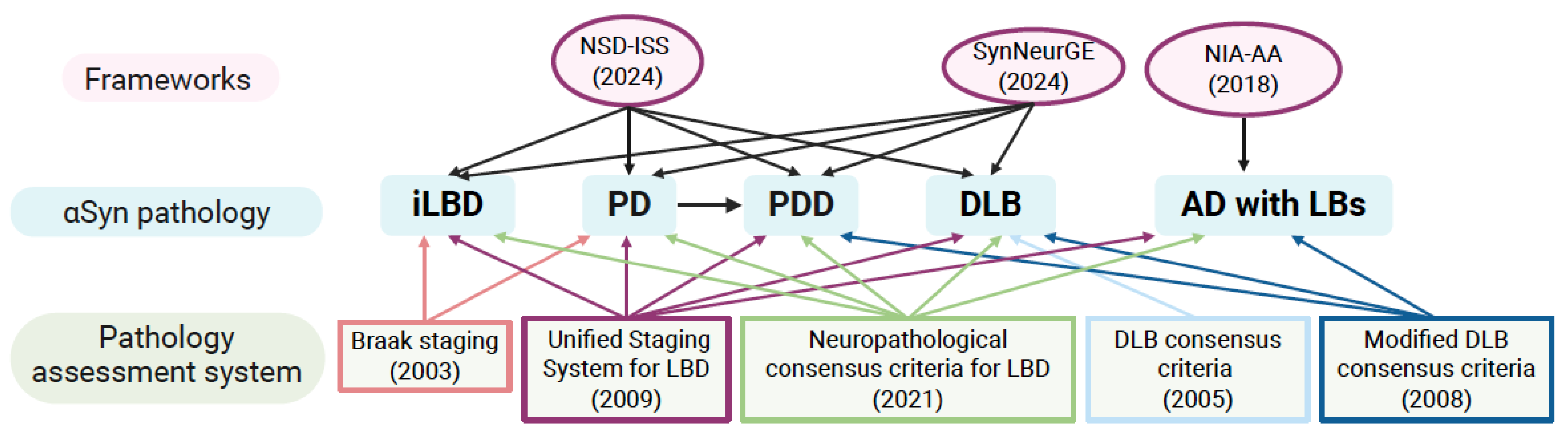
Figure 2.
Integrated features of α-synuclein pathology across different disorders. The schematic summarizes how multidimensional α-synuclein (αSyn) pathology differs in synucleinopathies and AD with Lewy pathology. Lewy body disorders include incidental Lewy body disease (iLBD), Parkinson’s disease (PD), Parkinson’s disease dementia (PDD), and dementia with Lewy bodies (DLB), containing characteristic neuronal Lewy bodies (LBs) and neurites (LNs), whereas multiple system atrophy (MSA) is defined by αSyn within oligodendrocytes, forming glial cytoplasmic (GCIs), glial nuclear inclusions (GNIs), as well as neuronal cytoplasmic (NCIs) and neuronal nuclear inclusions (NNIs). Alzheimer’s disease (AD) with LBs represents a mixed-protein disorder dominated by amyloid-β (Aβ) plaques and tau pathology, neurofibrillary tangles (NFTs). Created with BioRender.com.
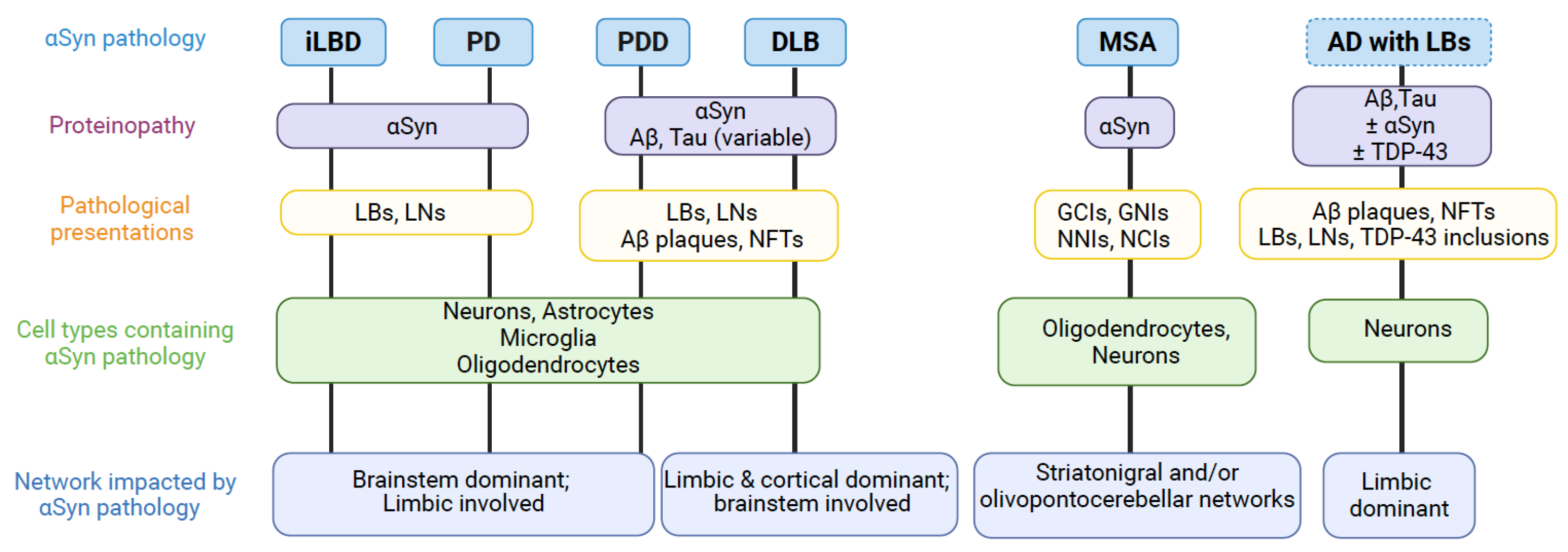
Disclaimer/Publisher’s Note: The statements, opinions and data contained in all publications are solely those of the individual author(s) and contributor(s) and not of MDPI and/or the editor(s). MDPI and/or the editor(s) disclaim responsibility for any injury to people or property resulting from any ideas, methods, instructions or products referred to in the content. |
© 2026 by the authors. Licensee MDPI, Basel, Switzerland. This article is an open access article distributed under the terms and conditions of the Creative Commons Attribution (CC BY) license (http://creativecommons.org/licenses/by/4.0/).
Copyright: This open access article is published under a Creative Commons CC BY 4.0 license, which permit the free download, distribution, and reuse, provided that the author and preprint are cited in any reuse.


