1. Introduction
Photosensitization reactions may be induced by numerous synthetic compounds including antibiotics, antihistamines, anti-inflammatory agents, antimalarials, cardiovascular drugs, central nervous system-acting pharmaceuticals, medications used in the management of multisystemic disorders, as well as by naturally occurring plant-derived bioactive substances. For both synthetic and natural agents, a close relationship exists between chemical structure and photosensitizing potential. These compounds typically contain aromatic chromophore groups or photolabile functional bonds capable of absorbing light energy, generating free radicals, and initiating light-induced cellular damage.
Naturally occurring photosensitizing compounds found within certain plant families exhibit considerable diversity in chemical structure and biosynthetic origin. Their formation is linked to distinct metabolic pathways, and they may also be classified according to their modes of biological activity. Photosensitization may be triggered by exposure to plants in natural vegetation (e.g. grasslands), cultivated agricultural crops, and species utilized in herbal medicine. Consequently, their effects are relevant to human nutrition, animal feeding systems, and complementary or alternative medical applications.
Plant compounds responsible for photosensitizing effects in both animals and humans are secondary metabolites that play functional roles in plant physiology and ecology. Their concentration and distribution within plant tissues show substantial variability and are not governed by the regulatory dynamics of primary metabolic processes essential for basic survival. Instead, their quantitative expression is strongly influenced by biotic and abiotic environmental factors, as well as by diverse stress conditions.
Environmental variables that fundamentally affect plant growth conditions, cultivability, and productivity also modify the concentration and distribution of nutritional constituents and secondary metabolites within a given plant species. In natural plant communities, such changes may additionally influence species distribution patterns within specific habitats. Under current climate change projections, for example, periodic water scarcity is expected to alter vegetation structure and species composition. Such shifts may increase the incidence of photosensitization-associated animal health disorders, both through conserved forage and during grazing.
Numerous secondary plant metabolites can cause dermal irritation without inducing true photosensitization (e.g. phorbol esters). The present review therefore focuses specifically on the mechanistic background of photosensitization, together with documented cases, causative agents, and plant taxa implicated in these reactions.
2. Role of Secondary Plant Metabolites
Whereas primary plant metabolites are universally essential for the maintenance of fundamental life processes and for the regulation of growth and development in all plant species [
1,
2,
3,
4]), secondary plant products (SPP) or secondary metabolites play predominant roles in chemical signaling and in defense against grazing herbivores, insects, pathogens, and competing plant taxa [
2,
5,
6]. Beyond their ecological functions, many secondary metabolites exert beneficial biological effects in both human and veterinary complementary medicine, including their utilization as medicinal plant products, nutritionally valuable feed components, or adjunct therapeutic agents.
The occurrence, concentration, and biological expression of secondary metabolites vary considerably among plant species and tissues. Owing to their extensive chemical diversity, these compounds may exert beneficial physiological effects. However, depending on their concentration and environmental context, they may also pose significant health risks.
SPPs synthesized by plants may act in grazing species as deterrents, irritants, or toxins. Nevertheless, behavioral avoidance mechanisms and physiological detoxification pathways in animals often mitigate the risk of irritation and overt poisoning [
7]. A smaller subset of SPPs can induce photosensitization and associated toxicity in both humans and grazing herbivores, either through direct contact with plant tissues or following ingestion [
8].
3. Photosensitization Induced by Secondary Metabolites
In animals, photosensitization manifests clinically as severe dermatitis resulting from heightened reactivity of skin cells and tissues following exposure to sunlight. The condition develops when animals ingest or encounter plants containing photodynamic pigments (e.g. chlorophyll derivatives) or specific secondary metabolites such as naphthodianthrones and furocoumarins that are activated by ultraviolet (UV) or visible radiation [
8,
9]. Comparable reactions may also be triggered by photoreactive microbial or fungal metabolites [
10,
11].
Clinical lesions are typically most severe in non-pigmented, sparsely haired, or hairless skin regions [
12]. Although the gross appearance may resemble solar dermatitis (sunburn), photosensitization represents a distinct pathophysiological entity. Its development is based on rapid photochemical reactions within skin cells triggered by both visible (400–700 nm) and ultraviolet (180–400 nm) radiation, representing a broader excitation spectrum than that associated with classical sunburn.
Both spectral ranges can activate plant-derived phototoxins, initiating photosensitization. In contrast, sunburn lesions primarily result from excessive UV exposure, particularly within the UV-A range (400–315 nm), which penetrates deeper dermal layers and contributes to cumulative photodamage and premature skin ageing [
13].
During photosensitization, photo-excited oxidation initiates a cascade of reactions within the skin cells of affected animals, leading to direct injury of proliferative cell layers [
8,
12]. Photodynamic oxidation generates reactive oxygen species (ROS), including free radicals and singlet oxygen, through light-driven processes. In plant tissues, these reactive intermediates may induce DNA degradation [
11,
13].
In herbivorous animals, the pathological manifestation of these cellular events is photosensitization, reflecting the accumulation of plant-derived photodynamic molecules within target tissues and the circulatory system. Certain photoreactive compounds may also form DNA adducts through direct molecular interactions, thereby inducing photosensitization via oxygen-independent photochemical pathways [
11].
Photosensitizing molecules are typically acquired during grazing and subsequently accumulate within the animal. Following systemic distribution or direct dermal contact, these compounds are localized in the skin, where they become activated upon exposure to sunlight. DNA damage arises through excitation induced by ultraviolet and/or visible radiation [
13,
14]. In addition to direct mechanisms, indirect photosensitization may develop secondary to hepatic dysfunction in grazing animals [
11].
4. Mechanism of Action
Plants, as photoautotrophic organisms, derive energy through the absorption of light by specialized light-harvesting pigment systems. Within the thylakoid membranes of plant chloroplasts, the photosynthetic electron transport chain generates ATP and NADPH via sequential electron transfer reactions. These energy carriers are subsequently utilized in the Calvin cycle for carbon dioxide fixation and reduction [
14].
During light absorption, only a proportion of pigment molecules enter an excited state capable of electron transfer. Among the most important excitable molecules are the photosynthetic pigments, particularly chlorophylls, which efficiently transfer photo-excited electrons to downstream acceptor molecules during the light-dependent phase of photosynthesis [
11].
Environmental and physiological perturbations may, however, enhance photosynthetic electron flux and activity [
15]. High light intensity represents a key example, leading to elevated production of reactive intermediates within plant tissues. Such conditions may simultaneously increase the photosensitizing potential of plants for grazing herbivores [
12].
In plants, photosensitisation is primarily associated with chlorophylls and their derivatives, although analogous photochemical reactions may occur extra-plant via other reactive molecules [
16].
The excited molecule (e.g. chlorophyll or its derivatives) is termed the photochemical activator (photosensitizer), whereas the molecule undergoing modification is referred to as the substrate or acceptor. All photochemical activators contain chromophore groups capable of absorbing light at specific wavelengths, thereby inducing intramolecular photochemical changes. These alterations subsequently affect associated biological substrates. When the photo-excited activator induces reactions involving molecular oxygen, the process is defined as photodynamic. Chlorophyll molecules located within intact chloroplast structures are normally protected from such photodynamic reactions [
14,
17]. In contrast, certain metabolites may become fully functional activators following metabolic transformation in acceptor tissues. Activated plant metabolites induce photosensitization via two principal photochemical pathways, classified mechanistically as Type I and Type II reactions [
16].
In Type I reactions, the excited activator interacts with the acceptor through electron transfer, generating radical ions in both molecules. Typically, the substrate donates an electron to the activator, producing a substrate cation radical (substrate⁺) and an activator anion radical (activator⁻), although the reverse may occur depending on the redox potentials of the interacting pair. In the presence of oxygen, both radical species may form oxygenated products, often resulting in oxidative consumption of the activator molecule. Alternatively, the extra electron of the activator anion radical may be transferred directly to molecular oxygen, generating superoxide radicals (O₂⁻•) while regenerating the ground-state activator.
In Type II reactions, the excited activator transfers excess energy directly to ground-state molecular oxygen, converting it into highly reactive singlet oxygen while the activator itself returns to the ground state. Singlet oxygen subsequently reacts with biological substrates, producing oxidized metabolites. Unlike Type I reactions, the activator is not consumed but cycles between excited and ground states.
In vascular plants, singlet oxygen formation is intrinsically linked to energy transfer processes of photo-excited molecules [
17]. Because chlorophylls function as natural photochemical activators, singlet oxygen generation is an ubiquitous phenomenon in plant tissues [
18,
19].
Beyond photosynthetic pigments, numerous secondary metabolites and phytotoxins produced by plant pathogens and microbes possess chromophoric structures capable of photochemical excitation. Photosensitizing secondary metabolites arise from diverse chemical classes, including: quinones, furocoumarins, polyacetylenes, thiophenes, benzofurans and chromenes. A shared functional property of these compounds is their ability to generate ROS upon photoactivation, particularly via Type II pathways leading to singlet oxygen production [
17]. This capability is largely attributable to structural features such as polycyclic aromatic ring systems and conjugated double bonds. These chromophoric configurations enable efficient light absorption and intramolecular electron rearrangements, resulting in ROS formation, including singlet oxygen, superoxide radicals, hydrogen peroxide, hydroxyl radicals [
20,
21].
The structural diversity, metabolic origins, and broad biogenetic distribution of plant photoreactive compounds suggest that these metabolites evolved multiple times independently during plant adaptive evolution. Their formation is closely linked to ecological defense strategies. Similarly, the evolution of ROS-mediated phototoxic mechanisms represents an important plant defense system against herbivores, pathogens, and pests [
6,
22].
In addition to phototoxic compounds, plants also synthesize widely distributed metabolites that protect cellular structures from photo-oxidative damage. Notable examples include flavonoids possessing antioxidant activity, which stabilize membranes and cellular components against photodynamic injury [
4]. A well-characterized compound is quercetin, a flavonoid derivative that functions as an efficient singlet oxygen quencher. Carotenoids provide further protection by directly quenching excited chlorophyll molecules and dissipating excess energy as heat, thereby limiting singlet oxygen formation [
23]. Additional quenching agents present in plant membranes including tocopherols, unsaturated fatty acids, specific proteins, water-soluble quenchers comprise, vitamin B6, ascorbate, various flavonoids [
4,
23]. Due to their photoprotective and antioxidant properties, plants rich in these compounds are widely utilized in the pharmaceutical and cosmetic industries. Their consumption or topical application is promoted for the protection of sensitive skin cells, mitigation of premature ageing, and reduction of harmful effects associated with ultraviolet radiation exposure.
5. Occurrence of Photosensitizing Compounds in Plants
The formation of ROS in response to environmental stressors is a universal phenomenon across living organisms, including plants. During plant development, elevated ROS production has been documented under conditions of high light intensity, temperature extremes, and exposure to environmental pollutants [
8,
18,
19]. Despite this, the photo-oxidative hazard posed by chlorophylls and their biosynthetic intermediates within intact plant tissues is considered negligible for grazing animals under normal circumstances [
8]. In contrast, the biosynthesis of light-absorbing secondary plant metabolites is often maximized under increased UV-A and UV-B radiation, conditions that may promote the development of photosensitization following ingestion or contact [
24].
In grazing livestock, the consumption of fresh green plant material represents the primary risk factor for the development of photosensitization when compared with hay or concentrate feeds [
25]. This increased risk is largely attributable to the high concentrations of chlorophyll and protochlorophyllide molecules present in actively growing tissues, both of which may contribute to photodynamic reactions. Additionally, fresh plant material frequently contains higher levels of heterocyclic secondary metabolites capable of inducing photosensitization. In certain cases, combined exposure to plant pigments and secondary metabolites has resulted in enhanced phototoxic responses in grazing animals, suggesting potential synergistic interactions following co-ingestion [
12]. Conversely, mechanical processing and preservation methods including drying, chopping, haymaking, or ensiling generally reduce phototoxicity and the likelihood of photosensitization after consumption. These observations are largely based on field and husbandry experience, as comparatively few studies have quantitatively analyzed differences in photosensitizing secondary metabolite content between fresh and processed forages. One earlier investigation compared phototoxic compound concentrations in processed versus fresh feed materials [
26]. The study demonstrated that sun-drying resulted in an approximately 80% reduction in hypericin content in
Hypericum perforatum (St John’s wort), highlighting the substantial impact of post-harvest processing on phototoxic risk.
Comparative analyses of the chemical composition of photosensitizing agents across different plant organs may provide valuable insight into the pathogenesis of photosensitization in grazing animals. However, the precise influence of individual biotic and abiotic environmental factors on the biosynthesis and accumulation of plant photosensitizers remains insufficiently characterized. Elucidating these relationships represents an important area for future research, particularly with respect to biosynthetic pathways of photodynamic compounds, environmental regulation of secondary metabolite production and tissue-specific accumulation patterns.
Understanding how global warming and climate change influence plant secondary metabolism – and specifically the production of photosensitizing compounds – is of growing veterinary and agricultural importance. Rising temperatures, intensified UV radiation, and drought stress are all projected to increase the concentration of secondary metabolites within green plant tissues [
4,
27]. Consequently, the incidence and severity of photosensitization, as well as broader plant-associated toxicities, may increase in grazing livestock populations [
28].
6. Development of Photosensitization in Animals
In susceptible animal skin, secondary plant metabolites are primarily responsible for the initiation of photosensitization reactions [
11,
14]. The integument of animals consists of three principal layers: the epidermis, dermis, and hypodermis (subcutis). The epidermis forms the outermost barrier, functioning as the first line of defense between the body and the external environment. It is composed of multiple cellular strata, some of which retain proliferative capacity. The outermost layer, the stratum corneum (SC), consists of keratinized, non-viable cells that are continuously desquamated and replenished by mitotically active cells originating from the stratum basale (SB). Continuous division within the SB ensures structural integrity of the SC layer [
29].
Damage to the epidermis, particularly the SC compromises the protective barrier against environmental insults, including ultraviolet radiation and xenobiotic penetration. Consequently, deeper tissues within the dermis and hypodermis become more susceptible to both photonic injury and chemical insult. Maintenance of epidermal integrity is therefore critical for cutaneous photoprotection. Cutaneous pigmentation also plays a protective role. Melanin present in darker skin absorbs and dissipates ultraviolet radiation, thereby reducing the risk of photosensitization [
12].
When photosensitizing plant compounds are present, significant pathological changes may develop within susceptible skin tissues, including irritation, dermal necrosis, secondary bacterial infection, severe trauma potentially culminating in mortality [
30]. In addition, endogenous amino acids within the skin, notably tryptophan, tyrosine, and histidine are particularly susceptible to photo-oxidation. Their oxidation may trigger pronounced inflammatory responses within tissues and vasculature, contributing to necrosis of affected regions [
8,
12].
Natural plant photosensitizers may reach the skin via two principal routes: direct plant–animal contact or systemic distribution via the bloodstream following ingestion [
11,
14]. These compounds exert cytotoxic (photocytotoxic) effects through direct cellular injury, or less commonly via immune-mediated (photoallergic) mechanisms.
The temporal onset of photocytotoxic photosensitization is highly variable. Clinical signs may develop a) within minutes following direct dermal exposure, b) several hours after systemic accumulation of primary photosensitizers or c) days later following activation of secondary photosensitizers (e.g. after hepatic injury and accumulation of phytoporphyrins in the skin). Photoallergic photosensitization may likewise manifest days after exposure via either contact or systemic pathways, although its precise pathogenesis in grazing livestock remains insufficiently characterized.
Following contact exposure, phototoxic compounds penetrate skin layers via two principal mechanisms. Polar compounds penetrate through active transport processes across the epidermis, less polar (lipophilic) compounds diffuse passively through tissue layers [
30]. Herbivore skin possesses several structural features that limit toxin penetration into sensitive subcutaneous tissues. The epidermis exhibits high resistance to passive transport of bioactive molecules. The keratinized SC layer forms a substantial barrier to lipophilic compounds, which therefore tend to accumulate within this layer. This accumulation may further impede diffusion of polar molecules into subepidermal tissues. Additionally, cutaneous lipids constitute an important physicochemical barrier. Removal or degradation of epidermal lipids increases dermal permeability to exogenous compounds [
31].
Phototoxic compounds may also reach cutaneous tissues via systemic circulation following gastrointestinal absorption. This represents the primary exposure route in livestock. In ingestion-associated cases, tissue injury develops either through direct deposition of bioactive molecules in the skin or accumulation of secondary metabolites following hepatic dysfunction [
5].
Sensitivity of grazing animals to dermally absorbed toxins and photosensitizers is influenced by multiple host and environmental factors, including species and breed, skin pigmentation, hair or wool density and thickness, age, general health status, ambient temperature, humidity, and precipitation [
8]. Hair coats and skin pigments provide photoprotection by limiting ultraviolet penetration into epidermal layers. Consequently, lesions most commonly develop in sparsely haired or non-pigmented regions, including periocular areas, ears, facial regions and muzzle, mammary glands, perineal and tail regions, coronary bands adjacent to the hoof wall [
32]. Light-colored animals and those with thin hair or fleece are more susceptible than heavily pigmented or densely coated counterparts [
33,
34]. Species-specific histological differences in skin architecture may also influence susceptibility [
30]. Young, debilitated, poorly pigmented, or hairless animals are generally more prone to cutaneous photosensitization than older, healthy, pigmented individuals.
Photosensitization in grazing animals is classified as either primary or secondary (hepatogenous) [
12]. In primary photosensitization, phototoxic plant compounds – or their metabolites – become biologically active following ingestion or direct contact. After gastrointestinal absorption, they circulate systemically and accumulate within cutaneous tissues [
8]. Primary photosensitization may also arise from abnormal porphyrin metabolism [
29]. Its typically acute and rapid clinical course suggests direct intestinal absorption and swift dermal accumulation of photodynamic compounds, with limited time for secondary biochemical or cholestatic changes to develop [
12]. Examples of primary photosensitizing agents include polycyclic compounds such as furocoumarin derivatives characteristic of the Apiaceae family and hypericin accumulating after ingestion of
Hypericum spp. (Hypericaceae).
Secondary photosensitization results from systemic accumulation of the photodynamic phytoporphyrin called phylloerythrin. Phylloerythrin is a normal metabolic by-product of chlorophyll degradation during digestion of green plant material in grazing animals. Following formation as an intermediate of chlorophyll catabolism, phylloerythrin is absorbed from the gastrointestinal tract (or rumen), enters systemic circulation, and is normally removed by the liver and excreted via bile.
When hepatic function is compromised – for example due to hepatotoxic secondary plant metabolites – phylloerythrin clearance is impaired. Elevated circulating concentrations then accumulate within the skin, where photoactivation induces tissue injury. Although the precise phototoxic mechanism of phytoporphyrins remains incompletely defined, experimental evidence indicates localization within the Golgi apparatus and mitochondria, suggesting these organelles as primary cellular targets [
35,
36].
Secondary photosensitizers induce hepatic dysfunction through mechanisms including cholestasis (inhibition of bile flow into the duodenum) or direct hepatocellular injury. In such cases, porphyrins and their derivatives fail to undergo biliary excretion, leading to systemic retention and abnormal porphyrin metabolism [
37]. Hepatogenous photosensitization therefore develops when ingestion of secondary plant metabolites results in liver disease or functional hepatic impairment [
8].
Primary photosensitization occurs less frequently in grazing livestock than hepatogenous forms [
12]. In addition to the plant taxa detailed in this review, numerous other species can induce both primary and secondary photosensitization syndromes [
38,
39,
40] However, only selected representative examples are discussed in detail in the present paper.
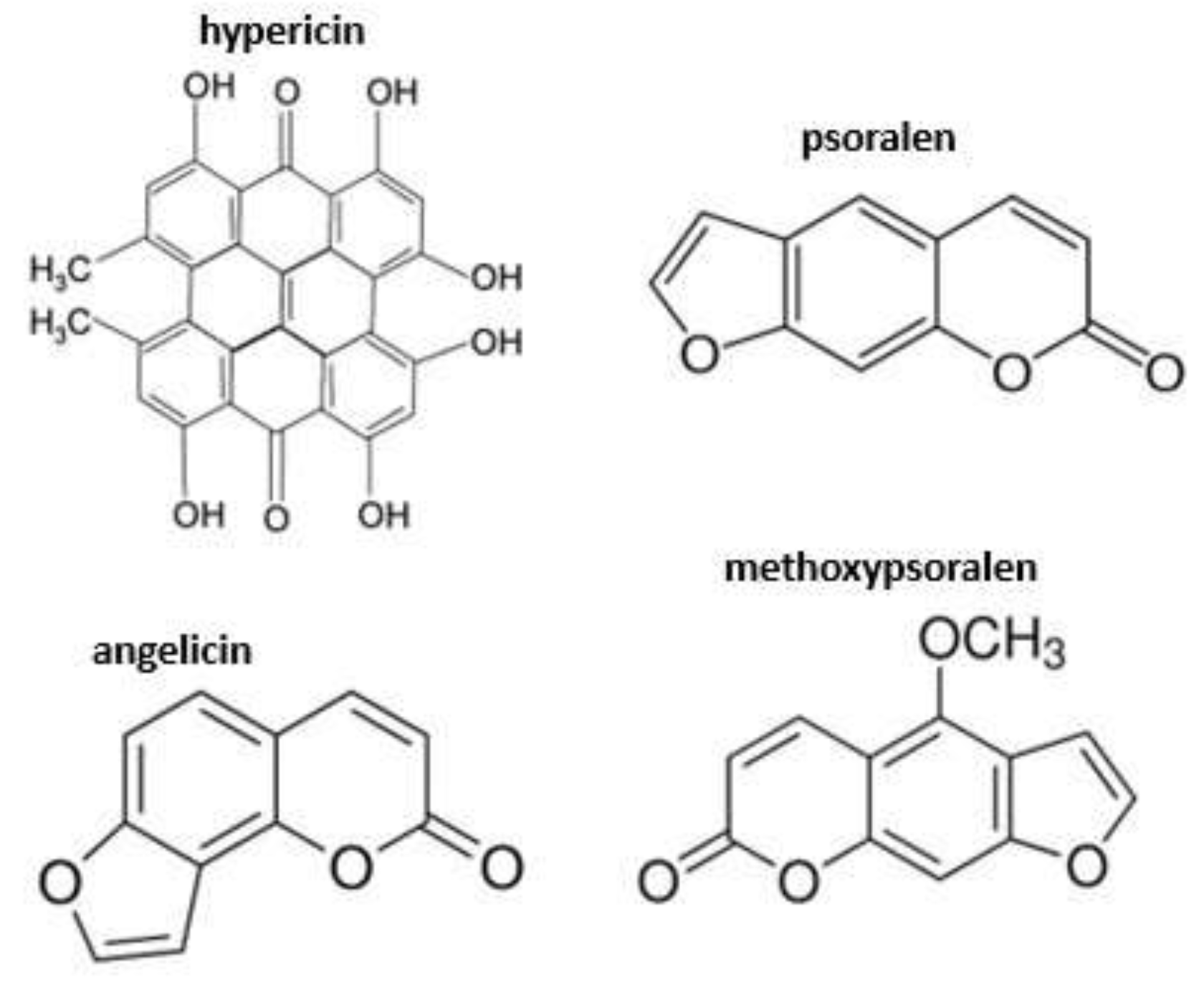
7. Plant Species and Compounds Causing Primary Photosensitization
Relatively few SPPs are classified as primary photosensitizing agents. This is partly attributable to the complex pathogenesis of photosensitization and to the methodological challenges associated with confirming compound-specific mechanisms of action and bioactive metabolite effects within animal tissues. Only a limited number of phytochemicals have been demonstrated to induce both primary and secondary (hepatogenous) photosensitization in grazing herbivores, whereas others are recognized exclusively as causative agents of secondary forms.
7.1. Hogweeds (Heracleum spp.)
Species of the genus Heracleum (family Apiaceae) are large herbaceous plants widely distributed across Europe. The three most prevalent species include Heracleum sphondylium, Heracleum mantegazzianum and Heracleum sosnowskyi.
Common hogweed (H. sphondylium) is native to Europe and typically occurs in moist meadows, forest margins, woodland clearings, and roadside habitats, often associated with nitrogen-rich soils. Its toxicity is lower than that of the larger invasive congeners, however, dermatitis may still occur. When incorporated into forage or encountered through direct contact, it may induce clinical disease, particularly in cattle.
The latter two species may reach heights of 3–5 meters and are classified as invasive in many regions. Their distribution is sporadic but locally dense, and eradication programs are implemented in several European countries due to ecological and public health concerns. Giant hogweed (H. mantegazzianum) is regarded as one of the most significant invasive plant health hazards in Europe.
Contact with
H. mantegazzianum followed by sunlight exposure induces severe cutaneous injury, clinically diagnosed as phototoxic dermatitis [
41,
42,
43]. The plant is characterized by large, serrated leaves, hollow stems covered with fine bristles and dark purple blotches, and prominent compound umbels up to 80 cm in diameter composed of small white flowers [
44]. Both living plants and cut plant material may trigger lesions. As a perennial species, giant hogweed poses substantial risks to both human and animal health via direct contact with photosensitizing sap, producing burns resembling thermal injury on sun-exposed skin [
41,
45]. The sap of
H. mantegazzianum contains photosensitizing furocoumarin derivatives [
44,
46]. These metabolites, particularly angelicin, occur at highest concentrations in leaves but are also present in stems and inflorescences [
47].
Ecologically, furocoumarins confer adaptive advantages through insecticidal activity and antimicrobial effects, thereby protecting plants from herbivory and pathogen attack [
48,
49,
50]. In mammalian cells, furocoumarins exert mutagenic and carcinogenic effects via direct DNA intercalation. Following integration, photoactivation induces covalent cross-linking of these tricyclic molecules. Subsequent reactions with pyrimidine bases on complementary DNA strands increase intra-strand cross-link formation [
51,
52,
53]. These cross-links induce apoptosis and inhibit cellular replication. Key furocoumarins identified in
H. mantegazzianum include angelicin, psoralen, methoxypsoralen, [
44] (
Figure 1). Severe dermal injury following sap contact is therefore attributable to profound disruption of cellular integrity within exposed skin tissues.
7.2. St John’s Wort (Hypericum perforatum)
Hypericum perforatum is a perennial species widely distributed throughout temperate regions. Numerous reports worldwide have documented primary photosensitization in livestock following grazing or ingestion of this plant [
33,
54,
55]. The principal phototoxic compound is hypericin (
Figure 1), a photocytotoxic secondary metabolite. Hypericin accumulates in specialized secretory glands within leaf tissues [
23,
26,
56,
57]. Threshold doses for photosensitization vary by species. In sheep, clinical disease has been reported at approximately 3 mg/kg dry matter intake [
33], whereas cattle exhibit comparatively higher tolerance [
58]. Clinical manifestations correspond to classical UV-associated photosensitization, including erythema of the muzzle, periocular tissues, and ears, oedema of exposed skin and elevated rectal temperature [
54].
Hypericin is a phenanthroperylene quinone capable of forming strong complexes with diverse protein targets. Owing to its photodynamic properties, it remains under investigation for therapeutic applications, including photodynamic therapy [
23,
59]. Upon photoactivation, hypericin generates multiple reactive oxygen species, including singlet oxygen (¹O₂), superoxide anions and hydrogen peroxide (H₂O₂) [
60,
61,
62,
63]. Each of these ROS can induce cell death in UV-A–exposed tissues.
Hypericin biosynthesis is enhanced under UV-B and full-spectrum UV exposure in both greenhouse-grown and field-grown plants. Longer-wavelength radiation may likewise promote reactive molecule formation in plants and grazing livestock. Because these wavelengths penetrate tissues more effectively, they contribute to increased photosensitivity and dermatitis severity [
64].
Hypericin-induced ROS causes cellular injury and loss within the skin via apoptosis, necrosis, and possibly autophagy pathways. Tissue damage depends on the spatial co-localization of ROS generation and cellular target molecules, as ROS diffusion is limited due to their high reactivity and short half-life [
65,
66,
67,
68,
69]. Hypericin binds preferentially to membranes of key intracellular organelles, including mitochondria, endoplasmic reticulum, Golgi apparatus and lysosomes [
63,
67,
70,
71,
72,
73,
74]. These structures are therefore considered primary subcellular targets of photodynamic injury. When photoactive compound accumulation coincides with light exposure, ROS-mediated degradation of proliferative cells within the SB ensues, producing clinically significant photosensitization in affected livestock. Removal of animals from pastures containing Hypericum spp. is typically sufficient to reduce systemic hypericin levels and mitigate further phototoxic reactions [
33,
44].
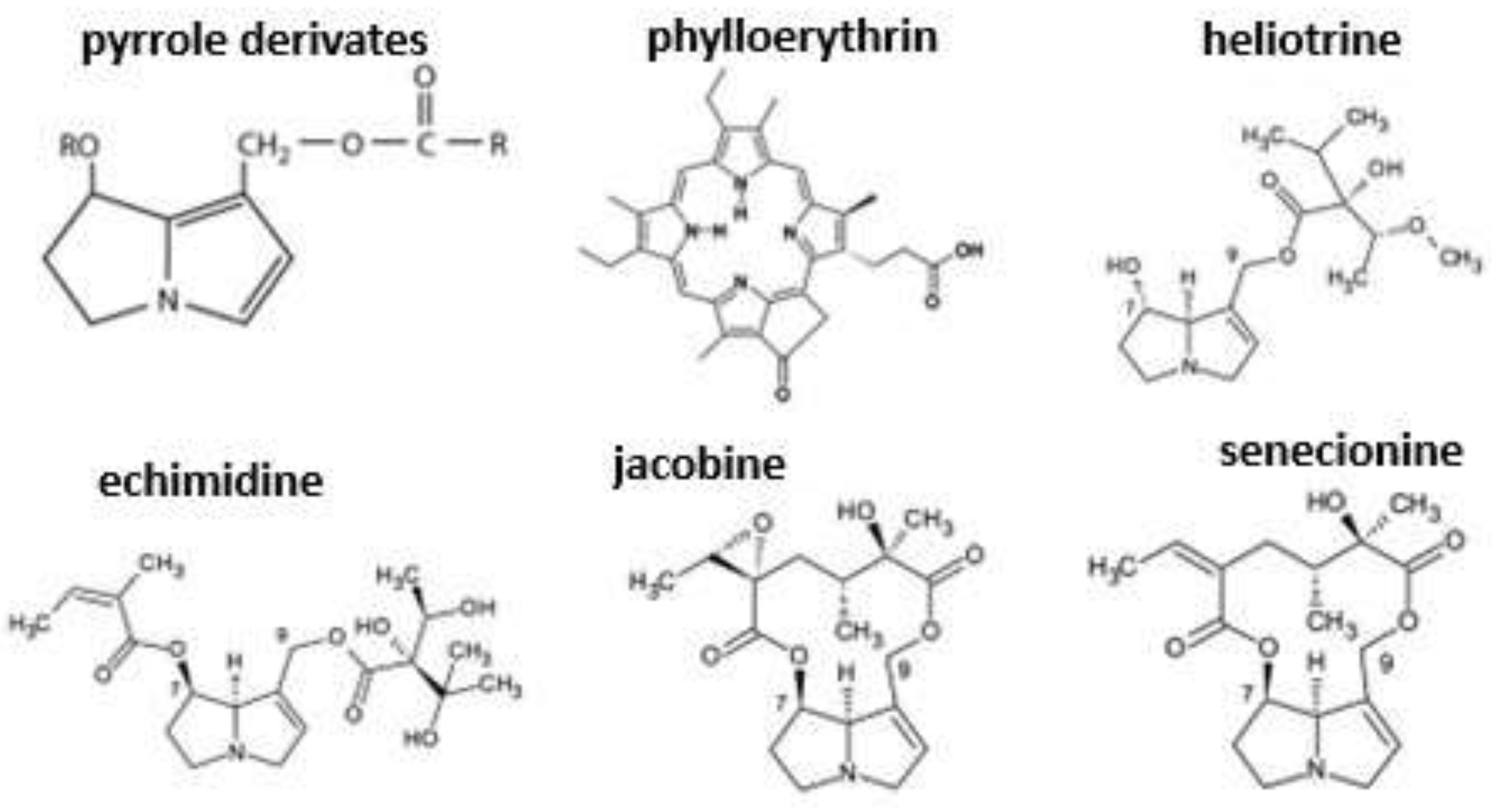
8. Plant Species Causing Secondary (Hepatogenous) Photosensitization and Their Compounds
Pyrrolizidine alkaloids (PAs), as secondary plant metabolites, induce significant hepatic injury in animals consuming plants containing high concentrations of these compounds [
8]. These metabolites may lead to hepatogenous photosensitization, which develops secondary to toxic liver damage. Unlike primary photosensitizers such as hypericin, pyrrolizidine alkaloids do not exert direct effects on biliary excretory processes. Nevertheless, the severity of hepatic injury they induce may indirectly impair these pathways, ultimately resulting in photosensitization.
PAs occur across numerous plant genera; approximately 3% of all plant species are known to contain at least one PA compound, with nearly 350 structural forms identified to date [
64,
75,
76]. Most plant species responsible for PA-associated hepatotoxicity in livestock belong predominantly to the families Boraginaceae and Asteraceae. The hepatotoxic and genotoxic potency of individual pyrrolizidine alkaloids varies considerably; however, many can induce severe acute and chronic hepatic insufficiency and dysfunction, frequently culminating in death.
PAs share a broadly similar mechanism of action in affected grazing animals. Following ingestion, they are metabolized within hepatocytes, particularly in the centrilobular (zone III) region of the liver. This region exhibits elevated enzymatic activity and lies furthest from vascular supply, rendering it especially vulnerable to toxic injury. Centrilobular hepatocytes possess high concentrations of enzymes catalyzing oxidative, reductive, and hydrolytic reactions. These enzymatic systems participate both in toxin biotransformation and in the generation of biologically active secondary metabolites [
77].
Structurally, PAs contain a pyrrolizidine nucleus that undergoes hepatic dehydrogenation, forming reactive pyrrolic metabolites, the principal cytotoxic derivatives of PAs [
78]. The cytotoxicity of pyrroles arises from covalent binding to DNA and cross-link formation with proteins and amino acids. These interactions produce both direct cytotoxic effects and potent inhibition of cellular mitosis [
75]. Due to their high reactivity, pyrrolic metabolites typically exert their toxic effects locally at the site of formation within hepatocytes.
Characteristic tissue-level lesions associated with PA hepatotoxicity include hepatocellular hypertrophy, hepatocellular necrosis, nuclear enlargement (megalocytosis) and progressive fibrosis [
8,
79]. DNA cross-linking disrupts normal mitotic processes, producing profound cellular dysplasia. Hepatocytes in PA-affected livers may become up to 30 times larger than normal, reflecting substantial DNA accumulation and failed cell division [
75]. Severe hepatic dysfunction may culminate in death. Both acute and chronic liver injury can also precipitate photosensitization due to impaired hepatic metabolism and biliary excretion of phototoxic compounds, leading to their systemic accumulation.
8.1. Ragworts (Senecio spp.)
Species of the genus
Senecio (family Asteraceae) are widely distributed in natural ecosystems and represent a major toxicological concern for livestock producers, owing to their pronounced toxicity in ruminants and horses [
8,
32]. Pyrrolizidine alkaloids present in
Senecio species are relatively resistant to microbial degradation within the gastrointestinal tract, permitting absorption into systemic circulation at higher concentrations. One determinant of toxicity variability lies in the marked structural diversity of alkaloid forms present among plant species.
A principal bioactive PA in
Senecio spp. is jacobine, which contains a macrocyclic (closed chain) diester structure. This contrasts with the open chain diester alkaloids, such as echimidine and heliotrine found in genera including
Echium and
Heliotropium. Another common PA in
Senecio species is senecionine, synthesized in the roots and translocated to aerial tissues during plant development (
Figure 2). Macrocyclic (closed chain) diester pyrrolizidine alkaloids exhibit substantially greater resistance to bacterial enzymatic degradation within the intestine. Consequently, they undergo enhanced metabolic activation to toxic pyrrolic intermediates within hepatocytes [
76].
The cytotoxic hepatic effects of PAs impair the normal degradation and biliary excretion of phylloerythrin, a chlorophyll-derived photodynamic pigment. Its systemic accumulation leads to cutaneous photosensitization in affected animals. Importantly, pyrrolizidine alkaloids do not exert direct phototoxic effects within the skin of grazing herbivores, in contrast to primary photosensitizers such as hypericin. Instead, dermal photosensitivity develops secondarily to hepatic dysfunction and subsequent retention of circulating photodynamic metabolites [
32].
9. Case Studies
In 2014, intoxication was reported in a flock of 27 Awassi sheep grazing on pasture heavily infested with
Hypericum perforatum. Twelve animals exhibited only elevated body temperature, whereas 15 showed edematous swelling of the head, eyelids and ears, accompanied by dermatitis typical of photosensitization on unpigmented and non-wool-covered areas, primarily the limbs and udder. Treatment consisted of corticosteroids, antihistamines and antibiotics, resulting in full clinical recovery [
80].
Although grazing animals generally avoid
Hypericum spp. under free-range conditions, outbreaks may occur under confined management. In a flock of 700 penned sheep, approximately 200 animals developed clinical signs within three days, as restricted feeding conditions enabled animals to graze the plants down to the stem base [
81].
A recent survey further demonstrated that Hypericum-associated photosensitization occurs more frequently in sheep than in cattle. In a population-level assessment of 8,200 animals, mortality among clinically affected individuals reached 14%. Clinical signs were most prevalent between May and September and included pruritus and cutaneous lesions. In addition to systemic therapy, topical zinc oxide formulations were applied to support wound healing [
82].
On a mixed sheep and cattle farm, animals grazed freely on pasture heavily contaminated with Senecio species. In sheep, initial clinical signs included weight loss, apathy and photosensitization lesions affecting the ears and nasal region. With progression of disease severity, euthanasia was required. Post-mortem examination revealed irregular hepatic architecture; in some cases, the liver appeared pale, firm and fibrotic, accompanied by gallbladder distension. Histopathology demonstrated marked degenerative changes, including hypertrophic Kupffer cells and hepatocellular enlargement (megalocytosis). Spongiform degeneration was also observed in cerebral tissues. Comparable clinical and pathological findings were described in affected cattle from the same farm [
83].
In cattle, chronic diarrhea, poor body condition and oedema of the dewlap are also frequently reported. Hepatic lesions typically include reduced liver size, grey discoloration and firm consistency, with fibrosis and bile duct hyperplasia evident on histological examination. Neuropathological lesions have been detected at the junction of grey and white matter [
84].
Senecio toxicity is not restricted to ruminants. In horses, clinical manifestations are similar; however, disease progression following ingestion of pyrrolizidine alkaloid-containing plants is rarely reversible, and euthanasia is often required [
85].
10. Conclusions
Secondary plant metabolites capable of inducing photosensitization may manifest as seasonal clinical syndromes in grazing livestock. The environmental and genotype-specific determinants governing the biosynthesis of light-absorbing bioactive molecules in plants remain incompletely understood. Nevertheless, such compounds occur in both cultivated forage species and invasive weeds, posing an ongoing risk to pasture-based production systems.
Within the animal organism, photosensitization represents a cascade of photodynamic reactions driven by light-absorbing molecules, typically heterocyclic or polyphenolic in nature. Primary photosensitization arises following ingestion or dermal exposure to photodynamic compounds, which increase cutaneous susceptibility to solar radiation. Clinical severity is heightened in animals with poorly pigmented or non-wool-covered skin, where natural photoprotection is limited.
Secondary (hepatogenous) photosensitization most commonly results from the accumulation of phylloerythrin due to impaired hepatic metabolism. Hepatotoxic plant compounds may induce severe liver damage, disrupting the normal excretion of chlorophyll degradation products from the bloodstream, thereby precipitating photosensitization in affected animals.
Author Contributions
Conceptualization, E.P.R. and D. Cs.; methodology, E.P.R.; investigation, E.P.R. and D. Cs.; writing—original draft preparation, E.P.R. and D. Cs.; writing—review and editing, E.P.R. and D. Cs. All authors have read and agreed to the published version of the manuscript.
Funding
This research received no external funding
Institutional Review Board Statement
Not applicable
Informed Consent Statement
Not applicable
Data Availability Statement
No new data were created or analyzed in this study. Data sharing is not applicable to this article.
Acknowledgments
During the preparation of this work the author(s) used ChatGPT to improve language and readability. After using this tool/service, the authors reviewed and edited the content as needed and take(s) full responsibility for the content of the publication.
Conflicts of Interest
The authors declare no conflicts of interest.
Abbreviations
The following abbreviations are used in this manuscript:
| PA |
pyrrolizidine alkaloids |
| ROS |
reactive oxygen species |
| UV |
ultraviolet |
| SC |
stratum corneum |
| SB |
stratum basale |
| SPP |
secondary plant products/metabolites |
References
- Croteau, R.; Kutchman, T.M.; Lewis, N.G. Natural products (secondary metabolites). In Biochemistry and Molecular Biology of Plants, 2nd ed.; Buchanan, B.B., Gruissem, W., Jones, R.L., Eds.; Wiley Blackwell: New Jersey, USA, 2000; pp. 1250–1319. [Google Scholar]
- Field, B.; Jordan, F.; Osbourn, A. First encounters - Deployment of defence-related natural products by plants. New Phytol 2006, 172, 193–207. [Google Scholar] [CrossRef] [PubMed]
- Pickett, J.A. New synthesis: Chemical ecology and sustainable food production. J. Chem. Ecol. 2012, 38, 1071–1071. [Google Scholar] [CrossRef] [PubMed]
- Weston, P.A.; Weston, L.A.; Hildebrand, P.A. Metabolic profiling in Echium plantagineum: Presence of bioactive pyrrolizidine alkaloids and naphthoquinones from accessions across Australia. Phytochem. Rev. 2013, 12, 831–837. [Google Scholar] [CrossRef]
- Weston, L.A.; Mathesius, U. Flavonoids: Their structure, biosynthesis and role in the rhizosphere, including allelopathy. J. Chem. Ecol. 2013, 39, 283–297. [Google Scholar] [CrossRef]
- Wink, M. Biochemistry of plant secondary metabolism, 2nd ed.; Wiley Blackwell: New Jersey, USA, 2000; pp. 1–16. [Google Scholar]
- Launchbaugh, K.L. Biochemical Aspects of Grazing Behaviour. In The ecology and management of grazing systems, 1st ed.; Hodgson, J., Illius, A.W., Eds.; CAB International: Wallingford, Oxfordshire, UK, 1996; pp. 159–184. [Google Scholar]
- Gupta, R.C. Veterinary Toxicology, 2nd ed.; Elseiver, London, UK, 2012; p. pp. 1154. [Google Scholar]
- Rowe, L.D. Photosensitization problems in livestock. Vet. Clin. N. Am.: Food. Anim. Pract. 1989, 5, 301–323. [Google Scholar] [CrossRef]
- McKenzie, R. Australia’s Poisonous Plants, Fungi and Cyanobacteria, 1st ed.; CSIRO Publishing: Collingwood, Australia, 2012; p. pp. 950. [Google Scholar]
- Spikes, J.D. Photosensitization, 2nd ed.; Plenum Press: New York, NY, USA, 1989; pp. 79–110. [Google Scholar]
- Knight, A.P.; Walter, R.G. A Guide to Plant Poisoning of Animals in North America, 1st ed.; Teton New Media: Jackson, WY, USA, 2001; p. pp. 367. [Google Scholar]
- Smith, E.; Kiss, F.; Porter, R.M.; Anstey, A.V. A review of UVA-mediated photosensitivity disorders. Photochem. Photobiol. Sci. 2012, 11, 199–206. [Google Scholar] [CrossRef]
- Yamori, W.; Sakata, N.; Suzuki, Y.; Shikanai, T.; Makino, A. Cyclic electron flow around photosystem I via chloroplast NAD(P)H dehydrogenase (NDH) complex performs a significant physiological role during photosynthesis and plant growth at low temperature in rice. Plant J. 2011, 68, 966–976. [Google Scholar] [CrossRef]
- Yamori, W. Improving photosynthesis to increase food and fuel production by biotechnological strategies in crops. J. Plant Biochem. Physiol. 2013, 1, pp. 113. [Google Scholar]
- Quinn, J.C.; Kessell, A.; Weston, L.A. Secondary plant products causing photosensitisation in grazing herbivores: their structure, activity and regulation. Intl. J. Mol. Sci. 2014, 15, 1441–1465. [Google Scholar] [CrossRef]
- Wallaart, T.E.; Pras, N.; Quax, W.J. Isolation and identification of dihydroartemisinic acid hydroperoxide from Artemisia annua: A novel biosynthetic precursor of artemisinin. J. Nat. Prod. 1999, 62, 1160–1162. [Google Scholar] [CrossRef]
- Ramel, F.; Birtic, S.; Cuine, S.; Triantaphylides, C.; Ravanat, J.L.; Havaux, M. Chemical quenching of singlet oxygen by carotenoids in plants. Plant Physiol. 2012, 158, 1267–1278. [Google Scholar] [CrossRef] [PubMed]
- Ramel, F.; Birtic, S.; Ginies, C.; Soubigou-Taconnat, L.; Triantaphylides, C.; Havaux, M. Carotenoid oxidation products are stress signals that mediate gene responses to singlet oxygen in plants. Proc. Natl. Acad. Sci. USA 2012, 109, 5535–5540. [Google Scholar] [PubMed]
- Alscher, R.G.; Donahue, J.L.; Cramer, C.L. Reactive oxygen species and antioxidants: Relationships in green cells. Physiol. Plant. 1997, 100, 224–233. [Google Scholar] [CrossRef]
- Apel, K.; Hirt, H. Reactive oxygen species: Metabolism, oxidative stress, and signal transduction. Annu. Rev. Plant Biol. 2004, 55, 373–399. [Google Scholar] [CrossRef]
- Wallaart, T.E.; Pras, N.; Quax, W.J. Seasonal variations of artemisinin and its biosynthetic precursors in tetraploid Artemisia annua plants compared with the diploid wild-type. Planta Med. 1999, 65, 723–728. [Google Scholar] [CrossRef]
- Theodossiou, T.A.; Hothersall, J.S.; De Witte, P.A.; Pantos, A.; Agostinis, P. The multifaceted photocytotoxic profile of hypericin. Mol. Pharm. 2009, 6, 1775–1789. [Google Scholar] [CrossRef]
- Brechner, M.L.; Albright, L.D.; Weston, L.A. Effects of UV-B on secondary metabolites of St. John’s Wort (Hypericum perforatum L.) grown in controlled environments. Photochem. Photobiol. 2011, 87, 680–684. [Google Scholar] [CrossRef]
- Moritz, R.; Aboling, S. Incidence of photosensitization in husbandry animals: A meta-study on the effects of feed diversity and feed choice. Agriculture 2024, 14, pp 1137. [Google Scholar] [CrossRef]
- Araya, O.S.; Ford, E.J.H. An investigation of the type of photosensitization caused by the ingestion of St John's Wort (Hypericum perforatum) by calves. J. Comp. Pathol. 1981, 91, 135–141. [Google Scholar] [CrossRef]
- Weston, L.A.; Ryan, P.R.; Watt, M. Mechanisms for cellular transport and release of allelochemicals from plant roots into the rhizosphere. J. Exp. Bot. 2012, 63, 3445–3454. [Google Scholar] [CrossRef]
- Puschner, B.; Chen, X.; Read, D.; Affolter, V.K. Alfalfa hay induced primary photosensitization in horses. Vet. J. 2016, 211, 32–38. [Google Scholar] [CrossRef] [PubMed]
- Monteiro-Riviere, N.A.; Bristol, D.G.; Manning, T.O.; Rogers, R.A.; Riviere, J.E. Interspecies and interregional analysis of the comparative histologic thickness and laser doppler blood-flow measurements at 5 cutaneous sites in 9 species. J. Invest. Dermatol. 1990, 95, 582–586. [Google Scholar] [CrossRef] [PubMed]
- Montiero-Riveiere, N. The Integument. In Dellmann’s Textbook of Veterinary Histology, 6th ed.; Eurell, J.A., Frappier, BlL., Eds.; Blackwell Press: Ames, IA, USA, 2006; pp. 320–349. [Google Scholar]
- Monteiro-Riviere, N.A.; Inman, A.O.; Mak, V.; Wertz, P.; Riviere, J.E. Effect of selective lipid extraction from different body regions on epidermal barrier function. Pharm. Res. 2001, 18, 992–998. [Google Scholar] [CrossRef]
- Stegelmeier, B.L. Equine photosensitization. Clin. Techn. Equine Pract. 2002, 1, 81–88. [Google Scholar] [CrossRef]
- Bourke, C.A. The effect of shade, shearing and wool type in the protection of Merino sheep from Hypericum perforatum (St. John’s wort) poisoning. Aust. Vet. J. 2003, 81, 494–498. [Google Scholar] [CrossRef]
- Bourke, C.A.; White, J.G. Reassessment of the toxicity of Hypericum perforatum (St. John’s wort) for cattle. Aust. Vet. J. 2004, 82, 707–710. [Google Scholar] [CrossRef]
- Campbell, W.M.; Dombroski, G.S.; Sharma, I.; Partridge, A.C.; Collett, M.G. Photodynamic chlorophyll a metabolites, including phytoporphyrin (phylloerythrin), in the blood of photosensitive livestock: Overview and measurement. N. Z. Vet. J. 2010, 58, 146–154. [Google Scholar] [CrossRef]
- Scheie, E.; Flaoyen, A.; Moan, J.; Berg, K. Phylloerythrin: Mechanisms for cellular uptake and location, photosensitisation and spectroscopic evaluation. N. Z. Vet. J. 2002, 50, 104–110. [Google Scholar] [CrossRef]
- Richarson, P.M. Plant and Fungal Toxins. In Handbook of Natural Toxins, 1st ed.; Keeler, R.F., Tu, A.T., Eds.; Marcel Dekker: New York, NY, USA, 1983; Volume 1. p. pp. 73. [Google Scholar]
- Stegelmeier, B.L.; Davis, T.Z.; Clayton, M.J. Plant-Induced Photosensitivity and Dermatitis in Livestock. Vet. Clin. North Am. Food Anim. Pract. 2020, 36, 725–733. [Google Scholar] [CrossRef] [PubMed]
- Collett, M.G. Photosensitisation diseases of animals: Classification and a weight of evidence approach to primary causes. Toxicon: X 2019, 3, 100012. [Google Scholar] [CrossRef] [PubMed]
- Kessell, A.; Ladmore, G.; Quinn, J. An outbreak of primary photosensitisation in lambs secondary to consumption of Biserrula pelecinus (biserrula). Aust. J. Vet. Sci. 2015, 93, 174–178. [Google Scholar] [CrossRef] [PubMed]
- Lagey, K.; Duinslaeger, L.; Vanderkelen, A. Burns induced by plants. Burns 1995, 21, 542–543. [Google Scholar] [CrossRef]
- Lehmann, P. Sun exposed skin disease. Clin. Dermatol. 2011, 29, 180–188. [Google Scholar] [CrossRef]
- Mathieu, A.; Jeanmougin, M.; Cavelierballoy, B.; Ribrioux, A.; Civatte, J. Phytodermatitis with Heracleum-Mantegazzianum. Ann. Dermatol. Vener. 1986, 113, 1005–1007. [Google Scholar]
- Tiley, G.E.D.; Dodd, F.S.; Wade, P.M. Heracleum mantegazzianum Sommier & Levier. J. Ecol. 1996, 84, 297–319. [Google Scholar] [CrossRef]
- Camm, E.; Buck, H.W.; Mitchell, J.C. Phytophotodermatitis from Heracleum mantegazzianum. Contact Dermat. 1976, 2, 68–72. [Google Scholar] [CrossRef]
- Musajo, L.; Rodighiero, G. The skin-photosensitizing furocoumarins. Experientia 1962, 18, 153–161. [Google Scholar] [CrossRef]
- Knudsen, E.A. Seasonal variations in the content of phototoxic compounds in giant hogweed. Contact Dermat. 1983, 9, 281–284. [Google Scholar] [CrossRef]
- Berenbaum, M. Patterns of Furanocoumarin distribution and insect herbivory in the Umbelliferae—Plant chemistry and community structure. Ecology 1981, 62, 1254–1266. [Google Scholar] [CrossRef]
- Berenbaum, M.; Feeny, P. Toxicity of angular furanocoumarins to swallowtail butterflies—Escalation in a co-evolutionary arms-race. Science 1981, 212, 927–929. [Google Scholar] [CrossRef] [PubMed]
- Sardari, S.; Mori, Y.; Horita, K.; Micetich, R.G.; Nishibe, S.; Daneshtalab, M. Synthesis and antifungal activity of coumarins and angular furanocoumarins. Bioorg. Med. Chem. 1999, 7, 1933–1940. [Google Scholar] [CrossRef] [PubMed]
- Bissonnette, L.; Arnason, J.T.; Smith, M.L. Real-time fluorescence-based detection of furanocoumarin photoadducts of DNA. Phytochem. Anal. 2008, 19, 342–347. [Google Scholar] [CrossRef] [PubMed]
- Kitamura, N.; Kohtani, S.; Nakagaki, R. Molecular aspects of furocoumarin reactions: Photophysics, photochemistry, photobiology, and structural analysis. J. Photochem. Photobiol. 2005, 6, 168–185. [Google Scholar] [CrossRef]
- Musajo, L.; Rodighie, G.; Breccia, A.; Dallacqu, F.; Malesani, G. Photoreaction between DNA and skin-photosensitizing furocoumarins studied using labelled bergapten. Experientia 1966, 22, pp. 75. [Google Scholar] [CrossRef] [PubMed]
- Bourke, C.A. Sunlight associated hyperthermia as a consistent and rapidly developing clinical sign in sheep intoxicated by St. John’s wort (Hypericum perforatum). Aust. Vet. J. 2000, 78, 483–488. [Google Scholar] [CrossRef]
- Cunningham, I.J. Photosensitivity disease in New Zealand. N. Z. J. Sci. Technol. 1947, 29, 207–213. [Google Scholar]
- Holzl, J.; Ostrowski, E. Analysis of the essential compounds of Hypericum perforatum. Planta Med. 1986, 52, 531–531. [Google Scholar] [CrossRef]
- Pace, N.; Mackinney, G. Hypericin, the photodynamic pigment from St. John’s wort. J. Am. Chem. Soc. 1941, 63, 2570–2574. [Google Scholar] [CrossRef]
- Bourke, C.A.; Rayward, D. Photosensitisation in dairy cattle grazing alligator weed (Alternanthera philoxeroides) infested pastures. Aust. Vet. J. 2003, 81, 361–362. [Google Scholar] [CrossRef]
- Agostinis, P.; Vantieghem, A.; Merlevede, W.; de Witte, P.A. Hypericin in cancer treatment: More light on the way. Int. J. Biochem. Cell Biol. 2002, 34, 221–241. [Google Scholar] [CrossRef] [PubMed]
- Ali, S.M.; Chee, S.K.; Yuen, G.Y.; Olivo, M. Hypericin induced death receptor-mediated apoptosis in photoactivated tumor cells. Int. J. Mol. Med. 2002, 9, 601–616. [Google Scholar] [CrossRef]
- Chen, B.; Xu, Y.; Roskams, T.; Delaey, E.; Agostinis, P.; Vandenheede, J.R.; de Witte, P. Efficacy of antitumoral photodynamic therapy with hypericin: relationship between biodistribution and photodynamic effects in the RIF-1 mouse tumor model. Int. J. Cancer 2001, 93, 275–282. [Google Scholar] [CrossRef]
- Diwu, Z.; Lown, J.W. Photosensitization with anticancer agents. 17. EPR studies of photodynamic action of hypericin: formation of semiquinone radical and activated oxygen species on illumination. Free Radic. Biol. Med. 1993, 14, 209–215. [Google Scholar] [CrossRef]
- Roslaniec, M.; Weitman, H.; Freeman, D.; Mazur, Y.; Ehrenberg, B. Liposome binding constants and singlet oxygen quantum yields of hypericin, tetrahydroxy helianthrone and their derivatives: Studies in organic solutions and in liposomes. J. Photochem. Photobiol. 2000, 57, 149–158. [Google Scholar] [CrossRef]
- Smith, L.W.; Culvenor, C.C.J. Plant sources of hepatotoxic pyrrolizidine alkaloids. J. Nat. Prod. 1981, 44, 129–152. [Google Scholar] [CrossRef]
- Ali, S.M.; Chee, S.K.; Yuen, G.Y.; Olivo, M. Hypocrellins and hypericin induced apoptosis in human tumor cells: A possible role of hydrogen peroxide. Int. J. Mol. Med. 2002, 9, 461–472. [Google Scholar] [CrossRef] [PubMed]
- Assefa, Z.; Vantieghem, A.; Declercq, W.; Vandenabeele, P.; Vandenheede, J.R.; Merlevede, W.; de Witte, P.; Agostinis, P. The activation of the c-Jun N-terminal kinase and p38 mitogen-activated protein kinase signaling pathways protects HeLa cells from apoptosis following photodynamic therapy with hypericin. J. Biol. Chem. 1999, 274, 8788–8796. [Google Scholar] [CrossRef]
- Buytaert, E.; Callewaert, G.; Hendrickx, N.; Scorrano, L.; Hartmann, D.; Missiaen, L.; Vandenheede, J.R.; Heirman, I..; Grooten, J.; Agostinis, P. Role of endoplasmic reticulum depletion and multidomain proapoptotic BAX and BAK proteins in shaping cell death after hypericin-mediated photodynamic therapy. FASEB J. 2006, 20, 756–758. [Google Scholar] [CrossRef] [PubMed]
- Semelakova, M.; Mikes, J.; Jendzelovsky, R.; Fedorocko, P. The pro-apoptotic and anti-invasive effects of hypericin-mediated photodynamic therapy are enhanced by hyperforin or aristoforin in HT-29 colon adenocarcinoma cells. J. Photochem. Photobiol. B 2012, 117, 115–125. [Google Scholar] [CrossRef] [PubMed]
- Vantieghem, A.; Assefa, Z.; Vandenabeele, P.; Declercq, W.; Courtois, S.; Vandenheede, J.R.; Merlevede, W.; de Witte, P.; Agostinis, P. Hypericin-induced photosensitization of HeLa cells leads to apoptosis or necrosis. Involvement of cytochrome c and procaspase-3 activation in the mechanism of apoptosis. FEBS Lett. 1998, 440, 19–24. [Google Scholar] [CrossRef]
- Ali, S.M.; Olivo, M. Bio-distribution and subcellular localization of Hypericin and its role in PDT induced apoptosis in cancer cells. Int. J. Oncol. 2002, 21, 531–540. [Google Scholar] [CrossRef]
- Chaloupka, R.; Obsil, T.; Plasek, J.; Sureau, F. The effect of hypericin and hypocrellin-A on lipid membranes and membrane potential of 3T3 fibroblasts. Biochim. Biophys. Acta, Biomemb 1999, 1418, 39–47. [Google Scholar] [CrossRef]
- Ho, Y.F.; Wu, M.H.; Cheng, B.H.; Chen, Y.W.; Shih, M.C. Lipid-mediated preferential localization of hypericin in lipid membranes. Biochim. Biophys. Acta, Biomemb 2009, 1788, 1287–1295. [Google Scholar] [CrossRef]
- Huntosova, V.; Nadova, Z.; Dzurova, L.; Jakusova, V.; Sureau, F.; Miskovsky, P. Cell death response of U87 glioma cells on hypericin photoactivation is mediated by dynamics of hypericin subcellular distribution and its aggregation in cellular organelles. Photochem. Photobiol. Sci. 2012, 11, 1428–1436. [Google Scholar] [CrossRef]
- Ritz, R.; Roser, F.; Radomski, N.; Strauss, W.S.; Tatagiba, M.; Gharabaghi, A. Subcellular colocalization of hypericin with respect to endoplasmic reticulum and Golgi apparatus in glioblastoma cells. Anticancer Res. 2008, 28, 2033–2038. [Google Scholar]
- Stegelmeier, B.L. Pyrrolizidine alkaloid-containing toxic plants (Senecio, Crotalaria, Cynoglossum, Amsinckia, Heliotropium, and Echium spp.). Vet. Clin. N. Am. Food A 2011, 27, 419–428. [Google Scholar] [CrossRef]
- Stegelmeier, B.L.; Edgar, J.A.; Colegate, S.M.; Gardner, D.R.; Schoch, T.K.; Coulombe, R.A.; Molyneux, R.J. Pyrrolizidine alkaloid plants, metabolism and toxicity. J. Nat. Toxins 1999, 8, 95–116. [Google Scholar]
- Adams, R. Veterinary Pharmacology and Therapeutics, 8th ed.; Iowa State University Press: Ames, IA, USA, 2001; p. pp. 1201. [Google Scholar]
- Mattocks, A.R. Chemistry and Toxicology of Pyrrolizidine Alkaloids, 1st ed.; Academic Press: Orlando, FL, USA, 1986; p. pp. 393. [Google Scholar]
- Cheeke, P.R. Toxicity and metabolism of pyrrolizidine alkaloids. J. Anim. Sci. 1988, 66, 2343–2350. [Google Scholar] [CrossRef] [PubMed]
- İçen, H.; Sekin, S.; Karataș, A.; Çakmak, F.; Vural, M. E. Hypericum perforatum toxication in Awassi sheep. YYU Veteriner Fakultesi Dergisi 2012, 23, 51–53. [Google Scholar]
- Schrader, A.; Schulz, O.; Völker, H.; Puls, H. Recent plant poisoning in ruminants of northern and eastern Germany. Communication from the practice for the practice. Berl. Munch. Tierarztl. Wochenschr. 2001, 114, 218–221. [Google Scholar] [PubMed]
- Mavromati, J.; Dervishi, M. The issue of intoxication in sheep from the consumption of plants with photodynamic content. J. Multidiscip. Eng. Sci. Tech. 2024, 11, 16509–16512. [Google Scholar]
- Giaretta, P.R.; Panziera, W.; Hammerschmitt, M.E.; Bianchi, R.M.; Galiza, G.J.; Wiethan, I.S.; Bazzi, T.; Barros, C.S. Clinical and pathological aspects of chronic Senecio spp. poisoning in sheep. Pesq. Vet. Bras. 2014, 34, 967–973. [Google Scholar] [CrossRef]
- Barraza, V.; Flores, M.M. Chronic pyrrolizidine alkaloid toxicosis in a heifer. Braz. J. Vet. Pathol. 2021, 14, 206–209. [Google Scholar] [CrossRef]
- Kopecka, A.; Novotna, T.; Svobodova, Z.; Drabkova, Z. Senecio ovatus poisoning in a horse - A case report. Vet. Med. 2024, 69, pp. 329. [Google Scholar] [CrossRef] [PubMed]
|
Disclaimer/Publisher’s Note: The statements, opinions and data contained in all publications are solely those of the individual author(s) and contributor(s) and not of MDPI and/or the editor(s). MDPI and/or the editor(s) disclaim responsibility for any injury to people or property resulting from any ideas, methods, instructions or products referred to in the content. |
© 2026 by the authors. Licensee MDPI, Basel, Switzerland. This article is an open access article distributed under the terms and conditions of the Creative Commons Attribution (CC BY) license (http://creativecommons.org/licenses/by/4.0/).