Submitted:
03 February 2026
Posted:
06 February 2026
You are already at the latest version
Abstract
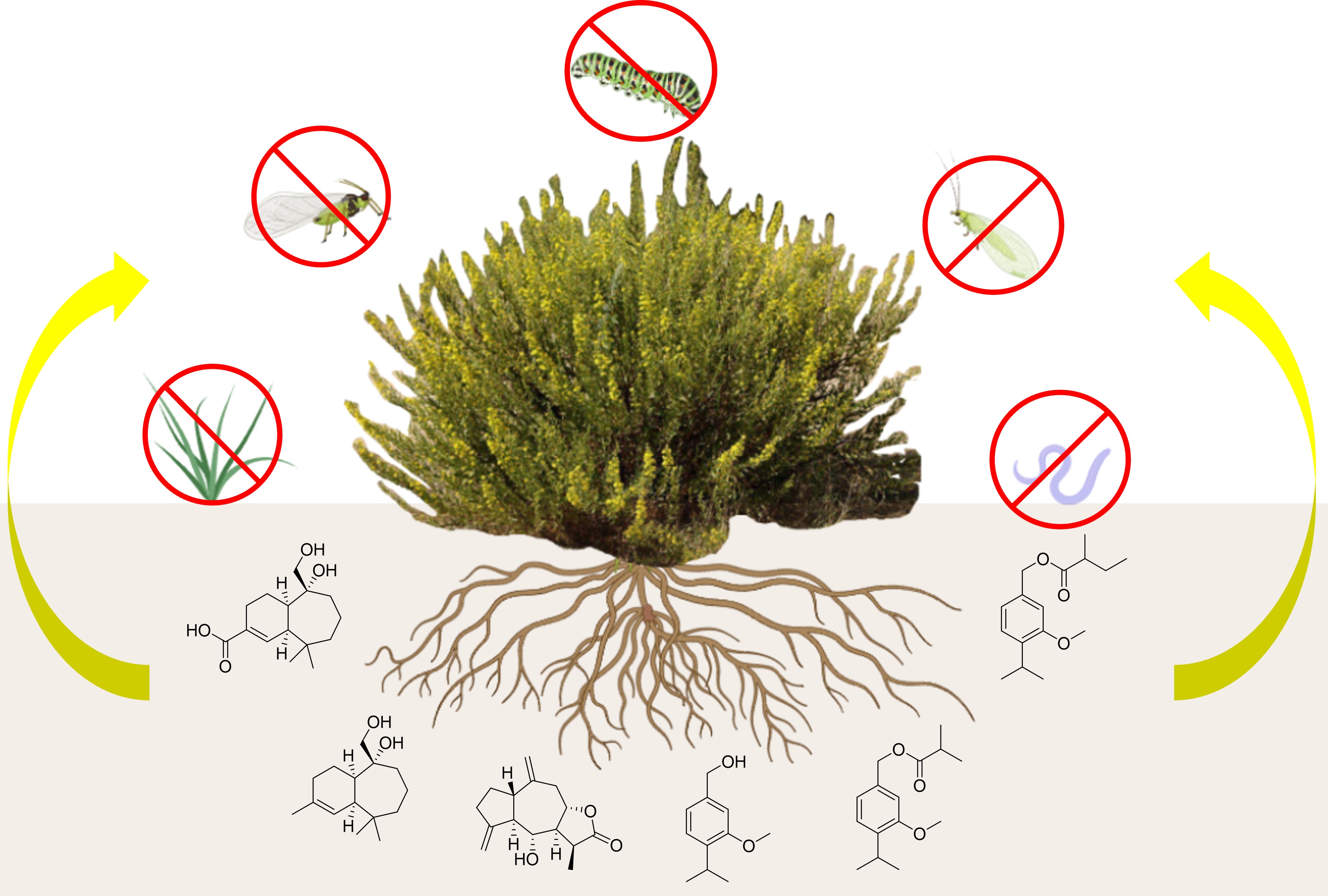
The natural products composition of the hexane and methyl tert-butyl ether extracts of Dittrichia viscosa roots have been examined. Eight terpenoids were identified by NMR and HRMS techniques, four of which (1, 5, 6 and 8) have not been previously reported as natural compounds. Of these eight compounds, four are thymol derivatives (1-4), two are guaianolides (5 and 7) and two are himachalanes (6 and 8). Additionally, the occurrence of himachalanes in this species has been noted for the first time. Furthermore, a study of the plant protection effects of some of these natural products and the chemical derivative 6a was carried out. Promising results have been obtained for compounds 1-3 and 6a as antifeedant against Spodoptera littoralis; 1-3 and 5 against Myzus persicae; 1-3 against Rhopalosiphum padi, and 4 as nematicide against Meloidogyne javanica. Finally, phytotoxic activity of 4, 5 and 6a against the monocotyledonous Lolium perenne has been also proved.
Keywords:
Dittrichia viscosa roots
; terpenes
; sesquiterpene lactones
; himachalenes
; biopesticides
1. Introduction
The exponential growth of the world population in recent decades makes it necessary to maximize agricultural production yields. In this context, the demand for pesticides that ensure a good harvest is continuously increasing, however, chemical pesticides can also have adverse effects on the environment and human health [1,2,3]. In this regard, biopesticides are natural products produced by organisms like bacteria, viruses, fungi or plants that are generally regarded as safe, efficient, highly targeted and biodegradable agents [4]. As a result of the gradual discontinuation of numerous chemical pesticides in Europe, the ongoing assessment of these chemicals and the increasing demand for organic food, the production of biopesticides has witnessed a substantial rise in recent years [5]. Indeed, in the framework of the European Green Pact and, in particular, its “Farm to Fork” strategy, the European Commission plans to reduce the use of chemical pesticides by 50% by 2030 (https://www.europarl.europa.eu/factsheets/en/sheet/78/los-productos-quimicos-ylosplaguicidas).
Within this specific context, our research group is dedicated to the research of novel biopesticides from plants. For this study, we selected Dittrichia viscosa (L.) Greuter, a woody plant with yellow flowers of Asteraceae family, which is distributed throughout the Mediterranean area and can be found in abandoned fields, roadsides, and disturbed areas due to its excellent colonizing ability [6].
D. viscosa has been used in traditional medicine due to its anti-inflammatory, antipyretic, antiseptic, sedative, antispasmodic, antidiarrheic, antimicrobial, and anthelmintic properties [7,8]. More recently, its insecticidal activity against pests such as the wheat weevil (Sitophilus granaries) has been confirmed [9], as well as its allelopathic [10] and antidepressant potential [11]. Furthermore, D. viscosa provides ecosystem functions [12]. For example, D. viscosa is a host plant for the aphid predator Macrolophus melanotoma in agroecosystems [13].
Encouraged by this range of reported bioactivities and potential applications, several studies of the chemical composition of the aerial parts of this plant have been carried out, revealing the presence of dozens of natural compounds [14,15,16]. However, little is known about the composition of roots, that has not been studied in detail and only some compounds such as chlorogenic acid, dicaffeoyl quinic acid, a few germacranolides, coumaric acid and p-cymene derivatives 2 and 3 are known to be present [17,18,19,20].
In this article, the results of a study of D. viscosa roots content (where new natural products 1, 5, 6 and 8 have been found) and its biopesticide activity against insect pests (Rhopalosiphum padi, Myzus persicae, Spodoptera littoralis) and a phytoparasitic nematode of economic importance (Meloidogyne javanica) are presented; as well as its herbicidal activity against the monocotyledonous ryegrass (Lolium perenne) and the dicotyledonous lettuce (Lactuca sativa).
The target species have been selected based on their economic importance and their availability in the laboratory. The insects S. littoralis and M. persicae can feed on a wide range of horticultural crops [21,22]. R. padi is a major cereal pest [23]. The root-knot nematode M. javanica is the most destructive among nematodes worldwide, due to the formation of root galls in the host [24,25]. Plant-parasitic nematodes are the most devastating group of plant pathogens worldwide, and their control is extremely challenging. Thus, in the last decade, much effort has been focused on the study of natural nematicidal agents for the management of root-knot nematodes, Meloidogyne spp., one of the most economically damaging genera on horticultural and field crops [26]. Perennial ryegrass (Lolium perenne) is an important cultivated grass species which becomes a very competitive weed when growing in cultivated crops (https://www.cropscience.bayer.co.nz/pests/weeds/ryegrass---perennial), and therefore is a good weed model to detect herbicidal effects. Lactuca satica (lettuce) is used to compare the selectivity of the phytotoxic effects between the monocotyledoneous ryegrass and a horticultural dicotyledoneous crop (lettuce).
2. Results
2.1. Extraction of D. viscosa Roots
Powdered roots of D. viscosa were successively extracted using a Soxhlet with hexane (H) and methyl tert-butyl ether (MTBE). Each extract was fractioned and the presence of compounds 1-8 could be established (compound 6a was obtained by acetylation of natural diol 6) (Figure 1).
Compounds 1, 5, 6, and 8 were found to be new natural products, whereas substances 2, 3, 4 and 7 were identified by comparing their spectroscopical data with those reported in the literature [20,27,28] (see NMR data in the Supporting Information (SI)). No biological activity has been previously reported for compounds 2, 3 and 4, however, sesquiterpene lactone 7 has been proven to possess anti-inflammatory and cytotoxic activity [29,30]. The spectroscopic data of compounds 5, 6 and 8 is shown in Table 1 and Table 2.
2.2. Structural Elucidation of New Natural Products
Compounds 1-3 were difficult to separate due to their high similarity, so they had to be subjected to several HPLC analysis to obtain enough of each product to achieve their identification (see experimental section).
Analysis of the NMR data of natural product 1 (Figures S1–S3 in the SI) confirmed this substance was an ester derivative of 3-methoxy-4-isopropylbenzylalcohol by comparison with data of those previously described 2-4 [20,27]. The esterifying moiety on the primary alcohol was determined to be a sec-butyl group, as inferred from the corresponding 1D TOCSY experiment after irradiating the methyl signal at δ 0.933 ppm 1H (see Figure S4 in the SI). Finally, compound 1 presented no optical rotation.
The molecular formula of compound 5 was C15H20O3 as deduced from the HRMS [M+H]+ ion at m/z 249.1491. This datum together with the analysis of its 1H and 13C NMR spectra (Table 1 and Table 2) evidenced that compound 5 was a tricyclic molecule with three unsaturations. The fact that two of these unsaturations are due to the presence of two di-substituted double bonds (δ 4.99; 5.10 and 4.93; 4.91 ppm 1H), together with the appearance of a carbonyl group at δ 179.14 ppm 13C (attributable to a saturated five-membered lactone), suggested that 5 was a sesquiterpene lactone, most likely a guaianolide [31,32]. Besides the lactone carbonyl group and the di-substituted olefin, the existence of two spin systems (Figure 2a) in compound 5 was deduced after the combined analysis of the 1H, 13C, HSQC, and 1D TOCSY NMR spectra (see SI, Figures S13–S15, S17 and S18). Finally, the complete planar structure was deduced by observing the correlations between the aforementioned partial structures in the HMBC spectrum (see Figure S16 at SI). The location of the lactone ring was confirmed after the HMBC correlations from H11 and H13 to C12. Other key HMBC correlations were those observed between H14 and C1, H2 and C5, H15 and C5, and H15 and C3 (Figure 2a).
Finally, the relative configuration of the chiral centers of the molecule was determined based on the analysis of 1D NOE experiments (Figures S19 and S20 in the SI). We previously computationally calculated the most stable conformation of 5 as detailed in the SI. The observed NOE correlations from H6 to H1, H8 and H13 suggested the same orientation of these hydrogens, while the correlations from H11 to H7 and H5 to H15 suggested a different orientation (Figure 2b). These data allowed us to propose the relative configuration for compound 5 as rel-1S,5R,6R,7S,8S,11S (https://doi.org/10.1351/goldbook.R05260).
The computationally calculated 13C NMR spectrum of 5, following the methodology outlined in the Experimental Section, is in complete agreement with the experimental data (rms = 1.18; Max abs. = 2.29), thereby confirming the structure of this compound. Furthermore, the absolute configuration of compound 5 was determined by calculating the theoretical optical rotation [α]D values using the Gaussian`16 software (see Experimental Section). Specific Rotation value obtained by the methodology described in the experimental section was -91.93 at 589 nm wavelength, which shares the sign of the experimental value ([α]D = - 40.12). Therefore, the absolute stereochemistry of this compound is established as 1S,5R,6R,7S,8S,11S. Noteworthy, to the best of our knowledge, this is the first time where the absolute configuration of guaianolides from D. viscosa is described.
The molecular formula of 6 was C15H26O2 as deduced from the HRMS [M-OH]+ ion at m/z 221.1905. The bicyclic structure was evidenced by considering this information together with the 1H NMR and 13C NMR data (Table 1 and Table 2). Furthermore, partial structures in this compound were inferred after the combined analysis of the 1H, 13C, HSQC, and 1D TOCSY NMR spectra (see SI, Figures S21–S23 and S25) (Figure 3a). Assignment of the two sp3 quaternary carbons was realized as shown based on the chemical shifts of the two singlet methyl groups (δ 0.88 and 1.01 ppm 1H). Also worthy of mention was the belonging of the methyl group at δ 1.68 ppm 1H and the olefinic proton at δ 5.61 ppm 1H to the same spin system as inferred from the analysis of the 1D TOCSY spectrum obtained after irradiating at δ 2.26 ppm 1H (see Figure S25 at SI).
Finally, the entire planar structure of 6 was elucidated after analyzing the correlations observed in the HMBC spectrum (see Figure S24 at SI), which allowed us to bond partial structures as shown on Figure 3a. Key HMBC correlations were those observed between H14 and C6, H1 and C11, H8 and C14, and H12 and C10 (Figure 3a).
The analysis of 1D NOE spectra (see Figures S26 and S27 at SI) revealed a cis-bonding of the bicyclic system, as evidenced by the correlation between protons H1 and H6, indicating that they are located on the same side of the molecule. However, 1D NOE experiments did not allow us to unambiguously establish the relative configuration at C-7. Thus, we resorted to computational methods to establish the configuration at this stereocenter. GIAO (Gauge-independent atomic orbital) NMR chemical shift calculations have become a reliable method to aid in the stereo structural elucidation of new natural products [33,34]. In addition, this methodology has made it possible to carry out structural revisions of numerous natural products [35]. Due to the flexibility of the 7-membered ring and the dependence of the conformation on chemical shifts, we applied the efficient protocol developed by Hehre et al., [36] to calculate chemical shifts for flexible natural products. Spartan'24 implemented a neural network routine within the multistep NMR Spectrum task developed by Hehre et al. that considerably improves its performance and accuracy of the obtained results.
After building compound 6 and its epimer at C-7 in Spartan’24, both stereoisomers were subjected to an NMR calculation protocol developed by Hehre et al. which is reported in detail in the experimental part. This protocol allows the prediction of 13C chemical shifts with an accuracy quantified using an overall rms (root-mean-square). The calculated rms deviation between experimental and calculated 13C chemical shifts for compound 6 was 1.3 ppm, with a Max. absolute of 2.7 corresponding at C-9, whereas for 6 epimer at C-7 the statistical term rmsd was 2.4 and with a Max. absolute of 3.7 (Table 3). These values definitively confirm the relative configuration for this molecule, as shown in Figure 4. In fact, the DP4 probability scores, calculated according to Goodman’s procedure [37], provide a value of 100% for R*-epimer. Based on these results, R* was predicted to be the relative configuration at C7 of compound 6.
For a more detailed interpretation of this study, consult the S7 and S8 tables in the SI in which the energies of each conformation of both epimers are shown, as well as their Boltzmann populations and the DP4 probability score of each of the conformations as a function of the proximity between the calculated and experimental chemical shifts. Thus, it can be observed that the two conformations of the R*-epimer with the best rmsd (δ13C root-mean-square deviation) correspond to some of the lowest energy ones; whereas, for the S*-epimer the conformation with the best rmsd corresponds to conformation 14 (M14), which shows a Max absolute of 4.18 and whose Bolzman weight is only 0.008 (Table 3 and Figure 4).
Once the spatial disposition of the natural product was computationally established, the observed NOEs effects in the corresponding 1D NOE experiments confirmed the conformation proposed for 6 (Figure 3b).
As happened with 5, the value of the specific rotation of compound 6 was calculated computationally. The optical rotation was computed on the two most populated conformations – M02 and M04 (see Tables S6 and S7 at the SI), to give an average [α]D value of +86.27 at 589 nm wavelength, which again shares the sign with the value calculated experimentally [α]D = + 53.26), allowing us to propose the absolute stereochemistry of this compound as 1S6R7S.
Finally, the molecular formula of the new natural product 8 was C15H24O4 as deduced from HRMS [M-OH]+ m/z 293.1751. This compound was identified by comparing its 1H and 13C NMR data with those of compound 6 (Table 1 and Table 2). Thus, while the C15 methyl in compound 6 (δ 1.68 ppm 1H, 26.60 ppm 13C) was no longer observed, a new carbonyl signal was observed at δ 171.61 ppm in 13C. These data, together with the significant deshielding experienced by H2 (from δ 5.61 ppm 1H in compound 6 to δ 7.32 ppm 1H in 8) prove the existence of a carboxylic acid at C3. As expected, compound 8 shared with diol 6 the same relative conformation as deduced from the analysis of the corresponding 1D NOE spectra (Figure 5). Thus, NOE correlations were observed from H6 to H14a and H1. Himachalane 8 was proposed to possess the same absolute configuration as himachalane 6.
2.3. Plant Protection Effects
The compounds isolated from D. viscosa roots 1+2+3, 4, 5 and the derivative 6a were tested against insect pests (R. padi, M. persicae, S. littoralis), and the root-knot nematode M. javanica and the plants ryegrass and lettuce.
Table 4 shows the insect antifeedant effects of the compounds tested (1-3, 4, 5-6, 5 and 6a). All these molecules showed species-dependent antifeedant effects except the monoterpene alcohol 4, which effects were below the established threshold for dose-response experiments (70% for antifeedant effects at the maximum dose tested). S. littoralis was moderately affected by the mixture 1+2+3 (EC50 = 32.35 μg/cm2) and himachalane 6a (EC50 = 22.10 μg/cm2). M. persicae was the most sensitive insect, affected by the mixture 1+2+3 (EC50 = 16.54 μg/cm2) and compound 5 (EC50 = 18.71μg/cm2), while R. padi was only affected by phenol derivatives 1+2+3 (EC50 = 19.14 μg/cm2).
Benzyl esters 1-3 are thymol derivatives. Thymol has been reported as an effective antifeedant to S. littoralis, M. persicae and R. padi (EC50 values of 21.0, 7.6 and 18.6 μg/cm2 respectively), while its precursor p-cymene was inactive [40], however, this is the first report on the antiffeedant effects of these type of phenols. Among the sesquiterpenes, the guaianolide sesquiterpene lactone 5 was active on M. persicae. Similarly, the structurally-related sesquiterpene lactone tomentosin was the active compound present in D. viscosa aerial parts, with antifeedant effects against M. persicae [41], while α- and γ-costic acids were identified in an insecticidal fraction of D. viscosa with contact toxicity against Sitophilus granarius adults [9]. Himachalane 6a was antifeedant to S. littoralis and M. persicae respectively. Himachalanes have been reported as being active on insects. For example, essential oil from Cedrus deodara, rich in β-himachalene (46%), was deterrent to Plutella xylostella [42]. Furthermore, himachalol and β-himachalene were toxic to the pulse beetle Callosobruchus analis [39].
The nematicidal effects of the compounds tested are shown in Table 5. Phenol derivative 4 was nematicidal against M. javanica J2 with a minimal lethal dose (MLD) of 0.5 mg/mL.
Phenols with a carboxyl group, such as salicylic and cumic acid resulted in effective nematicidal compounds, followed by isopropyl salicylate, and alkylated phenols with a p-isopropyl group (thymol and carvacrol). The nematicidal action of these compounds was further confirmed by their egg hatching inhibition effects [43]. Previous results have shown the nematicidal effect of extracts from D. viscosa aerial parts, containing costic and isocostic acids [44]. However, this is the first report on the presence of nematicidal compounds in D. viscosa roots.
D. viscosa root compounds (1+2+3, 4, 5 and 6a) were tested for phytotoxic effects against two model plant species, the monocotyledoneous ryegrass and the dycotiledoneous lettuce (L. perenne and L. sativa). All phytotoxic parameters measured for L. sativa had inhibition values < 20% (Figure 6), and therefore none of these compounds was considered phytotoxic to this plant. On the contrary, phytotoxic effects were found against L. perenne. Phenol derivative 4 showed dose-dependent phytotoxicity (72 and 37% root and leaf growth inhibition at 0.1 mg/mL, 42% root growth inhibition at 0.05 mg/mL), followed by 5 (41 and 46% root and leaf growth inhibition, 0.1mg/mL) and 6a (44.5 and 16% Conclusions, 0.1 mg/mL) (Figure 6).
The allelopathic effects of D. viscosa leaf extracts and dry biomass have been described [45,46]. The phytotoxic compounds identified included sesquiterpene lactones (inuloxins A, C and D) and α-costic acid, with herbicidal effects against parasitic weeds (Orobranche crenata and Cuscuta campestris) [47] and the dihydroflavanol 3-O-acetylpadmatin, that showed inhibition on the radicle growth of Orobranche cumana and Phelipanche ramosa [48]. The inhibition of the root elongation and growth of Lycopersicon esculentum and Lepidium sativum caused by Inuloxin A was attributed to an alteration of the cell redox system [49]. Furtermore, thymoxyacetic acid was phytotoxic in preemergence and thymol in postemergence on five plant species including L. sativa [50]. However, this is the first report on the allelopathic effects of D. viscosa root compounds 4, 5 and 6a.
3. Discussion
Dittrichia viscosa is a plant well known for its use in traditional medicine. Given its wide range of activities and the limited research on its roots, we have focused our study on analyzing the composition of D. viscosa roots and evaluating the pesticidal activity of the isolated compounds.
Roots of D viscosa revealed as the source of new natural compounds. The structures of these compounds were determined. Four new natural products were found, two of them being new himachalane sesquiterpenes – a scarce group of sesquiterpenes, with, to the best of our knowledge, less than twenty himachalanes (including seco-himachalanes) being reported so far. Noteworthy, the stereochemistry of these compounds was assigned with the aid of recently described computational protocols.
It is noteworthy that the terpenes found in the roots of D. viscosa are not found in other parts of the plant, which increases the structural diversity found in this species, a fact that could extend to other species.
Finally, the pesticidal activity of D. viscosa was assessed, and different compounds have shown interesting results as antifeedants against Spodoptera littoralis; Myzus persicae and Rhopalosiphum padi (pest insects), and as nematicides against Meloidogyne javanica. Additionally, some compounds showed allelopathic activity against the monocotyledonous Lolium perenne. Notably, to our knowledge, this is the first report of biological activity on natural products with himachalane skeleton, which may help to explain their biosynthesis in plants.
4. Materials and Methods
4.1. General Experimental Procedures
Optical rotations were measured on a Perkin-Elmer 141 polarimeter. IR spectra were recorded using an FT/IR-6200 spectrometer (JASCO Inc., Easton, MD, USA). NMR spectra were recorded with BRUKER Avance NEO (1H NMR 600 MHz), Varian Direct Drive (1H NMR 500 MHz/13C NMR 125 MHz) and BRUKER Nanobay Avance III HD (1H NMR 400 MHz) spectrometers. The multiplicity of signals is indicated using the following abbreviations: s = singlet, d = doublet, t = triplet, q = quartet, quint = quintuplet, hex = hexuplet, hep = heptuplet, bs = broad singlet, bd = broad doublet, bt = broad triplet, dd = doublet of doublets, dt = doublet of triplets, dq = doublet of quartets, dquint = doublet of quintets, td = triplet of doublets, ddd = doublet of doublets of doublets, m = multiplet. High-resolution mass spectra (HRMS) were determined on a BRUKER Autoflex mass spectrometer. Silica gel SDS 60 (35−70 μm) was used for column chromatography. Chromatography fractions and the acetylation reaction were monitored by thin-layer chromatography carried out on 0.25 mm E. Merck silica gel plates (60F-254) using UV light and a solution of phosphomolybdic acid in ethanol as the visualizing agent. Semipreparative HPLC separation was carried out on a column (5 μm Silica, 10×250 mm2) at a flow rate of 4.0 mL/min in an Agilent Series 1100 instrument.
4.2. Plant Material, Extraction and Isolation
Specimens of Dittrichia viscosa (GDA54164) were collected in Granada (37.208108, -3.622045, Spain) in September 2022.
Roots of D. viscosa were air-dried for 5 days and subsequently crushed (368 g). Then, they were extracted using a Soxhlet apparatus for 24 hours with hexane to obtain 2.6 g of extract (0.7% yield from dry roots), followed by MTBE, to get 2.9 g of extract (0.8% yield from dry roots).
A hexane extract portion (875 mg) was fractioned by column chromatography on silica gel using as eluent mixtures of H/MTBE of increasing polarity to, finally, obtain four fractions (FH1-FH6). Fraction FH1 (409 mg) was obtained using as eluent H/MTBE (6:1) and contained a mixture of geraniol, fatty acid esters and compounds 1, 2 and 3. FH1 was re- chromatographed (H/MTBE (95:5)) to separate a mixture of compounds 1, 2 and 3 in a 1:3:7 ratio (283 mg). A 15 mg fraction of this mixture was subjected to semipreparative HPLC (normal phase, H/MTBE 9:1) to obtain pure 1 (Rt = 8 min), 2 (Rt = 8.5 min) and 3 (Rt = 9 min). Fraction FH3 (156 mg H/MTBE (1:1)) was mainly composed by sterols, fatty acid esters and compound 4. Re-chromatography of this fraction with H/MTBE (2:1) led to the isolation of 46 mg of pure compound 4. FH4 (52 mg) was collected with MTBE and was constituted by fatty acids esters and compounds 5 and 6. FH4 was subjected to a second chromatography (H/MTBE (2:1)) to obtain a 2:1 mixture of 5 and 6 (19 mg). A 15 mg portion of this mixture was subjected to semipreparative HPLC (normal phase, H/MTBE 2:1) to give pure 5 (Rt= 8.0 min) and 6 (Rt= 9.6 min).
The MTBE extract (2.9 g) was also fractioned by column chromatography eluting with mixtures of H/MTBE of increasing polarity. Nine fractions were obtained (FE1-FE9). Fraction FE1 (313 mg) was obtained using as eluent H/MTBE (4:1) and was mainly composed by a mixture of fatty acids, geraniol and compounds 1, 2 and 3. Fraction FE5 (380 mg) was collected employing H/MTBE (1:1) and was constituted by a mixture of fatty acids esters and compounds 4, 5 and 6. FE5 was re-chromatographed using H/MTBE (2:1) to obtain 58 mg of compound 4 and 140 mg of a 2:1 mixture of 5 and 6. Finally, fraction FE7 (120 mg, MTBE) was mainly constituted by a mixture of fatty acids and compounds 7 and 8. FE7 was re-chromatographed with H/MTBE (1:3) obtaining 20 mg of a 2:1 mixture of 7 and 8. This mixture was subjected to semipreparative HPLC (normal phase, H/MTBE 1:4) to give 5 mg of pure 7 (Rt = 11.6 min), 3 mg of 8 (Rt = 13.8 min) and 4 mg of 7 and 8 mixture.
4.3. Derivatization of Natural Product 6
An acetylation of a mixture of 5 and 6 was done to isolate both natural products. 145 mg of 5 and 6 mix in proportion (2:1) were reacted with 1 mL of acetic anhydride and 1 mL of pyridine under inert atmosphere stirring for 50 min at 25 ºC. Then, the reaction content was mixed with ice and 30 mL of MTBE were added. Once the ice melted, the two phases were separated and the organic phase was washed with 2N HCl (15 mL x 3), saturated NaHCO3 solution (15 mL x 3) and brine (15 mL x 3) and dried with anhydrous Na2SO4. The solvent was removed, giving a residue, which was flash chromatographed (H/MTBE, 3:2) to give 87 mg of 5 and 46 mg of 6a.
4.4. Spectroscopical Data (1H and 13C NMR) of Natural Products 1-8
Compound 1. Colorless oil. [α]D = 00.0 (c 1.0, DCM). HRMS [M+H]+ m/z 265.1802 (calcd for C16H25O3, 265.1798). IR (ATR) νmax 2962, 2936, 2875, 1732, 1613, 1582, 1508, 1447, 1419, 1259, 1146, and 1042 cm-1. 1H NMR (400 MHz, CDCl3) δ 7.19 (d, J = 7.7 Hz, 1H), 6.91 (dd, J = 7.7 Hz, 1.7 Hz, 1H), 6.82 (d, 1.7 Hz, 1H), 5.09 (s, 2H), 3.83 (s, 3H), 3.30 (hept, J = 6.9 Hz, 1H), 2.43 (hex, J = 7.0 Hz, 1H) 1.71 (dquint, J = 14.8 Hz, 7.4 Hz, 1H), 1.50 (dquint, J = 14.8 Hz, 7.4 Hz, 1H) 1.20 (d, J = 6.9 Hz, 6H), 1.17 (d, J = 7 Hz, 3H), 0.91 (t, J = 7.4 Hz, 3H). 13C NMR (101 MHz, CDCl3) δ 176.64, 156.83, 137.02, 134.68, 126.11, 120.26, 110.11, 66.08, 55.36, 41.09, 26.81, 26.60, 22.63, 22.63, 16.62, 11.64.
Compound 2. Colorless oil. 1H NMR (400 MHz, CDCl3) δ 7.19 (d, J = 7.7 Hz, 1H), 6.91 (d, J = 7.7 Hz, 1H), 6.82 (s, 1H), 5.08 (s, 2H), 3.83 (s, 3H), 3.30 (hept, J = 6.9 Hz, 1H), 2.24 (d, J = 6.9 Hz, 2H) 2.13 (hept, J = 6.9 Hz, 1H), 1.20 (d, J = 6.9 Hz, 6H), 0.96 (d, J = 6.9 Hz, 6H). 13C NMR (101 MHz, CDCl3) δ 173.04, 156.85, 137.13, 134.51, 126.13, 120.48, 110.32, 66.17, 55.37, 43.46, 26.61, 25.75, 22.63, 22.43.
Compound 3. Colorless oil. 1H NMR (400 MHz, CDCl3): δ 7.19 (d, J = 7.7 Hz, 1H), 6.91 (d, J = 7.7 Hz, 1H), 6.82 (s, 1H), 5.08 (s, 2H), 3.83 (s, 3H), 3.30 (hept, J = 6.9 Hz, 1H), 2.60 (hept, J = 6.9 Hz, 1H), 1.20 (d, J = 7.0 Hz, 12H). 13C NMR (101 MHz, CDCl3) 177.02, 156.85, 137.03, 134.67, 126.12, 120.23, 110.08, 66.17, 55.36, 34.05, 26.62, 22.64, 19.02.
Compound 4. Colorless oil. 1H NMR (400 MHz, CDCl3): δ 7.19 (d, 1H), 6.91 (d, 1H), 6.90 (s, 1H), 4.66 (s, 2H), 3.85 (s, 3H), 3.31 (hept, J = 7.0 Hz, 1H), 1.21 (d, J = 7.0 Hz, 6H). 13C NMR (126 MHz, CDCl3): δ 157.01, 139.45, 136.62, 126.12, 119.07, 109.18, 65.55, 55.39, 26.58, 22.69.
Compound 5. Colorless oil. [α]D = - 40.12 (c 1.0, DCM). IR (ATR) νmax 3440, 2933, 2870, 1761, 1706, 1641, 1458, 1381, 1258, 1207, 1074, 1021, 985, and 894 cm-1. HRMS [M+H]+ m/z 249.1491 (calcd for C15H21O3, 249.1485). 1H and 13C NMR data is shown in Table 1 and Table 2.
Compound 6. Colorless oil. [α]D = + 53.26 (c 0.3, DCM). IR (ATR) νmax 3359, 2916, 1709, 1440, 1373, 1268, 109, and 795 cm-1. HRMS [M-OH]+ m/z 221.1905 (calcd for C15H25O, 221.1900). 1H and 13C NMR data is shown in Table 1 and Table 2.
Compound 6a. Colorless oil. [α]D = + 16.32 (c 1.0, DCM). 1H NMR (500 MHz, CDCl3): δ 5.61 (bd, J = 4.6 Hz, 1H), 4.07 (d, J = 11.3 Hz, 1H), 3.94 (d, J = 11.3 Hz, 1H), 2.29 (bs, 1H), 2.10 (s, 3H), 2.01 - 1.87 (m, 4H), 1.76 - 1.53 (m, 5H), 1.68 (s, 3H), 1.47 - 1.41 (m, 1H), 1.38 (ddd, J = 14.2, 5.8, 4.0 Hz, 1H), 1.00 (s, 3H), 0.87 (s, 3H). 13C NMR (126 MHz, CDCl3): δ 171.22, 133.46, 125.47, 76.42, 71.54, 46.06, 43.36, 40.79, 38.44, 33.64, 31.32, 30.82, 26.00, 23.61, 21.51, 20.98, 19.44. HRMS [M-OH]+ m/z 293.1751 (calcd for C17H25O4, 293.1747)
Compound 7. Colorless oil. 1H NMR (500 MHz, CDCl3): δ 5.01 (s, 1H), 4.95 (s, 1H), 4.39 (td, J = 10.4, 5.0 Hz, 1H), 3.18 (dd, J = 15.7, 5.0 Hz, 1H), 2.71 (quint, J = 7.5 Hz, 1H), 2.54 (dd, J = 15.7, 10.3 Hz, 1H), 2.23 (q, J = 10.0, 9.4 Hz, 1H) 2.12 (m, 1H), 2.00 - 1.85 (m, 2H), 1.85-1.65 (m, 4H), 1.63 (m, 1H) 1.22 (d, J = 7.4 Hz, 3H), 1.20 (s, 3H).
4.5. Computational Calculations of 13C NMR Spectrum and Optical Rotation
Calculations were performed with Spartan’24 (Wavefunction Inc., Irvine, CA, USA). To obtain the 13C NMR data by computational calculation, the automated protocol implemented in Spartan’24 was followed (37). This protocol consists of six steps: (I) Systematic conformational search using MMFF molecular mechanics, eliminating duplicate conformers and those with energy 40 kJ/mol above the global minimum; (II) geometric calculation using HF/3-21G, also eliminating duplicate conformers and those with energy higher than 40 kJ/mol above the global minimum; (III) energy calculation with the ωB97X-D/6-31G* model and removal of conformers above 15 kJ/mol with respect to the global minimum; (IV) geometric calculation with the ωB97X-D/6-31G* model and removal of conformers with energies higher than 10 kJ/mol from that of the global minimum; (V) energy calculation with the ωB97X-V/6-311+G(2df,2p)[6-311G*], and finally; (VI) the NMR calculations (following calculation of Boltzmann weights for conformationally flexible molecules) using the ωB97X-D/6-31G* method that has been corrected empirically based on the comparison of calculated and experimental 13C shifts for ~2000 rigid molecules. These corrections are on the order of 1−3 ppm.
Finally, compounds 5 and 6 in the best-fit conformer between experimental and calculated 13C NMR data were reoptimized with Gaussina’16 using DFT at the wb97xd/6-311+g(2d,p) level of theory [51,52], and the [α]D optical rotation was computed at the same level of theory [53]. To simulate the effect of the solvent used for the experimental measurements (dichlorometane), the iefpcm model were considered [54].
4.6. Biopesticide Assays
4.6.1. Antifeedant Activity
The insect colonies (Spodoptera littoralis, Myzus persicae and Rhopalosiphum padi) come from laboratory colonies reared on artificial diet and host plants (Capsicum annuum, Hordeum vulgare), respectively, at 22 ± 1 °C, >70% relative humidity and 16:8 h (L:D) photoperiod at ICA-CSIC.
The tests have been described before [43]. Briefly, the upper surface of leaf disks or fragments (1.0 cm2) of C. annuum and H. vulgare were treated with 10 µL of compound at an initial dose of 5 µg/µL (50 µg/cm2). Two sixth-instar S. littoralis larvae (>24 h after molting) per Petri dish (5 unities) or 10 apterous aphid adults (24–48 h old) placed in a 2×2 cm ventilated plastic box (20 unities) were allowed to feed at room temperature or in the growth chamber respectively. The experiments end at 75% larval consumption of paired control or treatment disks for S. littoralis or after 24 h for aphids. Each experiment was repeated 2 times. Feeding inhibition (%FI), based on the disk surface consumption (digitalized with https://imagej.nih.gov/ij/) [55], and aphid settling inhibition (%SI), based on the number of aphids on each leaf fragment, were calculated as % FI/SI = [1 − (T/C) × 100], where T and C represent feeding/settling on treated and control leaf disks, respectively. The significance of these effects was analyzed by the nonparametric Wilcoxon paired signed-rank test. Tests with an FI/SI > 70% were further tested in dose-response experiments (range of activities between 100 and <50%, minimum of 3 doses) to calculate their effective EC50 dose from linear regression analysis (% FI/SI on Log-dose, STATGRAPHICS Centurion XVI, version 16.1.02).
4.6.2. Nematicidal Activity
Meloidogyne javanica population was maintained on Solanum lycopersicum plants (var. Marmande) in pot cultures at 25º ± 1 C, 70% relative humidity. Egg masses of M. javanica were handpicked from infected tomato roots. Second-stage juveniles (J2) were obtained from hatched eggs by incubating egg masses in a water suspension at 25 ºC for 24 h.
The tests were carried out as described by Andres et al. [56]. Briefly, the compounds were dissolved in distilled water containing 5% of a DMSO-Tween solution (0.5% Tween 20 in DMSO) and evaluated. The initial concentration tested was 0.5 µg/µL. The nematicidal activity data are presented as percent dead J2 corrected according to Scheider-Orelli's formula. Effective lethal doses (LC50 and LC90) were calculated by Probit Analysis. Five serial dilutions were used to obtain the LC50 and LC90 and four replicates were used in each concentration.
4.6.3. Phytotoxic Effect
The experiments were conducted with Lactuca sativa cv. Teresa and Lolium perenne Nui seeds (donated by Fito-España and Battle-España, respectively). Filter paper disks (2.5 cm diam.) with 20 µL of the solvent (control) or the test compound (5 mg/mL in EtOH) were placed on 12-well plates (3 replicates / experiment), then 500 mL H2O/well and 10/5 seeds (L. sativa / L. perenne pre-soaked in distilled water for 12 h) were added to give a final concentration of 0.1 mg/mL in the well. Then the plates were covered and placed in a plant growth chamber (25°C, 70% RH, 16:8 L:D).
Germination was monitored for 6 days, and the root/leaf length measured at the end of the experiment (25 seedlings randomly selected for each experiment) with the application ImageJ (http://rsb.info.nih.gov./ij/) [55]. Serial dilutions (1:2) were carried out for tests resulting in inhibition > 50 % (respect to the control) for any parameter measured [56].
Supplementary Materials
The following supporting information can be downloaded at the website of this paper posted on Preprints.org, Figures S1–S44: NMR spectra; Table S1: Cartesian coordinates (mol2) of the conformer with the lowest energy out of all possible conformers of 5, Table S2: Comparation between the theorical 13C RMN of the lower energy conformer of 5 and the corresponding experimental values, Table S3: Lower energy conformations of 6, Table S4: Lower energy conformations of 6 epimer at C-7, Tables S5–S7: Cartesian coordinates.
Author Contributions
Conceptualization, J.F.Q.M., A.G.-C. and A.F.B; methodology, M.J.S.N., M.F.A. and A.G.-C.; NMR DFT calculation, J.L.L.P.; validation, J.F.Q.M., A.G.-C. and A.F.B; investigation M.J.S.N., A.G.; J.L.L.P. and M.F.A.; data curation, M.J.S.N.; J.F.Q.M., A.G.-C. and A.F.B.; writing—original draft preparation, M.J.S.N., M.F.A.,A.G.-C. and J.L.L.P.; writing—review and editing, J.F.Q.M., A.G.-C. and A.F.B; project administration, J.F.Q.M., A.G.-C. and A.F.B., funding acquisition, J.F.Q.M., A.G.-C. and A.F.B.
Funding
This research was funded by MINISTERIO DE CIENCIA (Spanish Ministry of Science and Innovation), grant numbers PID2019-106222RB-C32/SRA (State Research Agency, 10.13039/501100011033) and PID2019-106222RB-C31/SRA (State Research Agency, 10.13039/501100011033, and Unidad Asociada UGR-CSIC BIOPLAG.
Informed Consent Statement
Not applicable.
Data Availability Statement
All data supporting the findings of this study are available within the article.
Conflicts of Interest
The authors declare no conflicts of interest.
References
- Acheuk, F.; Basiouni, S.; Shehata, A. A.; Dick, K.; Hajri, H.; Lasram, S.; Yilmaz, M.; Emecki, M.; Tsiamis, G.; Spona-Friedl, M.; May-Simera, H.; Eisenreich, W.; Ntouglas, S. Status and prospects of botanical biopesticides in Europe and Mediterranean countries. Biomolecules 2022, 12, 311. [Google Scholar] [CrossRef]
- Lee, G. H.; Choi, K. C. Adverse effects of pesticides on the functions of immune system. Comp. Biochem. Physiol. C. 2020, 235, 108789. [Google Scholar] [CrossRef]
- Lo, C.C. Effect of pesticides on soil microbial community. J. Environ. Sci. Health B. 2010, 45(5), 348–359. [Google Scholar] [CrossRef]
- Villaverde, J. J.; Sandín-España, P.; Sevilla-Morán, B.; López-Goti, C.; Alonso-Prados, J. L. Biopesticides from natural products: Current development, legislative framework, and future trends. BioRes. 2016, 11(2), 5618–5640. [Google Scholar] [CrossRef]
- Kumar, S.; Singh, A. Biopesticides: present status and the future prospects. J. Fertil Pestic. 2015, 6(2), 100–129. [Google Scholar] [CrossRef]
- Zaki, M. Natural products from Dittrichia Viscosa (Mini-Review). RHAZES: Green Appl. Chem. 2020, 9, 30–46. [Google Scholar] [CrossRef]
- Grauso, L.; Cesarano, G.; Zotti, M.; Ranesi, M.; Sun, W.; Bonanomi, G.; Lanzotti, V. Exploring Dittrichia viscosa (L.) Greuter phytochemical diversity to explain its antimicrobial, nematicidal and insecticidal activity. Phytochem. Rev. 2020, 19, 659–689. [Google Scholar] [CrossRef]
- Barrero, A. F.; Herrador, M. M.; Arteaga, P.; Catalán, J. V. Dittrichia viscosa L. Greuter: Phytochemistry and biological activity. Nat. Prod. Commun. 2008, 3(11), 1799–1804. [Google Scholar] [CrossRef]
- Rotundo, G.; Paventi, G.; Barberio, A.; De Cristofaro, A.; Notardonato, I.; Russo, M. V.; Germinara, G. S. Biological activity of Dittrichia viscosa (L.) Greuter extracts against adult Sitophilus granarius (L.) (Coleoptera, Curculionidae) and identification of active compounds. Sci. Rep. 2019, 9(1), 6429. [Google Scholar] [CrossRef]
- Araniti, F.; Lupini, A.; Sunseri, F.; Abenavoli, M. R. Allelopatic potential of Dittrichia viscosa (L.) W. Greuter mediated by VOCs: A physiological and metabolomic approach. PLoS One 2017, 12(1), 170161. [Google Scholar] [CrossRef]
- Murlanova, K.; Cohen, N.; Pinkus, A.; Vinnikova, L.; Pletnikov, M.; Kirby, M.; Gorelick, J.; Drori, E.; Pinhasov, A. Antidepressant-like effects of a chlorogenic acid-and cynarine-enriched fraction from Dittrichia viscosa root extract. Sci. Rep. 2022, 12(1), 3647. [Google Scholar] [CrossRef] [PubMed]
- Sladonja, B.; Poljuha, D.; Krapac, M.; Uzelac, M.; Mikulic-Petkovsek, M. Dittrichia viscosa: Native-Non Native Invader. Diversity 2021, 13(8), 380. [Google Scholar] [CrossRef]
- Perdikis, D.; Favas, C.; Lykouressis, D.; Fantinou, A. Ecological Relationships between Non-Cultivated Plants and Insect Predators in Agroecosystems: The Case of Dittrichia viscosa (Asteraceae) and Macrolophus melanotoma (Hemiptera: Miridae). Acta Oecol. 2007, 31(3), 299–306. [Google Scholar] [CrossRef]
- Rhimi, W.; Ben Salem, I.; Immediato, D.; Saidi, M.; Boulila, A.; Cafarchia, C. Chemical composition, antibacterial and antifungal activities of crude Dittrichia viscosa (L.) greuter leaf extracts. Molecules 2017, 22(7), 942. [Google Scholar] [CrossRef]
- Al-Qudah, M.; Al-Jaber, H.; Mayyas, A.; Abu-Orabi, S.; Abu Zarga, M. Chemical compositions of the essential oil from the jordanian medicinal plant Dittrichia viscosa. Jordan J. Chem. 2010, 5(4), 343–348. [Google Scholar]
- Gharred, N.; Dbeibia, A.; Falconieri, D.; Hammami, S.; Piras, A.; Dridi-Dhaouadi, S. Chemical composition, antibacterial and antioxidant activities of essential oils from flowers, leaves and aerial parts of Tunisian Dittrichia viscosa. J. Essent. Oil Res. 2019, 31(6), 582–589. [Google Scholar] [CrossRef]
- Gökbulut, A.; Özhana, O.; Satılmiş, B.; Batçioğlu, K.; Günal, S.; Şarer, E. Antioxidant and antimicrobial activities, and phenolic compounds of selected Inula species from Turkey. Nat. Prod. Commun. 2013, 8(4), 475–478. [Google Scholar] [CrossRef]
- Trimech, I.; Weiss, E. K.; Chedea, V. S.; Marin, D.; Detsi, A.; Ioannou, E.; Roussi, V.; Kefalas, P. Evaluation of anti-oxidant and acetylcholinesterase activity and identification of polyphenolics of the invasive weed Dittrichia viscosa. Phytochem. Anal. 2014, 25(5), 421–428. [Google Scholar] [CrossRef] [PubMed]
- Bohlmann, F.; Gupta, R. K. Ineupatorolide-like sesquiterpene lactones from Dittrichia viscosa. Phytochemistry. 1982, 21(6), 1443–1445. [Google Scholar] [CrossRef]
- Shtacher, G.; Kashman, Y. Chemical investigation of volatile constituents of Inula viscosa Ait. Tetrahedron 1971, 27(6), 1343–1349. [Google Scholar] [CrossRef]
- Muñoz-Rodríguez, P.; Carruthers, T.; Wood, J. R.; Williams, B. R.; Weitemier, K.; Kronmiller, B.; Ellis, D.; Anglin, N. L.; Longway, L.; Harris, S. A.; Rausher, M. D.; Kelly, S.; Liston, A.; Scotland, R. W. Reconciling Conflicting Phylogenies in the Origin of Sweet Potato and Dispersal to Polynesia. Curr. Biol. 2018, 28(8), 1246–1256. [Google Scholar] [CrossRef]
- Kim, J. H.; Jander, G. Myzus persicae (green peach aphid) feeding on Arabidopsis induces the formation of a deterrent indole glucosinolate. Plant J. 2007, 49(6), 1008–1019. [Google Scholar] [CrossRef]
- Greenslade, A. F. C.; Ward, J. L.; Martin, J. L.; Corol, D. I.; Clark, S. J.; Smart, L. E.; Aradottir, G. I. Triticum monococcum lines with distinct metabolic phenotypes and phloem-based partial resistance to the bird cherry-oat aphid Rhopalosiphum padi. Ann. Appl. Biol. 2016, 168(3), 435–449. [Google Scholar] [CrossRef]
- Alekcevetch, J. C.; de Lima Passianotto, A. L.; Ferreira, E. G. C.; Dos Santos, A. B.; da Silva, D. C. G.; Dias, W. P.; Belzile, F.; Abdelnoor, R. V.; Marcelino-Guimaraes, F.C. Genome-wide association study for resistance to the Meloidogyne javanica causing root-knot nematode in soybean. Theor. Appl. Genet. 2021, 134(3), 777–792. [Google Scholar] [CrossRef]
- Chaudhary, S.; Dutta, T. K.; Tyagi, N.; Shivakumara, T. N.; Papolu, P. K.; Chobhe, K. A.; Rao, U. Host-induced silencing of Mi-msp-1 confers resistance to root-knot nematode Meloidogyne incognita in eggplant. Transgenic Res. 28 2019, 327–340. [Google Scholar] [CrossRef]
- Kesraoui, S.; Andrés, M. F.; Berrocal-Lobo, M.; Soudani, S.; Gonzalez-Coloma, A. Direct and Indirect Effects of Essential Oils for Sustainable Crop Protection. Plants 2022, 11(16), 2144. [Google Scholar] [CrossRef] [PubMed]
- Dhekne, V. V.; Rao, A. S. Selective Oxidations with Lead Tetraacetate; A Convenient Preparation of 3-Methoxy-4-iso-Propylbehzylalcohol. Synt. Commun. 1978, 8(3), 135–141. [Google Scholar] [CrossRef]
- Jakupovic, J.; Schuster, A.; Bohlmann, F.; King, R. M.; Lander, N. S. Sesquiterpene lactones from Gnephosis species. Phytochemistry 1988, 27(10), 3181–3185. [Google Scholar] [CrossRef]
- Cheng, X.; Zeng, Q.; Ren, J.; Qin, J.; Zhang, S.; Shen, Y.; Zhu, J.; Zhang, F.; Chang, R.; Zhu, Y.; Zhang, W.; Jin, H. Sesquiterpene lactones from Inula falconeri, a plant endemic to the Himalayas, as potential anti-inflammatory agents. Eur. J. Med. 2011, 46(11), 5408–5415. [Google Scholar] [CrossRef] [PubMed]
- Cheng, X. R.; Li, W. W.; Ren, J.; Zeng, Q.; Zhang, S. D.; Shen, Y. H.; Yan, S.-K.; Ye, J.; Jin, H.-Z.; Zhang, W.-D. Sesquiterpene lactones from Inula hookeri. Planta Med. 2012, 78(5), 465–471. [Google Scholar] [CrossRef]
- Da Silva, A. J.; Garcia, M.; Baker, P. M.; Rabi, J. A. 13C NMR spectra of natural products. 1—guaianolides. Org. Magn. Reson 1981, 16(3), 230–233. [Google Scholar] [CrossRef]
- Sülsen, V. P.; Martino, V. S. Sesquiterpene Lactones: Advances in their Chemistry and Biological Aspects, 1st ed.; Springer: Buenos Aires, 2018. [Google Scholar] [CrossRef]
- Ebihara, A.; Taguchi, R.; Jeelani, G.; Nozaki, T.; Suenaga, K.; Iwasaki, A. Kagimminols A and B, Cembrene-Type Diterpenes from an Okeania sp. Marine Cyanobacterium. J. Nat. Prod. 2024, 87(4), 16–23. [Google Scholar] [CrossRef]
- Nishiyama, M.; Tonouchi, A.; Maeda, H.; Hashimoto, M. DFT calculation–assisted stereo-structural assignment of arundifungin. Chirality 2020, 32(1), 17–31. [Google Scholar] [CrossRef]
- Sánchez-Martínez, H. A.; Morán-Pinzón, J. A.; Del Olmo Fernández, E.; Eguiluz, D. L.; Adserias Vistue, J. F.; López-Pérez, J. L.; Guerrero de Leon, E. Synergistic Combination of NAPROC-13 and NMR (13)C DFT Calculations: A Powerful Approach for Revising the Structure of Natural Products. J. Nat. Prod. 2023, 86(10), 294–303. [Google Scholar] [CrossRef] [PubMed]
- Hehre, W.; Klunzinger, P.; Deppmeier, B.; Driessen, A.; Uchida, N.; Hashimoto, M.; Fukushi, E.; Takata, Y. Efficient Protocol for Accurately Calculating 13 C Chemical Shifts of Conformationally Flexible Natural Products: Scope, Assessment, and Limitations. J. Nat. Prod. 2019, 82(8), 299–306. [Google Scholar] [CrossRef] [PubMed]
- Smith, S. G.; Goodman, J. M. Assigning stereochemistry to single diastereoisomers by GIAO NMR calculation: The DP4 probability. J. Am. Chem. Soc. 2010, 132(37), 46–59. [Google Scholar] [CrossRef] [PubMed]
- Chaudhary, B. S. Issues and Challenges in Geomatics Education in Haryana State, India. Int. Arch. Photogramm. Remote Sens. Spatial Inf. Sci. 2012, 39, 37–40. [Google Scholar] [CrossRef]
- Singh, D.; Agarwal, S. K. Insecticidal principles of himalayan cedarwood oil. J. Chem. Ecol. 1988, 14, 1145–1151. [Google Scholar] [CrossRef]
- Valcárcel, F.; Olmeda, A. S.; González, M. G.; Andrés, M. F.; Navarro-Rocha, J.; González-Coloma, A. Acaricidal and insect antifeedant effects of essential oils from selected aromatic plants and their main components. Front. Agron. 2022, 3, 66–80. [Google Scholar] [CrossRef]
- Mamoci, E.; Cavoski, I.; Andrés, M. F.; Díaz, C. E.; González-Coloma, A. Chemical Characterization of the Aphid Antifeedant Extracts from Dittrichia viscosa and Ferula communis. Biochem. Syst. Ecol. 2012, 43, 101–107. [Google Scholar] [CrossRef]
- Reddy, S. E.; Kirti Dolma, S.; Koundal, R.; Singh, B. Chemical composition and insecticidal activities of essential oils against diamondback moth, Plutella xylostella (L.) (Lepidoptera: Yponomeutidae). Nat. Prod. Res. 2016, 30(16), 1834–1838. [Google Scholar] [CrossRef]
- González-Coloma, A.; Andrés, M. F.; Contreras, R.; Zúñiga, G. E.; Díaz, C. E. Sustainable production of insecticidal compounds from Persea indica. Plants 2022, 11(3), 418. [Google Scholar] [CrossRef]
- Oka, Y.; Ben-Daniel, B. H.; Cohen, Y. Control of Meloidogyne javanica by formulations of Inula viscosa leaf extracts. J. Nematol. 2006, 38(1), 46–51. [Google Scholar]
- Boari, A.; Vurro, M.; Calabrese, G. J.; Mahmoud, M. N. Z.; Cazzato, E.; Fracchiolla, M. Evaluation of Dittrichia viscosa (L.) Greuter dried Biomass for weed management. Plants 2021, 10(1), 1–11. [Google Scholar] [CrossRef]
- Dor, E.; Hershenhorn, J. Allelopathic effects of Inula viscosa leaf extracts on weeds. Allelopathy J. 2012, 30(2), 281–290. [Google Scholar]
- Andolfi, A.; Zermane, N.; Cimmino, A.; Avolio, F.; Boari, A.; Vurro, M.; Evidente, A. Inuloxins A–D, phytotoxic bi-and tri-cyclic sesquiterpene lactones produced by Inula viscosa: Potential for broomrapes and field dodder management. Phytochemistry 2013, 86, 112–120. [Google Scholar] [CrossRef] [PubMed]
- Fernández-Aparicio, M.; Masi, M.; Cimmino, A.; Vilariño, S.; Evidente, A. Allelopathic effect of quercetin, a flavonoid from Fagopyrum esculentum roots in the radicle growth of Phelipanche ramosa: quercetin natural and semisynthetic analogues were used for a structure-activity relationship investigation. Plants 2021, 10(3), 543. [Google Scholar] [CrossRef] [PubMed]
- Villani, A.; Zonno, M. C.; de Leonardis, S.; Vurro, M.; Paciolla, C. Inuloxin A Inhibits Seedling Growth and Affects Redox System of Lycopersicon Esculentum Mill. and Lepidium Sativum L. Biomolecules 2022, 12(2). [Google Scholar] [CrossRef] [PubMed]
- de Oliveira Roberto, C.E.; Pinheiro, P.F.; de Assis Alves, T.; da Silva, J.A.; Praça-Fontes, M.M.; Soares, T.C.B. Phytogenotoxicity of thymol and semisynthetic thymoxyacetic acid in pre/post emergence of model plants and weeds. Environ. Sci. Pollut. 2023, 30, 38955–38969. [Google Scholar] [CrossRef]
- Frisch, M. J.; Trucks, G. W.; Schlegel, H. B.; Scuseria, G. E.; Robb, M. A.; Cheeseman, J. R.; et al. Gaussian 16 Revision C. 01, 2016. Gaussian Inc. Wallingford CT 1, 572.
- Lynch, B. J.; Zhao, Y.; Truhlar, D. G. Effectiveness of diffuse basis functions for calculating relative energies by density functional theory. J. Phys. Chem. A 2003, 107(9), 1384–1388. [Google Scholar] [CrossRef]
- Stephens, P. J.; Devlin, F. J.; Cheeseman, J. R.; Frisch, M. J. Calculation of Optical Rotation Using Density Functional Theory. J. Phys. Chem. A 2001, 105(22), 5356–5371. [Google Scholar] [CrossRef]
- Tomasi, J.; Mennucci, B.; Cammi, R. Quantum Mechanical Continuum Solvation Models. Chem. Rev. 2005, 105(8), 2999–3094. [Google Scholar] [CrossRef] [PubMed]
- Rueden, C. T.; Schindelin, J.; Hiner, M. C.; DeZonia, B. E.; Walter, A. E.; Arena, E. T.; Eliceiri, K. W. ImageJ2: ImageJ for the next generation of scientific image data. BMC Bioinform. 2017, 18, 529. [Google Scholar] [CrossRef]
- Ruiz-Vásquez, L.; Ruiz Mesia, L.; Caballero Ceferino, H. D.; Ruiz Mesia, W.; Andrés, M. F.; Díaz, C. E.; Gonzalez-Coloma, A. Antifungal and herbicidal potential of Piper essential oils from the Peruvian Amazonia. Plants 2022, 11(14), 1793. [Google Scholar] [CrossRef]
Figure 1.
Compounds 1-8 isolated from D. viscosa roots. Derivative 6a was obtained from natural 6 after acetylation.
Figure 1.
Compounds 1-8 isolated from D. viscosa roots. Derivative 6a was obtained from natural 6 after acetylation.
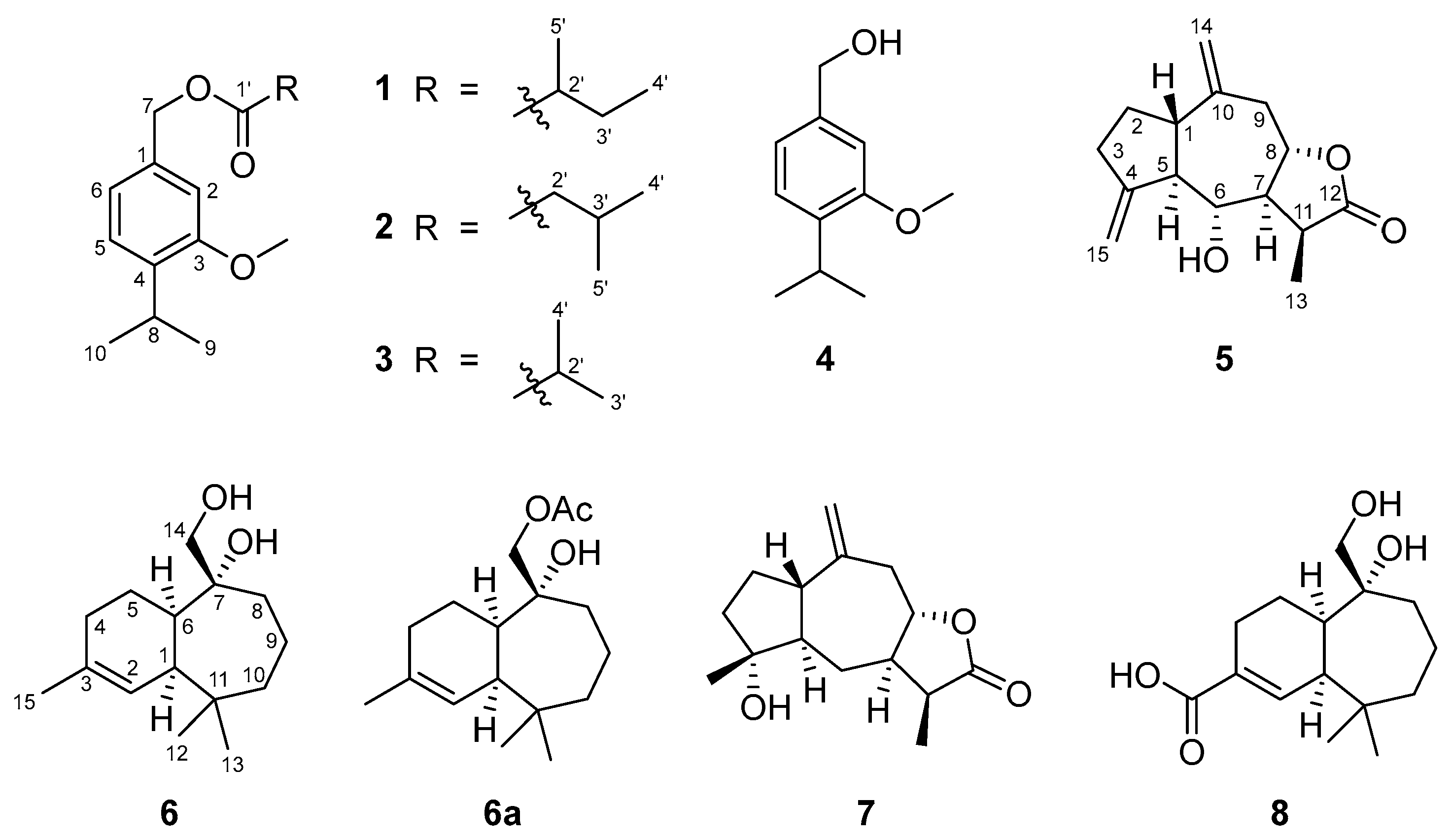
Figure 2.
a) Key TOCSY, HMBC and NOE correlations for compound 5. b) Selected 1D NOE correlations for compound 5.
Figure 2.
a) Key TOCSY, HMBC and NOE correlations for compound 5. b) Selected 1D NOE correlations for compound 5.
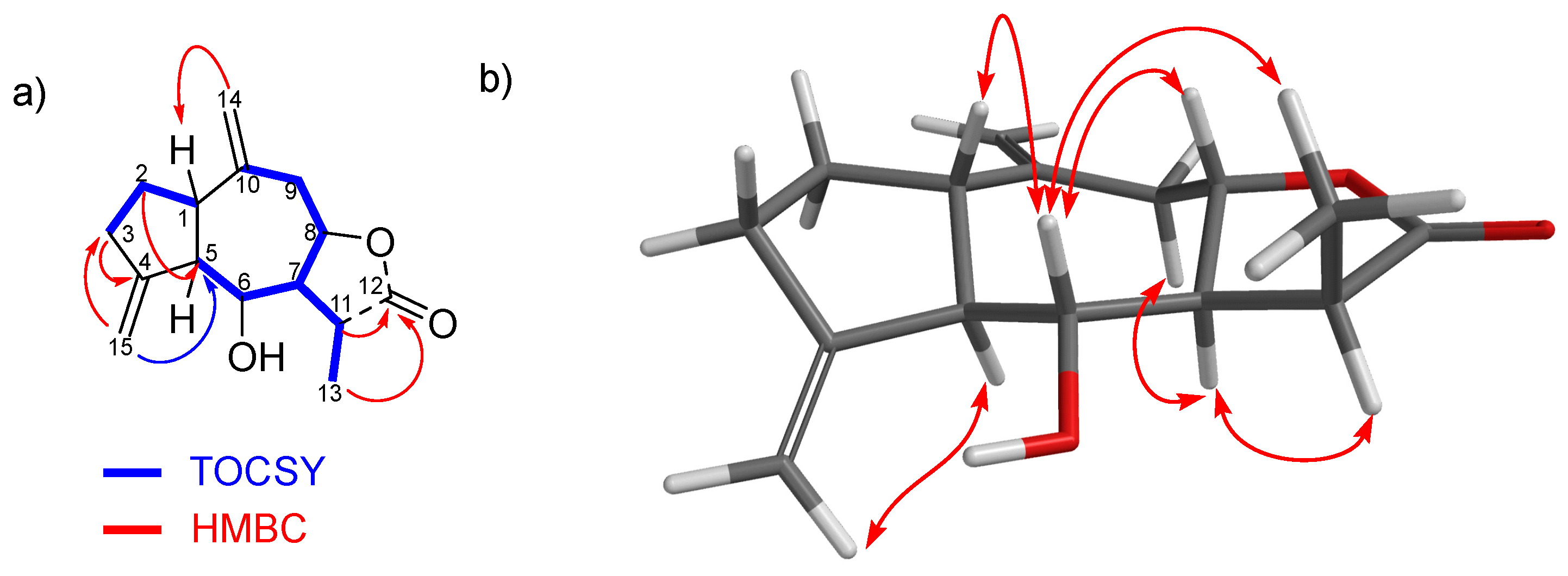
Figure 3.
a) Key TOCSY, HMBC and NOE correlations for compound 6. b) Selected 1D NOE correlations for compound 6.
Figure 3.
a) Key TOCSY, HMBC and NOE correlations for compound 6. b) Selected 1D NOE correlations for compound 6.
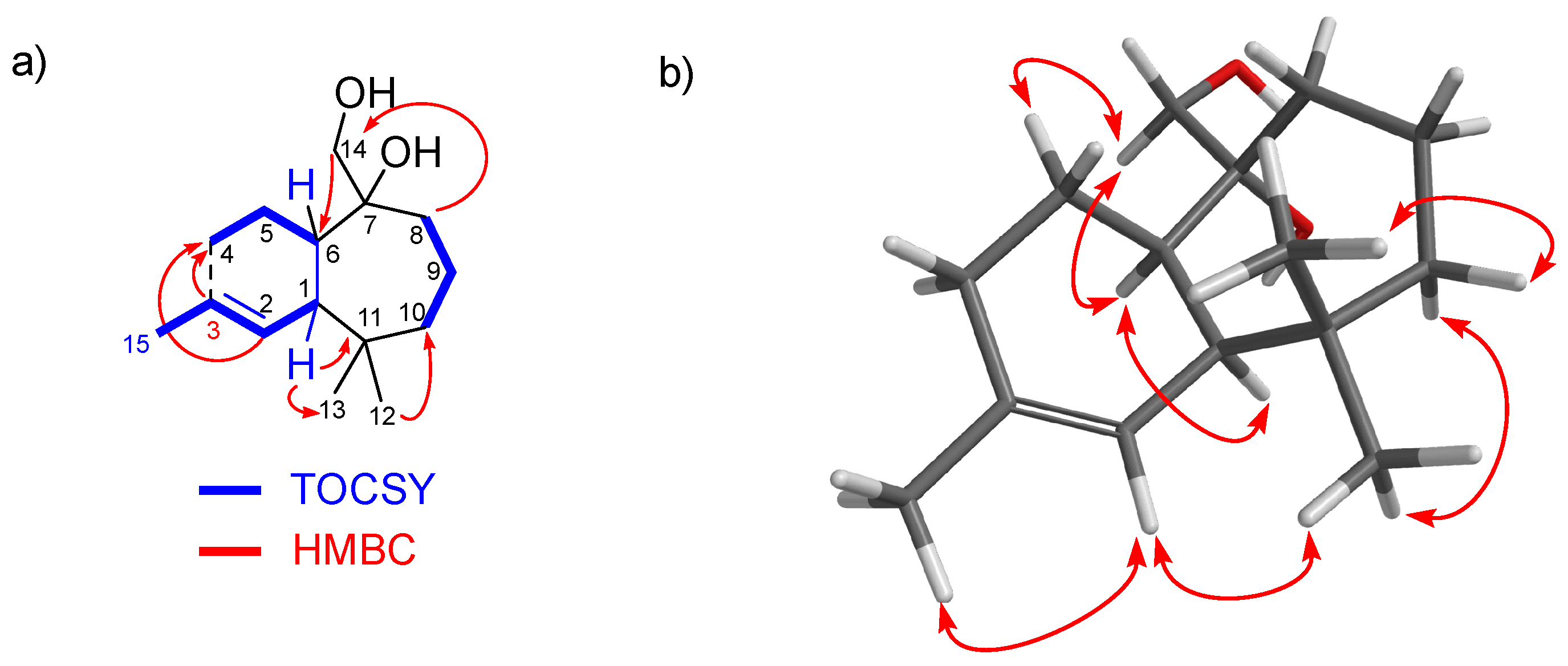
Figure 4.
Superimposition of the two computationally-calculated main conformations of 6. The blue one corresponds to conformation M01 of R*-epimer at C7. The green one corresponds to another conformation that shows 13C NMR data discordant with the experimental values.
Figure 4.
Superimposition of the two computationally-calculated main conformations of 6. The blue one corresponds to conformation M01 of R*-epimer at C7. The green one corresponds to another conformation that shows 13C NMR data discordant with the experimental values.
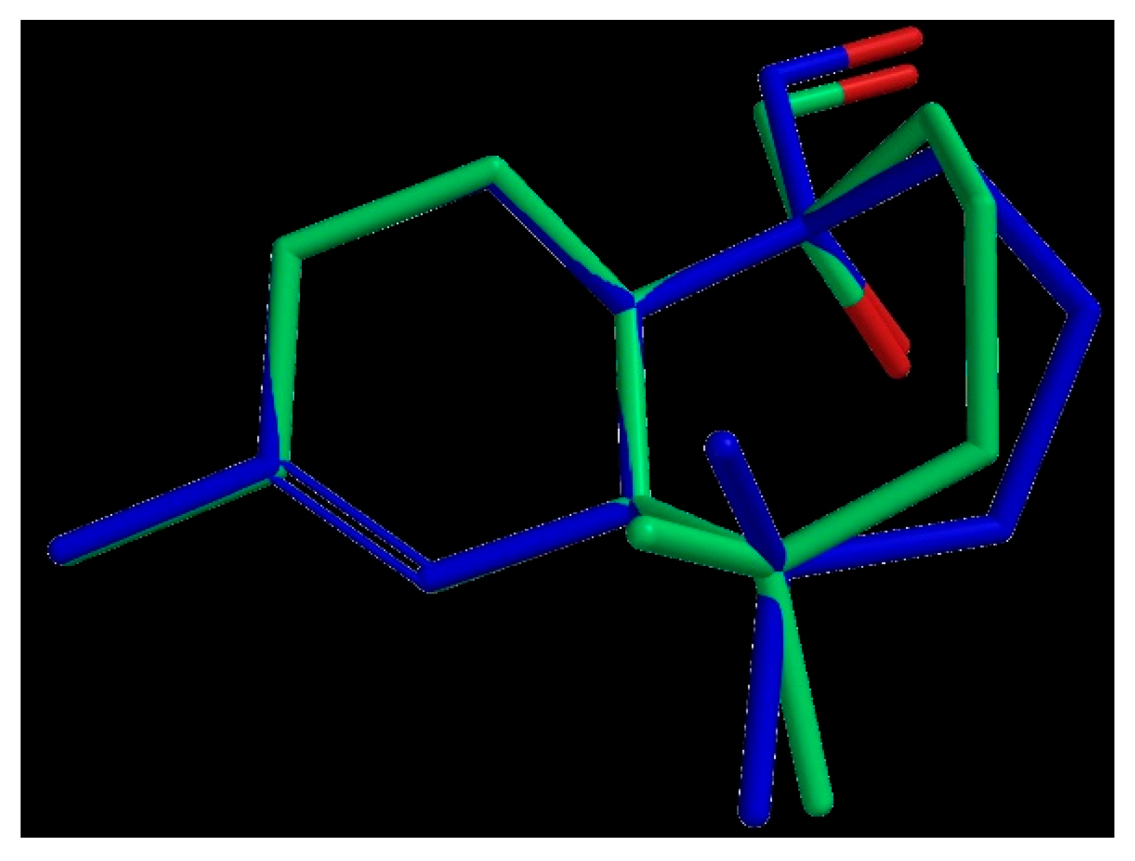
Figure 5.
Selected 1D NOE correlations for compound 8.
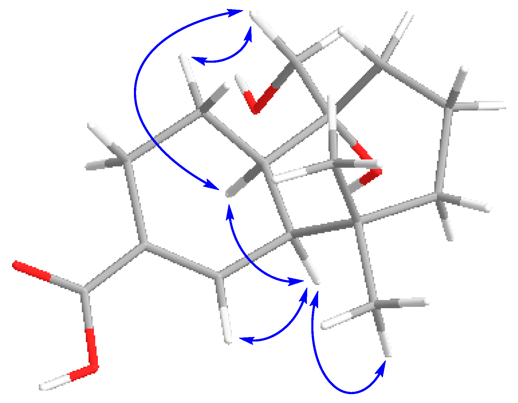
Figure 6.
Phytotoxicity (% relative to the control) of the compounds tested (1+2+3, 4, 5 and 6a) against Lolium perenne and Lactuca sativa. The dose tested was 0.1 mg/ml, and lower (0.05) for the active compounds (until the effects are < 50%).
Figure 6.
Phytotoxicity (% relative to the control) of the compounds tested (1+2+3, 4, 5 and 6a) against Lolium perenne and Lactuca sativa. The dose tested was 0.1 mg/ml, and lower (0.05) for the active compounds (until the effects are < 50%).
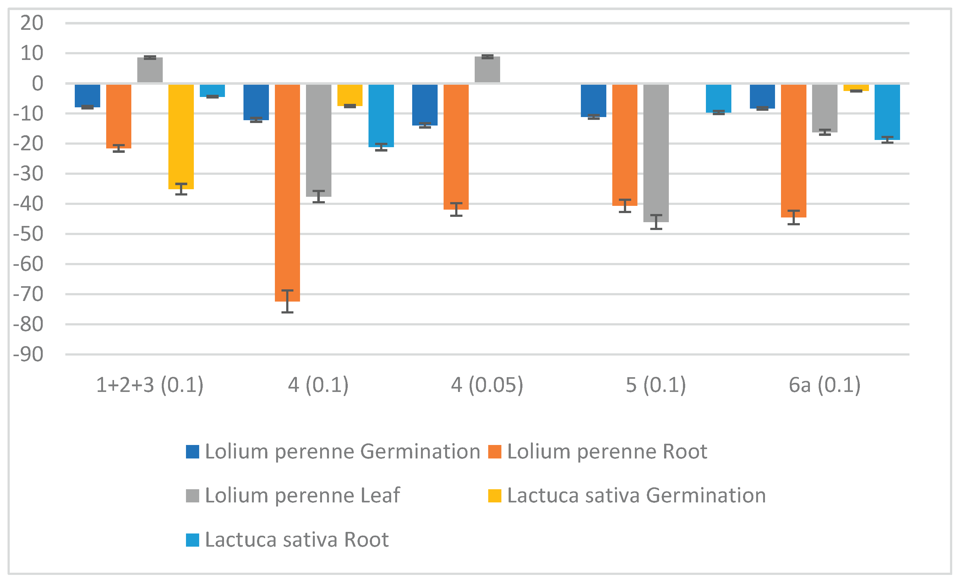

Table 1.
1H NMR spectroscopic data for compounds 5, 6 and 8 in CDCl3 (δ in ppm, J in Hz).
| Position | 5 | 6 | 8 |
|---|---|---|---|
| 1 | 2.23, m | 2.23, bs | 2.58, bs |
| 2a | 1.93, dt (11.1, 5.3) | 5.61, dd (5.6, 2.2) | 7.32, d (6.0) |
| 2b | 1.69, m | ||
| 3a | 2.40, m | ||
| 3b | 2.33, m | ||
| 4a | 2.01, bd (8.2) | 2.43, bd (18.6) | |
| 4b | 2.26, dt (18.6, 9.0) | ||
| 5a | 2.24, m | 1.70, m | 1.71, m |
| 5b | 1.44, m | 1.57, m | |
| 6 | 3.47, bt (9.5) | 1.96, m | 1.97, d (13.3) |
| 7 | 2.34, td (10.5, 7.6) | ||
| 8a | 4.54, td (11.1, 3.4) | 1.64, m | 1.66, m |
| 8b | 1.54, m | 1.54, m | |
| 9a | 3.17, dd (16.3, 3.1) | 1.69, m | 1.75, m |
| 9b | 2.63, dd (16.3, 11.6) | ||
| 10a | 1.90, m | 2.02, m | |
| 10b | 1.42, dq (14.4, 5.0) | 1.46, ddd (14.2, 6.6, 3.3) | |
| 11 | 2.92 p (7.7) | ||
| 12 | 1.01, s | 1.12, s | |
| 13a | 1.34, d (7.7) | 0.88, s | 0.89, s |
| 13b | |||
| 14a | 4.93, bs | 3.49, d (10.8) | 3.57, d (11.0) |
| 14b | 4.91, bs | 3.39, d (10.8) | 3.39, d (11.0) |
| 15a | 5.10, bs | 1.68, bs | |
| 15b | 4.99, bs |
Table 2.
13C NMR spectroscopic data for compounds 5, 6 and 8 in CDCl3 (δ in ppm).
| Position | 5 | 6 | 8 |
|---|---|---|---|
| 1 | 45.93 (CH) | 43.63 (CH) | 43.78 (CH) |
| 2 | 30.06 (CH2) | 125.31 (CH) | 146.49 (CH) |
| 3 | 34.20 (CH2) | 133.52 (C) | 128.93 (C) |
| 4 | 152.50 (C) | 31.22 (CH2) | 25.38 (CH2) |
| 5 | 54.94 (CH) | 21.27 (CH2) | 20.42 (CH2) |
| 6 | 71.19 (CH) | 45.86 (CH) | 45.92 (CH) |
| 7 | 54.83 (CH) | 77.49 (C) | 77.22 (C) |
| 8 | 76.66 (CH) | 30.49 (CH2) | 31.36 (CH2) |
| 9 | 39.64 (CH2) | 19.53 (CH2) | 19.27 (CH2) |
| 10 | 144.08 (C) | 41.05 (CH2) | 41.38 (CH2) |
| 11 | 39.06 (CH) | 38.47 (C) | 38.84 (C) |
| 12 | 179.14 (C) | 33.73 (CH3) | 33.50 (CH3) |
| 13 | 10.48 (CH3) | 25.74 (CH3) | 27.04 (CH3) |
| 14 | 110.67 (CH2) | 70.13 (CH2) | 69.65 (CH2) |
| 15 | 109.61 (CH2) | 26.60 (CH3) | 171.61 (C) |
Table 3.
Theorical 13C RMN data of the two possible C7 epimers of compound 6 and their comparison with the experimental values.
Table 3.
Theorical 13C RMN data of the two possible C7 epimers of compound 6 and their comparison with the experimental values.
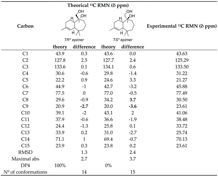
Max absolute expresses the largest deviations between the calculated and experimental chemical shifts; rmsd is a statistical parameter: δ13C root-mean-square deviation. The DP4 (%) probability score were calculated for the two epimers according to Goodman’s procedure.
Table 4.
13C NMR spectroscopic data for compounds 5, 6 and 8 in CDCl3 (δ in ppm).,l,l,l,nl,nl,l,lg,nl.
Table 4.
13C NMR spectroscopic data for compounds 5, 6 and 8 in CDCl3 (δ in ppm).,l,l,l,nl,nl,l,lg,nl.
| Compound | μg/cm2 | S. littoralis | M. persicae | R. padi |
|---|---|---|---|---|
| %FI2 (N = 6-10) | %SI2 (N = 20) | |||
| 1+2+3 | 50 | 73.13 ± 10.01 | 70.33 ± 7.72 | 71.30 ± 6.12 |
| EC501 | 32.35 (23.82-43.93) | 16.54 (9.17-29.84) | 19.14 (11.41-32.08) | |
| 4 | 50 | 41.70 ± 8.91 | 57.65 ± 8.49 | 56.53 ± 8.03 |
| EC501 | >50 | ≈50 | ≈50 | |
| 5 | 50 | 3.60 ± 3.65 | 81.69 ± 5.21 | 65.41 ± 5.45 |
| EC501 | >50 | 18.71 (14.00-25.01) | ≈50 | |
| 6a | 50 | 86.69 ± 4.26 | 55.31 ± 7.43 | 52.45 ± 7.38 |
| EC501 | 22.10 (15.34-31.84) | >50 | ≈50 | |
1 EC50: Effective dose to give 50% effect. 2%FI/SI = [1-(consumption/settling on treated disk / consumption/settling on control disk)] x 100.
Table 5.
In vitro nematicidal effects of compounds 1+2+3, 4, 5 and 6a against M. javanica J2 and minimal lethal dose (MLD).
Table 5.
In vitro nematicidal effects of compounds 1+2+3, 4, 5 and 6a against M. javanica J2 and minimal lethal dose (MLD).
| Compound | mg/mL | M. javanica | MLD |
|---|---|---|---|
| 1-3 | 0.5 | 25.83 ± 7.20 | >0.5 |
| 4 | 0.5 | 100.00 | 0.5 |
| 5 | 0.5 | 0.28 ± 0.12 | >0.5 |
| 6a | 0.5 | 0.00 | >0.5 |
Disclaimer/Publisher’s Note: The statements, opinions and data contained in all publications are solely those of the individual author(s) and contributor(s) and not of MDPI and/or the editor(s). MDPI and/or the editor(s) disclaim responsibility for any injury to people or property resulting from any ideas, methods, instructions or products referred to in the content. |
© 2026 by the authors. Licensee MDPI, Basel, Switzerland. This article is an open access article distributed under the terms and conditions of the Creative Commons Attribution (CC BY) license (http://creativecommons.org/licenses/by/4.0/).
Copyright: This open access article is published under a Creative Commons CC BY 4.0 license, which permit the free download, distribution, and reuse, provided that the author and preprint are cited in any reuse.


