

Submitted:
03 February 2026
Posted:
04 February 2026
You are already at the latest version
Abstract
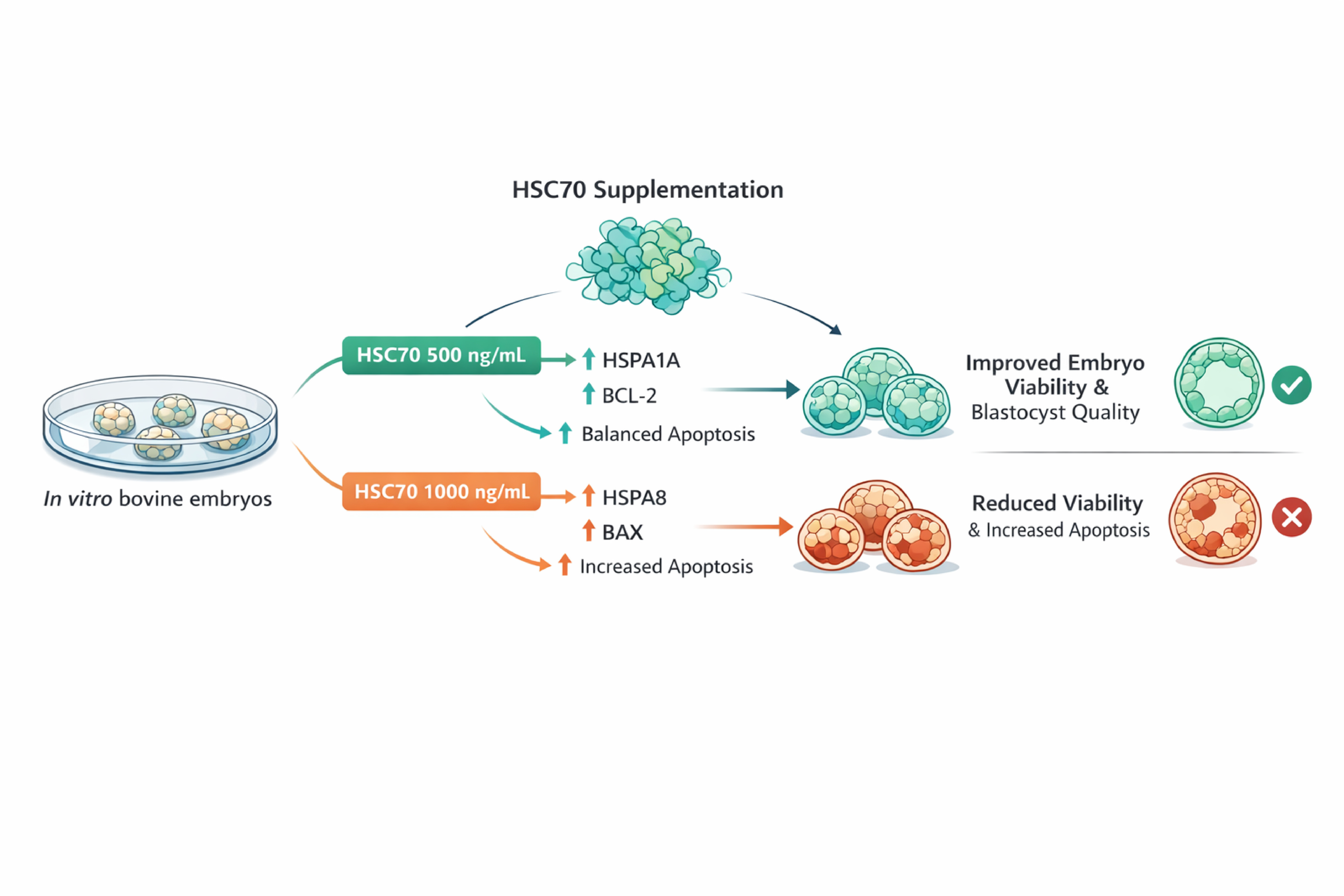
Endogenous heat shock cognate 73 kDa protein (HSC70) has an important role in early embryonic development. We assessed the effects of exogenous HSC70 on bovine embryo development and the expression of genes associated with apoptosis. Expression analyses of HSPA1A, HSPA8, BCL-2, and BAX genes were performed in bovine embryos in vivo on day 7 of development. The expression of HSPA1A and HSPA8 was associated with apop-totic gene (BCL-2 and BAX) expression in cultured bovine embryos in vitro that were sup-plemented with various concentrations (500 ng/mL or 1000 ng/ mL) of HSC70. The results indicated that the control group of in vitro embryos exhibited higher expression of the HSPA8, BAX, and BCL-2 genes compared with in vivo embryos (p ≤ 0.001). In vitro-produced embryos supplemented with 500 or 1000 ng/mL of HSC70 exhibited high-er expression of HSPA1A, HSPA8, BCL-2, and BAX genes compared with the control group (p ≤ 0.01). Embryos supplemented with 1000 ng/mL showed higher expression of the HSPA8 gene compared with the control group and the group supplemented with 500 ng/mL. However, embryos supplemented with 500 ng/mL exhibited more favorable char-acteristics (i.e., development stage and quality) compared with the control and 1000 ng/mL-treated groups. In conclusion, supplementation of bovine embryo culture media with 500 ng/mL recombinant HSC70 protein increased the expression of the HSPA1A and BCL-2 anti-apoptotic genes, resulting in an increase of the number of blastocysts produced in vitro.
Keywords:
bovine
; blastocyst
; HSC70
; apoptosis
; gene expression
1. Introduction
In vitro embryonic development is regulated by factors such as the origin of gametes, culture conditions, the microenvironment, and environmental stress. The post-fertilization culture period is a time when important developmental events occur that are a key determinant of blastocyst quality (Rizos et al., 2002). During vitro culture, embryos are exposed to various stresses that are not normally encountered within the bovine reproductive tract (Gordon, 2003).
Exogenous stress factors, such as exposure to light, high concentrations of oxygen, and culture medium composition, induce metabolic stress in oocytes, blastocysts, and reactive oxygen species (ROS) (Thiyagarajan and Valivittan, 2009). Lipid peroxidation, enzyme inactivation, protein modification, and DNA fragmentation may result in apoptosis and a developmental block of the oocyte to the blastocyst stages (Johnson and Nasr-Esfahani, 1994; Kowaltowski and Vercesi, 1999).
Apoptosis is an essential programmed cell death pathway in the development process. It eliminates redundant or superfluous cells to allow for normal patterning to proceed (Jacobson et al., 1997). Irreparably stressed and damaged cells also use this route for removal. Pathological stimuli, such as radiation, chemotherapy, and environmental toxicants, can initiate apoptosis in oocytes (Perez et al., 1997; Hu et al., 2001; Matikainen et al., 2001). Heat shock can also induce apoptosis in pre-implanted embryos (Paula Lopes and Hansen, 2002; Krininger et al., 2002; Paula Lopes and Hansen, 2002).
To improve the developmental proficiency of pre-implanted blastocysts, oocyte maturation and in vitro culture media have been supplemented with antioxidants, such as peroxiredoxin II (Fakruzzaman et al., 2015), resveratrol (Lee et al., 2010; Salzano et al., 2014), green tea polyphenols (Wang et al., 2007) and melatonin (Gao et al., 2012; Mohseni et al., 2012). This enhances the quality of the embryos through the reduction of ROS, increased expression of anti-apoptotic genes (BCL-2), and decreased expression of pro-apoptotic genes (BAX and caspase-3).
Moreover, molecular chaperones protect cells from damage caused by physical and chemical hazards, such as increased temperature (e.g., heat shock), anoxia, hypoxia, metabolic stress-associated cytokines, nitrogen oxide, ethanol, heavy metals, apoptosis-inducing agents, and other chemical denaturants and drugs (Smith et al., 1998; Tsukahara et al., 2000). Chaperones, such as the heat shock proteins (HSPs), play an important role in facilitating protein folding and maintaining normal protein structure and function (Liu et al., 2012).
HSPs are primarily classified based on their molecular weight in kilodaltons (kDa) as HSP110, HSP100, HSP90, HSP70, HSP60, HSP40, and the small HSP family (20 to 25 kDa). Within the HSP70 family, constitutively expressed, HSC70 (73 kDa), is slightly upregulated during stress conditions. Heat shock cognate protein 70 (HSC70) is particularly abundant in mammalian embryos (Dworniczak and Mirault, 1987). It participates in the uptake of proteins into the nucleus and other cellular organelles, such as the endoplasmic reticulum and mitochondria, and maintains the translocation-competent state of proteins destined for these locations (Chirico et al., 1988; Deshaies et al., 1988; Sheffield et al., 1990). HSC70 maintains protein homeostasis during normal and stress conditions, suppresses protein aggregation, and reactivates heat-denatured proteins (Liu et al., 2012). Because of these protective functions associated with HSC70 and the low percentage of embryos produced in vitro, we evaluated the effect of HSC70 supplementation on bovine embryo viability in vitro.
2. Materials and Methods
2.1. Animals
Simbrah cows (n = 6) were maintained under standard managerial practices in Centro de Investigación y Producción Agropecuaria de la Universidad Autónoma de Nuevo León were used for in vivo embryo production.
Experiments were performed in accordance with Facultad de Medicina Veterinaria y Zootecnia de la Universidad Autónoma de Nuevo León guidelines for the care and use of animals (01/2021). Maturation (IVM), fertilization (FCDM), early culture (CDM1), and late culture (CDM2) media were supplemented with fatty acid-free bovine serum albumin prepared at the National Center for Genetic Resources of the National Institute for Agricultural and Livestock Forestry Research (Jalisco, Mexico) according to data published by De La Torre Sanchez et al. (2006).
2.2. In Vivo Embryo Production
Estrus was synchronized using an intravaginal progesterone–releasing device (CIDR) (Pfizer, NY, USA) for 7 days. Cows were treated with 2.0 mg i.m. of Syntex estradiol benzoate (Zoetis, Mexico City, Mexico) at the time insertion of the CIDR device (day 0). On days 4, 5, and 6, 360 mg i.m. of Folltropin-V, follicle-stimulating hormone (Bioniche, Rockford, IL, USA) was administered. On day 6, the cows were treated with two doses (25 mg i.m. every 12 h) of dinoprost tromethamine (Lutalyse, Zoetis, Mexico City, Mexico). On day 7, the CIDR withdrawal time, another dose of follicle-stimulating hormone was administered. Artificial insemination was performed twice on day 8, in the morning and afternoon.
Seven days after artificial insemination, the embryos were recovered using two-way Foley catheters. ViGRO complete flush solution (Bioniche, Rockford, IL, USA) was introduced and the medium containing the embryos was obtained. Embryos were washed using the same solution and placed into Petri dishes. A stereoscopic microscope-assisted search for embryos and identification (stage of development and quality of embryos) was performed based on the Manual of the International Embryo Technology Society (IETS) guidelines. Subsequently, blastocysts were stored in pools of three per Eppendorf tube with 100 μl Trizol Reagent (Invitrogen, Carlsbad, CA) at 5 °C until RNA extraction and real-time quantitative reverse transcription (qPCR) analysis.
2.2. Oocyte Retrieval and In Vitro Maturation
Ovaries were obtained from the local abattoir and transported to the laboratory within 2 h in physiological saline (0.9% NaCl). Cumulus-oocyte complexes were recovered from follicles at a diameter of 3–6 mm using an 18-gage needle attached to a 10-mL syringe. Follicular fluid and the cell package were placed into a 50 ml conical tube at 38.5 °C. Selected cumulus-oocyte complexes with a uniform granular cytoplasm and surrounded by a multiple compact mass of cumular cells, were cultivated in IVM medium for 23 h at 38.5 °C in a humidified atmosphere of 5% CO2 in air. The medium included 2 µL/mL FSH (NIDDK-o FSH-20), 1 µL/mL LH (NIH-LH-S1), 5 µL/mL hCG (10,000 IU), 10 µL/mL β-estradiol (1 mg), cysteamine (0.1 mM), and 1 µL/mL epidermal growth factor (0.5 mg) (Sigma–Aldrich, St. Louis, MO, USA) based on previously published data (De La Torre Sanchez, 2004).
2.3. In Vitro Fertilization and Culture
Mature oocytes (n = 600) were transferred to FCDM medium for co-culture with spermatozoa. Semen was thawed and processed using a Percoll gradient (Sigma–Aldrich, St. Louis, MO, USA) (45:90%). Mature oocytes and sperm were incubated together at 5% CO2 in humidified air at 38.5 °C for 18 h prior to vitro culture.
After co-incubation, cumulus cells were removed using a pipette. The presumptive zygotes were washed, randomly divided (n = 160 per group), and transferred to 100 µL CDM1 medium supplemented with HSC70 (Sigma–Aldrich, St. Louis, MO, USA) at 500 or 1000 ng/mL. In addition, a control group was established without supplementation. The samples were incubated for 56 h at 38.5 °C in a humidified atmosphere of 5% CO2, 5% O2, and 90% N2 in air.
Embryos were transferred and cultured in CDM2 medium and supplemented with HSC70 (Sigma–Aldrich, St. Louis, MO, USA) (500 and 1000 ng/mL) or not (control group) according to the group in which they previously belonged. For gene expression assays, embryos were maintained for 48 h. For morphological analysis, embryos were maintained in culture for 120 h under the conditions described above. At the end of the culture period, blastocysts were identified and classified according to IETS manual numerical codes corresponding to the developmental stage and quality.
2.4. RNA Extraction Using a Modified Trizol Method and Complementary DNA (cDNA) Synthesis
Using a modified Trizol protocol (Pavani et al., 2015), RNA was isolated from three biological replicates. Embryos (≥8 cells, in pools of three) were added to Eppendorf tubes with 100 μl Trizol (Invitrogen, Carlsbad, CA). The samples were incubated at room temperature for 3 min. Next, 50 µl chloroform was added, the samples were inverted for 15 s, and then centrifuged at 12,000 g for 30 min at 4 °C. The aqueous phase was transferred to a fresh tube and RNA was precipitated with the addition of 2.5 volumes of isopropanol. Following centrifugation at 12,000 g for 30 min at 4 °C, the supernatant was discarded, the pellet was washed with 50 µl 70% ethanol, and centrifuged at 7,500 g for 5 min. Finally, the pellet was dried in an incubator for 30 min at 37 °C and dissolved in 20 µl of DEPC water.
The eluted RNA was reverse-transcribed into cDNA using the GoTaq Probe 2-step qRT-PCR System kit (Promega, Madison, WI, USA). Briefly, 5 µl RNA, 1 µl oligo dT (0.5 mg/µl), and 1 µl of random primer were added to each tube. Subsequently, the tubes were incubated in a heat block at 70 °C for 5 min and immediately placed on ice for at least 5 min. Then, 4.9 µl nuclease-free water, 4 µl GoScript 5X Reaction Buffer, 1.6 µl MgCl2, 1 µl dNTPs, 0.5 µl Recombinant RNasin Ribonuclease Inhibitor, and 1 µl GoScript Reverse Transcriptase were combined and incubated 25 °C for 5 min. Subsequently, the reaction was extended in a controlled-temperature heat block at 42 °C for 45 min and inactivated at 70 °C for 15 min. The resulting complementary DNA was quantified using a Quantus fluorometer (Promega, Madison, WI, USA) and stored at −20 °C until use.
2.5. Quantitative Real-Time PCR Analysis
Real-time RT-PCR was performed with an Applied Biosystems 7500/7500 Fast detection system (Thermo Fisher Scientific, Waltham, MA, USA). All primers and probes used were designed by Integrated DNA Technologies (Coral Ville, IA, USA). PrimeTime probes were synthesized and labeled with HEX and FAM (Table 1). The measurement of HSP1A, HSPA8, BAX, and BCL-2 mRNA was done in triplicate using the GoTaq Probe 2-step qRT-PCR System kit (Promega, Madison, WI, USA), in a 20 μl reaction mixture containing GoScript 5X reaction buffer, 25 mM MgCl2, 10 mM dNTP mix, 40 u/µL Recombinant RNasin ribonuclease inhibitor, GoScript Reverse Transcriptase, 500 µg/ml random primer, ROX, and 250 nM of probe.
The amplification conditions for all genes were as follows: 50 °C for 2 min, 95 °C for 10 min, followed by 40 cycles of 95 °C for 15 s, 60 °C for 15 s, and 60 °C for 15 s. The gene results were analyzed according to the comparative ΔΔCt method and are reported as relative expression or fold-difference in expression to a housekeeping gene (GAPDH).
2.6. Statistical Analysis
Gene expressions were analyzed using analysis of variance followed by multiple pairwise comparison tests. Dunn’s test was used to compare embryos in vitro versus in vivo, Bonferroni tests were used to compare excellent or good quality embryos with poor quality embryos, and Kruskal–Wallis tests were used to compare HSC70 protein-supplemented versus control embryos. GraphPad Prism version 4.0 software (GraphPad Software Inc., San Diego, CA, USA) was used to graph the results.
3. Results
3.1. Expression of HSPA1A and HSPA8 Genes and Their Relationship with Apoptotic Gene Expression in Bovine Embryos In Vivo and In Vitro
The basal expression levels of three of the four genes (HSPA8, BCL-2, and BAX) examined were significantly higher in vitro embryos compared with that of in vivo embryos (**p ≤ 0.001/ ** p ≤ 0.01) (Figure 1). HSPA1A did not show a significant difference in expression between in vitro and in vivo embryos.
The expression of the HSPA1A gene was significantly different (**p ≤ 0.001) between degenerate quality embryos compared with excellent or good quality embryos (transferable), and expression of the anti-apoptotic BCL-2 gene was higher in transferable embryos compared with degenerate quality embryos. In contrast, the expression of the pro-apoptotic BAX gene was higher in degenerate quality embryos compared with the treated group (Figure 2).
The relative gene expression differences in the embryos supplemented with 500 ng/mL of HSC70 protein compared with the control group were evaluated. There was a six-fold up-regulation of HSPA1A mRNA levels, three-fold up-regulation of HSPA8 mRNA levels, an almost three-fold up-regulation of BCL-2 mRNA levels, and a four-fold up-regulation of BAX mRNA levels. Following supplementation with 1000 ng/mL, a four-fold up-regulation of HSPA1A mRNA levels, eight-fold up-regulation of HSPA8 mRNA levels, an almost two-fold up-regulation of BCL-2 mRNA levels, and an almost five-fold up-regulation of BAX mRNA levels compared with the control group.
The results also indicated that there was higher expression of the HSPA1A gene, which encodes the stress-inducible protein, HSP70, in embryos supplemented with 500 and 1000 ng/mL of HSC70 (**p ≤ 0.01). Supplementation with high doses (1000 ng/mL HSC70) resulted in a marked difference in HSPA8 expression compared with the other treatments (control and 500 ng/mL; ***p ≤ 0.001).
The results for the apoptosis genes indicated that the anti-apoptotic BCL-2 gene exhibited significantly higher expression at low supplementation doses (500 ng/mL, * p ≤ 0.01); whereas the pro-apoptotic BAX gene showed higher expression in both treatment groups compared with the control group (*p ≤ 0.01) (Figure 3).
Embryos obtained for morphological analysis that were supplemented with 500 ng/mL of HSC70 exhibited improved developmental and quality characteristics, in which 57% of the resulting embryos (18% morulae, 41% blastocysts, and 41% expanded blastocysts) were categorized as excellent or good quality. This was followed by the control group, with 40% of the embryos (11% morulae, 28% blastocysts, 44% expanded blastocysts, and 17% hatched blastocysts) categorized as regular quality. When supplemented with 1000 ng/mL, 33% of the embryos (25% blastocysts with good quality, remainder degenerate embryos) were categorized as poor quality.
4. Discussion
HSPs are molecular chaperones that protect cells from physical or chemical damage (Smith et al., 1998; Tsukahara et al., 2000). They promote cell survival by regulating homeostasis (Rodriguez-Ariza et al., 2005). In this study, we investigated the effects of supplementation with the HSC70 protein on the viability and expression of apoptosis genes in bovine embryo cultures. HSPs are among the first proteins produced during embryonic development and have important cell functions. Although they have been studied extensively in humans and mice, there is little information on the function of genes that encode HSPs in bovine embryos (Zhang et al., 2011).
Identification of HSP genes that specifically contribute to embryo survival may provide an opportunity to enhance the protection of embryos in vitro against various toxic conditions and improve the gestation rates of cattle (Hansen, 2007). Embryos produced in vitro exhibit changes in gene expression that reflect the stress response to sub-optimal conditions. During embryonic development, conditions such as oxidative stress are associated with changes in the expression of genes, such as BAX and SOX (Rizos et al., 2002; Lonergan et al., 2003a; Lonergan et al., 2003b). In addition, it has been observed that a significantly higher expression of HSPA1A occurs in vitro-generated equine embryos and a lower-quality in in vivo embryos compared with higher quality in vivo-produced embryos (Mortensen et al., 2010). The results of this study are consistent with these characteristics, HSPA1A was differentially expressed between excellent or good quality embryos compared with degenerate quality embryos. Poorly developing embryos exhibited higher expression of HSP70. Relatively small changes (decreases or increases) in HSP expression can result in growth abnormalities and cell death (Nollen and Morimoto et al., 2002).
The results of this study with respect to differential expressions of HSPA8 (which encodes to heat shock cognate 70), BCL-2, and BAX genes between embryos obtained in vivo and those produced in vitro, are consistent with the data published by Wrenzycki et al. (2001). They hypothesized that embryos produced in vitro are subject to greater stress conditions during production, such as oxygen tension and the composition of the medium used during culture. Generally, the increase in oxygen tension during in vitro embryo production increases the generation of ROS, which causes DNA damage, lipoperoxidation, and oxidative modification of proteins that increase susceptibility to proteolysis (Johnson and Nasr-Esfahani, 1994; Wang et al., 2007). Therefore, antioxidants are used in oocyte maturation media and in vitro culture to improve the development of pre-implanted blastocysts (Wang et al., 2007; Salzano et al., 2014; Wang et al., 2014; Fakruzzaman et al., 2015; Rodrigues-Cunha et al., 2016).
Culture media supplemented with HSPs may be used to reduce the toxicity and aggregation of polyglutamine in cells with Huntington’s disease. Novoselova et al. (2005) purified HSP70 and HSC70 from bovine muscle, which were added to the culture media of cells transfected with the Huntington’s disease gene. Compared with the control group (without supplementation), the number of apoptotic cells decreased up to 50%, and apoptosis resulted from the aggregation of insoluble proteins. In the present study, we found that supplementation of embryo culture media with protein HSC70 promoted cell viability. HSC70 protein stimulated the expression of HSPA1A, HSPA8, BCL-2, and BAX compared with the control group. In addition, low dose (500 ng/mL) supplementation also resulted in higher expression of HSPA1A and in embryos with better characteristics at the time of stage and quality identification. These results may be attributed to the regulation of anti- and pro-apoptotic (BCL-2 and BAX, respectively) gene expression.
HSPs are molecular chaperones that are upregulated after cells are exposed to different stressors. They exhibit a protective effect that enables cells to survive potentially lethal conditions (Liu et al., 2012). HSP70 has an anti-apoptotic function (Lanneau et al., 2007; Arya et al., 2007), which we also observed by an almost three-fold up-regulation in BCL-2 mRNA in the group supplemented with 500 ng/mL compared with the control. This group exhibited better developmental and quality characteristics, which could be related to the anti-apoptotic effects. HSP70 interacts with the intrinsic and extrinsic pathways of apoptosis at several steps, which include inhibition of BAX translocation to the mitochondria, release of cytochrome c from mitochondria, apoptosome formation, and inhibition of caspase activation and activity. It also modulates the c-Jun N-terminal kinase, nuclear factor kappa B, and Akt signaling pathways in the apoptotic cascade.
The results of this study were consistent with that of Arya et al. (2007), in which HSC70 exhibited a pro-apoptotic effect. In the present study, we found that supplementation with high doses (1000 ng/mL) of HSC70 resulted in a low number of blastocysts, which is related to increased expression of the pro-apoptotic BAX gene.
5. Conclusions
The supplementation of bovine embryo culture media with 500 ng/mL recombinant protein HSC70 increased the expression of the HSPA1A gene, and the number of higher quality blastocysts produced in vitro.
Author Contributions
Conceptualization, A.J.G.A. and G.M.D.; methodology, D.E.Z.A. D.M.G.H and U.C.V; validation, R.C.V., A.J.G.A and D.E.Z.A.; formal analysis, U.C.V.; investigation, J.F.T.S. and S.P.R; resources, G.M.D.; data curation, A.J.G.A. D.M.G.H; writing—original draft preparation, A.J.G.A.; D.E.Z.A.; U.C.V. writing—review and editing, A.J.G.A.; D.E.Z.A.; U.C.V.; visualization, X.X.; supervision, R.C.V.; funding acquisition, G.M.D. All authors have read and agreed to the published version of the manuscript.
Funding
This research received no external funding.
Institutional Review Board Statement
Experiments were conducted in accordance with the guidelines for the care and use of laboratory animals of the Facultad de Medicina Veterinaria y Zootecnia, Universidad Autónoma de Nuevo León, and were approved under protocol number 01/2021.
Informed Consent Statement
Not applicable.
Data Availability Statement
The original contributions presented in this study are included in the article. Further inquiries can be directed to the corresponding author(s).
Acknowledgments
We thank the Aquatic-Livestock Laboratory of the National Center for Genetic Resources of the National Institute for Agricultural and Livestock Forestry Research (Tepatitlán de Morelos, Jalisco, México) for use of the facilities to perform the experiments in this study.
Conflicts of Interest
The authors declare no conflicts of interest.
References
- Arya, R; Mallik, M; Lakhotia, SC. Heat shock genes-integrating cell survival and death. J Biosci 2007, 32, 595–610. [Google Scholar] [CrossRef]
- Chirico, WJ; Walters, MG; Blobel, G. 70 K heat shock related proteins stimulate protein translocation into microsomes. Nature 1988, 332, 805–810. [Google Scholar] [CrossRef]
- De La Torre-Sanchez, JF; Preis, K; Seidel, GE, Jr. Metabolic regulation of in-vitro-produced bovine embryos. I. Effects of metabolic regulators at different glucose concentrations with embryos produced by semen from different bulls. Reprod Fertil Dev 2006, 18(5), 585–596. [Google Scholar] [CrossRef]
- De La Torre-Sanchez, JF. Regulation of glucose metabolism in bovine embryos. Unpublised doctoral dissertation thesis, Colorado State University, Fort Collins, Colorado, USA, 2004. [Google Scholar]
- Deshaies, RJ; Koch, BD; Werner-Washburne, M; Craig, EA; Schekman, R. A subfamily of stress proteins facilitates translocation of secretory and mitochondrial precursor polypeptides. Nature 1988, 332, 800–805. [Google Scholar] [CrossRef] [PubMed]
- Dworniczak, B; Mirault, ME. Structure and expression of a human gene coding for a 71 kd heat shock “cognate” protein. Nucleic Acids Res 1987, 15, 5181–5197. [Google Scholar] [CrossRef]
- Fakruzzaman, M; Ghanem, N; Bang, JI; Ha, AN; Lee, KL; Sohn, SH; Wang, Z; Lee, DS; Kong, IK. Effect of peroxiredoxin II on the quality and mitochondrial activity of pre-implantation bovine embryos. Anim Reprod Sci 2015, 159, 172–183. [Google Scholar] [CrossRef] [PubMed]
- Gao, C; Han, HB; Tian, XZ; Tan, DX; Wang, L; Zhou, GB; Zhu, SE; Liu, GS. Melatonin promotes embryonic development and reduces reactive oxygen species in vitrified mouse 2-cell embryos. J Pineal Res 2012, 52, 305–311. [Google Scholar] [CrossRef]
- Gordon, I. Laboratory production of cattle embryos, 2nd ed; CAB International: Wallingford, Oxon, UK, 2003; p. 273. [Google Scholar]
- Hansen, PJ. To be or not to be. Determinants of embryonic survival following heat shock. Theriogenology 2007, 68, 40–48. [Google Scholar] [CrossRef] [PubMed]
- Hu, X; Christian, PJ; Thompson, KE; Sipes, IG; Hoyer, PB. Apoptosis induced in rats by 4-vinylcyclohexene diepoxide is associated with activation of the caspase cascade. Biol Reprod 2001, 65, 87–93. [Google Scholar] [CrossRef]
- Jacobson, MD; Weil, M; Raff, MC. Programmed cell death in animal development. Cell 1997, 88, 347–354. [Google Scholar] [CrossRef]
- Johnson, M; Nasr-Esfahani, M. Radical solutions and cultural problems: could free oxygen radicals be responsible for the impaired development of preimplantation mammalian embryos in vitro? Bioessays 1994, 16, 31–38. [Google Scholar] [CrossRef]
- Kowaltowski, AJ; Vercesi, AE. Mitochondrial damage induced by conditions of oxidative stress. Free Radic Biol Med 1999, 26, 463–471. [Google Scholar] [CrossRef] [PubMed]
- Krininger, CE, III; Stephens, SH; Hansen, PJ. Developmental changes in inhibitory effects of arsenic and heat shock on growth of preimplantation bovine embryos. Mol Reprod Dev 2002, 63, 335–340. [Google Scholar] [CrossRef] [PubMed]
- Lanneau, D; de Thonel, A; Maurel, S; Didelot, C; Garrido, C. Apoptosis versus cell differentiation: role of heat shock proteins HSP90, HSP70 and HSP27. Prion 2007, 1, 53–60. [Google Scholar] [CrossRef]
- Lee, K; Wang, C; Chaille, JM; Machaty, Z. Effect of resveratrol on the development of porcine embryos produced in vitro. J Reprod Dev 2010, 56, 330–335. [Google Scholar] [CrossRef]
- Liu, T; Daniels, CK; Cao, S. Comprehensive review on the HSC70 functions, interactions with related molecules and involvement in clinical diseases and therapeutic potential. Pharmacol Ther 2012, 136, 354–374. [Google Scholar] [CrossRef] [PubMed]
- Lonergan, P; Rizos, D; Gutiérrez-Adán, A; Moreira, PM; Pintado, B; de la Fuente, J. Temporal divergence in the pattern of messenger RNA expression in bovine embryos cultured from the zygote to blastocyst stage in vitro or in vivo. Biol Reprod 2003a, 69, 1424–1431. [Google Scholar] [CrossRef]
- Lonergan, P; Rizos, D; Kanka, J; Nemcova, L; Mbaye, AM; Kingston, M; Wade, M; Duffy, P; Boland, MP. Temporal sensitivity of bovine embryos to culture environment after fertilization and the implications for blastocyst quality. Reproduction 2003b, 126, 337–346. [Google Scholar] [CrossRef]
- Matikainen, TM; Perez, GI; Jurisicova, A; Schlezinger, JJ; Ryu, H-Y; Pru, JK; Sakai, T; Korsmeyer, SJ; Casper, RF; Sherr, DH; Tilly, JL. Aromatic hydrocarbon receptor-driven Bax gene expression is required for premature ovarian failure caused by biohazardous environmental chemicals. Nat Genet 2001, 28, 335–360. [Google Scholar] [CrossRef]
- Mohseni, M; Mihandoost, E; Shirazi, A; Sepehrizadeh, Z; Bazzaz, JT; Ghazi-khansari, M. Melatonin may play a role in modulation of bax and bcl-2 expression levels to protect rat peripheral blood lymphocytes from gamma irradiation-induced apoptosis. Mutat Res 2012, 738–739, 19–27. [Google Scholar] [CrossRef]
- Mortensen, CJ; Choi, Y-H; Ing, NH; Kraemer, DC; Vogelsang, MM; Katrin-Hinrichs, K. Heat shock protein 70 gene expression in equine blastocysts after exposure of oocytes to high temperatures in vitro or in vivo after exercise of donor mares. Theriogenology 2010, 74, 374–383. [Google Scholar] [CrossRef]
- Nollen, EA; Morimoto, RI. Chaperoning signaling pathways: molecular chaperones as stress-sensing heat shock proteins. J Cell Sci 2002, 115, 2809–2816. [Google Scholar] [CrossRef]
- Novoselova, TV; Margulis, BA; Novoselov, SS; Sapozhnikov, AM; van der Spuy, J; Cheetham, ME; Guzhova, IV. Treatment with extracellular HSP70/HSC70 protein can reduce polyglutamine toxicity and aggregation. J Neurochem 2005, 94, 597–606. [Google Scholar] [CrossRef] [PubMed]
- Paula-Lopes, FF; Hansen, PJ. Apoptosis is an adaptive response in bovine preimplantation embryos that facilitates survival after heat shock. Biochem Biophys Res Commun 2002, 295, 37–42. [Google Scholar] [CrossRef]
- Paula-Lopes, FF; Hansen, PJ. Heat-shock induced apoptosis in bovine preimplantation embryos is a developmentally-regulated phenomenon. Biol Reprod 2002, 66, 1169–1177. [Google Scholar] [CrossRef]
- Pavani, KC; Baron, EE; Faheem, M; Chaveiro, A; Moreira Da Silva, F. Optimisation of total RNA extraction from bovine oocytes and embryos for gene expression studies and effects of cryoprotectants on total RNA extraction. Cytol Genet 2015, 49, 232–239. [Google Scholar] [CrossRef]
- Perez, GI; Knudson, CM; Leykin, L; Korsmeyer, SJ; Tilly, JL. Apoptosis associated signaling pathways are required for chemotherapy-mediated female germ cell destruction. Nat Med 1997, 3, 1228–1232. [Google Scholar] [CrossRef]
- Rizos, D; Lonergan, P; Boland, MP; Arroyo-Garcia, R; Pintado, B; de la Fuente, J; Gutiérrez-Adán, A. Analysis of differential mRNA expression between bovine blastocyst produced in different culture systems: implications for blastocyst quality. Biol Reprod 2002, 66, 589–595. [Google Scholar] [CrossRef] [PubMed]
- Rodrigues-Cunha, MC; Mesquita, LG; Bressan, F; del Collado, M; Balieiro, JCC; Schwarz, KRL; de Castro, FC; Watanabe, OY; Watanabe, YF; de Alencar Coelho, L; Leal, CLV. Effects of melatonin during IVM in defined médium on oocyte meiosis, oxidative stress, and subsequent embryo development. Theriogenology 2016, 86, 1685–1694. [Google Scholar] [CrossRef]
- Rodriguez-Ariza, A; López-Sánchez, LM; González, R; Corrales, FJ; López, P; Bernardos, A; Muntané, J. Altered protein expression and protein nitration pattern during D-galactosamine-induced cell death in human hepatocytes: a proteomic analysis. Liver Int 2005, 25, 1259–1269. [Google Scholar] [CrossRef]
- Salzano, A; Albero, G; Zullo, G; Neglia, G; Abdel-Wahab, A; Bifulco, G; Zicarelli, L; Gasparrini, B. Effect of resveratrol supplementation during culture on the quality and cryotolerance of bovine in vitro produced embryos. Anim Reprod Sci 2014, 151, 91–96. [Google Scholar] [CrossRef]
- Sheffield, WP; Shore, GC; Randall, SK. Mitochondrial precursor protein. Effects of 70-kilodalton heat shock protein on polypeptide folding, aggregation and import competence. J Biol Chem 1990, 265, 11069–11076. [Google Scholar] [CrossRef] [PubMed]
- Smith, DF; Whitesell, L; Katsanis, E. Molecular chaperones: biology and prospects for pharmacological intervention. Pharmacol Rev 1998, 50, 493–514. [Google Scholar] [CrossRef]
- Thiyagarajan, B; Valivittan, K. Ameliorating effect of vitamin E on in vitro development of preimplantation buffalo embryos. J Assist Reprod Genet 2009, 26, 217–225. [Google Scholar] [CrossRef]
- Tsukahara, F; Yoshioka, T; Muraki, T. Molecular and functional characterization of HSC54, a novel variant of human heat-shock cognate protein 70. Mol Pharmacol 2000, 58, 1257–1263. [Google Scholar] [CrossRef]
- Wang, F; Tian, X; Zhang, L; He, C; Ji, P; Li, Y. Beneficial effect of resveratrol on bovine oocyte maturation and subsequent embryonic development after in vitro fertilization. Fertil Steril 2014, 101, 577–586. [Google Scholar] [CrossRef] [PubMed]
- Wrenzycki, C; Herrmann, D; Keskintepe, L; Martins, Jr A; Sirisathien, S; Brackett, B; Niemann, H. Effects of culture system and protein supplementation on mRNA expression in preimplantation bovine embryos. Hum Reprod 2001, 16, 893–901. [Google Scholar] [CrossRef]
- Wang, ZG; Yu, SD; Xu, ZR. Improvement in bovine embryo production in vitro by treatment with green tea polyphenols during in vitro maturation of oocyte. Anim Reprod Sci 2007, 100, 22–31. [Google Scholar] [CrossRef] [PubMed]
- Zhang, B; Peñagaricano, F; Driver, A; Chen, H; Khatib, H. Differential expression of heat shock protein genes and their splice variants in bovine preimplantation embryos. J Dairy Sci 2011, 94, 4174–4182. [Google Scholar] [CrossRef]
Figure 1.
Baseline expression of study genes (HSPA1A, HSPA8, BCL-2, BAX) in vitro versus in vivo embryos. Data are expressed as relative mRNA expression units (x±sd). Asterisks indicate a significant difference (***p < 0.001/ ** p < 0.01).
Figure 1.
Baseline expression of study genes (HSPA1A, HSPA8, BCL-2, BAX) in vitro versus in vivo embryos. Data are expressed as relative mRNA expression units (x±sd). Asterisks indicate a significant difference (***p < 0.001/ ** p < 0.01).

Figure 2.
Baseline expression of study genes, based on embryonic quality. Data are expressed as relative mRNA expression units (X ± SD). Asterisks indicate a significant difference (***p < 0.001).
Figure 2.
Baseline expression of study genes, based on embryonic quality. Data are expressed as relative mRNA expression units (X ± SD). Asterisks indicate a significant difference (***p < 0.001).

Figure 3.
Gene expression of in vitro embryos supplemented with 500 or 1000 ng/mL HSC70, compared with the control group (no supplementation). Data are expressed as relative mRNA expression units (x ± SD). Asterisks indicate a significant difference (***p < 0.001/ * p < 0.05).
Figure 3.
Gene expression of in vitro embryos supplemented with 500 or 1000 ng/mL HSC70, compared with the control group (no supplementation). Data are expressed as relative mRNA expression units (x ± SD). Asterisks indicate a significant difference (***p < 0.001/ * p < 0.05).


Table 1.
Primer and probe sequences and amplicon sizes for target and reference genes used in this study.
Table 1.
Primer and probe sequences and amplicon sizes for target and reference genes used in this study.
| Gene | Accession number | Primer and probe sequences | Size (bp) |
| HSPA1A | NM_174550.1 | F:CACCATTGAGGAGGTGGATTAG R:TAGCTGATGGCTGATGAAAGG P:FAM/ATGGAGACT/Zen/GTTGGGATCCAAGGC |
128 |
| HSPA8 | NM-174345.4 | F:CCAGGTTGCTGACTCTTTCA R:GGAAGACACCCACACAAGAATA P:FAM/TGCAGTTGG/Zen/CATTGATCTTGGCAC |
96 |
| BAX | NM_173894.1 | F:CGAGTTGATCAGGACCATCAT R:ATGTGGGTGTCCCAAAGTAG P:HEX/TCGAAGGAA/Zen/GTCCAATGTCCAGCC |
579 |
| BCL-2 | NM_001166486.1 | F:GATTTCTCCTGGCTGTCTCTG R:GCCTGTGGGCTTCACTTAT P:FAM/TTGCATCAC/Zen/CCTGGGTGCCTA T |
792 |
| GAPDH | NM_001034034.2 | F:TGAGATCAAGAAGGTGGTGAAG } R:GCATCGAAGGTAGAAGAGTGAG P:FAM/CCAGGTTGT/Zen/CTCCTGCGACTTCAA |
82 |
Disclaimer/Publisher’s Note: The statements, opinions and data contained in all publications are solely those of the individual author(s) and contributor(s) and not of MDPI and/or the editor(s). MDPI and/or the editor(s) disclaim responsibility for any injury to people or property resulting from any ideas, methods, instructions or products referred to in the content. |
© 2026 by the authors. Licensee MDPI, Basel, Switzerland. This article is an open access article distributed under the terms and conditions of the Creative Commons Attribution (CC BY) license (http://creativecommons.org/licenses/by/4.0/).
Copyright: This open access article is published under a Creative Commons CC BY 4.0 license, which permit the free download, distribution, and reuse, provided that the author and preprint are cited in any reuse.