Submitted:
03 February 2026
Posted:
05 February 2026
You are already at the latest version
Abstract
The liver possesses a remarkable regenerative capacity following injury, a process fundamentally orchestrated by the dynamic extracellular matrix (ECM). Far beyond a passive scaffold, the liver matrisome functions as an integrative mechano-biochemical circuit. It comprises a core structural network and regulatory non-core components that together establish a dynamic niche. This niche stores and releases mitogenic cues, transmits mechanical forces, and coordinates multicellular crosstalk. Through receptors like integrins and mechanosensitive channels, ECM-derived signals converge on key pathways, including Hippo-YAP/TAZ and Wnt/β-catenin, to drive hepatocyte proliferation and tissue restructuring. The balance between matrix stabilization and remodeling dictates the outcome, guiding physiological regeneration versus fibrotic progression. Consequently, the ECM emerges as a central therapeutic target and a blueprint for engineering strategies aimed at restoring liver function. Strategies to recalibrate its composition, mechanics, and remodeling, from pharmacological inhibitors to bioengineered decellularized ECM scaffolds, hold transformative potential for steering liver repair and combating chronic disease.
Keywords:
ECM remodeling
; liver regeneration
; matrix–cell signaling
1. Introduction
The liver plays essential roles in systemic metabolism, detoxification and immune homeostasis. In response to acute injury or partial tissue loss, it displays a remarkable capacity for regeneration [1]. Central to this process is the proliferative expansion of hepatocytes, the primary parenchymal cells that restore liver mass and function [2]. Following partial hepatectomy or acute toxic insult, quiescent hepatocytes rapidly re-enter the cell cycle to replace the lost tissue. At the same time, non-parenchymal cells (NPCs), including sinusoidal endothelial cells (SECs), Kupffer cells (KCs), liver stellate cells (HSCs) and biliary epithelial cells (BECs), coordinate angiogenic, inflammatory and remodeling programs that collectively sustain hepatocyte proliferation and tissue restructuring [3,4,5]. This highly orchestrated response is governed by temporally regulated cytokine and growth factor networks, together with precise paracrine communication among liver cell populations [6].
Among the diverse regulatory cues guiding liver repair, the extracellular matrix (ECM) plays an indispensable role [5]. Far from being a static scaffold, the liver ECM constitutes a dynamic structural and biochemical environment that actively directs hepatocyte behavior during regeneration [7,8]. Following liver injury, ECM remodeling critically influences the balance between tissue restoration and pathological repair by influencing cell proliferation, migration and tissue organization [7,9,10]. Appropriate ECM remodeling facilitates hepatocyte regeneration and architectural recovery, whereas excessive or disordered matrix accumulation disrupts these processes and promotes fibrotic progression [9,11]. Thus, the ECM serves not only as a structural framework but also as a central regulator of liver regeneration, ultimately determining whether injury leads to effective repair or chronic fibrosis.
Given that the ECM supports physiological regeneration while also driving fibrotic pathology when dysregulated, a deeper understanding of its dynamic regulation is crucial for the development of novel therapeutic strategies. This review considers the ECM across three interrelated dimensions: its composition, its structural features, and the surrounding multicellular microenvironment, and outlines how these elements collectively govern hepatocyte proliferation, regeneration, and tissue restoration following injury.
2. Dynamic Roles of the Core and Non-Core Liver Matrisomal Components in Balancing Liver Regeneration and Fibrosis
The liver ECM orchestrates liver regeneration through a hierarchical partnership between its core and non-core matrisome components. The core matrisome establishes the structural and biochemical foundation, whereas the non-core matrisome refines this framework through enzymatic remodeling, affiliated protein activity and spatiotemporal regulation of signaling molecules. Together, these elements function as a biomechanical and biochemical integrator that converts injury cues into regenerative programs. The balance this system maintains, defined by the interplay between stability and plasticity, ultimately shapes the outcome of liver repair and positions the ECM as a central regulator of regeneration versus fibrosis (Figure 1).
3.1. Core Matrisome
The liver core matrisome, which consists of collagens, glycoproteins and proteoglycans, forms the fundamental structural and biochemical foundation for liver regeneration [8,12,13]. During the early regenerative phase, activated HSCs and periportal hepatocytes rapidly synthesize fibrillar collagens, such as types I and III, to assemble a provisional scaffold that preserves parenchymal architecture and transmits mechanosensory cues through integrin focal adhesion and cytoskeletal signaling pathways [13,14,15]. This transient collagenous framework not only provides tensile support but also acts as a reservoir for growth factors including hepatocyte growth factor (HGF) and epidermal growth factor (EGF), enabling spatially controlled release that promotes synchronized hepatocyte proliferation [16,17,18]. Concurrently, ECM glycoproteins, most prominently fibronectin, laminins and tenascin C, coordinate cell adhesion, migration and survival [19,20]. Fibronectin engages integrin α5β1 to activate cytoprotective signaling, whereas laminins and elastin help restore lobular polarity and mechanical elasticity, forming fibrillar conduits that guide the movement of hepatocytes and progenitor cells [21]. Proteoglycans such as decorin (DCN), perlecan and versican (VCNA) further refine the regenerative niche by temporally sequestering and releasing mitogenic factors and by buffering profibrotic mediators, including transforming growth factor-β (TGF-β) [22,23].
Collectively, these core ECM components form a dynamic and adaptive matrix that integrates biochemical gradients with mechanical cues to sustain tissue plasticity and functional recovery [18,24]. The balance between structural stability and matrix remodeling is a defining feature of successful regeneration, and it distinguishes physiological repair from maladaptive fibrotic responses [25]. The precision with which the core matrisome governs this transition highlights its role not as a passive scaffold but as an active conductor in the orchestration of liver regeneration, coordinating the restoration of liver structure and function.
3.2. Non-Core Matrisome
Beyond the core matrisome, the non-core matrisome encompasses a broad spectrum of ECM regulators, ECM-affiliated proteins and matrix-bound secreted factors that collectively fine-tune the regenerative niche [26]. Matrix metalloproteinases (MMPs) and their tissue inhibitors (TIMPs) form a tightly regulated proteolytic system that controls the rate of ECM turnover, releasing sequestered mitogens while preventing excessive degradation and structural collapse [27,28]. Complementing this process, lysyl oxidase family enzymes (LOX and LOXL isoforms) catalyze collagen cross-linking to enhance mechanical stability while preserving the pliability required for cell migration and tissue reorganization [29,30,31]. ECM-affiliated proteins such as galectin-3 (GAL3), annexin A2 (ANXA2) and secreted protein acidic and rich in cysteine (SPARC) function as molecular modulators of cell-matrix adhesion, immune-cell recruitment and angiogenic remodeling, thereby linking biochemical cues to tissue morphogenesis [32,33,34]. Simultaneously, the ECM serves as a dynamic reservoir for secreted growth factors such as HGF, TGF-β, vascular endothelial growth factor (VEGF) and platelet-derived growth factor (PDGF), with their bioavailability regulated by proteolytic activity and mechanical inputs [26,35]. The controlled release of these ligands in space and time provides the precision necessary for synchronized hepatocyte proliferation and vascular repair [36].
Collectively, these non-core matrisome components transform the ECM into an information-rich signaling interface that integrates proteolytic remodeling, mechanical feedback, and paracrine crosstalk [37]. Through these mechanisms, they critically influence the balance between adaptive regeneration and pathological scar formation . Thus, deciphering how non-core matrisome elements dynamically recalibrate matrix composition and mechanics may unlock novel strategies to reprogram fibrotic ECM toward a pro-regenerative state.
3. Liver ECM Orchestrates Regeneration Through a Mechano-Biochemical Circuit
The ECM that emerges after liver injury is not a passive scaffold but a dynamic and instructive niche that integrates mechanical and biochemical cues to guide tissue regeneration [24,38,39]. Activation of LOX family enzymes (LOX and LOXL1–4) promotes covalent cross-linking of type I and type III collagens, strengthening the newly formed matrix and modulating its mechanical properties [40,41]. This stabilization preserves the cohesion of the regenerating parenchyma while avoiding excessive stiffening that would favor fibrotic progression [42,43]. In parallel, MMPs such as MMP-2 and MMP-9, along with related proteases, remodel the ECM by cleaving overly stabilized fibrils and releasing matrix-bound morphogens, including HGF, VEGF and epidermal growth factor (EGF) [12,42]. The dynamic balance between LOX-mediated stabilization and MMP-driven remodeling defines an optimal microenvironment that harmonizes stiffness, porosity and ligand availability to support hepatocyte adhesion, polarization and coordinated proliferation [41,44].
Hepatocytes and NPCs sense these physical cues through specialized mechanosensors. Integrins (e.g., α5β1, αvβ3) clustered in focal adhesions recruit focal adhesion kinase (FAK) and Proto-oncogene tyrosine-protein kinase Src, activating the RAS-RAF-MEK-ERK and PI3K-AKT-mTOR cascades to promote anabolic growth and survival [40,45]. Concurrently, integrin engagement stimulates RhoA–ROCK–mediated actomyosin contractility, amplifying FAK signaling in a positive feedback loop that refines focal adhesion maturation and strength [46]. Mechanical shear and tensile strain are also sensed by ion channels such as Piezo1 and TRPV4; subsequent Ca²⁺ influx activates calcineurin-NFAT and CaMKII/PKC-MAPK pathways, reinforcing proliferation-associated transcription and promoting Yes-associated protein/transcriptional coactivator with PDZ-binding motif (YAP/TAZ) dephosphorylation [47,48]. Central to this network is the Hippo-YAP/TAZ axis: increased cytoskeletal tension or matrix rigidity suppresses MST1/2-LATS1/2 kinase activity, enabling YAP and TAZ to translocate into the nucleus; there, they partner with TEA domain transcription factor family (TEAD) transcription factors to drive expression of pro-proliferative genes such as Cyclin D1, connective tissue growth factor (CTGF), Cysteine-rich angiogenic inducer 61 (Cyr61), and amphiregulin (AREG) to induce pro-proliferative genes such as Cyclin D1, CTGF, Cyr61, and AREG [46,49]. This mechanical circuitry further converges with canonical Wnt/β-catenin signaling, destruction complex (Axin/GSK3β) and stabilizing β-catenin, which then cooperates with T cell factor (TCF), and is further potentiated by YAP/TAZ activation, to promote transcriptional synergy that drives hepatocytes through the G1/S transition and restores liver mass [40,44].
Mechanical homeostasis is equally crucial during the resolution phase. Persistent LOX activity or excessive cross-linking locks the ECM into a rigid, fibrotic state, restricting hepatocyte migration and sustaining HSC activation [43]. Conversely, broad MMP inhibition compromises matrix clearance and growth factor release [12]. Disruption of integrin-FAK signaling or enforced Hippo pathway activation impairs YAP/TAZ-dependent transcription and delays tissue recovery [34]. Restoring optimal matrix compliance, achieved through balanced LOX/MMP activity and tuned actomyosin tension, reactivates FAK-ERK/AKT, YAP/TAZ-TEAD, and β-catenin-TCF programs in a coordinated manner [34,43]. Together, these insights support a unified framework in which the liver ECM functions as an integrated mechano-biochemical circuit, synchronizing matrix stiffness, calcium flux and transcriptional crosstalk to orchestrate regeneration [50]. The liver exemplifies, perhaps more clearly than any other organ, that successful repair depends not only on biochemistry but also on biomechanics [51,52].
Taken together, the liver's regenerative capability is fundamentally guided by a mechano-biochemical network centered on the dynamic ECM (Figure 2). This system establishes a tunable mechanical microenvironment through balanced LOX- and MMP-mediated remodeling, creating an optimal physical niche for hepatocyte proliferation. Mechanical cues are transduced via integrin-mediated pathways and mechanosensitive ion channels, converging on key transcriptional regulators including YAP/TAZ, β-catenin, and ERK-dependent signaling to coordinate proliferative and morphogenetic programs. This regulatory network enables transient physical stimuli to elicit sustained pro-regenerative responses, yet the same mechanisms can promote fibrosis and pathogenesis under conditions of sustained mechanical imbalance. Future therapeutic strategies aimed at restoring regenerative capacity will need to target this intricate mechanochemical code that bridges physical cues with biological outcomes in liver repair.
4. Liver ECM Coordinates Liver Regeneration Through Multicellular Crosstalk
Following acute liver injury or partial hepatectomy, the liver ECM rapidly and coordinately transitions from a structural scaffold into a dynamic signaling hub. This reprogrammed microenvironment integrates biochemical, mechanical, and immunological cues to direct multicellular regeneration [51,52]. Its transient remodeling is sustained through intricate crosstalk among hepatocytes, NPCs, and BECs, each contributing distinct ECM-modifying and -sensing capabilities [53,54] (Figure 3).
Liver SECs act as early orchestrators of the regenerative niche. Through the release of angiocrine factors, including Wnt2, HGF, and VEGF, they stimulate hepatocyte proliferation and remodel the surrounding matrix through uPA- and MMP-dependent pathways [55,56,57,58]. These secreted factors are sequestered within the ECM by heparan sulfate proteoglycans and are subsequently released in response to local mechanical and proteolytic signals, creating spatially resolved mitogenic gradients that align with sinusoidal flow [59,60].
Concurrently, HSCs sense alterations in ECM stiffness and composition through integrin-FAK and YAP/TAZ-mediated mechanotransduction [61,62]. This prompts their transition into a transiently activated, pro-regenerative phenotype. In this state, HSCs deposit fibronectin, laminin, and matricellular proteins such as SPARC and osteopontin (OPN), which provide both adhesive ligands and mechanical elasticity to facilitate hepatocyte spreading, ductular cell migration, and vascular stabilization [63,64,65,66]. These HSCs-derived matrices also act as molecular scaffolds that coordinate growth-factor presentation and regulate endothelial permeability, thereby linking mechanical feedback to metabolic and transcriptional reprogramming in regenerating hepatocytes [67,68].
BECs, traditionally regarded as passive ductal components, are now recognized as pivotal contributors to ECM remodeling and regenerative patterning. Upon injury, BECs upregulate the secretion of matricrine mediators such as AREG, OPN, and CTGF, which bind ECM glycosaminoglycans and enhance hepatocyte proliferation at the ductal-parenchymal interface [69,70,71,72]. Simultaneously, BECs release MMPs and TIMPs, enabling finely tuned ECM turnover that facilitates ductular-reaction expansion and the migration of progenitor-like cells into periportal zones [73,74,75]. Crosstalk between BECs and KCs further amplifies this process: KCs-derived Wnt ligands sustain BECs-to-hepatocytes differentiation, whereas BECs-derived cytokines such as IL-6 and OPN recruit KCs and shape their activation state toward a reparative phenotype [6,76,77].
KCs and infiltrating monocyte-derived macrophages serve as temporal conductors of ECM homeostasis. During the early inflammatory phase, these macrophages release TNF-α, MMP-9, and MMP-12 to degrade necrotic ECM and release matrix-bound mitogens such as HGF and VEGF [78,79,80]. As regeneration proceeds, macrophages shift to an IL-10–dominant, M2-like state, promoting ECM reassembly through induction of collagen cross-linking enzymes (LOX family) and secretion of anti-fibrotic mediators [81,82,83]. This biphasic pattern of ECM degradation and reconstitution ensures that hepatocytes and BECs are exposed to temporally ordered mechanical and biochemical cues—initially permissive for proliferation, subsequently restrictive to facilitate termination and remodeling [84,85,86].
Ultimately, these intercellular interactions convert the ECM into a self-regulating mechano-biochemical network. Through integrin signaling, cytoskeletal tension and mechanosensitive transcriptional programs such as YAP/TAZ and β-catenin, hepatocytes and NPCs continuously interpret and reshape their matrix context. The resulting regenerative circuit, which integrates endothelial angiocrine output, HSC mechanotransduction, macrophage-driven remodeling and cholangiocyte plasticity, precisely restores liver architecture and function. In essence, liver regeneration exemplifies a paradigm in which ECM dynamics act as both the language and the logic of multicellular coordination, translating injury-induced physical and molecular perturbations into an ordered program of tissue reconstruction.
5. ECM as a Therapeutic Target and Engineering Blueprint Orchestrating Liver Regeneration
Recent advances in liver regenerative medicine increasingly recognize the ECM not merely as a structural scaffold, but as a therapeutic target and engineering template capable of directing cellular behavior and tissue repair [87,88]. Modulating ECM composition, cross-linking, and mechanical properties has emerged as a powerful strategy to promote liver regeneration after injury or resection (Table 1).
Pharmacologic targeting of fibrogenic signaling constitutes a major class of ECM-directed therapies. Within this framework, selected interventions targeting the TGF-β axis exert direct pro-regenerative effects in addition to antifibrotic activity. As a master regulator of hepatic stellate cell activation and SMAD2/3-driven matrix deposition, TGF-β signaling is antagonized by recombinant decorin, which sequesters extracellular ligands, and by LY2157299, a selective TGF-β receptor I inhibitor, both of which are associated with enhanced hepatocyte proliferation and accelerated liver mass recovery following partial hepatectomy or toxic injury [89,90,91]. Other modulators of this pathway, including SB525334, phosphocreatine, 3-HBI and astaxanthin, primarily attenuate fibrogenic and oxidative injury, thereby relieving microenvironmental constraints that permit, rather than directly induce, endogenous regeneration [92,93,94,95]. Therapeutic targeting of ECM biomechanics further modulates regenerative competence. Collagen cross-linking mediated by LOX and LOXL enzymes drives matrix stiffening and limits tissue compliance, thereby constraining hepatocyte renewal [29]. Selective inhibition of LOXL2 using AB0023 or GS341 directly couples matrix softening with hepatocyte proliferation, whereas broader LOX inhibition predominantly suppresses pathological remodeling and injury, exerting indirect pro-regenerative effects[31,96,97,98]. Dual LOXL2/LOXL3 inhibition with PXS-5153A similarly improves matrix compliance, establishing a biomechanical milieu permissive for regeneration [99]. Dynamic regulation of ECM turnover constitutes an additional axis of regenerative control. Augmentation of matrix degradation through MMP-13 gene delivery or MMP-9 suppression promotes hepatocyte cell-cycle re-entry and liver regeneration, in part by releasing matrix-sequestered mitogenic cues and restoring ECM plasticity [100,101,102,103]. Finally, canonical regenerative signaling pathways are tightly interwoven with ECM remodeling. Normalization of aberrant Wnt activity using DKK1 or sFRP5 restores hepatocyte proliferative capacity, while disruption of β-catenin-CBP-dependent transcription with ICG-001 robustly enhances hepatocyte proliferation across experimental systems [104,105,106]. PRI-724 demonstrates early clinical activity in HCV-associated fibrosis, consistent with enhanced regenerative competence despite limited direct proliferation readouts [107,108]. Recombinant BMP-7 represents a bona fide pro-regenerative cue following partial hepatectomy, acting through stromal reprogramming and coordinated ECM remodeling to support hepatocyte renewal [109].
In parallel, bioengineering strategies increasingly seek to reconstruct the instructive roles of liver ECM. Among these, decellularized ECM (dECM) hydrogels and scaffolds are the most established platforms, retaining organ-specific biochemical complexity and supporting hepatocyte adhesion, vascularization and ductal morphogenesis in preclinical models [26,110,111]. Advanced dECM configurations, such as cryogelated scaffolds and nanoparticle-functionalized ECM hydrogels, restore liver architecture while modulating local immune responses [112]. Injectable immunoregulatory hydrogels promote hepatoprotective macrophage phenotypes in ischemia–reperfusion models, whereas THBS1-enriched dECM bioinks enhance liver organoid maturation by recapitulating developmental cues [113,114]. Other formulations, including self-assembling dECM adhesives, bioengineered lobular constructs, and hydrogels optimized for hepatocyte transplantation or culture, further expand the regenerative toolkit [115,116,117,118]. As dECM platforms evolve in biological specificity and design flexibility, they are increasingly viewed not as passive scaffolds, but as programmable matrices capable of directing liver repair.
Collectively, these ECM-centered molecular and engineering innovations converge on a common principle: effective liver regeneration depends on the restoration of a compliant, growth-factor–responsive, and dynamically remodeled ECM. By simultaneously recalibrating biochemical signaling and mechanical feedback, ECM-targeted therapeutics and biomimetic scaffolds hold transformative potential for clinical translation—offering regenerative platforms that not only repair but actively reprogram the injured liver toward physiological homeostasis.
6. Discussion
Liver regeneration exemplifies a remarkable capacity for tissue restoration in adults, with the ECM now recognized as a central regulatory platform rather than a passive scaffold. Acting as an integrative mechano-biochemical circuit, the ECM converts structural, cellular and molecular inputs into spatially and temporally organized programs that determine regenerative success or failure, balancing functional repair against fibrotic progression. Looking ahead, a major frontier lies in decoding the ECM as a multicomponent signaling system. Future research can move beyond cataloging individual matrix constituents and instead elucidate how biochemical cues, mechanical forces and intercellular communication are coherently integrated within the matrix microenvironment. Systems-level insights enabled by advanced omics, bioinformatics and engineered in vitro models will be critical for predicting and therapeutically steering regenerative outcomes with precision.
Translationally, the ECM is rapidly emerging as both a therapeutic target and an engineering blueprint. Strategies that recalibrate matrix stiffness, restore growth factor bioavailability or interfere with pathological cross-linking show promise in redirecting fibrotic microenvironments toward regeneration. At the same time, biofabricated scaffolds such as dECM hydrogels, zonated bioprinted constructs and EV-functionalized matrices increasingly recapitulate the native matrix niche, providing new avenues for organ repair and replacement. The convergence of matrix biology with mechanobiology, biomaterials science and computational modeling is opening transformative opportunities for the treatment of liver disease. By leveraging the ECM not only as a target but also as an instructional tool, regenerative hepatology can shift from a reactive discipline to a proactive one, designing microenvironments that do not merely respond to injury but actively orchestrate tissue reconstruction.
Author Contributions
H.M. drafted the manuscript with contributions from W.W.; W.C. and H.Y. contributed to the study conception and design and provided critical revisions to the manuscript; all authors reviewed, edited, and approved the final version for submission.
Funding
this work was supported by National Natural Science Foundation of China (82470646 [to WC], 82430023 [to HY], 82170613 [to WC], 82130018 [to HY]), and Young Talent Funds of Chinese Institutes for Medical Research (CX23YQB08 [to WC]).
Institutional Review Board Statement
Not applicable.
Informed Consent Statement
Not applicable.
Data Availability Statement
No new data were created or analyzed in this study.
Acknowledgments
We sincerely thank all participants for their valuable contributions to this study.
Conflicts of Interest
The authors declare no conflicts of interest.
Abbreviations
The following abbreviations are used in this manuscript:
| ANXA2 | Annexin A2 |
| AREG | Amphiregulin |
| BDL | Bile duct ligation |
| BECs | Biliary epithelial cells |
| BMP-7 | Bone morphogenetic protein 7 |
| BAPN | β-aminopropionitrile |
| CTGF | Connective tissue growth factor |
| CYR61 | Cysteine-rich angiogenic inducer 61 |
| dECM | Decellularized extracellular matrix |
| DCN | Decorin |
| DDC | 3,5-diethoxycarbonyl-1,4-dihydrocollidine model |
| ECM | Extracellular matrix |
| EGF | Epidermal growth factor |
| EV | Extracellular vesicle |
| FAK | Focal adhesion kinase |
| GAL3 | Galectin-3 |
| GSK3β | Glycogen synthase kinase 3 beta |
| HA | Hyaluronic acid |
| HCV | Hepatitis C virus |
| HGF | Hepatocyte growth factor |
| HSC | Hepatic stellate cell |
| KC | Kupffer cell |
| LOX/LOXL | Lysyl oxidase / lysyl oxidase-like enzymes |
| LSEC | Liver sinusoidal endothelial cells |
| MMPs | Matrix metalloproteinases |
| NASH | Non-alcoholic steatohepatitis |
| NFAT | Nuclear factor of activated T cells |
| OPN | Osteopontin |
| PDGF | Platelet-derived growth factor |
| PHx | Partial hepatectomy |
| RAF | Rapidly accelerated fibrosarcoma kinase |
| RAS | Rat sarcoma family GTPase |
| SPARC | Secreted protein acidic and rich in cysteine |
| SPP1 | Secreted phosphoprotein 1 |
| TAA | Thioacetamide |
| TCF | T cell factor |
| TEAD | TEA domain transcription factor family |
| TIMPs | Tissue inhibitors of metalloproteinases |
| TRPV4 | Transient receptor potential vanilloid 4 |
| VCAN | Versican |
| VEGF | Vascular endothelial growth factor |
References
- Liu, Q.; Wang, S.; Fu, J.; Chen, Y.; Xu, J.; Wei, W.; Song, H.; Zhao, X.; Wang, H. Liver Regeneration after Injury: Mechanisms, Cellular Interactions and Therapeutic Innovations. Clin Transl Med 2024, 14(8), e1812. [Google Scholar] [CrossRef]
- Michalopoulos, G. K.; Bhushan, B. Liver Regeneration: Biological and Pathological Mechanisms and Implications. Nat Rev Gastroenterol Hepatol 2021, 18(1), 40–55. [Google Scholar] [CrossRef] [PubMed]
- Mak, K. M.; Shin, D. W. Liver Sinusoids versus Central Veins: Structures, Markers, Angiocrines, and Roles in Liver Regeneration and Homeostasis. Anat Rec (Hoboken) 2021, 304(8), 1661–1691. [Google Scholar] [CrossRef]
- Hammerich, L.; Tacke, F. Liver Inflammatory Responses in Liver Fibrosis. Nat Rev Gastroenterol Hepatol 2023, 20(10), 633–646. [Google Scholar] [CrossRef] [PubMed]
- Geng, Y.; Schwabe, R. F. Liver Stellate Cell Heterogeneity: Functional Aspects and Therapeutic Implications. Hepatology 2025. [Google Scholar] [CrossRef]
- Shu, W.; Yang, M.; Yang, J.; Lin, S.; Wei, X.; Xu, X. Cellular Crosstalk during Liver Regeneration: Unity in Diversity. Cell Commun Signal 2022, 20(1), 117. [Google Scholar] [CrossRef] [PubMed]
- Matsuda, M.; Seki, E. The Liver Fibrosis Niche: Novel Insights into the Interplay between Fibrosis-Composing Mesenchymal Cells, Immune Cells, Endothelial Cells, and Extracellular Matrix. Food Chem Toxicol 2020, 143, 111556. [Google Scholar] [CrossRef]
- Ortiz, C.; Schierwagen, R.; Schaefer, L.; Klein, S.; Trepat, X.; Trebicka, J. Extracellular Matrix Remodeling in Chronic Liver Disease. Curr Tissue Microenviron Rep 2021, 2(3), 41–52. [Google Scholar] [CrossRef]
- Berumen, J.; Baglieri, J.; Kisseleva, T.; Mekeel, K. Liver Fibrosis: Pathophysiology and Clinical Implications. WIREs Mech Dis 2021, 13(1), e1499. [Google Scholar] [CrossRef]
- Zhou, X.; Xing, Z.; Dong, R.; Zhang, X.; Liang, X.; Lu, Z.; Yang, G. Cell Function Experiments and Bioinformatics Analysis Jointly Revealed the Antineoplastic Effect of Lumican on Hepatocellular Carcinoma. Phenomics 2025, 5(3), 252–269. [Google Scholar] [CrossRef]
- Chen, W.; Sun, Y.; Chen, S.; Ge, X.; Zhang, W.; Zhang, N.; Wu, X.; Song, Z.; Han, H.; Desert, R.; Yan, X.; Yang, A.; Das, S.; Athavale, D.; Nieto, N.; You, H. Matrisome Gene-Based Subclassification of Patients with Liver Fibrosis Identifies Clinical and Molecular Heterogeneities. Hepatology 2023, 78(4), 1118–1132. [Google Scholar] [CrossRef]
- Ma, X.; Huang, T.; Chen, X.; Li, Q.; Liao, M.; Fu, L.; Huang, J.; Yuan, K.; Wang, Z.; Zeng, Y. Molecular Mechanisms in Liver Repair and Regeneration: From Physiology to Therapeutics. Signal Transduct Target Ther 2025, 10(1), 63. [Google Scholar] [CrossRef]
- Zhao, Y.-Q.; Deng, X.-W.; Xu, G.-Q.; Lin, J.; Lu, H.-Z.; Chen, J. Mechanical Homeostasis Imbalance in Liver Stellate Cells Activation and Liver Fibrosis. Front Mol Biosci 2023, 10, 1183808. [Google Scholar] [CrossRef]
- Ishikawa, J.; Takeo, M.; Iwadate, A.; Koya, J.; Kihira, M.; Oshima, M.; Suzuki, Y.; Taniguchi, K.; Kobayashi, A.; Tsuji, T. Mechanical Homeostasis of Liver Sinusoid Is Involved in the Initiation and Termination of Liver Regeneration. Commun Biol 2021, 4(1), 409. [Google Scholar] [CrossRef]
- Zheng, X.; Liu, W.; Xiang, J.; Liu, P.; Ke, M.; Wang, B.; Wu, R.; Lv, Y. Collagen I Promotes Hepatocellular Carcinoma Cell Proliferation by Regulating Integrin Β1/FAK Signaling Pathway in Nonalcoholic Fatty Liver. Oncotarget 2017, 8(56), 95586–95595. [Google Scholar] [CrossRef]
- Hayes, A. J.; Farrugia, B. L.; Biose, I. J.; Bix, G. J.; Melrose, J. Perlecan, a Multi-Functional, Cell-Instructive, Matrix-Stabilizing Proteoglycan with Roles in Tissue Development Has Relevance to Connective Tissue Repair and Regeneration. Front Cell Dev Biol 2022, 10, 856261. [Google Scholar] [CrossRef]
- Hynes, R. O. The Extracellular Matrix: Not Just Pretty Fibrils. Science 2009, 326(5957), 1216–1219. [Google Scholar] [CrossRef]
- Adamek, B.; Zalewska-Ziob, M.; Strzelczyk, J. K.; Kasperczyk, J.; Wołkowska-Pokrywa, K.; Spausta, G.; Hudziec, E.; Wiczkowski, A.; Świętochowska, E.; Kukla, M.; Ostrowska, Z. Hepatocyte Growth Factor and Epidermal Growth Factor Activity during Later Stages of Rat Liver Regeneration upon Interferon α-2b Influence. Clin Exp Hepatol 2017, 3(1), 9–15. [Google Scholar] [CrossRef]
- Midwood, K. S.; Orend, G. The Role of Tenascin-C in Tissue Injury and Tumorigenesis. J Cell Commun Signal 2009, 3(3–4), 287–310. [Google Scholar] [CrossRef]
- Nirwane, A.; Yao, Y. Laminins and Their Receptors in the CNS. Biol Rev Camb Philos Soc 2019, 94(1), 283–306. [Google Scholar] [CrossRef]
- Gonzalez-Molina, J.; Zhang, X.; Borghesan, M.; Mendonça da Silva, J.; Awan, M.; Fuller, B.; Gavara, N.; Selden, C. Extracellular Fluid Viscosity Enhances Liver Cancer Cell Mechanosensing and Migration. Biomaterials 2018, 177, 113–124. [Google Scholar] [CrossRef]
- Hardingham, T. E.; Fosang, A. J. Proteoglycans: Many Forms and Many Functions. FASEB J 1992, 6(3), 861–870. [Google Scholar] [CrossRef]
- Schaefer, L.; Iozzo, R. V. Biological Functions of the Small Leucine-Rich Proteoglycans: From Genetics to Signal Transduction. J Biol Chem 2008, 283(31), 21305–21309. [Google Scholar] [CrossRef]
- Humphrey, J. D.; Dufresne, E. R.; Schwartz, M. A. Mechanotransduction and Extracellular Matrix Homeostasis. Nat Rev Mol Cell Biol 2014, 15(12), 802–812. [Google Scholar] [CrossRef]
- Karsdal, M. A.; Nielsen, M. J.; Sand, J. M.; Henriksen, K.; Genovese, F.; Bay-Jensen, A.-C.; Smith, V.; Adamkewicz, J. I.; Christiansen, C.; Leeming, D. J. Extracellular Matrix Remodeling: The Common Denominator in Connective Tissue Diseases. Possibilities for Evaluation and Current Understanding of the Matrix as More than a Passive Architecture, but a Key Player in Tissue Failure. Assay Drug Dev Technol 2013, 11(2), 70–92. [Google Scholar] [CrossRef]
- Zhang, W.; Zhang, N.; Wu, W.; Li, H.; You, H.; Chen, W. Atlas of Mildly and Highly Insoluble Matrisome Driving Liver Fibrosis. Front Pharmacol 2024, 15, 1435359. [Google Scholar] [CrossRef]
- Naim, A.; Pan, Q.; Baig, M. S. Matrix Metalloproteinases (MMPs) in Liver Diseases. J Clin Exp Hepatol 2017, 7(4), 367–372. [Google Scholar] [CrossRef]
- Chen, K.; Xu, M.; Lu, F.; He, Y. Development of Matrix Metalloproteinases-Mediated Extracellular Matrix Remodeling in Regenerative Medicine: A Mini Review. Tissue Eng Regen Med 2023, 20(5), 661–670. [Google Scholar] [CrossRef]
- Chen, W.; Yang, A.; Jia, J.; Popov, Y. V.; Schuppan, D.; You, H. Lysyl Oxidase (LOX) Family Members: Rationale and Their Potential as Therapeutic Targets for Liver Fibrosis. Hepatology 2020, 72(2), 729–741. [Google Scholar] [CrossRef]
- Zhang, N.; Yang, A.; Zhang, W.; Li, H.; Xu, A.; Yan, X.; Han, Q.; Wang, B.; You, H.; Chen, W. Crosstalk of Lysyl Oxidase-like 1 and Lysyl Oxidase Prolongs Their Half-Lives and Regulates Liver Fibrosis through Notch Signal. Hepatology Communications 2024, 8(4). [Google Scholar] [CrossRef]
- Ikenaga, N.; Peng, Z.-W.; Vaid, K. A.; Liu, S. B.; Yoshida, S.; Sverdlov, D. Y.; Mikels-Vigdal, A.; Smith, V.; Schuppan, D.; Popov, Y. V. Selective Targeting of Lysyl Oxidase-like 2 (LOXL2) Suppresses Liver Fibrosis Progression and Accelerates Its Reversal. Gut 2017, 66(9), 1697–1708. [Google Scholar] [CrossRef]
- Hsieh, W.-C.; Mackinnon, A. C.; Lu, W.-Y.; Jung, J.; Boulter, L.; Henderson, N. C.; Simpson, K. J.; Schotanus, B.; Wojtacha, D.; Bird, T. G.; Medine, C. N.; Hay, D. C.; Sethi, T.; Iredale, J. P.; Forbes, S. J. Galectin-3 Regulates Liver Progenitor Cell Expansion during Liver Injury. Gut 2015, 64(2), 312–321. [Google Scholar] [CrossRef] [PubMed]
- Feng, M.; He, Y.; Wang, H. Role of the Annexin a Protein Family in Liver Diseases: Insights and Therapeutic Opportunities. Front Pharmacol 2025, 16, 1569927. [Google Scholar] [CrossRef]
- Wu, H.; Zhou, M.; Jin, Q.; Wang, X.; Xu, Y.; Li, M.; Chen, S.; Tang, Q.; Wang, Q.; Hu, B.; Wu, H.; Xiao, M.; Qu, L.; Zhang, Q.; Liu, J. The Upregulation of Annexin A2 by TLR4 Pathway Facilitates Lipid Accumulation and Liver Injury via Blocking AMPK/mTOR-Mediated Autophagy Flux during the Development of Non-Alcoholic Fatty Liver Disease. Hepatol Int 2024, 18(4), 1144–1157. [Google Scholar] [CrossRef]
- Akhtam, R.; Nuraliyevna, S. N.; Kadham, M. J.; Mirzakhamitovna, K. S.; Tursunaliyevna, R. M.; Shakhnoz, K.; Shakhzod, T.; Otabek, B.; Baxtiyorovich, M. I.; Shakhboskhanovna, A. F.; Zulxumorxon, B.; Isroilovna, I. M.; Khodji-Akbarovna, N. R. Biomarkers in Liver Regeneration. Clin Chim Acta 2025, 576, 120413. [Google Scholar] [CrossRef]
- Wen, Y.; Ju, C. New Insights into Liver Injury and Regeneration from Single-Cell Transcriptomics. eGastroenterology 2025, 3(3), e100202. [Google Scholar] [CrossRef]
- Zhang, C.; Sun, C.; Zhao, Y.; Ye, B.; Yu, G. Signaling Pathways of Liver Regeneration: Biological Mechanisms and Implications. iScience 2024, 27(1), 108683. [Google Scholar] [CrossRef]
- Sharip, A.; Kunz, J. Mechanosignaling via Integrins: Pivotal Players in Liver Fibrosis Progression and Therapy. Cells 2025, 14(4), 266. [Google Scholar] [CrossRef]
- Katoh, K. Integrin and Its Associated Proteins as a Mediator for Mechano-Signal Transduction. Biomolecules 2025, 15(2), 166. [Google Scholar] [CrossRef]
- Di, X.; Gao, X.; Peng, L.; Ai, J.; Jin, X.; Qi, S.; Li, H.; Wang, K.; Luo, D. Cellular Mechanotransduction in Health and Diseases: From Molecular Mechanism to Therapeutic Targets. Signal Transduct Target Ther 2023, 8(1), 282. [Google Scholar] [CrossRef]
- Vallet, S. D.; Ricard-Blum, S. Lysyl Oxidases: From Enzyme Activity to Extracellular Matrix Cross-Links. Essays Biochem 2019, 63(3), 349–364. [Google Scholar] [CrossRef] [PubMed]
- Karnawat, K.; Parthasarathy, R.; Sakhrie, M.; Karthik, H.; Krishna, K. V.; Balachander, G. M. Building in Vitro Models for Mechanistic Understanding of Liver Regeneration in Chronic Liver Diseases. J Mater Chem B 2024, 12(32), 7669–7691. [Google Scholar] [CrossRef]
- Chen, T.; Oh, S.; Gregory, S.; Shen, X.; Diehl, A. M. Single-Cell Omics Analysis Reveals Functional Diversification of Hepatocytes during Liver Regeneration. JCI Insight 2020, 5(22), e141024, 141024. [Google Scholar] [CrossRef] [PubMed]
- Guo, T.; Wantono, C.; Tan, Y.; Deng, F.; Duan, T.; Liu, D. Regulators, Functions, and Mechanotransduction Pathways of Matrix Stiffness in Liver Disease. Front Physiol 2023, 14, 1098129. [Google Scholar] [CrossRef]
- Shah, H.; Guddati, M. N. Towards Linking Histological Changes to Liver Viscoelasticity: A Hybrid Analytical-Computational Micromechanics Approach. Phys Med Biol 2025, 70(4). [Google Scholar] [CrossRef]
- Ye, B.; Yue, M.; Chen, H.; Sun, C.; Shao, Y.; Jin, Q.; Zhang, C.; Yu, G. YAP/TAZ as Master Regulators in Liver Regeneration and Disease: Insights into Mechanisms and Therapeutic Targets. Mol Biol Rep 2025, 52(1), 78. [Google Scholar] [CrossRef]
- Sichler, A.; Hüser, N.; Janssen, K.-P. Boosting Liver Regeneration: Kinase Inhibitor as a New Tool to Prevent Liver Failure. Signal Transduct Target Ther 2024, 9(1), 168. [Google Scholar] [CrossRef]
- Liu, S.; Xu, X.; Fang, Z.; Ning, Y.; Deng, B.; Pan, X.; He, Y.; Yang, Z.; Huang, K.; Li, J. Piezo1 Impairs Hepatocellular Tumor Growth via Deregulation of the MAPK-Mediated YAP Signaling Pathway. Cell Calcium 2021, 95, 102367. [Google Scholar] [CrossRef]
- Ji, C.; McCulloch, C. A. TRPV4 Integrates Matrix Mechanosensing with Ca2+ Signaling to Regulate Extracellular Matrix Remodeling. FEBS J 2021, 288(20), 5867–5887. [Google Scholar] [CrossRef]
- Yang, C.; Tibbitt, M. W.; Basta, L.; Anseth, K. S. Mechanical Memory and Dosing Influence Stem Cell Fate. Nature Mater 2014, 13(6), 645–652. [Google Scholar] [CrossRef]
- Chen, G.; Xia, B.; Fu, Q.; Huang, X.; Wang, F.; Chen, Z.; Lv, Y. Matrix Mechanics as Regulatory Factors and Therapeutic Targets in Liver Fibrosis. Int J Biol Sci 2019, 15(12), 2509–2521. [Google Scholar] [CrossRef]
- Campana, L.; Esser, H.; Huch, M.; Forbes, S. Liver Regeneration and Inflammation: From Fundamental Science to Clinical Applications. Nat Rev Mol Cell Biol 2021, 22(9), 608–624. [Google Scholar] [CrossRef]
- Michalopoulos, G. K. Hepatostat: Liver Regeneration and Normal Liver Tissue Maintenance. Hepatology 2017, 65(4), 1384–1392. [Google Scholar] [CrossRef]
- Zhang, L.; Theise, N.; Chua, M.; Reid, L. M. The Stem Cell Niche of Human Livers: Symmetry between Development and Regeneration. Hepatology 2008, 48(5), 1598–1607. [Google Scholar] [CrossRef]
- Tirnitz-Parker, J. E. E.; Forbes, S. J.; Olynyk, J. K.; Ramm, G. A. Cellular Plasticity in Liver Regeneration: Spotlight on Cholangiocytes. Hepatology 2019, 69(5), 2286–2289. [Google Scholar] [CrossRef]
- Ding, B.-S.; Nolan, D. J.; Butler, J. M.; James, D.; Babazadeh, A. O.; Rosenwaks, Z.; Mittal, V.; Kobayashi, H.; Shido, K.; Lyden, D.; Sato, T. N.; Rabbany, S. Y.; Rafii, S. Inductive Angiocrine Signals from Sinusoidal Endothelium Are Required for Liver Regeneration. Nature 2010, 468(7321), 310–315. [Google Scholar] [CrossRef]
- Wang, W.-L.; Zheng, X.-L.; Li, Q.-S.; Liu, W.-Y.; Hu, L.-S.; Sha, H.-C.; Guo, K.; Lv, Y.; Wang, B. The Effect of Aging on VEGF/VEGFR2 Signal Pathway Genes Expression in Rat Liver Sinusoidal Endothelial Cell. Mol Cell Biochem 2021, 476(1), 269–277. [Google Scholar] [CrossRef]
- Cenciarini, M.; Uccelli, A.; Mangili, F.; Grunewald, M.; Bersini, S. Microvascular Health as a Key Determinant of Organismal Aging. Adv Sci (Weinh) 2025, e08659. [Google Scholar] [CrossRef] [PubMed]
- Nakamura, T.; Sakai, K.; Nakamura, T.; Matsumoto, K. Hepatocyte Growth Factor Twenty Years on: Much More than a Growth Factor. J Gastroenterol Hepatol 2011, 26 Suppl 1, 188–202. [Google Scholar] [CrossRef] [PubMed]
- Duarte, S.; Baber, J.; Fujii, T.; Coito, A. J. Matrix Metalloproteinases in Liver Injury, Repair and Fibrosis. Matrix Biol 2015, 44–46, 147–156. [Google Scholar] [CrossRef]
- Reynaert, H.; Chavez, M.; Geerts, A. Vascular Endothelial Growth Factor and Liver Regeneration. Journal of Hepatology 2001, 34(5), 759–761. [Google Scholar] [CrossRef]
- Dupont, S.; Morsut, L.; Aragona, M.; Enzo, E.; Giulitti, S.; Cordenonsi, M.; Zanconato, F.; Le Digabel, J.; Forcato, M.; Bicciato, S.; Elvassore, N.; Piccolo, S. Role of YAP/TAZ in Mechanotransduction. Nature 2011, 474(7350), 179–183. [Google Scholar] [CrossRef]
- Yan, Q.; Sage, E. H. SPARC, a Matricellular Glycoprotein with Important Biological Functions. J Histochem Cytochem 1999, 47(12), 1495–1506. [Google Scholar] [CrossRef]
- Neubauer, K.; Saile, B.; Ramadori, G. Liver Fibrosis and Altered Matrix Synthesis. Can J Gastroenterol 2001, 15(3), 187–193. [Google Scholar] [CrossRef] [PubMed]
- Schwabe, R. F.; Brenner, D. A. Liver Stellate Cells: Balancing Homeostasis, Hepatoprotection and Fibrogenesis in Health and Disease. Nat Rev Gastroenterol Hepatol 2025, 22(7), 481–499. [Google Scholar] [CrossRef] [PubMed]
- Stamati, K.; Priestley, J. V.; Mudera, V.; Cheema, U. Laminin Promotes Vascular Network Formation in 3D in Vitro Collagen Scaffolds by Regulating VEGF Uptake. Exp Cell Res 2014, 327(1), 68–77. [Google Scholar] [CrossRef] [PubMed]
- Liu, R.; Scimeca, M.; Sun, Q.; Melino, G.; Mauriello, A.; Shao, C.; Centre, TOR; Shi, Y.; Piacentini, M.; Tisone, G.; Agostini, M. Harnessing Metabolism of Liver Macrophages to Aid Liver Regeneration. Cell Death Dis 2023, 14(8), 574. [Google Scholar] [CrossRef]
- Vining, K. H.; Mooney, D. J. Mechanical Forces Direct Stem Cell Behaviour in Development and Regeneration. Nat Rev Mol Cell Biol 2017, 18(12), 728–742. [Google Scholar] [CrossRef]
- Kukan, M.; Haddad, P. S. Role of Hepatocytes and Bile Duct Cells in Preservation-Reperfusion Injury of Liver Grafts. Liver Transpl 2001, 7(5), 381–400. [Google Scholar] [CrossRef]
- Gadd, V. L.; Aleksieva, N.; Forbes, S. J. Epithelial Plasticity during Liver Injury and Regeneration. Cell Stem Cell 2020, 27(4), 557–573. [Google Scholar] [CrossRef]
- Michalopoulos, G. K.; Khan, Z. Liver Regeneration, Growth Factors, and Amphiregulin. Gastroenterology 2005, 128(2), 503–506. [Google Scholar] [CrossRef]
- Wang, X.; Lopategi, A.; Ge, X.; Lu, Y.; Kitamura, N.; Urtasun, R.; Leung, T.-M.; Fiel, M. I.; Nieto, N. Osteopontin Induces Ductular Reaction Contributing to Liver Fibrosis. Gut 2014, 63(11), 1805–1818. [Google Scholar] [CrossRef]
- Rodrigo-Torres, D.; Affò, S.; Coll, M.; Morales-Ibanez, O.; Millán, C.; Blaya, D.; Alvarez-Guaita, A.; Rentero, C.; Lozano, J. J.; Maestro, M. A.; Solar, M.; Arroyo, V.; Caballería, J.; van Grunsven, L. A.; Enrich, C.; Ginès, P.; Bataller, R.; Sancho-Bru, P. The Biliary Epithelium Gives Rise to Liver Progenitor Cells. Hepatology 2014, 60(4), 1367–1377. [Google Scholar] [CrossRef]
- Williams, M. J.; Clouston, A. D.; Forbes, S. J. Links between Liver Fibrosis, Ductular Reaction, and Progenitor Cell Expansion. Gastroenterology 2014, 146(2), 349–356. [Google Scholar] [CrossRef]
- Elchaninov, A.; Vishnyakova, P.; Glinkina, V.; Fatkhudinov, T.; Sukhikh, G. Liver Regeneration as a Model for Studying Cellular Plasticity in Mammals: The Roles of Hepatocytes and Cholangiocytes. Cells 2025, 14(15), 1129. [Google Scholar] [CrossRef]
- Luo, Z.; Peng, W.; Xu, Y.; Xie, Y.; Liu, Y.; Lu, H.; Cao, Y.; Hu, J. Exosomal OTULIN from M2 Macrophages Promotes the Recovery of Spinal Cord Injuries via Stimulating Wnt/β-Catenin Pathway-Mediated Vascular Regeneration. Acta Biomater 2021, 136, 519–532. [Google Scholar] [CrossRef]
- Chen, L.-P.; Cai, M.; Zhang, Q.-H.; Li, Z.-L.; Qian, Y.-Y.; Bai, H.-W.; Wei, X.; Shi, B.-Y.; Dong, J.-H. Activation of Interleukin-6/STAT3 in Rat Cholangiocyte Proliferation Induced by Lipopolysaccharide. Dig Dis Sci 2009, 54(3), 547–554. [Google Scholar] [CrossRef]
- Li, W.; Chang, N.; Li, L. Heterogeneity and Function of Kupffer Cells in Liver Injury. Front Immunol 2022, 13, 940867. [Google Scholar] [CrossRef] [PubMed]
- Sun, Y.-Y.; Li, X.-F.; Meng, X.-M.; Huang, C.; Zhang, L.; Li, J. Macrophage Phenotype in Liver Injury and Repair. Scand J Immunol 2017, 85(3), 166–174. [Google Scholar] [CrossRef]
- Korchilava, B.; Khachidze, T.; Megrelishvili, N.; Svanadze, L.; Kakabadze, M.; Tsomaia, K.; Jintcharadze, M.; Kordzaia, D. Liver Regeneration after Partial Hepatectomy: Triggers and Mechanisms. World J Hepatol 2025, 17(7), 107378. [Google Scholar] [CrossRef]
- Xiao, Q.; Ge, G. Lysyl Oxidase, Extracellular Matrix Remodeling and Cancer Metastasis. Cancer Microenviron 2012, 5(3), 261–273. [Google Scholar] [CrossRef] [PubMed]
- Louis, H.; Le Moine, O.; Goldman, M.; Devière, J. Modulation of Liver Injury by Interleukin-10. Acta Gastroenterol Belg 2003, 66(1), 7–14. [Google Scholar]
- Wynn, T. A.; Vannella, K. M. Macrophages in Tissue Repair, Regeneration, and Fibrosis. Immunity 2016, 44(3), 450–462. [Google Scholar] [CrossRef]
- Naba, A. Mechanisms of Assembly and Remodelling of the Extracellular Matrix. Nat Rev Mol Cell Biol 2024, 25(11), 865–885. [Google Scholar] [CrossRef]
- Daley, W. P.; Peters, S. B.; Larsen, M. Extracellular Matrix Dynamics in Development and Regenerative Medicine. J Cell Sci 2008, 121 Pt 3, 255–264. [Google Scholar] [CrossRef]
- Michalopoulos, G. K. Liver Regeneration: Molecular Mechanisms of Growth Control. FASEB J 1990, 4(2), 176–187. [Google Scholar] [CrossRef]
- Vogel, V. Unraveling the Mechanobiology of Extracellular Matrix. Annu Rev Physiol 2018, 80, 353–387. [Google Scholar] [CrossRef]
- Martinez-Hernandez, A.; Amenta, P. S. The Extracellular Matrix in Liver Regeneration. FASEB J 1995, 9(14), 1401–1410. [Google Scholar] [CrossRef]
- Ma, R.; Chen, J.; Li, Z.; Tang, J.; Wang, Y.; Cai, X. Decorin Accelerates the Liver Regeneration after Partial Hepatectomy in Fibrotic Mice. Chin Med J (Engl) 2014, 127(14), 2679–2685. [Google Scholar] [PubMed]
- Masuda, A.; Nakamura, T.; Abe, M.; Iwamoto, H.; Sakaue, T.; Tanaka, T.; Suzuki, H.; Koga, H.; Torimura, T. Promotion of Liver Regeneration and Anti-fibrotic Effects of the TGF-β Receptor Kinase Inhibitor Galunisertib in CCl4-treated Mice. Int J Mol Med 2020, 46(1), 427–438. [Google Scholar] [CrossRef] [PubMed]
- Zhang, B.; Meng, F.; Liu, Y.; Yuan, Y.; Wang, J.; Wu, D.; Cui, Y.; Zhang, S.; Guo, H.; Liang, S.; Wang, W.; Klos, M.; Morgenstern, S.; Liu, Y.; Sun, L.; Ma, K.; Liu, X.; Wang, Y.; Han, J.; Yang, G.; Zheng, C.; Li, X.; Zhou, S.; Ji, C.; Bai, Q.; Wang, J.; Liu, L. Inhibition of TGFβ1 Accelerates Regeneration of Fibrotic Rat Liver Elicited by a Novel Two-Staged Hepatectomy. Theranostics 2021, 11(10), 4743–4758. [Google Scholar] [CrossRef]
- Wang, F. H.; Qaed, E.; Aldahmash, W.; Mahyoub, M. A.; Al-Mutairi, D. S.; Tang, Z.; Almoiliqy, M. Phosphocreatine Alleviates Liver Fibrosis in Diabetic Mice by Targeting TGF-β/Smad and α-SMA Pathways. Tissue Cell 2025, 96, 103013. [Google Scholar] [CrossRef]
- Shen, M.; Zheng, Y.; Tu, J.; Zhao, F. Mechanism of Astaxanthin-Mediated TGF-β/SMAD Signaling Pathway in the Activation of LX-2 Cells and Anti-Liver Fibrosis. Journal of Radiation Research and Applied Sciences 2025, 18(3), 101713. [Google Scholar] [CrossRef]
- Khongpiroon, C.; Buakaew, W.; Brindley, P. J.; Potikanond, S.; Daowtak, K.; Thongsri, Y.; Potup, P.; Usuwanthim, K. Effect of 3-HBI on Liver Fibrosis via the TGF-β/SMAD2/3 Pathway on the Human Liver Stellate Cell Model. IJMS 2025, 26(13), 6022. [Google Scholar] [CrossRef]
- Ding, C.; Liu, B.; Yu, T.; Wang, Z.; Peng, J.; Gu, Y.; Li, Z. SIRT7 Protects against Liver Fibrosis by Suppressing Stellate Cell Activation via TGF-β/SMAD2/3 Pathway. Biomedicine & Pharmacotherapy 2024, 180, 117477. [Google Scholar] [CrossRef]
- Klepfish, M.; Gross, T.; Vugman, M.; Afratis, N. A.; Havusha-Laufer, S.; Brazowski, E.; Solomonov, I.; Varol, C.; Sagi, I. LOXL2 Inhibition Paves the Way for Macrophage-Mediated Collagen Degradation in Liver Fibrosis. Front. Immunol. 2020, 11, 480. [Google Scholar] [CrossRef]
- Chaudhari, N.; Findlay, A. D.; Stevenson, A. W.; Clemons, T. D.; Yao, Y.; Joshi, A.; Sayyar, S.; Wallace, G.; Rea, S.; Toshniwal, P.; Deng, Z.; Melton, P. E.; Hortin, N.; Iyer, K. S.; Jarolimek, W.; Wood, F. M.; Fear, M. W. Topical Application of an Irreversible Small Molecule Inhibitor of Lysyl Oxidases Ameliorates Skin Scarring and Fibrosis. Nat Commun 2022, 13(1), 5555. [Google Scholar] [CrossRef]
- Liu, S. B.; Ikenaga, N.; Peng, Z.; Sverdlov, D. Y.; Greenstein, A.; Smith, V.; Schuppan, D.; Popov, Y. Lysyl Oxidase Activity Contributes to Collagen Stabilization during Liver Fibrosis Progression and Limits Spontaneous Fibrosis Reversal in Mice. The FASEB Journal 2016, 30(4), 1599–1609. [Google Scholar] [CrossRef] [PubMed]
- Schilter, H.; Findlay, A. D.; Perryman, L.; Yow, T. TT.; Moses, J.; Zahoor, A.; Turner, C. I.; Deodhar, M.; Foot, J. S.; Zhou, W.; Greco, A.; Joshi, A.; Rayner, B.; Townsend, S.; Buson, A.; Jarolimek, W. The Lysyl Oxidase like 2/3 Enzymatic Inhibitor, PXS-5153A, Reduces Crosslinks and Ameliorates Fibrosis. J Cell Mol Med 2019, 23(3), 1759–1770. [Google Scholar] [CrossRef] [PubMed]
- Kim, T.-H.; Mars, W. M.; Stolz, D. B.; Michalopoulos, G. K. Expression and Activation of Pro-MMP-2 and pro-MMP-9 during Rat Liver Regeneration. Hepatology 2000, 31(1), 75–82. [Google Scholar] [CrossRef]
- Yokoo, T.; Kamimura, K.; Nozawa, R.; Sugita, M.; Shibata, O.; Kobayashi, Y.; Abe, H.; Miura, H.; Ohtsuka, M.; Terai, S. Therapeutic Effect of Hydrodynamics-Based Delivery of Matrix Metalloproteinase-13 Gene on Thioacetamide-Induced Liver Fibrosis in Rats. Advances in Cell and Gene Therapy 2023, 2023, 1–8. [Google Scholar] [CrossRef]
- Endo, H.; Niioka, M.; Sugioka, Y.; Itoh, J.; Kameyama, K.; Okazaki, I.; Ala-Aho, R.; Kähäri, V.-M.; Watanabe, T. Matrix Metalloproteinase-13 Promotes Recovery from Experimental Liver Cirrhosis in Rats. Pathobiology 2011, 78(5), 239–252. [Google Scholar] [CrossRef]
- Wang, X.; Maretti-Mira, A. C.; Wang, L.; DeLeve, L. D. Liver-Selective MMP-9 Inhibition in the Rat Eliminates Ischemia-Reperfusion Injury and Accelerates Liver Regeneration. Hepatology 2019, 69(1), 314–328. [Google Scholar] [CrossRef]
- Cheng, J. H.; She, H.; Han, Y.-P.; Wang, J.; Xiong, S.; Asahina, K.; Tsukamoto, H. Wnt Antagonism Inhibits Liver Stellate Cell Activation and Liver Fibrosis. American Journal of Physiology-Gastrointestinal and Liver Physiology 2008, 294(1), G39–G49. [Google Scholar] [CrossRef] [PubMed]
- Chatani, N.; Kamada, Y.; Kizu, T.; Ogura, S.; Furuta, K.; Egawa, M.; Hamano, M.; Ezaki, H.; Kiso, S.; Shimono, A.; Ouchi, N.; Yoshida, Y.; Takehara, T. Secreted Frizzled-Related Protein 5 (Sfrp5) Decreases Liver Stellate Cell Activation and Liver Fibrosis. Liver Int 2015, 35(8), 2017–2026. [Google Scholar] [CrossRef]
- Akcora, B. Ö.; Storm, G.; Bansal, R. Inhibition of Canonical WNT Signaling Pathway by β-Catenin/CBP Inhibitor ICG-001 Ameliorates Liver Fibrosis in Vivo through Suppression of Stromal CXCL12. Biochim Biophys Acta Mol Basis Dis 2018, 1864(3), 804–818. [Google Scholar] [CrossRef]
- Duspara, K.; Bojanic, K.; Pejic, J. I.; Kuna, L.; Kolaric, T. O.; Nincevic, V.; Smolic, R.; Vcev, A.; Glasnovic, M.; Curcic, I. B.; Smolic, M. Targeting the Wnt Signaling Pathway in Liver Fibrosis for Drug Options: An Update. J Clin Transl Hepatol 2021, 9(6), 960–971. [Google Scholar] [CrossRef]
- Kimura, K.; Ikoma, A.; Shibakawa, M.; Shimoda, S.; Harada, K.; Saio, M.; Imamura, J.; Osawa, Y.; Kimura, M.; Nishikawa, K.; Okusaka, T.; Morita, S.; Inoue, K.; Kanto, T.; Todaka, K.; Nakanishi, Y.; Kohara, M.; Mizokami, M. Safety, Tolerability, and Preliminary Efficacy of the Anti-Fibrotic Small Molecule PRI-724, a CBP/β-Catenin Inhibitor, in Patients with Hepatitis C Virus-Related Cirrhosis: A Single-Center, Open-Label, Dose Escalation Phase 1 Trial. EBioMedicine 2017, 23, 79–87. [Google Scholar] [CrossRef]
- Sugimoto, H.; Yang, C.; LeBleu, V. S.; Soubasakos, M. A.; Giraldo, M.; Zeisberg, M.; Kalluri, R. BMP-7 Functions as a Novel Hormone to Facilitate Liver Regeneration. FASEB J 2007, 21(1), 256–264. [Google Scholar] [CrossRef]
- Zhang, Y.; Li, L.; Dong, L.; Cheng, Y.; Huang, X.; Xue, B.; Jiang, C.; Cao, Y.; Yang, J. Hydrogel-Based Strategies for Liver Tissue Engineering. Chem Bio Eng. 2024, 1(11), 887–915. [Google Scholar] [CrossRef]
- Chen, W.; Zhang, W.; Zhang, N.; Chen, S.; Huang, T.; You, H. Pipeline for Precise Insoluble Matrisome Coverage in Tissue Extracellular Matrices. Front Bioeng Biotechnol 2023, 11, 1135936. [Google Scholar] [CrossRef]
- Loneker, A. E.; Faulk, D. M.; Hussey, G. S.; D’Amore, A.; Badylak, S. F. Solubilized Liver Extracellular Matrix Maintains Primary Rat Hepatocyte Phenotype In-Vitro. J Biomed Mater Res A 2016, 104(4), 957–965. [Google Scholar] [CrossRef] [PubMed]
- Li, S.; Liang, C.; Jiang, W.; Deng, J.; Gu, R.; Li, W.; Tian, F.; Tang, L.; Sun, H. Tissue-Specific Hydrogels Ameliorate Liver Ischemia/Reperfusion Injury in Rats by Regulating Macrophage Polarization via TLR4/NF-κB Signaling. ACS Biomater Sci Eng 2021, 7(4), 1552–1563. [Google Scholar] [CrossRef]
- Xu, Z.-Y.; Wang, M.; Shi, J.-Y.; Liu, Y.; Yu, C.; Zhang, X.-Y.; Zhang, C.-W.; He, Q.-F.; Pan, C.; Zhou, J.; Xiao, H.; Cao, H.-Y.; Ma, Y. Engineering a Dynamic Extracellular Matrix Using Thrombospondin-1 to Propel Hepatocyte Organoids Reprogramming and Improve Mouse Liver Regeneration Post-Transplantation. Mater Today Bio 2025, 32, 101700. [Google Scholar] [CrossRef]
- Udagawa, D.; Nagata, S.; Yagi, H.; Nishi, K.; Morisaku, T.; Adachi, S.; Nakano, Y.; Tanaka, M.; Hori, S.; Hasegawa, Y.; Abe, Y.; Kitago, M.; Kitagawa, Y. A Novel Approach to Orthotopic Hepatocyte Transplantation Engineered with Liver Hydrogel for Fibrotic Livers, Enhancing Cell-Cell Interaction and Angiogenesis. Cell Transplant 2024, 33, 9636897241253700. [Google Scholar] [CrossRef]
- Damania, A.; Kumar, A.; Teotia, A. K.; Kimura, H.; Kamihira, M.; Ijima, H.; Sarin, S. K.; Kumar, A. Decellularized Liver Matrix-Modified Cryogel Scaffolds as Potential Hepatocyte Carriers in Bioartificial Liver Support Systems and Implantable Liver Constructs. ACS Appl Mater Interfaces 2018, 10(1), 114–126. [Google Scholar] [CrossRef]
- Zou, C.-Y.; Han, C.; Xiong, M.; Hu, J.-J.; Jiang, Y.-L.; Zhang, X.-Z.; Li, Y.-X.; Zhao, L.-M.; Song, Y.-T.; Zhang, Q.-Y.; Li, Q.-J.; Nie, R.; Zhang, Y.-Q.; Li-Ling, J.; Xie, H.-Q. All-in-One Extracellular Matrix-Based Powders with Instant Self-Assembly and Multiple Bioactivities Integrate Hemostasis and in-Situ Tissue Functional Repair. Bioact Mater 2025, 50, 215–231. [Google Scholar] [CrossRef]
- Zhang, J.; Chen, X.; Chai, Y.; Jin, Y.; Li, F.; Zhuo, C.; Xu, Y.; Wang, H.; Ju, E.; Lao, Y.-H.; Xie, X.; Li, M.; Tao, Y. Mesenchymal Stromal/Stem Cell Spheroid-Derived Extracellular Vesicles Advance the Therapeutic Efficacy of 3D-Printed Vascularized Artificial Liver Lobules in Liver Failure Treatment. Bioactive Materials 2025, 49, 121–139. [Google Scholar] [CrossRef]
Figure 1.
The liver matrisome as an integrative regulator of liver regeneration.
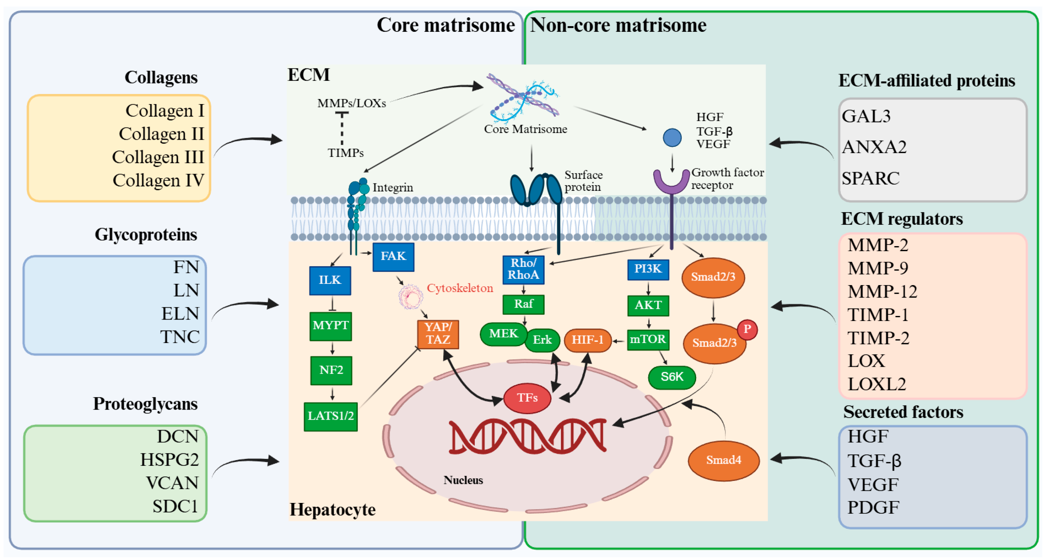
Figure 2.
Mechanical signaling networks in liver regeneration.
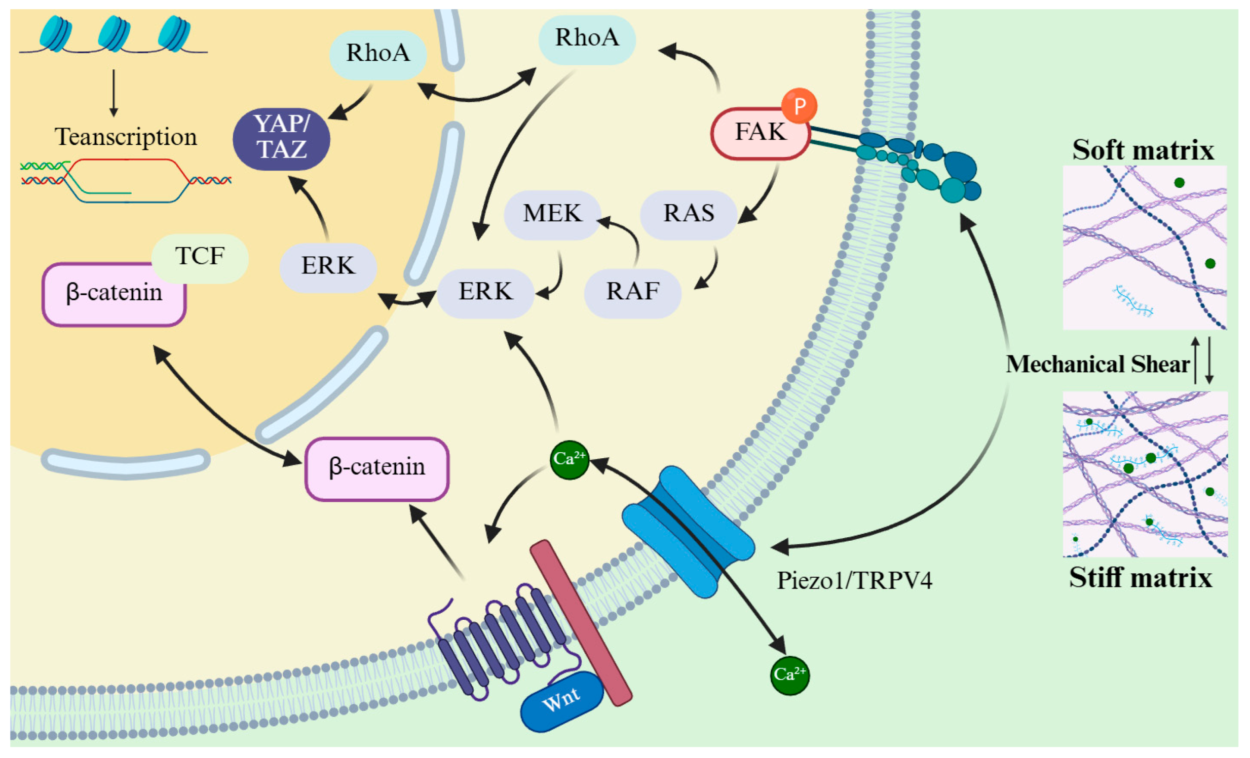
Figure 3.
Multicellular crosstalk mediated by the liver ECM during regeneration.
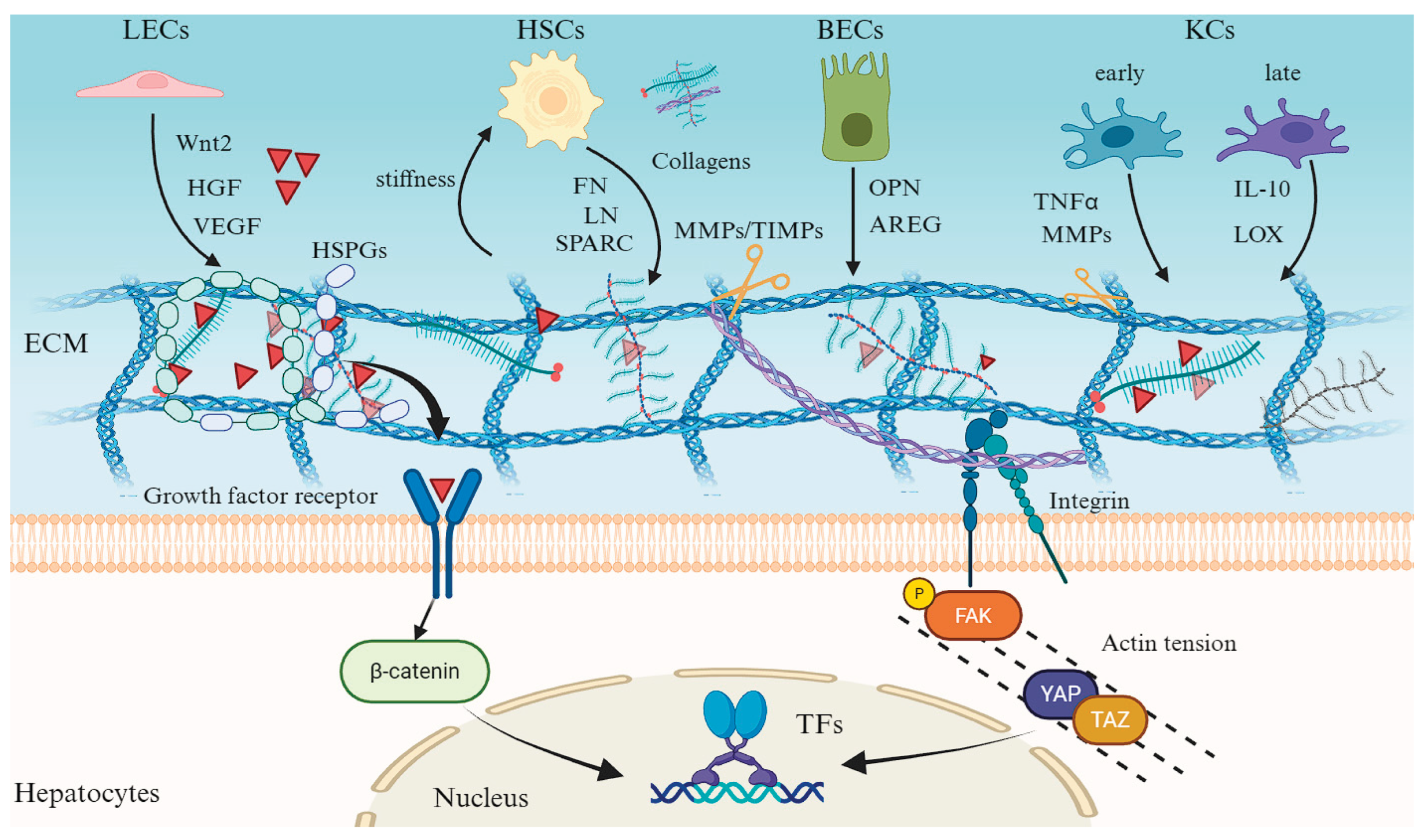

Table 1.
Summary of ECM-targeted strategies for liver regeneration.
| Target | Intervention | Models | Effect | References |
|---|---|---|---|---|
| TGF-β | Recombinant decorin (binds to TGF-β) | PHx or CCl₄ mouse models | ++ | [89] |
| LY2157299 | PHx or CCl₄ mouse models | + | [90,91] | |
| LOXl2 | AB0023 | TAA or DDC mouse models | +++ | [31] |
| GS341 | CCl₄ mouse models | ++ | [96] | |
| MMP-13 MMP-9 |
||||
| MMP13-encoding plasmids (pBGI-MMP13) | TAA mouse models | ++ | [101] | |
| MMP-9 antisense oligonucleotides | PHx rat models | ++ | [103] | |
| Wnt3a/4/5a Wnt5a |
DKK1 | BDL mouse models, primary mouse HSCs | ++ | [104] |
| sFRP5 | CCl₄ mouse models | + | [105] | |
| Wnt pathway | ICG-001 | CCl₄ mouse models, LX-2 cells, primary human fibroblasts | ++ | [106] |
| PRI-724 | Human HCV | + | [108] | |
| BMP-7 | Recombinant human BMP-7 (rhBMP-7) | PHx mouse models | + | |
| dECM | ||||
| Injectable hydrogel | Rat models of liver ischemia/reperfusion injury | ++ | [113] | |
| L-ECM hydrogel | TAA rat models | ++ | [115] | |
| dECM-Cryogel implantable scaffold | Liver failure rat models | + | [116] | |
| Self-assembling dECM hydrogel adhesive | Rabbit models of hemorrhage liver injury | ++ | [117] | |
| dECM-based bioengineered lobules | Mouse models of acute liver failure | ++ | [118] | |
| THBS1-dECM | PHx mouse models, primary mouse hepatocytes | +++ | [114] | |
| L-ECM hydrogel | Rat primary hepatocytes | ++ | [112] |
* the observed effect was categorized into three levels based on the percentage of response: + (0–25%), ++ (26–50%), +++ (51–75%).
Disclaimer/Publisher’s Note: The statements, opinions and data contained in all publications are solely those of the individual author(s) and contributor(s) and not of MDPI and/or the editor(s). MDPI and/or the editor(s) disclaim responsibility for any injury to people or property resulting from any ideas, methods, instructions or products referred to in the content. |
© 2026 by the authors. Licensee MDPI, Basel, Switzerland. This article is an open access article distributed under the terms and conditions of the Creative Commons Attribution (CC BY) license (http://creativecommons.org/licenses/by/4.0/).
Copyright: This open access article is published under a Creative Commons CC BY 4.0 license, which permit the free download, distribution, and reuse, provided that the author and preprint are cited in any reuse.


