
Submitted:
28 January 2026
Posted:
28 January 2026
You are already at the latest version
Abstract
The circulation of viruses of medical and veterinary importance is monitored to evaluate risks to both human and animal health. Among the species most commonly used in surveillance programs, the wild boar (Sus scrofa) plays a key role due to its high popu-lation density and its contacts with rural swine herds in the areas under investigation. In this study, molecular and serological analyses were carried out on wild boar samples collected from the regions of Apulia, Basilicata, Campania, and Calabria. The aim of this study is assessing the circulation of Influenza viruses, porcine Circovirus type 2 (PCV-2), Flaviviruses, and Aujeszky’s disease virus (ADV). Molecular analyses confirmed the presence of Influenza virus and PCV-2 in the wild population, while serological moni-toring detected specific antibodies against Influenza viruses, Flaviviruses, and ADV. The overall objective of the study is to deepen the understanding of virus circulation in the wildlife of Southern Italy, using the wild boar as an additional sentinel species for epidemiological surveillance activities. The distinctive feature of the project lies in its multi-pathogen approach applied to a reference population distributed across a parti-cularly wide geographical area, allowing for the simultaneous assessment of the circu-lation of different viruses over a large territory.
Keywords:
viral diseases
; wild boars
; Southern Italy
1. Introduction
Wild boar (Sus scrofa), belonging to the Suidae family, is characterized by a generalist and opportunistic behaviour [1]. Owing to its marked ecological plasticity, the species is capable of adapting to a broad spectrum of habitats, tolerating substantial anthropogenic pressures and exploiting food resources of human-made origin[1,2]. The species’ high reproductive capacity, flexible diet, and the reduced impact of hunting have enabled an exponential increase in its population over recent decades[3,4]. Recent studies have documented a substantial expansion of the species across Europe, both numerically and geographically, with increasingly significant repercussions for agriculture, the environment, road safety, and public health [4,5]. Wild boars are animals that move in groups of variable size [6]. Specifically, females live in groups with their offspring, while males are solitary, although in autumn and winter they tend to display gregarious behaviour [7]. Social interactions in this species facilitate interspecies virus transmission and contribute to increasing viral circulation. Moreover, the growing interaction between wild boars, urban environments, and domestic animals further heightens health risks, making integrated management and surveillance strategies necessary [8]. Wild boar acts as a potential reservoir for numerous zoonotic pathogens, posing a threat to both livestock and human health [6]. Its scavenger behaviour, combined with a variable diet and frequent exposure to faecal material, makes it an ideal sentinel species for monitoring zoonotic diseases. Among the most feared zoonoses, those caused by Influenza viruses represent a concrete threat, considering their demonstrated enzootic circulation in Italy [9,10] and the ability of these viruses to cross species barriers [9,10,11]. Suids, in particular, play a crucial role as a “mixing vessel” host species, as co-infection with avian, swine and human influenza strains can promote genetic reassortment and the emergence of new viral variants with unknown antigenic and pathogenic characteristics [12,13]. In this context, virological monitoring of wild suid populations, such as wild boars, represents a strategic tool within a One Health approach, useful for the early identification of potentially zoonotic emerging strains. In view of these considerations, it is equally important to extend virological surveillance of wild boars to other zoonotic pathogens, such as Flaviviruses. Some members of this genus, including West Nile virus (WNV) and USUTU virus (USUTUV), are known for their ability to infect a wide range of incidental hosts, including humans and wild boars[14,15] Recent studies conducted in Italy have detected anti-Flavivirus antibodies in wild boars, suggesting previous exposure [16]. However, since this evidence derives exclusively from serological investigations, there is a clear need to deepen the research using molecular approaches and across a broader geographical scale. In this context, health surveillance of wild boars assumes a strategic role not only for zoonotic pathogens but also for those of veterinary relevance. Aujeszky’s disease virus (Suid Herpesvirus 1, ADV) is endemic in many areas of Italy and is capable of infecting various domestic and wild species [14,16]. Although wild boars represent the main reservoir of the virus in the wild, the increasing interaction with livestock and hunting dogs raises the risk of interspecies transmission, making monitoring necessary within the framework of veterinary public health programs [17]. In addition to the zoonotic viruses and those of veterinary relevance already discussed, Porcine Circovirus type 2 (PCV2) has also been frequently detected in wild boar populations in Italy and other European countries [18,19]. The circulation of PCV-2 in the wild, although it does not pose a direct zoonotic risk, represents a threat to animal health, as it may affect pig farms through possible direct or indirect interactions [20]. Several studies have confirmed the co-circulation of PCV2 with other swine viruses (e.g., SuHV-1 and PPV1) in wild boars, suggesting the possibility of mixed infections, which can potentially increase the pathogenicity and spread of viral strains [22]. This highlights the importance of integrated virological monitoring extended also to non-zoonotic pathogens with high production impact. In the present study, molecular and serological monitoring was conducted on a wild boar population with the aim of assessing the circulation of both zoonotic viruses (with potential direct impact on public health) and non-zoonotic viruses of veterinary relevance, which can cause significant economic losses in the swine industry. The investigation focused in particular on geographical areas where, to date, epidemiological data on the presence of these pathogens in wildlife are limited or lacking.
2. Materials and Methods
The study was conducted in a geographical area including the provinces of Foggia, BAT, and Bari for Apulia; Matera and Potenza for Basilicata; Salerno for Campania; and Catanzaro and Cosenza for Calabria (Figure 1). Sampling was carried out over a two-year period (2023–2024) during the implementation of the “Extraordinary Plan for the Capture, Culling, and Disposal of Wild Boars” aimed at controlling African swine fever (ASF), at the wild game slaughterhouse located in the municipality of Tito (PZ), Basilicata (Figure 2). This facility regularly receives wild boars’ samples culled during hunting activities from the provinces. During each sampling session (conducted during hunting periods and months compatible with the circulation of the diseases under investigation), biological samples were collected from animals undergoing slaughter (Figure 2 and Figure 3). More in details, nasal swabs (n = 444), lung tissue (n = 346), and muscle tissue (n = 339) were collected and subsequently subjected to laboratory analyses. Overall, the study included a total of 636 individual wild boars, from which different types of biological samples were collected. Specifically, some animals were sampled exclusively for muscle tissue, others only for lung tissue, others solely for swabs, or, in some cases, a combination of these tissues. The geographical origin of each animal was recorded at the time of delivery to the slaughterhouse and used for sample georeferencing. The specimens showed marked heterogeneity in terms of geographic origin, with distribution across numerous municipalities within the eight provinces under study. The distribution of the specimens was predominantly concentrated in the Basilicata region, with a high density in the province of Potenza (48%), where environmental conditions, particularly the widespread presence of forested habitats, are especially favourable for the presence and proliferation of wild boars [21].
2.1. Serological Investigation
For the serological investigation, meat juice obtained from wild boar muscle was used. Specifically, considering that at the sampling site wild boar heads and viscera were available, the masseter muscle was collected from the head. Muscle samples were subjected to repeated freezing and thawing cycles to obtain the release of meat juice, until a minimum volume of 1 mL per sample was achieved [22]. The resulting extract represents a tissue transudate, rich in proteins and antibodies, although generally at lower concentrations compared to serum. This method is particularly useful in the context of passive surveillance at slaughterhouses, due to the easy availability of muscle tissue from carcasses obtained during hunting activities or slaughter. The presence of specific antibodies against Influenza A virus, Flavivirus, and ADV was investigated using a competitive ELISA, performed according to the instructions provided by the diagnostic kit manufacturer and read spectrophotometrically.
2.2. Nucleic Acid Extraction
Molecular monitoring was conducted through three main steps: sample preparation, nucleic acid extraction, and PCR amplification. For preparation of samples, 0.5 g of lung tissue was homogenized in 2 mL Safe-Lock Eppendorf tubes containing 1 mL of phosphate-buffered saline (PBS, pH 7.4). Subsequently, 0.5 mL of the homogenate was transferred to a new tube containing 0.5 mL of lysis buffer and incubated on ice at 4 °C for 10 minutes. The samples were then centrifuged at 8,000 rpm for 5 minutes. A volume of 300 µL of the supernatant was finally used for automated nucleic acid extraction using the TanBead® system, following the manufacturer’s protocol. For nasal swabs, each swab was resuspended in 1 mL of PBS. A volume of 300 µL of the resulting suspension was then directly transferred to the TanBead automated extraction system, according to the manufacturer’s instructions. The extracts were stored at -80 °C until further use.
2.3. Molecular Detection of Influenza Viruses
The extracts obtained were analyzed for the detection of the M gene of Influenza A viruses using Real-Time Quantitative Reverse Transcription Polymerase Chain Reaction (RT-qPCR). The protocol described by Spackman et al., 2002, was adopted, which utilizes the following oligonucleotides: Forward primer M+25: 5’-AGA TGA GTC TTC TAA CCG AGG TCG-3’; Reverse primer M–124: 5’-TGC AAA AAC ATC TTC AAG TCT CTG-3’; Probe M+64: 5’-FAM-TCA GGC CCC CTC AAA GCC GA-TAMRA-3’, where FAM (6-carboxyfluorescein) is the reporter fluorophore and TAMRA (6-carboxytetramethylrhodamine) is the quencher [24]. Reactions were performed using the AccuStart™ One-Step RT-qPCR SuperMix kit. Each reaction was prepared in a final volume of 20 µL, containing: 0.2 µL of extracted RNA, 4 µL of RT-qPCR SuperMix, 12.6 µL of nuclease-free water, 0.4 µL of Reference Dye (50×), and 0.5 µL of the probe (0.2 µM) and primer (0.4 µM) mix. The thermal cycling conditions were as follows: Reverse transcription at 55 °C for 15 minutes; reverse transcriptase inactivation and initial denaturation at 95 °C for 3 minutes; amplification (45 cycles) with denaturation at 95 °C for 10 seconds and annealing/extension at 60 °C for 60 seconds. Fluorescence signal acquisition was performed at the end of each annealing/extension phase. Samples with a cycle threshold (Ct) value below 36 were considered positive.
2.4. Molecular Detection of Flaviviruses
For the molecular identification of Flaviviruses, the Pan-Flavi qRT-PCR assay was used, targeting the amplification of a conserved fragment of the NS5 gene, in order to simultaneously detect a broad range of viruses belonging to the Flavivirus genus [23]. The oligonucleotide sequences used in the assay were as follows: Forward primer: 5’-TACAACATGATGGGGAARAGAGARAA-3’; Reverse primer: 5’-GTGTCCCAGCNGCKGTGTCATCWGC-3’; Probe: 5’-FAM-TGGTWYATGTGGYTNGGRGC-BBQ-3’, where FAM is the reporter fluorophore and BBQ (BlackBerry Quencher) serves as the quencher. Reactions were performed using the AccuStart™ One-Step RT-qPCR SuperMix kit (Quantabio). Each reaction was prepared in a total volume of 20 µL, consisting of: 2 µL of extracted RNA, 5 µL of RT-qPCR SuperMix, 12 µL of nuclease-free water, and 1 µL of probe mix (containing the two primers at a concentration of 10 pmol and the probe at a concentration of 5 pmol). The thermal cycling protocol was set as follows: Reverse transcription at 55 °C for 10 minutes; initial activation and denaturation at 95 °C for 1 minute; amplification cycle (45 cycles) with denaturation at 95 °C for 10 seconds and annealing/extension at 60 °C for 60 seconds. Fluorescence was acquired at the end of each annealing/extension phase. Results were interpreted based on the obtained Ct values.
2.5. Molecular Detection of PCV2
The molecular detection of PCV2 was performed following the protocol described by Kim HR et al., 2017 [18]. The decision to analyze the samples for PCV2 was motivated by the clinical and economic relevance of this genotype in the livestock production context [18]. For Real-Time qPCR, the specific primer pair PCV-2-For (5′-CCAGGAGGGCGTTSTGACT-3′) and PCV-2-Rev (5′-CGYTACCGYTGGAGAAGGAA-3′) was used, together with the labelled probe PCV-2-P (FAM-5′-AATGGCATCTTCAACACCCGCCTCT-3′-TAMRA). Reactions were performed using the TaqMan™ Universal PCR Master Mix kit (Applied Biosystems™). Each 20 µL reaction was prepared with the following composition: 3 µL of extracted DNA, 10 µL of master mix, 4 µL of nuclease-free water, 1 µL of forward primer (10 µM), 1 µL of reverse primer (10 µM), and 1 µL of probe (5 µM). The thermal cycling protocol used for amplification was: initial denaturation at 95 °C for 10 minutes, followed by 40 cycles of denaturation at 95 °C for 30 seconds, annealing at 55.1 °C for 30 seconds, and extension at 72 °C for 30 seconds. This approach allowed sensitive and specific detection of PCV-2 in the investigated samples.
3. Results
The available data, in addition to the geographical origin of the samples, included sex and body weight. Regarding sex, of the total sample of 636 individuals, 309 were males (48.7%), 315 were females (49.5%), and 12 individuals (1.9%) had undetermined sex. The distribution between males and females was therefore essentially balanced, with an almost 50:50 ratio for each sex (Figure 3a). With respect to body weight, there was a clear predominance of adult animals, most of which weighed over 50 kg (70%) (Figure 3b).
Serological analyses, 28 out of 337 tested subjects were positive for Influenza virus (8.31%, 95% CI: 5,81-11,75%), primarily distributed in the provinces of Potenza (18/337; 5,34%, 95%CI: 3,40-8,28%)followed by Matera (4/337; 1.19%, 95% CI: 0,46-3,01%), Foggia (4/337; 1.19%, 95% CI: 0,46-3,01), Catanzaro (1/337; 0.30%, 95% CI: 0,05-1,66%), and Salerno (1/337; 0.30%, 95% CI: 0,05-1,66%). Among the 28 positive samples, 23 (82.14%, 95% CI: 64,41-92,12%) were collected during the February–April period, in agreement with the molecular survey data. The weight of positive subjects ranged widely from 18 to 87 kg. Regarding sex, 9 subjects (32.14%, 95% CI: 17,93-50,66%) were males and 19 (67.86%, 95% CI: 49,34-82,07%) females. The detection of anti-Flavivirus antibodies revealed a single positive sample out of 337 (0.30%, 95% CI: 0,05-1,66%), collected in the province of Foggia. This sample was subsequently confirmed as positive for WNV. ADV was detected in 4 out of 153 tested samples (2.61%, 95% CI: 1,02-6,53%), all of which originated from the province of Potenza. All positive subjects were males, and no correlation with body weight was observed. It should be noted that only samples from Basilicata were included for ADV analysis; consequently, no data are available from Puglia region.
Regarding molecular analysis detected the presence of Influenza viruses in 5 out of 429 animals examined (1.17%, 95% CI: 0,50-2,70%). The positive subjects had a body weight ranging from 44 to 60 kg, corresponding to an estimated age of approximately 8 to 14 months [24]. This distribution indicates a higher frequency of positivity in young–
subadult individuals, suggesting a possible association between age and susceptibility to infection. Regarding sex, three females and two males tested positive, suggesting a balanced distribution between the two genders, with no apparent correlation. All infected individuals originated from the Basilicata region, with a territorial distribution of 2 animals from the province of Matera (40%) and 3 from the province of Potenza (60%). Concerning Flaviviruses, no positive cases were detected in the analysed samples, indicating an absence of detectable viral circulation during the study period and in the areas considered. Regarding PCV2, 66 out of 223 examined subjects tested positive (29.6%, 95% CI: 23,99-35,89%). The geographical distribution of positive cases showed a higher concentration in the province of Potenza (41/66; 62.12%, 95% CI: 50,06-72,85%), followed by Matera (16/66; 24.2%, 95% CI: 15,51-35,81%) and Catanzaro (11/66; 16.7%, 95% CI: 9,57-27,43%). The sex distribution revealed a substantial parity between females (32/66; 48.5%, 95% CI: 36,85-60,29%) and males (34/66; 51.52%, 95% CI: 39,71- 63,15%), suggesting that sex is not a significant risk factor for infection. The weight of positive subjects ranged from 28 to 121 kg, with no apparent correlation between body mass and the likelihood of positivity.
4. Discussion
Viral circulation in wildlife populations plays a crucial role in emerging epidemiological systems, as these populations can act as reservoirs, sentinel indicators, and bridges to livestock populations [25,26]. In particular, the analysis of viral presence in wildlife species is strategically important from both an animal health and a public health perspective. For this reason, the combined use of molecular and serological investigations allows not only the detection of active infections but also the reconstruction of past exposure within the population, highlighting temporal dynamics that are often invisible through virological analysis alone [27]. In the literature, numerous studies support the persistence of specific antibodies in animal serum for periods that can extend for months or longer, allowing the reconstruction of previous viral circulation within the population [28]. Moreover, seroprevalence has been demonstrated in other regions of Italy [29,30]; however, no studies have previously addressed the regions investigated in this research. In our work, PCR analyses confirmed the active circulation of Influenza viruses in hunted wild boars, particularly during the February–April 2024 period, whereas serology revealed a wider distribution of positivity. Overall, the data on Influenza virus positivity indicate a correlation with the seasonality of viral circulation, mainly concentrated in February–April. Additionally, the distribution of positive individuals by weight suggests that infection predominantly affects sub-adult animals. From a geographical perspective, most positive samples originated from the Basilicata region, especially from the province of Potenza; however, this finding may reflect the higher number of samples collected from this area rather than a real higher viral circulation. It should be emphasized that the wild boar organ samples analyzed in this study were obtained through an opportunistic sampling approach, based on the availability of biological materials at the slaughterhouses. As a consequence, the geographical distribution of samples is unbalanced, with a higher proportion originating from the province of Potenza, so this his sampling strategy can represent a limit of data representativeness. Therefore, the results should be interpreted as an estimation deriving from the analyzed samples rather than a measure of the true regional prevalence.
However, regarding Flaviviruses, no positive cases were detected by PCR, while serological analysis revealed past exposure in a single individual (antibodies directed against Flavivirus). This pattern is fully consistent with previous reports in the literature [27]. The data therefore indicate past exposure to the virus but do not provide evidence of active viral circulation during the sampling period. The observed seropositivity should be interpreted as reflecting prior contact with the pathogen, while the absence of molecular detections does not allow the demonstration of ongoing viral replication.
Regarding PCV-2, recent studies conducted in the Basilicata and Campania regions reported a prevalence of 27% in wild boars in Basilicata [19] and 47.3% in Campania [18], with evidence of the dominant genotype 2. Our findings are fully consistent with these studies, confirming that genotype 2 is widespread in southern Italy and that the wild population may represent an important reservoir. The agreement between our results and the literature indicates that viral circulation is stable and well-established in the area. Similarly, to what was observed for Influenza virus, a higher number of PCV-2 positive samples were detected in the province of Potenza. As previously discussed, this finding may be related to the larger number of samples analysed from this province. Regarding antibodies detected through the serological component, our study revealed seropositivity for ADV. Previous investigations in Italy reported, for example, a seroprevalence of 23.85% for ADV in wild boars in the Campania region during the 2016–2017 hunting season [31]. Our results confirm the circulation of these agents in the study area and indicate a territorial correlation, as all positive samples originated from the province of Potenza. Additionally, all positive subjects were male. The overall analysis did not reveal any significant associations among the investigated pathogens. Positive cases were mutually exclusive, and no coinfections were detected in any of the examined animals. The operational choice to include four regions of Southern Italy (Apulia, Basilicata, Campania, and Calabria) and to simultaneously investigate multiple viral diseases gives our study a distinctive contribution. Sampling wild boars across a wide geographic area—characterized by diverse environmental conditions, altitudes, and hunting pressures—allows for a more representative snapshot of viral circulation in wildlife in Southern Italy. Our findings highlight the importance of an integrated multi-pathogen molecular and serological approach, which is also valuable in studying potential overlaps and interactions between viral agents. It is important to note that not all samples were tested for the full panel of pathogens, due to insufficient biological material. Nevertheless, the results obtained provide a solid foundation for future prospective investigations, which could include studies on the genetic characterization and variability of the detected pathogens, and highlight the importance of an integrated multipathogen approach based on molecular and serological analyses to assess potential overlaps and interactions among different viral agents.
5. Conclusions
The present study documents the circulation of influenza viruses, Flaviviruses, ADV, and the persistent presence of PCV-2 in wild boar populations, reflecting patterns already observed in wildlife surveillance programs across Europe. The strengths of the study lie in its broad spatial coverage, the simultaneous assessment of multiple viral pathogens, and the combined use of serological and molecular diagnostic approaches. It is important to note that sampling was performed opportunistically, based on the availability of biological material at slaughterhouses, and that not all samples could be tested for the full panel of pathogens due to limited material. Moreover, the study did not include sequencing or genetic characterization of viral strains, representing a methodological limitation. Nevertheless, the results provide a solid foundation for future prospective studies aimed at addressing these gaps through more representative sampling, comprehensive molecular analyses, and investigations into the genetic variability of isolated viral strains. Overall, this study makes a significant contribution to the understanding of pathogen ecology in free-ranging Suidae populations and provides a basis for future research on long-term epidemiological dynamics and interspecific transmission risks at the wildlife–livestock interface.
Funding
his research was supported by EU funding within the NextGenerationEU-MUR PNRR Extended Partnership initiative on Emerging Infectious Diseases (Project no. PE00000007, INF-ACT).
References
- Pittiglio, C.; Khomenko, S.; Beltran-Alcrudo, D. Wild boar mapping using population-density statistics: From polygons to high resolution raster maps. PLoS One 2018, 13. [Google Scholar]
- Lewis, J.S.; Farnsworth, M.L.; Burdett, C.L.; Theobald, D.M.; Gray, M.; Miller, R.S. Biotic and abiotic factors predicting the global distribution and population density of an invasive large mammal. Sci Rep. 2017, 7. [Google Scholar] [CrossRef]
- Torres, R.T.; Fernandes, J.; Carvalho, J.; Cunha, M.V.; Caetano, T.; Mendo, S.; et al. Wild boar as a reservoir of antimicrobial resistance. Vol. 717, Science of the Total Environment. Elsevier B.V.; 2020.
- Massei, G.; Kindberg, J.; Licoppe, A.; Gačić, D.; Šprem, N.; Kamler, J.; et al. Wild boar populations up, numbers of hunters down? A review of trends and implications for Europe. Pest Manag Sci. 2015, 71, 492–500. [Google Scholar] [CrossRef]
- Bieber, C.; Ruf, T. Population dynamics in wild boar Sus scrofa: Ecology, elasticity of growth rate and implications for the management of pulsed resource consumers. Journal of Applied Ecology 2005, 42, 1203–1213. [Google Scholar] [CrossRef]
- Ruiz-Fons, F.; Segalés, J.; Gortázar, C. A review of viral diseases of the European wild boar: Effects of population dynamics and reservoir rôle. Veterinary Journal 2008, 176, 158–169. [Google Scholar] [CrossRef] [PubMed]
- Rosell, C.; Navàs, F.; Romero, S.; De Dalmases, I. ACTIVITY PATTERNS AND SOCIAL ORGANIZATION OF WILD BOAR (Sus scrofa, L.) IN A WETLAND ENVIRONMENT: PRELIMINARY DATA ON THE EFFECTS OF SHOOTING INDIVIDUALS. [CrossRef]
- Vicente, J.; Ruiz-Fons, F.; Vidal, D.; Höfle, U.; Acevedo, P.; Villanúa, D.; et al. Serosurvey of Aujeszky’s disease virus infection in European wild boar in Spain. Veterinary Record 2005, 156, 408–412. [Google Scholar] [CrossRef]
- Mostafa, A.; Naguib, M.M.; Nogales, A.; Barre, R.S.; Stewart, J.P.; García-Sastre, A.; et al. Avian influenza A (H5N1) virus in dairy cattle: origin, evolution, and cross-species transmission. Vol. 15, mBio. American Society for Microbiology; 2024.
- Graziosi, G.; Lupini, C.; Catelli, E.; Carnaccini, S. Highly Pathogenic Avian Influenza (HPAI) H5 Clade 2.3.4.4b Virus Infection in Birds and Mammals. Animals 2024, 14. [Google Scholar] [CrossRef]
- Liang, Y. Pathogenicity and virulence of influenza. Vol. 14, Virulence. Taylor and Francis Ltd.; 2023.
- Abdelwhab, E.M.; Mettenleiter, T.C. Zoonotic Animal Influenza Virus and Potential Mixing Vessel Hosts. Viruses 2023, 15. [Google Scholar] [CrossRef]
- AbuBakar, U.; Amrani, L.; Kamarulzaman, F.A.; Karsani, S.A.; Hassandarvish, P.; Khairat, J.E. Avian Influenza Virus Tropism in Humans. Viruses 2023, 15. [Google Scholar] [CrossRef]
- Ferrara, G.; Longobardi, C.; D’ambrosi, F.; Amoroso, M.G.; D’alessio, N.; Damiano, S.; et al. Aujeszky’s disease in south-Italian wild boars (Sus Scrofa): A serological survey. Animals 2021, 11. [Google Scholar] [CrossRef] [PubMed]
- Veljović, L.; Paunović, M.; Glišić, D.; Šolaja, S.; Zurovac Sapundžić, Z.; Maletić, J.; et al. Wild Mammals as Sentinels for West Nile Virus Circulation: Evidence from Serbia. Pathogens 2025, 14. [Google Scholar] [CrossRef] [PubMed]
- Müller, A.; Melo, N.; González-Barrio, D.; Pinto, M.V.; Ruiz-Fons, F. Aujeszky’s disease in hunted wild boar (Sus scrofa) in the iberian peninsula. J Wildl Dis. 2021, 57, 543–552. [Google Scholar]
- Ciarello, F.P.; Moreno, A.; Miragliotta, N.; Antonino, A.; Fiasconaro, M.; Purpari, G.; et al. Aujeszky’s disease in hunting dogs after the ingestion of wild boar raw meat in Sicily (Italy): clinical, diagnostic and phylogenetic features. BMC Vet Res. 2022, 18. [Google Scholar] [CrossRef]
- Amoroso, M.G.; Serra, F.; Esposito, C.; D’alessio, N.; Ferrara, G.; Cioffi, B.; et al. Prevalence of infection with porcine circovirus types 2 and 3 in the wild boar population in the campania region (Southern italy). Animals 2021, 11. [Google Scholar] [CrossRef]
- Fanelli, A.; Pellegrini, F.; Camero, M.; Catella, C.; Buonavoglia, D.; Fusco, G.; et al. Genetic Diversity of Porcine Circovirus Types 2 and 3 in Wild Boar in Italy. Animals 2022, 12. [Google Scholar] [CrossRef]
- Gillespie, J.; Opriessnig, T.; Meng, X.J.; Pelzer, K.; Buechner-Maxwell, V. Porcine circovirus type 2 and porcine circovirus-associated disease. Journal of Veterinary Internal Medicine 2009, 23, 1151–1163. [Google Scholar] [CrossRef]
- Cillis, G.; Statuto, D.; Picuno, P. Historical gis as a tool for monitoring, preserving and planning forest landscape: A case study in a mediterranean region. Land 2021, 10. [Google Scholar] [CrossRef]
- Poonsuk, K.; Cheng, T.Y.; Ji, J.; Zimmerman, J.; Giménez-Lirola, L. Detection of porcine epidemic diarrhea virus (PEDV) IgG and IgA in muscle tissue exudate (“meat juice”) specimens. Porcine Health Manag 2018, 4. [Google Scholar]
- Patel, P.; Landt, O.; Kaiser, M.; Faye, O.; Koppe, T.; Lass, U.; et al. Development of one-step quantitative reverse transcription PCR for the rapid detection of flaviviruses. Virol J. 2013, 10. [Google Scholar] [CrossRef]
- Zmijewski, T.; Modzelewska-Kapituła, M. The influence of age and sex on carcass characteristics and chemical composition of the longissimus thoracis et lumborum muscle in wild boars (Sus scrofa). Arch Anim Breed 2021, 64. [Google Scholar] [CrossRef]
- Pandit, P.S.; Doyle, M.M.; Smart, K.M.; Young, C.C.W.; Drape, G.W.; Johnson, C.K. Predicting wildlife reservoirs and global vulnerability to zoonotic Flaviviruses. Nat Commun. 2018, 9. [Google Scholar] [CrossRef] [PubMed]
- Ojeyinka, O.T.; Omaghomi, T.T. Wildlife as sentinels for emerging zoonotic diseases: A review of surveillance systems in the USA. World Journal of Advanced Research and Reviews 2024, 21, 768–776. [Google Scholar] [CrossRef]
- Petruccelli, A.; Zottola, T.; Ferrara, G.; Iovane, V.; Di Russo, C.; Pagnini, U.; et al. West nile virus and related flavivirus in european wild boar (Sus scrofa), latium region, Italy: A retrospective study. Animals 2020, 10. [Google Scholar] [CrossRef] [PubMed]
- Pepin, K.M.; Pedersen, K.; Wan, X.F.; Cunningham, F.L.; Webb, C.T.; Wilber, M.Q. Individual-Level Antibody Dynamics Reveal Potential Drivers of Influenza A Seasonality in Wild Pig Populations. In Integrative and Comparative Biology; Oxford University Press, 2019; pp. 1231–1242. [Google Scholar]
- De Marco, M.A.; Cotti, C.; Raffini, E.; Frasnelli, M.; Prosperi, A.; Zanni, I.; et al. Long-Term Serological Investigations of Influenza A Virus in Free-Living Wild Boars (Sus scrofa) from Northern Italy (2007–2014). Microorganisms 2022, 10. [Google Scholar]
- Delogu, M.; Cotti, C.; Vaccari, G.; Raffini, E.; Frasnelli, M.; Nicoloso, S.; et al. Serologic and virologic evidence of influenza a viruses in wild boars (Sus scrofa) from two different locations in Italy. J Wildl Dis. 2019, 55, 158–163. [Google Scholar]
- Ferrara, G; Longobardi, C; D’ambrosi, F; Amoroso, MG; D’alessio, N; Damiano, S; et al. Aujeszky’s disease in south-Italian wild boars (Sus Scrofa): A serological survey. Animals 2021, 11. [Google Scholar] [CrossRef]
Figure 1.
Investigated area: The figure shows the geographical area covered by the study, including the regions of Southern Italy: Apulia, Basilicata, Campania, and Calabria. The municipalities where sampling was carried out are indicated by yellow dots. Data visualization generated using Flourish.
Figure 1.
Investigated area: The figure shows the geographical area covered by the study, including the regions of Southern Italy: Apulia, Basilicata, Campania, and Calabria. The municipalities where sampling was carried out are indicated by yellow dots. Data visualization generated using Flourish.
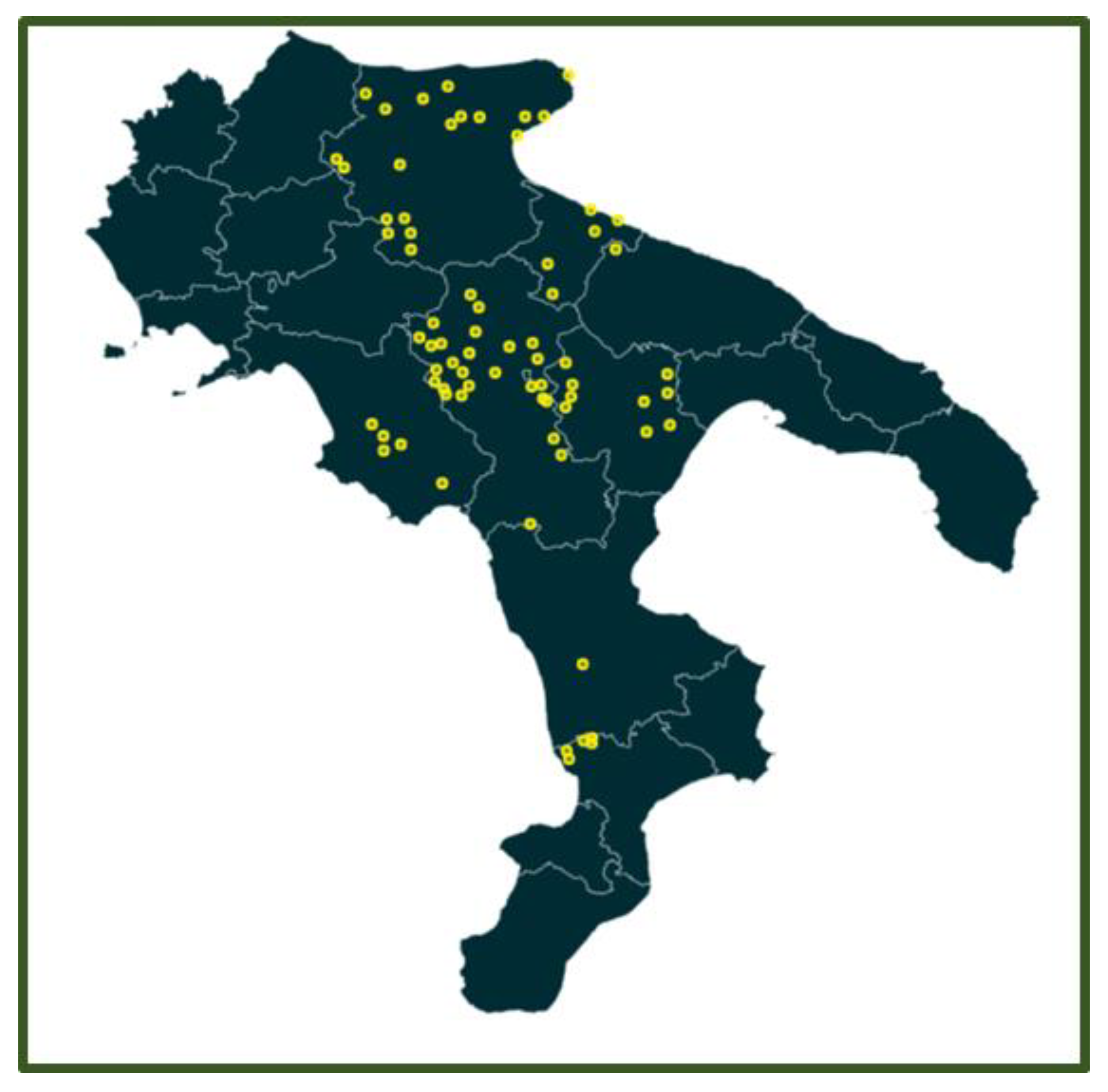
Figure 2.
Sus scrofa during slaughtering activities at the Tito (PZ) slaughterhouse: [ a] The picture shows the post-evisceration phase, with the viscera (heart, lungs, liver) temporarily suspended to facilitate sanitary inspection and the collection of samples for diagnostic analyses.; [ b] Wild boars heads post-evisceration, used for masseter muscle sampling and nasal swab collection.
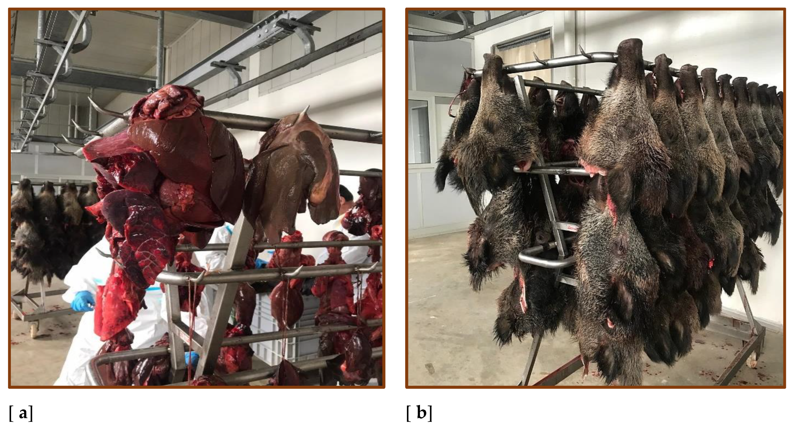
Figure 3.
Distribution of wild boar samples by sex and weight: [a] Pie chart showing the distribution of samples by sex: approximately half of the samples were collected from female wild boars, half from males, and a portion from individuals with indeterminate sex due to unavailable data.; [b] Pie chart showing the distribution of samples by weight classes: 0–25 kg, 25–50 kg, and over 50 kg.
Disclaimer/Publisher’s Note: The statements, opinions and data contained in all publications are solely those of the individual author(s) and contributor(s) and not of MDPI and/or the editor(s). MDPI and/or the editor(s) disclaim responsibility for any injury to people or property resulting from any ideas, methods, instructions or products referred to in the content. |
© 2026 by the authors. Licensee MDPI, Basel, Switzerland. This article is an open access article distributed under the terms and conditions of the Creative Commons Attribution (CC BY) license (http://creativecommons.org/licenses/by/4.0/).
Copyright: This open access article is published under a Creative Commons CC BY 4.0 license, which permit the free download, distribution, and reuse, provided that the author and preprint are cited in any reuse.


