Preprint
Article

This version is not peer-reviewed.

Activities of Some Cannabinoids as Predicted by Molecular Docking Computation and Confirmed by Cell Calcium Assay

Submitted:

26 January 2026

Posted:

27 January 2026

You are already at the latest version

Abstract

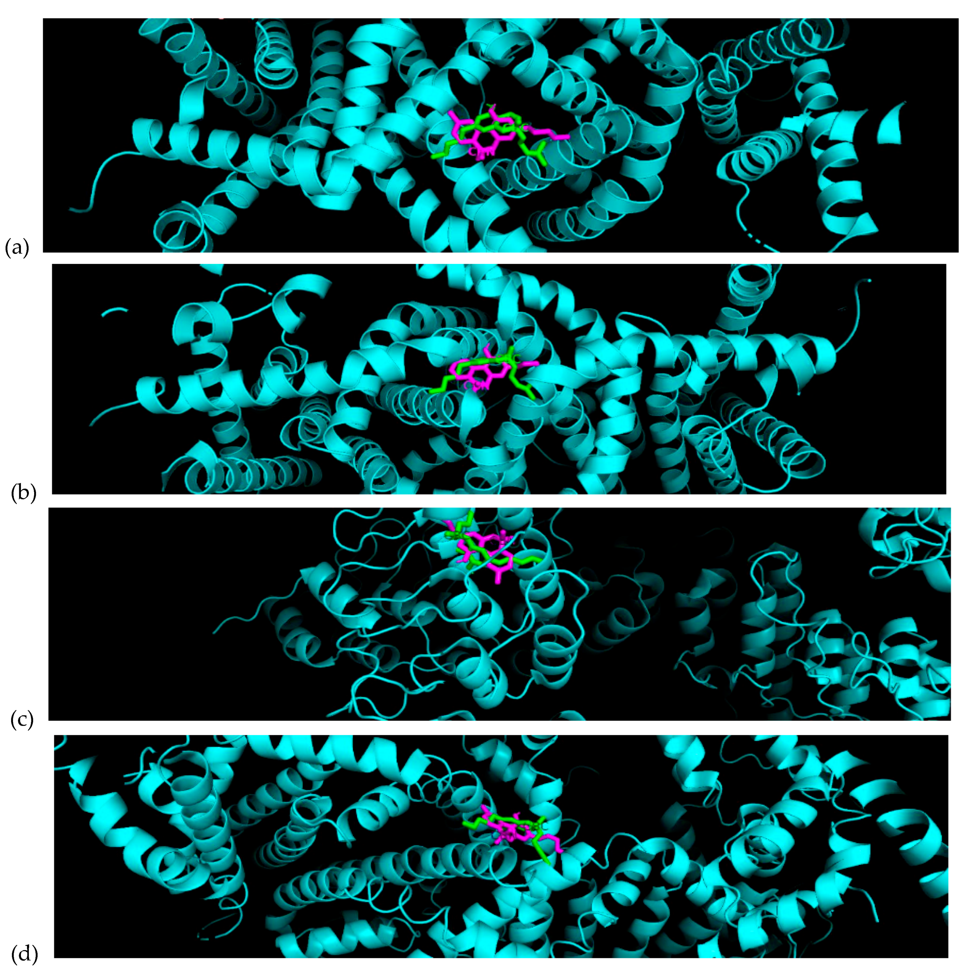
Many cannabinoids are derived from Cannabis and exhibit a diverse range of pharmacological properties. Predictions of bioactivities of these compounds were conducted by molecular docking computation on two transient receptor potential (TRP) receptors (TRPV1 and TRPC5) found on human glioma (U-87 MG) cells. These predictions were experimentally confirmed by monitoring changes in intracellular calcium concentration in U-87 MG cells treated with cannabinol (CBN), cannabichromene (CBC), and cannabicyclol (CBL), as measured using a fluorescence microplate reader. The results indicate that CBN and CBC are bioactive, whereas CBL exhibits minimal activity. These findings are consistent with predictions obtained from molecular docking computation based on AutoDock Vina.

Keywords: 
;  ;  ;  ;  

Introduction

The global demand for medical-grade Cannabis has drastically increased patient interest in the use of cannabinoid-based substances. Due to their limited psychoactivity, cannabinoids such as CBN and CBL have demonstrated efficacy in several pathological conditions, including inflammatory and neurodegenerative diseases, epilepsy, autoimmune disorders such as multiple sclerosis and arthritis, schizophrenia, and cancer [1]. Cannabinoids have been reported to trigger TRPV-dependent autophagic processes in glioma stem-like cells, thereby abolishing chemoresistance to carmustine [3]. In addition, combinations of cannabinoids with cytotoxic drugs such as temozolomide, carmustine, or doxorubicin have been shown to enhance drug activity and uptake in human glioblastoma multiforme (GBM) cells [2].
Cannabinoids have also been demonstrated to modulate glioma progression through multiple mechanisms, including regulation of cell proliferation, invasion, apoptosis, and autophagy pathways, independent of classical cannabinoid receptors [6,19,20,21].
Beyond pharmacological characterization, cell-based biosensing and cell-monitoring assays enable quantitative detection of functional intracellular responses to chemical stimuli. Intracellular calcium is a robust biosensing parameter that reflects ion channel activity and downstream signaling, and fluorescence microplate monitoring provides a convenient route to high-throughput functional sensing. In this work, we adopt the Fluo-4 microplate assay as a cell-monitoring biosensing platform, and we integrate it with molecular docking to guide compound selection and interpretation.
GBM is the most common and aggressive malignant brain tumor, accounting for approximately 50% of all gliomas and 15.6% of all primary brain and central nervous system tumors [4,5]. Calcium-permeable transient receptor potential (TRP) channels play a critical role in glioma cell proliferation and translocation. In particular, TRPC1 has been shown to be a prerequisite for glioma cell proliferation and migration [7,9,11]. Elevated expression levels of TRPV1, TRPV2, TRPC1, TRPC6, TRPM2, TRPM3, TRPM7, and TRPM8 have also been reported in GBM tissues [12].
Beyond TRPV and TRPC channels, intracellular calcium signaling plays a central role in glioma cell survival, migration, and neuroprotection, making calcium homeostasis an attractive therapeutic target in cancer research [8,10,13]. Several studies have demonstrated that non-psychoactive cannabinoids such as cannabidiol can induce apoptosis, inhibit proliferation, and suppress invasion in glioma and other cancer cell lines through multi-target mechanisms [22,23,24,25,26,27,28]. Cannabinoids have also been reported to directly interact with TRP channels, including TRPV1, thereby regulating intracellular calcium dynamics and downstream signaling pathways associated with tumor progression [29,30,31,32].
Targeting calcium channel activity and intracellular calcium accumulation has therefore emerged as a promising strategy for cancer therapy. The present study was designed to investigate the pharmacological potential of selected cannabinoids in U-87 MG glioma cells based on intracellular calcium measurements. Previous studies have reported significant calcium changes in U-87 MG cells treated with curcumin [33,34], resveratrol [34], and capsaicin [34]. Predictions were also obtained using molecular docking computations based on AutoDock Vina, and a strong correlation between intracellular calcium measurements and molecular docking predictions has been previously established [34].

Experimental Section

Reagents

A fluorescent calcium probe, Fluo-4 AM ester (50 µg, Molecular Probes, Eugene, OR) was dissolved in dimethyl sulfoxide (DMSO, 99.9%, Sigma–Aldrich) to prepare a stock solution of 1 µg µL⁻¹. Prior to use, the stock solution was freshly diluted in Hanks’ balanced salt solution (HBSS, Invitrogen Corp., Grand Island, NY) to yield a working concentration of 5.0 µM. Due to its light sensitivity, Fluo-4 AM was stored in the dark at −20 °C [15].
Cannabinol (CBN), cannabichromene (CBC), and cannabicyclol (CBL) were purchased from Cerilliant Corporation (Round Rock, TX) as drug enforcementa-exempt solutions (1 mg mL⁻¹ in methanol).
Ionomycin calcium salt (Sigma Chemical Co.) was dissolved in DMSO and diluted in HBSS containing 1 mM CaCl₂ to prepare working solutions of 10 mg mL⁻¹ [14].

Cell Samples

U-87 MG cells (ATCC, USA) were obtained from cryopreserved storage and were maintained in DMEM/high glucose, pyruvate (ThermoFisher Scientific), containing 10% FBS (ThermoFisher Scientific), and 1% penicillin (Stemcell Technologies) in a 5% CO2 atmosphere at 37 °C. The doubling time for the cell line was approximately 39 h; hence, the growth medium was changed two times a week, and cells were passaged once a week. We used 0.05% trypsin-EDTA (ThermoFisher Scientific) to detach cells, and 0.4% trypan blue solution (ThermoFisher Scientific) to ensure the viability of the cell under the microscope. Dead cells will become stained after the addition of trypan blue and live cells can be observed as bight circles.

Instrument

Fluorescence measurements were performed using a microplate reader (M200, Tecan, Switzerland). Intracellular calcium concentration was calculated using the following equation 1 [17]:
Preprints 196165 i001
where F represents fluorescence intensity after reagent addition, Fₘᵢₙ is the background fluorescence, and Fₘₐₓ is the maximum fluorescence obtained following ionomycin treatment. The dissociation constant (Kₑ) for Fluo-4 is 0.35 µM [18].

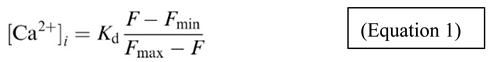
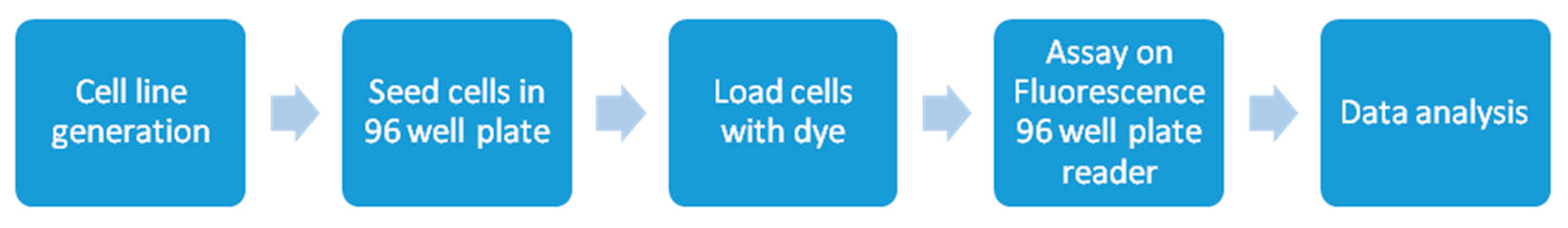
Cell Calcium Bulk Analysis

Bulk analysis of intracellular calcium was performed using a 96-well microplate coupled with fluorescence detection. This method enables rapid and reproducible measurement of cytosolic calcium changes following chemical stimulation. The overall experimental workflow is illustrated schematically in Figure 1.

3.2. Loading Cells on a 96-Well Plate

After reaching adequate confluency, cells were detached using trypsin-EDTA. A 50 µL aliquot of cell suspension was mixed with 50 µL of trypan blue solution and counted using a hemocytometer. Live cells were identified as unstained bright circular objects under microscopic observation. Cell density was calculated using standard hemocytometer protocols, and 10 000 viable cells were seeded into each well of a black, clear-bottom 96-well cell culture plate. Plates were incubated overnight to allow cell attachment.

3.3. Fluo-4 AM Dye Loading

After cell attachment, the culture medium was removed and cells were washed with HBSS. Fluo-4 AM working solution (5.0 µM, 20 µL per well) was added, followed by incubation for 15 min at room temperature and an additional 15 min at 37 °C to allow dye uptake and intracellular ester hydrolysis. Excess dye was removed by washing with HBSS prior to fluorescence measurements.

3.4. Fluorescence Assay on a 96-Well Plate

Fluorescence measurements were performed in a black, clear-bottom COC-coated 96-well plate at a cell density of 10 000 cells per well. Measurements were carried out using the bottom-reading mode with fluorescence excitation at 470 nm and emission at 530 nm. Instrument settings included 25 flashes and an integration time of 20 µs. The fluorescence gain was maintained between 170 and 190, and they were corrected for Fmin, F and Fmax obtained in each experiment, by the relationship between fluorescence intensity and gain as depicted in Figure 2.
Fluorescence intensity was recorded in three stages: resting fluorescence (F₀), fluorescence after reagent addition (F), and maximal fluorescence (Fₘₐₓ) following ionomycin treatment (final concentration 20 µM). This three-point acquisition provides a rapid functional readout of stimulus-invoked Ca²⁺ responses suitable for cell monitoring and screening in a microplate format. Background fluorescence (Fₘᵢₙ) was measured in cell-free wells. All experiments were performed in triplicate. Data are reported as mean ± standard deviation (SD), and fold-change was calculated relative to resting [Ca²⁺] for each condition.

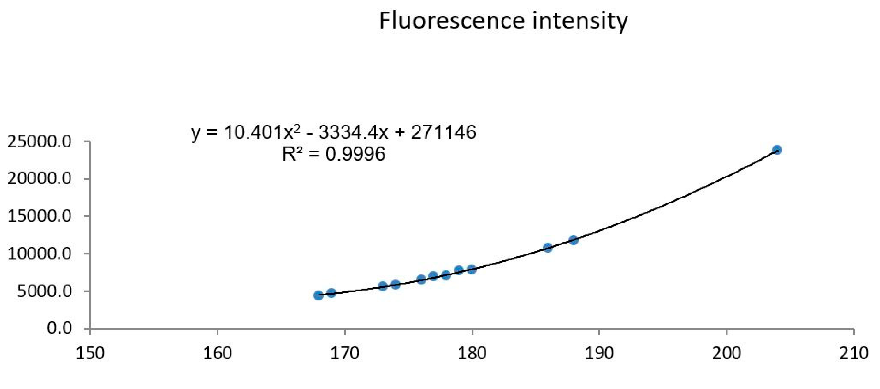
Data Analysis and Results

The intracellular Ca²⁺ biosensing assay produced a clear functional dynamic range across the tested cannabinoids, with fold responses spanning from minimal change (CBL) to strong activation (CBN/CBC). Measurements were performed in triplicate, and the low SD values indicate good reproducibility of the microplate fluorescence readout under constant instrument settings (gain correction). These characteristics support the use of the assay as a cell-monitoring biosensor for screening stimulus-evoked Ca²⁺ responses. Changes in intracellular calcium concentration were calculated using Equation (1). Calcium concentrations (nM) and fold changes relative to resting levels are summarized in Table 1. CBC and CBN induced pronounced increases in intracellular calcium concentration, whereas CBL produced only minimal changes.
To correlate the computational prediction with the cell-monitoring readout, molecular docking was used as a screening and ranking tool for TRPV1 and TRPC5 channels. The cannabinoids CBN and CBC showed stronger predicted binding affinities than CBL across the tested channel structures (Table 2), and this ranking correlated with the experimental calcium responses (Table 1), where CBN/CBC produced pronounced Ca²⁺ elevations and CBL showed minimal effect. The predicted channel binding poses and interaction patterns are summarized in Figure 3 and Figure 4, supporting a mechanistic interpretation consistent with the biosensing results.

Conclusion

A fluorescence-based intracellular calcium assay was implemented as a cell-monitoring biosensing platform to evaluate cannabinoid-induced functional responses in U-87 MG glioma cells. CBC and CBN produced pronounced intracellular Ca²⁺ elevations, whereas CBL induced minimal responses, demonstrating the assay’s ability to discriminate bioactivity based on functional signaling output. The strong agreement between experimental Ca²⁺ readouts and molecular docking predictions highlights the value of integrating computational screening with cell-based biosensing as an efficient workflow for identifying bioactive compounds.

Author Contributions

conceptualization, PL; methodology, PL, BS, SR, AR; formal analysis, PL, BS, SR, AR; investigation, PL, BS, SR, AR; resources, PL; writing-original, BS, SR, AR, writing-review, PL, BS, SR, AR; supervision, PL; project admin, PL; funding acquisition, PL.

Funding

Funding by NSERC of Canada as said in the paper, grant number is RGPIN-2022-03320

Institutional Review Board Statement

Not applicable

Data Availability Statement

The original contributions presented in this study are included in the article. Further inquiries can be directed to the corresponding author(s).

Acknowledgments

We thank the funding from Natural Sciences and Engineering Research Council of Canada.

Conflicts of Interest

The authors declare no conflict of interest

References

  1. Pisanti, S; Malfitano, AM; Ciaglia, E; Lamberti, A; Ranieri, R; Cuomo, G; et al. Cannabidiol: State of the art and new challenges for therapeutic applications. Pharmacology & therapeutics 2017, 175, 133–50. [Google Scholar]
  2. Nabissi, M; Morelli, MB; Santoni, M; Santoni, G. Triggering of the TRPV2 channel by cannabidiol sensitizes glioblastoma cells to cytotoxic chemotherapeutic agents. Carcinogenesis 2013, 34(1), 48–57. [Google Scholar] [PubMed]
  3. Nabissi, M; Morelli, MB; Amantini, C; Liberati, S; Santoni, M; Ricci-Vitiani, L; et al. Cannabidiol stimulates Aml-1a-dependent glial differentiation and inhibits glioma stem-like cells proliferation by inducing autophagy in a TRPV2-dependent manner. International journal of cancer 2015, 137(8), 1855–69. [Google Scholar] [CrossRef] [PubMed]
  4. Rice, T; Lachance, DH; Molinaro, AM; Eckel-Passow, JE; Walsh, KM; Barnholtz-Sloan, J; et al. Understanding inherited genetic risk of adult glioma – a review. Neuro-Oncology Practice 2015, 3(1), 10–6. [Google Scholar] [CrossRef]
  5. Dumitru, CA; Sandalcioglu, IE; Karsak, M. Cannabinoids in Glioblastoma Therapy: New Applications for Old Drugs. Frontiers in molecular neuroscience 2018, 11, 159. [Google Scholar]
  6. Velasco, G; Carracedo, A; Blázquez, C; Lorente, M; Aguado, T; Haro, A; et al. Cannabinoids and gliomas. Molecular neurobiology 2007, 36(1), 60–7. [Google Scholar] [CrossRef]
  7. Nimmrich, V; Gross, G. P/Q-type calcium channel modulators. Br J Pharmacol. 2012, 167(4), 741–59. [Google Scholar]
  8. Montana, V; Sontheimer, H. Bradykinin promotes the chemotactic invasion of primary brain tumors. The Journal of neuroscience: the official journal of the Society for Neuroscience 2011, 31(13), 4858–67. [Google Scholar] [CrossRef]
  9. Watkins, S; Sontheimer, H. Unique biology of gliomas: challenges and opportunities. Trends in neurosciences 2012, 35(9), 546–56. [Google Scholar] [CrossRef]
  10. Bomben, VC; Sontheimer, HW. Inhibition of transient receptor potential canonical channels impairs cytokinesis in human malignant gliomas. Cell proliferation 2008, 41(1), 98–121. [Google Scholar] [CrossRef]
  11. Cuddapah, VA; Turner, KL; Sontheimer, H. Calcium entry via TRPC1 channels activates chloride currents in human glioma cells. Cell calcium 2013, 53(3), 187–94. [Google Scholar] [CrossRef]
  12. Alptekin, M; Eroglu, S; Tutar, E; Sencan, S; Geyik, MA; Ulasli, M; et al. Gene expressions of TRP channels in glioblastoma multiforme and relation with survival. Tumour biology: the journal of the International Society for Oncodevelopmental Biology and Medicine 2015, 36(12), 9209–13. [Google Scholar] [CrossRef] [PubMed]
  13. Duncan, RS; Goad, DL; Grillo, MA; Kaja, S; Payne, AJ; Koulen, P. Control of intracellular calcium signaling as a neuroprotective strategy. Molecules (Basel, Switzerland) 2010, 15(3), 1168–95. [Google Scholar] [CrossRef] [PubMed]
  14. Li, X; Li, PC. Microfluidic selection and retention of a single cardiac myocyte, on-chip dye loading, cell contraction by chemical stimulation, and quantitative fluorescent analysis of intracellular calcium. Analytical chemistry 2005, 77(14), 4315–22. [Google Scholar] [CrossRef]
  15. Li, X; Xue, X; Li, PCH. Real-time detection of the early event of cytotoxicity of herbal ingredients on single leukemia cells studied in a microfluidic biochip. Integrative Biology 2009, 1(1), 90–8. [Google Scholar] [CrossRef]
  16. Huang, H. C.; Chang, P.; Lu, S. Y.; Zheng, B. W.; Jiang, Z. F. Protection of curcumin against amyloid-beta-induced cell damage and death involves the prevention from NMDA receptor-mediated intracellular Ca2+ elevation. Journal of receptor and signal transduction research 2015, 35(5), 450–7. [Google Scholar] [CrossRef]
  17. Takahashi, A.; Camacho, P.; Lechleiter, J. D.; Herman, B. Measurement of intracellular calcium. Physiological Reviews 1999, 79(4), 1089–125. [Google Scholar] [CrossRef] [PubMed]
  18. Gee, KR; Brown, KA; Chen, WN; Bishop-Stewart, J; Gray, D; Johnson, I. Chemical and physiological characterization of fluo-4 Ca(2+)-indicator dyes. Cell calcium 2000, 27(2), 97–106. [Google Scholar] [CrossRef]
  19. Fraguas-Sánchez, AI; Martín-Sabroso, C; Torres-Suárez, AI. Insights into the effects of the endocannabinoid system in cancer: a review. Br J Pharmacol. 2018, 175(13), 2566–80. [Google Scholar] [CrossRef]
  20. Hinz, B; Ramer, R. Anti-tumour actions of cannabinoids. Br J Pharmacol. 2019, 176(10), 1384–94. [Google Scholar] [CrossRef]
  21. Ramer, R; Hinz, B. Cannabinoids as Anticancer Drugs. Advances in pharmacology (San Diego, Calif) 2017, 80, 397–436. [Google Scholar] [PubMed]
  22. Ligresti, A; Moriello, AS; Starowicz, K; Matias, I; Pisanti, S; De Petrocellis, L; et al. Antitumor activity of plant cannabinoids with emphasis on the effect of cannabidiol on human breast carcinoma. The Journal of pharmacology and experimental therapeutics 2006, 318(3), 1375–87. [Google Scholar] [CrossRef] [PubMed]
  23. Shrivastava, A; Kuzontkoski, PM; Groopman, JE; Prasad, A. Cannabidiol induces programmed cell death in breast cancer cells by coordinating the cross-talk between apoptosis and autophagy. Molecular cancer therapeutics 2011, 10(7), 1161–72. [Google Scholar] [CrossRef] [PubMed]
  24. Sultan, AS; Marie, MA; Sheweita, SA. Novel mechanism of cannabidiol-induced apoptosis in breast cancer cell lines. Breast (Edinburgh, Scotland) 2018, 41, 34–41. [Google Scholar] [CrossRef]
  25. Massi, P; Vaccani, A; Ceruti, S; Colombo, A; Abbracchio, MP; Parolaro, D. Antitumor effects of cannabidiol, a nonpsychoactive cannabinoid, on human glioma cell lines. The Journal of pharmacology and experimental therapeutics 2004, 308(3), 838–45. [Google Scholar] [CrossRef]
  26. Vaccani, A; Massi, P; Colombo, A; Rubino, T; Parolaro, D. Cannabidiol inhibits human glioma cell migration through a cannabinoid receptor-independent mechanism. Br J Pharmacol. 2005, 144(8), 1032–6. [Google Scholar] [CrossRef]
  27. Massi, P; Valenti, M; Vaccani, A; Gasperi, V; Perletti, G; Marras, E; et al. 5-Lipoxygenase and anandamide hydrolase (FAAH) mediate the antitumor activity of cannabidiol, a non-psychoactive cannabinoid. Journal of neurochemistry 2008, 104(4), 1091–100. [Google Scholar] [CrossRef]
  28. Solinas, M; Massi, P; Cinquina, V; Valenti, M; Bolognini, D; Gariboldi, M; et al. Cannabidiol, a non-psychoactive cannabinoid compound, inhibits proliferation and invasion in U87-MG and T98G glioma cells through a multitarget effect. PloS one 2013, 8(10), e76918. [Google Scholar] [CrossRef]
  29. De Petrocellis, L; Ligresti, A; Moriello, AS; Allarà, M; Bisogno, T; Petrosino, S; et al. Effects of cannabinoids and cannabinoid-enriched Cannabis extracts on TRP channels and endocannabinoid metabolic enzymes. Br J Pharmacol. 2011, 163(7), 1479–94. [Google Scholar] [CrossRef]
  30. Bisogno, T; Hanus, L; De Petrocellis, L; Tchilibon, S; Ponde, DE; Brandi, I; et al. Molecular targets for cannabidiol and its synthetic analogues: effect on vanilloid VR1 receptors and on the cellular uptake and enzymatic hydrolysis of anandamide. Br J Pharmacol. 2001, 134(4), 845–52. [Google Scholar] [CrossRef]
  31. Lu, J; Ju, YT; Li, C; Hua, FZ; Xu, GH; Hu, YH. Effect of TRPV1 combined with lidocaine on cell state and apoptosis of U87-MG glioma cell lines. Asian Pacific journal of tropical medicine 2016, 9(3), 288–92. [Google Scholar] [CrossRef]
  32. Kárai, LJ; Russell, JT; Iadarola, MJ; Oláh, Z. Vanilloid receptor 1 regulates multiple calcium compartments and contributes to Ca2+-induced Ca2+ release in sensory neurons. The Journal of biological chemistry 2004, 279(16), 16377–87. [Google Scholar] [CrossRef] [PubMed]
  33. Noghabi, Hamideh Sharifi; Ahmed, Abdul Q.; Li, Paul C. H. Intracellular Calcium Increases due to Curcumin Measured using a Single-Cell Biochip. Anal. Lett. 2021, 54, 2769–2776. [Google Scholar] [CrossRef]
  34. Pflaum-Jaeger, Natali; Shahbod, Bardia; Rahimi, Abolfazl; Li, Paul C.H. Responses to herbal compounds in brain cancer cells: two cell-calcium assays and a molecular docking computation study. Anal. Lett. 2025, 58, 2338–2350. [Google Scholar] [CrossRef]
Figure 1. Different steps of cell calcium bulk analysis.
Preprints 196165 g001
Figure 2. Relationship between fluorescent intensity and microplate reader gain.
Preprints 196165 g002
Figure 3. Predicted docking poses of cannabinoids in TRP ion channels generated using Autodock Vina. (a) Rat TRPV1 (PDB ID: 3J5Q), (b) Rat TRPV1 (PDB ID: 3J5R), (c) Human TRPV1 (PDB ID: 6L93), (d) Human TRPC5 (PDB ID: 6YSN). Receptor structures are shown in cyan ribbon representation, and cannabinoid ligands are shown in magenta stick format. In all cases, the ligands occupy the hydrophobic transmembrane binding pockets consistent with known ligand interaction regions, supporting a potential modulatory role in calcium-permeable channels.
Preprints 196165 g003
Figure 4. Two-dimensional interaction maps illustrating predicted molecular contacts and interactions between cannabinol (CBN) and human TRP channels. (a) Human TRPV1 (PDB ID: 6L93), (b) Human TRPC5 (PDB ID: 6YSN). These molecular interaction maps provide structural context that supports the experimentally observed Ca²⁺ responses obtained from the cell-monitoring assay.
Preprints 196165 g004
Table 1. Changes in [Ca2+] when 100 μM of reagents (CBC, CBN, CBL) were added.
Compound Resting [Ca²⁺] (nM) After Treatment [Ca²⁺] (nM) Fold Increase
CBN 445 ± 8 2144 ± 47 4.8
CBC 463 ± 6 2295 ± 31 4.9
CBL 311 ± 40 361 ± 15 1.2
Table 2. Binding affinity of different cannabinoids on two major TRPV1 ion channels (of rat and human origins).
Ion Channel PDB ID CBC (kcal/mol) CBL (kcal/mol) CBN (kcal/mol)
Rat TRPV1 3J5Q −5.7 −6.0 −5.2
Rat TRPV1 3J5R −7.6 −8.3 −8.3
Human TRPV1 6L93 −7.4 −7.9 −8.0
Human TRPC5 6YSN −4.2 −4.5 −5.0±
Disclaimer/Publisher’s Note: The statements, opinions and data contained in all publications are solely those of the individual author(s) and contributor(s) and not of MDPI and/or the editor(s). MDPI and/or the editor(s) disclaim responsibility for any injury to people or property resulting from any ideas, methods, instructions or products referred to in the content.
Copyright: This open access article is published under a Creative Commons CC BY 4.0 license, which permit the free download, distribution, and reuse, provided that the author and preprint are cited in any reuse.
Prerpints.org logo

Preprints.org is a free preprint server supported by MDPI in Basel, Switzerland.

Subscribe

Disclaimer

Terms of Use

Privacy Policy

Privacy Settings

© 2026 MDPI (Basel, Switzerland) unless otherwise stated