Preprint
Article

This version is not peer-reviewed.

Epidemiological and Clinical Insights from 68 Veterinarian-Reported Cases of Feline Infectious Peritonitis During the Documented 2023 FIP Epizootic in Cyprus

A peer-reviewed version of this preprint was published in:
Pathogens 2026, 15(5), 499. https://doi.org/10.3390/pathogens15050499

Submitted:

19 January 2026

Posted:

21 January 2026

Read the latest preprint version here

Abstract

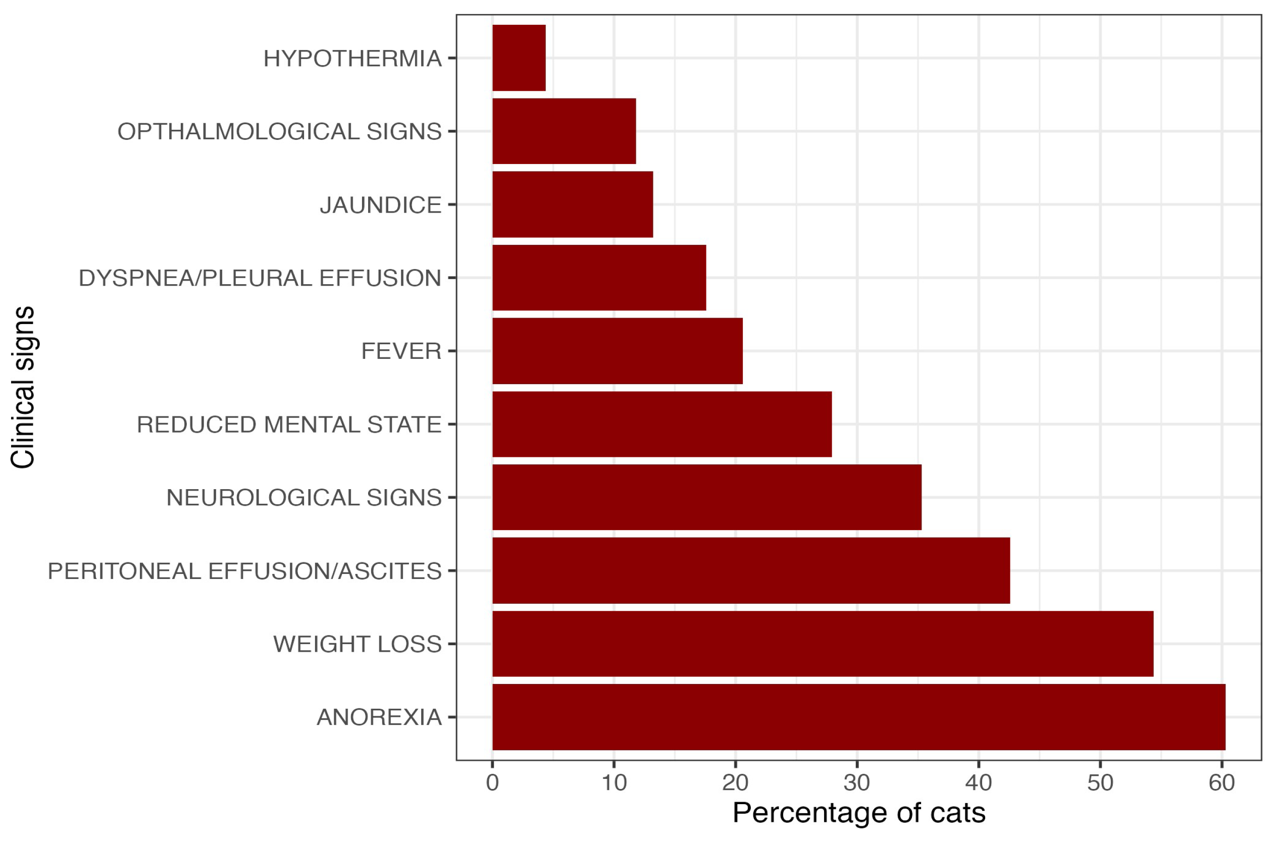
In 2023, Cyprus experienced a large-scale epizootic of feline infectious peritonitis (FIP) associated with a novel feline coronavirus, FCoV-23. While molecular investigations have elucidated its recombinant origin, field-based clinical and epidemiological data remain limited. A prospective study used a structured 30-item questionnaire embedded in veterinary management software to characterize FIP cases. Data were voluntarily submitted by registered veterinarians across Cyprus for cases identified between late 2022 and 2025. Data from 68 cases reported by 22 clinics (response rate 21.0%) were analyzed. Affected cats were older than typically reported for FIP (mean age of 3.9 years; median = 3.0; range 0.4y - 12.9y, SD = 3.41), with most cases documented from Limassol (51.5%) and Nicosia (25.0%). The most frequently reported clinical signs were anorexia (60.3%) and weight loss (54.4%), while neurological involvement was documented in 35.3% of cases. An albumin-to-globulin ratio <0.8 was observed in 86.8% of cats tested. Antiviral (GS-441524 or molnupiravir) therapy was administered in 92.2% of cases, with reported clinical improvement in 88.9%. These findings demonstrate the value of questionnaire-based reporting for documenting outbreak-associated disease patterns and confirm the change in viral tropism seen in FCoV-23 infection as an increased prevalence of neurological cases compared to ‘traditional’ FIP.

Keywords: 
;  ;  ;  ;  ;  ;  

1. Introduction

Feline infectious peritonitis (FIP) is a fatal viral disease that is found worldwide [1,2,3]. To date it remains one of the most important and challenging infectious diseases of cats, with significant clinical and epidemiological relevance worldwide. FIP is caused by virulent mutated variants of feline coronavirus (FCoV)1 [4,5]. Despite decades of study, its multifactorial pathogenesis, diverse clinical presentation, and high mortality continue to complicate diagnosis, prevention, and control [6,7,8]. Recent epizootic events, such as the large-scale outbreak reported in Cyprus in 2023 [9,10,11,12,13], have renewed scientific interest in the mechanisms of FCoV evolution and transmission, and in the conditions that may favor the emergence of highly pathogenic variants.
FCoV is highly prevalent in environments with multiple cats, with infection rate as high as 90% where cats are housed together [14,15,16,17,18]. FCoV is distinguished into two pathotypes: the low-virulence Feline Enteric Coronavirus (FECV) and the highly pathogenic Feline Infectious Peritonitis Virus (FIPV).
FCoV infection begins after the oral ingestion of the virus, as the primary route of transmission is the fecal-oral route, usually through contact with contaminated cat litter, grooming, and fomites such as litter scoops or hairbrushes [19,20]. Following oral ingestion, the virus moves and replicates in the epithelial cells of the small intestine, mainly the ileum and the colon [21], leading to viral shedding in feces [22]. Most FCoV infections remain subclinical, although mild enteritis may occur, especially where co-infections are present [22,23,24]. FIP is believed to develop due to mutation of FECV to FIPV, which is known as the “internal mutation theory”. The mutation from FECV to FIPV alters cell tropism from intestinal cells to macrophages which enables the mutated virus to replicate within circulating monocytes and activated macrophages, facilitating its systemic spread through these infected immune cells to the body tissues [21,25,26]. Clinically, this manifests as the effusive (or wet) form of FIP, characterized by fluid accumulation in the peritoneal, pleural, and/or pericardial cavities. In more chronic or localized cases extensive perivascular pyogranulomatous lesions develop within affected organs. This is referred to as the non-effusive (or dry) form of FIP, with clinical signs depending on which organs are involved. Granuloma formation most commonly occurs in the spleen, liver, kidneys, intestines, heart, lungs, eyes, central nervous system, and spinal cord, resulting in a wide spectrum of clinical manifestations [19,27,28,29,30,31]. In addition, many cats develop clinical signs that are a combination of or transition from non-effusive to effusive disease [33,34,35,36,37,38].
The shifting of cell tropism from enterocytes to macrophages can also affect transmission dynamics. More specifically, while FECV spreads primarily via the fecal-oral route, FIPV has generally limited transmission potential between cats as it rarely retains the capacity to replicate effectively within intestinal enterocytes [38]. Until recently, outbreaks of FIP that resulted from direct FIPV transmission were regarded as rare, isolated events [34,35] with limited epidemiological significance, reinforcing the prevailing belief that FIPV transmission between cats was minimal. This belief was changed when an outbreak of FIP was documented in 2023 in the island of Cyprus, a European Country in the Eastern Mediterranean Sea[9,11,12]. In the first half of 2023, the number of reverse transcriptase-polymerase chain reaction (RT-PCR) - confirmed FIP cases, based on samples from body cavity fluids, peritoneal lymph node aspirates, and tissue biopsies, rose dramatically marking a more than 20-fold increase compared to 2022 [9]. Nevertheless, the cat population in Cyprus is predominantly consistent of cared for stray cats and shelter cats. As a result, only a small proportion of the affected cats that presented to the primary care practice, had extensive diagnostics leading to RT-PCR confirmation [41]. Reports from the Pancyprian Veterinary Association (PVA) estimated that over 8000 cats were affected in the first half of 2023 [36]. The outbreak was found to involve a newly identified recombinant FCoV, named FCoV-23, which was seen to cause severe illness in both stray and domestic cats. Genetic analysis revealed that FCoV-23 had acquired a spike protein from a highly virulent canine coronavirus, the Pantropic Canine Coronavirus (P-CCoV) [11,13,43]. The spike protein exchange observed in FCoV-23 is reminiscent of similar recombination mechanisms seen in other coronaviruses, such as SARS-CoV-2 [44], which have led to increased transmissibility and virulence [13,45].
Previous studies by our group have described the molecular features and geographic distribution of RT-PCR-confirmed FIP cases associated with the 2023–2024 epizootic in Cyprus[9,11,12,13,43]. However, systematically collected data from routine clinical practice, reflecting how cases were identified and presented to veterinarians during the outbreak, remain limited. The present study addresses this gap by analyzing prospectively collected data obtained through a structured questionnaire administered to veterinary clinics across Cyprus. By documenting clinician-reported FIP cases encountered in clinical settings during the epizootic, this study provides a complementary field-based perspective to existing laboratory and surveillance focused investigations.

2. Materials and Methods

2.1. Data Collection

Beginning in March 2023, the at the time Vice-President of the Pancyprian Veterinary Association (PVA), author DE, contacted authors CA, DGM, ML, and SM, [members of the newly established FCoV-23 International Research Consortium (FCoV-23 IRC), of which CA, DGM, DE, ML, SM and MEF are founding members] to request the documentation and investigation of the ongoing FIP outbreak in Cyprus. Amongst other measures, the authors created a questionnaire (included as supplementary material) which primarily aimed to document the characteristics of the FIP outbreak from the perspective of the practicing veterinarians. With the facilitation of the PVA an open invitation was extended to all registered companion animal or mixed practice veterinary clinics in Cyprus island-wide (this term refers to the areas that are under the effective control of the government of the Cyprus Republic) in the areas of Cyprus that the government of the Republic of Cyprus represents, using the Vet Clinic Pro® practice management system2. The invitation encouraged veterinarians to contribute to data collection about FIP cases identified from 2022 onward. Veterinarians were contacted via an official email distributed by PVA, which included comprehensive instructions, guidance documents, and study information. The data collection was managed through a structured questionnaire integrated into the veterinary management software Vet Clinic Pro®.
The questionnaire was comprised of 30 questions and organized into three sections. The first section aimed to obtain information regarding the veterinary practice, including practice demographics and the unique identifier associated with the practice's software management system. The second section aimed to capture epidemiological and background clinical information for each feline patient. Variables recorded included patient age, sex, neutering status, breed, housing status (e.g., indoor/outdoor, multi-cat household), recent exposure to potential stressors, and reported contact with other cats, allowing characterization of individual and environmental level factors relevant to disease occurrence. The third section aimed to document detailed clinical data, including observed clinical signs, results from laboratory and imaging investigations, confirmation of FIP by RT-PCR testing, and information on therapeutic interventions administered. Relevant diagnostic findings were entered directly into the questionnaire using data extracted from the patient’s electronic medical records via the same practice’s software management system.
To ensure consistency and quality of data collection the authors, with the facilitation of the PVA, organized a dedicated online seminar prior to the initiation of the questionnaire (May 2023). During this online seminar, the objectives of the data collection process were outlined, including an overview of the evolving outbreak and its epidemiological significance. At that time, the presentation focused on the documented increase in FIP cases, as information on the clinical manifestations of the outbreak was still limited. Comprehensive instructions were provided to participating veterinarians, detailing the procedures for accurate completion of the questionnaire, and clarifying inclusion criteria for case identification. The online seminar also addressed best practices for clinical assessment and specimen collection to optimize diagnostic accuracy and data reliability. Veterinarians were encouraged to report cases from late 2022 onwards. All veterinarians were asked to read the consent declaration and indicate their agreement by ticking the consent box before completing the electronic questionnaire.
Case definitions were divided into two categories:
Suspected cases were domestic feline patients with compatible clinical appearance3 [36] (e.g., loss of appetite, loss of weight, fever, peritoneal effusion, pleural effusion, pericardial effusion, ophthalmological and/or neurological signs), Category A cases. While those with additional compatible laboratory findings (e.g., hyperglobulinemia, hyperbilirubinemia, albumin to globulin ratio (A/G) <0,5) were Category B cases.
Confirmed cases were described by the reporting veterinarian as suspected cases with a positive FCoV RT-PCR result on a sample collected from either peritoneal effusion, pleural effusion, pericardial effusion, cerebrospinal fluid (CSF), aqueous fluid cell suspension, from fine needle aspiration biopsy (FNAB) or tissue biopsy. Additionally, positive cases were considered those with positive FCoV immunohistochemistry on tissue biopsy with compatible pathological findings for FIP.
The case definitions and description were used to encourage the participating veterinarians to record data for all suspected cases, including the ones that were not definitively confirmed. This is in line with the European Advisory Board of Cat Diseases (ABCD) FIP diagnostic tool algorithms[46]

2.2. Data storage and analysis

Data was securely stored on a server accessible exclusively to the software operator. Reports containing questionnaire responses, along with additional files and data, were exported in Microsoft Excel [47] format for analysis. Additional files - including blood tests and diagnostic imaging - were exported as individual files for each test, labeled with unique identifiers to ensure accurate patient tracking. Data analysis was performed using R Statistical Software version 4.4.2 [48]. Data manipulation, descriptive statistics and visualisation were performed using the tidyverse collection of packages [49].

3. Results

3.1. Questionnaire response

Out of 105 veterinary clinics contacted (n=105 veterinary clinics use the veterinary management software Vet Clinic Pro® out of 150 companion animal veterinary clinics island-wide in total), 22 responded, giving a response percentage of 21.0%. These clinics were spread across five districts in Cyprus: ten in Limassol (45.5%), six in Nicosia (27.3%), three in Ammochostos (13.6%), two in Larnaca (9.0%) and one in Paphos (4.5%).
A total of 77 cases of FIP were reported. However, seven cases were excluded due to incomplete questionnaires, leaving 70 cases for analysis. Of these, two cases were also excluded as the reported occurrence was before the outbreak was declared (one case in February 2022 and another in July of 2022), resulting in 68 cases for the final analysis. The cases covered a period from November 2022 to March 2025 (Figure 1) with the majority of the cases being reported between January 2023 and October 2023. Although all case data were collected prospectively, 9 cases were reported in the questionnaire at a different time from when the cases occurred.
The completeness of data varied across the many variables; however, most fields had low rates of missing data (<5%).

3.2. Case signalment and clinical history, plus geographical distribution of cases

The mean age of the cats in this population was 3.9 years (median = 3.0; min = 0.4, max = 12.9, SD = 3.41), indicating that most of the reported cases were young adults. Male cats accounted for 40/68 (58.8%) of the cases and female cats for 28/68 (41.2%). Most cats were neutered (55/68; 80.9%). The majority of cats were domestic short hair (56/68; 82.4%), followed by domestic long hair (10/68; 14.7%), while British shorthair and Maine coon breeds represented one case each (1.5%). Regarding living environment, 30/68 (44.1%) cats lived both indoors and outdoors, 18/68 (26.5%) were kept exclusively outdoors, 12/68 (17.6%) were stray cats, and 8/68 (11.8%) were kept strictly indoors. The majority of the cats (65/68; 95.6%) were reported to have contact with other cats; however, two of the indoor-only cats did not have contact with other cats. The third cat that was declared not to have any contact with other cats, was reported as indoor/outdoor cat. Over half of the cases originated from the Limassol district (35/68; 51.5%), followed by Nicosia (17/68; 25.0%), Ammochostos (10/68; 14.7%), then Larnaca and Paphos (3/68; 4.4% each).

3.3. Categorization and clinical presentation

Based on the classification scheme detailed in the M&M section, there were more Suspect cases (Category B) 40/68 (58.8%), followed by Confirmed cases (Category C)15/68 (22.1%) then by Suspect cases (Category A) 13/68 (19.1%).
Among suspected cases without further laboratory confirmation (Category A; n = 13), Peritoneal effusion caused ascites was the most frequently observed clinical finding, reported in 10/13 cases (76.9%). Anorexia was recorded in 8/13 cats (61.5%), while neurological abnormalities, fever, weight loss, and reduced mental state were each documented in 4/13 cases (30.8%). Other clinical signs were infrequently observed in this category.
The largest group of cases was diagnosed after clinical examination and in-house laboratory testing without a definite confirmation by an RT-PCR (Category B; n = 40). In this group, anorexia and weight loss were the most prevalent findings, each present in 24/40 cases (60.0%). Neurological signs were also commonly observed (17/40; 42.5%), followed by reduced mental state (12/40; 30.0%). Peritoneal effusion/ascites were identified in 14/40 cases (35.0%). Fever and ophthalmological signs consistent with uveitis were each recorded in 8/40 cats (20.0%), while dyspnea attributable to pleural effusion and jaundice were observed in 7/40 cases each (17.5%). Hypothermia was uncommon, occurring in only 2/40 cases (5.0%).
Cases with molecular confirmation by RT-PCR (Category C; n = 15) exhibited a clinical presentation similar to that observed in Category B. Anorexia and weight loss were again the most frequent clinical signs, each noted in 9/15 cases (60.0%). Peritoneal effusion/ascites were present in 5/15 cats (33.3%). Dyspnea due to pleural effusion was observed in 4/15 cases (26.7%), whereas neurological signs and reduced mental state were each documented in 3/15 cases (20.0%). Fever and jaundice were relatively uncommon, reported in 2/15 (13.3%) and 1/15 (6.7%) cases, respectively.
Across all cases (Suspected and Confirmed; Figure 2), the most frequent clinical signs were anorexia in 41/68 (60.3%) and weight loss in 37/68 (54.4%). Ascites was reported in 29/68 (42.6%), and neurological signs in 24/68 (35.3%). Additional findings included reduced mental state in 19/68 (27.9%), fever in 14/68 (20.6%), and dyspnea in 12/68 (17.6%). Jaundice was recorded in 9/68 (13.2%), ocular involvement (uveitis) in 8/68 (11.8%), and hypothermia in 3/68 (4.4%).

3.4. Co-infections and stressors

Of the cats tested for co-infections, Feline Leukaemia Virus (FeLV) antigen and Feline Immunodeficiency Virus (FIV) antibody (36/68; 52.9%), all were FeLV negative. FIV positivity was detected in 5/36 (13.9%). The retrovirus status was unknown or untested in 32/68 (47.1%) of the cats. The only other co-infection reported was toxoplasmosis, which was reported to be positive by serology in 2/68 (2.9%) cats. Only one of those two cases was an FIP-confirmed RT-PCR (Category C) case. Most of the cats were not receiving any pharmaceutical treatment upon the time of their presentation (62/68; 91.2%).
A potential stress-related event prior to FIP-associated disease onset was reported in 14/68 (20.6%) of cats. This was most commonly a surgical procedure under general anesthesia (e.g., neutering; 6/14; 42.8%), followed by relocation (2/14; 14.3%), teeth cleaning (2/14; 14.3%), or other events (e.g., unspecified; 2/14; 14.3%), and travel or veterinary visits (1/14 each; 7.1%).

3.5. Hematology and biochemistry

A total of 53 cases had hematology and/or serum biochemistry results included (but this had been performed by different point of care in-house analyzers). Among these, 46/53 (86.8%) showed an albumin-to-globulin (A/G) ratio < 0.8, making this the most consistent finding. Hyperglobulinemia was reported in 32/53 (60.4%) cases. Leukocytosis was present in 25/53 (47.2%), anemia in 18/53 (34.0%), and hyperproteinemia in 18/53 (34.0%). Hyperbilirubinemia was reported in 14/53 (26.4%), elevated Alanine Aminotransferase (ALT) in 5/53 (9.4%), and increased Blood Urea Nitrogen (BUN) and/or Creatinine in 4/53 (7.5%). Protein electrophoresis was performed in 31/68 (45.6%) cases with the majority showed an increase in isolated gamma-globulins (24/31; 77.4%), followed by combined alpha- and gamma- increases (5/31; 16.1%), and isolated alpha increases (2/31; 6.5%). Among cases with increased gamma globulins, 19/24 (79.2%) had an A/G ratio < 0.8, and 7/31 (22.6%) had FIP confirmed by RT-PCR, including 4/31 (12.9%) from effusion fluid and 3/31 (9.7%) from FNAB. The combination of hyperglobulinemia and a low albumin-to-globulin (A/G) ratio was observed in 31 cases and varied according to clinical presentation. Among these, 11 cases (35.5%) had effusive-only FIP form, 11 cases (35.5%) had neurological involvement, 5 cases (16.1%) were non-effusive, 2 cases (6.5%) had concurrent effusive and ophthalmic involvement, and 2 cases (6.5%) presented with combined neurological and ophthalmic forms.

3.6. Diagnostic imaging

In-house ultrasonographic examination was reported in 52/68 (76.5%) of cats; there were no abnormal findings in 32/68 of those (61.5%). Radiographs were available for 46/68 (67.6%) cases; abnormal findings were reported in 18/68 of those (39.1%)

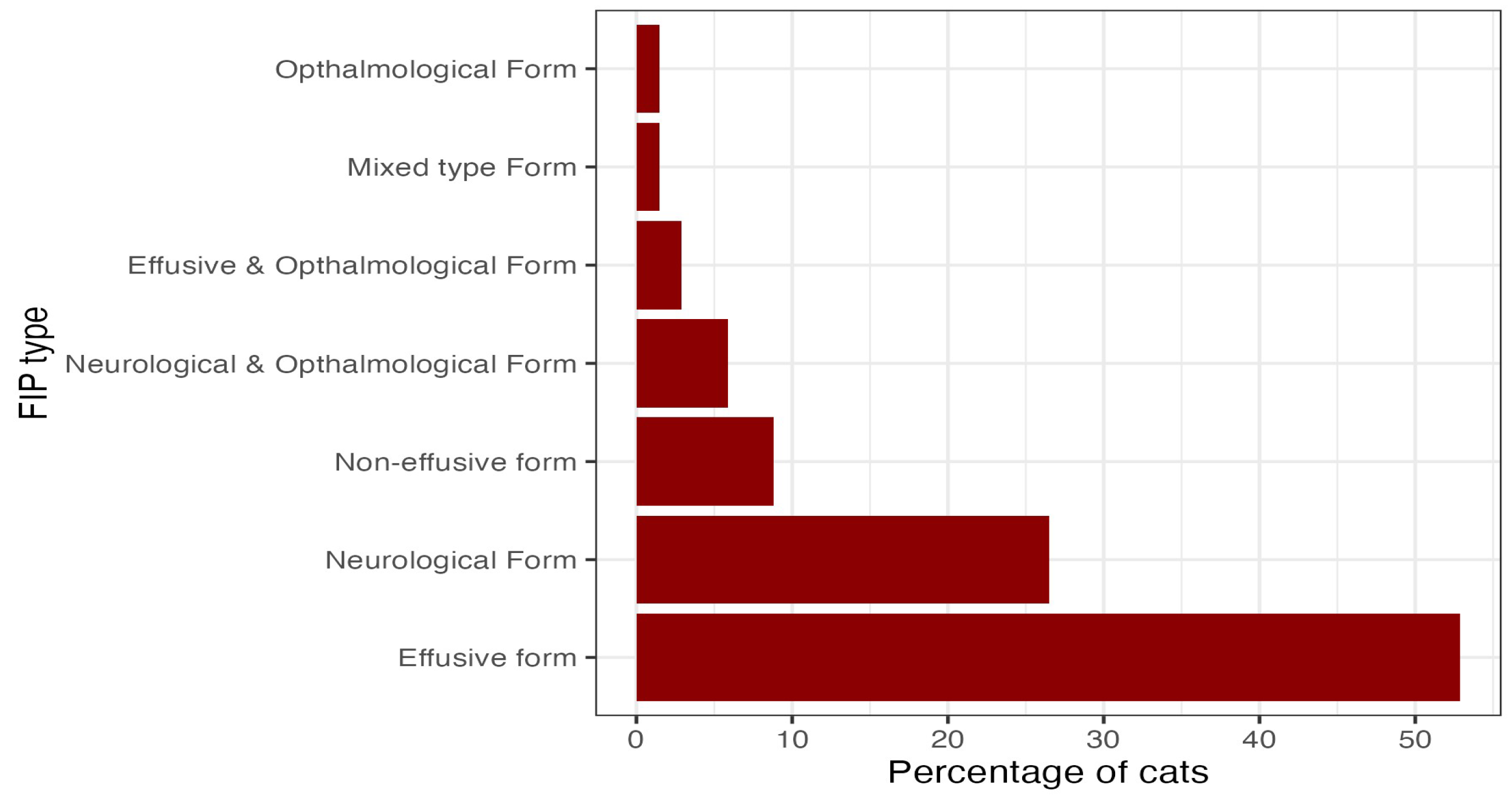
3.7. FIP diagnosis and form

A confirmed diagnosis of FIP by RT-PCR was obtained in 15/68 cases (22.1%). Of these, 11/68 cases (16.2%) occurred from effusion samples, while 4/68 cases (5.9%) occurred from FNAB samples. Based on clinical signs, clinical pathology and diagnostic imaging findings, the effusive form was the most frequently observed presentation, accounting for 36/68 (52.9%) cases. Neurological involvement was identified in 18/68 (26.5%) cases. The non-effusive form was less common, recorded in 6/68 (8.8%) cases. Combined neurological and ophthalmological involvement was observed in 4/68 (5.9%) cases, while concurrent effusive and ophthalmological presentations were identified in 2/68 (2.9%) cases. Mixed-type presentation was recorded in a single case (1.5%), as was the ophthalmological involvement (1.5%).
Figure 1. Graph presenting the types of FIP reported in the Cyprus outbreak as determined by the clinical signs, clinical pathology and diagnostic imaging findings reported on the questionnaires by the participating veterinarians (with the frequencies reported in percentages).
Preprints 195094 g003

3.8. Treatment and outcome

Treatment was undertaken in 63/68 cases (92.3%), Euthanasia was selected in 3/68 cases (4.4%), and no treatment was recorded in 2/68 cases (2.9%). The most frequently reported therapy was oral GS-441524, administered in 33/63 treated cases (53.2%), followed by oral molnupiravir in 21/62 cases (33.3%), injectable remdesivir in 5/63 cases (7.9%), and a combination of injectable remdesivir with oral molnupiravir in 4/63 cases (6.3%). Most cases were treated with unlicensed products (43/63; 68.3%), whereas licensed formulations were used in 19/63 cases (30.2%).
Overall, a positive clinical response to treatment was reported in 56/62 cases (90.3%), while no clinical improvement was documented in 6/62 cases (9.7%). Only one case did not have treatment response feedback which was excluded from the analysis leaving 62 cases analyzed. All cases treated with a combination of injectable remdesivir, and oral molnupiravir demonstrated clinical improvement (4/4; 100%), as did all cats receiving remdesivir monotherapy (5/5; 100%). Among cats treated with oral GS-441524, a positive clinical response was observed in 30/33 cases (90.9%), whereas no response to treatment was reported in 3/33 cases (9.1%). For cases receiving oral molnupiravir monotherapy, clinical improvement was documented in 17/20 cases (85.0%), while 3/20 cases (15.0%) showed no response to treatment.

4. Discussion

The present study provides new information about the essential characteristics of the FIP outbreak in Cyprus in 2023, documented through the use of a standardized, structured questionnaire completed in part by practicing veterinarians in real-time while the outbreak was evolving. The outbreak was caused by a new FCoV recombinant called FCoV-23; the molecular nature of this change and the epidemiology of the outbreak have already been described [9,11,13,50]; the current work provides complementary evidence from the field, documenting how the outbreak was perceived and diagnosed by general practitioners in Cyprus. This study clearly supports the usefulness of structured questionnaires as a practical epidemiological tool for rapidly capturing real-time clinical observations during emerging infectious disease outbreaks.
The response rate of the study was 21.0% (22 clinics answered the questionnaire out of 105 contacted). Although modest, this response rate is comparable to those reported in questionnaire-based studies involving veterinary professionals, particularly in companion animal infectious disease research requiring detailed clinical data. Reported response rates in the veterinary literature vary widely depending on study design and target population [51,52,53,54], reflecting the practical challenges of achieving high participation in voluntary clinic-based surveys. Non-response bias cannot be excluded and should be considered when interpreting the results. An interesting finding of this study is that only 9 out of 68 cases were reported in a different time from the time of the case presentation. This distinction underscores that the level of engagement among practicing veterinarians in documenting the outbreak remained high throughout the epidemic phase and did not diminish after the disease became endemic. Moreover, the data for these nine cases had been collected prospectively in real time, although reported at a later stage, thereby minimizing recall bias in the recorded information.
Most cases were recorded in Limassol and Nicosia with the most probable explanation for this being the higher number of veterinary facilities in those two areas. However, this distribution may also reflect the population density of cats, with higher numbers of feeding stations, and greater interaction between owned and free-roaming cats in those two areas of Cyprus.
The signalment of cats with traditional FIP is typically young adult, and non-pedigree, with a male predominance [34,35,37,55,56]. However, the mean age of the cats in this outbreak was 3.9 years, which is higher than the typical 1–2 years reported elsewhere [36,37] but still lower than the findings reported in the first paper that described this outbreak where the mean age was 4.6 years [9,11,12]. A possible reason for the higher age in the cats in the Cyprus outbreak, which then reduced over time, is likely because cats of all ages were immune-naïve and therefore susceptible when recombinant FCoV-23 first infected the cats on the island. However, with time, the older cats had met the virus and were resistant to it, hence only young immune-naïve kittens developed clinical FIP as the outbreak progressed. Also, while pedigree cats make up the majority (up to 70%) of traditional FIP cases[34,36,37] few pedigree cats were seen in the current outbreak. This is because Cyprus is known for the large numbers of unowned cat population [41]. Even though a number of pure breed cats exist in Cyprus, the vast majority of owned cats are domestic shorthaired and domestic longhaired cats adopted from the streets[57].
The overwhelming presence of cats with prior contact with other cats (95.6%) reinforces the recognized role of social and environmental transmission in FCoV maintenance [58], and also supports the direct transmission dynamics of FCoV-23 which caused the epizootic [11,12,13]. An additional noteworthy observation was that 14/68 cats were reported to have experienced a stressor preceding the onset of clinical signs. This finding supports the recognized association between stress and the development of FIP. The identification of stress prior to disease manifestation aligns with existing knowledge regarding risk factors that may contribute to the development of FIP in susceptible individuals [59,60,61].
The clinical manifestations recorded in this study were largely consistent with previous publications on FIP, including anorexia, weight loss, lethargy, and effusive peritoneal disease [34,35,37,62]. However, the relatively high frequency of neurological involvement (35.3%) deserves attention. This proportion exceeds the 10–30% range reported in earlier large population studies[34,36,37] suggesting a potential shift in the disease phenotype associated with FCoV-23 [11,13]. This finding aligns with the experimental and genomic evidence that recombinant FCoV-23 carries spike protein regions derived from P-CCoV, known for its systemic and potential neurotropic presentation [63,64,65,66,67,68]. The increased neurotropism of FCoV-23 reported in other studies describing this epizootic [9,11,12,13] may explain the higher proportion of neurological cases reported in this study. Similar shifts in virulence following recombination events have been documented in canine and porcine coronaviruses [68,69,70,71], supporting the hypothesis that FCoV-23 represents a novel, epizootically capable variant [11,50].
Diagnostic approaches reported by the veterinary practitioners on Cyprus reflected the practical challenges of field conditions. Only 22.1% (15/68) of cases were RT-PCR-confirmed as FIP, with most diagnoses being based on the combination of clinical signs and in-house laboratory test findings. This is consistent with patterns reported in several large clinical studies, where the cost and accessibility of molecular tests limit their use [34,55,72]. This situation is particularly evident in countries where companion animal health insurance systems are not established and where large populations of stray animals exist, such as on Cyprus [41,57], resulting in financial and logistical limitations that restrict the routine use of advanced diagnostic methods [73,74,75]. Despite this, most cases (55/68; 80.9%) included in-house analyzer blood examinations, reflecting the standard of animal care in Cyprus. The high prevalence of hyperglobulinemia and low serum albumin-to-globulin ratios (A/G <0.8) mirrors classical FIP profiles and supports their continued diagnostic value [55,59]. A raised serum gamma-globulin concentration was the most common protein electrophoretic pattern (77.4%), consistent with the immunopathogenic nature of the disease [2,76,77]. Also, in line with the current literature is the finding that hyperglobulinemia and A/G ratio are not dependent on the FIP form[34,36]
Therapeutically, Cyprus was the first European Country that approved three different options for the treatment of FIP in cats, injectable Remdesivir and GS441524 50mg tablets (BOVA UK Ltd, United Kingdom) and molnupiravir (Lagevrio™, Merck Sharp & Dohme B.V.) [78]. The study confirms the widespread adoption of these antiviral drugs for the treatment of FIP, as 92% of the cases received one of these agents, or a combination, with an overall reported clinical improvement rate of 88.9%. These figures are consistent with published efficacy data of the antiviral drugs on FIP [33,37,79,80,81]. However, since 68.3% of the cats were given unlicensed preparations, this highlights the ethical and legal ambiguity surrounding these formulations[82,83]. The information gathered for the licensed molnupiravir preparation referenced in the study (Lagevrio™, Merck Sharp & Dohme B.V.) is useful for the knowledge of the use of this medication for the treatment of FIP in cats which was specifically permitted for this use in Cyprus [78]. It’s administration to 25 cases, four of which were also given initial remdesivir, with 84% treatment success, agrees with a number of studies where cats with FIP were treated with generic formulations of molnupiravir, resulting in high survival rates of 78–92% [84,85,86,87,88,89], highlighting the drug’s effectiveness. The successful treatment of cats with FIP in Cyprus demonstrates the rapid dissemination of treatment protocols among veterinary practitioners during the outbreak. It exemplifies effective professional communication and adaptability in response to crisis conditions as well as the crucial role that the PVA played in this crisis-management.
A major strength of this work lies in its partially prospective design, with almost real-time data collection. Once the outbreak was discovered, the authors, with the help of the PVA, asked veterinary practitioners on Cyprus to prospectively complete questionnaires for cases of FIP they had seen since the beginning of 2023. They were then asked to prospectively collect data for all new cases while the outbreak was ongoing; this minimized recall bias and allowed for a more accurate reflection of temporal and geographic trends. Unlike fully retrospective or laboratory-based studies, this approach provides a ground-level view of the dynamics in clinical practice. Moreover, the island-wide coverage achieved through voluntary participation of veterinarians across all districts was something that occurred for the first time in Cyprus in the field of companion animals.
Another key strength is the integration of clinical, epidemiological, and therapeutic data in one dataset. This allows holistic interpretation of how the outbreak manifested, evolved, and was managed.
Finally, this study demonstrates the feasibility and value of a questionnaire-based veterinary reporting model, which could be replicated for future similar events in companion animal medicine, especially where a systematic surveillance program does not exist.
Despite these strengths, several limitations must be acknowledged. The voluntary and self-reported nature of the questionnaire may have introduced selection bias: clinics that experienced more cases or were more aware of the outbreak may have been more likely to participate. If their diagnostic protocols differed from under-represented clinics, this could have biased the data. Second, diagnostic heterogeneity represents a significant limitation. The reliance on clinical and in-house diagnostic findings rather than molecular confirmation in most cases means that misclassification cannot be entirely excluded. Although clinical patterns were strongly consistent with FIP, some overlap with other systemic diseases is possible.

5. Conclusions

In summary, this study is part of the ongoing investigation of the large-scale epizootic of FIP associated with the novel recombinant feline coronavirus, FCoV-23, which started in Cyprus in late 2022. The cats were older than typical FIP, and mainly non-pedigree cats, with neurological involvement seen more commonly than with typical FIP (35% vs 10-30%). The latter supports the change in viral tropism seen with FCoV-23 infection. Diagnostic approaches reported by the veterinary practitioners on Cyprus reflected the practical challenges of field conditions, with only 22.1% of cases being confirmed as FIP by RT-PCR; however, a combination of clinical signs and in-house laboratory test findings were used in the majority (80.9%) of cases, reflecting the standard of animal care in Cyprus. Antiviral therapy was administered to most cats (92.2%), primarily using GS-441524 or molnupiravir (the latter in the form of Lagevrio™), with clinical improvement in the majority (88.9%). This study clearly supports the usefulness of structured questionnaires as a practical epidemiological tool for rapidly capturing real-time clinical observations during emerging infectious disease outbreaks.

Supplementary Materials

The following supporting information can be downloaded at the website of this paper posted on preprints.org.

Funding

This research received no external funding. The APC was funded by Morris Animal Foundation, award number: DE25FE-204.

Institutional Review Board Statement

The study was reviewed and ethically approved by the Cyprus National Bioethics Committee (approval number: EEBK.ΕΠ.2024.01.240, DATE: 04/09/2024, 18062025).

Data Availability Statement

The data that support the findings of this study are available from the corresponding author upon reasonable request.

Acknowledgments

The authors extend their heartfelt gratitude to the veterinary practitioners across Cyprus who generously and voluntarily contributed to this study. Their dedication in completing the questionnaire and sharing valuable clinical and epidemiological data during a challenging outbreak period was crucial. Their commitment not only made the successful documentation of the outbreak possible but also played a key role in enabling a swift response and effective management of this epizootic. The authors extend their sincere gratitude to the Pancyprian Veterinary Association (PVA) for its unwavering support throughout the study. The Association provided substantial assistance in disseminating the questionnaire and played a key role in organizing the preparatory online seminar, which contributed significantly to the smooth implementation of the study. In addition, the PVA was instrumental in the crisis management and coordinated response to this outbreak, and its contribution merits explicit acknowledgment. The authors also wish to thank the Federation of the Companion Animal Veterinary Associations (FECAVA) for raising awareness of the outbreak at the time it occurred and for the support provided. The authors are also grateful to the Veterinary Services of Cyprus and the Ministry of Agriculture, Rural Development and Environment for their ongoing collaboration, guidance, and steadfast support throughout the course of this research. Sincere appreciation is expressed to the Morris Animal Foundation (MAF), EveryCat Health Foundation (EHF), and the Governing Council of the Cat Fancy (GCCF) Foundation. In particular, this work was supported by the Morris Animal Foundation/EveryCat Health Foundation and the FCoV23 International Research Consortium (IRC) under award number DE25FE-204. The authors are especially thankful for the contributions of Dr. Drew Weigner, Dr. Vicki Thayer, and Professor Gregg Dean, all of whom are members of the FCoV-23 IRC. Their involvement, along with the broader support of the board network for FIP and their pivotal roles in the establishment of the FCoV23 International Research Consortium (IRC), has been invaluable. These organizations and individuals have made significant contributions to advancing research on FIP worldwide, and their efforts are deeply appreciated.

Conflicts of Interest

There are no conflicts of interest reported for this study.

Abbreviations

The following abbreviations are used in this manuscript:
MDPI Multidisciplinary Digital Publishing Institute
DOAJ Directory of open access journals
FIP Feline Infectious Peritonitis
FIV Feline Immunodeficiency Virus
FeLV Feline Leukaemia Virus
CCoV Canine Coronavirus
P-CCoV Pantropic Canine Coronavirus
RNA Ribonucleic acid
RT-PCR Real Time Polymerase Chain Reaction
ALT Alanine Aminotransferase
BUN Blood Urea Nitrogen
PVA Pancyprian Veterinary Association

Notes

1
ICTV, classifies FCoV in the order of Nidovirales, the family of Coronaviridae, the subfamily of Orthocoronavirinae, the Genus Alphacoronavirus, the Subgenus of Tegacovirus
2
D.S. Compass Solutions LTD
3
European Advisory Board of Cat Diseases (ABCD) Guidelines on Feline Coronavirus and Feline Infectious Peritonitis. https://www.abcdcatsvets.org/guideline-for-feline-infectious-peritonitis/Preprints.org (www.preprints.org) | NOT PEER-REVIEWED | Posted: 21 January 2026 doi:10.20944/preprints202601.1567.v1 © 2025

References

  1. Holgzinek, M.C.; Ostermaus, A.D.M.E. Archives of Virology The Girolofy and Pathogenesis of Feline Infectious Peritonitis Brief Review; 1979; Vol. 59. [Google Scholar]
  2. Felten, S.; Hartmann, K. Diagnosis of Feline Infectious Peritonitis: A Review of the Current Literature. Viruses 2019, 11. [Google Scholar] [CrossRef] [PubMed]
  3. Pedersen, N.C.; Boyle, J.F.; Floyd, K.; Fudge, A.; Barker, J.; Niels Pedersen, B.C. An Enteric Coronavirus Infection of Cats and Its An Enteric Coronavirus Infection of Cats and Its Relationship to Feline Infectious Peritonitis Relationship to Feline Infectious Peritonitis.
  4. Jaimes, J.A.; Millet, J.K.; Stout, A.E.; André, N.M.; Whittaker, G.R. A Tale of Two Viruses: The Distinct Spike Glycoproteins of Feline Coronaviruses. Viruses 2020, 12. [Google Scholar] [CrossRef] [PubMed]
  5. Lauzi, S.; Stranieri, A.; Giordano, A.; Luzzago, C.; Zehender, G.; Paltrinieri, S.; Ebranati, E. Origin and Transmission of Feline Coronavirus Type I in Domestic Cats from Northern Italy: A Phylogeographic Approach. Vet. Microbiol. 2020, 244. [Google Scholar] [CrossRef]
  6. Pedersen, N.C. An Update on Feline Infectious Peritonitis: Virology and Immunopathogenesis. Veterinary Journal 2014, 201, 123–132. [Google Scholar] [CrossRef]
  7. Kipar, A.; Meli, M.L. Feline Infectious Peritonitis: Still an Enigma? Vet. Pathol. 2014, 51, 505–526. [Google Scholar] [CrossRef]
  8. Solikhah, T.I.; Agustin, Q.A.D.; Damaratri, R.A.; Siwi, D.A.F.; Rafi’uttaqi, G.N.; Hartadi, V.A.; Solikhah, G.P. A Review of Feline Infectious Peritonitis Virus Infection. Vet. World 2024, 17, 2417. [Google Scholar] [CrossRef]
  9. Attipa, C.; Gunn-Moore, D.; Mazeri, S.; Epaminondas, D.; Lyraki, M.; Hardas, A.; Loukaidou, S.; Gentil, M. Concerning Feline Infectious Peritonitis Outbreak in Cyprus. Veterinary Record 2023, 192, 449–450. [Google Scholar] [CrossRef]
  10. Attipa, C.; Gunn-Moore, D.; Mazeri, S.; Epaminondas, D.; Lyraki, M.; Hardas, A.; Loukaidou, S.; Gentil, M. Concerning Feline Infectious Peritonitis Outbreak in Cyprus. Veterinary Record 2023, 192, 449–450. [Google Scholar] [CrossRef]
  11. Attipa, C.; Warr, A.S.; Epaminondas, D.; O’Shea, M.; Hanton, A.J.; Fletcher, S.; Malbon, A.; Lyraki, M.; Hammond, R.; Hardas, A.; et al. Feline Infectious Peritonitis Epizootic Caused by a Recombinant Coronavirus. Nature 2025, 645, 228–234. [Google Scholar] [CrossRef] [PubMed]
  12. Attipa, C.; Warr, A.S.; Epaminondas, D.; O’Shea, M.; Hanton, A.J.; Fletcher, S.; Malbon, A.; Lyraki, M.; Hammond, R.; Hardas, A.; et al. Emergence and Spread of Feline Infectious Peritonitis Due to a Highly Pathogenic Canine/Feline Recombinant Coronavirus 2023.
  13. Warr, A.; Attipa, C.; Gunn-Moore, D.; Tait-Burkard, C. FCoV-23 Causing FIP in a Cat Imported to the UK from Cyprus. Veterinary Record 2023, 193, 414–415. [Google Scholar] [CrossRef]
  14. Sharif, S.; Arshad, S.S.; Hair-Bejo, M.; Omar, A.R.; Zeenathul, N.A.; Hafidz, M.A. Prevalence of Feline Coronavirus in Two Cat Populations in Malaysia. J. Feline Med. Surg. 2009, 11, 1031–1034. [Google Scholar] [CrossRef]
  15. Felten, S.; Klein-Richers, U.; Hofmann-Lehmann, R.; Bergmann, M.; Unterer, S.; Leutenegger, C.M.; Hartmann, K. Correlation of Feline Coronavirus Shedding in Feces with Coronavirus Antibody Titer. Pathogens 2020, 9, 1–13. [Google Scholar] [CrossRef] [PubMed]
  16. Herrewegh, A.A.P.M.; De Groot, R.J.; Cepica, A.; Egberink, H.F.; Horzinek, M.C.; Rottier, P.J.M. Detection of Feline Coronavirus RNA in Feces, Tissues, and Body Fluids of Naturally Infected Cats by Reverse Transcriptase PCR; 1995; Vol. 33. [Google Scholar]
  17. Bell, E.T.; Toribio, J.A.L.M.L.; White, J.D.; Malik, R.; Norris, J.M. Seroprevalence Study of Feline Coronavirus in Owned and Feral Cats in Sydney, Australia. Aust. Vet. J. 2006, 84, 74–81. [Google Scholar] [CrossRef] [PubMed]
  18. Kokkinaki, K.C.G.; Saridomichelakis, M.N.; Mylonakis, M.E.; Leontides, L.; Xenoulis, P.G. Seroprevalence of and Risk Factors for Feline Coronavirus Infection in Cats from Greece. Comp. Immunol. Microbiol. Infect. Dis. 2023, 94. [Google Scholar] [CrossRef]
  19. Pedersen, N.C.; Eckstrand, C.; Liu, H.; Leutenegger, C.; Murphy, B. Levels of Feline Infectious Peritonitis Virus in Blood, Effusions, and Various Tissues and the Role of Lymphopenia in Disease Outcome Following Experimental Infection. Vet. Microbiol. 2015, 175, 157–166. [Google Scholar] [CrossRef]
  20. Stranieri, A.; Probo, M.; Pisu, M.C.; Fioletti, A.; Meazzi, S.; Gelain, M.E.; Bonsembiante, F.; Lauzi, S.; Paltrinieri, S. Preliminary Investigation on Feline Coronavirus Presence in the Reproductive Tract of the Tom Cat as a Potential Route of Viral Transmission. J. Feline Med. Surg. 2020, 22, 178–185. [Google Scholar] [CrossRef] [PubMed]
  21. Gao, Y.Y.; Wang, Q.; Liang, X.Y.; Zhang, S.; Bao, D.; Zhao, H.; Li, S.B.; Wang, K.; Hu, G.X.; Gao, F.S. An Updated Review of Feline Coronavirus: Mind the Two Biotypes. Virus Res. 2023, 326. [Google Scholar] [CrossRef]
  22. Pedersen, N.C.; Boyle, J.F.; Floyd, K.; Fudge, A.; Barker, J.; Niels Pedersen, B.C. An Enteric Coronavirus Infection of Cats and Its An Enteric Coronavirus Infection of Cats and Its Relationship to Feline Infectious Peritonitis Relationship to Feline Infectious Peritonitis; 1981. [Google Scholar]
  23. Addie, DD. J.O. Control of Feline Coronavirus Infection in Kittens. Veterinary Record 1990, 7, 164–164. [Google Scholar]
  24. Jaimes, J.A.; Millet, J.K.; Stout, A.E.; André, N.M.; Whittaker, G.R. A Tale of Two Viruses: The Distinct Spike Glycoproteins of Feline Coronaviruses. Viruses 2020, 12. [Google Scholar] [CrossRef]
  25. Pedersen, N.C.; Liu, H.; Dodd, K.A.; Pesavento, P.A. Significance of Coronavirus Mutants in Feces and Diseased Tissues of Cats Suffering from Feline Infectious Peritonitis. Viruses 2009, 1, 166–184. [Google Scholar] [CrossRef]
  26. Myrrha, L.W.; Silva, F.M.F.; Vidigal, P.M.P.; Resende, M.; Bressan, G.C.; Fietto, J.L.R.; Santos, M.R.; Silva, L.M.N.; Assao, V.S.; Silva-Júnior, A.; et al. Feline Coronavirus Isolates from a Part of Brazil: Insights into Molecular Epidemiology and Phylogeny Inferred from the 7b Gene. J. Vet. Med. Sci. 2019, 81, 1455–1460. [Google Scholar] [CrossRef] [PubMed]
  27. Kimble, B.; Coggins, S.J.; Norris, J.M.; Thompson, M.F.; Govendir, M. Quantification of GS-441524 Concentration in Feline Plasma Using High Performance Liquid Chromatography with Fluorescence Detection. Veterinary Quarterly 2023, 43, 1–9. [Google Scholar] [CrossRef] [PubMed]
  28. Taylor, S.S.; Coggins, S.; Barker, E.N.; Gunn-Moore, D.; Jeevaratnam, K.; Norris, J.M.; Hughes, D.; Stacey, E.; MacFarlane, L.; O’Brien, C.; et al. Retrospective Study and Outcome of 307 Cats with Feline Infectious Peritonitis Treated with Legally Sourced Veterinary Compounded Preparations of Remdesivir and GS-441524 (2020–2022). J. Feline Med. Surg. 2023, 25. [Google Scholar] [CrossRef]
  29. Riemer, F.; Kuehner, K.A.; Ritz, S.; Sauter-Louis, C.; Hartmann, K. Clinical and Laboratory Features of Cats with Feline Infectious Peritonitis – a Retrospective Study of 231 Confirmed Cases (2000–2010). J. Feline Med. Surg. 2016, 18, 348–356. [Google Scholar] [CrossRef]
  30. Pedersen, N.C. An Update on Feline Infectious Peritonitis: Virology and Immunopathogenesis. Veterinary Journal 2014, 201, 123–132. [Google Scholar] [CrossRef] [PubMed]
  31. Pedersen, N.C.; Allen, C.E.; Lyons, L.A. Pathogenesis of Feline Enteric Coronavirus Infection. J. Feline Med. Surg. 2008, 10, 529–541. [Google Scholar] [CrossRef]
  32. Hardwick, J.J.; Ioannides-Hoey, C.S.F.K.; Finch, N.; Black, V. Bicavitary Effusion in Cats: Retrospective Analysis of Signalment, Clinical Investigations, Diagnosis and Outcome. J. Feline Med. Surg. 2024, 26. [Google Scholar] [CrossRef]
  33. Dickinson, P.J.; Bannasch, M.; Thomasy, S.M.; Murthy, V.D.; Vernau, K.M.; Liepnieks, M.; Montgomery, E.; Knickelbein, K.E.; Murphy, B.; Pedersen, N.C. Antiviral Treatment Using the Adenosine Nucleoside Analogue GS-441524 in Cats with Clinically Diagnosed Neurological Feline Infectious Peritonitis. J. Vet. Intern. Med. 2020, 34, 1587–1593. [Google Scholar] [CrossRef]
  34. Riemer, F.; Kuehner, K.A.; Ritz, S.; Sauter-Louis, C.; Hartmann, K. Clinical and Laboratory Features of Cats with Feline Infectious Peritonitis – a Retrospective Study of 231 Confirmed Cases (2000–2010). J. Feline Med. Surg. 2016, 18, 348–356. [Google Scholar] [CrossRef] [PubMed]
  35. Tasker, S. Diagnosis of Feline Infectious Peritonitis: Update on Evidence Supporting Available Tests. J. Feline Med. Surg. 2018, 20, 228–243. [Google Scholar] [CrossRef]
  36. Tasker, S.; Addie, D.D.; Egberink, H.; Hofmann-Lehmann, R.; Hosie, M.J.; Truyen, U.; Belák, S.; Boucraut-Baralon, C.; Frymus, T.; Lloret, A.; et al. Feline Infectious Peritonitis: European Advisory Board on Cat Diseases Guidelines. Viruses 2023, 15. [Google Scholar] [CrossRef]
  37. Taylor, S.S.; Coggins, S.; Barker, E.N.; Gunn-Moore, D.; Jeevaratnam, K.; Norris, J.M.; Hughes, D.; Stacey, E.; MacFarlane, L.; O’Brien, C.; et al. Retrospective Study and Outcome of 307 Cats with Feline Infectious Peritonitis Treated with Legally Sourced Veterinary Compounded Preparations of Remdesivir and GS-441524 (2020–2022). J. Feline Med. Surg. 2023, 25. [Google Scholar] [CrossRef]
  38. Gao, Y.Y.; Wang, Q.; Liang, X.Y.; Zhang, S.; Bao, D.; Zhao, H.; Li, S.B.; Wang, K.; Hu, G.X.; Gao, F.S. An Updated Review of Feline Coronavirus: Mind the Two Biotypes. Virus Res. 2023, 326. [Google Scholar] [CrossRef]
  39. Barker, E.N.; Tasker, S.; Gruffydd-Jones, T.J.; Tuplin, C.K.; Burton, K.; Porter, E.; Day, M.J.; Harley, R.; Fews, D.; Helps, C.R.; et al. Phylogenetic Analysis of Feline Coronavirus Strains in an Epizootic Outbreak of Feline Infectious Peritonitis. J. Vet. Intern. Med. 2013, 27, 445–450. [Google Scholar] [CrossRef]
  40. Wang, Y.T.; Su, B.L.; Hsieh, L.E.; Chueh, L.L. An Outbreak of Feline Infectious Peritonitis in a Taiwanese Shelter: Epidemiologic and Molecular Evidence for Horizontal Transmission of a Novel Type II Feline Coronavirus. Vet. Res. 2013, 44. [Google Scholar] [CrossRef]
  41. I-cat care Cat Welfare and Population Management: 10-Year Strategic Framework for Cyprus. Available online: https://icatcare.org/resources/icatcare_10-year_strategic_framework_cyprus.pdf (accessed on 9 January 2026).
  42. Cyprus News Agency Around 8,000 Clinical FIP Cases This Year in Cyprus, Veterinary Association Says. Available online: https://cna.org.cy/en/article/5263722/around-8-000-clinical-fip-cases-this-year-in-cyprus-veterinary-association-says (accessed on 30 December 2025).
  43. Tortorici, M.A.; Choi, A.; Gibson, C.A.; Lee, J.; Brown, J.T.; Stewart, C.; Joshi, A.; Harari, S.; Willoughby, I.; Treichel, C.; et al. Loss of FCoV-23 Spike Domain 0 Enhances Fusogenicity and Entry Kinetics. Nature 2025, 645, 235–243. [Google Scholar] [CrossRef] [PubMed]
  44. Paltrinieri, S.; Giordano, A.; Stranieri, A.; Lauzi, S. Feline Infectious Peritonitis (FIP) and Coronavirus Disease 19 (COVID-19): Are They Similar? Transbound. Emerg. Dis. 2021, 68, 1786–1799. [Google Scholar] [CrossRef] [PubMed]
  45. Attipa, C.; Warr, A.S.; Epaminondas, D.; O’Shea, M.; Hanton, A.J.; Fletcher, S.; Malbon, A.; Lyraki, M.; Hammond, R.; Hardas, A.; et al. Feline Infectious Peritonitis Epizootic Caused by a Recombinant Coronavirus. Nature 2025. [Google Scholar] [CrossRef]
  46. Tasker, S.; Addie, D.D.; Egberink, H.; Hartmann, K.; Hofmann-Lehmann, R.; Hosie, M.J.; Truyen, U.; Belak, S.; Boucraut-Baralon, C.; Frymus, T. European Advisory Board on Cat Diseases: FIP Diagnostic Tools. Available online: https://www.abcdcatsvets.org/wp-content/uploads/2022/11/TOOL_FIP_Feline_infectious_peritonitis_December_2021_EN.pdf (accessed on 11 January 2026).
  47. Microsoft Corporation Microsoft Excel 2025.
  48. R: What Is R? Available online: https://www.r-project.org/about.html (accessed on 26 December 2025).
  49. Wickham, H.; Averick, M.; Bryan, J.; Chang, W.; D’, L.; Mcgowan, A.; François, R.; Grolemund, G.; Hayes, A.; Henry, L.; et al. Welcome to the Tidyverse. J. Open Source Softw. 2019, 4, 1686. [Google Scholar] [CrossRef]
  50. Tortorici, M.A.; Choi, A.; Gibson, C.A.; Lee, J.; Brown, J.T.; Stewart, C.; Joshi, A.; Harari, S.; Willoughby, I.; Treichel, C.; et al. Loss of FCoV-23 Spike Domain 0 Enhances Fusogenicity and Entry Kinetics. Nature 2025, 645, 235–243. [Google Scholar] [CrossRef]
  51. Nielsen, T.D.; Dean, R.S.; Robinson, N.J.; Massey, A.; Brennan, M.L. Survey of the UK Veterinary Profession: Common Species and Conditions Nominated by Veterinarians in Practice. Vet. Rec. 2014, 174, 324. [Google Scholar] [CrossRef] [PubMed]
  52. Jessen, L.R.; Sørensen, T.M.; Lilja, Z.L.; Kristensen, M.; Hald, T.; Damborg, P. Cross-Sectional Survey on the Use and Impact of the Danish National Antibiotic Use Guidelines for Companion Animal Practice. Acta Vet. Scand. 2017, 59, 81. [Google Scholar] [CrossRef] [PubMed]
  53. Sousa, A.; de Rago, L.; Pinho, J.O.; Estrela, M.; Coelho, A.C.; Oliveira, P.A.; Figueiras, A.; Roque, F.; Herdeiro, M.T. Understanding How Veterinarians’ Knowledge, Attitudes, and Practices Influence Antibiotic Prescription: A Systematic Review of Survey Studies. BMC Veterinary Research 2025, 21, 543. [Google Scholar] [CrossRef]
  54. Robin, C.; Bettridge, J.; McMaster, F. Zoonotic Disease Risk Perceptions in the British Veterinary Profession. Prev. Vet. Med. 2017, 136, 39–48. [Google Scholar] [CrossRef]
  55. Pedersen, N.C. An Update on Feline Infectious Peritonitis: Diagnostics and Therapeutics. Veterinary Journal 2014, 201, 133–141. [Google Scholar] [CrossRef]
  56. Chan, I.; Dowsey, A.; Lait, P.; Tasker, S.; Blackwell, E.; Helps, C.R.; Barker, E.N. Prevalence and Risk Factors for Common Respiratory Pathogens within a Cohort of Pet Cats in the UK. Journal of Small Animal Practice 2023, 64, 552–560. [Google Scholar] [CrossRef]
  57. Cats: Lovable Pets with a Big Downside for Nature - BirdLife Cyprus. Available online: https://birdlifecyprus.org/cats-lovable-pets-with-a-big-downside-for-nature/ (accessed on 9 January 2026).
  58. Addie, D.; Belák, S.; Boucraut-Baralon, C.; Egberink, H.; Frymus, T.; Gruffydd-Jones, T.; Hartmann, K.; Hosie, M.J.; Lloret, A.; Lutz, H.; et al. Feline Infectious Peritonitis ABCD Guidelines on Prevention and Management. J. Feline Med. Surg. 2009, 11, 594–604. [Google Scholar] [CrossRef]
  59. Hartmann, K. Feline Infectious Peritonitis. Veterinary Clinics of North America - Small Animal Practice 2005, 35, 39–79. [Google Scholar] [CrossRef]
  60. Hu, T.; Zhang, H.; Zhang, X.; Hong, X.; Zhang, T. Prevalence and Risk Factors Associated with Feline Infectious Peritonitis (FIP) in Mainland China between 2008 and 2023: A Systematic Review and Meta-Analysis. Animals (Basel) 2024, 14. [Google Scholar] [CrossRef]
  61. Chen, C.H.; Chang, C.C.; Chen, W.C.; Lee, Y.J. Evaluation of Chronic Stress Status and Quality of Life in Cats Suffering from Chronic Kidney Disease and Suspected Feline Infectious Peritonitis Based on Hair Cortisol Concentration Analysis and a Questionnaire. Vet. Q. 2024, 44, 1–9. [Google Scholar] [CrossRef]
  62. Rissi, D.R. A Retrospective Study of the Neuropathology and Diagnosis of Naturally Occurring Feline Infectious Peritonitis. Journal of Veterinary Diagnostic Investigation 2018, 30, 392–399. [Google Scholar] [CrossRef]
  63. Zappulli, V.; Ferro, S.; Bonsembiante, F.; Brocca, G.; Calore, A.; Cavicchioli, L.; Centelleghe, C.; Corazzola, G.; De Vreese, S.; Gelain, M.E.; et al. Pathology of Coronavirus Infections: A Review of Lesions in Animals in the One-Health Perspective. Animals 2020, 10, 2377. [Google Scholar] [CrossRef]
  64. Alfano, F.; Fusco, G.; Mari, V.; Occhiogrosso, L.; Miletti, G.; Brunetti, R.; Galiero, G.; Desario, C.; Cirilli, M.; Decaro, N. Circulation of Pantropic Canine Coronavirus in Autochthonous and Imported Dogs, Italy. Transbound. In Emerg. Dis.; PAGE:STRING:ARTICLE/CHAPTER, 2020; Volume 67, pp. 1991–1999. [Google Scholar] [CrossRef]
  65. Buonavoglia, A.; Pellegrini, F.; Decaro, N.; Galgano, M.; Pratelli, A. A One Health Perspective on Canine Coronavirus: A Wolf in Sheep’s Clothing? Microorganisms 2023, 11, 921. [Google Scholar] [CrossRef] [PubMed]
  66. Buonavoglia, C.; Decaro, N.; Martella, V.; Elia, G.; Campolo, M.; Desario, C.; Castagnaro, M.; Tempesta, M. Canine Coronavirus Highly Pathogenic for Dogs. Emerg. Infect. Dis. 2006, 12, 492. [Google Scholar] [CrossRef] [PubMed]
  67. Ntafis, V.; Mari, V.; Decaro, N.; Papanastassopoulou, M.; Pardali, D.; Rallis, T.S.; Kanellos, T.; Buonavoglia, C.; Xylouri, E. Canine Coronavirus, Greece. Molecular Analysis and Genetic Diversity Characterization. Infection, Genetics and Evolution 2013, 16, 129–136. [Google Scholar] [CrossRef]
  68. Decaro, N.; Mari, V.; von Reitzenstein, M.; Lucente, M.S.; Cirone, F.; Elia, G.; Martella, V.; King, V.L.; Di Bello, A.; Varello, K.; et al. A Pantropic Canine Coronavirus Genetically Related to the Prototype Isolate CB/05. Vet. Microbiol. 2012, 159, 239–244. [Google Scholar] [CrossRef] [PubMed]
  69. Pratelli, A. The Evolutionary Processes of Canine Coronaviruses. Adv. Virol. 2011, 2011, 562831. [Google Scholar] [CrossRef]
  70. Lin, C.M.; Saif, L.J.; Marthaler, D.; Wang, Q. Evolution, Antigenicity and Pathogenicity of Global Porcine Epidemic Diarrhea Virus Strains. Virus Res. 2016, 226, 20. [Google Scholar] [CrossRef]
  71. Su, S.; Wong, G.; Shi, W.; Liu, J.; Lai, A.C.K.; Zhou, J.; Liu, W.; Bi, Y.; Gao, G.F. Epidemiology, Genetic Recombination, and Pathogenesis of Coronaviruses. Trends Microbiol. 2016, 24, 490–502. [Google Scholar] [CrossRef] [PubMed]
  72. Felten, S.; Klein-Richers, U.; Hofmann-Lehmann, R.; Bergmann, M.; Unterer, S.; Leutenegger, C.M.; Hartmann, K. Correlation of Feline Coronavirus Shedding in Feces with Coronavirus Antibody Titer. Pathogens 2020, 9, 1–13. [Google Scholar] [CrossRef]
  73. Pasteur, K.; Diana, A.; Yatcilla, J.K.; Barnard, S.; Croney, C.C. Access to Veterinary Care: Evaluating Working Definitions, Barriers, and Implications for Animal Welfare. Front. Vet. Sci. 2024, 11, 1335410. [Google Scholar] [CrossRef]
  74. Becker, M.; Volk, H.; Kunzmann, P. Is Pet Health Insurance Able to Improve Veterinary Care? Why Pet Health Insurance for Dogs and Cats Has Limits: An Ethical Consideration on Pet Health Insurance. Animals 2022, 12, 1728. [Google Scholar] [CrossRef]
  75. Springer, S.; Lund, T.B.; Grimm, H.; Kristensen, A.T.; Corr, S.A.; Sandøe, P. Comparing Veterinarians’ Attitudes to and the Potential Influence of Pet Health Insurance in Austria, Denmark and the UK. In Veterinary Record; ISSUE:ISSUE:DOI, 2022; Volume 190, p. no. [Google Scholar] [CrossRef]
  76. Taylor, S.S.; Tappin, S.W.; Dodkin, S.J.; Papasouliotis, K.; Casamian-Sorrosal, D.; Tasker, S. Serum Protein Electrophoresis in 155 Cats. J. Feline Med. Surg. 2010, 12, 643–653. [Google Scholar] [CrossRef]
  77. Stranieri, A.; Giordano, A.; Bo, S.; Braghiroli, C.; Paltrinieri, S. Frequency of Electrophoretic Changes Consistent with Feline Infectious Peritonitis in Two Different Time Periods (2004–2009 vs 2013–2014). J. Feline Med. Surg. WEBSITE:WEBSITE:SAGE;REQUESTEDJOURNAL:JOURNAL:JFMA. 2017, 19, 880–887. [Google Scholar] [CrossRef] [PubMed]
  78. FECAVA, 2023 Feline Infectious Peritonitis (FIP) Treatment Allowed in Cyprus after FCoV-2023-Outbreak - FECAVA. Available online: https://www.fecava.org/news-and-events/news/feline-infectious-peritonitis-fip-treatment-allowed-in-cyprus/ (accessed on 7 November 2025).
  79. Pedersen, N.C.; Perron, M.; Bannasch, M.; Montgomery, E.; Murakami, E.; Liepnieks, M.; Liu, H. Efficacy and Safety of the Nucleoside Analog GS-441524 for Treatment of Cats with Naturally Occurring Feline Infectious Peritonitis. J. Feline Med. Surg. 2019, 21, 271–281. [Google Scholar] [CrossRef] [PubMed]
  80. Černá, P.; Wittenburg, L.; Hawley, J.; Willis, M.; Siegenthaler, B.; Lappin, M.R. Pharmacokinetics of Molnupiravir in Cats with Naturally Occurring Feline Infectious Peritonitis. Pathogens 2025, 14. [Google Scholar] [CrossRef]
  81. Kimble, B.; Coggins, S.J.; Norris, J.M.; Thompson, M.F.; Govendir, M. Quantification of GS-441524 Concentration in Feline Plasma Using High Performance Liquid Chromatography with Fluorescence Detection. Veterinary Quarterly 2023, 43, 1–9. [Google Scholar] [CrossRef]
  82. Kent, A.M.; Guan, S.; Jacque, N.; Novicoff, W.; Evans, S.J.M. Unlicensed Antiviral Products Used for the At-Home Treatment of Feline Infectious Peritonitis Contain GS-441524 at Significantly Different Amounts than Advertised. J. Am. Vet. Med. Assoc. 2024, 262, 489–497. [Google Scholar] [CrossRef] [PubMed]
  83. Jones, S.; Novicoff, W.; Nadeau, J.; Evans, S. Unlicensed Gs-441524-like Antiviral Therapy Can Be Effective for at-Home Treatment of Feline Infectious Peritonitis. Animals 2021, 11. [Google Scholar] [CrossRef]
  84. Cerna, P.; Dow, S.; Hawley, J.; Willis, M.; Lappin, M.R. EXPRESS: Clinical Trial of Molnupiravir with or without an Oral Immune Stimulant as a First-Line Treatment of Feline Infectious Peritonitis. J. Feline Med. Surg. 2025. [Google Scholar] [CrossRef]
  85. Roy, M.; Jacque, N.; Novicoff, W.; Li, E.; Negash, R.; Evans, S.J.M. Unlicensed Molnupiravir Is an Effective Rescue Treatment Following Failure of Unlicensed GS-441524-like Therapy for Cats with Suspected Feline Infectious Peritonitis. Pathogens 2022, 11. [Google Scholar] [CrossRef]
  86. Clark, T.M.; Coggins, S.J.; Korman, R.; King, J.; Malik, R. Treatment of Feline Infectious Peritonitis in Cats with Molnupiravir: Clinical Observations and Outcomes for 54 Cases. Aust. Vet. J. 2025, 103, 339–353. [Google Scholar] [CrossRef] [PubMed]
  87. Sase, O. Molnupiravir Treatment of 18 Cats with Feline Infectious Peritonitis: A Case Series. J. Vet. Intern. Med. 2023, 37, 1876–1880. [Google Scholar] [CrossRef]
  88. Reagan, K.L.; Brostoff, T.; Pires, J.; Rose, A.; Castillo, D.; Murphy, B.G. Open Label Clinical Trial of Orally Administered Molnupiravir as a First-Line Treatment for Naturally Occurring Effusive Feline Infectious Peritonitis. J. Vet. Intern. Med. 2024, 38, 3087–3094. [Google Scholar] [CrossRef]
  89. Sase, O.; Iwami, T.; Sasaki, T.; Sano, T. GS-441524 and Molnupiravir Are Similarly Effective for the Treatment of Cats with Feline Infectious Peritonitis. Front. Vet. Sci. 2024, 11. [Google Scholar] [CrossRef]
Figure 1. Timeline of the reported cases of feline infectious peritonitis (FIP) occurring in Cyprus between November 2022 and January 2025.
Preprints 195094 g001
Figure 2. Graph presenting the clinical signs of FIP reported in the Cyprus outbreak (with the frequencies reported in percentages). The data came from the questionnaires submitted by the participating veterinarians.
Preprints 195094 g002
Disclaimer/Publisher’s Note: The statements, opinions and data contained in all publications are solely those of the individual author(s) and contributor(s) and not of MDPI and/or the editor(s). MDPI and/or the editor(s) disclaim responsibility for any injury to people or property resulting from any ideas, methods, instructions or products referred to in the content.
Copyright: This open access article is published under a Creative Commons CC BY 4.0 license, which permit the free download, distribution, and reuse, provided that the author and preprint are cited in any reuse.
Prerpints.org logo

Preprints.org is a free preprint server supported by MDPI in Basel, Switzerland.

Subscribe

Disclaimer

Terms of Use

Privacy Policy

Privacy Settings

© 2026 MDPI (Basel, Switzerland) unless otherwise stated