Preprint
Article

This version is not peer-reviewed.

Cobalt Coordination Networks Based on the Linker (Phenazine-5,10-diyl)di- and Tetrabenzoate

Submitted:

16 January 2026

Posted:

20 January 2026

You are already at the latest version

Abstract

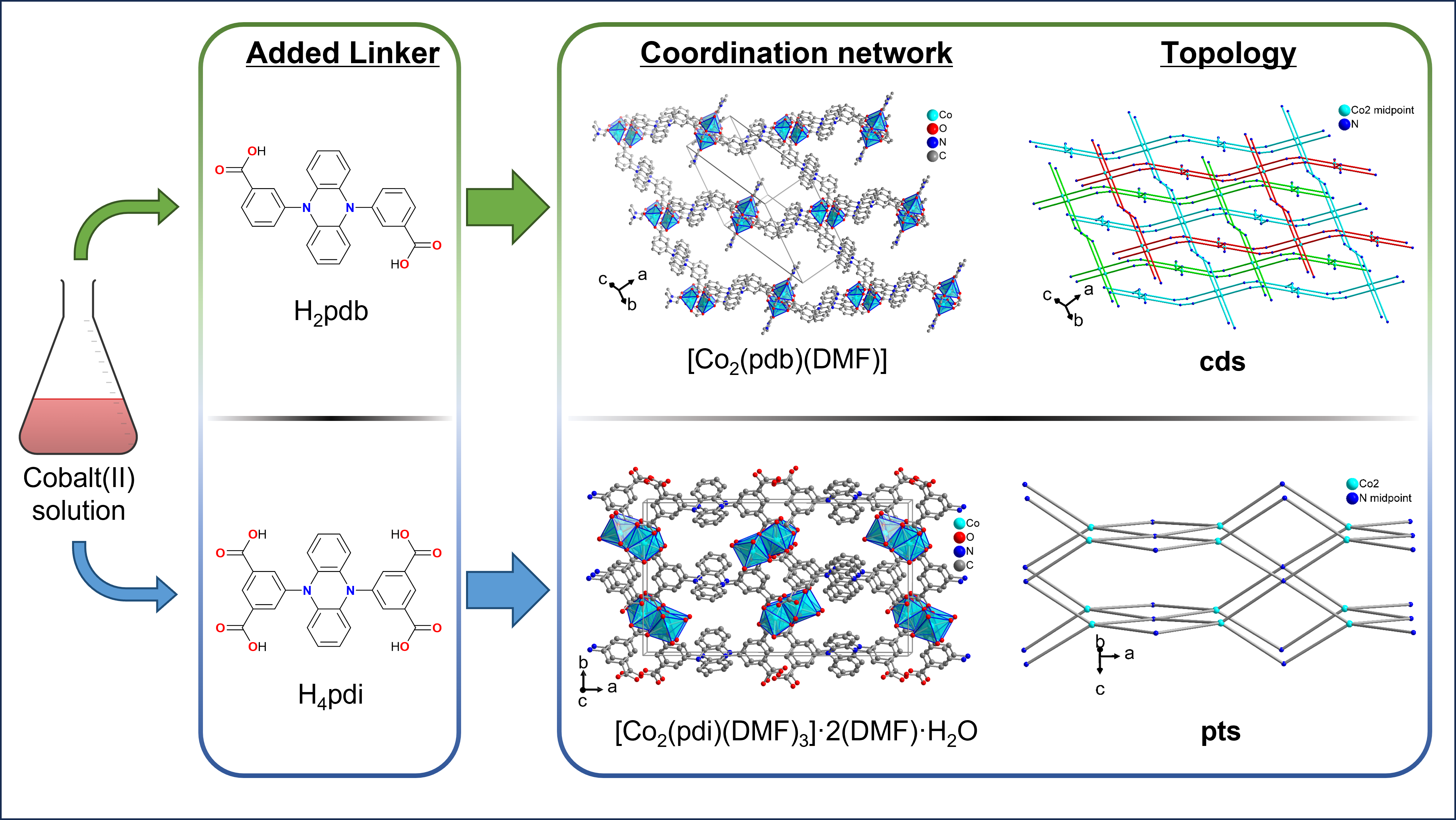
The crystal structures of the cobalt(II) metal-organic frameworks or coordination networks [Co(pdb)(DMF)] and [Co2(pdi)(DMF)3]·2(DMF)·H2O (H2pdb = 3,3′-(phenazine-5,10-diyl)dibenzoic acid, H4pdi = 5,5′-(phenazine-5,10-diyl)diisophthalic acid, DMF = N,N-dimethylformamide) were synthesized solvothermally from cobalt(II) nitrate and the free acid of the linker in DMF. In catena-[(N,N-dimethylformamide)-μ4-3,3′-(phenazine-5,10-diyl)dibenzoate-cobalt(II)], [Co(pdb)(DMF)], the Co2 handles as secondary building units are surrounded by four carboxylate groups from four linkers in a paddle-wheel arrangement giving a three-dimensional (3D) network with cds (or CdSO4) topology in which the wide openings are filled by two symmetry related nets to a threefold interpenetrated structure. In catena-[tris(N,N-dimethylformamide)-μ8-5,5′-(phenazine-5,10-diyl)diisophthalate-dicobalt(II)] bis(N,N-dimethylformamide) hydrate, [Co2(pdi)(DMF)3]·2(DMF)·H2O, there are two different Co atoms from which only Co2 is connected to each of the four carboxyl groups of the tetracarboxyl linker and, thus, is responsible for the 3D network formation. The network topology in [Co2(pdi)(DMF)3] is pts (or platinum(II) sulfide) when taking the Co2 atom as a tetrahedral and the linker as a square-planar fourfold node which is, however, inverse from the common square-planar metal and tetrahedral linker nodes in PtS and most pts topologies.

Keywords: 
;  ;  ;  ;  

1. Introduction

Incorporating redox-active carboxylate linkers—such as those based on phenazine—into the construction of new coordination polymers (CPs) and metal–organic frameworks (MOFs) offers expanded opportunities to harness redox behavior [1,2,3,4]. Phenazine derivatives contain a dibenzo-fused pyrazine core and are well known for their intrinsic redox activity. They can be readily reduced to 5,10-dihydrophenazines, which, together with subsequent transformations, give access to a wide range of dihydrophenazine derivatives (Scheme 1) [5]. Dihydrophenazines (DHPs), particularly N,N′-substituted diaryl-phenazin-5,10-diyls, are electron-rich species that generate stable radical cations upon irradiation, heating, electrochemical oxidation, or treatment with suitable chemical oxidants [6]. Owing to their pronounced redox activity and the stability of their radical cations [7,8], DHPs display distinctive optical, electronic, magnetic, and catalytic characteristics [9,10,11]. Several diaryl-substituted DHPs have been identified as effective photoredox catalysts for visible-light-mediated atom transfer radical polymerization [12,12,13,15,16,17].
Dihydrophenazine-based organic polymers [9,10,18], MOFs [10,19,20] and coordination cages [21] have been investigated for their electrochemical behavior [22,23,24] and have been explored as heterogeneous catalysts. While previous publications have reported on phenazine-based covalent organic frameworks (COFs) used for catalytic and optoelectronic applications [25,26,27], DHP-containing 2D and 3D covalent organic frameworks have demonstrated high efficiency as heterogeneous photocatalysts for the radical ring-opening polymerization of vinylcyclopropanes [18]. The UiO-type MOF [Zr63-O)43-OH)4(pzdb)6] (pzdb = 4,4′-(phenazine-5,10-diyl)dibenzoate)) [10] has been successfully utilized as a heterogeneous donor component to enhance catalytic electron-donor–acceptor photoactivation. Similarly, the framework [Zn2(pzdb)2(dabco)]·4DMF (dabco = 1,4-diazabicyclo [2.2.2]octane) has been applied as a heterogeneous catalyst for aza-Diels–Alder reactions [28].
Recently, UV/Vis/NIR spectroelectrochemistry revealed that the pzdb linker is the primary redox site in the coordination polymers [Zn(pzdb)(DEF)2] and [Co(Hpzdp)2(DEF)2] upon electrochemical oxidation or chemical oxidation with SbCl5 based on the same color changes as in Me2pzdb together with EPR signals typical of ligand-based radical cations. However, the coordination polymers decomposed upon the pzdb linker oxidation [29].
Here we report the synthesis and characterization of two new coordination networks based on the DHP-derived redox-active linkers H2pdb and H4pdi.

2. Materials and Methods

All chemicals were purchased from commercial suppliers and used without further purification (see Supplementary Material, SM, Section S1 for details). Deionized water was employed in all procedures involving water using a Sartorius Arium Mini water purifier. Single-crystal X-ray diffraction data were collected on a Rigaku XtaLAB Synergy S instrument (Rigaku, Tokyo, Japan) equipped with a PhotonJet Cu Kα radiation source (λ = 1.54184 Å) and a hybrid pixel array detector. Suitable crystals were selected under a Leica M80 polarized-light microscope (Leica, Wetzlar, Germany) and mounted on a cryo-loop in oil. Data processing—including unit-cell refinement, data reduction, and absorption correction—was carried out with CrysAlisPro. Structures were solved and refined in Olex2 using SHELXT and SHELXL, respectively [30,31,32]. Molecular graphics were generated using Diamond 5 software [33].
Powder X-ray diffraction (PXRD) data were collected at room temperature on a Rigaku Mini-Flex600 diffractometer (Rigaku, Tokyo, Japan) (600 W, 40 kV, 15 mA) using Cu Kα radiation (λ = 1.54184 Å). The samples were dried for 12 h at 60 °C under vacuum. PXRD patterns were normalized to the intensity of the most intense peak. Simulated PXRD patterns were obtained from MERCURY 2020.3.0 using the single-crystal XRD data [34].
Fourier-transform infrared (FT-IR) spectra were recorded between 500 and 4000 cm−1 using a Bruker TENSOR 37 spectrometer (Bruker, Billerica, MA, USA) in ATR mode (Platinum ATR-QL, Diamond). Nuclear magnetic resonance (NMR) spectra were measured on a Bruker Avance III 300 spectrometer (Bruker, Billerica, MA, USA) operating at 300 MHz for 1H NMR and 150 MHz for 13C NMR. Electron impact (EI) mass spectra were obtained using a Thermo Finnigan Trace DSQ spectrometer (Thermo Fisher Scientific).
N2 sorption measurements were carried out with a Belsorp Max II (Microtrac, MRB, Haan, Germany) volumetric gas sorption analyzer at 77 K. Samples were pre-dried for 12 h at 60 °C under vacuum and then activated at the sorption analyzer under turbomolecular pump vacuum for 3 h at 130 °C.
Thermogravimetric analysis (TGA) was carried out in air and under nitrogen on a Netzsch TG209 F3 Tarsus instrument (Netzsch, Selb, Germany) at a heating rate of 10 K min−1 up to 1000 °C. Melting points were measured in open capillaries using a Büchi Melting Point B-540 apparatus (Büchi Labortechnik AG, Flawil, Switzerland).

2.1. Synthesis of 5,10-dihydrophenazine (Scheme S1)

Phenazine (4.00 g, 22.2 mmol) was dissolved in 50 mL of ethanol in a 500 mL round-bottom flask. Sodium dithionite (38.7 g, 222 mmol) was dissolved in 250 mL of deionized water. The two solutions were then combined in the flask, which was heated to 95 °C under stirring and reflux for 3 h. After the flask was cooled to room temperature the reaction mixture was filtered, and the product was dried under vacuum for 40 minutes. Finally, the product was stored under a protective N2 atmosphere. Yield: 3.69 g (91%). 1H-NMR (300 MHz, DMSO-d6): δ [ppm] = 7.29 (s, 2 H), 6.25 (dd, J = 6.7, 3.4 Hz, 4 H), 6.01 (dd, J = 6.7, 3.5 Hz, 4 H).

2.2. Synthesis of dimethyl 3,3′-(phenazine-5,10-diyl)dibenzoate (Scheme S2)

A 100 mL three-neck flask was charged with 1.00 g of 5,10-dihydrophenazine (5.50 mmol), 2.60 g of 3-methyl 3-bromobenzoate (12.1 mmol), and 1.52 g of potassium carbonate (16.5 mmol). After sealing the flask, a solution of 0.062 g of palladium(II)-acetate (0.27 mmol) and 0.3 mL of tri-tert-butylphosphine dissolved in 60 mL of xylene was added. The reaction mixture was heated under stirring to reflux for 48 hours. After cooling to room temperature, 100 mL of water was added to the reaction mixture and it was extracted with 3 x 150 mL of dichlormethane. The combined organic phases were washed with 150 mL of brine solution (60 g NaCl in 300 mL water) and were dried over magnesium sulfate. The organic solution was concentrated using a rotary evaporator. After adding 5 mL of DCM and 40 mL of n-hexane, the product was recrystallized in the refrigerator. The precipitate was filtered off and dried under vacuum. Yield: 1.83 g (74%). 1H-NMR (300 MHz, CDCl3): δ [ppm] = 8.20 – 8.08 (m, 4 H), 7.71 (t, J = 9.0, 6.0 Hz, 2 H), 7.65 – 7.59 (m, 4 H), 6.28 (dd, J = 6.7, 3.4 Hz, 4 H), 5.58 (dd, J = 6.7, 3.4 Hz, 4 H), 3.94 (s, 6 H). %). 13C{1H}-NMR (600 MHz, CDCl3): δ [ppm] = 166.16, 140.34, 136.31, 136.16, 133.73, 132.93, 131.51, 129.49, 121.13, 112.62, 52.36. [HR-ESI-MS] m/z = 450.16 (calculated for 12C28 1H22 14N2 16O4 450.16). IR (ATR): υ [cm-1] = 3430, 3076, 3037, 3006, 2953, 2841, 2625, 2579, 1911, 1719, 1660, 1629, 1609, 1596, 1580, 1484, 1440, 1391, 1347, 1287, 1261, 1192, 1173, 1158, 1129, 1098, 1078, 1062, 991, 949, 928, 889, 838, 813, 794, 733, 693, 678, 643, 618, 558. Mp. = 255 °C.

2.3. Synthesis of 3,3′-(phenazine-5,10-diyl)dibenzoic acid (H2pdb) (Scheme S3)

A solution of 1.72 g (3.82 mmol) of dimethyl 3,3′-(phenazine-5,10-diyl)dibenzoate in 25 mL of 1,4-dioxane was prepared in a 100 mL flask. Then, 1.60 g of lithium hydroxide (38.2 mmol) and 15 mL of water were added to the reaction mixture, which was then heated to 100 °C for 24 hours. After cooling to room temperature, the mixture was neutralized with concentrated hydrochloric acid (~1.9 mL). The precipitation was then filtered and dried under vacuum. Yield: 1.53 g (91%). 1H-NMR (300 MHz, DMSO-d6): δ [ppm] = 13.07 (s, 2 H), 8.08 (dt, 2 H), 7.92 (t, J = 9.0, 6.0 Hz, 2 H), 7.82 (t, J = 9.0, 6.0 Hz, 2 H), 7,73 (dt, 2 H), 6.30 (dd, J = 6.7, 3.4 Hz, 4 H), 5.52 (dd, J = 6.7, 3.4 Hz, 4 H). 13C{1H}-NMR (600 MHz, DMSO-d6): δ [ppm] = 166.47, 139.94, 135.75, 134.75, 134.74, 132.00, 131.70, 129.25, 121.19, 112.49. [HR-ESI-MS] m/z = 422.13 (calculated for 12C26 1H18 14N2 16O4 422.13). IR (ATR): υ [cm-1] = 3386, 3067, 2994, 2857, 2814, 2662, 2540, 1979, 1909, 1865, 1821, 1748, 1683, 1609, 1597, 1581, 1483, 1443, 1411, 1388, 1344, 1280, 1183, 1160, 1138, 1103, 1082, 1061, 1002, 990, 965, 928, 912, 838, 819, 757, 734, 693, 669, 643, 617, 568. Mp. > 350 °C.

2.4. Synthesis of Tetramethyl 5,5′-(phenazine-5,10-diyl)diisophthalate (Scheme S4)

A 100 mL three-neck flask was charged with 1.00 g of 5,10-dihydrophenazine (5.50 mmol), 2.60 g of dimethyl 5-bromoisophthalate (12.1 mmol), and 1.52 g of potassium carbonate (16.5 mmol). After sealing the flask, a solution of 0.062 g of palladium(II)-acetate (0.27 mmol) and 0.3 mL of tri-tert-butylphosphine dissolved in 60 mL of xylene were added. The reaction mixture was heated under stirring to reflux for 48 hours. After cooling to room temperature, 100 mL of water was added to the reaction mixture and it was extracted with 3 x 150 mL of dichlormethane. The combined organic phases were washed with 150 mL of brine solution (60 g NaCl in 300 mL water) and were dried over magnesium sulfate. The organic solution was concentrated using a rotary evaporator. After adding 5 mL of DCM and 40 mL of n-hexane, the product was recrystallized in the refrigerator. The precipitate was filtered off and dried under vacuum. Yield: 2.12 g (68%). 1H-NMR (300 MHz, CDCl3): δ [ppm] = 8.80 (t, J = 3.0, 3.0 Hz, 2 H), 8.32 (d, 4 H), 6.32 (dd, J = 6.7, 3.4 Hz, 4 H), 5.59 (dd, J = 6.7, 3.4 Hz, 4 H), 3.98 (s, 12 H). 13C{1H}-NMR (600 MHz, CDCl3): δ [ppm] = 165.47, 141.00, 137.72, 135.96, 134.35, 130.68, 121.66, 113.12, 52.79. [HR-ESI-MS] m/z = 566.17 (calculated for 12C32 1H26 14N2 16O8 566.17). IR (ATR): υ [cm-1] = 3431, 3088, 3004, 2954, 2846, 1718, 1590, 1544, 1489, 1434, 1398, 1355, 1307, 1287, 1242, 1199, 1167, 1137, 1105, 1065, 998, 950, 938, 918, 898, 876, 834, 806, 784, 753, 741, 723, 686, 664, 618, 562. Mp. = 336 °C.

2.5. Synthesis of 5,5′-(phenazine-5,10-diyl)diisophthalic acid (H4pdi) (Scheme S5)

A solution of 1.00 g (1.77 mmol) of tetramethyl 5,5′-(phenazine-5,10-diyl)diisophthalate in 25 mL of 1,4-dioxane was prepared in a 100 mL flask. Then, 0.74 g of lithium hydroxide (17.7 mmol) and 15 mL of water were added to the reaction mixture, which was heated to 100 °C for 24 hours. After cooling to room temperature, the mixture was neutralized with concentrated hydrochloric acid (~1.5 mL). The precipitation was then filtered and dried under vacuum. Yield: 0.71 g (79%). 1H-NMR (300 MHz, D2O): δ [ppm] = 8.33 (t, J = 3.0, 3.0 Hz, 2 H), 7.89 (d, 4 H), 6.30 (dd, J = 6.7, 3.4 Hz, 4 H), 5.67 (dd, J = 6.7, 3.4 Hz, 4 H). 13C{1H}-NMR (600 MHz, D2O): δ [ppm] = 165174.03, 140.06, 139.67, 136.08, 134.08, 128.94, 121.48, 113.00. IR (ATR): υ [cm-1] = 3384, 3079, 3063, 2981, 2861, 2824, 2665, 2616, 2572, 2540, 1891, 1865, 1723, 1694, 1635, 1591, 1552, 1488, 1455, 1423, 1351, 1311, 1279, 1244, 1192, 1159, 1109, 1064, 1003, 957, 925, 847, 790, 756, 729, 693, 681, 661, 615, 602. Mp. > 350 °C.

2.6. Synthesis of Catena-[(N,N-dimethylformamide)-μ4-3,3′-(phenazine-5,10-diyl)dibenzoate-cobalt(II)], [Co(pdb)(DMF)]

15.0 mg of H2pdb (0.034 mmol) and 20.0 mg of cobalt nitrate hexahydrate (0.068 mmol) were placed in a glass tube. Then, 2 mL of DMF were added to completely dissolve the solids. The tubes were sealed and placed in an oven at 100 °C for two days. Dark red crystals were obtained as a final product. The crystals were washed with DMF and dried under vacuum. Yield: 11 mg (56%).

2.6. Synthesis of Catena-[tris(N,N-dimethylformamide)-μ8-5,5′-(phenazine-5,10-diyl)diisophthalate-dicobalt(II)] bis(N,N-dimethylformamide) hydrate [Co2(pdi)(DMF)3]·2(DMF)·H2O

17.9 mg of H4pdi (0.035 mmol) and 20 mg of cobalt nitrate hexahydrate (0.070 mmol) were placed in a glass tube. Then, 2 mL of DMF were added to completely dissolve the solids. The tubes were sealed and placed in an oven at 100 °C for two days. Dark red crystals were obtained as a final product. The crystals were washed with DMF and dried under vacuum. Yield: 22 mg (62%).

3. Results and Discussion

The linkers syntheses started with the reduction of phenazine with sodium dithionate to 5,10-dihydrophenazine (DHP, Scheme 1, Scheme S1) [1]. DHP is a light-green solid and was stored under a nitrogen atmosphere because of its easy re-oxidation in air. 1H NMR spectroscopy (Figure S1) confirmed the product with high purity. In the next reaction step, dimethyl 3,3′-(phenazine-5,10-diyl)dibenzoate [3,3′-(phenazine-5,10-diyl)dibenzoate dimethyl ester] or tetramethyl-5,5′-(phenazine-5,10-diyl)diisophthalate were synthesized (Scheme S2 or S4) through Buchwald-Hartwig coupling [19,35,36]. The highest yields and purest products were obtained with palladium(II) acetate as the catalyst, tri-tert-butylphosphine as the ligand and potassium carbonate as the base. The products are yellow or red powders, respectively and their constitution and high purity is confirmed by NMR spectroscopy (Figures S2–S5).
Finally, the methyl esters were hydrolyzed with LiOH in a 1,4-dioxane/H2O mixture to give 3,3′-(phenazine-5,10-diyl)dibenzoic acid as a light-yellow solid (H2pdb, Scheme S3) or 5,5′-(phenazine-5,10-diyl)diisophthalic acid as a dark green solid (H4pdi, Scheme S5). Again the 1H-NMR spectra are consistent with the constitutions (Figures S6–S9). The bands in the infrared spectra of the esters and the free acids can be assigned (Figures S10 and S11).
Various futile syntheses attempts were performed with zirconium(IV) chloride, zirconium oxychloride octahydrate, aluminum chloride hexahydrate, copper(II) chloride monohydrate, nickel(II) sulfate hexahydrate and nickel(II) chloride hexahydrate with H2pdb and H4pdi in N,Nˈ-dimethylformamide giving no or no crystalline products. Needle-shaped crystals from zinc nitrate hexahydrate with H2pdb in a molar ratio of 1:3 at 120 °C after two days were too small for single crystal X-ray diffractometry. Only in the reaction from cobalt(II) nitrate hexahydrate with H2pdb and with H4pdi, both in a molar ratio of 2:1 at 100 °C good quality crystals were formed after two days (Figures S12 and S13). The anion plays a certain role as identical reactions of cobalt(II) sulfate heptahydrate and cobalt(II) chloride hexahydrate yielded no solid products.
The experimental powder X-ray diffraction patterns of the bulk crystallized cobalt compounds matched well with the simulated patterns derived from the crystal structures of [Co(pdb)(DMF)] and [Co2(pdi)(DMF)3]·2(DMF)·H2O, confirming their phase purity (Section S5, Figures S15 and S16).

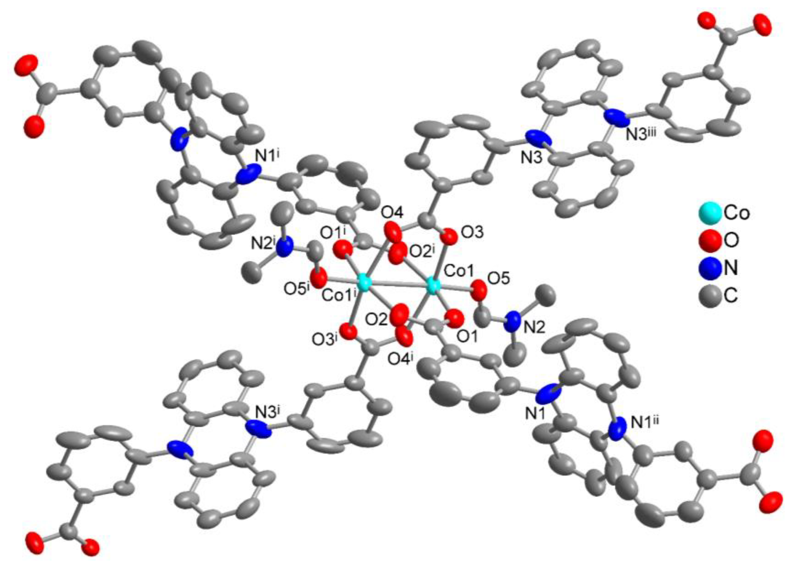
3.1. Crystal Structures of [Co(pdb)(DMF)]

Figure 1 shows the extended asymmetric unit and cobalt coordination environment of [Co(pdb)(DMF)] and Table 1 lists the crystallographic data. The space group of this compound is I2/a. It belongs to the monoclinic crystal system, and the asymmetric unit consists of one cobalt(II) ion, two half pdb linkers and a DMF ligand. The pdb linker with N1 has a 2-fold rotation axis passing through the phenazine core perpendicular to the ring plane. The linker with N3 has an inversion center at the phenazine ring centroid. The cobalt(II) ion is square-pyramidally surrounded by five oxygen atoms and forms a Co2 handle as the secondary building unit (SBU) with another symmetry-equivalent cobalt(II) ion. Together this Co2 handle is surrounded by four carboxylate groups from four linkers in a paddle-wheel arrangement. Each pdb ligand acts as a tetradentate bridging ligand and connects two Co2 handles, being thereby bound to four cobalt atoms. The axial directions of the paddle-wheel unit bind to the oxygen atoms of the monodentate DMF ligands.
The selected bond lengths and bond angles can be found in Table 2. The bond lengths of the four cobalt-oxygen bonds in the equatorial plane range from 2.00 Å to 2.09 Å, the bond length of the cobalt-oxygen(DMF) bond in the axial direction is approximately 2.03 Å. The cobalt-cobalt distance in the Co2 handle is 2.81 Å. The O-Co-O bond angles between the cis-positioned O atoms of the pdb linkers in the paddle-wheel unit are all approximately 90°, while the trans-O-Co-O angles are significantly smaller (~164°) because the Co···Co separation (2.81 Å) is larger than the O···O separation (~2.23 Å) in a carboxyl group so that the Co atom is placed “above” the equatorial plane of the four oxygen atoms. Consequently, the O-Co-O5(DMF) bond angle is also significantly greater than 90°.
The Co2 handles or SBUs are then linked into a three-dimensional network with cds topology (Figure 2). In cds (or CdSO4) topology [37,41,42,43], each SBU is connected to four neighboring nodes via the linkers. If one starts with the chain shown in Figure 2a where alternatingly each SBU connects to a parallel chain top-to-bottom and front-to-back then the 3D network in Figure 2b is obtained.
The single network has wide openings, several Ångströms across (Figure S13), even taking into account the space-filling van der Waals surface (Figure 2c). These openings are filled with two symmetry related nets through interpenetration giving a threefold interpenetrated structure with cds topology (Figure 3). Consequently, no voids remain and there is no porosity in the network. A threefold interpenetrated cds topology was described in 3D-[Co(pam)(bpa)(H2O)2]·DMF (H2pam = pamoic acid, bpa = 1,2-di(4-pyridyl)ethylene, DMF = N,N’-dimethylformamide) [45], Twofold interpenetrated cds net topologies were found in [Zn2(BDC)(BPP)Cl2] (BDC = benzene-1,4-dicarboxylate, BPP = 1,3-bis(4-pyridyl)propane) [46] and in [Cu(ceb)(bpmp)]·H2O (ceb = 4-(carboxylatoethyl)benzoato, bpmp = 1,4-bis(pyridin-4-ylmethyl)piperazine) [47]. Further examples for interpenetrated networks with a cds topology are [Zn(Br-1,3-bdc)(NI-mbpy-34)] (Br-1,3-bdc = 5-bromobenzene-1,3-dicarboxylate, NI-mbpy-34 = N-(pyridin-3-ylmethyl)-4-(pyridin-4-yl)-1,8-naphthalimide) [48] and [M(μ-TBG)(μ-H2O)(H2O)2]·2H2O (M = Cu, Co and H2TBG = terephthaloylbisglycine) [49]. A non-interpenetrated 3D network with cds topology was reported for [trans-Ni(H2O)2(μ-4,4′-bpy)2](ClO4)2 (4,4′-bpy = 4,4′-bipyridine) [50].
The TGA curve of [Co(pdb)(DMF)] is shown in Figure S17. The mass loss starts below 100 °C due to the removal of the DMF ligand (theor. 13 wt%) and is slightly smaller because the samples were measured after drying for 12 h at 60 °C under vacuum. The solvent removal transits without a plateau into the network decomposition of [Co(pdb)] which begins at ~200 °C and the sample is completely decomposed at ~410 °C with a concomitant mass loss of 80 wt% (theor. 76 wt%). The final solid residue at 1000 °C of 14 wt% corresponds to CoO (theor. 14 wt% for CoO, 11 wt% for Co).

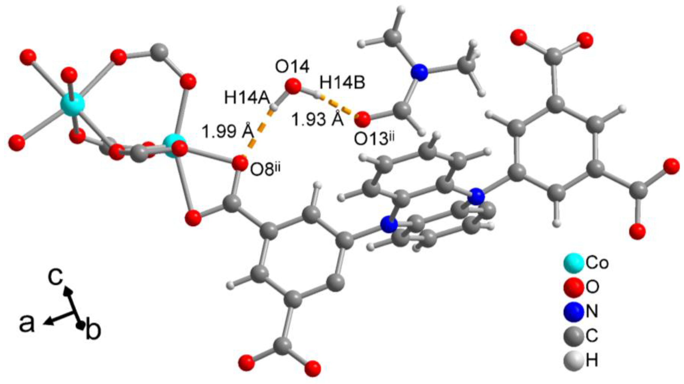
3.2. Crystal Structure of [Co2(pdi)(DMF)3]·2(DMF)·H2O

Figure 3 depicts the extended asymmetric unit and cobalt coordination environment of [Co2(pdi)(DMF)3]·2(DMF)·H2O and Table 1 gives the crystallographic data. The space group of this compound is Pna21 in the orthorhombic crystal system. The asymmetric unit contains two crystallographically different cobalt(II) ions, a full pdi linker, three coordinated DMF ligands, two non-coordinated DMF and a water molecule of solvation. Both cobalt(II) ions are six-fold coordinated and surrounded by six oxygen atoms and form again a Co2 handle as the secondary building unit. The Co1 ion is coordinated by the three DMF ligands and three carboxyl oxygen atoms of three different linkers in the facial configuration. Co2 is coordinated by four different pdi linkers with two of them chelating and two monodentate. Each of the bridging linkers is connected to Co2, which can be seen as the primary metal node. Out of the four different carboxyl groups of the linker two are monodentate bridging (O1-C-O2, O3-C-O4) between the two Co ions, one is bidentate bridging between Co1 and Co2 and at the same time chelating to Co2 (O5-C-O6 as κO:κO,κO’) and the last one is bidentate chelating to Co2 (O7-C-O8). From the perspective of the linker, each pdi ligand is bound to seven cobalt atoms, i.e., acts as a heptadentate bridging ligand and connects four Co2 handles. Three handles are connected with both cobalt atoms, one handle only through one Co atom. Selected bond lengths and angles are listed in Table 3.
In [Co2(pdi)(DMF)3]·2(DMF)·H2O, crystal water and non-coordinated DMF solvent molecules are incorporated by hydrogen bonds in the crystal lattice. The hydrogen bonds of the crystal water molecule are shown in Figure 4. The hydrogen bond O14–H14A∙∙∙O8ii is between the water molecule and a cobalt coordinating carboxyl oxygen atom of the pdi linker; the hydrogen bond O14–H14B∙∙∙O13ii is a bridge to a non-coordinated DMF molecule. These two hydrogen bonds are of medium strength, as the distance between the hydrogen atom and the accepting oxygen atom is between 1.9 and 2.0 Å in both cases.
The Co-pdi bridging action gives rise to a three-dimensional network (Figure 5). In this metal-ligand 3D network there would be open channels (Figure 5a), which are, however, occupied by the coordinated DMF ligands together with the DMF and H2O solvent molecules of crystallization (Figure 5b). When the free solvent molecules are omitted in the network, only small voids remain (Figure 6).
After activation no N2 gas uptake could be detected with a volumetric gas sorption analyzer, hence there is no N2 accessible porosity in the [Co2(pdi)(DMF)3] network. In view of the tight framework packing the removal of the free DMF was probably not fully achieved, the framework partially collapsed and the potential voids were too small.
The network topology in [Co2(pdi)(DMF)3] is pts (or platinum(II) sulfide) [37,38,39,40] when taking the Co2 atom as a tetrahedral and the linker as a square-planar fourfold node (Figure 5c). From the two Co atoms only Co2 is connected to each of the four carboxyl groups of the linker and is responsible for the 3D network formation. Compared to the PtS structure, which is composed of square planar Pt(II) and tetrahedral sulfide atoms, the pts topology in [Co2(pdi)(DMF)3] is reversed, such as that the metal node is tetrahedral and the linker square planar.
A thermogravimetric analysis (Figure S18) starts with the mass loss below 100 °C with the loss of crystal water (theor. 2 wt%) and the loss of the two free DMF molecules (theor. 14.5 wt%) from the pre-dried sample (12 h at 60 °C under vacuum). Without a pronounced plateou a mass loss of ~22 wt% continues between 100 to ~300 °C and involves the loss of the coordinated three DMF ligands (theor. 22 wt%). Again without a plateau the final decomposition occurs above ~300 °C and extends up to 560 °C with a mass loss of ~58 wt%. This last step involves degradation of the organic linker (theor. 50 wt%), consistent with the steep decline observed in the TGA curve. At temperatures above 560 °C, a stable residue of 17–18 wt% remains, in agreement with the dicobalt content as CoO (theor. 15 wt%, theor. 12 wt% for Co).

4. Conclusions

Two cobalt(II) coordination networks based on the dihydrophenazine-derived dicarboxylate and tetracarboxylate linkers H2pdb and H4pdi were synthesized solvothermally and structurally characterized. Crystalline products were obtained only with cobalt(II) nitrate, highlighting the sensitivity of framework formation to the metal ion and its counterion when using rigid, multitopic carboxylate linkers.
In [Co(pdb)(DMF)], dinuclear cobalt paddle-wheel units are linked by tetradentate pdb ligands into a three-dimensional cds net that exhibits threefold interpenetration and results in a densely packed framework. In contrast, [Co2(pdi)(DMF)3]·2(DMF)·H2O contains two crystallographically distinct cobalt(II) centers, of which only one propagates the three-dimensional network, giving rise to an inverse pts topology with tetrahedral metal nodes and square-planar linker nodes.
These compounds demonstrate how the connectivity and geometry of dihydrophenazine-based carboxylate ligands govern secondary building unit formation, network topology, and interpenetration in cobalt coordination polymers.

Supplementary Materials

The following supporting information can be downloaded at the website of this paper posted on Preprints.org, Section S1: Chemicals used; Section S2: Reaction schemes for ligand syntheses; Section S3: NMR spectra; Section S4: IR spectra; Section S5 Crystal photographs and crystal structure images; Section S6: PXRD; Section S7: Thermogravimetric analyses.

Author Contributions

Conceptualization, A.V. and C.J.; methodology, A.V.; software, A.V.; validation, A.V., X.L. and N.J.; formal analysis, A.V., X.L., N.J., P.S. and D.G.; investigation, A.V., X.L., N.J., P.S. and D.G.; resources, C.J.; data curation, A.V., X.L. and N.J.; writing—original draft preparation, A.V. and C.J.; writing—review and editing, A.V. and C.J.; visualization, A.V. and C.J.; supervision, C.J.; project administration, C.J.; funding acquisition, C.J. All authors have read and agreed to the published version of the manuscript.

Funding

Funding was provided by Deutsche Forschungsgemeinschaft (DFG), under grant 440366605 (for the Rigaku diffractometer). This work was supported by the Deutsche Forschungsgemeinschaft (DFG, German Research Foundation) – 396890929 / GRK 2482.

Data Availability Statement

The data presented in this study are available upon request from the corresponding author. The CCDC numbers 2522790-2522791 for compounds [Co(pdb)(DMF)] and [Co2(pdi)(DMF)3]·2(DMF)·H2O, respectively, contain the supplementary crystallographic data reported in this paper. These data can be obtained free of charge from the Cambridge Crystallographic Data Center via www.ccdc.cam.ac.uk/data_request/cif (accessed on 14 January 2026).

Acknowledgments

The authors would like to thank Birgit Tommes for providing the IR measurements. We also thank the Center for Molecular and Structural Analytics at Heinrich Heine University (CeMSA@HHU) for recording the mass spectrometric and NMR spectrometric data.

Conflicts of Interest

The authors declare no conflicts of interest.

References

  1. Feng, S.; Wang, L.; Tian, L.; Liu, Y.; Hu, K.; Xu, H.; Wang, H.; Hua, J. Leveraging phenazine and dihydrophenazine redox dynamics in conjugated microporous polymers for high-efficiency overall photosynthesis of hydrogen peroxide. Chem. Sci. 2024, 15, 11972–11980. [Google Scholar] [CrossRef] [PubMed]
  2. Ding, B.; Solomon, M.B.; Leong, C.F.; D’Alessandro, D.M. Redox-active ligands: Recent advances towards their incorporation into coordination polymers and metal-organic frameworks. Coord. Chem. Rev. 2021, 439, 213891. [Google Scholar] [CrossRef]
  3. Liao, P.-Q.; Shen, J.-Q.; Zhang, J.-P. Metal–organic frameworks for electrocatalysis. Coord. Chem. Rev. 2018, 373, 22–48. [Google Scholar] [CrossRef]
  4. D’Alessandro, D.M. Exploiting redox activity in metal–organic frameworks: concepts, trends and perspectives. Chem. Commun. 2016, 52, 8957–8971. [Google Scholar] [CrossRef]
  5. Lee, J.; Shizu, K.; Tanaka, H.; Nakanotani, H.; Yasuda, T.; Kaji, H.; Adachi, C. Controlled emission colors and singlet–triplet energy gaps of dihydrophenazine-based thermally activated delayed fluorescence emitters. J. Mater. Chem. C 2015, 3, 2175–2181. [Google Scholar] [CrossRef]
  6. Dulov, D.A.; Bogdanov, A.V.; Dorofeev, S.G.; Magdesieva, T.V. N,N0 -Diaryldihydrophenazines as a Sustainable and Cost-Effective Alternative to Precious Metal Complexes in the PhotoredoxCatalyzed Alkylation of Aryl Alkyl Ketones. Molecules 2023, 28, 221. [Google Scholar] [CrossRef]
  7. Corbin, D.A.; McCarthy, B.G.; van de Lindt, Z.; Miyake, G.M. Radical Cations of Phenoxazine and Dihydrophenazine Photoredox Catalysts and Their Role as Deactivators in Organocatalyzed Atom Transfer Radical Polymerization. Macromolecules 2021, 54, 4726–4738. [Google Scholar] [CrossRef] [PubMed]
  8. Suzuki, S.; Takeda, T.; Kuratsu, M.; Kozaki, M.; Sato, K.; Shiomi, D.; Takui, T.; Okada, K. Pyrene-dihydrophenazine bis(radical cation) in a singlet ground state. Org. Lett. 2009, 11, 2816–2818. [Google Scholar] [CrossRef]
  9. Unglaube, F.; Hünemörder, P.; Guo, X.; Chen, Z.; Wang, D.; Mejía, E. Phenazine Radical Cations as Efficient Homogeneous and Heterogeneous Catalysts for the Cross-Dehydrogenative Aza-Henry Reaction. Helv. Chim. Acta 2020, 103, e2000184. [Google Scholar] [CrossRef]
  10. Lin, J.; Ouyang, J.; Liu, T.; Li, F.; Sung, H.H.-Y.M.; Williams, I.; Quan, Y. Metal-organic framework boosts heterogeneous electron donor–acceptor catalysis. Nat. Commun. 2023, 14, 7757. [Google Scholar] [CrossRef]
  11. Hong, Q.-Y.; Huang, B.; Wu, M.-X.; Xu, L.; Zhao, X.-L.; Shi, X.; Yang, H.-B. Redox-Active Dihydrophenazine-Based Macrocycle: Synthesis, Conformation-Adaptive Behavior and Host-Guest Complexation with Tetracyanoquinodimethane. Chin. J. Chem. 2024, 42, 1895–1900. [Google Scholar] [CrossRef]
  12. Theriot, J. C.; Lim, C.-H.; Yang, H.; Ryan, M. D.; Musgrave, C. B.; Miyake, G. M. Organocatalyzed atom transfer radical polymerization driven by visible light. Science 2016, 352, 1082–1086. [Google Scholar] [CrossRef]
  13. Cheng, Y.; Li, Y.-X.; Liu, C.-H.; Zhu, Y.-Y.; Lin, W. Diaryl Dihydrophenazine-Based Porous Organic Polymers Enhance Synergistic Catalysis in Visible-Light-Driven Organic Transformations. Angew. Chem. Int. Ed. 2023, 62, e202310470. [Google Scholar] [CrossRef]
  14. Corbin, D.A.; Puffer, K.O.; Chism, K.A.; Cole, J.P.; Theriot, J.C.; McCarthy, B.G.; Buss, B.L.; Lim, C.-H.; Lincoln, S.R.; Newell, B.S.; Miyake, G.M. Radical Addition to N,N-Diaryl Dihydrophenazine Photoredox Catalysts and Implications in Photoinduced Organocatalyzed Atom Transfer Radical Polymerization. Macromol. 2021, 54, 10, 4507–4516. [Google Scholar] [CrossRef]
  15. Cole, J. P.; Federico, C. R.; Lim, C.-H.; Miyake, G. M. Photoinduced organocatalyzed atom transfer radical polymerization using low ppm catalyst loading. Macromol. 2019, 52, 747−754. [Google Scholar] [CrossRef]
  16. Su, X.; Jessop, P. G.; Cunningham, M. F. Versatility of organocatalyzed atom transfer radical polymerization and CO2- switching for preparing both hydrophobic and hydrophilic polymers with the recycling of a photocatalyst. Macromol. 2019, 52, 6725− 6733. [Google Scholar] [CrossRef]
  17. McCarthy, B.; Sartor, S.; Cole, J.; Damrauer, N.; Miyake, G. M. Solvent effects and side reactions in organocatalyzed atom transfer radical polymerization for enabling the controlled polymerization of acrylates catalyzed by diaryl dihydrophenazines. Macromol. 2020, 53, 9208−9219. [Google Scholar] [CrossRef]
  18. Wang, K.; Kang, X.; Yuan, C.; Han, X.; Liu, Y.; Cui, Y. Porous 2D and 3D Covalent Organic Frameworks with Dimensionality-Dependent Photocatalytic Activity in Promoting Radical Ring-Opening Polymerization. Angew. Chem. Int. Ed. 2021, 60, 19466–19476. [Google Scholar] [CrossRef] [PubMed]
  19. Kochetygov, I.; Roth, J.; Espín, J.; Pache, S.; Justin, A.; Schertenleib, T.; Taheri, N.; Chernyshov, D.; Queen, W. L. A Simple, Transition Metal Catalyst-Free Method for the Design of Complex Organic Building Blocks Used to Construct Porous Metal-Organic-Frameworks. Angew. Chem. Int. Ed. 2023, 62, e202215595. [Google Scholar] [CrossRef] [PubMed]
  20. Zhang, S.; Ma, X.-F.; Yang, Y.; Ren, H.; Li, W.; Liu, Y.; Xi, C.; Zheng, L.; Miao, Z. Phenazine-functionalized Zn-based metal organic framework for efficient visible light photocatalytic sulfides. J. Solid State Chem. 2025, 351, 125525. [Google Scholar] [CrossRef]
  21. Jiang, W. L.; Huang, B.; Zhao, X. L.; Shi, X.; Yang, H. B. Strong halide anion binding within the cavity of a conformation-adaptive phenazine-based Pd2L4 cage. Chem. 2023, 9, 2655–2668. [Google Scholar] [CrossRef]
  22. Zhang, H.; Tanga, X.; Gu, C. Dihydrophenazine linked porous organic polymers for high capacitance and energy density pseudocapacitive electrodes and devices. J. Mater. Chem. A 2021, 9, 4984–4989. [Google Scholar] [CrossRef]
  23. Vitaku, E.; Gannett, C.N.; Carpenter, K.L.; Shen, L.; Abruna, H.D.; Dichtel, W.R. Phenazine- Based Covalent Organic Framework Cathode Materials with High Energy and Power Densities. J. Am. Chem. Soc. 2020, 142, 16−20. [Google Scholar] [CrossRef] [PubMed]
  24. Wu, M.-X.; Hong, Q.-Y.; Li, M.; Jiang, W.-L.; Huang, B.; Lu, S.; Wang, H.; Yang, H.-B.; Zhao, X.-L.; Shi, X. Self-assembly of conformation-adaptive dihydrophenazine-based coordination cages. Chem. Commun. 2024, 60, 1184–1187. [Google Scholar] [CrossRef] [PubMed]
  25. Yu, J.; Cheng, L.; Zhang, X.; Shi, X.; Wang, H.-g. Integrating p-type phenazine into covalent triazine framework to achieve co-storage of cations and anions for quasi-solid-state dual-ion batteries. Chem. Eng. J. 2024, 489, 151320. [Google Scholar] [CrossRef]
  26. Guo, J.; Xu, Y.; Jin, S.; Chen, L.; Kaji, T.; Honsho, Y.; Addicoat, M.A.; Kim, J.; Saeki, A.; Ihee, H.; Seki, S.; Irle, S.; Hiramoto, M.; Gao, J.; Jiang, D. Conjugated organic framework with three-dimensionally ordered stable structure and delocalized π clouds. Nat. Commun. 2013, 4, 2736. [Google Scholar] [CrossRef] [PubMed]
  27. Sun, H.; Ji, H.; Qiao, D.; Xu, Y.; Qu, X.; Qi, Y.; Feng, Z.; Zhang, X.; Zhang, F.; Wang, R.; Dong, B. Vinylene-linked covalent organic frameworks based on phenanthroline for visible-light-driven bifunctional photocatalytic water splitting. Chem. Eng. J. 2025, 507, 160448. [Google Scholar] [CrossRef]
  28. Jiang, W.-L.; Huang, B.; Wu, M.-X.; Zhu, Y.-K.; Zhao, X.-L.; Shi, X.; Yang, H.-B. Post-Synthetic Modification of Metal-Organic Frameworks Bearing Phenazine Radical Cations for aza-Diels-Alder Reactions. Chem. Asian. J. 2021, 16, 3985–3992. [Google Scholar] [CrossRef]
  29. Püschel, D.; Nau, M.; Assahub, N.; Beglau, T.H.Y.; Hufnagel, N.; Jordan, D.; Heinen, T.; Strothmann, T.; Suta, M.; Winter, R.F.; Janiak, C. Zinc and Cobalt Coordination Polymers Based on the Redox-Active Linker 4,4′-(Phenazine-5,10-diyl)dibenzoate: Structures and Electrochemical Properties. Eur. J. Inorg. Chem. 2025, 28, e202500270. [Google Scholar] [CrossRef]
  30. Dolomanov, O.V.; Bourhis, L.J.; Gildea, R.J.; Howard, J.A.K.; Puschmann, H. OLEX2: A complete structure solution, refinement and analysis program. J. Appl. Crystallogr. 2009, 42, 339–341. [Google Scholar] [CrossRef]
  31. Sheldrick, G.M. SHELXT—Integrated space-group and crystal-structure determination. Acta Crystallogr. A 2015, 71, 3–8. [Google Scholar] [CrossRef]
  32. Sheldrick, G.M. Crystal structure refinement with SHELXL. Acta Crystallogr. C 2015, 71, 3–8. [Google Scholar] [CrossRef]
  33. Brandenburg, K. Diamond, Version 5.0.0; Crystal and Molecular Structure Visualization, Crystal Impact; K. Brandenburg & H. Putz Gbr: Bonn, Germany, 1997–2023.
  34. Macrae, C.F.; Sovago, I.; Cottrell, S.J.; Galek, P.T.A.; McCabe, P.; Pidcock, E.; Platings, M.; Shields, G.P.; Stevens, J.S.; Towler, M.; et al. Mercury 4.0: From visualization to analysis, design and prediction. J. Appl. Crystallogr. 2020, 53, 226–235. [Google Scholar] [CrossRef]
  35. Dai, G.; He, Y.; Niu, Z.; He, P.; Zhang, C.; Zhao, Y.; Zhang, X.; Zhou, H. A Dual-Ion Organic Symmetric Battery Constructed from Phenazine-Based Artificial Bipolar Molecules. Angew. Chem. Int. Ed. 2019, 58, 9902–9906. [Google Scholar] [CrossRef] [PubMed]
  36. Koyama, D.; Dale, H.J.A.; Orr-Ewing, A.J. Ultrafast Observation of a Photoredox Reaction Mechanism: Photoinitiation in Organocatalyzed Atom-Transfer Radical Polymerization. J. Am. Chem. Soc. 2018, 140, 4, 1285–1293. [Google Scholar] [CrossRef] [PubMed]
  37. Delgado-Friedrichs, O.; O’Keeffe, M.; Yaghi, O.M. Three-periodic nets and tilings: edge-transitive binodal structures. Acta Cryst. 2006, A62, 350–355. [Google Scholar] [CrossRef]
  38. Zhang, J.; Xue, Y.-S.; Liang, L.-L.; Ren, S.-B.; Li, Y.-Z.; Du, H.-B.; You, X.-Z. Porous Coordination Polymers of Transition Metal Sulfides with PtS Topology Built on a Semirigid Tetrahedral Linker. Inorg. Chem. 2010, 49, 7685–7691. [Google Scholar] [CrossRef]
  39. Yuan, A.-H.; Lu, R.-Q.; Zhou, H.; Chen, Y.-Y.; Li, Y.-Z. Three unique two-fold interpenetrated three-dimensional networks with PtS-type topology constructed from [M(CN)4] 2 (M ¼ Ni, Pd, Pt) as ‘‘square-planar’’ building blocks. CrystEngComm 2010, 12, 1382–1384. [Google Scholar] [CrossRef]
  40. Murphy, M.J.; D’Alessandro, D.M.; Kepert, C.J. A porous Mn(V) coordination framework with PtS topology: assessment of the influence of a terminal nitride on CO2 sorption. Dalton Trans. 2013, 42, 13308–13310. [Google Scholar] [CrossRef] [PubMed]
  41. Friedrichs, O.D.; O’Keeffe, M.; Yaghi, O.M. The CdSO4, rutile, cooperite and quartz dual nets: Interpenetration and catenation. Solid State Sci. 2003, 5, 73–78. [Google Scholar] [CrossRef]
  42. Friedrichs, O.D.; O’Keeffe, M. Three-periodic tilings and nets: Face-transitive tilings and edge-transitive nets revisited. Acta Cryst. A 2007, 63, 344–347. [Google Scholar] [CrossRef]
  43. Moulton, B.; Abourahma, H.; Bradner, M.W.; Lu, J.; McManus, G.J.; Zaworotko, M.J. A new 65.8 topology and a distorted 65.8 CdSO4 topology: Two new supramolecular isomers of [M2(bdc)2(L)2]n coordination polymers. Chem. Commun. 2003, 1342–1343. [Google Scholar] [CrossRef]
  44. Ortín-Rubio, B.; Rostoll-Berenguer, J.; Vila, C.; Proserpio, D.M.; Guillerm, V.; Juanhuix, J.; Imaz, I.; Maspoch, D. Net-clipping as a top-down approach for the prediction of topologies of MOFs built from reduced-symmetry linkers. Chem. Sci. 2023, 14, 12984–12994. [Google Scholar] [CrossRef]
  45. Gao, H., Lou, X. H., Li, Q. T., Du, W. J., & Xu, C. (2015). Three-Fold Interpenetrated CdSO4 Topology in a Co(II) Coordination Polymer Constructed From Pamoic Acid and N-Containing Auxiliary Ligand. Synthesis and Reactivity in Inorganic, Metal-Organic, and Nano-Metal Chemistry 2015, 45, 865–868. [CrossRef]
  46. Zhang, J.; Chen, Y.-B.; Ji, Z.; Qin, Y.-Y.; Yao, Y.-G. A rare twofold interpenetrated cds topology in a Zn-organic polymer [Zn2(BDC)(BPP)Cl2]n. Inorg. Chem. Commun. 2006, 9, 449–451. [Google Scholar] [CrossRef]
  47. Gaskin, G.J.; LaDuca, R.L. Poly[[[μ-1,4-bis(pyridin-4-ylmethyl)piperazine][μ-4-(2-carboxylatoethyl)benzoato]copper(II)] monohydrate], a coordination polymer with twofold interpenetrated cds topology networks. IUCrData 2023, 8, x230855. [Google Scholar] [CrossRef] [PubMed]
  48. Tsai, M.-J.; Liao, K.-S.; Wu, J.-Y. A Water-Stable 2-Fold Interpenetrating cds Net as a Bifunctional Fluorescence-Responsive Sensor for Selective Detection of Cr(III) and Cr(VI) Ions. Nanomaterials 2022, 12, 158. [Google Scholar] [CrossRef]
  49. Kostaki, G., E.; Casella, L.; Hadjiliadis, N.; Monzani, E.; Kourkoumelis, N.; Plakatouras, J., C. Interpenetrated networks from a novel nanometer-sized pseudopeptidic ligand, bridging water, and transition metal ions with cds topology. Chem. Commun. 2005, 3859–3861. [CrossRef] [PubMed]
  50. Shi, Z.-C.; Wang, X.; Drozd, V.; Raptis, R.G. A Foldable Metal–Organic Framework with cds Topology Assembled via Four-Connected Square-Planar Single Ni2+-Ion Nodes and Linear Bidentate Linkers. Crystals 2024, 14, 40. [Google Scholar] [CrossRef]
Scheme 1. Synthesis route to the free acids of the H2pdb = 3,3′-(phenazine-5,10-diyl)dibenzoic acid and H4pdi = 5,5′-(phenazine-5,10-diyl)diisophthalic acid. For details see Materials and Methods and Section S2 in the Supplementary Materials.
Preprints 194671 sch001
Figure 1. Expanded asymmetric unit for [Co(pdb)(DMF)], symmetry code: (i) −x+1/2, −y+3/2, −z+3/2; (ii) −x+3/2, y, −z+1; (iii) −x, −y+1, −z+1 (50% thermal ellipsoids). Hydrogen atoms are not shown for clarity.
Preprints 194671 g001
Figure 2. (a) Chain section of the 3D network in [Co(pdb)(DMF)] which is linked to parallel chains top-to-bottom and front-to-back to give the full 3D network in (b) with cds topology. Hydrogen atoms are not shown for clarity, the Co coordination is presented as polyhedra. (c) Space-filling view of the network with hydrogen atoms included.
Preprints 194671 g002aPreprints 194671 g002b
Figure 3. (a) The threefold interpenetrated structure of [Co(pdb)(DMF)]. Each network is shown in a different color. (b) Schematic representation of the threefold interpenetrated cds net in [Co(pdb)(DMF)] by taking the midpoint of the Co2 handle as a fourfold node. The rods connect the Co2 midpoints and the phenazine ring nitrogen atoms.
Preprints 194671 g003
Figure 3. Expanded asymmetric unit for [Co2(pdi)(DMF)3]·2(DMF)·H2O, symmetry code: (i) −x+1/2, −y+3/2, −z+3/2; (ii) −x+3/2, y, −z+1; (iii) −x, −y+1, −z+1 (50% thermal ellipsoids). Hydrogen atoms are not shown for clarity.
Preprints 194671 g004
Figure 4. Hydrogen bonds from the crystal water molecule in [Co2(pdi)(DMF)3]·2(DMF)·H2O.
Preprints 194671 g005
Figure 5. 3D network of [Co2(pdi)(DMF)3]·2(DMF)·H2O. (a) Hydrogen atoms, all DMF and H2O molecules are omitted for clarity. (b) Hydrogen atoms, all DMF and H2O molecules are shown within the unit cell. (c) Schematic representation of the pts net in [Co2(pdi)(DMF)3]by taking the Co2 atom and the linker fourfold nodes. The rods connect the Co2 atom and the midpoint of the phenazine ring nitrogen atoms.
Preprints 194671 g006
Figure 6. Space-filling view of the network [Co2(pdi)(DMF)3] with the coordinated DMF ligands to illustrate remaining small voids when the DMF and H2O solvent of crystallization were omitted.
Preprints 194671 g007
Table 1. Crystal data for [Co(pdb)(DMF)] and [Co2(pdi)(DMF)3]·2(DMF)·H2O.
[Co(pdb)(DMF)] [Co2(pdi)(DMF)3]·2(DMF)·H2O
empirical formula C29H23CoN3O5 C43H51Co2N7O14
mol wt (g mol–1) 552.43 1007.76
temperature (K) 150 150
crystal system monoclinic orthorhombic
space group I2/a Pna21
a (Å) 9.5099 (6) 26.9491 (5)
b (Å) 18.7609 (15) 15.5325 (3)
c (Å) 27.3129 (19) 11.4627 (2)
α (deg) 90 90
β (deg) 91.587 (6) 90
γ (deg) 90 90
Volume, V (Å3) 4871.1 (6) 4798.14 (15)
Z, Z′ 8, 1 4, 1
Dcalc (g/cm3) 1.507 1.395
μ (mm–1) 5.922 6.02
F(000) 2280 2096
crystal size [mm3] 0.09 × 0.05 × 0.04 0.1 × 0.07 × 0.05
wavelength (Å) 1.54184 1.54184
No. of unique reflections 4840 8579
No. of total reflections 27266 64849
No. of parameters 349 608
Rint 0.1268 0.0715
R[F2 > 2σ(F2)] (a) 0.0632 0.0445
wR[F2 > 2σ(F2)] (a) 0.1270 0.1091
R, wR(F2) [all data] (a) 0.1248, 0.1523 0.0494, 0.1116
S [all data] (a) 1.073 1.041
Δρmax, Δρmin (e·Å−3) (b) 0.678, −0.471 0.722, −0.365
CCDC no. 2522790 2522791
(a) R = [Σ(||Fo| − |Fc||)/Σ|Fo|]; wR= [Σ[w(Fo2 − Fc2)2]/Σ[w(Fo2)2]]1/2. Goodness-of-fit S = [Σ[w(Fo2 − Fc2)2]/(n − p)]1/2. (b) Largest difference peak and hole.
Table 2. Selected bond lengths (Å) and angles (°) for [Co(pdb)(DMF)].(a).
Co1—O1 2.006 (4) O1—Co1—O5 98.90 (15)
Co1—O2i 2.023 (4) O1—Co1—O2i 163.69 (16)
Co1—O3 2.023 (3) O2i—Co1—O4i 89.53 (15)
Co1—O4i 2.081 (3) O2i—Co1—O5 97.18 (15)
Co1—O5 2.027 (3) O3—Co1—O4i 163.52 (13)
Co1···Co1i 2.8068 (14) O3—Co1—O5 100.55 (13)
O3—Co1—O2i 89.66 (13)
O1—Co1—O3 90.06 (13) O5—Co1—O4i 95.88 (13)
O1—Co1—O4i 86.15 (14)
a) Symmetry code: i = −x+1/2, −y+3/2, −z+3/2.
Table 3. Selected bond lengths (Å) and angles (°) for [Co2(pdi)(DMF)3]·2(DMF)·H2O.(a).
Co1—O1 2.072 (3) O10—Co1—O5i 91.01 (16)
Co1—O3ii 2.040 (3) O10—Co1—O11 91.32 (16)
Co1—O5i 2.105 (3) O10—Co1—O1 89.36 (16)
Co1—O9 2.128 (4) O10—Co1—O9 91.85 (17)
Co1—O10 2.069 (4) O11—Co1—O5i 93.69 (14)
Co1—O11 2.097 (3) O11—Co1—O9 86.46 (15)
Co2—O2 1.993 (3)
Co2—O4ii 2.037 (3) O2—Co2—O5i 106.59 (15)
Co2—O5i 2.105 (3) O2—Co2—O4ii 94.93 (15)
Co2—O6i 2.270 (4) O2—Co2—O7iii 153.89 (13)
Co2—O7iii 2.265 (4) O2—Co2—O8iii 93.13 (14)
Co2—O8iii 2.050 (3) O2—Co2—O6i 94.23 (15)
O4ii—Co2—O5i 100.40 (13)
O1—Co1—O5i 97.97 (13) O4ii—Co2—O7iii 95.87 (14)
O1—Co1—O11 168.31 (14) O4ii—Co2—O8iii 108.82 (15)
O1—Co1—O9 81.86 (14) O4ii—Co2—O6i 160.17 (13)
O3ii—Co1—O5i 86.19 (14) O5i—Co2—O7iii 94.72 (13)
O3ii—Co1—O11 88.10 (15) O5i—Co2—O6i 60.06 (12)
O3ii—Co1—O1 91.80 (15) O8iii—Co2—O5i 143.07 (13)
O3ii—Co1—O9 90.95 (15) O8iii—Co2—O7iii 60.87 (13)
O3ii—Co1—O10 177.10 (16) O8iii—Co2—O6i 88.18 (14)
Co1—Co2 3.368 (1) Co2iv—O5—Co1iv 106.27 (14)
(a) Symmetry code: i = −x+1/2, −y+3/2, −z+3/2; ii = −x+3/2, y, −z+1; iii = −x, −y+1, −z+1.
Disclaimer/Publisher’s Note: The statements, opinions and data contained in all publications are solely those of the individual author(s) and contributor(s) and not of MDPI and/or the editor(s). MDPI and/or the editor(s) disclaim responsibility for any injury to people or property resulting from any ideas, methods, instructions or products referred to in the content.
Copyright: This open access article is published under a Creative Commons CC BY 4.0 license, which permit the free download, distribution, and reuse, provided that the author and preprint are cited in any reuse.
Prerpints.org logo

Preprints.org is a free preprint server supported by MDPI in Basel, Switzerland.

Subscribe

Disclaimer

Terms of Use

Privacy Policy

Privacy Settings

© 2026 MDPI (Basel, Switzerland) unless otherwise stated