Submitted:
12 February 2026
Posted:
12 February 2026
You are already at the latest version
Abstract
Background: A group of patients with untreated unilateral vestibular schwannoma (UVS) was observed in previous clinical trials, and the results indicated a reduction in the vestibulo-ocular reflex (VOR) on the side of lesion. However, in a subset of patients, a loss of VOR gain was also observed on the contralateral (non-tumor) side, which may indicate the presence of contralateral neural crosstalk. Methods: To understand our previous clinical findings, the present study has expanded its population to investigate whether these unexpected findings are recognized in a significantly larger population of patients with UVS (n=640). Retrospectively, mean VOR gains of all semicircular canals (SCC) were obtained using video head impulse tests (vHIT) and compared between ipsi- and contralateral side of lesion. To eliminate any potential bias resulting from procedural effects, vHIT data was also obtained from a control group of 72 healthy subjects. Results: As expected, a VOR gain reduction was identified on the side of lesion in a substantial proportion of patients with UVS, varying ranging from 19.4% (anterior SCC) to 39.7% (posterior SCC). More interesting was the observation of a significant proportion of patients (21.9%) exhibiting a significant VOR reduction in posterior semicircular canal on the contralateral side, with a strong correlation with the ipsilateral side (r = 0.70). In relation to this phenomenon, our data further demonstrates that possible crosstalk of the superior branch of the vestibular nerve is of less influence on the contralateral side VOR gains compared to that of the inferior branch. Conclusion: Firstly, a reduced VOR gain in the contralateral posterior semicircular canals was found. Secondly, correlations between the inferior vestibular branches in UVS patients were comparable to the control group. These results may support the interactions such as bilateral commissural connectivity between vestibular nuclei.
Keywords:
vestibular schwannoma
; VOR
; video head impulse test (vHIT)
; unilateral vestibular loss
; neural crosstalk
; inhibitory commissural vestibular pathways
1. Introduction
Vestibular schwannomas (VS) are slow-growing benign tumors originating from Schwann cells, the myelin producing cells of the vestibular nerve sheath [1,2,3]. VS most commonly arise within the internal auditory canal or cerebellopontine angle and only rarely extend into the labyrinthine space [4,5]. Factors associated with tumor growth include external canalicular extension, larger tumor size, presence of cystic components, and clinical symptoms such as imbalance [6]. After 10 years of follow-up, growth is observed in approximately 25% of smaller tumors, increasing to 42% in larger tumors [7]. Although a substantial percentage of VS show a general tendency for growth after diagnosis, prior growth does not reliably predict subsequent behavior. Tumors may accelerate, decelerate, or stabilize into an indolent state. Notably, recent studies even report tumor shrinkage in up to 34% of cases, underscoring the heterogeneous natural tumor course of the VS. [8,9]. The existence, frequency and degree of the symptoms may vary greatly due to the tumor's heterogenic growth characteristics, size, and location. Due to compression of the VS, the eight cranial nerve may be damaged, leading to the most common symptoms such as sensorineural hearing loss, vertigo, tinnitus, aural fullness, headache, and disequilibrium [10,11]. Other mass effect symptoms may arise due to compression of other involved neural structures, such as the cerebellum, facial nerve, and trigeminal nerve [12]. In accordance with symptoms, studies also stated that audiological and vestibular test results can be various as mainly depending on the tumor type, location or size [13,14,15,16,17,18].
In the clinical practice, because VS tumors generally grow slowly, patients rarely suffer from acute symptoms. The gradual progression allows the central vestibular system to develop an adaptation mechanism that decreases the VS-related symptoms' effect on daily life, which in consequence, has relatively less impact on quality of life (QoL) of VS patients [18]. Nevertheless, Kjærsgaard et al [19] reported that a loss of more than three vestibular end-organs in VS patients correlates with an increased DHI scores (Dizziness Handicap Inventory), reflecting a severe handicap, while two or less defected vestibular end-organs correlate with a moderate dizziness handicap.
Several objective vestibular test batteries provide clinicians the opportunity to evaluate the function of the vestibular apparatus. At least one or more objective and/or electrophysiological vestibular tests are usually applied, typically the caloric reflex test, the rotary chair test, the vestibular evoked myogenic potential (VEMP) test, and/or video head impulse test (HIT). In contrast to other vestibular tests, the latter is a very fast and relatively easy test method to assess VOR, and forms part of most standard clinical equipment, opening up the possibility to obtain quantitative data on the functionality of the vestibulo-ocular reflex (VOR) for each semicircular canal (SCC) in the frequency range between 5 - 7 Hz [13]. Thus, vHIT is even used as diagnostic criteria of different vestibular disorders in recent years [20,21,22].
In the past, typically, vestibular functions of patients with a unilateral vestibular schwannoma (UVS) were conventionally assessed by oculomotor test, caloric reflex test, and, if available, rotary chair testing [23,24,25]. Then more recent studies has extended the test batteries with Vestibular Evoked Myogenic Potential (VEMP) recordings and/or the video-Head Impulse Test (vHIT) to evaluate peripheral vestibular end organs in UVS patients [18,26,27]. However, these studies have mainly been focusing on the impact of VS on loss of vestibular function on the ipsilateral side of the lesion [18,28] or on its relation with age, tumor size, duration of disease or hearing loss [28,29,30]. Thus, many studies have reported decreased vestibular function in the tumor side (TS) [31,32], but the vestibular function in the non-tumor side (NTS) mostly has been remained less explained or even ignored. To our knowledge, the number of studies interpreting both the ipsi- versus contralateral side in patients with UVS is therefore very limited or only includes a small number of observations [17,29,30,33,34]. Lee et al [30] have investigated ipsi- and contralateral VOR reduction in a relatively large group of 101 patients with UVS. Although they reported bilateral VOR data, unfortunately no correlations were reported and/or regression analyses were executed to investigate possible interaction between both sides, since the main goal of their study was to investigate the relation between clinical assessment and tumor size. To our best knowledge, no studies evaluating the bilateral effect of the UVS investigating similar effects in a control group are known in the current literature.
Rationale and Aim of the Present Study
As for bilateral effect of the UVS tumors, merely a retrospective analysis of clinical data obtained between 2014 and 2015 in a small group patients with untreated UVS revealed a reduction in the VOR on the ipsilateral side, i.e., the site of the lesion (unpublished data). Notably, this reduction was also observed on the contralateral (non-tumor) side in eight patients. The limited number of studies restricts the recognition of possible within-subject correlations of VOR functionality between bilateral semicircular canals as a result of UVS. Therefore, the main objective of this study is to examine the vestibulo-ocular reflex (VOR) in patients with unilateral vestibular schwannomas (UVS) on the ipsilateral side (i.e., the side of the VS) and its impact on the contralateral non-tumor side in comparison to neural bilateral connectivity in a healthy control group without any vestibular dysfunction.
Firstly, we performed within-subject comparisons in UVS patients, i.e. comparing the VOR gain at the TS with that at the NTS for each individual SCC.
Next, we investigated whether the presence of a VS has an effect on the VOR gain at the NTS, and if so, to what extent.
Because we wanted to rule out any bias resulting from measurement procedures and/or hardware/software, we will perform exactly the same measurements on a group of healthy subjects. Interaural (within-group) correlations for both the control and the UVS group calculated and compared.
Although not a primary goal, we will investigate whether, in addition to possible differences in neural connectivity between the same bilateral SCCs, there are also VOR differences of the SCCs originating from the inferior vs. superior branch of the eight nerve. Despite certainly interesting, we would like to emphasize that the current study does not focus on a possible relationship between VOR gain and the characteristics of a UVS, but only on investigating possible mutual correlations between the reactivity of TS versus NTS, independent of tumor size and location, obtained with vHIT.
2. Materials and Methods
A retrospective investigation was conducted on the database of our tertiary referral center from 2014 to 2024. The objective of this investigation was to identify patients with unilateral vestibular schwannoma (UVS). The present study included a total of 640 patients with UVS (mean age: 58;1 ± 13;2 years) and a control group of 72 normal hearing, healthy adults (mean age: 43;9 ± 14;9 years). The clinical diagnosis of unilateral vestibular schwannomas (UVS) in all patients was made and subsequently confirmed by magnetic resonance imaging (MRI). Patients with other known vestibular pathologies documented in the medical records were excluded from the study. In 325 patients (50.8%), the vestibular schwannoma was diagnosed on the right side (see Table 1, patient characteristics).
Given that the study in patients was an observational, retrospective nature and concerned the analysis of medical data obtained from patient records, it was deemed exempt from the provisions of the Medical Research Involving Human Subjects Act by the Local Ethical Committee. Normative data was acquired in a group of healthy adults with normal hearing (i.e. auditory thresholds for octave frequencies from 250 to 8000 Hz were ≤ 15 dBHL). This research adhered to the ethical principles for medical research of the World Medical Association according to the Declaration of Helsinki. Approval was received from the institutional local human research and ethics committee (dossier NL87231.091.24) and informed consent was obtained from all individual participants of the control group.
2.1. Measurement Procedure
Subjects were asked to sit straight in a chair in front of a high-speed camera (vHIT Ulmer II device, Synapsys S.A.R.L., France). All participants underwent complete vHIT testing, i.e. responses were randomly obtained from all 6 semicircular canals. A high-speed camera was positioned in the middle, 90 cm away from the subject and 100 cm away from the wall to capture the eye movements during the head impulse (fsample= 100 Hz, i.e. frames per second). Subjects were asked to, dependent on which SCC to be tested, visually focus at one of the three colored dots on the wall, placed one centrally, one at 20 degrees from central to the right and one at 20 degrees to the left. The central fixation point was utilized for lateral canal assessment, while the right-sided and left-sided fixation target dots were employed to isolate the left anterior/right posterior and right anterior/left posterior canal planes, respectively. For every semicircular canal, at least seven correct impulses were captured in the direction of each horizontal or vertical plane to stimulating each individual semicircular canal. Head impulses were manually executed using an impulse with small angle movements between 150-250, similar to clinical execution of the ‘head thrust’ test [35,36]. Responses were automatically accepted or rejected by the vHIT software (vHIT Ulmer II, vs. 3.1.1.0), using a cut-off value for the head impulse velocity > 1500/sec, so that only sufficiently fast impulses above 150°/sec were included for analyses. For each impulse per semicircular canal, the gain was calculated, defined as the ratio between the output (eye movement) and the input (head movement) signal, calculated at the start of the impulse in a predefined region, defined by time interval [t0;t1], with t0 = tacc - 40 ms and t1 = tacc + 80 ms, where t0 = start of head movement, and tacc = time of head acceleration peak. The mean gains of each SCC with their standard deviations were used for analyses.
2.2. Analysis
The data for UVS patients was classified as tumor-side (TS, ipsilateral) and non-tumor-side (NTS, contralateral). To report the proportion of compromised VOR gains, we have applied the widely used cut-off values of 0.7 for vertical and 0.8 for lateral SCCs [17,37].
Within-subject comparisons were made for VOR gains between the opposite same SCCs in patients with UVS. The same procedure was replicated for the control group. Finally, data of the control group data were compared with those of the UVS patient group. All statistical analyses were performed in SPSS for Windows 27.0 (IBM Corp., Armonk, NY). Shapiro-Wilk test was conducted to test the normality of data. The homogeneity of variances was tested by the Levene test. Independent-samples t-tests were conducted to analyze gender, and control vs. UVS group differences with respect to same SCC pairs. One-way ANOVA and simple regression analysis were performed with Bonferroni corrections to evaluate the effect of the tumor on the non-tumor side. In a sub-group of 475 UVS patients, a two-way analysis of covariance (ANCOVA) was conducted including effect size to evaluate the effects of group and nerve branch on VOR gains with age as a covariate. To evaluate the differences in correlation patterns between groups, the relationships between VOR gains (TS vs. NTS for the tumor group; right vs. left side for the control group) were first assessed using partial Pearson correlation analysis controlling for age. Partial Pearson correlation coefficients (r) and sample sizes were noted. Subsequently, the statistical significance of the difference between the correlation coefficients of the two independent groups was analyzed using Fisher’s r-to-z transformation. Statistical significance was defined at the 5% level.
3. Results
In all UVS patients, VOR gains of all six horizontal and vertical canals were obtained (n = 640). Additionally, six-canal vHIT recordings were also obtained in all healthy control subjects (n = 72). Typical examples of VOR gain per SCC are shown in Figure 1.
3.1. UVS Group: Tumor Side (TS) vs. Non-Tumor Side (NTS)
In the UVS group, most VOR gain reductions were found in the ipsilateral tumor side for the posterior SCC (i.e. Gain < 0.7), followed by a gain reduction in the lateral and anterior SCC (i.e. Gain < 0.8 and < 0.7, respectively). Remarkably, also in the non-tumor side, a reduction of gain (Gain < 0.7) was found in a significant proportion of patients for the posterior SCC, i.e. 21.9% of the total population, while the lateral and anterior SCC showed less reduction: see Table 2. Note that all mentioned percentages are based on the absolute number of gain reductions for each SCC of all UVS patients (n = 640).
An overview of all raw data obtained in the UVS group for all three semicircular canals on tumor side and contralateral non-tumor side is shown in Figure 2.
For absolute VOR gain analysis, no statistical differences were found between females and males in both UVS and control groups regarding the same canal pairs on the same sides (independent-samples t-tests, all p > .05), VOR gains were pooled across gender for all subsequent analyses.
All combinations of SCCs on the TS and NTS in the UVS group were tested for between- and within-SCC gain differences (One-way ANOVA: F(5,3834)=165.111, p < .001). Post-hoc tests with Bonferroni correction showed that posterior SCCs differed significantly from the other canals (p < .05). In contrast, on each side (TS and NTS), the anterior and lateral SCC gains were similar (p > .05). However, all corresponding SCC pairs showed significant differences when comparing the TS and NTS (p < .001), with the TS consistently showing lower gains for all pairs. An overview of absolute mean VOR gains of each SCC is shown for the UVS patients and the control group in Table 3.
Additionally, to estimate how much tumor-side (TS) VOR gain affects non-tumor-side (NTS) VOR gain, a simple linear regression analysis was performed on the same canal pairs. Each TS semicircular canal (SCC) showed a significant effect on its each corresponding NTS canal pair, with F(1,638) = 142.80, F(1,638) = 248.63, and F(1,638) = 592.70, for anterior, lateral, and posterior canals, respectively. For the anterior canal, an absolute VOR gain reduction of 0.10 in TS was associated with 0.03 decrease in NTS (β = 0.25, 95% CI [0.21, 0.30], R2 = 0.18); for the lateral canal, a VOR gain reduction of 0.10 in TS was associated with 0.02 decrease in NTS (β = 0.19, 95% CI [0.17, 0.22], R2 = 0.28); for the posterior canal, a VOR gain reduction of 0.10 in TS was associated with 0.05 gain decrease in NTS (β = 0.50, 95% CI [0.46, 0.54], R2 = 0.48).
For comparing SCC gains of the UVS group with those of the control group, UVS group was reduced to a smaller population that consisted of patients belonging to the same age range as the control group in order to reduce any potential confounding by age. Hence, from now on, all statistical analyses were performed with this reduced population (n = 475 UVS patients) by including only the participants within the age range of the control group, i.e. 20–67 years. The mean age of UVS group now was 52.7 ± 10.4 years vs. 43.9 ± 14.9 years in the control group.
Subsequently, one-way ANOVA was conducted with Bonferroni-adjusted post hoc comparisons (F(5,2844) = 113.851, p < .001) to evaluate between- and within-semicircular canal (SCC) differences in VOR gain in the UVS group (see Table 4b). Results revealed that only the anterior and lateral SCC gains on the same side (i.e., within-TS and within-NTS) did not differ significantly (p > .05), whereas all VOR gains between all other canal-pairs show significant differences (all p < .001).
An overview of the distributions per SCC including Box-whisker plots for the control and the UVS group is shown in Figure 3. In the control group, no differences were found between the SCCs of the left versus right side (p > 0.05), but in contrast, the UVS group showed significant differences between TS and NTS for all canals (p < .001).
To examine between- and within–semicircular canal (SCC) differences in VOR gain in the control group, a one-way ANOVA was performed (F(5,426) = 11.926, p < .001) and post-hoc pairwise comparisons with Bonferroni adjustment revealing that posterior SCC gains differed significantly from anterior and lateral SCC gains on both the ipsilateral and contralateral sides. No significant bilateral (right - left) differences were observed for the anterior or lateral canals, and also the bilateral posterior canal gains did not show different VOR gain (p > .05). With respect to analyze the possibility of bilateral crosstalk originated from the superior branch of the eight nerve in subsequent analyses, gains of anterior and lateral canal on the same side were pooled to evaluate possible differences.
3.2. Interactions Between Bilateral Superior and Inferior Branches
For branch comparisons, VOR data from anterior and lateral were combined (superior branch) versus posterior SCC (inferior branch). A two-way ANCOVA was conducted to examine the effects of Group (three levels: TS, NTS, control) and Branch (two levels: inferior and superior) on VOR gains, and ‘Age’ as covariate: see Table 5.
Follow-up simple effects analysis based on estimated marginal means revealed distinct patterns across groups. On the TS, the inferior branch (mean = .722) showed substantially lower VOR gains compared to the superior branch (mean = .865), indicating a greater deficit in the inferior vestibular nerve (see Table 6). A similar pattern was also observed on NTS: inferior branch (mean .821) vs. superior branch (mean: .961). In contrast, the control group exhibited relatively preserved gains in both branches, with a much smaller disparity between the superior (mean = 1.00) and inferior (mean = .94) branches. Pairwise comparisons confirmed that while the inferior branch gains were significantly lower in the TS compared to the control group (p < .001), the superior branch gains were relatively more preserved.
Age-corrected partial Pearson’s correlation coefficients were calculated to assess the relationships between canal pairs in both groups (Table 7).
In the UVS group, significant correlations were observed between TS and NTS for the anterior (r = 0.39), lateral ( r = 0.55), and posterior (r = 0.70) semicircular canals (all p < .001; Figure 4).
Similarly, the control group showed significant correlations between the right and left sides for all three canals: anterior (r = 0.63), lateral (r = 0.87), and posterior (r = 0.73) (all p < .001; Figure 5).
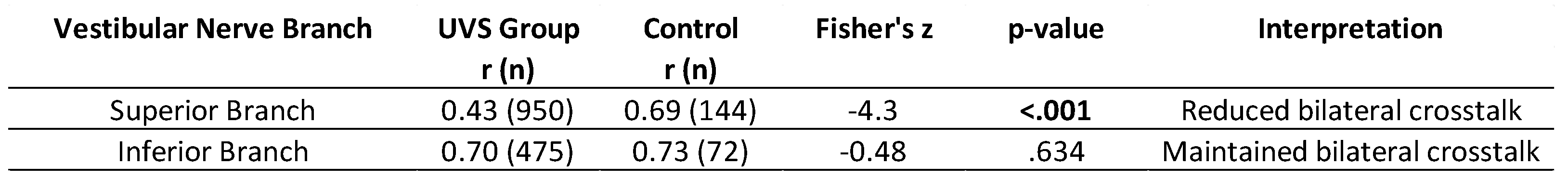
To compare the relationship of VOR gains between groups while accounting for the potential confounding effect of age, partial Pearson’s correlation coefficients were first calculated for each branch. Subsequently, these partial correlation coefficients were compared between the UVS and control groups using Fisher’s r-to-z transformation (see Table 8). Both anterior and lateral SCCs showed significantly reduced correlations in UVS tumor group (see Table 7). Analysis also revealed a significant difference in the correlation coefficients of the superior branch between groups (z = -4.3, p < .001), indicating reduced bilateral crosstalk in the UVS group (r = 0.43) compared to the control group (r = 0.69). In contrast, no significant difference was observed for the inferior branches (z = -0.48, p = .634), suggesting that the amount of possible bilateral crosstalk remains in this branch, despite the presence of the tumor.
4. Discussion
In this study, we have compared the functionality of the vestibular-ocular reflex of each semicircular canal between the non-tumor side and the tumor side in patients with unilateral vestibular schwannoma. The distinguishing characteristic of our research is the substantial size of our data set (n = 640 UVS) and the methodology we employed: in addition to the tumor group, a healthy control group (n = 72) was included to ensure the reliability of the results obtained in UVS patients through interaural comparisons and correlation analyses. In essence, the primary objective of the control group data analysis was to perform a form of cross-checking rather than a direct comparison of the results obtained from the tumor and healthy group. The present study's methodology is believed to contribute to its originality and to a more profound understanding of how the vestibular system reorganizes itself in unilateral pathologies.
Statistical analyses in control group revealed no differences in VOR gains for gender and side, and age effect, which is in agreement with other studies [38,39,40,41]: we did not find an age effect for ages up to 67 years. Significant differences were found in the UVS group, showing a reduction of the VOR on the tumor side compared to the contralateral non-tumor side, a result that has been described by previous studies, since it is well known that VOR gains on the side of the lesion might decrease in the presence of a VS [31,32]. Besides VOR gains on TS, most of the studies ignore the NTS in VS patients. The number of studies mentioning contralateral VOR impairment in VS is therefore rather limited [17,30,42].
The present data are also in conformity with previous (unpublished) preliminary studies in a smaller group of UVS patients [43,44]. Caloric vestibular reflex reactivity and vHIT responses were obtained resulting in abnormal caloric reflexes (i.e. slow phase nystagmus velocities < 10 degrees/sec) in 49% of the patients on the side of lesion. However, in 7.5% of the patients with normal caloric reactivity, vHIT outcomes revealed an isolated posterior canal dysfunction, highlighting the added value of vHIT testing in patients with UVS when compared with caloric reflex testing, as vHIT assesses the vertical canals not covered by caloric testing [43].
Other studies, [17,28,29,31] have also investigated the function of the vertical SCC in a relatively small number of patients with UVS. The study by Fujiwara et al. [31] reported VOR reactivity in 15 patients with UVS, as determined by vHIT. The objective of their study was to investigate the functionality of the vertical SCC in UVS patients. The results of their study demonstrated a reduction in both the lateral VOR and the posterior VOR on the TS. However, the anterior VOR appeared to be less affected by the unilateral UVS, of which it was noted that the vertical canals presented a greater challenge during examination, attributable to their unusual oblique vertical impulse movements. Nevertheless, in the absence of further elucidation from the authors, we recognized that their data also exhibited a dysfunction of the posterior SCC on the unaffected side in 2 out of 15 subjects, which is in accordance with our data from a considerably larger population. Comparable reduction of the posterior VOR gain on the NTS was also reported by others [17,29,30] although these studies were more focused on the sensitivity of different vestibular tests (caloric reflex, cVEMP, vHIT) related to TS and tumor volume without further exploration of the posterior canal gain reductions on the NTS and/or analyses of gain differences between and/or within SCCs.
Our UVS group data revealed significantly lower VOR gains in the posterior SCC compared to the other SCCs. Moreover, it showed that posterior SCC gain reduction on NTS (n = 140, 21.9%) is remarkably more sensitive for the presence of a UVS. An additional visualization of a comparison between TS and NTS is shown as paired plots for each SCC as Supplementary Material: it is clearly visible that the density of the VOR gains on the NTS for the lateral SCC is higher than it is for the anterior and posterior SCC. Since this gain reduction was found on both sides, it immediately raised the question whether these outcomes might be caused by variations in the practical execution of impulse movements in three different planes of the six semicircular canals, in particular for the posterior SCC impulse movements on both sides. To exclude the effect of any procedural bias such as variations in impulse movements executed by clinicians (e.g. unconscious preferences for impulse directions), asymmetrical neck muscle stiffness of patients, hard/software sensitivity of the recording device or other in/external factors causing variability, we have performed the same measurement procedures under exactly the same conditions in a group of healthy subjects (control group).
Contrary to our initial expectations, we observed that the control group exhibited significant correlations between the same bilateral SCCs (Fig. 5). However, we think that this is of negligible relevance, given that the range within the control group is evidently constrained (> 0.8 and > 0.7 for lateral and vertical SCC, respectively), with no discernible interaural variations. In contrast, the VOR gains in the UVS group exhibited a greater range (Table 3 and Table 4), thereby rendering the correlations more meaningful and relevant for clinical interpretation.
Although we claim that the presence of a VS affects the contralateral NTS in UVS patients, it should also be recognized that the response variability in the vertical SCCs was also seen in the control group, albeit to a lesser extent (Fig. 3): posterior SCCs seems to follow the same pattern in both groups. This might imply that response variability of vertical SCCs in itself appears to be more susceptible to variations resulting from external factors such e.g. the practical execution of the vertical head impulse movements that are generally more difficult to perform than horizontal impulse movements [45]. Consequently, this may mean that a small portion of the variability found in the UVS group is the result of bias due to practical recording of vHIT movements. Nevertheless, the present results found are of such magnitude that they will not undermine the interaural interdependence that we have observed in UVS patients.
4.1. Inferior vs. Superior Branch Possible Crosstalk Between TS and NTS
Although we didn’t analyze the actual location of VS tumors on the n. VIII, we compared VOR gains originating from the superior-branch-linked SCCs with the inferior branch and how those might affect the contralateral side by calculating the gain reduction ratio for each branch. Even in the control group, we found lower VOR gains in the posterior SCC (see Table 4). Our UVS data showed that the most compromised SCC appeared to be the posterior SCC on both tumor and non-tumor sides (Table 4b). The fact that the posterior SCCs on both sides appear to be most sensitive to a decrease in VOR gain suggests that, in addition to a neural bilateral dependence, the inferior branch of the cochleovestibular nerve might also play a role here, since it is responsible for the innervation of the posterior canal. After all, it is not entirely unlikely that the presence of a UVS apparently affects the VOR of the lateral and anterior canals less than that of the posterior canal, since it is known that the majority of vestibular schwannomas develop in the inferior branch of the vestibular nerve [46].
By comparing the impact of the vestibular schwannoma on the NTS, our data demonstrate that the inferior branch of the n. VIII may appear to be more sensitive to bilateral neural interactions than the superior branch (e.g. Table 7).
4.2. Contribution of Interactions Between the Bilateral Vestibular Nuclei?
On the tumor side, a reduced number of type I hair cells and fibers has been reported [47]. Other histological research has also shown that an isolated schwannoma on the superior vestibular branch of the 8th nerve can cause selective degeneration of neuroepithelia in the lateral and anterior [48] or only in the posterior SCC [49]. It is known that transient fast head movements, such as those performed during vHITs, will activate irregular afferent fibers that are mostly linked to these type I hair cells [50,51]. As a result, this histological change would lead to a decrease in VOR gain.
Besides hair cell damage, neural changes may also play an important compensatory role in unilateral pathologies. A study in macaques that underwent unilateral labyrinthectomy [46] showed that neural reorganization of afferents occurs following surgery, resulting in a greater number of irregular fibers after the damage. Similar compensatory processes might also take place in patients with UVS as a result of the loss of type I hair cells, which are more sensitive to phasic-encoding, i.e. fast movements.
The commissural inhibitory system interconnecting the bilateral medial vestibular nuclei (MVN) represents the first crossing area within the vestibular system [52]. These commissural fibers regulate excitability balance between the two nuclei through GABAergic neurotransmission, establishing baseline excitability and shaping neuronal output [53]. The commissural inhibitory system also increases the sensitivity to angular head movements [54,55], and even mediates the inhibitory signals for angular VOR velocity storage [56,57]. However, in patients with unilateral vestibular loss, commissural inhibitory input from the lesioned side is substantially reduced, causing insufficient inhibitory tone and compensatory hyperactivity of the intact side [58]. The ipsilesional MVN faces two converging problems: a decreased excitatory afferent input from the damaged labyrinth and an enhanced commissural inhibition from the hyperactive contralateral side. Logically, this dual influence should silence the ipsilesional MVN; however, the vestibular system prevents such collapse through homeostatic GABAergic adaptation[53,59].
Based on these outcomes, we hypothesize that GABA-receptor efficacy might be down-regulated on the lesioned side while it is up-regulated on the intact side due to the UVS. With respect to the ipsilesional MVN, this molecular reorganization may also diminish commissural inhibition and simultaneously enhance self-inhibition of hyperactive contralesional MVN. Through this rebalancing of GABAergic tone and receptor sensitivity, the bilateral vestibular system can attempt to restore excitability homeostasis—a process known as vestibular compensation. This commissural inhibitory system may be the underlying cause of the correlations we found between tumor and non-tumor sides. In addition, the increasing growth of VS tumors might continuously trigger commissural compensation mechanisms. In parallel with our hypothesis, it is noteworthy that the strong correlations we found between the p-SCCs (inferior branches) on the right and left sides in our healthy subjects were retained among VS patients. This suggests that the posterior SCCs might play the most important role in vestibular compensation. The hypothesized crosstalk may also help mitigate the impact of VS on balance in daily life, since it is also shown that QoL of UVS patients could be maintained, even in long-standing large tumors [60].
4.3. Strengths and Limitations
The distinguishing characteristic of this study is its inclusion of a substantial number of patients with UVS, a feature that differentiates it from the majority of previous studies in this field. Furthermore, the present study ascertained VOR gains of all SCCs in a substantial group of healthy subjects, thereby enabling the differences of subtle disparities in VOR gain between the three SCCs in the control group. This study also offers the advantage to compare the abnormal VORs exhibited by UVS patients, both at TS and NTS, with the mean gains observed in a healthy population within the relevant SCC. Consequently, VOR gains could be comparatively analyzed for each SCC, with the same SCC the other side, as well as with that of the other SCC on the same side for both groups.
Nonetheless, it is acknowledged that the scope of the present study is limited to the functionality of the VOR of the individual and combined SCCs based on vHIT outcomes, disregarding the role of otolith functionality and VS characteristics. Although the present design was not focused on establishing a correlation between the predictive value of the vHIT and impairment of a specific branch of the eight nerve, a comparative analysis of the reactivity of SCCs innervated by either superior or inferior branch of the eighth nerve, was additionally conducted, but could be further reinforced if this was supplemented with data of otolith functionality. Our data might suggest that, in the majority of our population, the VS might be located inferiorly, but we should realize that the current study lacks radiological information regarding the size and location of the VS. Other limitations of the present study are the duration of tumor presence on VOR gains and the relationship between saccades and VOR gains that have not been taken into account, since it is possible to also analyze the saccades in NTS among UVS patients [42]. Generally, it is good to be aware that UVS might lead to more pathological changes on neural fibers that might indirectly affect VOR gains. Additional research is necessary to investigate possible crosstalk between NT and NTS considering VS characteristics such as size, tempo of growth and location.
5. Conclusions
To the best of our knowledge, this study is the only one reporting the vestibulo-ocular reflex (VOR) functionality on the non-tumor side in a large population of patients with untreated unilateral vestibular schwannomas (n=640). Patients with unilateral vestibular schwannoma show that 39.7% of them exhibit a reduced VOR gain in the ipsilateral posterior SCC. However, the most noteworthy finding is that, in addition to the impaired vestibular reactivity on the ipsilateral tumor side, the presence of a vestibular schwannoma also reveals an impact on the contralateral posterior SCC on the non-tumor side in 21.9% of patients. While both lateral and anterior SCCs also demonstrate a positive correlation with their contralateral equivalents, the most significant and strongest correlation was identified between the two posterior semicircular canals (r = 0.70).
Furthermore, our age-corrected data also suggest that lesions resulting in a decrease of VOR gain on the ipsilateral superior branch of the vestibular nerve seems to lead to a lower gain on the contralateral side. This difference may be explained by the effects of possible present interactions such as commissural pathways between the vestibular nuclei on the tumor and non-tumor sides at the brainstem level.
Generally, our research outcomes reveal that clinicians should be aware that VOR on the contralateral side may also be partially impaired in a significant number of UVS patients, albeit unnoticed.
Supplementary Materials
Author Contributions
Conceptualization, AB; methodology, AB; formal analysis, AB and ME; investigation, AB; data curation, AB and ME; writing original draft preparation, AB; writing review and editing, AB, ME, SS, TJ and HK; supervision, AB; project administration, AB; All authors have read and agreed to the published version of the manuscript.
Funding
This research received no external funding.
Institutional Review Board Statement
This research adhered to the ethical principles for medical research of the World Medical Association according to the Declaration of Helsinki. Approval was received from the institutional local human research and ethics committee of the Radboud University Medical Center Nijmegen under file number NL87231.091.24 and informed consent was obtained from all individual participants of the control group. With respect to patient data, obtained from medical records, according to the Medical Ethical Research Involving Human Subjects Act, ethical approval was not required due to the retrospective nature and anonymization.
Informed Consent Statement:. Informed consent was obtained from all healthy subjects involved in the study.
Data Availability Statement
The datasets generated for this study are available on request to the corresponding author.
Acknowledgments
The authors want to acknowledge Anne Voncken, Karin Krommenhoek and Jacquelien Jilissen for their assistance with data acquisition.
Conflicts of Interest
The authors declare no conflicts of interests or any funding from third parties or manufacturers of the products that have been used in this research.
Abbreviations
The following abbreviations are used in this manuscript:
|
GABA MVN NTS |
gamma-aminobutyric acid medial vestibular nucleus non-tumor side |
| SCC | semicircular canal |
| TS | tumor side |
| UVS | unilateral vestibular schwannoma |
| vHIT | video head impulse test |
| VOR | vestibulo-ocular reflex |
| VS | vestibular schwannoma |
References
- Verocay, J. Multipie Gaschwulste als systemerkrankung am nervosen Apparate. Festschrift fur chiari 1908, 387. [Google Scholar]
- Antoni, N. Über rückenmarkstumoren und neurofibrome: studien zur pathologischen anatomie und embryogenese. Über rückenmarkstumoren und neurofibrome: studien zur pathologischen anatomie und embryogenese 1920, 435–435. [Google Scholar]
- Joshi, R. Learning from eponyms: Jose Verocay and Verocay bodies, Antoni A and B areas, Nils Antoni and Schwannomas. Indian Dermatol Online J 2012, 3, 215–219. [Google Scholar] [CrossRef] [PubMed]
- Tieleman, A.; Casselman, J.W.; Somers, T.; Delanote, J.; Kuhweide, R.; Ghekiere, J.; De Foer, B.; Offeciers, E.F. Imaging of intralabyrinthine schwannomas: a retrospective study of 52 cases with emphasis on lesion growth. AJNR Am J Neuroradiol 2008, 29, 898–905. [Google Scholar] [CrossRef] [PubMed]
- Salzman, K.L.; Childs, A.M.; Davidson, H.C.; Kennedy, R.J.; Shelton, C.; Harnsberger, H.R. Intralabyrinthine schwannomas: imaging diagnosis and classification. AJNR Am J Neuroradiol 2012, 33, 104–109. [Google Scholar] [CrossRef] [PubMed]
- Yang, C.; Alvarado, D.; Ravindran, P.K.; Keizer, M.E.; Hovinga, K.; Broen, M.P.G.; Kunst, H.P.M.; Temel, Y. Untreated Vestibular Schwannoma: Analysis of the Determinants of Growth. Cancers (Basel) 2024, 16. [Google Scholar] [CrossRef]
- Reznitsky, M.; Petersen, M.; West, N.; Stangerup, S.E.; Cayé-Thomasen, P. The natural history of vestibular schwannoma growth-prospective 40-year data from an unselected national cohort. Neuro Oncol 2021, 23, 827–836. [Google Scholar] [CrossRef]
- Schouten, S.M.; Cornelissen, S.; Langenhuizen, P.; Jansen, T.T.G.; Mulder, J.J.S.; Derks, J.; Verheul, J.B.; Kunst, H.P.M. Wait-and-scan management in sporadic Koos grade 4 vestibular schwannomas: A longitudinal volumetric study. Neurooncol Adv 2024, 6, vdad144. [Google Scholar] [CrossRef]
- Marinelli, J.P.; Schnurman, Z.; Killeen, D.E.; Nassiri, A.M.; Hunter, J.B.; Lees, K.A.; Lohse, C.M.; Roland, J.T.; Golfinos, J.G.; Kondziolka, D.; et al. Long-term natural history and patterns of sporadic vestibular schwannoma growth: A multi-institutional volumetric analysis of 952 patients. Neuro Oncol 2022, 24, 1298–1306. [Google Scholar] [CrossRef]
- Huang, X.; Xu, J.; Xu, M.; Zhou, L.F.; Zhang, R.; Lang, L.; Xu, Q.; Zhong, P.; Chen, M.; Wang, Y.; et al. Clinical features of intracranial vestibular schwannomas. Oncol Lett 2013, 5, 57–62. [Google Scholar] [CrossRef]
- Halliday, J.; Rutherford, S.A.; McCabe, M.G.; Evans, D.G. An update on the diagnosis and treatment of vestibular schwannoma. Expert Rev Neurother 2018, 18, 29–39. [Google Scholar] [CrossRef] [PubMed]
- Rosahl, S.; Bohr, C.; Lell, M.; Hamm, K.; Iro, H. Diagnostics and therapy of vestibular schwannomas - an interdisciplinary challenge. GMS Curr Top Otorhinolaryngol Head Neck Surg 2017, 16, Doc03. [Google Scholar] [CrossRef] [PubMed]
- Constanzo, F.; Teixeira, B.C.A.; Sens, P.; Ramina, R. Video Head Impulse Test in Vestibular Schwannoma: Relevance of Size and Cystic Component on Vestibular Impairment. Otol Neurotol 2019, 40, 511–516. [Google Scholar] [CrossRef]
- Lee, S.U.; Bae, Y.J.; Kim, H.J.; Choi, J.Y.; Song, J.J.; Choi, B.Y.; Choi, B.S.; Koo, J.W.; Kim, J.S. Intralabyrinthine Schwannoma: Distinct Features for Differential Diagnosis. Front Neurol 2019, 10, 750. [Google Scholar] [CrossRef] [PubMed]
- Teixeira, B.C.d.A.; Constanzo, F.; Sens, P.; Ramina, R.; Escuissato, D.L. Brainstem hyperintensity in patients with vestibular schwannoma is associated with labyrinth signal on magnetic resonance imaging but not vestibulocochlear tests. The Neuroradiology Journal 2021, 34, 180–186. [Google Scholar] [CrossRef]
- Constanzo, F.; Teixeira, B.C.A.; Sens, P.; Escuissato, D.; Ramina, R. Relationship between Signal Intensity of the Labyrinth and Cochleovestibular Testing and Morphologic Features of Vestibular Schwannoma. J Neurol Surg B Skull Base 2022, 83, e208–e215. [Google Scholar] [CrossRef]
- Nilsen, K.S.; Nordahl, S.H.G.; Berge, J.E.; Dhayalan, D.; Goplen, F.K. Vestibular Tests Related to Tumor Volume in 137 Patients With Small to Medium-Sized Vestibular Schwannoma. Otolaryngol Head Neck Surg 2023, 169, 1268–1275. [Google Scholar] [CrossRef]
- West, N.C.; Groth, J.B.; Caye-Thomasen, P. Does Location of Intralabyrinthine Vestibular Schwannoma Determine Objective and Subjective Vestibular Function? Otol Neurotol 2024, 45, 319–325. [Google Scholar] [CrossRef]
- Kjærsgaard, J.B.; Szeremet, M.; Hougaard, D.D. Vestibular Deficits Correlating to Dizziness Handicap Inventory Score, Hearing Loss, and Tumor Size in a Danish Cohort of Vestibular Schwannoma Patients. Otology & Neurotology 2019, 40. [Google Scholar] [CrossRef]
- Agrawal, Y.; Van de Berg, R.; Wuyts, F.; Walther, L.; Magnusson, M.; Oh, E.; Sharpe, M.; Strupp, M. Presbyvestibulopathy: Diagnostic criteria Consensus document of the classification committee of the Bárány Society. J Vestib Res 2019, 29, 161–170. [Google Scholar] [CrossRef]
- Strupp, M.; Bisdorff, A.; Furman, J.; Hornibrook, J.; Jahn, K.; Maire, R.; Newman-Toker, D.; Magnusson, M. Acute unilateral vestibulopathy/vestibular neuritis: Diagnostic criteria. J Vestib Res 2022, 32, 389–406. [Google Scholar] [CrossRef] [PubMed]
- Strupp, M.; Kim, J.S.; Murofushi, T.; Straumann, D.; Jen, J.C.; Rosengren, S.M.; Della Santina, C.C.; Kingma, H. Bilateral vestibulopathy: Diagnostic criteria Consensus document of the Classification Committee of the Bárány Society. J Vestib Res 2017, 27, 177–189. [Google Scholar] [CrossRef] [PubMed]
- Moretz, W.H., Jr.; Orchik, D.J.; Shea, J.J., Jr.; Emmett, J.R. Low-frequency harmonic acceleration in the evaluation of patients with intracanalicular and cerebellopontine angle tumors. Otolaryngol Head Neck Surg 1986, 95, 324–332. [Google Scholar] [CrossRef] [PubMed]
- Nilsen, K.S.; Lund-Johansen, M.; Nordahl, S.H.G.; Finnkirk, M.; Goplen, F.K. Long-term Effects of Conservative Management of Vestibular Schwannoma on Dizziness, Balance, and Caloric Function. Otolaryngol Head Neck Surg 2019, 161, 846–851. [Google Scholar] [CrossRef]
- Abboud, T.; Regelsberger, J.; Matschke, J.; Jowett, N.; Westphal, M.; Dalchow, C. Long-term vestibulocochlear functional outcome following retro-sigmoid approach to resection of vestibular schwannoma. Eur Arch Otorhinolaryngol 2016, 273, 719–725. [Google Scholar] [CrossRef]
- Ogawa, Y.; Otsuka, K.; Inagaki, T.; Nagai, N.; Itani, S.; Kondo, T.; Kohno, M.; Suzuki, M. Comparison of cervical vestibular evoked potentials evoked by air-conducted sound and bone-conducted vibration in vestibular Schwannoma patients. Acta Otolaryngol 2018, 138, 898–903. [Google Scholar] [CrossRef]
- Rahne, T.; Plontke, S.K.; Fröhlich, L.; Strauss, C. Optimized preoperative determination of nerve of origin in patients with vestibular schwannoma. Scientific Reports 2021, 11, 8608. [Google Scholar] [CrossRef]
- Fujiwara, K.; Morita, S.; Fukuda, A.; Akamatsu, H.; Yanagi, H.; Hoshino, K.; Nakamaru, Y.; Kano, S.; Homma, A. Analysis of semicircular canal function as evaluated by video Head Impulse Test in patients with vestibular schwannoma. J Vestib Res 2020, 30, 101–108. [Google Scholar] [CrossRef]
- Taylor, R.L.; Kong, J.; Flanagan, S.; Pogson, J.; Croxson, G.; Pohl, D.; Welgampola, M.S. Prevalence of vestibular dysfunction in patients with vestibular schwannoma using video head-impulses and vestibular-evoked potentials. Journal of Neurology 2015, 262, 1228–1237. [Google Scholar] [CrossRef]
- Lee, J.W.Y.; Hassannia, F.; Rutka, J.A. Contralesional High-Acceleration Vestibulo-Ocular Reflex Function in Vestibular Schwannoma. Otology & Neurotology 2021, 42, e1106–e1110. [Google Scholar] [CrossRef]
- Fujiwara, K.; Yanagi, H.; Morita, S.; Hoshino, K.; Fukuda, A.; Nakamaru, Y.; Homma, A. Evaluation of Vertical Semicircular Canal Function in Patients With Vestibular Schwannoma. Ann Otol Rhinol Laryngol 2019, 128, 113–120. [Google Scholar] [CrossRef] [PubMed]
- Kontorinis, G.; Tailor, H.; Tikka, T.; Slim, M.A.M. Six-canal video head impulse test in patients with labyrinthine and retrolabyrinthine pathology: detecting vestibulo-ocular reflex deficits. J Laryngol Otol 2023, 137, 398–403. [Google Scholar] [CrossRef] [PubMed]
- Amiraraghi, N.; Gaggini, M.; Crowther, J.A.; Locke, R.; Taylor, W.; Kontorinis, G. Benefits of pre-labyrinthectomy intratympanic gentamicin: contralateral vestibular responses. J Laryngol Otol 2019, 133, 668–673. [Google Scholar] [CrossRef] [PubMed]
- Guerra Jiménez, G.; Pérez Fernández, N. Reduction in posterior semicircular canal gain by age in video head impulse testing. Observational study. Acta Otorrinolaringol Esp 2016, 67, 15–22. [Google Scholar] [CrossRef]
- Halmagyi, G.M.; Chen, L.; MacDougall, H.G.; Weber, K.P.; McGarvie, L.A.; Curthoys, I.S. The Video Head Impulse Test. Front Neurol 2017, 8, 258. [Google Scholar] [CrossRef]
- Halmagyi, G.M.; Curthoys, I.S. A Clinical Sign of Canal Paresis. Archives of Neurology 1988, 45, 737–739. [Google Scholar] [CrossRef]
- Curthoys, I.S.; Manzari, L. Clinical application of the head impulse test of semicircular canal function. Hearing, Balance and Communication 2017, 15, 113–126. [Google Scholar] [CrossRef]
- Emekci, T.; Uğur, K.Ş.; Cengiz, D.U.; Men Kılınç, F. Normative values for semicircular canal function with the video head impulse test (vHIT) in healthy adolescents. Acta oto-laryngologica 2021, 141, 141–146. [Google Scholar] [CrossRef]
- Li, C.; Layman, A.J.; Geary, R.; Anson, E.; Carey, J.P.; Ferrucci, L.; Agrawal, Y. Epidemiology of vestibulo-ocular reflex function: data from the Baltimore Longitudinal Study of Aging. Otol Neurotol 2015, 36, 267–272. [Google Scholar] [CrossRef]
- Matiño-Soler, E.; Esteller-More, E.; Martin-Sanchez, J.-C.; Martinez-Sanchez, J.-M.; Perez-Fernandez, N. Normative data on angular vestibulo-ocular responses in the yaw axis measured using the video head impulse test. Otology & Neurotology 2015, 36, 466–471. [Google Scholar] [CrossRef]
- Treviño-González, J.L.; Maldonado-Chapa, F.; González-Cantú, A.; Soto-Galindo, G.A.; Ángel, J.A.M.-d. Age adjusted normative data for Video Head Impulse Test in healthy subjects. American Journal of Otolaryngology 2021, 42, 103160. [Google Scholar] [CrossRef] [PubMed]
- Batuecas-Caletrio, A.; Santa Cruz-Ruiz, S.; Muñoz-Herrera, A.; Perez-Fernandez, N. The map of dizziness in vestibular schwannoma. Laryngoscope 2015, 125, 2784–2789. [Google Scholar] [CrossRef] [PubMed]
- Beynon, A. Surplus value of the video-head impulse test in patients with vestibular schwannoma. J. Neurol. Sci. 2015, 357, e174. [Google Scholar] [CrossRef]
- Beynon, A.J. Isolated and combined semicircular canal dysfunction in patients with unilateral vestibular schwannoma. In Proceedings of the 7th International Symposium on Menière's Disease & Inner Ear Disorders, Rome, Italy, October 17-20, 2015. [Google Scholar]
- Karabin, M.J.; Harrell, R.G.; Sparto, P.J.; Furman, J.M.; Redfern, M.S. Head and vestibular kinematics during vertical semicircular canal impulses. Journal of Vestibular Research 2023, 33, 367–376. [Google Scholar] [CrossRef]
- Khrais, T.; Romano, G.; Sanna, M. Nerve origin of vestibular schwannoma: a prospective study. The Journal of Laryngology & Otology 2008, 122, 128–131. [Google Scholar] [CrossRef]
- Hızlı, Ö.; Cureoglu, S.; Kaya, S.; Schachern, P.A.; Paparella, M.M.; Adams, M.E. Quantitative Vestibular Labyrinthine Otopathology in Temporal Bones with Vestibular Schwannoma. Otolaryngol Head Neck Surg 2016, 154, 150–156. [Google Scholar] [CrossRef]
- Møller, M.N.; Hansen, S.; Caye-Thomasen, P. Peripheral Vestibular System Disease in Vestibular Schwannomas: A Human Temporal Bone Study. Otology & Neurotology 2015, 36, 1547–1553. [Google Scholar] [CrossRef]
- Moura, L.F.P.D. Inner Ear Pathology in Acoustic Neurinoma. Archives of Otolaryngology 1967, 85, 125–133. [Google Scholar] [CrossRef]
- Hullar, T.E.; Della Santina, C.C.; Hirvonen, T.; Lasker, D.M.; Carey, J.P.; Minor, L.B. Responses of irregularly discharging chinchilla semicircular canal vestibular-nerve afferents during high-frequency head rotations. J Neurophysiol 2005, 93, 2777–2786. [Google Scholar] [CrossRef]
- McCaslin, D.L.; Rivas, A.; Jacobson, G.P.; Bennett, M.L. The dissociation of video head impulse test (vHIT) and bithermal caloric test results provide topological localization of vestibular system impairment in patients with "definite" Ménière's disease. Am J Audiol 2015, 24, 1–10. [Google Scholar] [CrossRef]
- Dieterich, M.; Brandt, T. The bilateral central vestibular system: its pathways, functions, and disorders. Annals of the New York Academy of Sciences 2015, 1343, 10–26. [Google Scholar] [CrossRef] [PubMed]
- Yamanaka, T.; Him, A.; Cameron, S.A.; Dutia, M.B. Rapid compensatory changes in GABA receptor efficacy in rat vestibular neurones after unilateral labyrinthectomy. J Physiol 2000, 523 Pt 2, 413–424. [Google Scholar] [CrossRef] [PubMed]
- Shimazu, H.; Precht, W. Inhibition of central vestibular neurons from the contralateral labyrinth and its mediating pathway. J Neurophysiol 1966, 29, 467–492. [Google Scholar] [CrossRef] [PubMed]
- Markham, C.H.; Yagi, T.; Curthoys, I.S. The contribution of the contralateral labyrinth to second order vestibular neuronal activity in the cat. Brain Res 1977, 138, 99–109. [Google Scholar] [CrossRef]
- Wearne, S.; Raphan, T.; Cohen, B. Contribution of vestibular commissural pathways to spatial orientation of the angular vestibuloocular reflex. J Neurophysiol 1997, 78, 1193–1197. [Google Scholar] [CrossRef]
- Holstein, G.R.; Martinelli, G.P.; Wearne, S.; Cohen, B. Ultrastructure of vestibular commissural neurons related to velocity storage in the monkey. Neuroscience 1999, 93, 155–170. [Google Scholar] [CrossRef]
- Bergquist, F.; Ludwig, M.; Dutia, M.B. Role of the commissural inhibitory system in vestibular compensation in the rat. The Journal of Physiology 2008, 586, 4441–4452. [Google Scholar] [CrossRef]
- Graham, B.P.; Dutia, M.B. Cellular basis of vestibular compensation: analysis and modelling of the role of the commissural inhibitory system. Exp Brain Res 2001, 137, 387–396. [Google Scholar] [CrossRef]
- Wagner, J.N.; Glaser, M.; Wowra, B.; Muacevic, A.; Goldbrunner, R.; Cnyrim, C.; Tonn, J.C.; Strupp, M. Vestibular function and quality of life in vestibular schwannoma: does size matter? Front Neurol 2011, 2, 55. [Google Scholar] [CrossRef]
Figure 1.
Examples of VOR gains output of all SCCs in three subjects: a) patient with UVS on the left side, showing reduced VOR gains in both posterior SCCs (TS and NTS); b) patient with UVS on the left side, showing reduced VOR gains in all SCCs on TS and in the posterior SCC on NTS; c) control subject, showing normal VOR gains in all SCCs on both sides. Table values in green and red represent normal and abnormal gains, respectively.
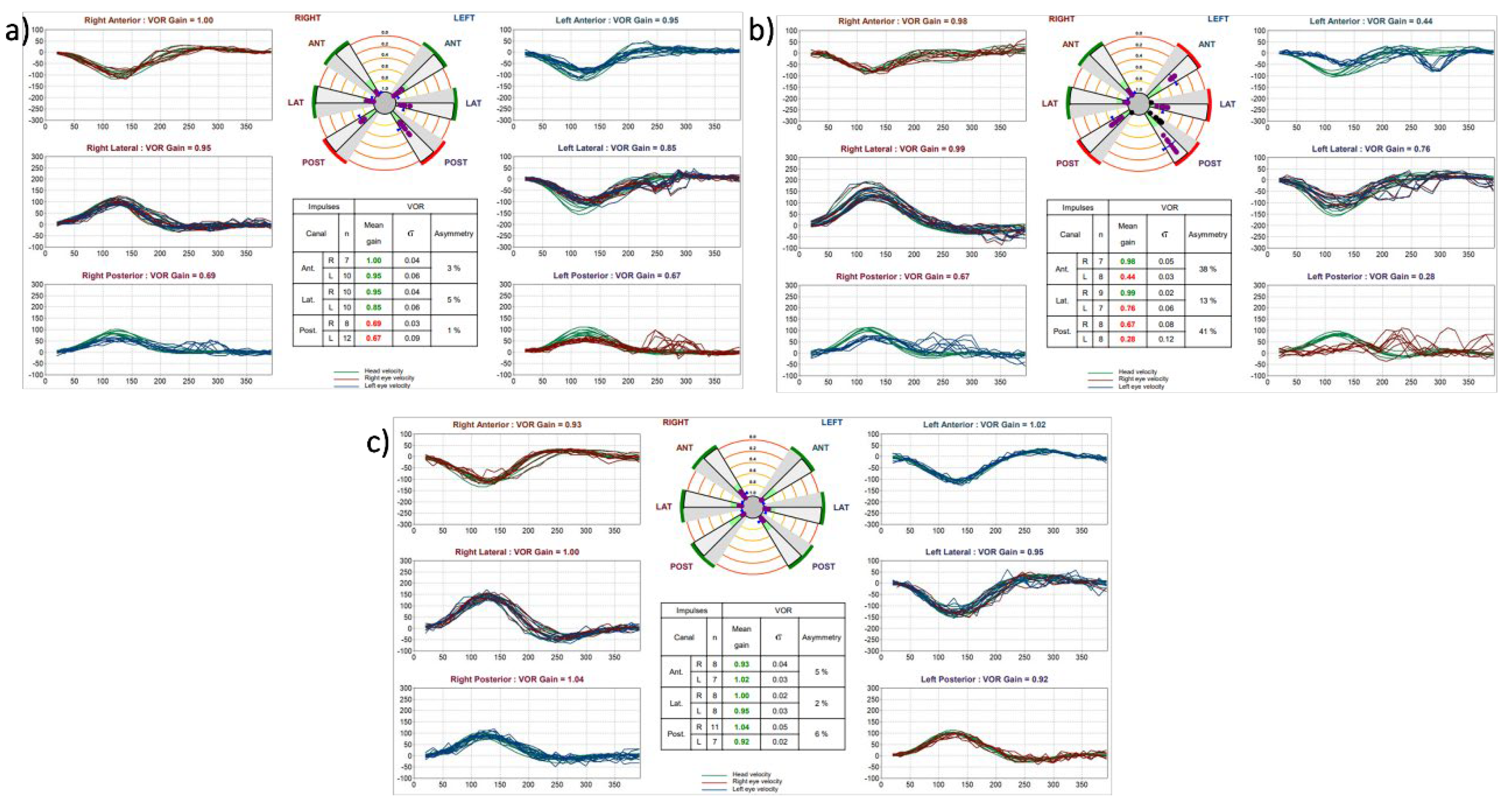
Figure 2.
Scatter plots of VOR gain of non-tumor side (NTS) as a function of tumor side (TS) for the anterior (a-SCC), lateral (l-SCC), and posterior semicircular canals (p-SCC) of 640 patients with UVS.
Figure 2.
Scatter plots of VOR gain of non-tumor side (NTS) as a function of tumor side (TS) for the anterior (a-SCC), lateral (l-SCC), and posterior semicircular canals (p-SCC) of 640 patients with UVS.
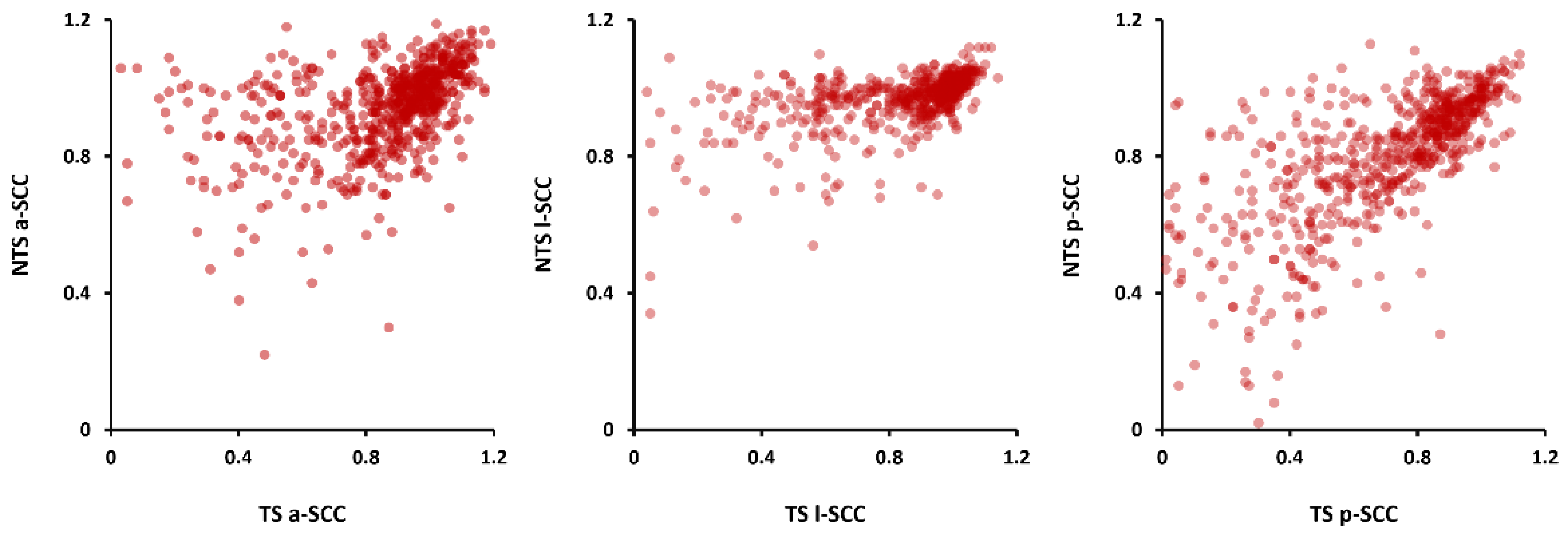
Figure 3.
Box-whisker plots including distribution of each semicircular canal on both sides in a) the control group (left) and UVS group (right): a-SCC: anterior semicircular canal, l-SCC: lateral semicircular canal, p-SCC: posterior semicircular canal, TS: tumor side, NTS: non-tumor side. A comparison of the VORs of the same SCCs from both sides revealed significant disparities in the UVS patients (* p < .001, n = 475).
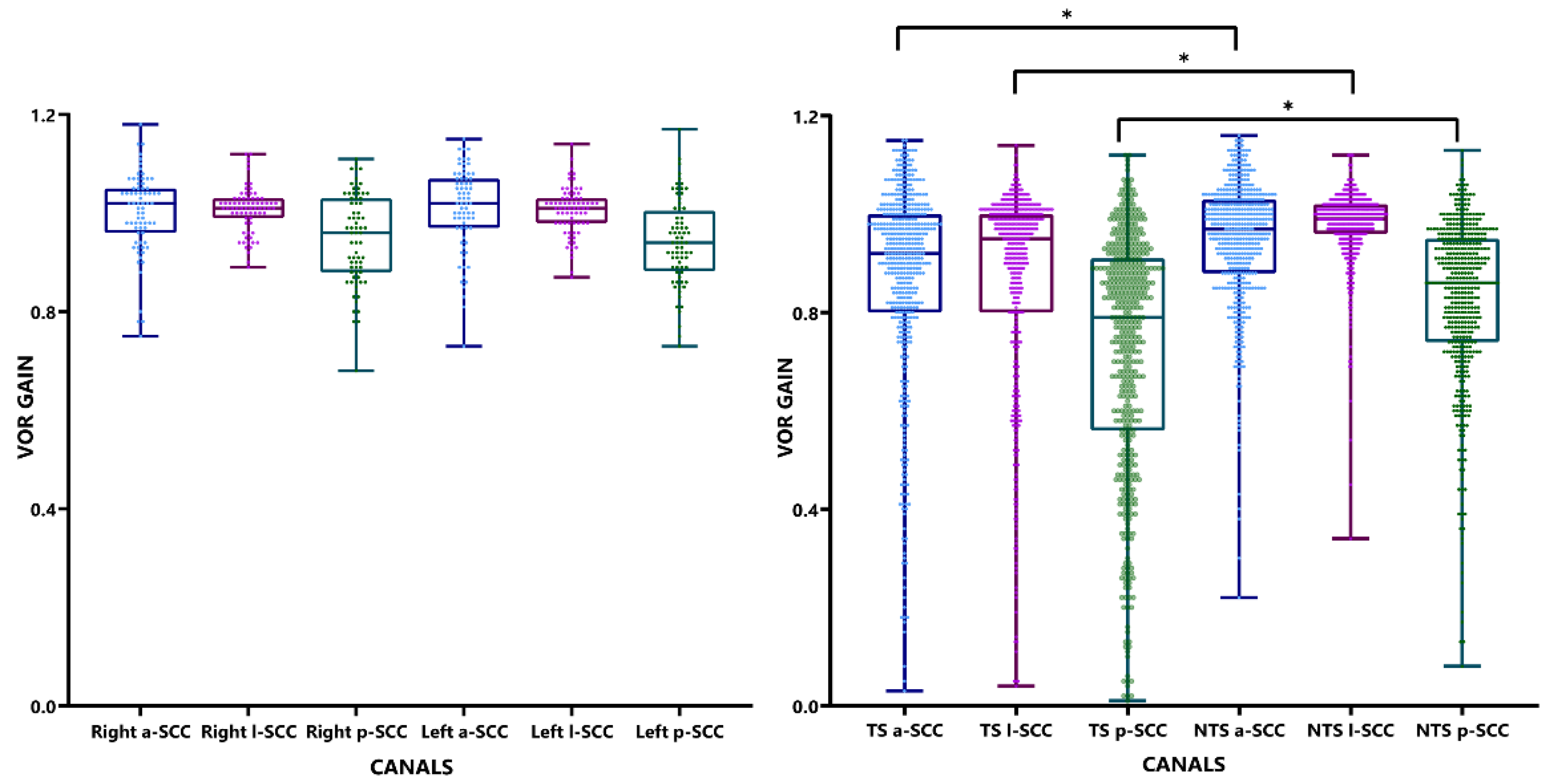
Figure 4.
Scatter plots of VOR gain of non-tumor side (NTS) as a function of tumor side (TS) for the anterior (a-SCC), lateral (l-SCC), and posterior semicircular canals (p-SCC) of 475 patients with UVS and correlations coefficients between same SCC pairs.
Figure 4.
Scatter plots of VOR gain of non-tumor side (NTS) as a function of tumor side (TS) for the anterior (a-SCC), lateral (l-SCC), and posterior semicircular canals (p-SCC) of 475 patients with UVS and correlations coefficients between same SCC pairs.
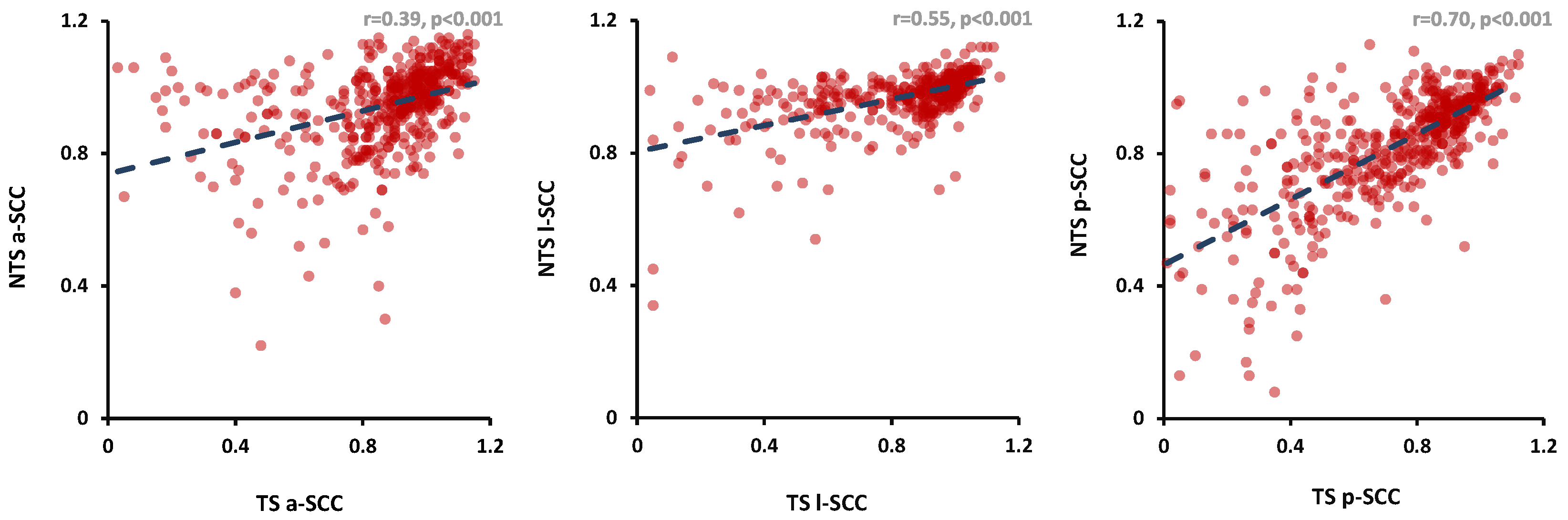
Figure 5.
Scatter plots of VOR gains of right versus left side for the anterior (a-SCC), lateral (l-SCC), and posterior semicircular canals (p-SCC) of 72 healthy subjects and correlations coefficients between same SCC pairs.
Figure 5.
Scatter plots of VOR gains of right versus left side for the anterior (a-SCC), lateral (l-SCC), and posterior semicircular canals (p-SCC) of 72 healthy subjects and correlations coefficients between same SCC pairs.
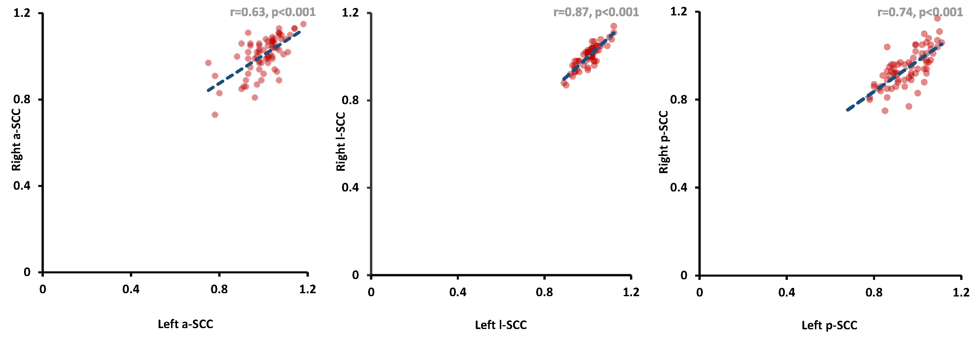

Table 1.
Overview patient and control group characteristics (n=640, age in years).
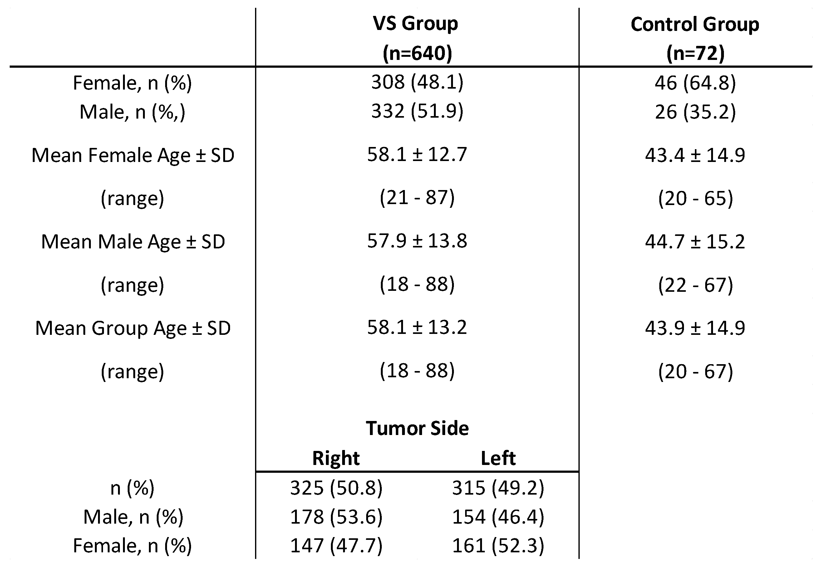
Table 2.
Frequency table of compromised VOR gains (i.e. < 0.7 for vertical, 0.8 for lateral SCC) per semicircular canal for tumor side (TS) and non-tumor side (NTS) for UVS group (n=640).
Table 2.
Frequency table of compromised VOR gains (i.e. < 0.7 for vertical, 0.8 for lateral SCC) per semicircular canal for tumor side (TS) and non-tumor side (NTS) for UVS group (n=640).
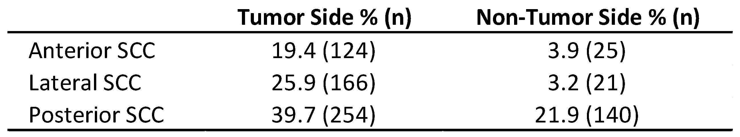
Table 3.
Absolute mean VOR gains of all six semicircular canals of UVS patients (left): tumor side vs. non-tumor side (n=640).
Table 3.
Absolute mean VOR gains of all six semicircular canals of UVS patients (left): tumor side vs. non-tumor side (n=640).
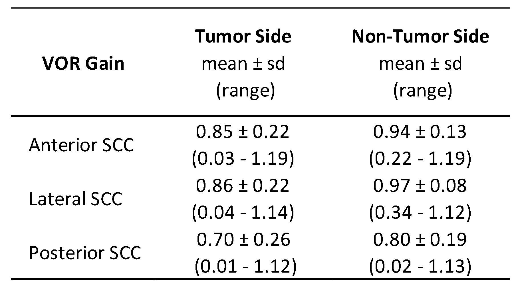
Table 4.
Absolute mean VOR gains of all separate SCC in a) control group (n = 72) and (b) age-adjusted UVS group (n=475).
Table 4.
Absolute mean VOR gains of all separate SCC in a) control group (n = 72) and (b) age-adjusted UVS group (n=475).
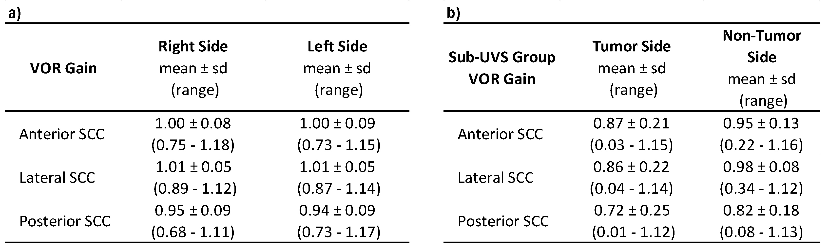
Table 5.
Two-way ANCOVA analysis: UVS (n = 475) vs. control group (n = 72).
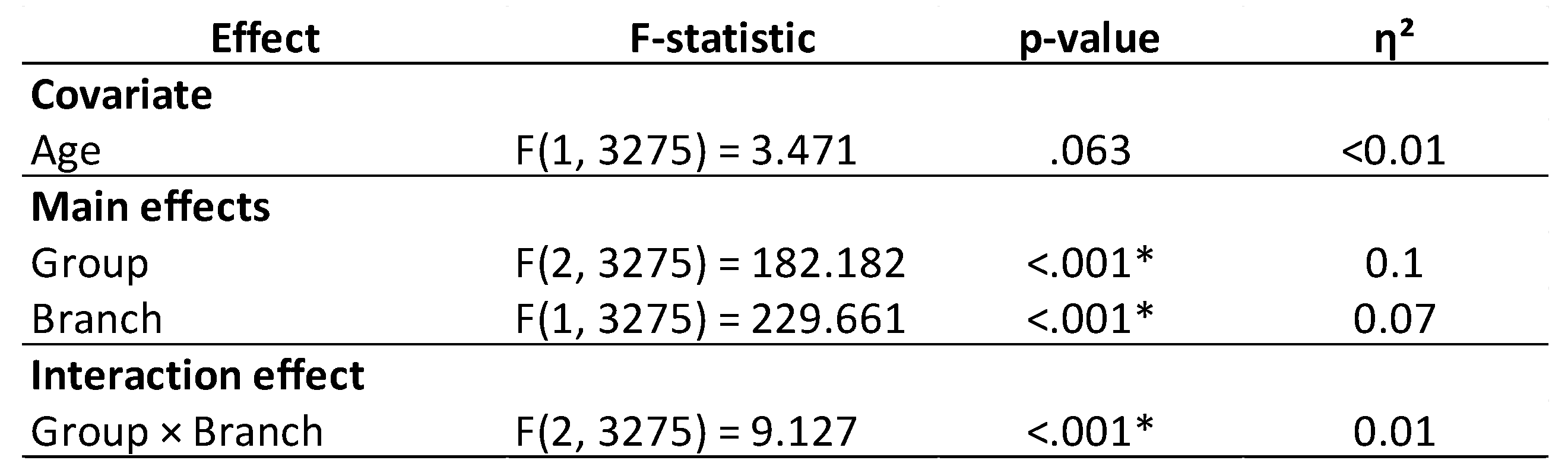
Table 6.
Mean VOR gain for inferior and superior branches of n. VIII of UVS and control group (ANCOVA, adjusted for covariate ‘Age’ evaluated at 51.55 years). Disparity = Superior - Inferior branch difference within each group.
Table 6.
Mean VOR gain for inferior and superior branches of n. VIII of UVS and control group (ANCOVA, adjusted for covariate ‘Age’ evaluated at 51.55 years). Disparity = Superior - Inferior branch difference within each group.
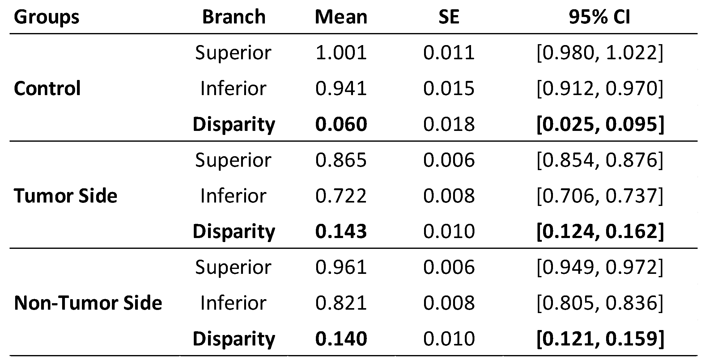
Table 7.
Pearson’s partial correlation coefficients based on age correction for all three semicircular canals a) between tumor side (TS) vs. non-tumor side (NTS) (p < .001, n = 475) for UVS patients and b) between right and left side for control group (p < .001, n = 72).
Table 7.
Pearson’s partial correlation coefficients based on age correction for all three semicircular canals a) between tumor side (TS) vs. non-tumor side (NTS) (p < .001, n = 475) for UVS patients and b) between right and left side for control group (p < .001, n = 72).
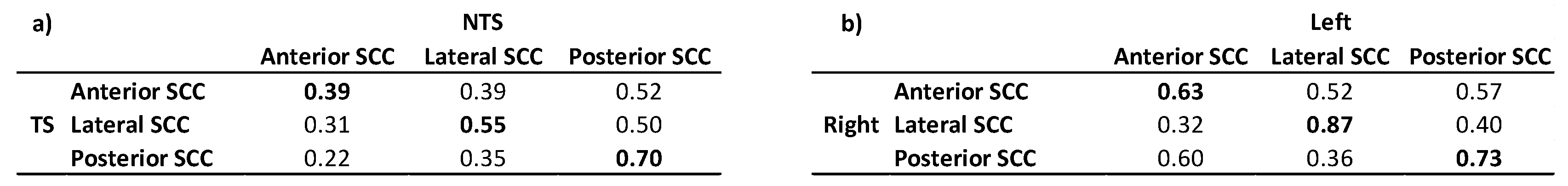
Table 8.
Comparison of branch correlations within groups with Fisher’s z transformation.
Disclaimer/Publisher’s Note: The statements, opinions and data contained in all publications are solely those of the individual author(s) and contributor(s) and not of MDPI and/or the editor(s). MDPI and/or the editor(s) disclaim responsibility for any injury to people or property resulting from any ideas, methods, instructions or products referred to in the content. |
© 2026 by the authors. Licensee MDPI, Basel, Switzerland. This article is an open access article distributed under the terms and conditions of the Creative Commons Attribution (CC BY) license (http://creativecommons.org/licenses/by/4.0/).
Copyright: This open access article is published under a Creative Commons CC BY 4.0 license, which permit the free download, distribution, and reuse, provided that the author and preprint are cited in any reuse.


