
Submitted:
02 January 2026
Posted:
06 January 2026
You are already at the latest version
Abstract
Background: Adult growth hormone deficiency (GHD) is linked to increased cardio-vascular and metabolic risk due to oxidative stress (OS), endothelial dysfunction, and adverse body composition. Long-term systemic effects of recombinant human growth hormone (rhGH) therapy remain insufficiently defined. This study assessed the impact of 24-month rhGH replacement on OS, vascular markers, body composition, and bone mineral density (BMD) in adults with severe GHD.
Methods: Fifteen adults with confirmed GHD received rhGH for 24 months. Serum insulin-like growth factor -1 (IGF-1), oxidized LDL (Ox-LDL), thioredoxin (Trx), 8-oxoguanine DNA glycosylase (OGG1), E-selectin, ICAM-1, and VCAM-1 were meas-ured at baseline, 12, and 24 months. Body composition and BMD were evaluated by DXA.
Results: IGF-1 increased significantly at 12 and 24 months (p < 0.001). Ox-LDL decreased markedly (p < 0.00001), while Trx and OGG1 increased (p < 0.05). Levels of E-selectin, ICAM-1, and VCAM-1 declined, indicating improved endothelial function. Lean body mass and BMD (lumbar spine and femoral neck) increased, whereas body fat percentage decreased. Lipid profiles were unchanged. Significant correlations were observed be-tween vascular markers and adiposity, and between BMD, triglycerides, and IGF-1.
Conclusion: A 24-month rhGH therapy improves redox balance, vascular function, and body composition in adults with severe GHD, supporting the use of redox and vascular biomarkers to monitor treatment efficacy.
Keywords:
growth hormone deficiency
; oxidized low-density lipoprotein
; thioredoxin
; vascular cell adhesion molecule-1
; intercellular adhesion molecule-1
; 8-oxoguanine DNA glycosylase 1
1. Introduction
Growth hormone deficiency (GHD) is a clinically recognized syndrome characterized by inadequate secretion of growth hormone (GH). In adults, it may represent the persistence of childhood-onset deficiency (CO-GHD) or arise de novo as adult-onset GHD (AO-GHD), with an estimated prevalence of approximately 2–3 cases per 10,000 individuals [1]. AO-GHD is consistently associated with increased cardiovascular risk, driven by dyslipidemia, visceral adiposity, endothelial dysfunction, and chronic low-grade inflammation [2,3,4]. Beyond these metabolic disturbances, GHD is marked by a broadened profile of vascular abnormalities that further amplify cardiovascular vulnerability. A central feature of its pathophysiology is endothelial dysfunction, reflected by reduced nitric oxide (NO) bioavailability and upregulated expression of adhesion molecules, including selectins, which collectively contribute to early atherogenic processes.
As demonstrated in our previous study and supported by existing literature, patients typically exhibit an atherogenic lipid profile—marked by elevated low-density lipoprotein cholesterol (LDL-C) and triglycerides (TG), alongside reduced high-density lipoprotein cholesterol (HDL-C) - together with increased arterial stiffness, oxidative stress (OS), and diminished NO bioavailability [5]. These factors collectively accelerate atherogenesis [6,7,8]. Considering the central role of atherosclerosis in cardiovascular disease (CVD), heart failure, and thrombotic complications, understanding its underlying mechanisms in GHD remains essential. The interplay between oxidized low-density lipoprotein (ox-LDL), thioredoxin (Trx), vascular cell adhesion molecule-1 (VCAM-1), intercellular adhesion molecule-1 (ICAM-1), and E-selectin offers valuable mechanistic insights into atherosclerosis pathogenesis within the GHD context. Ox-LDL contributes significantly to endothelial dysfunction, inflammation, and plaque formation by reducing NO availability and increasing OS [9,10,11]. Additionally, it induces the expression of adhesion molecules, facilitating leukocyte adhesion and transmigration into the vascular intima, thereby promoting inflammation and plaque progression [12]. Trx, a major endogenous antioxidant, counteracts OS by scavenging reactive oxygen species (ROS), thereby protecting endothelial integrity and limiting oxidative damage [13,14]. Moreover, it inhibits pro-inflammatory pathways involved in atherogenesis [15]. Adhesion molecules VCAM-1, ICAM-1, and E-selectin are upregulated in response to oxidative and inflammatory stimuli, enhancing immune cell recruitment to the endothelium and aggravating vascular inflammation and remodeling [16,17,18]. Furthermore, 8-oxoguanine DNA glycosylase 1 (OGG1) plays a critical role in the base excision repair pathway by removing oxidatively damaged guanine from DNA, thus preserving genomic stability [19]. Reduced OGG1 activity has been associated with increased susceptibility to doxorubicin-induced cardiotoxicity, accelerated vascular aging, and worsened endothelial dysfunction in atherosclerosis models [20]. OGG1 deficiency leads to accumulation of oxidative DNA lesions, intensifying vascular inflammation and contributing to CVD pathogenesis [21]. Given the pivotal role of atherosclerosis in CVD, heart failure, and thrombotic events, elucidating molecular mechanisms of vascular dysfunction in GHD remains a research priority. Although recombinant human growth hormone (rhGH) therapy has demonstrated improvements in metabolic and vascular parameters, the specific contributions of Trx, OGG1, VCAM-1, ICAM-1, and E-selectin—and their interactions with lipid metabolism and body composition—are yet to be fully defined [22,23,24]. Consequently, data on endothelial and OS biomarkers in this setting remain limited.
This study aimed to evaluate the effects of 24-month rhGH therapy on cardiovascular and metabolic parameters in adults with severe GHD, with particular focus on biomarkers of OS and endothelial dysfunction, including ox-LDL, Trx, OGG1, VCAM-1, ICAM-1, and E-selectin. By characterizing changes in these markers during long-term treatment, the research seeks to enhance understanding of the mechanisms linking GHD to increased cardiovascular risk. The findings may facilitate integration of these biomarkers into clinical monitoring, enabling improved risk stratification and supporting personalized therapeutic strategies to reduce long-term CVD risk in this population.
2. Results
2.1. Biochemical Analysis
2.1.1. Lipid Profile
No statistically significant changes in the lipid profile were observed (Table 1).
2.1.2. IGF-1
Serum insulin-like growth factor-1 (IGF-1) concentrations increased significantly after both 12 and 24 months of rhGH therapy compared with baseline values (p < 0.001 for both time points; Table 1).
2.1.3. Ox-LDL
2.1.4. Thx
2.1.4. OGG-1
A statistically significant increase in OGG-1 concentration was observed after 24 months of therapy compared with baseline (p < 0.05; Table 1).
2.1.4. E-selectin and P-selectin
2.1.4. I-CAM1 and VCAM-1
Body Composition Evaluation
Total body mass changed significantly after 24 months of therapy (p = 0.05). Fat mass (%) decreased significantly at 12 and 24 months compared with baseline (p = 0.04 and p < 0.001, respectively), although an increase in absolute fat mass (g) was noted at 24 months (p = 0.05). Lean mass (g) increased significantly after 24 months of treatment (p = 0.0008). Significant improvements were also observed in L1–L4 BMD, femoral neck BMD, and femoral neck Z-score at 24 months compared with baseline (p = 0.04, p = 0.0002, and p = 0.0018, respectively). However, no statistically significant changes in BMC were detected (Table 2).
2.2. Correlations
2.2.1. Ox-LDL and Related Correlations
A significant positive correlation was observed between ox-LDL and P-selectin after 24 months of therapy (p = 0.02, R = 0.60). Additionally, ox-LDL correlated positively with lean mass (g) at 12 months (p = 0.02, R = 0.63) (Table 3).
2.2.2. Endothelial Adhesion Molecules
E-selectin demonstrated significant positive correlations with VCAM-1 (p = 0.05, R = 0.57) and waist circumference (p = 0.04, R = 0.57), as well as a negative correlation with HDL-C at 24 months (p = 0.01, R = –0.67). It also correlated positively with BMI (p = 0.02, R = 0.64) and tissue mass (g) (p = 0.03, R = 0.63) at 24 months, and with fat mass (g) at 12 months (p = 0.04, R = 0.57). VCAM-1 was positively associated with glucose concentration at 24 months (p = 0.03, R = 0.58). ICAM-1 levels correlated positively with BMI (p = 0.04, R = 0.55) and hip circumference (p = 0.02, R = 0.61) at 24 months, as well as with waist circumference at 18 months (p = 0.03, R = 0.58). A significant negative correlation with LDL-C was also observed (p = 0.04, R = –0.57). The above correlations are presents in Table 3.
2.2.3. Redox-Related Marker
Trx showed positive correlations with tissue mass (g) and fat mass (g) (p = 0.04, R = 0.71; p = 0.01, R = 0.71, respectively), and a negative correlation with serum vitamin D levels (p = 0.01, R = –0.69) (Table 3).
2.2.4. Lipid Profile Correlations
LDL-C demonstrated inverse correlations with the L1–L4 Z-score (p = 0.03, R = –0.58) and with lean mass (g) at 24 months (p = 0.01, R = –0.33). HDL-C correlated negatively with IGF-1 (p = 0.02, R = –0.59) and with lean mass (g) at both 12 months (p < 0.01, R = –0.73) and 24 months (p = 0.01, R = –0.68). Moreover, positive associations were found between TG levels and both the L1–L4 T-score (p = 0.03, R = 0.60) and L1–L4 Z-score (p = 0.05, R = 0.54) at 24 months (Table 3).
2.2.5. Body Composition and Bone Parameters
Lean mass (g) showed consistent positive correlations with L1–L4 BMD, femoral neck BMD, and femoral neck T-score at both 12 and 24 months (all p < 0.05). At 12 months, additional correlations were observed between lean mass and the L1–L4 T-score (p = 0.01), L1–L4 Z-score (p = 0.04), and femoral neck Z-score (p = 0.01) (Table 3).
3. Discussion
Authors should discuss the results and how they can be interpreted from the perspective of previous studies and of the working hypotheses. The findings and their implications should be discussed in the broadest context possible. Future research directions may also be highlighted.
In patients with GHD, numerous studies have demonstrated vascular alterations typical of early atherosclerosis. The most frequently reported findings include impaired endothelium-dependent vasodilation, increased intima–media thickness (IMT), elevated indices of arterial stiffness, and heightened vascular inflammatory activity [25,26]. These changes are driven by upregulated pro-inflammatory cytokine activity, increased expression of adhesion molecules, enhanced leukocyte adhesion and infiltration within the vascular wall, and lipid abnormalities—such as low HDL and elevated LDL—that further exacerbate endothelial inflammation. Collectively, these disturbances promote a distinctly pro-atherogenic and pro-thrombotic endothelial phenotype. Treatment with rhGH in adults with GHD has been shown to improve endothelial function, enhance NO bioavailability, reduce IMT and arterial stiffness, and ameliorate lipid profiles while diminishing vascular inflammation [25].
By evaluating a combination of traditional biochemical markers and less commonly assessed redox and vascular indicators, our findings contribute to a more integrated understanding of GH’s therapeutic impact. An early and important finding of this study was the marked rise in IGF-1 concentrations at both 12 and 24 months (p < 0.001), demonstrating the effective biological action of rhGH and its broad metabolic influence. IGF-1, as the main mediator of GH effects, plays a crucial role in reducing inflammation and OS by suppressing pro-inflammatory cytokines such as IL-6 and TNF-α and by boosting antioxidant defenses, including enzymes like superoxide dismutase (SOD) [27,28,29]. These protective mechanisms help maintain endothelial health and slow down the development of atherosclerosis. It is worth noting that obesity—particularly with a BMI between 30 and 35 kg/m²—is linked to decreased IGF-1 levels, likely due to factors such as fatty liver, increased insulin levels, and changes in IGF-binding proteins [30,31]. Conversely, some studies, including that of Andersen et al., have reported normal or even elevated IGF-1 levels in individuals with obesity [32]. In this context, given that 26.6% of our study participants were classified as obese, IGF-1 values were interpreted using a –2 SDS cutoff, in accordance with current clinical guidelines [33]. Numerous prior investigations have consistently demonstrated substantial increases in serum IGF-1 levels following rhGH therapy in adults with GHD, underscoring the biological efficacy of treatment. Bengtsson et al. reported a significant rise in IGF-1 after long-term rhGH administration, particularly among male patients, accompanied by improvements in body water and overall body composition [34]. Similarly, Christiansen et al. observed early increases in IGF-1, lean body mass, and total body water, together with reductions in fat mass after just 12 months of GH therapy (p < 0.001) [35]. These findings are consistent with our results, which demonstrated a significant increase in IGF-1 levels at both 12 and 24 months (p < 0.001), further supporting IGF-1 as a robust early biomarker of metabolic and vascular responsiveness to rhGH in adults with GHD.
Body composition analysis in our study confirmed the dual anabolic and lipolytic effects of rhGH therapy. Lean mass increased significantly (p = 0.0008), while fat percentage decreased markedly (p < 0.001), indicating an improved muscle-to-fat ratio. Interestingly, total fat mass showed a slight but significant increase (p = 0.05), which may reflect redistribution between visceral and subcutaneous compartments or compensatory adaptations in fat storage. These findings are consistent with the results of Johannsson et al. and Salomon et al., who reported improved body composition following GH replacement, although they contrast with observations by Jørgensen et al., who documented sustained reductions in total fat mass without evidence of redistribution [36,37,38]. The positive correlations observed between P-selectin and both BMI and fat mass (R = 0.64 and 0.57, respectively) further highlight the inflammatory burden imposed by adiposity, emphasizing the role of adipose tissue as an active endocrine organ contributing to vascular activation.
Skeletal outcomes were similarly favorable. BMD increased significantly at both the lumbar spine (L1–L4; p = 0.04) and femoral neck (p = 0.0002), aligning with findings from Rahim et al., who demonstrated enhanced bone turnover and mineralization during GH therapy [39]. Strong positive correlations between lean mass and BMD (e.g., R = 0.78 for L1–L4) reinforce the interdependence between muscle and bone tissue, particularly in the context of GHD, where mechanical loading and anabolic signaling are impaired. In contrast, BMC remained unchanged, supporting previous evidence that early skeletal responses to GH are driven predominantly by improvements in bone architecture and density rather than by increases in absolute mineral content. This distinction is especially relevant during the initial phases of treatment, when structural remodeling precedes mineral accrual. Additionally, the positive correlations between TG and both L1–L4 T- and Z-scores (R = 0.60 and 0.54) suggest that lipid availability may influence bone metabolism, potentially through pathways involving PPAR activation or adipokine signaling, as proposed by Zhou et al. [40]. The inverse associations observed between HDL and both IGF-1 and lean mass further illustrate the complex systemic effects of GH on lipid transport, energy utilization, and tissue remodeling.
A following notable finding in our study was the marked reduction in Ox-LDL concentrations observed after both 12 and 24 months of rhGH therapy (p < 0.00001), highlighting a potential protective effect of GH on vascular health. Ox-LDL is a key mediator of atherogenesis, promoting endothelial dysfunction, inflammation, and foam cell formation [41,42]. Its decline suggests improved oxidative balance and reduced lipid peroxidation, which are consistent with previously reported GH-mediated enhancements in endothelial function. Colao et al. demonstrated that long-term GH replacement in GHD patients significantly improved endothelial-dependent vasodilation, partly via increased NO bioavailability and attenuation of OS markers [43]. Similarly, Wu et al. described how GH and IGF-1 signaling can downregulate ROS production and inflammatory responses in the vascular endothelium, thereby mitigating Ox-LDL-induced injury [44]. These findings support the hypothesis that rhGH therapy not only corrects hormonal deficiency but also exerts systemic antioxidant effects, which may contribute to the reduced cardiovascular risk observed in treated GHD patients. The consistent decline in Ox-LDL levels throughout the study period reinforces its potential utility as a sensitive biomarker of vascular response to GH replacement therapy.
The increase in Trx levels observed after 24 months of rhGH therapy (p < 0.05), together with a concurrent decrease in Ox-LDL, indicates a strengthened antioxidant response to treatment. Trx is a key intracellular redox regulator that protects cells from oxidative injury by reducing ROS, maintaining protein thiol homeostasis, and modulating redox-sensitive signaling pathways [45]. Therefore, the rise in Trx suggests that rhGH therapy may enhance endogenous antioxidant defenses, potentially counteracting OS associated with GHD. This interpretation is consistent with findings by Andoh et al., who demonstrated a protective role of Trx against oxidative injury in vascular tissue, particularly through its ability to preserve endothelial integrity and limit lipid peroxidation [46]. The simultaneous reduction in Ox-LDL further reinforces this protective profile, as Ox-LDL is a well-established marker and mediator of atherosclerotic progression. Lower Ox-LDL levels may reflect decreased oxidative modification of lipids, improved lipoprotein handling, or enhanced scavenging of ROS—all of which support the notion that rhGH treatment mitigates vascular OS.
The inverse correlation between Trx and vitamin D (R = –0.69; p = 0.01) observed in our cohort may indicate a complex hormonal and nutritional interplay in redox regulation. Although the underlying mechanism remains unclear, one possibility is that lower vitamin D levels stimulate compensatory upregulation of antioxidant pathways such as the Trx system. Alternatively, vitamin D may modulate redox signaling indirectly through effects on inflammation, calcium homeostasis, or adipose tissue function, thereby influencing Trx expression. Further mechanistic studies are needed to elucidate whether this relationship is causal, compensatory, or merely reflective of broader metabolic alterations in adults with GHD undergoing rhGH therapy.
Our findings suggest a potential role of GH in DNA repair mechanisms, as evidenced by a significant increase in OGG1 levels after 24 months of therapy (p < 0.05). OGG1, a key glycosylase involved in the excision of oxidized guanine lesions, is upregulated in response to OS [47]. The observed positive correlation between Ox-LDL and OGG1 supports the notion that lipid peroxidation may trigger compensatory DNA repair pathways. This aligns with data from González-Duarte et al., who reported reduced OGG1 activity in GHD patients and linked it to increased cardiovascular vulnerability [48]. Similar trends were also described by Mancini et al., who observed impaired redox-related DNA repair in untreated GHD, suggesting that GH replacement may partially restore genomic stability under oxidative conditions [49].
Despite clear improvements in redox status and endothelial markers, no significant changes were observed in traditional lipid parameters (LDL, HDL, TG) over the course of therapy. This is consistent with findings by Rudling et al., who proposed that GH modulates lipid metabolism primarily through hepatic LDL receptor expression and regulation of VLDL turnover—effects that may not be fully captured by standard fasting lipid profiles [50]. Interestingly, we observed an inverse correlation between IGF-1 and HDL levels (p=0.02; R = –0.59), which contrasts with the results of Capaldo et al., who documented an increase in HDL following GH replacement [51]. Additionally, HDL showed a strong negative correlation with lean mass at both 12 and 24 months (p<0.01; R = –0.73 and p=0.01; R=–0.68, respectively), suggesting a complex, potentially bidirectional interaction between anabolic processes and lipid remodeling. This may reflect a shift in lipid distribution or function rather than concentration, emphasizing the need to interpret static lipid values within a broader metabolic context.
Endothelial function improved significantly in our study, as indicated by the reduction in circulating adhesion molecules E-selectin, ICAM-1, and VCAM-1. These molecules play key roles in leukocyte adhesion, transmigration, and the propagation of vascular inflammation, and are well-established markers of endothelial dysfunction. Their decline following rhGH therapy suggests a restoration of endothelial homeostasis and a reduction in subclinical vascular inflammation. Our results are consistent with previous reports by Christ et al. and Pfeifer et al., who observed time-dependent improvements in endothelial markers in adult GHD patients treated with GH [52,53]. These effects have been attributed to enhanced NO bioavailability, reduced OS, and improved insulin sensitivity—factors that collectively support endothelial repair. However, Franco et al. found no significant changes in E-selectin levels after 12 months of treatment, highlighting potential variability based on therapy duration, baseline inflammation, or individual response [54]. It is possible that improvements in endothelial biomarkers occur gradually and may only become evident with prolonged GH exposure. Additionally, early effects of GH on vascular function may be functional (e.g., NO-mediated vasodilation) rather than structural (e.g., reduced expression of adhesion molecules), which could explain the delayed changes observed in circulating markers.
Correlation analyses in our study further highlight the pro-inflammatory role of adiposity in endothelial dysfunction. E-selectin showed a positive association with waist circumference and VCAM-1, and a negative correlation with HDL (R = –0.67, p = 0.01), suggesting that central fat accumulation contributes to vascular inflammation and impaired lipid handling. Similarly, ICAM-1 correlated positively with BMI and waist-to-hip ratio, and negatively with LDL (R = –0.57), indicating that increased adiposity may promote chronic low-grade inflammation while altering lipid transport or clearance. These findings are consistent with earlier studies by Mulhem et al. and Matsumoto et al., which identified elevated adhesion molecule levels as early indicators of cardiovascular risk in individuals with obesity or metabolic syndrome [55,56]. The observed positive correlation between VCAM-1 and fasting glucose in our cohort reinforces the link between glycemic dysregulation and endothelial stress, suggesting that metabolic disturbances may amplify vascular inflammation through upregulation of adhesion molecules. Collectively, these associations underscore the complex crosstalk between adipose tissue, lipid metabolism, and endothelial activation in GHD.
Summarizing, this study demonstrates that 24-month rhGH therapy in adults with severe GHD improves redox status, endothelial function, and body composition. Significant reductions in Ox-LDL and adhesion molecules, together with increases in Trx and OGG1, indicate that rhGH replacement exerts antioxidant and vascular-protective effects that extend beyond its classical metabolic actions. Positive changes in BMD and lean mass further support its anabolic and skeletal benefits. The strengths of this study include its multidimensional evaluation of biochemical, vascular, and structural parameters, as well as the incorporation of less commonly investigated markers such as Trx and OGG1. However, several limitations should be acknowledged. The relatively small sample size and lack of a placebo-controlled group reduce the generalizability of the results, and the observational design precludes causal inference. Variability in lipid responses may reflect unmeasured confounders, including diet, physical activity, and genetic predisposition. In addition, the absence of detailed lipoprotein subfraction assessment and inflammatory cytokine profiling limits the resolution with which GH-related metabolic and vascular changes can be characterized. Contemporary research on the molecular effects of rhGH in adult GHD remains limited, with much of the existing evidence derived from earlier trials lacking mechanistic depth.
Despite these limitations, the present work advances current understanding by integrating vascular, redox, and genomic markers, offering new insights into the potential mechanisms underlying the long-term systemic benefits of rhGH replacement. Further studies with larger cohorts and mechanistic endpoints are warranted to validate and expand upon these findings.
4. Materials and Methods
4.1. Studied Population
The study was conducted at the Department of Endocrinology, Diabetology, and Internal Medicine, Medical University of Bialystok, Poland, and was supported by grant APK.002.393.2021. A total of 15 participants (4 females, 11 males), aged 18–60 years, with a confirmed diagnosis of GHD were enrolled. Severe GHD was diagnosed based on clinical features, low IGF-1 concentrations—defined as values below −2 standard deviation scores (SDS) from the age- and sex-adjusted reference range, in accordance with current clinical guidelines—and peak GH levels below 3 ng/mL during hypoglycemia induced by insulin and/or glucagon stimulation tests. Hormonal assessments were performed after appropriate replacement of cortisol, thyroxine, and sex steroids, as per current recommendations. Three patients were diagnosed with de novo AO-GHD, while the remaining individuals had a history of CO-GHD and had received rhGH therapy during childhood, which had been discontinued between 1 and 20 years prior to the study. Fourteen participants had multiple pituitary hormone deficiencies (MPHD), while one patient had isolated GHD, confirmed by two GH stimulation tests (Table 4). Participants had no history of cardiovascular events, no current diagnosis of CVD, and were not receiving any medications known to influence cardiovascular risk (e.g., statins, ezetimibe, or antiplatelet therapy). In accordance with World Health Organization (WHO) guidelines, all patients were advised to engage in at least 150 minutes of moderate-intensity physical activity per week, distributed over several days, or at least 75 minutes of vigorous activity, or an equivalent combination using a 2:1 ratio (e.g., 75 minutes of vigorous plus 150 minutes of moderate activity). Obese individuals received dietary counseling to follow a calorie-reduction diet targeting a daily energy deficit of 500–1000 kcal. Two male patients (aged 18 and 25 years) had type 1 diabetes, diagnosed 6 months before study enrollment and treated with intensive functional insulin therapy using insulin analogs. Both achieved adequate metabolic control (glycated hemoglobin (HbA1c) < 6.5%). Exclusion criteria included poor general condition, uncontrolled diabetes (HbA1c > 7%), pre-proliferative or proliferative diabetic retinopathy, pregnancy, and any history of malignancy. All patients initiated rhGH therapy at starting doses of 0.2 mg/day (males) and 0.3 mg/day (females), with dose titration based on IGF-1 concentrations. The mean daily maintenance doses were 0.4 mg/day for men and 0.5 mg/day for women. Personalized rhGH substitution was well tolerated, and no adverse events were reported during the treatment period. Anthropometric measurements, including height and weight, were performed using standardized instruments. BMI was calculated by dividing body weight (kg) by height squared (m²). BMD and body composition were assessed using DXA. The patients were non-smokers, did not abuse alcohol, and had no other conditions affecting peripheral markers assessment. These data were collected from medical history, physical examination, and patient records. Venous blood samples (5.5 mL) were taken after fasting, centrifuged, and serum stored at -80°C.
4.2. Biochemical Measurement
Serum concentrations of IGF-1, total cholesterol (CHOL), LDL-C, HDL-C, TG, ox-LDL, Trx, OGG1, VCAM-1, ICAM-1, P-selectin, and E-selectin were assessed at baseline, after 12 months, and following completion of the 24-month rhGH therapy.
Serum IGF-1 levels were measured using the electrochemiluminescence immunoassay (ECLIA) method on a Roche Cobas e411 analyzer (Roche Diagnostics, 05061313 190; Sussex, UK) in accordance with the manufacturer’s protocol.
Lipid profile parameters, including CHOL, LDL-C, HDL-C, and TG, were determined using the enzymatic colorimetric method on a Roche Cobas c111 analyzer (Roche Diagnostics, Basel, Switzerland), with the following reagent kits: CHOL: 03039773190; LDL-C: 03039073390; HDL-C: 03037973219; TG: 03039773190.
OS and endothelial markers were quantified using commercially available enzyme-linked immunosorbent assay (ELISA) kits, according to the manufacturers’ instructions: ox-LDL (Novus Biologicals, Bio-Techne, NBP3-18794; Minneapolis, MN, USA), Trx (XpressBio, XPEH0291; Frederick, MD, USA), OGG1 (Cloud-Clone Corp., SEC704Hu; Wuhan, China), VCAM-1 (Cloud-Clone Corp., SEA547Hu; Wuhan, China), ICAM-1 (Cloud-Clone Corp., SEA548Ca; Wuhan, China), and E-selectin (Cloud-Clone Corp., SEA029Hu; Wuhan, China).
All samples were analyzed in duplicate. Intra- and inter-assay variability were within the acceptable range specified by the respective manufacturers.
Statistical Analysis
Statistical analyses were performed using GraphPad Prism 9.0 software. Statistical significance was set at p<0.05. Data distribution was assessed with the Shapiro-Wilk test, indicating non-normal distribution. Consequently, nonparametric tests, including the Mann-Whitney (**) and Kruskal-Wallis (*) tests, were used for inter-group comparisons. Spearman correlation analysis was conducted to evaluate relationships between parameters. Observational statistical analysis involved repeated-measures ANOVA and post hoc tests with Bonferroni corrections.
Dual-Energy X-Ray Absorptiometry and Body Composition
Body composition was assessed using DXA with a Hologic medical body analyzer (Hologic Inc., USA). This method allows for the precise measurement of BMD, bone mineral content (BMC), body mass, total body water (TBW), fat mass, lean mass, and BMI.
5. Conclusions
This study confirms that 24 months of rhGH therapy in adults with severe GHD leads to sustained increases in IGF-1 levels, demonstrating effective hormonal replacement and biological responsiveness. GH therapy was associated with significant improvements in OS markers, including reductions in Ox-LDL and increases in Trx and OGG1, suggesting enhanced antioxidant capacity and activation of DNA repair pathways. Long-term treatment also resulted in decreased concentrations of endothelial adhesion molecules (E-selectin, ICAM-1, VCAM-1), indicating improved endothelial function and reduced vascular inflammation. Anabolic effects were reflected by increases in lean body mass and BMD at both the lumbar spine and femoral neck. While body fat percentage declined significantly, total fat mass showed variable responses, likely reflecting inter-individual differences in fat redistribution or metabolic adaptation. Classical lipid parameters (LDL, HDL, TG) remained unchanged, underscoring the need for more sensitive markers to capture GH-related changes in lipid metabolism. Correlations between lipid indices, adiposity, inflammatory markers, and endothelial function highlight the complex interplay among GH action, metabolic status, and cardiovascular risk.
Collectively, these findings demonstrate the broad multisystem benefits of rhGH therapy in adult GHD and provide new insights into its potential mechanisms, particularly in redox regulation, vascular protection, and tissue remodeling.
Supplementary Materials
The following supporting information can be downloaded at the website of this paper posted on Preprints.org.
Author Contributions
Conceptualization, M.K., A.B., A.PK.; methodology, M.K., A.PK., K.S., A.A., and A.B.; software, A.B. J.H. and M.K.; validation, M.K. and A.B.; formal analysis, M.K., A.B; investigation, M.K. M.Z and A.PK.; resources, M.K., A.B., and A.P.K.; data curation, M.K. and A.B.; writing—original draft preparation, M.K. and A.B; writing—review and editing, M.K., and A.P.K.; visualization, M.K. and A.B.; supervision, A.J.K., and A.PK.; project administration, M.K.; funding acquisition, M.K.
Funding
This research was funded by internal financing of the Medical University of Bialystok, grant number (APK.002.393.2021).
Institutional Review Board Statement
The study was conducted in accordance with the Declaration of Helsinki and approved by the Institutional Review Board. The Bioethics Committee of the Medical University of Bialystok approved the protocol for specimen collection.
Informed Consent Statement
Informed consent was obtained from all subjects involved in the study. Written informed consent has been obtained from the patient(s) to publish this paper.
Data Availability Statement
The datasets analyzed during the current study are available from the corresponding author upon reasonable request.
Conflicts of Interest
The authors declare no conflicts of interest.
Abbreviations
The following abbreviations are used in this manuscript: IGF-1, insulin-like growth factor 1; Ox-LDL, oxidized low-density lipoprotein; Trx, thioredoxin; VCAM-1, vascular cell adhesion molecule-1; ICAM-1, intercellular adhesion molecule-1; OGG-1, 8-oxoguanine DNA glycosylase 1; LDL, low-density lipoprotein; HDL, high-density lipoprotein; TG, triglycerides; BMC, bone mineral content; BMD, bone mineral density; BMI, body mass index; GHD, growth hormone deficiency; rhGH, recombinant human growth hormone; P, patient; F, female; M, male; HCT, hydrocortisone; L, levothyroxine; Es/Pg, estrogen/progesterone; D, desmopressin; T, testosterone; CPH, congenital pituitary hypoplasia; CPGP, craniopharyngioma postsurgical; ES, empty sella; NFPM, non-functioning pituitary macroadenoma; CO-GHD, childhood-onset growth hormone deficiency; I, idiopathic; “−”, minus.
References
- Brooke, AM; Monson, JP. Adult growth hormone deficiency. Clin Med (Lond) 2003, 3(1), 15–9. [Google Scholar] [CrossRef]
- Ratku, B; Sebestyén, V; Erdei, A; Nagy, EV; Szabó, Z; Somodi, S. Effects of adult growth hormone deficiency and replacement therapy on the cardiometabolic risk profile. Pituitary 2022, 25(2), 211–228. [Google Scholar] [CrossRef]
- Giovannini, L; Tirabassi, G; Muscogiuri, G; Di Somma, C; Colao, A; Balercia, G. Impact of adult growth hormone deficiency on metabolic profile and cardiovascular risk [Review]. Endocr J 2015, 62(12), 1037–48. [Google Scholar] [CrossRef] [PubMed]
- Lombardi, G; Di Somma, C; Grasso, LF; Savanelli, MC; Colao, A; Pivonello, R. The cardiovascular system in growth hormone excess and growth hormone deficiency. J Endocrinol Invest 2012, 35(11), 1021–9. [Google Scholar] [CrossRef]
- Kościuszko, M; Buczyńska, A; Hryniewicka, J; Jankowska, D; Adamska, A; Siewko, K; Jacewicz-Święcka, M; Zaniuk, M; Krętowski, AJ; Popławska-Kita, A. Early Cardiovascular and Metabolic Benefits of rhGH Therapy in Adult Patients with Severe Growth Hormone Deficiency: Impact on Oxidative Stress Parameters. Int J Mol Sci. 2025, 26(12), 5434. [Google Scholar] [CrossRef]
- Herman, R; Janez, A; Mikhailidis, DP; Poredos, P; Blinc, A; Sabovic, M; Studen, KB; Schernthaner, GH; Anagnostis, P; Antignani, PL; Jensterle, M. Growth Hormone, Atherosclerosis and Peripheral Arterial Disease: Exploring the Spectrum from Acromegaly to Growth Hormone Deficiency. Curr Vasc Pharmacol. 2024, 22(1), 28–35. [Google Scholar] [CrossRef] [PubMed]
- McGrath, S; Morris, M; Bouloux, PM. Growth hormone deficiency and atherosclerosis--is there a link? Growth Horm IGF Res 1999, 9 Suppl A, 9–13. [Google Scholar] [CrossRef]
- Binay, C; Simsek, E; Yıldırım, A; Kosger, P; Demiral, M; Kılıç, Z. Growth hormone and the risk of atherosclerosis in growth hormone-deficient children. Growth Horm IGF Res 2015, 25(6), 294–7. [Google Scholar] [CrossRef] [PubMed]
- Munno, M; Mallia, A; Greco, A; Modafferi, G; Banfi, C; Eligini, S. Radical Oxygen Species, Oxidized Low-Density Lipoproteins, and Lectin-like Oxidized Low-Density Lipoprotein Receptor 1: A Vicious Circle in Atherosclerotic Process. Antioxidants (Basel) 2024, 13(5), 583. [Google Scholar] [CrossRef]
- Cominacini, L; Rigoni, A; Pasini, AF; Garbin, U; Davoli, A; Campagnola, M; Pastorino, AM; Lo Cascio, V; Sawamura, T. The binding of oxidized low density lipoprotein (ox-LDL) to ox-LDL receptor-1 reduces the intracellular concentration of nitric oxide in endothelial cells through an increased production of superoxide. J Biol Chem. 2001, 276(17), 13750–5. [Google Scholar] [CrossRef]
- Wang, H; Wang, ZH; Kong, J; Yang, MY; Jiang, GH; Wang, XP; Zhong, M; Zhang, Y; Deng, JT; Zhang, W. Oxidized low-density lipoprotein-dependent platelet-derived microvesicles trigger procoagulant effects and amplify oxidative stress. Mol Med. 2012, 18(1), 159–66. [Google Scholar] [CrossRef]
- Cominacini, L; Garbin, U; Pasini, AF; Davoli, A; Campagnola, M; Contessi, GB; Pastorino, AM; Lo Cascio, V. Antioxidants inhibit the expression of intercellular cell adhesion molecule-1 and vascular cell adhesion molecule-1 induced by oxidized LDL on human umbilical vein endothelial cells. Free Radic Biol Med. 1997, 22(1-2), 117–27. [Google Scholar] [CrossRef]
- Altschmied, J; Haendeler, J. Thioredoxin-1 and endothelial cell aging: role in cardiovascular diseases. Antioxid Redox Signal 2009, 11(7), 1733–40. [Google Scholar] [CrossRef] [PubMed]
- Mukherjee, A; Martin, SG. The thioredoxin system: a key target in tumour and endothelial cells. Br J Radiol 2008, 81 Spec No 1, S57–68. [Google Scholar] [CrossRef]
- Wang, Y; Ji, N; Gong, X; Ni, S; Xu, L; Zhang, H. Thioredoxin-1 attenuates atherosclerosis development through inhibiting NLRP3 inflammasome. Endocrine 2020, 70(1), 65–70. [Google Scholar] [CrossRef]
- Davies, MJ; Gordon, JL; Gearing, AJ; Pigott, R; Woolf, N; Katz, D; Kyriakopoulos, A. The expression of the adhesion molecules ICAM-1, VCAM-1, PECAM, and E-selectin in human atherosclerosis. J Pathol 1993, 171(3), 223–9. [Google Scholar] [CrossRef]
- Demerath, E; Towne, B; Blangero, J; Siervogel, RM. The relationship of soluble ICAM-1, VCAM-1, P-selectin and E-selectin to cardiovascular disease risk factors in healthy men and women. Ann Hum Biol. 2001, 28(6), 664–78. [Google Scholar] [CrossRef]
- Jude, EB; Douglas, JT; Anderson, SG; Young, MJ; Boulton, AJ. Circulating cellular adhesion molecules ICAM-1, VCAM-1, P- and E-selectin in the prediction of cardiovascular disease in diabetes mellitus. Eur J Intern Med 2002, 13(3), 185–189. [Google Scholar] [CrossRef] [PubMed]
- de Sousa, MML; Ye, J; Luna, L; Hildrestrand, G; Bjørås, K; Scheffler, K; Bjørås, M. Impact of Oxidative DNA Damage and the Role of DNA Glycosylases in Neurological Dysfunction. Int J Mol Sci. 2021, 22(23), 12924. [Google Scholar] [CrossRef] [PubMed]
- Anene-Nzelu, CG; Li, PY; Luu, TDA; Ng, SL; Tiang, Z; Pan, B; Tan, WLW; Ackers-Johnson, M; Chen, CK; Lim, YP; Qin, RWM; Chua, WW; Yi, LX; Foo, RS; Nakabeppu, Y. 8-Oxoguanine DNA Glycosylase (OGG1) Deficiency Exacerbates Doxorubicin-Induced Cardiac Dysfunction. Oxid Med Cell Longev 2022, 2022, 9180267. [Google Scholar] [CrossRef]
- Alomair, A; Alamri, A; Shaik, J; Aljafari, S; Ba Abdullah, M; Alanazi, M. Association between polymorphisms of the DNA repair genes RAD51 and OGG1 and risk of cardiovascular disease. Mol Med Rep.;Epub 2024, 29(3), 53. [Google Scholar] [CrossRef]
- Yi, C; Wang, SR; Zhang, SY; Yu, SJ; Jiang, CX; Zhi, MH; Huang, Y. Effects of recombinant human growth hormone on acute lung injury in endotoxemic rats. Inflamm Res 2006, 55(11), 491–7. [Google Scholar] [CrossRef]
- Hansen, TK; Fisker, S; Dall, R; Ledet, T; Jørgensen, JO; Rasmussen, LM. Growth hormone increases vascular cell adhesion molecule 1 expression: in vivo and in vitro evidence. J Clin Endocrinol Metab 2004, 89(2), 909–16. [Google Scholar] [CrossRef]
- Ishikawa, M; Toyomura, J; Yagi, T; Kuboki, K; Morita, T; Sugihara, H; Hirose, T; Minami, S; Yoshino, G. Role of growth hormone signaling pathways in the development of atherosclerosis. Growth Horm IGF Res. 2020, 53-54, 101334. [Google Scholar] [CrossRef]
- Soares, DV; Spina, LD; de Lima Oliveira Brasil, RR; da Silva, EM; Lobo, PM; Salles, E; Coeli, CM; Conceição, FL; Vaisman, M. Carotid artery intima-media thickness and lipid profile in adults with growth hormone deficiency after long-term growth hormone replacement. Metabolism 2005, 54(3), 321–9. [Google Scholar] [CrossRef]
- Reed, ML; Merriam, GR; Kargi, AY. Adult growth hormone deficiency - benefits, side effects, and risks of growth hormone replacement. Front Endocrinol (Lausanne) 2013, 4, 64. [Google Scholar] [CrossRef]
- Higashi, Y; Sukhanov, S; Anwar, A; Shai, SY; Delafontaine, P. IGF-1, oxidative stress and atheroprotection. Trends Endocrinol Metab 2010, 21(4), 245–54. [Google Scholar] [CrossRef]
- Street, ME; de'Angelis, G; Camacho-Hübner, C; Giovannelli, G; Ziveri, MA; Bacchini, PL; Bernasconi, S; Sansebastiano, G; Savage, MO. Relationships between serum IGF-1, IGFBP-2, interleukin-1beta and interleukin-6 in inflammatory bowel disease. Horm Res. 2004, 61(4), 159–64. [Google Scholar] [CrossRef] [PubMed]
- Chábová, V; Perusicová, J; Tesar, V; Zabka, J; Merta, M; Rychlík, T; Zima, T; Bradová, V. Vztah plazmatických hladin IGF-I, leptinu a TNF-alfa u diabetiků [Relation between plasma levels of IGF-I, leptin and TNF-alpha in diabetics]. Cas Lek Cesk 1999, 138(7), 217–9. [Google Scholar] [PubMed]
- Haywood, NJ; Slater, TA; Matthews, CJ; Wheatcroft, SB. The insulin like growth factor and binding protein family: Novel therapeutic targets in obesity & diabetes. Mol Metab 2019, 19, 86–96. [Google Scholar] [CrossRef]
- Al-Samerria, S; Radovick, S. Exploring the Therapeutic Potential of Targeting GH and IGF-1 in the Management of Obesity: Insights from the Interplay between These Hormones and Metabolism. Int J Mol Sci. 2023, 24(11), 9556. [Google Scholar] [CrossRef] [PubMed]
- Andersen, M; Brixen, K; Hagen, C; Frystyk, J; Nielsen, TL. Positive associations between serum levels of IGF-I and subcutaneous fat depots in young men. The Odense Androgen Study. Growth Horm IGF Res 2012, 22(3-4), 139–45. [Google Scholar] [CrossRef]
- Bidlingmaier, M; Friedrich, N; Emeny, RT; Spranger, J; Wolthers, OD; Roswall, J; Körner, A; Obermayer-Pietsch, B; Hübener, C; Dahlgren, J; Frystyk, J; Pfeiffer, AF; Doering, A; Bielohuby, M; Wallaschofski, H; Arafat, AM. Reference intervals for insulin-like growth factor-1 (igf-i) from birth to senescence: results from a multicenter study using a new automated chemiluminescence IGF-I immunoassay conforming to recent international recommendations. J Clin Endocrinol Metab 2014, 99(5), 1712–21. [Google Scholar] [CrossRef]
- Bengtsson, BA; Johannsson, G; Shalet, SM; Simpson, H; Sonken, PH. Treatment of growth hormone deficiency in adults. J Clin Endocrinol Metab 2000, 85(3), 933–42. [Google Scholar] [CrossRef]
- Christiansen, JS; Jørgensen, JO; Pedersen, SA; Møller, J; Jørgensen, J; Skakkeboek, NE. Effects of growth hormone on body composition in adults. Horm Res. 1990, 33 Suppl 4, 61–4. [Google Scholar] [CrossRef]
- Johannsson, G; Rosén, T; Bosaeus, I; Sjöström, L; Bengtsson, BA. Two years of growth hormone (GH) treatment increases bone mineral content and density in hypopituitary patients with adult-onset GH deficiency. J Clin Endocrinol Metab 1996, 81(8), 2865–73. [Google Scholar] [CrossRef]
- Salomon, F; Cuneo, RC; Hesp, R; Sönksen, PH. The effects of treatment with recombinant human growth hormone on body composition and metabolism in adults with growth hormone deficiency. N Engl J Med 1989, 321(26), 1797–803. [Google Scholar] [CrossRef]
- Jørgensen, JO; et al. Growth hormone therapy in adults: metabolic effects and dose-response relationships. J Clin Endocrinol Metab 1994, 78(5), 1058–1065. [Google Scholar] [CrossRef]
- Rahim, A; Holmes, SJ; Adams, JE; Shalet, SM. Long-term change in the bone mineral density of adults with adult onset growth hormone (GH) deficiency in response to short or long-term GH replacement therapy. Clin Endocrinol (Oxf) 1998, 48(4), 463–9. [Google Scholar] [CrossRef] [PubMed]
- Zhou, R; Guo, Q; Xiao, Y; Guo, Q; Huang, Y; Li, C; Luo, X. Endocrine role of bone in the regulation of energy metabolism. Bone Res 2021, 9(1), 25. [Google Scholar] [CrossRef] [PubMed]
- Xing, Y; Lin, X. Challenges and advances in the management of inflammation in atherosclerosis. J Adv Res. Epub. 2025, 71, 317–335. [Google Scholar] [CrossRef]
- Thangasparan, S; Kamisah, Y; Ugusman, A; Mohamad Anuar, NN; Ibrahim, N'. Unravelling the Mechanisms of Oxidised Low-Density Lipoprotein in Cardiovascular Health: Current Evidence from In Vitro and In Vivo Studies. Int J Mol Sci. 2024, 25(24), 13292. [Google Scholar] [CrossRef]
- Colao, A; Di Somma, C; Savanelli, MC; De Leo, M; Lombardi, G. Beginning to end: cardiovascular implications of growth hormone (GH) deficiency and GH therapy. Growth Horm IGF Res 2006, 16 Suppl A, S41–8. [Google Scholar] [CrossRef]
- Wu, Y.; Wang, Q.; Cheng, L.; et al. IGF-1 reduces the apoptosis of endothelial progenitor cells induced by oxidized low-density lipoprotein by the suppressing caspasse-3 activity. Cell Res 2008, 18 Suppl 1, S159. [Google Scholar] [CrossRef]
- Oberacker, T; Kraft, L; Schanz, M; Latus, J; Schricker, S. The Importance of Thioredoxin-1 in Health and Disease. Antioxidants (Basel) 2023, 12(5), 1078. [Google Scholar] [CrossRef] [PubMed]
- Andoh, T; Chock, PB; Chiueh, CC. The roles of thioredoxin in protection against oxidative stress-induced apoptosis in SH-SY5Y cells. J Biol Chem. 2002, 277(12), 9655–60. [Google Scholar] [CrossRef] [PubMed]
- Zhong, R; Zhang, W; Qu, X; Xiang, Y; Ji, M. Molecular Duality of OGG1: From Genomic Guardian to Redox-Sensitive Modulator in Diseases. Antioxidants (Basel) 2025, 14(8), 980. [Google Scholar] [CrossRef]
- González-Duarte, D; Madrazo-Atutxa, A; Soto-Moreno, A; Leal-Cerro, A. Measurement of oxidative stress and endothelial dysfunction in patients with hypopituitarism and severe deficiency adult growth hormone deficiency. Pituitary 2012, 15(4), 589–97. [Google Scholar] [CrossRef] [PubMed]
- Mancini, A; Di Segni, C; Bruno, C; Olivieri, G; Guidi, F; Silvestrini, A; Meucci, E; Orlando, P; Silvestri, S; Tiano, L; Pontecorvi, A. Oxidative stress in adult growth hormone deficiency: different plasma antioxidant patterns in comparison with metabolic syndrome. Endocrine 2018, 59(1), 130–136. [Google Scholar] [CrossRef]
- Rudling, M; Angelin, B. Growth hormone reduces plasma cholesterol in LDL receptor-deficient mice. FASEB J 2001, 15(8), 1350–6. [Google Scholar] [CrossRef]
- Capaldo, B; Patti, L; Oliviero, U; Longobardi, S; Pardo, F; Vitale, F; Fazio, S; Di Rella, F; Biondi, B; Lombardi, G; Saccà, L. Increased arterial intima-media thickness in childhood-onset growth hormone deficiency. J Clin Endocrinol Metab 1997, 82(5), 1378–81. [Google Scholar] [CrossRef] [PubMed]
- Christ, ER; et al. Endothelial function in growth hormone-deficient adults before and after growth hormone replacement therapy. J Clin Endocrinol Metab. 1999, 84(12), 4531–4535. [Google Scholar] [CrossRef]
- Pfeifer, M; et al. Growth hormone replacement improves vascular reactivity in growth hormone-deficient adults. J Clin Endocrinol Metab. 1999, 84(6), 1834–1839. [Google Scholar] [CrossRef]
- Franco, C; Andersson, B; Lönn, L; Bengtsson, BA; Svensson, J; Johannsson, G. Growth hormone reduces inflammation in postmenopausal women with abdominal obesity: a 12-month, randomized, placebo-controlled trial. J Clin Endocrinol Metab 2007, 92(7), 2644–7. [Google Scholar] [CrossRef] [PubMed]
- Mulhem, A; Moulla, Y; Klöting, N; Ebert, T; Tönjes, A; Fasshauer, M; Dietrich, A; Schön, MR; Stumvoll, M; Richter, V; Blüher, M. Circulating cell adhesion molecules in metabolically healthy obesity. Int J Obes (Lond) 2021, 45(2), 331–336. [Google Scholar] [CrossRef]
- Matsumoto, K; Sera, Y; Abe, Y; Tominaga, T; Horikami, K; Hirao, K; Ueki, Y; Miyake, S. High serum concentrations of soluble E-selectin correlate with obesity but not fat distribution in patients with type 2 diabetes mellitus. Metabolism 2002, 51(7), 932–4. [Google Scholar] [CrossRef]
Figure 1.
Longitudinal changes in ox-LDL, Trx, E-selectin, P-selectin, ICAM-1, and VCAM-1 during 24-month rhGH therapy at baseline, 12 months, and 24 months. Statistical significance is indicated by brackets; p < 0.05 (), p < 0.01 (), p < 0.001 (), p < 0.0001 (****) vs. baseline.
Figure 1.
Longitudinal changes in ox-LDL, Trx, E-selectin, P-selectin, ICAM-1, and VCAM-1 during 24-month rhGH therapy at baseline, 12 months, and 24 months. Statistical significance is indicated by brackets; p < 0.05 (), p < 0.01 (), p < 0.001 (), p < 0.0001 (****) vs. baseline.
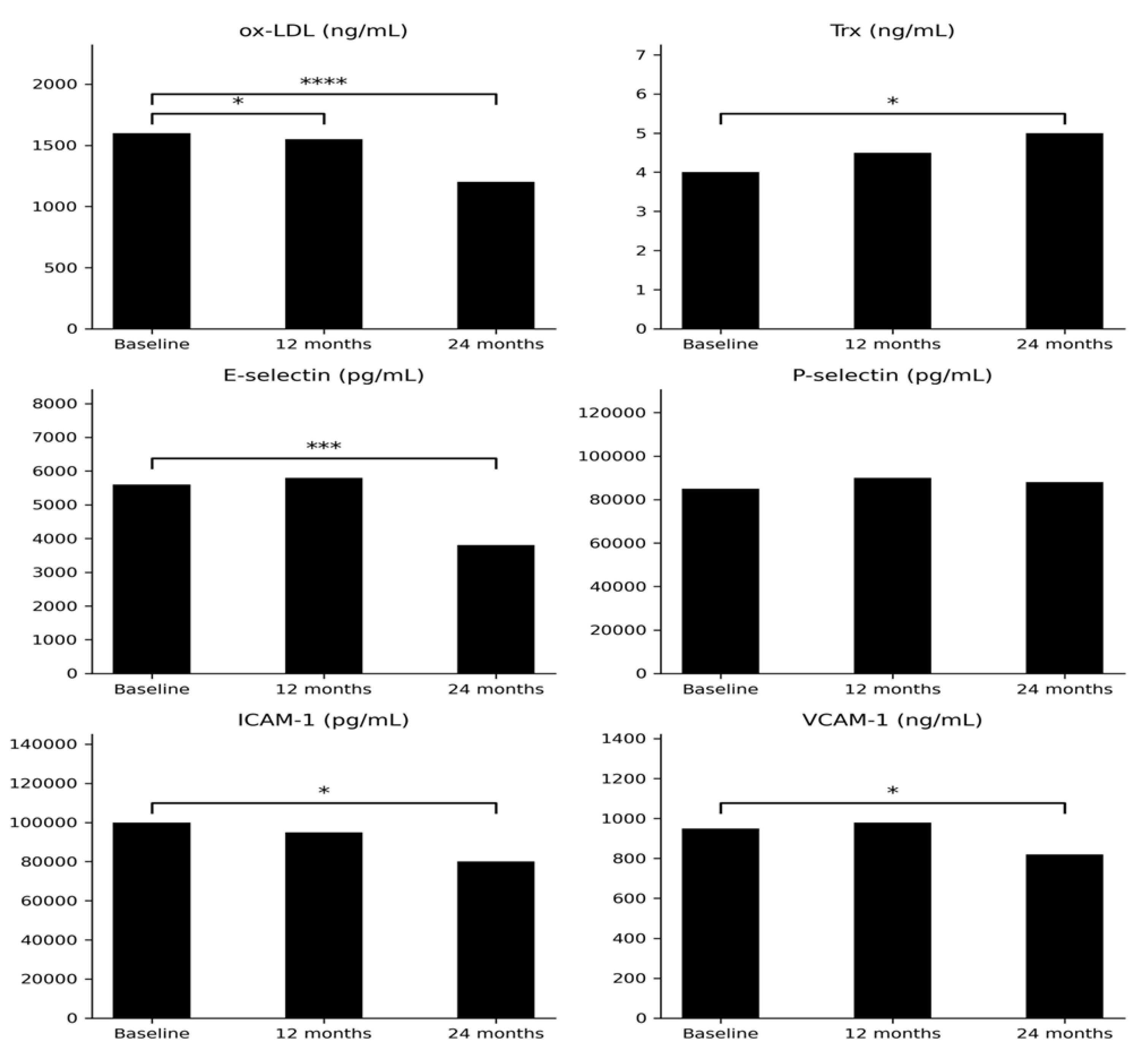

Table 1.
The investigated biochemical parameters in the studied group.
| Parameters | |||||
|---|---|---|---|---|---|
| Initially | After 12 mth | After 24 mth | p* value (0 vs 12 mth) |
p** value (0 vs 24 mth) |
|
| IGF-1 (ng/mL) | 47.07 (8.57-138.8) |
155.1 (36.04-265.1) |
153.19 (58.02-303.9) |
<0.001 | <0.001 |
|
Ox-LDL (ng/mL) |
1643 (1233-1910) |
1623 (1066-2154) |
1187 (919.5-1887) |
<0.05 | <0.00001 |
|
Trx (ng/mL) |
3.90 (2.66-6.88) |
4.59 (2.18-11.6) |
5.1 (2.7-10.2) |
0.38 | <0.05 |
|
E-selectin (pg/mL) |
5736 (1043-14414) |
5649 (2040-9500) |
3828 (717.4-7692) |
0.54 | <0.001 |
|
P-selectin (pg/mL) |
83493 (35819-147352) |
95089 (37656-188437) |
91296 (41620-121511) |
0.20 | 0.37 |
|
ICAM-1 (pg/mL) |
99269 (44598-130792) |
102415 (38572-146950) |
79565 (23092-129154) |
0.79 | <0.05 |
| VCAM-1 (ng/mL) | 953.8 (753.1-1258) |
1001 (761-1394) |
838.1 (532-993.7) |
0.62 | =0.05 |
|
OGG-1 (ng/mL) |
4.14 (2.53-5.7) |
5.42 (2.4-8.63) |
6.8 (3.7-12.8) |
0.65 | <0.05 |
|
Cholesterol (mg/dL) |
201 (114-302) |
199 (114-295) |
200.4 (114-302) |
0.69 | 0.13 |
|
LDL (mg/dL) |
126 (65-219) |
131 (58-216) |
127 (65-219) |
0.20 | 0.49 |
|
HDL (mg/dL) |
43 (24-85) |
50 (27-80) |
46.6 (24-79) |
0.20 | 0.09 |
|
TG (mg/dL) |
120 (51-684) |
120.5 (45-326) |
153.3 (51-684) |
0.67 | 0.35 |
Abbreviations: IGF-1: insulin like growth factor type 1; Ox-LDL: oxidized low-density lipoprotein, Trx; thioredoxin; VCAM-1: vascular cell adhesion molecule-1; ICAM-1: intercellular adhesion molecule-1; OGG-1: 8-oxoguanine DNA glycosylase 1; LDL: low-density lipoprotein; HDL: high-density lipoprotein; TG: triglycerides.
Table 2.
The investigated bioimpedance parameters in the studied group.
| Parameters | |||||
|---|---|---|---|---|---|
| Initially | After 12 mth | After 24 mth | p* value (0 vs 12 mth) |
p** value (0 vs 24 mth) |
|
| Total mass (kg) | 78.6 (39.6-167.3) |
78.0 (62.3-156) |
86.9 (59.8-167.3) |
0.51 | 0.05 |
| Tissue fat % | 37.5 (27.4-50.4) |
38.4 (26.7-48.7) |
39.7 (27.4-60.4) |
0.04 | 0.001 |
| Fat tissue (g) | 28434 (13891-82462) |
29937 (16939-67385) |
33868 (16881-82462) |
0.08 | 0.05 |
| Lean mass (g) | 48646 (23996-81228) |
45550 (36530-84977) |
50021 (34296-81228) |
0.49 | 0.0008 |
| BMC | 2547 (1261-3778) |
2568 (1770-3650) |
2731 (1844-3778) |
0.42 | 0.72 |
| L1-L4 BMD | 1.09 (0.8-1.6) |
1.1 (0.9-1.5) |
1.2 (0.9-1.6) |
0.73 | 0.04 |
| L1-L4 T score | -1.1 (-3.4-3.2) |
-0.3 (-2.0-2.4) |
-0.1 (-2.5-3.2) |
0.17 | 0.1 |
| L1-L4 Z score | -1.1 (-3.7-3.0) |
-0.9 (-2.3-2.0) |
-0.3 (-2.2-3.0) |
0.73 | 0.13 |
| Neck BMD | 0.95 (0.7-1.4) |
0.96 (0.77-1.5) |
1.04 (0.74-1.4) |
0.29 | 0.0002 |
| Neck T score | -0.8 (-2.1-2.3) |
-0.6 (-1.9-2.5) |
-0.63 (-2.1-2.3) |
0.79 | 0.92 |
| Neck Z score | -0.9 (-2.2-2.0) |
-1.0 (-2.0-2.3) |
-0.43 (-2.1-2.0) |
0.72 | 0.0018 |
Abbreviations: BMC: bone mineral content; BMD: bone mineral density; “-“: minus.
Table 3.
Spearman`s correlation coefficients between parameters and other metabolic markers in group initially and during the treatment.
Table 3.
Spearman`s correlation coefficients between parameters and other metabolic markers in group initially and during the treatment.
| Parameters | Initially | After 12 months | After 24 months |
|---|---|---|---|
| Ox-LDL (ng/mL) vs P-selectin (pg/mL) | NS | NS | p=0.02 R=0.60 |
| Ox-LDL (ng/mL) vs Lean (g) | NS | p=0.02 R=0.63 |
NS |
| E-selectin (pg/mL) vs VCAM-1 (ng/mL) | NS | NS | p=0.05 R=0.57 |
| E-selectin (pg/mL) vs HDL (mg/dL) | NS | NS | p=0.01 R=-0.67 |
| E-selectin (pg/mL) vs Waist (cm) | NS | NS | p=0.04 R=0.57 |
| Trx (ng/mL) vs Tissue (g) | NS | NS | p=0.04 R=0.71 |
| Trx (ng/mL) vs Vitamin D (mg/dL) | NS | NS | p=0.01 R=-0.69 |
| Trx (ng/mL) vs Fat (g) | NS | NS | p=0.01 R=0.71 |
| ICAM-1 (pg/mL) vs BMI (kg/m2) | NS | NS | p=0.04 R=0.55 |
| ICAM-1 (pg/mL) vs Waist (cm) | NS | p=0.03 R=0.58 |
p=0.04 R=0.57 |
| ICAM-1 (pg/mL) vs Hip (cm) | NS | NS | p=0.02 R=0.61 |
| ICAM-1 (pg/mL) vs LDL (mg/L) | NS | NS | p=0.04 R=-0.57 |
| P-selectin (pg/mL) vs BMI (kg/m2) | NS | NS | p=0.02 R=0.64 |
| P-selectin (pg/mL) vs Tissue (g) | NS | NS | p=0.03 R=0.63 |
| P-selectin (pg/mL) vs Fat (g) | NS | p=0.04 R=0.57 |
NS |
| VCAM-1 (ng/mL) vs glucose (mg/dL) | NS | NS | p=0.03 R=0.58 |
| LDL (mg/dL) vs L1-L4 Z-score | NS | NS | p=0.03 R=-0.58 |
| LDL (mg/dL) vs Lean (g) | NS | NS | p=0.01 R=-0.33 |
| HDL (mg/dL) vs IGF-1 (ng/mL) | NS | NS | p=0.02 R=-0.59 |
| HDL (mg/dL) vs Lean (g) | NS | p<0.01 R=-0.73 |
p=0.01 R=-0.68 |
| TG (mg/dL) vs L1-L4 T-score | NS | NS | p=0.03 R=0.60 |
| TG (mg/dL) vs L1-L4 Z-score | NS | NS | p=0.05 R=0.54 |
| Lean (g) vs L1-L4 BMD | NS | p<0.001 R=0.78 |
p=0.05 R=0.55 |
| Lean (g) vs L1-L4 T-score | NS | p=0.01 R=0.67 |
NS |
| Lean (g) vs L1-L4 Z-score | NS | p=0.04 R=0.53 |
NS |
| Lean (g) vs Neck BMD | NS | p<0.01 R=0.72 |
p=0.01 R=0.69 |
| Lean (g) vs Neck T-score | NS | p=0.02 R=0.68 |
p=0.03 R=0.60 |
| Lean (g) vs Neck Z-score | NS | p=0.01 R=0.67 |
NS |
Abbreviations: IGF-1: insulin like growth factor type 1; Ox-LDL: oxidized low-density lipoprotein, Trx; thioredoxin; VCAM-1: vascular cell adhesion molecule-1; ICAM-1: intercellular adhesion molecule-1; OGG-1: 8-oxoguanine DNA glycosylase 1; LDL: low-density lipoprotein; HDL: high-density lipoprotein; TG: triglycerides; BMC: bone mineral content; BMD: bone mineral density; “-“: minus; BMI: body mass index.
Table 4.
The investigated biochemical parameters in the studied group.
| Sex | Age (years) | Treatment (before rhGH) |
Dose of rhGH | Etiology GHD | IGF-1 (ng/ml) initially | BMI (kg/m2) initially |
CO-GHD in history | |
|---|---|---|---|---|---|---|---|---|
| P1 | F | 41 | HCT, L, D, Es/Pg | 0.5 mg | CPGP | 68.6 | 30.9 | + |
| P2 | M | 25 | L,T | 0.5 mg | NFPM | 62.8 | 24.8 | + |
| P3 | M | 18 | T | 0.4 mg | CPH | 27.3 | 22.8 | + |
| P4 | F | 26 | HCT, L, Es/Pg | 0.6 mg | CPH | 40.1 | 29.0 | + |
| P5 | M | 19 | D, L, T, HCT | 0.3 mg | CPGP | 74.8 | 34.9 | + |
| P6 | F | 60 | HCT, L | 0.4 mg | ES | 15.11 | 24.3 | - |
| P7 | M | 20 | L, HCT, T | 0.3 mg | CPH | 91.8 | 28.1 | + |
| P8 | M | 23 | - | 0.3 mg | I | 138.8 | 25.9 | - |
| P9 | F | 38 | L, HCT, Es/Pg | 0.5 mg | NFPM | 47.07 | 24.9 | - |
| P10 | M | 18 | T | 0.2 mg | I | 120.2 | 20.4 | + |
| P11 | M | 28 | L, HCT, T, D | 0.3 mg | CPGP | 22.6 | 27.1 | + |
| P12 | M | 42 | L, T, D | 0.3 mg | CPGP | 63.0 | 54.1 | + |
| P13 | M | 36 | HCT, L, T | 0.5 mg | CPGP | 48.9 | 21.5 | + |
| P14 | M | 18 | L, HCT, D, T | 0.7 mg | CPGP | 54.4 | 24.4 | + |
| P15 | M | 25 | L, HCT, T | 0.5 mg | CPGP | 8.6 | 35.8 | + |
Abbreviations: GHD: growth hormone deficiency; rhGH: recombinant human growth hormone; P: patient; F: female; M: male; HCT: hydrocortisone; L: levothyroxine; Es/Pg: estrogen/progesterone; D: desmopressin; T: testosterone; CPH: congenital pituitary hypoplasia; CPGP: craniopharyngioma postsurgical; ES: empty sella; NFPM: non-functioning pituitary macroadenoma; CO-GHD - childhood-onset growth hormone deficiency; I: idiopathic; IGF-1: insulin like growth factor type 1; BMI: body mass index.
Disclaimer/Publisher’s Note: The statements, opinions and data contained in all publications are solely those of the individual author(s) and contributor(s) and not of MDPI and/or the editor(s). MDPI and/or the editor(s) disclaim responsibility for any injury to people or property resulting from any ideas, methods, instructions or products referred to in the content. |
© 2026 by the authors. Licensee MDPI, Basel, Switzerland. This article is an open access article distributed under the terms and conditions of the Creative Commons Attribution (CC BY) license.
Copyright: This open access article is published under a Creative Commons CC BY 4.0 license, which permit the free download, distribution, and reuse, provided that the author and preprint are cited in any reuse.


