
Submitted:
30 December 2025
Posted:
31 December 2025
You are already at the latest version
Abstract
Background We hypothesize that galantamine extracted from Narcissus species may have a protective effect against organophosphate-neurotoxicity, with lower environmental impact.
ObjectiveIn vitro testing of the neuroprotective effect of these compounds onneuronal models exposed to an organophosphorus insecticide (diazinon), considering its acute neurotoxicity.
Materials and methodsCytotoxicity and protective effect of these compounds in concentrations close to toxic (60µg/ml) and therapeutic (12; 6µg/ml) ones were testedin vitrousing the MTT cell viability assay kit. Statistical analysis was used to test whether the differences between the cell viability of the groups were statistically significant.
ResultsThe toxicity values of natural galantamine were higher than those of the synthetic one in concentration of 60µg/ml (p=0.002) and comparable inconcentrations of 6 and 12µg/ml(p=0.06 and p=0.5).Highlighting of the neuroprotective effect by assessing cell viability in the case of exposure of the rat hippocampal neurons cell line to the organophosphorus compound in concentrations of 240 µg/mL, with pretreatment with the studied compounds, emphasized comparable values.
In case of exposure to lower concentrations of diazinonthe neuroprotective effect of natural galantamine at a concentration of 12μg/ml is higher than that of synthetic galantamine (p=0.03),andlower than that of the synthetic compound in the case of concentrations of 6 and 60μg/ml (p=0.009 and respectively p=0.002,).
Conclusions The protective effect offered by galantamine obtained from the N. poeticusextract was superior to the synthetic compound under experimental conditions at a concentration of 12μg/ml,with lower environmental impact.
Keywords:
preventive antidote
; galantamine
; Narcissus extracts
; organophosphates
1. Introduction
Neurotoxic organophosphorus compounds are a class of highly toxic substances with a structure of phosphoric acid esters [1]. They contain two identical or different organic residues and an inorganic or organic active group in the molecule, linked to a pentavalent phosphorus atom. In turn, the central pentavalent phosphorus atom may be bonded to an oxygen or sulfur atom (phosphates, phosphonates, thionphosphates, thionphosphonates)[1,2]. The main mechanism of action of neurotoxic organophosphorus compounds is the irreversible inhibition, through phosphorylation, of the active site of acetylcholinesterase (AChE), the enzyme responsible for the hydrolysis of acetylcholine at the synapses of the cholinergic system [3,4,5]. This results in an excess of acetylcholine at the synapses of the cholinergic system. The cholinergic system includes all synapses at which transmission is mediated by acetylcholine, as well as structures whose functionality is dependent on it [1,2,3]. Cholinergic mediation is characteristic for the following categories of synapses: synapses at effector sites that are innervated by parasympathetic postganglionic fibers; synapses at sympathetic and parasympathetic ganglia and chromaffin cells of the adrenal medulla, innervated by preganglionic autonomic nerve fibers; cholinergic synapses at the neuromuscular junction, innervated by somatic motor neurons; synapses at the level of the central nervous system (motoneurons in the anterior horns of the medulla) and the central nervous system (hippocampus, medial and lateral geniculate bodies of the thalamus, caudate nucleus, hypothalamus, auditory and visual cortex) [6,7,8,9].
Emergency medical countermeasures in case of exposure to neurotoxic chemical compounds consist in administrating preventive (pyridostigmine) and curative treatment (obidoxime, pralidoxime, HI-6), and anticonvulsant compounds (benzodiazepines: midazolam, diazepam, avizafone) [10,11,12,13,14,15].
Preventive treatment consists in the administration of pyridostigmine, a reversible acetylcholinesterase inhibitor which, administered pre-exposure, can protect the enzyme [16,17,18]. This is the standard option, which, in combination with curative antidotes, is able to increase their therapeutic efficacy and thus survival [19,20,21]. Due to low lipophilicity and low oral bioavailability, pyridostigmine cannot cross the blood-brain barrier, thus being unable to antagonize the neurotoxic effects of organophosphorus compounds [22,23,24]. Also, the narrow therapeutic window allows toxic effects to occur at doses that are closer to therapeutic ones [25,26].
Studies in the literature have shown that galantamine, a reversible cholinesterase inhibitor with neuroprotective properties due to its ability to cross the blood-brain barrier, could be a superior therapeutic alternative to pyridostigmine in neurotoxic chemical intoxication [27,28,29].
Galantamine is approved by the EMA and FDA for the treatment of mild and moderate forms of Alzheimer's disease and has proven efficacy and safety [30,31,32,33,34,35]. The commercial products known as Reminyl, Galantamine or Galsya are packaged as 4, 8 and 16 mg capsules, or as oral solution; they are obtained by chemical synthesis with rather high production costs [33,34,35,36,37]. A less costly alternative could be to obtain galantamine from natural extracts [38,39,40,41], an environmentally friendly method that follows the principles of green chemistry [42].
In this general context, we hypothesise that galantamine extracted from Narcissus species may have a protective effect (compared to the synthetic version) against organophosphate-induced toxicity, with lower environmental impact.
The objectives of the experimental study consist in the identification and quantification of galantamine obtained from natural extracts belonging to various species of Narcissus, Amaryllidacae family, and in vitro testing of its toxicity and therapeutic efficacy compared to the synthetic compound containing galantamine.
2. Materials and Methods:
2.1. Materials and Reagents
- Galantamine standard, product of MERCK Germany, purity 99.8%, CAS No 1953-04-4;
- Diazinon PESTANAL, product of MERCK Germany, purity 99%, CAS No 33-41;
- Acetonitrile standard HPLC, product of MERCK Germany, purity 99.5%, CAS No 75-05-8;
- Trifluoroacetic acid standard, product of MERCK Germany, purity 99%, CAS No 76-05-1;
- Formic acid standard HPLC, product of MERCK Germany, purity 98.9%, CAS No 64-18-6;
- Ethanol gradient grade, product of MERCK Germany, purity 99.8 %, CAS No 64-17-15;
- Methanol standard HPLC, product of MERCK Germany, purity 99.9%, CAS No 67-57-15;
- MTT cell proliferation assay kit produced by Sigma Aldrich Germany, product number CT 01.
2.2. Cell Lines
- NIH3T3 mouse embryonic fibroblast cell line, product of ATCC-USA catalogue number CRL-1658;
- Rat neuronal cell line (rat hippocampal cell line), product of Lonza Bioscience Switzerland catalogue number R HY-501.
2.3. Equipment
- Arc HPLC high-performance liquid chromatography system (Waters, Milford, MA, USA);
- En Sight™ Multimode Microplate Reader (Perkin Elmer USA);
- Centrifugal partition chromatography system (Armen, France).
2.4. Methods
The natural dried extracts were prepared from bulbs of controlled origin from various species of Narcissus: Narcissus poeticus (coded as sample P4), Narcissus pseudonarcissus (coded as sample P5), and Narcissus double (coded as sample P6).
These extracts were selected because they displayed a significant galantamine content in previous experimental studies. The dried version is closer to the future conditioning form of the preventive antidote. The natural dried extracts were obtained using two methods:
- Maceration (MAC). The method consists in keeping the samples for 3 days at room temperature, with occasional shaking;
- Ultrasound assisted extraction (UAE) using a Bradsonic MH1800 bath at 50°C.
2.5. Identification of Galantamine from Dried Natural Extracts Using HPLC Analysis
HPLC-PAD analysis was carried out using an Arc HPLC High Performance Liquid Chromatography System (Waters, Milford, MA, USA).
2.6. Reaction Conditions of the Chromatographic System
- A Symmetry C18 column (250 х 4.6 mm, particle size 5 μm – Waters, Milford, MA, USA) was used, with the oven temperature set at 30 °C.
- The injection volume was 20 μL.
- The mobile phases were: (A) 0.1% formic acid in water and (B) 0.1% formic acid in acetonitrile.
-The elution gradient was 2-15% mobile phase B for 10 min and the elution flow rate was 1 mL/min.
- A wavelength of 288 nm was used for the detection and quantification of galantamine.
- The retention time in each sample was compared with the retention time of the galantamine standard, obtained at 5.8 min.
2.7. Advanced Natural Galantamine Isolation Tests Using Centrifugal Partition Chromatography
Centrifugal partition chromatography (CPC), also known as counter-current chromatography (CCC) or preparative HPLC, is a technique used in pilot studies whose main purpose is the quantitative isolation of molecules from complex mixtures. The novelty of this technique is that CPC does not require the solid stationary phase column, but uses a mixture of immiscible liquids which function as the mobile and stationary phases. One liquid serves as the mobile phase or eluent, while the other as the stationary phase. The stationary phase is retained in the rotor through the application of a centrifugal field. The Centrifugal Partition Chromatography (CPC) system used in the study was a SCPC-250-B equipment (Armen, France).
Method and conditions of reaction within the operating framework of the CPC system:
- Loading the column with 100% of the upper stationary phase was performed in descending mode; the reaction conditions of this phase were as follows: volume 30 mL/min, speed 500 rpm, and time: 10 min;
- Balancing the column with 100% of the lower mobile phase was performed in descending mode; the reaction conditions of this phase were as follows: volume: 8 mL/min, speed 1600 rpm, and time: 15 min; injection volume – 2 mL of extract;
- Lower mobile phase elution was performed in descending mode;
- Galantamine detection wavelength: 288 nm;
- Extrusion with upper stationary phase volume: 30 mL/min, time: 12 min.
In vitro tests for highlighting the galantamine-induced cytotoxicity and its protective effect in case of exposure to neurotoxic organophosphorus compounds.
The experimental protocol follows the insert attached to the MTT cell viability kit; they were performed according to the regulations of ISO Standard 10993-5/2009 with the approval of the Institutional Ethics Committee no 437/24.08.2024.
Working solutions were prepared as follows:
- Diazinon PESTANAL – organophosphorus insecticide, irreversible inhibitor of acetylcholinesterase, similar in structure and toxicological properties to neurotoxic chemical compounds, from which solutions were prepared in concentrations of 240 and 24 µg/mL. These concentrations were considered in literature to be close to the median lethal in vitro concentration of this toxic compound, and a fraction (1/10) of it.
- Galantamine – obtained from the dried extract of N. poeticus, from which solutions were prepared in concentrations close to toxic (60 µg/ml) and therapeutic (12, 6 µg/ml) ones.
- Galantamine obtained through chemical synthesis (Sigma – Germany product), from which solutions were prepared in the same concentrations.
Cell lines used:
The cell lines used in the study are correlated with the main toxicity of cholinesterase inhibition (NIH/3T3 mouse embryonic fibroblast cell line) and the specific acute neurotoxicity of organophosphorus insecticides (the rat hippocampal neuron R HY – 501cell neuron line), the class to which diazinon belongs. The hippocampal neuron cell line was chosen because it is known in the literature that in this region there is a large number of cholinergic synapses, with acetylcholine as the neurotransmitter.
- NIH/3T3 mouse embryonic fibroblast cell line, product of ATCC USA
The fibroblast cell line was cultured in 25 cm2 cell culture flasks, using Dulbecco's Modified Eagle's Medium (DMEM) supplemented with 10% fetal bovine serum (FBS) and 50 μg/mL of gentamicin. The cells in 25 cm2 culture flasks were subjected to trypsinization with 0.025% Trypsin-EDTA and centrifuged, and the pellet was resuspended in 5 mL complete DMEM medium. The cell suspension was cultured on a microplate with 96 flat-bottomed wells, 200 μL/well (2.14x10⁵ cells/well) and incubated in the CO2 incubator at 37 0C for 24 hours with the studied compounds and diazinon. 20 μL MTT medium/well (10% of the volume of medium) was added, followed by incubation for 4 hours at 37° C. After the 4 hours, the microplate was removed from the incubator, the medium was removed, and the MTT solvent was added. The optical density was read at a wavelength of 570 nm, within maximum one hour from the addition of the solvent, using the multimode reader (En Sight™ Multimode Microplate Reader, PerkinElmer).
-The rat hippocampal neuron R HY-501 cell line
It was cultured (according to the protocol in the insert) directly on cell microplates with 96 wells specially treated with poly-D-lysine. The microplate cultivation was performed using a concentration of 2x10⁵ cells/well and was followed by incubation in CO2 atmosphere (5%) at 37° C for 96 hours for optimal cell adhesion.
Subsequently, the galantamine solutions obtained from the extract and using chemical synthesis were applied to the plates and the MTT cell proliferation kit was used.
The samples were read in triplicate (n=3). Standard deviation was calculatedThe results were expressed as percentages. Statistical analysis was used to test whether the differences between the cell viability of the groups were statistically significant.
3. Results and Discussions
Obtaining dried natural extracts from bulbs of controlled origin from the Amaryllidaceae species: Narcissus poeticus (hereinafter referred to as sample P4), Narcissus pseudonarcissus (hereinafter referred to as sample P5) and Narcissus double (hereinafter referred to as sample P6). The comparative results are shown in Table 1.
The sample of Narcissus poeticus (P4) exhibited the highest amount of dry extract (10.65 g/100g bulbs) using the maceration technique. This extract will be used to make the preventive antidote.
3.1. Identification of Galantamine in Dried Natural Extracts Using the HPLC Analysis
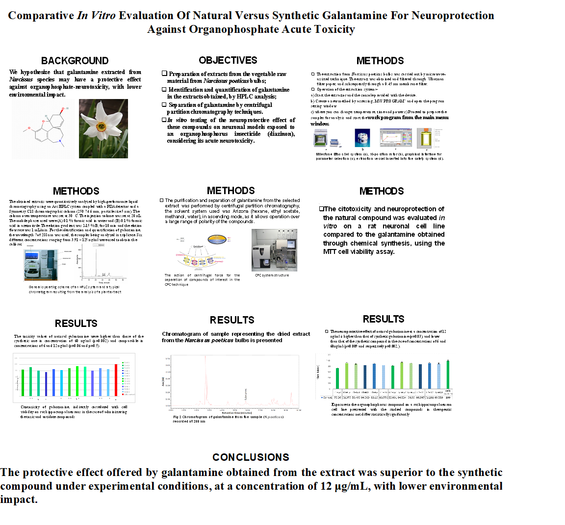
Six different concentrations ranging from 3.91–125 μg/mL were used to obtain the calibration curve and regression equation (y = 1.09x +2.8343). The linearity showed a correlation coefficient (R2) value of 0.99998. In the case of galantamine, the value of the limit of detection (LOD) was 1.012 μg/mL, and the value of the limit of quantification (LOQ) was 3.068 μg/mL. A chromatographic analysis was performed in triplicate for each sample. The retention time was 5.8 minutes. The identification of the parent compound was carried out using mass spectrometry (MS – mode associated with the used HPLC system). The chromatogram of the reference standard of synthetic galantamine is presented in Figure 1.
The chromatogram of sample P4, representing the dried extract from the Narcissus poeticus bulbs obtained by maceration (MAC) is presented in Figure 2.
The identification of the compound with a similar retention time to that of the galantamine standard was carried out using mass spectrometry
The results of the HPLC quantification of galantamine from Narcissus extracts obtained by maceration and ultrasound-assisted extraction methods are shown in Table 2.
3.2. Advanced Isolation of Galantamine Using Centrifugal Partition Chromatography
Using this technique in the case of the Narcissus poeticus extract, the separation of galantamine was performed in the time range of 6.8-12 minutes. When the galantamine peak occurred, the separation and collection of the respective fraction in the above-mentioned time range was initiated. Subsequently, identification in fractions collected at the initial (6.8 min) and final (10 min) times, by using HPLC analysis of galantamine, was performed.
The percentage of recovery of galantamine separated in the collection fraction was calculated (Table 3).
Testing the cytotoxicity indirectly correlated with cell viability of galantamine obtained from natural extracts and that obtained by chemical synthesis on NIH3T3 mouse embryonic fibroblast cell line by applying MTT cell viability kit (Figure 3)
The mean value of the cytotoxicity of diazinon was 44.37%±0.3 for the concentration of 240 μg/mL and 53.46%±0.23 for the concentration of 24μg/mL.
The mean values of cell viability indirectly correlated with cytotoxicity were the following: at a galantamine concentration of 60 μg/ml, the average value of natural galantamine obtained from the dry extract of Narcissus poeticus extract was 88.63%±0.3, while for galantamine obtained by chemical synthesis it was 91.47%±0.2; the concentration of 12 μg/ml, highlights a cell viability average value of 97.77%±0.8 in the case of the natural compound and 97.27%±0.7 in the case of galantamine obtained by chemical synthesis; the average value of cell viability of natural galantamine atconcentrations of 6 μg/ml was 99.79%±0.2; in the case of galantamine obtained by chemical synthesis, the average value of cell viability was 98.31%±0.4.
The mechanism of the organophosphorus compound Diazinon’s toxicity was similar to that of the chemical neurotoxic agents with military use: inhibition of acetylcholinesterase, oxidative aggression and apoptosis.
Dry extracts were obtained from the bulbs of Amaryllidaceae, namely three Narcissus species (Narcissus poeticus, Narcissu. pseudonarcissus, Narcissus double) using maceration and ultrasound-assisted technique.
Quantification by HPLC analysis revealed that the dry extracts obtained by maceration showed a higher concentration of galantamine than those obtained by ultrasound-assisted extraction.
The dry extract from Narcissus poeticus showed higher galantamine values than those in Narcissus pseudonarcissus and Narcissus double.
The recovery of galantamine from the fraction collected from the extract of Narcissus poeticus, using the technique of partition chromatography, was achieved almost completely.
When discussing cytotoxicity, indirectly correlated with cell viability, of galantamine from the dry extract and that of galantamine obtained by chemical synthesis by testing the cell viability using the MTT cell proliferation test on NIH3T3 mouse embryonic fibroblast cell lines, the following aspects should be considered, in accordance with the statistical analysis: toxicity of galantamine obtained from Narcissus poeticus extract, in concentrations of 60 μg/mL, is higher than that of galantamine obtained by chemical synthesis (p=0.003); mean values of natural galantamine toxicity in concentrations of 6 and 12μg/ml, did not differ in a statistically significant manner from those of the chemical compound (p=0.07);
Evaluation, on NIH3T3 mouse embryonic fibroblast cell line, of the protective effect of galantamine obtained from natural extracts compared to that obtained by chemical synthesisby applying MTT test (Figure 4).
The evaluation of the protective effect directly correlated with cell viability, in case of exposure to the organophosphorus compound in concentrations of 240 μg/mL on mouse fibroblasts pretreated with the studied compounds in different concentrations, highlighted the following aspects: at concentrations of 60 μg/mL, the mean value of cell viability was 83.05%±0.52 in the case of galantamine obtained from Narcissus poeticus extract and 75.30%±0.45 in the case of galantamine obtained by chemical synthesis, in the case of concentrations of 12 μg/mL, the mean value of cell viability was 90.64%±0.43 for natural galantamine and 84.62%±0.26 for galantamine obtained by chemical synthesis; at concentrations of 6 μg/mL, the mean value of cell viability was 80.59%±0.33 in the case of galantamine from dry extract and 81.63%±0.54 in the case of galantamine obtained by chemical synthesis.
The evaluation of the protective effect, in case of exposure to the organophosphorus compound in concentrations of 24 μg/mL of mouse fibroblasts pretreated with the studied compounds highlighted the following aspects: at concentrations of 60 μg/mL the mean value of cell viability was 87.72%±0.33; in the case of galantamine obtained from Narcissus poeticus extract and 80.47%±0.18; in the case of galantamine obtained by chemical synthesis, the mean value was 94.29%±0.72 for natural galantamine and 87.84%±0.50 in the case of synthetic one, at concentrations of 12 μg/mL; at concentrations of 6 μg/mL, the average value of cell viability was 92.65%±0.53 for natural galantamine and 85.1%±0.82 in the case of galantamine obtained by chemical synthesis.
In the case the NIH3T3 mouse embryonic fibroblast cell line that was pretreated with the antidote compounds and exposed to the organophosphorus compound in concentrations of 240 µg/mL, which is close to lethal toxic concentration. The protective effect was emphasized by the following results in accordance with statistical analysis: galantamine in the dry extract, at a concentration of 12 µg/mL, resulted in higher cell viability and, implicitly, higher protective effect than that of galantamine obtained by chemical synthesis (p=0.003); due to the fact that the extract of Narcissus. poeticus may contain, in addition to galantamine, others antioxidants that could potentiate its activity. the protective effect did not differ significantly between the two compounds in concentration of 6 µg/mL (p=0,07); galantamine in the dry extract, at a concentration of 60 µg/mL, resulted in higher cell viability and protective effect than that of galantamine obtained by chemical synthesis (p=0,002) but it is more toxic; therefore, this concentration could not represent a starting point for an antidote formulation option.
In case of exposure to the organophosphate in concentrations of 24 µg/mL, representing values ten times lower than the lethal toxic concentrations, the following should be noted: galantamine in the dry extract at a concentration of 6,12 and 60 µg/mL resulted in higher protective effect than that of galantamine obtained by chemical synthesis;
-galantamine in the dry extract at a concentration of 60 µg/mL resulted in higher cell viability than galantamine obtained by chemical synthesis.
The protective effects of galantamine in the case of these concentrations of diazinon cannot therefore constitute a starting point for future research, because protection at the highest dose of the toxic compound is considered effective.
Emphasis of the cytotoxicity indirectly correlated with cell viability of galantamine from the dry extract and that of galantamine obtained by chemical synthesis by testing cell viability using the MTT cell viability test on rat neuronal cell line (R HY -501-rat hippocampal cell line) (Figure 5).
The mean value of cytotoxicity of galantamine obtained from dry extract at concentrations of 60 μg/mL was 74.09%±0.32; the mean value of galantamine obtained by chemical synthesis was 81.30%±0.15.the mean value of cytotoxicity of natural galantamine in concentrations of 12 μg/mL was 84.67%±0.46, the mean value of galantamine obtained by chemical synthesis was 84.57%±0.6. The mean value of cytotoxicity of natural galantamine in concentrations of 6 μg/mL was 91.63%±0.07. The mean value of cell viability of galantamine obtained by chemical synthesis was 88.90%±0.55. The results revealed that: the toxicity of galantamine obtained from natural extract in concentrations of 60 μg/mL is higher than that of galantamine obtained by chemical synthesis (p=0.002); the toxicity of galantamine obtained from Narcissus poeticus extract in concentrations of 6 and 12 μg/mL is comparable to that of galantamine obtained using chemical synthesis ( the average values did not differ in a statistically significant manner for concentrations of 6 and 12 µg/mL (p=0.06 and respectively p=0.5;).
Emphasis of the neuroprotective effect of galantamine from the natural extract and that obtained using chemical synthesis by using the MTT cell proliferation test on a rat neuronal cell line (R HY -501-rat hippocampal cell line) is represented in Figure 6.
The evaluation of the neuroprotective effect in the case of exposure to the organophosphorus compound in concentrations of 240 μg/mL on rat hippocampal neurons cell line, which was pretreated with the antidote compounds in concentrations of 60 μg/mL, revealed mean values of cell viability of 73.36%±0.2 (galantamine from natural extract) and 84.06±0.5 (galantamine obtained through chemical synthesis).
Exposure to the organophosphorus compound in concentrations of 240 μg/mL in a rat hippocampal neurons cell line pretreated with the antidote compounds in concentrations of 12 μg/mL revealed mean values of cell viability of 91.37%±0.4 (galantamine from natural extract) and 89.16 ± 0.5 (galantamine obtained by chemical synthesis). The same concentration of organophosphate in rat hippocampal neurons cell line pretreated with the antidotes in concentrations of 6 μg/mL revealed mean values of cell viability of 88.46%±0.53 (galantamine from natural extract) and 83.77±0.42 (galantamine by chemical synthesis). In the case of exposure to the organophosphorus compound in concentrations of 24 μg/mL on rat hippocampal neurons cell line pretreated with the antidote compounds in concentrations of 60 μg/mL revealed mean values of cell viability of 82.03%±0.61 (galantamine from natural extract) and 86.84±0.43 (galantamine by chemical synthesis). Exposure to the same concentration of organophosphorus compound of a rat hippocampal neurons cell line pretreated with the antidote compounds in concentrations of 12 μg/mL revealed mean values of 94.83%±0.20 for galantamine from natural extract and 91.29±0.15 for the synthetic one. In case of pretreatment at concentration of concentration of 6 μg/mL the main values of cell viability were 90.55%±0.43 in case of galantamine from natural extract and 89.55±0.62 for galantamine using chemical synthesis.
A cell line of hippocampal neurons was chosen for the study, as this region of the paleocortex is known to contain a high number of cholinergic synapses and high concentrations of the cholinergic neurotransmitter acetylcholine. The central acetylcholinesterase, involved in the degradation of acetylcholine, is considered the major mechanism of the immediate neurotoxicity of organophosphorus insecticides. Highlighting of the neuroprotective effect by assessing cell viability in the case of exposure of the rat hippocampal neurons cell line to the organophosphorus compound in concentrations of 240 µg/mL, with pretreatment with the studied compounds, at concentration of 12 µg/mL emphasized comparable values of the neuroprotective effect in the case of the two studied compounds (p=0.4)
The evaluation of the protective effect by assessing cell viability in the case of exposure of the rat hippocampal neurons cell line to the organophosphorus compound in concentrations of 24 µg/mL, with pretreatment with the studied compounds, emphasized the following aspects: the protective effect offered by natural galantamine at concentration of 12 μg/mL is higher than that of the synthetic one (p=0.03). These values were lower than those of the synthetic compound in the case of concentrations of 6 and 60 μg/mL, (p=0.009 and p=0.002, respectively).
4. Conclusions
-The mean percentage values of cell viability induced on human fibroblasts and rat neuronal cell by the two concentrations of the organophosphorus compound, tested and pretreated with the three concentrations of galantamine extracts, are correlated in direct proportion to the concentrations used.
-Neuronal cells in the hippocampus are more sensitive than fibroblasts to the action of the organophosphorus compound and treatment.
-High concentrations of galantamine from dry extracts are more toxic than concentrations of galantamine obtained through chemical synthesis.
-The protective effect offered by galantamine obtained from the extract was superior to the synthetic compound under experimental conditions, at a concentration of 12 μg/mL, with lower environmental impact.
Author Contributions
Conceptualization, CGS, CAS, MST, Data curation, CAS, CGS, DMP, AS, CVP, MG, MST, Formal analysis, BC, MDC, FT, AS, CVP; Investigation, CGS, CAS, MG, MST; Methodology, DMP, MDC, FT, CVP, MG Project administration, CAS; Software, AS; Validation, CGS, BC, MDC, FT, MSTand; Writing – original draft, CGS, CAS, MST.
Funding
The project was financed from the budget of the "CHEMPREVENT" project within the Research and Development Plan of Ministry of Defence.
Conflicts of Interest
The authors declare no conflict of interest.
References
- K. Sinha and A. Sharma, Organophosphate poisoning a review, Medical Journal of Indonesia, 2003 vol. 12, no. 2, p. 120.
- S. M. Bester, M. A. Guelta, J. Cheung et al., Structural insights of stereospecific inhibition of human acetylcholinesterase by VX and subsequent reactivation by HI-6, Chemical Research in Toxicology, 2018 vol. 31, no. 12, pp. 1405–1417. [CrossRef]
- Mirjana Colovic, Tamara Lasarevicz Pasti, Daniela Carstic et al.: Acetylcholinesterase Inhibitors: Pharmacology and Toxicology. Current Neuropharmacology · 2013 May, 11(3) 315-35. [CrossRef]
- J. Misik, Ruzena Pavlikova, Jiri Cabal, et al. Acute toxicity of some nerve agents and pesticides in rats Drug Chem. Toxicol. 2014, Vol.38: 32-36. [CrossRef]
- Organisation for the prohibition of chemical weapons, 2019. https://www.opcw.org/.
- Maria Alozi, Mutasem Rawas-Qalaji: Treating organophosphates poisoning: management challenges and potential solutions Crit Rev Toxicol. 2020, 50:764-779. [CrossRef]
- Mona A. H. Yehia, Sabah G. El-Banna, Aly B. Okab: Diazinon toxicity affects histophysiological and biochemical parameters in rabbits Experimental-and-toxicologic-pathology 2007 vol 59 issue 3-4, p. 215-225.
- Dyah Ayu O. A., Zulfa Aulia Aulanni, Fajar Sotiq Permata: The study of organophosphates Diazinon toxicity toward liver histopathology and malonaldehyde serum levels on rats. Veterinary Biomedical and Clinical Journal Vet Bio Clin J. 2019 Vol. 1, 15. [CrossRef]
- Saeed Samarghandian, Tahereh Farkhondeh, Shahnaz Yousefizadeh: Toxicity Evaluation of the Subacute Diazinon in Aged Male Rats: Hematological Aspects. Cardiovasc Hematol Disord Drug Targets. 2020, vol 20, 198-201. [CrossRef]
- Elspeth J., Hulse James, D. Haslam, Stevan R. Emmet, Tom Woolley: Organophosphorus nerve agent poisoning: managing the poisoned patient British Journal of Anaesthesia 2019 Volume. 123: 457-463. [CrossRef]
- Sakib Aman, Shrebash Paul, Fazle Rabbi Chowdhury: Management of Organophosphorus Poisoning: Standard Treatment and Beyond. Crit Care Clin. 2021, Vol 37:673-686. [CrossRef]
- Aroniadou-Anderjaska, V. Apland, J. P. Figueiredo, T.H. De Araujo Furtado, Braga, M. F. Acetylcholinesterase inhibitors as weapons of mass destruction: History, mechanism of action, and medical countermeasures. Neuropharmacology. 2020, Vol 15, 181 108298. [CrossRef]
- Lukas Gorecki: Ondrej Soukup, Jan Korabecn: Countermeasures in organophosphorus intoxication: pitfalls and prospects Trends in Pharmacological Sciences 2022 Volume 43 pag. 593-606. [CrossRef]
- Bajgar J., Fusek J., Hrdina V., Patocka J., Vachek J. Acute toxicities of 2-dialkylaminoalkyl-(dialkyamido)-fluoro-phosphates. Physiol. Res. 1992; Vol 41 pag 399–402.
- Kamil Kuca, Jorge AlbertoValle da Silva, Eugenie Nepovimova, Ngo Pham , Wenda Wu, Martin Valis, Qinghua Wu, Tanos Celmar: Pralidoxime-like reactivator with increased lipophilicity - Molecular modeling and in vitro study Chemico-Biological Interactions 2023 Volume 385, 1. [CrossRef]
- Trond Myhrer, Pål Aas: Pretreatment and prophylaxis against nerve agent poisoning: Are undesirable behavioral side effects unavoidable? J.neubiorev. 2016 Volume 71, pag. 657-670. [CrossRef]
- Nikolina Maček Hrvat, Zrinka Kovarik: Counteracting poisoning with chemical warfare nerve agents Arh Hig Rada Toksikol. 2020, Vol 31 pag 266-284. [CrossRef]
- Lorke, D. E., Petroianu, G. A: Reversible cholinesterase inhibitors as pretreatment for exposure to organophosphates a review. J. Appl. Toxicol. 2018 vol 39 issue 1. Pag. 101-116. [CrossRef]
- Petroianu G. A., Nurulain S.M., Hasan M.Y., et al. Reversible cholinesterase inhibitors as pre-treatment for exposure to organophosphates: assessment using azinphos-methyl. J. Appl. Toxicol. 2014 Vol 35 issue 5 pag 493-499. [CrossRef]
- Amourette C., Lamproglou I., Barbier L., et al.: Gulf War illness: Effects of repeated stress and pyridostigmine treatment on blood-brain barrier permeability and cholinesterase activity in rat brain. Behav. Brain Res. 2009, 203, pag. 207-214. DOI. 10.1016/j.bbr.2009.05.002.
- Dames M. Madsen, Charles G. Hurst, Roger Macintosh, Dames A. Romano: Clinical Considerations in the Use of Pyridostigmine Bromide as Pretreatment for Nerve-agent Exposure U.S. Army Medical Research Institute in Chemical Defense Usamricd- SP-03-01 2003 pag 1-35. [CrossRef]
- Dietrich E. Lorke, Syed M. Nurulain, Mohamed Y. Hasan, Kamil Kuča, Georg A. Petroianu Oximes as pretreatment before acute exposure to paraoxon Journal of Applied Toxicology Vol 39 issue 11 pag 1505-1515 . [CrossRef]
- Xia, D. Y., Wang L. X., Pei S. Q The inhibition and protection of cholinesterase by physostigmine and pyridostigmine against Soman poisoning in vivo Fundam. Appl. Toxicol. Volume 1, Issue 2, March 1981, Pages 217–221. [CrossRef]
- Wigenstam, E. Artursson, A. Bucht, L.Thors: Pharmacological prophylaxis with pyridostigmine bromide against nerve agents adversely impact on airway function in an ex vivo rat precision-cut lung slice model Toxicology Mechanisms and Methods 2023 Volume 33, 9:732-740. [CrossRef]
- Walday P., A. P., Haider T, Fonnum,F.: Effect of pyridostigmine pre-treatment, HI-6 and Toxogonin treatment on rat tracheal smooth muscle response to cholinergic stimulation after organophosphorus inhalation exposure. Arch. Toxicol. 1993 67:212-219. [CrossRef]
- L. Brunton, J. S. Lazo, and I. L. O. Buxton, Goodman &Gilman’s The Pharmacological Basis of Therapeutics, McGraw-Hill, New York, NY, USA, 14th edition, 2022. [Google Books].
- T. H. Indu, D. Raja, B. Manjunatha, S.Ponnusankar: Can Galantamine act as an antidote for organophosphate poisoning? A Review. JSS Medical College, Indian J Pharm Sci 2016;78(4):428-435 20165700151041721000136,10.4172/pharmaceutical-sciences.1000136.
- Gabriella Marucci, Michela Buccioni, Diego Dal Ben, Catia Lambertucci, Rosaria Volpini: Francesco Amenta Efficacy of acetylcholinesterase inhibitors in Alzheimer's disease. Neuropharmacology 2021, 1:190. [CrossRef]
- Zueva I., José Dias, Sofya Lushchekina, et al.: New evidence for dual binding site inhibitors of acetylcholinesterase as improved drugs for treatment of Alzheimer’s disease. Neuropharmacology. 2019 DOI 155:131-141. 10.1016/j.neuropharm.05.025.
- Malcolm Lane, D Arice Carter, Joseph D. Pescrille, et al.: Oral Pretreatment with Galantamine Effectively Mitigates the Acute Toxicity of a SupraLethal Dose of Soman in Cynomolgus Monkeys Post-treated with. Conventional Antidotes Journal of Pharmacology and Experimental Therapeutics. 2020, Vol 375(1) pag. 115-126. [CrossRef]
- 31 Rama Rao Golime, Meehir Palit, J Acharya, D K Dubey: Neuroprotective Effects of Galantamine on Nerve Agent-Induced Neuroglial and Biochemical Changes Neurotox Res. 2018, 33:738-748. [CrossRef]
- Farlow M: Clinical pharmacokinetics of galantamine. Clin Pharmacokinet. 42:1383-1392. [CrossRef]
- Van Beijsterveldt L, Geerts R., Verhaeghe T., Willems B., Bode W., Lavrijsen K., Meuldermans W: Pharmacokinetics and tissue distribution of galantamine and galantamine-related radioactivity after single intravenous and oral administration in the rat. Arzneimittelforschung. 01 May 1991, 30(5):447-455. PMID: 1865992. [CrossRef]
- Alexander, M.Ajazuddin, R.J. Patel, S.SarafRecent expansion of pharmaceutical nanotechnologies and targeting strategies in the field of phytopharmaceuticals for the delivery of herbal extracts and bioactives J. Contr. Release, 241(2016), pp.110-124. [CrossRef]
- Marta Pérez-Gómez Moreta, Natalia Burgos-Alonso, María Torrecilla, José Marco-Contelles, Cristina Bruzos-Cidón: Efficacy of Acetylcholinesterase Inhibitors on Cognitive Function in Alzheimer's Disease. Review of Reviews Biomedicine Nov. 2021, Nov 15;9(11):1689. [CrossRef]
- Edna F, R Pereira: Yasco Aracava, Manickavasagom Alkondon, Miriam Akkerman, Istvan Merchenthaler, Edson X Albuquerque: Molecular and cellular actions of galantamine: clinical implications for treatment of organophosphorus poisoning. J Mol Neurosci. 2010, 40:196-203. [CrossRef]
- Vaibhav Rathi-Galantamine Naturally Occurring Chemicals Against Alzheimer's DiseaseChapter3.1.4 2021:83-92. [CrossRef]
- Aarti Yadav, Surender Singh Yadav, Sandeep Singh: Rajesh Dabur Natural products: Potential therapeutic agents to prevent skeletal muscle atrophy European Journal of Pharmacology. 2022, 15:174995. [CrossRef]
- Yu Pong Ng, Terry Cho Tsun Or, Nancy Y: Plant alkaloids as drug leads for Alzheimer's disease Neurochemistry International Volume 2015 89: 260-270. [CrossRef]
- Michael Heinrich, Hooi Lee Teoh: Galanthamine from snowdrop the development of a modern drug against Alzheimer’s disease from local Caucasian knowledge Journal of Ethnopharmacology 2004 Volume. 92:147-162. [CrossRef]
- Rezaul Islam, Shopnil Akash, Mohammed Murshedul Islam, Nadia Sarkar, Ajoy Kumer, Sandip Chakraborty, Kuldeep Dhama, Majed Ahmed Al-Shaeri, Yasir Anwar, Polrat Wilairatana, Abdur Rauf, Ibrahim F. Halawani, Fuad M. Alzahrani, Haroon Khan: Alkaloids as drug leads in Alzheimer's treatment: Mechanistic and therapeutic insights Brain Research Volume. 1834,1:2024-148886. [CrossRef]
- J. Egea.D. Martín-de-Saavedra, E. Parada, A. Romero , L. del Barrio, A.O. Rosa, A.G. García, M. G. López: Galantamine elicits neuroprotection by inhibiting iNOS, NADPH oxidase and ROS in hippocampal slices stressed with anoxia/reoxygenation. Neuropharmacology 2012 Vol 311 DOI1082-1090. [CrossRef]
Figure 1.
Chromatogram of galantamine reference standard recorded at 288 nm.
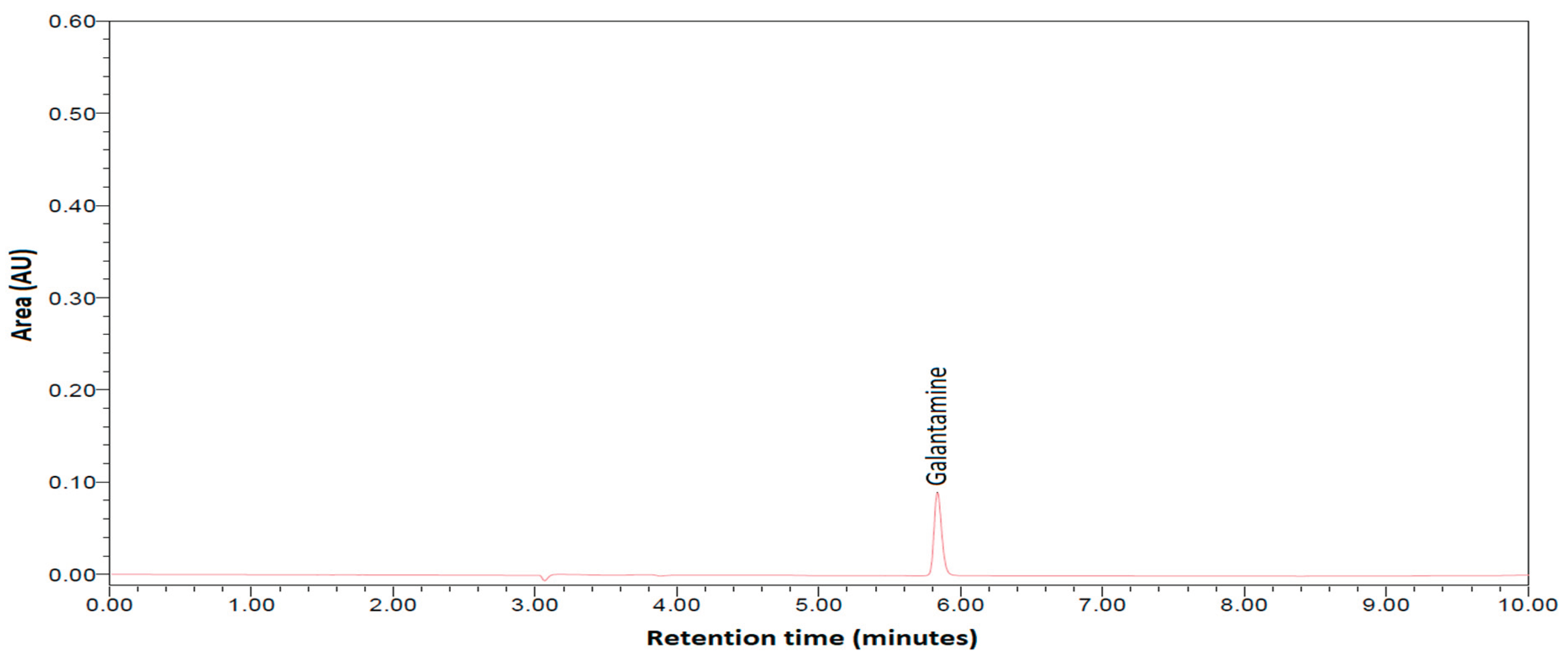
Figure 2.
Chromatogram of galantamine from the P4 sample (N. poeticus) recorded at 288 nm.
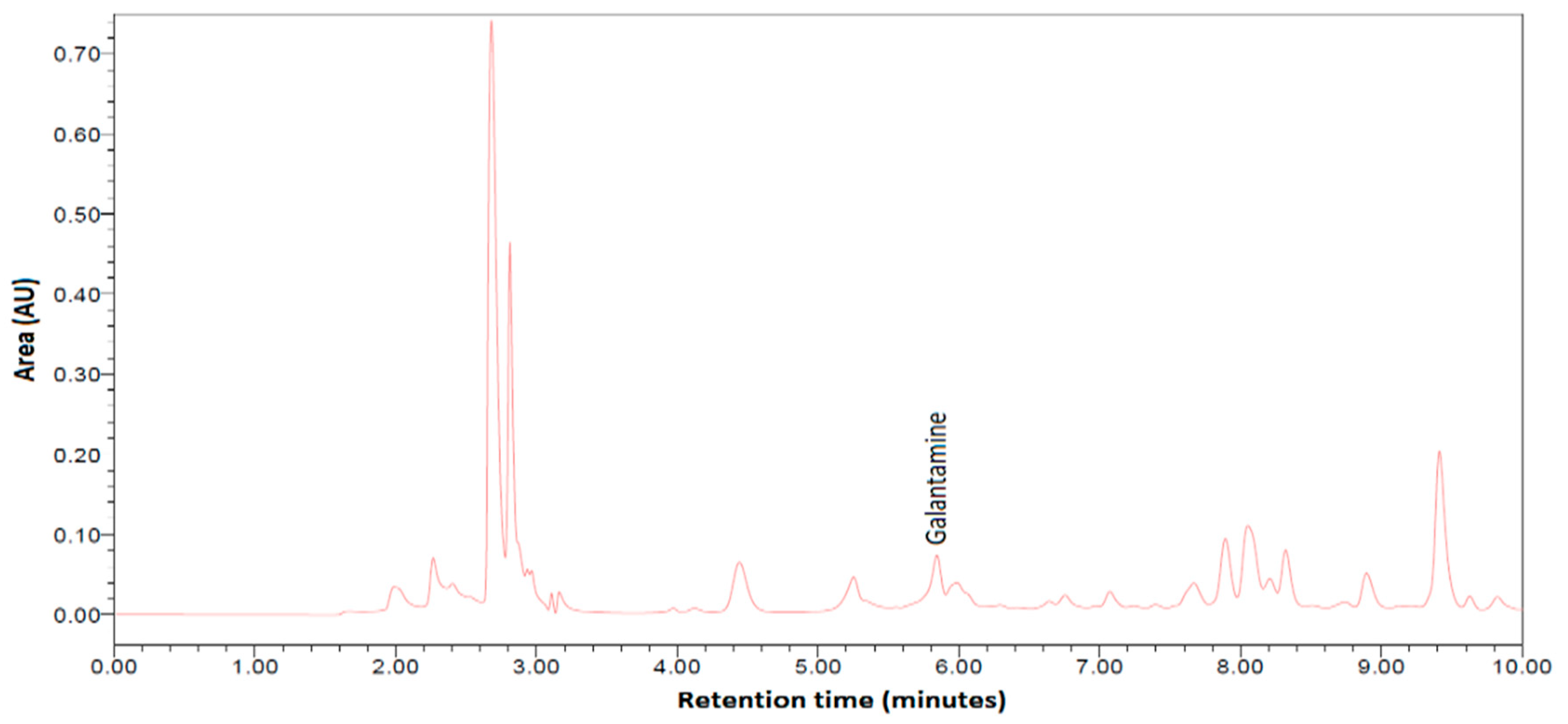
Figure 3.
Citotoxicity indirectly correlated with cell viability of galantamine on human fibroblasts in the case of the administration of the toxicant and the antidote compounds. Legend: T1 - diazinon concentration of 240 μg/mL; T2 - diazinon concentration of 24 μg/mL; G1 - galantamine from natural extract 60 μg/mL; G2 - galantamine from natural extract 12 μg/mL; G3 - galantamine from natural extract 6 μg/mL; R1 - galantamine obtained by chemical synthesis 60 μg/mL; R2 - galantamine obtained by chemical synthesis 12 μg/mL; R3 - galantamine obtained by chemical synthesis 6 μg/m.
Figure 4.
The protective effect of the natural versus synthetic galantamine. Legend: T1 - diazinon concentration 240 μg/mL; T2 - diazinon concentration 24 μg/mL; G1 - galantamine from natural extract 60 μg/mL; G2 - galantamine from natural extract 12 μg/mL; G3 - galantamine from natural extract 6 μg/mL; R1 - galantamine by chemical synthesis 60 μg/mL; R2 - galantamine by chemical synthesis 12 μg/mL; R3 - galantamine by chemical synthesis 6 μg/mL.
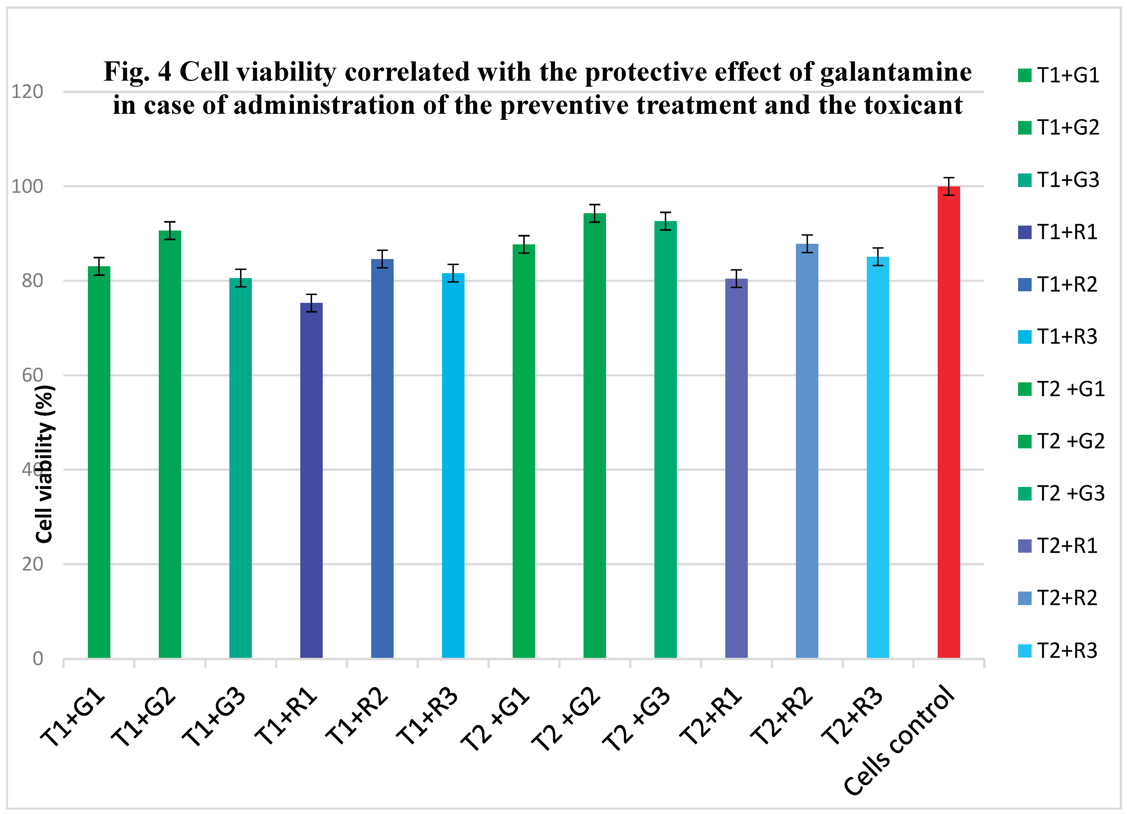
Figure 5.
Citotoxicity of galantamine, indirectly correlated with cell viability on rat hippocampal neurons in the case of administering the toxic and antidote compounds. Legend: T1 - diazinon concentration 240 μg/mL; T2 - diazinon concentration 24 μg/mL; G1 - galantamine from natural extract 60 μg/mL; G2 - galantamine from natural extract 12 μg/mL; G3 - galantamine from natural extract 6 μg/mL; R1 - galantamine by chemical synthesis 60 μg/mL; R2 - galantamine by chemical synthesis 12 μg/mL; R3 - galantamine by chemical synthesis 6 μg/mL.
Figure 6.
Cell viability concerning the protective effect of galantamine on rat hippocampal neurons in the case of the administration of the toxicant together with the antidote compounds. Legend; T1 - diazinon conc 240 μg/mL; T2 - diazinon conc 24 μg/mL; G1 - galantamine from natural extract 60 μg/mL; G2 - galantamine from natural extract 12 μg/mL; G3 - galantamine from natural extract 6 μg/mL; R1 - galantamine using chemical synthesis 60 μg/mL; R2 - galantamine using chemical synthesis 12 μg/mL; R3 - galantamine using chemical synthesis 6 μg/mL.
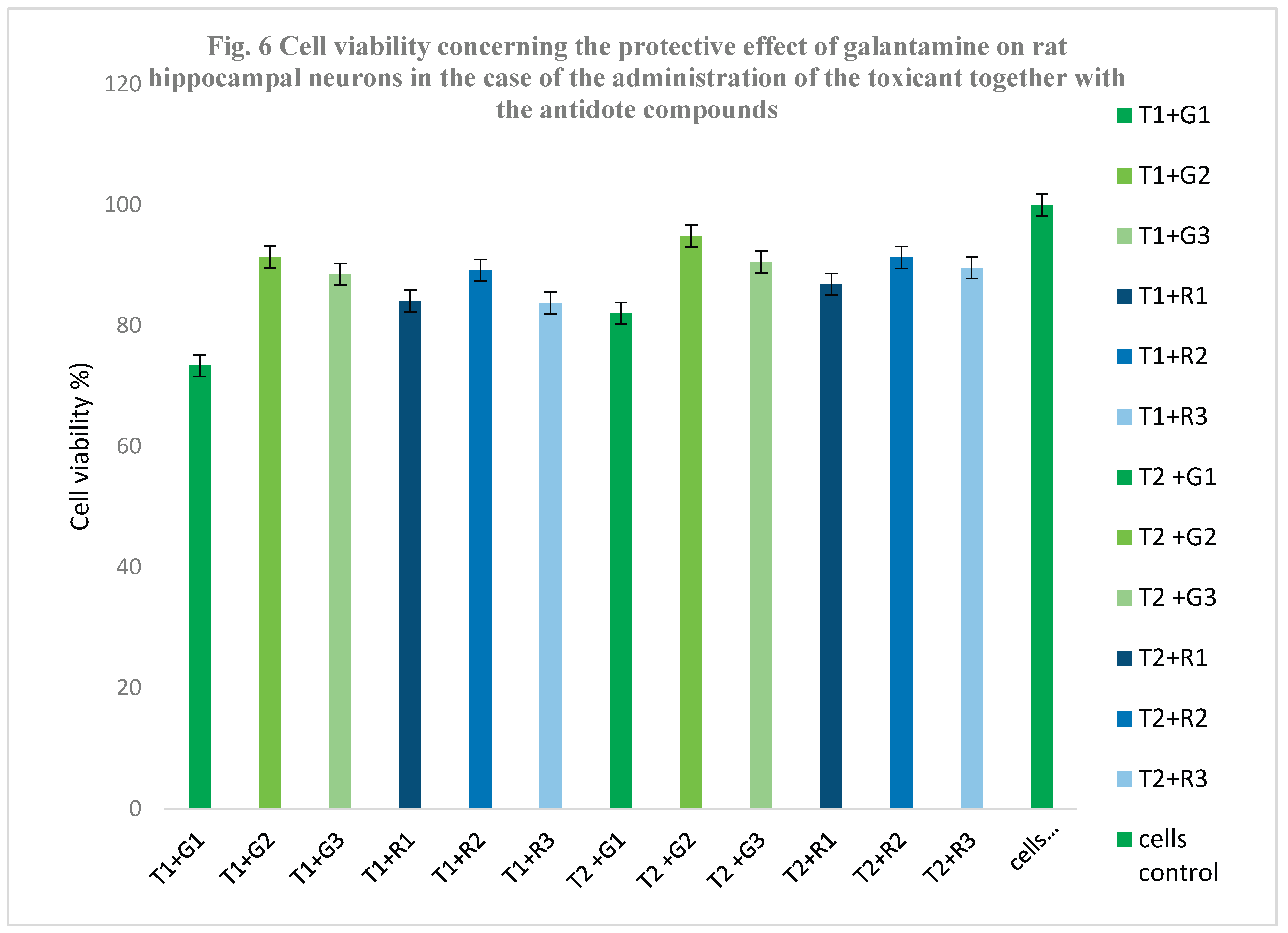

Table 1.
Dried extracts obtained by maceration (MAE) and ultrasound-assisted technique (UAE).
| Current Number | Dry extract sample (P) | Mass of maceration dried extract (MAC) (g)/100 g bulbs | Mass ultrasonic dried extract (UAE) (g)/100 g bulbs |
| 1 | Narcissus poeticus (P4) | 10.65 ± 0.02 | 10.25 ± 0.01 |
| 2 | N.pseudonarcissus (P5) | 10.02 ± 0.007 | 9.24 ± 0.008 |
| 3 | Narcissus double (P6) | 10.04 ± 0.05 | 9.13 ± 0.003 |
Table 2.
Identification and quantification of galantamine in the dried Narcissus extracts.
| Current number | Sample code | Galantamine (mg/100 mg dry extract) |
| 1 | P4 MAC SOLID | 7.928±0.016 |
| 2 | P5 MAC SOLID | 2.114±0.024 |
| 3 | P6 MAC SOLID | 5.893±0.004 |
| 4 | P4 UAE SOLID | 2.682±0.017 |
| 5 | P5 UAE SOLID | 0.768±0.014 |
| 6 | P6 UAE SOLID | 1.597±0.067 |
Legend: MAC – Obtaining natural extracts using the maceration technique; UAE – Obtaining natural extracts using the ultrasound assisted technique; P4 extract obtained from Narcissus poeticus; P5 extract obtained from Narcissus pseudonarcissus; P6 extract obtained from Narcissus double.
Table 3.
Initial and final mean values from the advanced separation of galantamine using the CPC technique.
Table 3.
Initial and final mean values from the advanced separation of galantamine using the CPC technique.
| Current Number | Initial value of galantamine in the fraction collected by the method described above (mg/dL) | Final value of galantamine in the fraction collected by the method described above (mg/dl) | Percentage of galantamine recovered (%) |
| 1 | 2.352±0.0025 | 2.231 ±0.0016 | 94.89 |
Disclaimer/Publisher’s Note: The statements, opinions and data contained in all publications are solely those of the individual author(s) and contributor(s) and not of MDPI and/or the editor(s). MDPI and/or the editor(s) disclaim responsibility for any injury to people or property resulting from any ideas, methods, instructions or products referred to in the content. |
© 2026 by the authors. Licensee MDPI, Basel, Switzerland. This article is an open access article distributed under the terms and conditions of the Creative Commons Attribution (CC BY) license.
Copyright: This open access article is published under a Creative Commons CC BY 4.0 license, which permit the free download, distribution, and reuse, provided that the author and preprint are cited in any reuse.


