Preprint
Review

This version is not peer-reviewed.

A Biophysical Framework for Neurodegeneration: Prioritizing Protein Homeostasis Over Aggregate Toxicity

Submitted:

30 December 2025

Posted:

31 December 2025

You are already at the latest version

Abstract

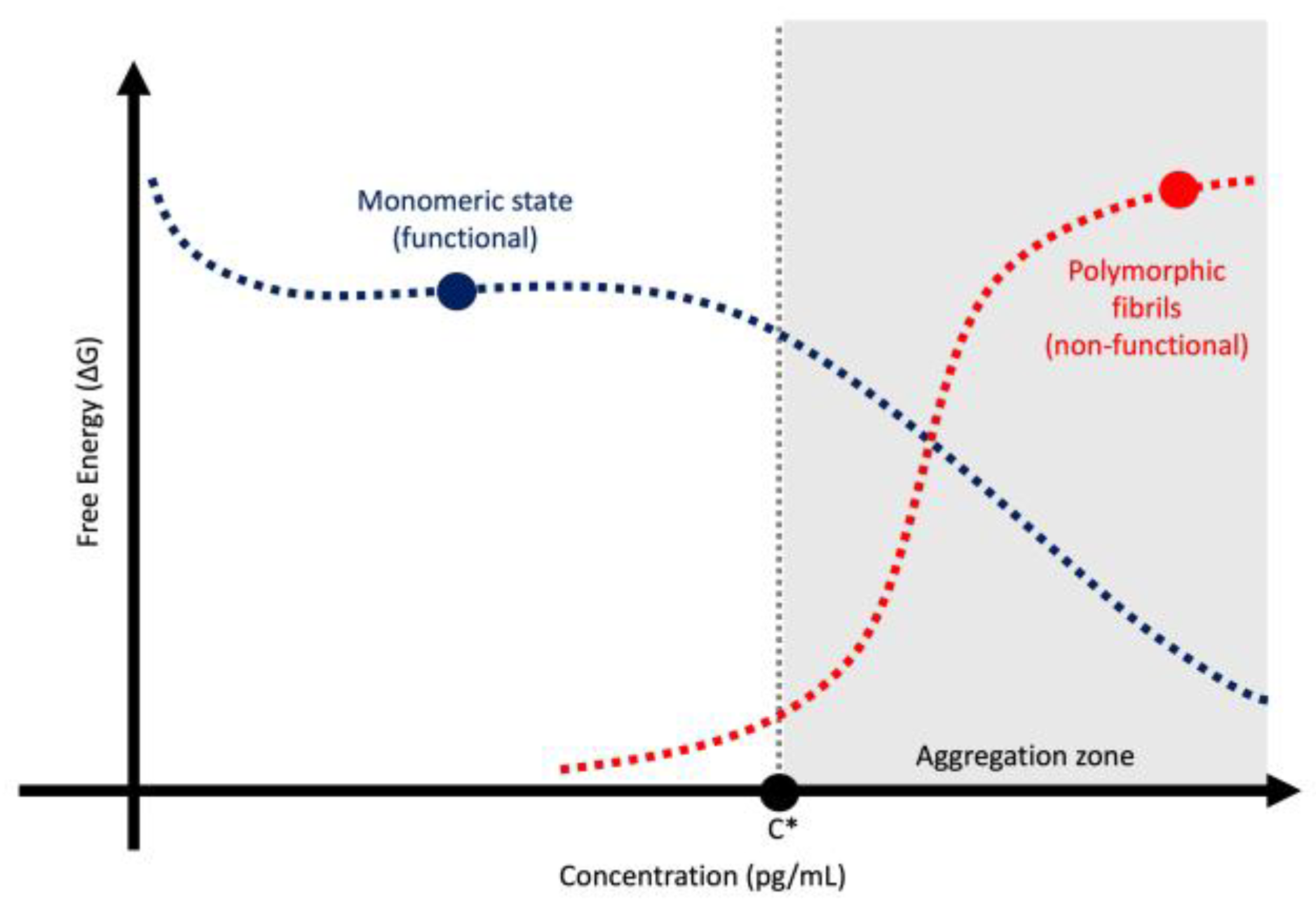
Neurodegenerative research has long hypothesized that aggregated proteins such as amyloid‑β (Aβ), tau, and α‑synuclein (αSyn) are intrinsically toxic and are directly associated with the etiologies of Alzheimer’s disease (AD) and Parkinson’s disease (PD). However, emerging scientific evidence challenges this view. Plasma p‑tau217 shows weak correlation with cognitive severity, αSyn seed amplification assays provide only binary diagnostic support, and anti‑amyloid monoclonal antibodies yield modest short-term benefit while increasing amyloid-related imaging abnormality (ARIA) risk. Postmortem pathology and fluid biomarkers explain only a limited amount of variance in clinical outcomes, undermining their role as surrogate endpoints. We propose a biophysical framework in which aggregation reflects a supersaturation-driven phase transition that signals depletion of soluble, functional monomers rather than the emergence of toxic species. Within this paradigm, amyloid plaques, neurofibrillary tangles, and Lewy bodies represent tombstones of lost protein function, and neurodegeneration occurs when monomer supply falls below neuronal demand. This shift has practical implications for biomarker interpretation, staging, and therapeutic design. Future directions include quantifying monomer flux using stable-isotope labeling kinetics (SILK), integrating supply and demand ratios, and prioritizing mechanism-testing trials that restore protein homeostasis rather than indiscriminately clear aggregates. By reframing pathology as a marker of stress rather than a maker of disease, this approach may enable more effective precision therapeutics based on human biology.

Keywords: 
;  ;  ;  ;  ;  ;  ;  ;  ;  

1. Introduction

For over three decades, research into AD and PD has been dominated by two central scientific hypotheses: first, that aggregated proteins (Aβ, tau, and αSyn) are intrinsically toxic; and second, that these aggregates accumulate in predictable spatiotemporal cascades that mirror clinical progression [1,2,3]. These premises have shaped biomarker development, informed “biological” definitions of disease, and driven therapeutic strategies aimed at clearing pathological aggregates [4,5]. Despite billions of dollars invested and numerous clinical trials, translation into durable, clinically meaningful disease modification remains elusive [6]. Instead, contradictory findings have accumulated, challenging the reductionist view that pathology equals pathogenesis [7,8].
Several observations highlight this disconnect between pathology-based biomarkers and clinical expression. In AD, plasma p-tau217, a leading biomarker of tau pathology, shows substantial mismatch with clinical severity, with nearly half of patients deviating from expected correlations between biomarker levels and cognitive impairment [9,10]. Similarly, in PD, the αSyn-SAA, hailed as a breakthrough diagnostic tool, provides only a binary readout and fails to predict disease stage, progression, or treatment response [11]. Furthermore, OLE data for anti-Aβ mAbs, such as lecanemab and donanemab, reveal an acceleration of cognitive decline compared to initial trial phases, contradicting claims of sustained benefit [12,13]. Negative results from disease-modifying trials targeting synucleinopathies reinforce the need to revisit foundational hypotheses [11,14].
An emerging biophysical framework offers an alternative interpretation (Figure 1)(Table 1). Proteins implicated in neurodegeneration do not replicate like prions [15]; instead, they undergo phase transitions into polymorphic amyloid structures under conditions of supersaturation and catalytic stress [16,17]. This behavior reflects general principles of protein folding and polymer physics, where aggregation is a thermodynamically driven phase transition under supersaturation rather than an information-preserving replication process [18,19]. Aggregation in this context represents a loss of function of soluble, monomeric proteins rather than the gain of a toxic entity [2,20]. Pathology, therefore, is better understood as a “marker” of brain reactivity to diverse insults rather than a “maker” of disease [21]. This paradigm shift has profound implications because it challenges the validity of using aggregate-based biomarkers as surrogates for disease severity, questions the logic of aggregate-clearing therapies, and redirects focus toward restoring protein homeostasis. In this review, we synthesize evidence supporting and contesting this model, propose principles to guide future research, and outline its therapeutic and diagnostic implications.

2. Biomarker–Clinical Mismatch in AD

2.1. The p-tau217 “Mismatch”: When Clinical Severity Diverges from Pathology

Recent studies have demonstrated that plasma levels of p-tau181, p-tau217, and p-tau231 do not consistently align with clinical severity (Table 2)[22,23,24]. Analyses of large cohorts reveal that nearly half of individuals deviate substantially from the expected correlation between p-tau217 concentrations and cognitive impairment measured by the CDR-SB [9,25,26]. This discordance has prompted the classification of patients into “vulnerable” and “resilient” subgroups, defined by worse or better clinical performance than predicted by biomarker levels [9]. While this approach acknowledges heterogeneity, it raises fundamental questions about the validity of p-tau217 as a surrogate for disease severity. Although p-tau217 demonstrates high diagnostic accuracy (AUC >0.90) for AD [9,27], its translation into severity staging remains limited. Multicenter head-to-head studies confirm assay performance equivalence but highlight challenges in standardization and harmonization that limit cut-point portability across platforms [28,29]. Furthermore, memory-clinic cohorts report some prognostic value for MCI-to-AD conversion, yet effect sizes vary by assay and context, reinforcing the need for caution against surrogate determinism [30].
To reconcile these discrepancies, investigators have proposed that co-pathologies, such as αSyn and TDP-43, may modulate clinical trajectories [31]. However, empirical evidence supporting this hypothesis remains weak. Differences in αSyn-SAA positivity across subgroups are minimal and lack a biologically plausible gradient [32], while imaging surrogates for TDP-43 pathology yield effect sizes near statistical significance [33,34].
If tau pathology were linearly causal for cognitive decline, plasma p-tau217 should exhibit strong predictive validity for clinical severity. Instead, regression analyses reveal substantial scatter, undermining this assumption and indicating that p-tau217 primarily reflects the presence of aggregated tau rather than the functional integrity of neuronal networks [9,26]. This interpretation aligns with emerging evidence that pathological tau represents a loss of normal protein function rather than an intrinsically neurotoxic entity (Figure 2) [35,36].
Alternative explanations emphasize the complexity of neurodegenerative biology. Cognitive outcomes may depend on nonlinear interactions among multiple factors, including compensatory mechanisms, neuroinflammation, vascular comorbidities, and cognitive reserve [37]. Even modest differences in secondary pathologies could exert additive or synergistic effects, particularly in advanced disease stages. Nevertheless, the magnitude of unexplained variance underscores the limitations of single biomarkers as definitive indicators of disease severity.
Contrasting roles of p-tau highlight the risk of conflating pathological markers with causal mechanisms. Plasma p-tau217 is also elevated in premature infants, where it appears to serve a protective, homeostatic function during neurodevelopment [38]. These observations suggest that tau phosphorylation may represent an adaptive response to cellular stress rather than an intrinsically pathogenic event [39].
Collectively, these observations caution against overreliance on p-tau217 as a stand-alone measure of progression. They highlight the need for multimodal approaches that integrate structural imaging, functional assessments, and molecular markers to capture the dynamic interplay between pathology and clinical expression [35]. Above all, they support a conceptual shift that pathology should be interpreted as a reactive marker of brain stress rather than a deterministic maker of disease (Table 3) [7,9,26,40,41,42].

2.2. Resilience and Compensation: Pathology as Tombstone of Function

An emerging conceptual framework suggests that clinical trajectories in AD arise from dynamic biological systems rather than a simple, linear accumulation of pathology. These systems include compensatory mechanisms, network redundancy, and homeostatic responses that help maintain cognitive function despite ongoing molecular changes [37]. Within this view, pathological tau aggregates are not inherently toxic but represent the “tombstone” of monomeric, functional tau. Cognitive decline may result from the depletion of soluble tau, which is required for microtubule stabilization and synaptic integrity, rather than from the aggregate burden itself [43]. Experimental evidence confirms that soluble tau is essential for maintaining neuronal architecture and synaptic function, and its loss compromises microtubule stability [44]. Similarly, endogenous Aβ is necessary for hippocampal synaptic plasticity and memory formation, indicating that aggregation-driven depletion of Aβ may impair cognition [45]. When cognition diverges positively from tau biomarker levels, compensatory processes may remain intact; conversely, negative divergence may indicate exhaustion of these reserves [46].
Evidence supporting this interpretation comes from studies demonstrating that tau pathology often precedes symptoms by decades and that many individuals with high tau burden remain cognitively intact [47,48]. This resilience highlights the role of adaptive mechanisms that buffer against pathological stress. However, countervailing data from experimental models indicate that soluble tau oligomers can exert synaptotoxic effects [49], and human imaging studies consistently show associations between tau PET signal and cognitive decline [50,51]. These correlations, though statistically significant, typically explain only a modest proportion of variance in clinical outcomes, suggesting that tau aggregation alone is insufficient to account for disease expression [52].
The implications for biomarker research are significant. Current paradigms rely heavily on aggregate-based measures, such as p-tau217 or tau PET, as proxies for disease severity [53]. However, without quantifying the pool of functional monomeric tau, conclusions drawn from aggregate levels risk misattributing causality. Future studies should incorporate assays that measure both soluble and aggregated tau species, enabling a more accurate assessment of protein homeostasis and its relationship to cognitive resilience [54]. This approach aligns with emerging biophysical models that interpret aggregation as a reactive phase transition rather than a primary pathogenic driver [35].

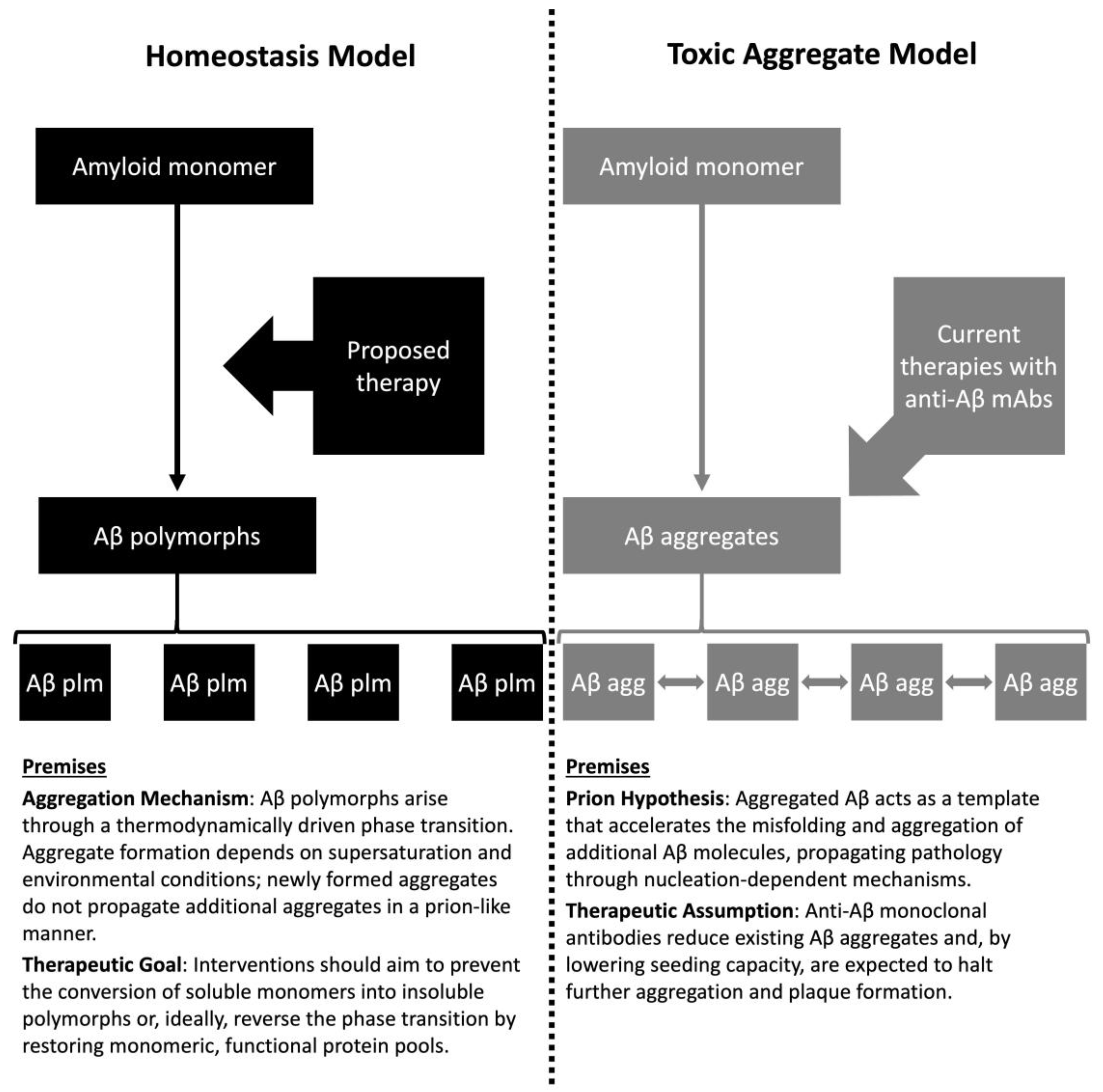
3. Reinterpreting Aβ Biology: From Toxic Waste to Essential Peptide

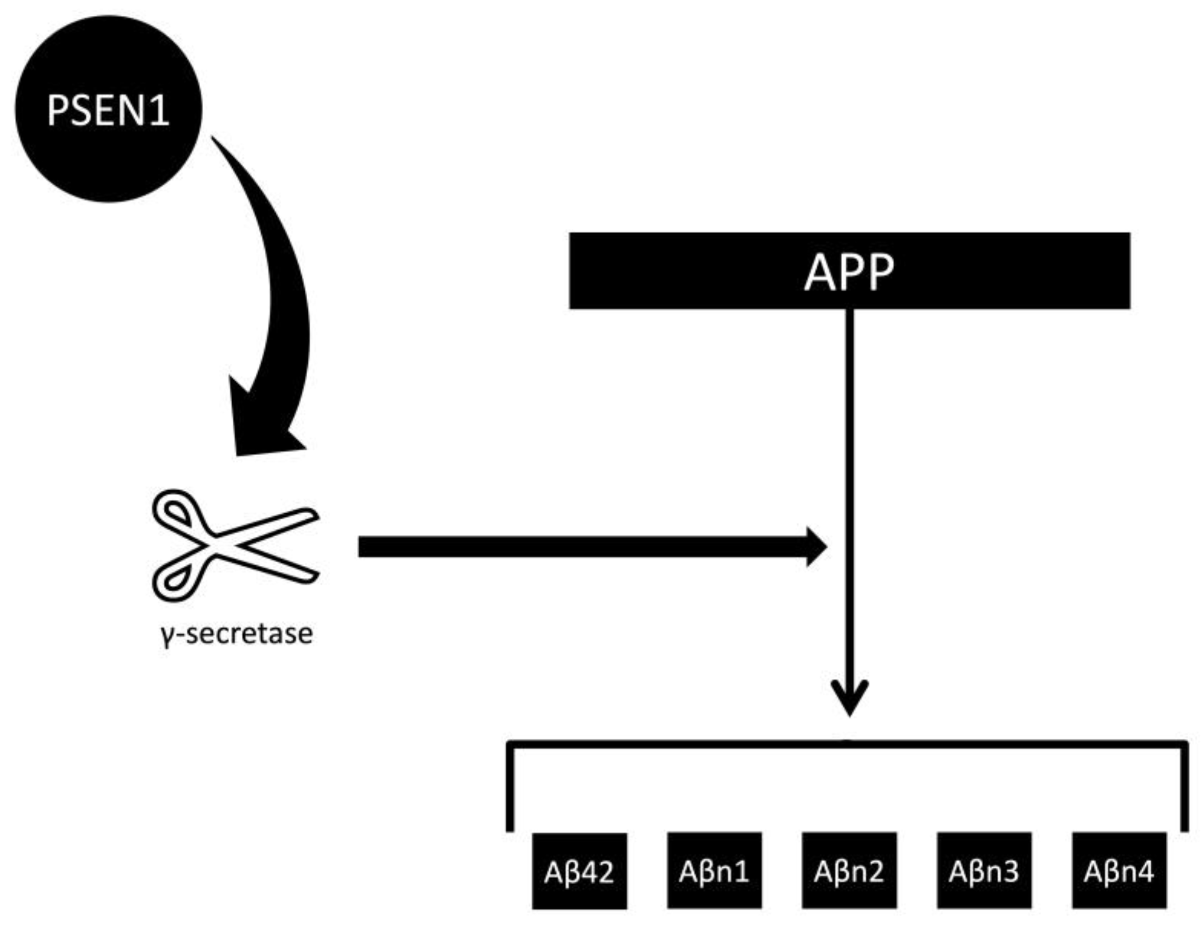
3.1. The Aβ42 Depletion Hypothesis and γ-Secretase Function

The classical Aβ cascade hypothesis posits that an excess of Aβ42 initiates tau aggregation and neurodegeneration [55]. However, increasing evidence from human studies indicates that soluble CSF Aβ42 is reduced, not elevated, in AD [56], and that pathogenic PSEN1 mutations can decrease γ-secretase activity, resulting in lower Aβ42 production (Figure 3)[57,58]. These reductions correlate with earlier onset, accelerated cognitive decline, and greater cortical atrophy [59,60,61]. This pattern challenges the assumption that Aβ42 excess is the primary driver of AD [62,63]. Zaretsky et al. demonstrated that patients with AD exhibit a markedly increased rate of soluble Aβ42 removal from CSF compared to controls, reinforcing the idea that aggregation reflects the depletion of functional monomers rather than the accumulation of toxic species [64].
Therapeutic interventions aimed at suppressing Aβ42 have produced paradoxical outcomes. γ-Secretase inhibitors, developed to reduce Aβ42 production, consistently worsened cognition compared with placebo in randomized trials [65]. In contrast, some anti-Aβ monoclonal antibodies, while intended to clear plaques, have been associated with increases in CSF Aβ42—and these increases correlate with modest clinical benefit [66,67].
APOE4 homozygosity (E4/E4) confers a markedly higher lifetime risk of AD—approximately 60% by age 85—compared to the neutral risk associated with E3/E3 [68]. Interestingly, amyloid and tau PET patterns appear similar across genotypes, yet E4/E4 carriers exhibit a rapid and early conversion of soluble Aβ42 to insoluble amyloid, resulting in profound depletion of CSF Aβ42 (Figure 4)(Appendix A)[42,69,70]. This accelerated phase transition may compromise the homeostatic functions of soluble Aβ42, thereby predisposing to neurodegeneration [7]. Aβ42 participates in synaptic signaling, neuroprotection, and trophic support (Aβ42 participates in synaptic signaling, neuroprotection, and trophic support [71]. Aggregation into plaques likely represents a loss of function rather than a gain of toxicity. Restoring Aβ42 within physiological ranges could therefore be protective [72], whereas chronic suppression may exacerbate neuronal vulnerability [59,66]. This interpretation is reinforced by observational studies showing that higher CSF Aβ42 levels are associated with better cognitive outcomes, even in Aβ-positive individuals [73].
Mounting evidence shifts focus from plaque burden to depletion of soluble Aβ42. In ADAD, CSF Aβ42 declines decades before symptoms, indicating that monomer loss more closely tracks disease emergence than aggregate load [60]. At the population level, amyloid PET positivity rises with age in cognitively normal individuals, while many oldest-old remain resilient despite substantial pathology, exposing the limited diagnostic specificity of plaques [74,75]. Therapeutically, β-secretase and γ-secretase inhibitors consistently worsened cognition and function [65,76]. Also, meta-regression across about 26,000 participants shows that increases in CSF Aβ42 independently predict slower decline, even after adjusting for amyloid PET [7]. Modeling and observational data converge on a protective role for higher CSF Aβ42 [30].
Although oligomers may exert synaptotoxic effects, the clinical correlation between oligomer burden and cognitive decline remains inconsistent. Resilience appears most strongly linked to soluble Aβ42 rather than plaque clearance [42,73].
These insights carry essential implications for trial design. Stratification by baseline CSF Aβ42 may identify subgroups most likely to benefit from interventions that restore physiological peptide levels. Moreover, reliance on the Aβ42/40 ratio as a biomarker may be misleading, as this metric obscures the absolute depletion of Aβ42, which is pathophysiologically relevant [7,42]. A recent computational modeling study demonstrated that elevated CSF Aβ42 significantly lowered the risk of preclinical Alzheimer’s in younger adults, supporting its role as a resilience marker rather than a pathogenic signal [30].
Several drugs shown to increase Aβ42 levels in vitro have demonstrated mixed clinical outcomes across the AD continuum (Figure 5)[77,78,79,80]. Indomethacin, a COX-1 inhibitor, raised Aβ42 by up to 250% [77] and showed positive trends on ADAS-Cog and MMSE in two small trials of mild-to-moderate AD patients [81,82]. Fenofibrate, a PPAR-α agonist, increased Aβ42 by up to 900% in vitro [78,83] but failed to reduce dementia risk in a seven-year observational study of 6,830 older adults [84]. Metformin, an AMPK activator, elevated Aβ42 by 250% [79] and was associated with better cognitive performance in longitudinal MCI cohorts and a dose-dependent reduction in AD incidence in a nine-year population study of over 14,000 individuals with diabetes [85,86]. Sacubitril, a neprilysin inhibitor, produced a modest 20% increase in Aβ42 [80] and was associated with a 52.5% lower three-year incidence of AD compared with ACEI/ARB therapy in a large heart failure cohort [87].

3.2. OLE Illusions and Survivor Bias Under Anti-Aβ mAbs

OLE studies of anti-Aβ mAbs such as lecanemab and donanemab have been widely interpreted as demonstrating “increasing benefit” with prolonged exposure. However, detailed analyses of cognitive trajectories challenge this interpretation (Table 4) [66,67,88,89,90]. When the slopes of CDR-SB change are examined using original randomized-trial comparators, the rate of decline accelerates during the OLE phase, and the drug-placebo gap narrows substantially [66,67]. Apparent widening of treatment effects emerges only when compared with external historical cohorts, such as those from the Alzheimer’s Disease Neuroimaging Initiative (ADNI), which exhibit steeper, non-concurrent decline [91], potentially introducing bias and inflating perceived benefit [67,92].
Attrition further complicates interpretation. By the end of the OLE phase, only about half of the original participants remain, with retention disproportionately favoring individuals who tolerate therapy and decline more slowly [67]. This survivor bias creates a misleading impression of sustained efficacy, as participants with rapid progression or adverse events are systematically excluded from later analyses.
These findings raise critical questions about the mechanistic rationale for plaque clearance as a driver of clinical benefit. If Aβ removal were disease-modifying, one would expect persistent slowing of decline rather than slope acceleration. Instead, the observed pattern aligns poorly with claims of causal benefit and suggests that early differences may reflect symptomatic or statistical artifacts rather than durable modification [66,93].
Counterarguments emphasize the nonlinearity of AD progression, noting that extrapolation from early trial phases using straight-line assumptions may misrepresent natural disease trajectories. Proponents argue that even modest early slowing could translate into clinically meaningful delay for select subgroups, provided that ARIAs are managed effectively [88]. However, this interpretation remains speculative without concurrent placebo controls beyond the blinded phase.
Safety considerations further temper enthusiasm for routine clinical use. National and international guidelines have cautioned against widespread adoption of lecanemab and donanemab outside research settings [94], citing modest symptomatic effects and increased risk of ARIA, which can be severe or life-threatening [8]. In comparative analyses, established symptomatic agents such as donepezil continue to offer superior tolerability and, in many cases, comparable or greater effect sizes on global cognition [95,96].

3.3. Aβ as Protector: Evidence Against Intrinsic Neurotoxicity

Emerging evidence challenges the long-standing assumption that Aβ accumulation is intrinsically neurotoxic. A recent Human Connectome Project study reported that higher Aβ load in cognitively healthy older adults correlated with better cognitive performance, cardiorespiratory fitness, tissue integrity, and cerebral perfusion—findings inconsistent with the presumed pathogenicity of Aβ [97]. These observations support an alternative interpretation in which Aβ serves protective or homeostatic functions, and its aggregation reflects a reactive phase transition rather than a primary driver of neurodegeneration [35,98]. Within this framework, AD may arise not from excess Aβ but from failure to maintain physiological levels of soluble Aβ42, a peptide essential for synaptic signaling and trophic support [7]. Therapeutic strategies aimed at indiscriminate Aβ clearance risk exacerbating this deficit, underscoring the need to prioritize restoration of protein homeostasis over removal of aggregates.

4. αSyn: What Seed Amplification Assays Can and Cannot Tell Us

4.1. Binary Diagnostic Support, Not a Quantitative Severity Readout

The αSyn-SAA has emerged as a highly sensitive tool for detecting misfolded αSyn aggregates in CSF, offering strong diagnostic support for PD [40]. Its clinical utility lies in confirming the presence of Lewy pathology in symptomatic individuals. However, attempts to extract quantitative information from kinetic parameters—such as lag phase, time-to-threshold (TTT), maximum fluorescence (Fmax), and slope—are conceptually flawed (Figure 6). The assay operates under conditions of supersaturation, where aggregation kinetics are governed by reagent concentration and incubation parameters rather than patient biology [41,99]. Experimental evidence demonstrates that altering laboratory conditions can significantly change kinetic profiles on identical samples [100,101], underscoring that these readouts reflect assay behavior rather than disease severity [102].
These limitations have significant implications. Kinetic parameters should not be interpreted as biomarkers of progression, prognosis, or treatment response. αSyn-SAA is inherently binary, answering whether aggregated αSyn is present or absent in the tested specimen. Positive results do not “biologically define” PD in asymptomatic individuals and cannot serve as a basis for staging frameworks [103]. Indeed, proposals such as the Neuronal Synuclein Disease–Integrated Staging System (NSD-ISS), which classify disease stages based on αSyn-SAA positivity, have shown poor correlation with established progression markers, skewed stage distributions, and even “stage regression” following symptomatic levodopa therapy [11].
Counterarguments cite studies that use endpoint dilution series to estimate seed concentration and mitigate matrix effects, suggesting a potential for semi-quantitative interpretation [104]. While these approaches improve analytical robustness, mechanistic extrapolation to clinical severity remains unvalidated. Furthermore, cross-seeding phenomena and surface catalysis introduce additional confounders, limiting specificity and reproducibility [105,106,107].
αSyn-SAA should be used as a supporting diagnostic tool in symptomatic individuals, not as a surrogate for disease staging or progression. Overextending its significance risks misclassification, unnecessary anxiety, and inappropriate trial enrollment. Future research should prioritize integrating αSyn-SAA with multimodal biomarkers rather than relying on kinetic artifacts for prognostic inference [40].

4.2. Protective Role and the Paradox of Pathology-Based Severity

Recent findings challenge the assumption that aggregated or depleted proteins are intrinsically toxic. In CBS, patients positive for CSF αSyn-SAA exhibited milder symptoms, slower progression, and lower NfL levels—features typically associated with reduced neurodegeneration [108]. Similarly, longitudinal analyses in PD cohorts show that lower CSF concentrations of C-truncated αSyn predict faster MMSE decline and worse outcomes, suggesting that soluble αSyn may play a protective rather than a pathogenic role [33]. These paradoxical associations underscore the conceptual flaw of equating aggregate detection or protein depletion with biological severity. Consistent with biophysical models, aggregation reflects loss of functional monomeric protein rather than the emergence of a toxic species [35].
Recent analyses refute the Braak hypothesis [109], which posits that αSyn aggregation into Lewy bodies is both necessary and sufficient for PD progression. Postmortem studies demonstrate no consistent correlation between Braak stage, age, or disease duration, and, paradoxically, greater Lewy body burden in the substantia nigra associates with better neuronal preservation [110], suggesting that aggregation may serve a compensatory rather than a toxic role [111].

4.3. Pathology Without Toxicity: Signals from Peripheral Autonomic Nerves

Recent skin biopsy studies in patients with Lewy body disorders and in individuals with neurological sequelae after SARS-CoV-2 infection have revealed αSyn deposition in peripheral autonomic fibers, without evidence of neuronal loss or noradrenergic deficiency [112]. These findings challenge the long-standing assumption that aggregated αSyn is intrinsically neurotoxic. Instead, they suggest that αSyn accumulation may represent a reactive process, potentially triggered by postinfectious immune or inflammatory responses rather than a primary pathogenic driver.
The absence of structural degeneration in nerves harboring αSyn inclusions aligns with broader evidence that protein aggregates often serve as markers of cellular stress rather than direct mediators of toxicity [17]. This interpretation is consistent with biophysical models in which aggregation reflects a phase transition driven by supersaturation and surface catalysis, resulting in loss of soluble, functional protein rather than the emergence of a toxic species [35].
A counterargument posits that local deposition could still exert harm through microenvironmental mechanisms, such as disrupting axonal transport or impairing synaptic signaling [113]. However, the consistent lack of correlation between αSyn burden and neuronal loss in these studies argues against a simple “toxic load” model [112]. If aggregates were inherently pathogenic, one would expect a dose-dependent relationship between deposition and structural degeneration, which has not been observed.
The therapeutic implications are significant. Strategies focused solely on clearing aggregates may fail to address the underlying biological deficit—the depletion of monomeric, functional αSyn required for normal synaptic and vesicular processes. Interventions that restore protein homeostasis, rather than indiscriminately removing aggregates, may offer a more rational approach to disease modification [35].

5. Prion Paradigms, Strains, and the Physics of Aggregation

5.1. Replication vs. Precipitation: Why Prionization Analogies Mislead

The extension of prion paradigms to AD and PD has led to the hypothesis that misfolded proteins propagate as conformational “strains” with phenotypic specificity, akin to infectious prions [16,114]. This concept underpins the notion of templated growth and transmissibility of Aβ, tau, and αSyn aggregates. However, human data consistently show weak correlations between aggregate burden and clinical phenotype or neurodegeneration (Table 5) [106,115,116,117,118,119,120,121,122,123,124]. For example, tau and Aβ pathology account for only a small fraction of the variance in hippocampal atrophy and cognitive decline in AD cohorts [74]. These observations challenge the assumption that strain fidelity drives disease expression.
From a biophysical perspective, brain proteins do not replicate in the manner of nucleic acids or prions. Instead, they undergo phase transitions under supersaturation, forming diverse polymorphic fibrils stabilized by a cross-β sheet architecture [35,125]. Supersaturation lowers the nucleation barrier for precipitation of monomeric proteins into their pathological state [126]. Aggregation is governed by thermodynamic principles and surface catalysis rather than information-preserving templating. This interpretation is consistent with Anfinsen’s dogma, which asserts that protein folding and structural transitions are determined by thermodynamic stability rather than external templates [127]. Amyloid formation, therefore, obeys universal physical laws of phase transitions, which are driven by concentration and energy minimization rather than prion-like replication of conformational information. Structural studies confirm that amyloid fibrils exhibit significant polymorphism under varying environmental conditions, with distinct cross-β architectures documented across preparations [128,129]. Electron microscopy studies reveal extensive polymorphism among Aβ fibrils, contradicting the idea of uniform strain propagation [111,130]. This heterogeneity aligns with clinical variability and undermines deterministic models of strain-specific phenotypes [131]. The phase transition can be active or passive, where the spreading of pathology in neurodegenerative disorders reflects the transition of normal proteins [132].
This biophysical framework contrasts sharply with the traditional clinicopathologic model. Whereas the conventional view treats amyloid deposition as a causal driver of neurodegeneration [133], the biophysical perspective interprets amyloid as an effect of phase transition and monomer depletion [119]. Pathogenesis shifts from an emphasis on aggregate accumulation to the loss of soluble, functional protein. Conformational diversity is understood as polymorphism rather than strain specificity [134], and the propagation of pathology reflects passive nucleation rather than active replication [135]. These distinctions carry therapeutic implications: instead of prioritizing amyloid clearance, interventions should aim to restore monomeric protein homeostasis and prevent supersaturation-driven transitions.
Counterarguments cite evidence from prion diseases and experimental models demonstrating nucleation-dependent elongation and strain transmissibility under controlled conditions [17,136]. While these phenomena are well documented in vitro and in animal systems, extrapolation to common neurodegenerative disorders requires caution. Unlike prion diseases, AD and PD are characterized by progressive brain atrophy rather than tissue expansion [137], and their clinical heterogeneity resists mapping onto discrete strain identities [16,17].
Describing aggregates as passively precipitating polymorphs better reflects physical principles and observed biological complexity. Persisting with prionization analogies risks conceptual overreach and misdirected therapeutic strategies focused on blocking “spread” rather than restoring protein homeostasis [35,130]. A shift toward biophysical models that emphasize loss of soluble, functional protein may provide a more rational foundation for disease-modifying interventions.

5.2. Kinetic vs Thermodynamic Control in Amyloid Formation

Amyloid aggregation is precipitation, not replication [119]. Under thermodynamic control, proteins that exceed a supersaturation threshold lower the nucleation barrier and minimize free energy by transitioning from soluble monomers to insoluble cross-β assemblies [19,118]. Kinetic pathways such as secondary nucleation, branching, and fragmentation modulate the rate and extent of assembly but do not encode or maintain biological “strain fidelity [115,116].” The resulting fibril polymorphism is selected by environmental conditions (pH [138], ionic strength [120], protein concentration [139]) and catalytic interfaces (membranes, nanoparticles), which lower nucleation barriers and bias pathway choice [120,121]. Consistent with Anfinsen’s dogma, protein structural transitions are determined by thermodynamic stability in a given environment rather than by external informational templates (Figure 7)(Table 6) [3,19,33,42,54,140,141]; amyloid thus obeys universal phase-transition physics driven by concentration and energy minimization, not prion-like replication of conformational information [18]. Therefore, the transition from the monomeric state is exergonic, meaning it occurs spontaneously and releases free energy (ΔG).

6. Infection, Surface Catalysis, and Protein Depletion

Emerging evidence indicates that viral exposures, including SARS-CoV-2 and herpes simplex virus type 1 (HSV-1), may induce Aβ aggregation in human CSF through surface-mediated catalysis, independent of nucleic acids or replication [21]. This mechanism reflects a general principle whereby microbial surfaces lower nucleation barriers, accelerating the phase transition of soluble proteins into insoluble aggregates [142]. Experimental studies demonstrate that such aggregation is accompanied by a marked depletion of soluble proteins in CSF, suggesting a plausible route to functional impairment via the loss of monomeric protein pools rather than the direct toxicity of aggregates [21,143]. These observations align with broader evidence that surfaces, including nanoparticles and biological interfaces, facilitate amyloid nucleation by lowering the energy barrier for aggregation [141,144]. This reinforces the concept that viral particles act as catalytic platforms rather than informational templates, consistent with the thermodynamic model of phase transition. Importantly, enrolling patients with AD based on HSV-1 IgG/IgM serostatus does not imply that the virus is actively driving dementia, and a recent trial of valacyclovir was ineffective in preventing cognitive decline [145].
This interpretation reframes the conventional “toxic aggregate” narrative. Rather than viewing Aβ deposition as the primary pathogenic agent, these findings support a model in which damage arises from what is lost (functional, soluble proteins) rather than from what accumulates. The depletion of monomeric proteins compromises essential cellular processes, including synaptic signaling and structural maintenance, and may represent the critical event in neurodegeneration [35].
Counterarguments emphasize that non-selective protein depletion is unlikely to be protective and may, in fact, be inherently detrimental [146]. However, proponents of the adaptive hypothesis argue that aggregation could initially serve as part of an innate immune response, helping to sequester potentially harmful species or microbial components [147]. Pathology may emerge only when compensatory mechanisms fail to replenish the soluble protein pool [148]. Consistent with this view, AMP behavior of Aβ in vivo and in vitro indicates that aggregation may be initiated by infectious triggers as part of innate defense [149], with subsequent depletion of soluble monomer representing the proximate biological deficit [150,151].
The clinical implications are clear. Therapeutic strategies should prioritize addressing upstream triggers, such as active infections or inflammatory states, rather than focusing exclusively on aggregate clearance. Anti-aggregation approaches that do not restore monomeric protein function risk being insufficient or even harmful. Future interventions may need to combine pathogen-targeted therapies with strategies to maintain or replenish functional protein homeostasis [35,148].

7. Precision Medicine in PD and AD: From Epidemiology to Hypothesis-Testing Trials

7.1. The Pharmaco-Epidemiology Disconnect

Epidemiology-based therapeutic pipelines have repeatedly failed to deliver disease-modifying treatments for PD [5]. Observational associations, such as reduced PD risk among caffeine or urate consumers, have inspired numerous interventional trials, yet these efforts have yielded null results [152]. This disconnect underscores a critical limitation: correlation does not imply causation, and risk modifiers identified in population studies may not translate into clinically meaningful interventions. The field must pivot from association-driven molecule testing toward hypothesis-driven strategies that interrogate disease mechanisms. Trials should aim to falsify mechanistic models, such as the hypothesis that restoring monomeric protein pools can reverse neurodegeneration, rather than merely repurposing compounds based on epidemiologic signals [153].

7.2. Three Guiding Principles for PD Research

A proposed framework for precision medicine in PD articulates three principles. First, quantify both monomeric and aggregated αSyn to capture the balance between functional and pathological protein states [154]. Second, prioritize human data over animal surrogates for therapeutic insights, as preclinical models often fail to replicate the complexity of human disease biology [35]. Third, design trials to test mechanisms rather than molecules, ensuring that interventions are evaluated against falsifiable hypotheses rather than surrogate biomarker shifts [155]. These principles emphasize protein homeostasis over aggregate clearance and encourage pathway-specific approaches, such as targeting vesicle trafficking, mitochondrial stability, or microglial regulation, rather than global suppression strategies, such as LRRK2 kinase inhibition, which may overlook compensatory network effects [156].
Counterarguments highlight that targeted pathway modulation may underperform if redundancy within biological networks blunts therapeutic impact [157]. Conversely, global inhibition could theoretically capture broader benefit, albeit at the cost of increased off-target effects. Only rigorously designed trials can adjudicate these competing hypotheses. Crucially, trial designs must avoid biomarker surrogatism and prioritize clinically meaningful endpoints, such as functional outcomes and quality of life, over isolated molecular changes [153,156,158].

8. Clinical Staging and the Risks of Surrogate Determinism

The Neuronal Synuclein Disease–Integrated Staging System (NSD-ISS) proposed for PD exemplifies surrogate determinism—the assumption that a biomarker equates to disease. This framework anchors staging to αSyn-SAA positivity, presuming that detection of misfolded αSyn defines biological PD [159]. However, application of NSD-ISS to the Parkinson’s Progression Markers Initiative (PPMI) cohort revealed significant conceptual and empirical flaws [160]. Stage distributions were skewed, with more than 70% of participants classified at advanced stages at baseline, and nearly half exhibited “stage regression” following symptomatic therapy with levodopa, a non-disease-modifying intervention [11,103]. Furthermore, αSyn-SAA positivity showed poor correlation with established progression markers such as NfL and dopaminergic imaging [40], undermining its validity as a staging anchor.
A similar issue arises in AD, where the Alzheimer’s Association 2024 biological-clinical staging framework integrates Aβ and tau biomarkers with cognitive categories [161]. Analyses of large cohorts demonstrate poor alignment between biomarker-defined stages and clinical severity in the majority of Aβ-positive individuals [74,162]. To reconcile these discrepancies, proponents invoke co-pathology or cognitive resilience [163], effectively moving conceptual goalposts rather than acknowledging model limitations. Such adjustments risk perpetuating frameworks that lack predictive accuracy and clinical utility.
These examples illustrate the dangers of surrogate determinism: labeling asymptomatic individuals as diseased based solely on biomarker positivity, without longitudinal validation or evidence of imminent clinical conversion. Such an approach not only inflates disease prevalence but also exposes individuals to unnecessary anxiety and potential overtreatment [164]. Staging systems must be empirically validated against prospective outcomes, prioritize patient benefit over conceptual elegance, and avoid conflating pathological markers with pathogenesis [11,74].
Proposed Stage 0 and Its Conceptual Challenges
Recent revisions to the Alzheimer’s Association staging framework have suggested adding Stage 0, defined as genetically determined Alzheimer’s disease (ADAD or DSAD) in individuals who are clinically asymptomatic and biomarker-negative [161]. The rationale is that these individuals “have the disease from birth,” before any measurable pathologic change, and that transition to Stage 1 occurs when a Core 1 biomarker becomes abnormal. While this proposal aims to capture the earliest point in the biological continuum, it raises significant conceptual and practical concerns.
Labeling genotype alone as “disease” conflates immutable risk with active pathophysiology. Unlike biomarker-positive preclinical stages, Stage 0 lacks evidence of ongoing biological processes and therefore cannot inform prognosis or therapeutic response. This approach risks expanding disease definitions beyond empirical validation, increasing anxiety, and potential overtreatment without demonstrable benefit. Moreover, it exemplifies surrogate determinism—the assumption that a marker equates to disease—despite the absence of clinical or biomarker expression [35,74].
Although analogous frameworks exist in Huntington’s disease (HD-ISS), in which Stage 0 reflects fully penetrant genetic mutations [165], applying this logic to AD ignores key differences in penetrance, variability, and the timing of biomarker conversion in ADAD and DSAD cohorts [60,68]. Before incorporating Stage 0 into clinical or research paradigms, rigorous longitudinal validation and ethical safeguards are essential.

9. Therapeutic Lessons Across Neurodegenerations

9.1. Anti-Aβ mAbs: Mechanism of Modest Benefit and Safety Trade-Offs

Meta-analyses of anti-amyloid trials indicate that increases in CSF Aβ42—not reductions in plaque burden—are independently associated with slower cognitive and clinical decline [7]. This observation suggests that any benefit from anti-Aβ mAbs such as lecanemab or donanemab may stem from restoring soluble Aβ42 rather than clearing insoluble aggregates. Conversely, OLE data and independent guideline assessments highlight the limited clinical relevance of these therapies and their elevated risk of ARIA [8,12,66], which can include cerebral edema and hemorrhage. If Aβ42 restoration is indeed beneficial, direct replacement strategies or approaches that maintain physiological peptide levels may outperform aggregate clearance while avoiding ARIA-related complications [35,67].

9.2. PD Anti-αSyn Strategies and Bayesian Re-Analyses

Re-analyses of prasinezumab trials using Bayesian methods have found no clinically meaningful difference versus placebo, even in subgroups characterized as “rapid progressors [166].” These findings challenge post hoc claims of benefit and, together with the limitations of αSyn-SAA and evidence of peripheral αSyn deposition without neurodegeneration, question the rationale for aggregate-targeting strategies in synucleinopathies. Without interventions that restore monomeric αSyn function, therapies focused solely on aggregate removal may fail to address the underlying biological deficit [40].

9.3. Lessons From Other Neurodegenerations

Gene therapy for Huntington’s disease provides an instructive parallel. Recent trials have demonstrated disease slowing despite paradoxical increases in CSF mutant huntingtin levels following treatment [165]. This pattern mirrors observations in AD, where “successful” interventions may increase levels of functional protein pools rather than decrease them, supporting a loss-of-function mechanism as a common theme across neurodegenerative disorders. These insights underscore the need to shift therapeutic development toward strategies that restore protein homeostasis rather than indiscriminately eliminate aggregates.

10. Counter-Arguments and Open Questions

A substantial body of literature implicates soluble oligomers of Aβ, tau, and αSyn in synaptic toxicity and neuronal dysfunction [167]. Experimental models demonstrate that oligomers can disrupt membrane integrity, impair synaptic transmission, and activate inflammatory cascades [168,169]. However, clinical evidence supporting a dominant, monotonic toxicity model remains limited. Prospective correlations between oligomer burden and patient trajectories are inconsistent, and therapeutic strategies aimed at reducing oligomers have not produced robust clinical benefit in randomized trials [88]. While oligomers may exert local toxicity, their inconsistent correlation with clinical decline and failure of oligomer-targeting therapies suggest they are contributory, not primary drivers.
The concept of prion-like spread offers mechanistic analogies but risks over-interpretation when applied to common neurodegenerative disorders. Although transmissibility and strain fidelity are well documented in prion diseases and experimental systems [17], extrapolation to AD and PD is problematic. Human data reveal extensive polymorphism among aggregates and weak correlations between strain characteristics and clinical phenotypes [130]. Moreover, the progressive brain atrophy observed in AD and PD contrasts with the proliferative pathology typical of prion disorders, underscoring fundamental biological differences.
Inflammation and autoimmunity have been proposed as alternative drivers of neurodegeneration, reframing AD as an autoimmune condition. However, gold-standard demonstrations—such as disease induction via transfer of pathogenic antibodies or immune cells—are absent [170]. Elevated immune markers in AD likely reflect homeostatic responses to injury rather than primary pathogenic mechanisms. Misinterpreting these signals risks misdirecting therapeutic development toward immunosuppression, which may exacerbate vulnerability rather than confer benefit.
Finally, the role of aggregation itself remains context dependent. Transient aggregation may serve adaptive functions, such as sequestering misfolded proteins or microbial components during stress. Conversely, overwhelming surface catalysis can precipitate non-selective depletion of soluble proteins, compromising essential cellular processes [21]. The critical clinical variable may therefore be the net balance of monomeric protein relative to physiological demand, rather than aggregate load per se.

11. Practical Implications and Future Directions

11.1. Biomarker Use

Pathology biomarkers such as plasma p-tau217, amyloid PET, and αSyn-SAA should be interpreted as reactive markers, not definitive indicators of disease presence or progression. They identify pathological states but do not reliably predict clinical severity or trajectory. Consequently, these biomarkers should never be used in isolation to define disease or stage progression without clinical correlation [9,171]. For AD, absolute CSF Aβ42 concentrations should be prioritized over Aβ42/40 ratios for risk assessment, as ratios can obscure physiologically relevant Aβ42 depletion [42,172]. Similarly, p-tau217 must be interpreted with caution, given its substantial mismatch with cognitive outcomes in large cohorts [26,27]. In PD, αSyn-SAA should serve as a diagnostic adjunct in symptomatic individuals; labeling asymptomatic positives as “pre-diseased” risks overdiagnosis and unnecessary anxiety [40,41].

11.2. Quantifying Monomer Flux in Humans

A shift from static biomarker concentrations toward dynamic flux can directly test whether loss of functional monomer predicts clinical trajectories better than aggregate burden. Stable-isotope labeling kinetics in humans already demonstrate measurable production and turnover of Aβ and tau, and ADAD data show decades-long changes preceding symptoms, enabling parameterization of compartmental models linking neuronal, interstitial, and CSF pools [54,60]. Increased removal rates of soluble Aβ42 in AD provide human evidence consistent with depletion rather than excess, supporting the relevance of a ΔMonomer index defined by the balance of production, aggregation, and clearance (Figure 8) [19,54,64,118,140]. Population imaging studies reveal modest explanatory power of amyloid and tau burden for clinical variance, underscoring the need to test whether ΔMonomer outperforms plaque/tau PET in predicting 12–24-month cognitive and functional decline [27,52,74].
Mechanism-testing trials could stratify participants by baseline soluble monomer levels and use dynamic flux metrics, such as the ΔMonomer index derived from SILK, as primary endpoints rather than aggregate clearance. This approach enables falsifiable hypotheses about whether restoring physiological monomer pools can slow or reverse neurodegeneration, moving beyond surrogate biomarker shifts toward biologically grounded interventions. However, SILK currently faces practical limitations in clinical settings, including high cost, the need for specialized mass spectrometry infrastructure, limited availability outside research centers, and logistical complexity for repeated sampling, which restricts scalability for large trials [173].

11.3. Mapping Neuronal Demand Relative to Supply

Clinical resilience and mismatch between pathology and cognition imply that demand-side pressures shape outcomes alongside monomer supply. Longitudinal resilience analyses and cognitive reserve data point to compensatory buffering that can sustain performance despite pathology, motivating a supply/NDI ratio that integrates soluble protein availability with an NDI derived from connectivity, electrophysiology, and microtubule burden proxies [37,46]. Tau PET patterns mirror neuroanatomical variability but typically explain only a fraction of outcome variance; physiologic tau is essential for microtubule stability and synaptic integrity, so decline should follow regional supply-demand rather than aggregate load alone [43,51].

11.4. Therapeutic-Strategy Trials

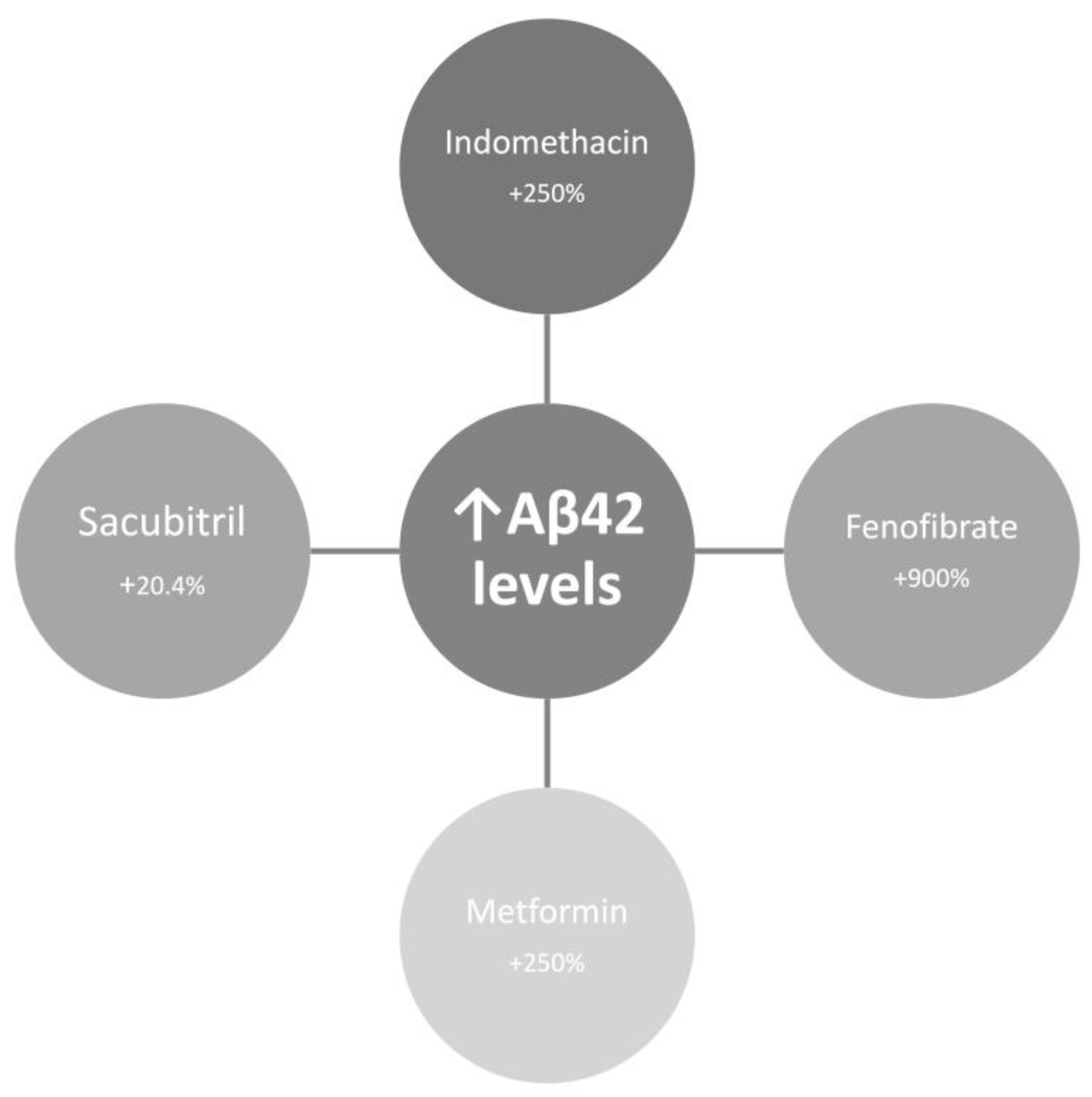
11.4.1. CSF Aβ42 Augmentation

One possible strategy is a mechanism-testing trial in Aβ-positive MCI that titrates intrathecal Aβ42 toward a physiologic CSF range observed in cognitively normal individuals (approximately 200–500 pg/mL), as AD cohorts consistently show depletion below 200 pg/mL [42,140]. The primary mechanistic endpoint should be ΔMonomer, calculated from stable-isotope labeling kinetics to capture production, clearance, and aggregation fluxes. Clinical interpretation must focus on whether absolute CSF Aβ42 levels that increase correlate with slower cognitive decline, given human evidence that higher soluble Aβ42 predicts resilience and improved trajectories [7].

11.4.2. Neutralizing Surface Catalysis

Use human CSF matrices to quantify how viral particles form protein coronas that catalyze amyloid aggregation and drive broad soluble protein depletion, then test neutralization strategies that preserve absolute monomer pools (Aβ42, αSyn) under the same ex vivo conditions [21,143]. Mechanistic controls should include nanoparticles known to nucleate fibrillation and coatings that reduce catalytic interfaces, with orthogonal measurements to confirm monomer preservation [141,144]. In vivo, map environmental determinants of aggregation by integrating non-invasive measures of tissue pH and ionic milieu that bias polymorphism and secondary nucleation with regional pathology readouts, acknowledging that tau PET captures patterns but explains limited clinical variance, and that α-synuclein seed amplification is binary and not quantitative for severity [51,120,121].

12. Conclusions

Neurodegeneration should be reframed as a failure of protein homeostasis, in which the depletion of functional monomeric proteins, rather than the accumulation of intrinsically toxic aggregates, compromises neuronal integrity. Aggregates are tombstones of lost function, formed through reactive phase transitions driven by supersaturation and catalytic stress. Evidence from biomarkers–clinical mismatches, trial failures, and monomer flux studies supports this biophysical model, which explains heterogeneity more effectively than the toxic-aggregate doctrine. This shift demands therapeutic strategies that restore monomer supply and prevent pathological phase transitions, rather than focusing solely on aggregate clearance. Future research must prioritize mechanism-testing trials using dynamic monomer flux metrics to determine whether replenishing physiological protein pools can halt or reverse neurodegeneration, defining the next frontier in precision therapeutics.

Author Contributions

Conceptualization, J.P.R. and A.L.F.C.; methodology, A.L.F.C.; software, A.L.F.C.; validation, A.L.F.C. and J.P.R.; formal analysis, A.L.F.C.; investigation, A.L.F.C.; resources, A.L.F.C.; data curation, J.P.R.; writing—original draft preparation, J.P.R.; writing—review and editing, J.P.R.; visualization, A.L.F.C.; supervision, A.L.F.C.; project administration, A.L.F.C.; funding acquisition, J.P.R. All authors have read and agreed to the published version of the manuscript.

Funding

This research received no external funding.

Institutional Review Board Statement

Not applicable.

Informed Consent Statement

Not applicable.

Data Availability Statement

All the data are presented in the manuscript.

Conflicts of Interest

The authors declare no conflicts of interest.

Abbreviations

The following abbreviations are used in this manuscript:
AD Alzheimer’s disease
ADAD Autosomal dominant Alzheimer's Disease
Anti-Aβ mAbs Anti-amyloid-β monoclonal antibodies
Amyloid-β
ARIA Amyloid-related imaging abnormalities
CBS Corticobasal syndrome
CDR-SB Clinical dementia rating–sum of boxes
CSF Cerebrospinal fluid
DSAD Down syndrome-associated Alzheimer's Disease
LRRK2 Leucine-rich repeat kinase 2
MCI Mild cognitive impairment
NDI Neuronal demand index
NfL Neurofilament light chain
OLE Open-label extension
PD Parkinson’s disease
PSEN1 Presenilin-1
SAA Seed amplification assays
SILK Stable isotope labelling kinetics
TDP-43 TAR DNA-binding protein 43
α-Synuclein αSyn

Appendix A

Table A1. CSF and plasma Aβ biomarkers across AD stages.
Matrix CSF Aβ42/Aβ40 CSF Aβ42/Aβ40 (normalized) Plasma Aβ42/Aβ40 Plasma Aβ42/Aβ40 (normalized)
Control 0.093 100 0.132 100
Preclinical AD 0.076 81.6 0.124 93.9
MCI due to AD 0.064 68.4 0.120 90.9
AD Dementia 0.056 59.8 0.119 89.8
Assay Electrochemiluminescence immunoassay Immunoprecipitation-Mass Spectrometry
Reference Hansson et al. (2018) [42]
Wojdała et al. (2023) [70]
Nakamura et al. (2018) [69]
Wojdała et al. (2023) [70]

References

  1. Bigi, A.; Lombardo, E.; Cascella, R.; Cecchi, C. The Toxicity of Protein Aggregates: New Insights into the Mechanisms. Int J Mol Sci 2023, 24. [Google Scholar] [CrossRef]
  2. Lovestone, S.; McLoughlin, D.M. Protein Aggregates and Dementia: Is There a Common Toxicity? J Neurol Neurosurg Psychiatry 2002, 72, 152–161. [Google Scholar] [CrossRef]
  3. Tsoi, P.S.; Quan, M.D.; Ferreon, J.C.; Ferreon, A.C.M. Aggregation of Disordered Proteins Associated with Neurodegeneration. Int J Mol Sci 2023, 24. [Google Scholar] [CrossRef]
  4. Chopra, A.; Lang, A.E.; Höglinger, G.; Outeiro, T.F. Towards a Biological Diagnosis of PD. Parkinsonism Relat Disord 2024, 122, 106078. [Google Scholar] [CrossRef]
  5. Pitton Rissardo, J.; Fornari Caprara, A. Disease-Modifying Therapies for Parkinson’s Disease: Biological Mechanisms, Pharmacological Strategies, and Clinical Pipeline. Preprints 2025. [Google Scholar] [CrossRef]
  6. Keefe, R.S.E.; Kraemer, H.C.; Epstein, R.S.; Frank, E.; Haynes, G.; Laughren, T.P.; McNulty, J.; Reed, S.D.; Sanchez, J.; Leon, A.C. Defining a Clinically Meaningful Effect for the Design and Interpretation of Randomized Controlled Trials. Innov Clin Neurosci 2013, 10, 4S–19S. [Google Scholar] [PubMed]
  7. Abanto, J.; Dwivedi, A.K.; Imbimbo, B.P.; Espay, A.J. Increases in Amyloid-Β42 Slow Cognitive and Clinical Decline in Alzheimer’s Disease Trials. Brain 2024, 147, 3513–3521. [Google Scholar] [CrossRef] [PubMed]
  8. Fabrizi, E.; Ancidoni, A.; Locuratolo, N.; Piscopo, P.; Della Gatta, F.; Salemme, S.; Pani, S.M.; Marconi, D.; Vignatelli, L.; Sagliocca, L.; et al. The Italian Guideline on Diagnosis and Treatment of Dementia and Mild Cognitive Impairment. Age Ageing 2024, 53. [Google Scholar] [CrossRef]
  9. Ashton, N.J.; Brum, W.S.; Di Molfetta, G.; Benedet, A.L.; Arslan, B.; Jonaitis, E.; Langhough, R.E.; Cody, K.; Wilson, R.; Carlsson, C.M.; et al. Diagnostic Accuracy of a Plasma Phosphorylated Tau 217 Immunoassay for Alzheimer Disease Pathology. JAMA Neurol 2024, 81, 255–263. [Google Scholar] [CrossRef]
  10. Benedet, A.L.; Arslan, B.; Tan, K.; Huber, H.; Pola, I.; Di Molfetta, G.; Kvartsberg, H.; Dolado, A.O.; Janelidze, S.; Blennow, K.; et al. Diagnostic Value of Serum P-Tau217 in Alzheimer Disease: Equal to Plasma in Levels and Clinical Utility? Clin Chem 2025, hvaf162. [Google Scholar] [CrossRef] [PubMed]
  11. Espay, A.J.; Cardoso, F.; Frucht, S.J.; Imarisio, A.; Halliday, G.M.; Lees, A.J. Refutation of the αSyn-SAA-Based Staging for Parkinson’s Progression (Neuronal α-Synuclein Disease-Integrated Staging System [NSD-ISS]). Mov Disord 2025, 40, 1744–1745. [Google Scholar] [CrossRef]
  12. Alkhalifa, A.E.; Al Mokhlf, A.; Ali, H.; Al-Ghraiybah, N.F.; Syropoulou, V. Anti-Amyloid Monoclonal Antibodies for Alzheimer’s Disease: Evidence, ARIA Risk, and Precision Patient Selection. J Pers Med 2025, 15. [Google Scholar] [CrossRef]
  13. Espay, A.J.; Kepp, K.P.; Herrup, K. Lecanemab and Donanemab as Therapies for Alzheimer’s Disease: An Illustrated Perspective on the Data. eNeuro 2024, 11. [Google Scholar] [CrossRef]
  14. Pitton Rissardo, J.; Fornari Caprara, A.L. Disease-Modifying Trials in Parkinson’s Disease: Challenges, Lessons, and Future Directions. Preprints 2025. [Google Scholar] [CrossRef]
  15. Prusiner, S.B. Prions. Proc Natl Acad Sci U S A 1998, 95, 13363–13383. [Google Scholar] [CrossRef]
  16. Condello, C.; Westaway, D.; Prusiner, S.B. Expanding the Prion Paradigm to Include Alzheimer and Parkinson Diseases. JAMA Neurol 2024, 81, 1023–1024. [Google Scholar] [CrossRef] [PubMed]
  17. Brundin, P.; Melki, R. Prying into the Prion Hypothesis for Parkinson’s Disease. J Neurosci 2017, 37, 9808–9818. [Google Scholar] [CrossRef]
  18. Dill, K.A.; MacCallum, J.L. The Protein-Folding Problem, 50 Years On. Science 2012, 338, 1042–1046. [Google Scholar] [CrossRef] [PubMed]
  19. Knowles, T.P.J.; Waudby, C.A.; Devlin, G.L.; Cohen, S.I.A.; Aguzzi, A.; Vendruscolo, M.; Terentjev, E.M.; Welland, M.E.; Dobson, C.M. An Analytical Solution to the Kinetics of Breakable Filament Assembly. Science 2009, 326, 1533–1537. [Google Scholar] [CrossRef]
  20. Outeiro, T.F.; Höglinger, G.; Lang, A.E.; Vieira, T.C.R.G. Protein Misfolding: Understanding Biology to Classify and Treat Synucleinopathies. J Neural Transm (Vienna) 2025, 132, 1645–1654. [Google Scholar] [CrossRef]
  21. Christ, W.; Kapell, S.; Sobkowiak, M.J.; Mermelekas, G.; Evertsson, B.; Sork, H.; Saher, O.; Bazaz, S.; Gustafsson, O.; Cardenas, E.I.; et al. SARS-CoV-2 and HSV-1 Induce Amyloid Aggregation in Human CSF Resulting in Drastic Soluble Protein Depletion. ACS Chem Neurosci 2024, 15, 4095–4104. [Google Scholar] [CrossRef]
  22. Duncan, G.B.; Dickson, S.P.; Kaplan, J.M.; Johnson, S.B.; Duke, T.M.; Dayley, C.W.; Hendrix, S.B.; Altstiel, L.D.; Mallinckrodt, C.H. Leveraging Recent Advances in Plasma Biomarkers to Optimize Early Proof of Concept Trials in Alzheimer’s Disease. Alzheimers Dement (N Y) 2025, 11, e70183. [Google Scholar] [CrossRef]
  23. Feizpour, A.; Doré, V.; Doecke, J.D.; Saad, Z.S.; Triana-Baltzer, G.; Slemmon, R.; Maruff, P.; Krishnadas, N.; Bourgeat, P.; Huang, K.; et al. Two-Year Prognostic Utility of Plasma P217+tau across the Alzheimer’s Continuum. J Prev Alzheimers Dis 2023, 10, 828–836. [Google Scholar] [CrossRef] [PubMed]
  24. Ashton, N.J.; Pascoal, T.A.; Karikari, T.K.; Benedet, A.L.; Lantero-Rodriguez, J.; Brinkmalm, G.; Snellman, A.; Schöll, M.; Troakes, C.; Hye, A.; et al. Plasma P-Tau231: A New Biomarker for Incipient Alzheimer’s Disease Pathology. Acta Neuropathol 2021, 141, 709–724. [Google Scholar] [CrossRef]
  25. Brown, C.A.; Mundada, N.S.; Cousins, K.A.Q.; Sadeghpour, N.; Lyu, X.; McGrew, E.; Korecka, M.; Chen-Plotkin, A.; Xie, L.; Wisse, L.E.M.; et al. Tau-Clinical Mismatch Identifies Individuals with Co-Pathology and Predicts Clinical Trajectory. medRxiv : the preprint server for health sciences 2025. [Google Scholar]
  26. Palmqvist, S.; Janelidze, S.; Quiroz, Y.T.; Zetterberg, H.; Lopera, F.; Stomrud, E.; Su, Y.; Chen, Y.; Serrano, G.E.; Leuzy, A.; et al. Discriminative Accuracy of Plasma Phospho-Tau217 for Alzheimer Disease vs Other Neurodegenerative Disorders. JAMA 2020, 324, 772–781. [Google Scholar] [CrossRef] [PubMed]
  27. Palmqvist, S.; Tideman, P.; Mattsson-Carlgren, N.; Schindler, S.E.; Smith, R.; Ossenkoppele, R.; Calling, S.; West, T.; Monane, M.; Verghese, P.B.; et al. Blood Biomarkers to Detect Alzheimer Disease in Primary Care and Secondary Care. JAMA 2024, 332, 1245–1257. [Google Scholar] [CrossRef]
  28. Arslan, B.; Gobom, J.; Andreasson, U.; Gonzalez-Ortiz, F.; Ashton, N.J.; Zetterberg, H.; Kvartsberg, H. Comparative Analysis of Plasma P-Tau217 Immunoassays: Challenges for Standardization and Harmonization. Clin Chem Lab Med 2025. [Google Scholar] [CrossRef]
  29. Liu, Y.; Guo, Y.; Yu, J. Blood-Based Biomarkers of Alzheimer’s Disease: Standardization and Comprehensiveness. Neuroprotection 2024, 2, 246–254. [Google Scholar] [CrossRef]
  30. Anyaiwe, O.; Nataraj, N.; Gudikandula, B.S. Computational Risk Stratification of Preclinical Alzheimer’s in Younger Adults. Diagnostics (Basel) 2025, 15. [Google Scholar] [CrossRef]
  31. Pitton Rissardo, J.; McGarry, A.; Shi, Y.; Fornari Caprara, A.L.; Kannarkat, G.T. Alpha-Synuclein Neurobiology in Parkinson’s Disease: A Comprehensive Review of Its Role, Mechanisms, and Therapeutic Perspectives. Brain Sciences 2025, 15, 1260. [Google Scholar] [CrossRef]
  32. Shahnawaz, M.; Mukherjee, A.; Pritzkow, S.; Mendez, N.; Rabadia, P.; Liu, X.; Hu, B.; Schmeichel, A.; Singer, W.; Wu, G.; et al. Discriminating α-Synuclein Strains in Parkinson’s Disease and Multiple System Atrophy. Nature 2020, 578, 273–277. [Google Scholar] [CrossRef] [PubMed]
  33. Bellomo, G.; Stoops, E.; Vanbrabant, J.; Demeyer, L.; Francois, C.; Vanhooren, M.; Ma, Y.; Farris, C.M.; Concha-Marambio, L.; Paolini Paoletti, F.; et al. Phosphorylated α-Synuclein in CSF and Plasma Does Not Reflect Synucleinopathy. NPJ Parkinsons Dis 2025, 11, 232. [Google Scholar] [CrossRef]
  34. Carlos, A.F.; Tosakulwong, N.; Weigand, S.D.; Senjem, M.L.; Schwarz, C.G.; Knopman, D.S.; Boeve, B.F.; Petersen, R.C.; Nguyen, A.T.; Reichard, R.R.; et al. TDP-43 Pathology Effect on Volume and Flortaucipir Uptake in Alzheimer’s Disease. Alzheimers Dement 2023, 19, 2343–2354. [Google Scholar] [CrossRef]
  35. Espay, A.J.; Sturchio, A.; Imarisio, A.; Hill, E.J.; Williamson, B.; Montemagno, K.; Hoffmann, C.; Roy, H.L.; Milovanovic, D.; Manfredsson, F.P. Physics of Protein Aggregation in Normal and Accelerated Brain Aging. Bioessays 2025, 47, e70030. [Google Scholar] [CrossRef] [PubMed]
  36. Ezzat, K.; Sturchio, A.; Espay, A.J. The Shift to a Proteinopenia Paradigm in Neurodegeneration. Handb Clin Neurol 2023, 193, 23–32. [Google Scholar] [CrossRef] [PubMed]
  37. Wilson, R.S.; Yu, L.; Lamar, M.; Schneider, J.A.; Boyle, P.A.; Bennett, D.A. Education and Cognitive Reserve in Old Age. Neurology 2019, 92, e1041–e1050. [Google Scholar] [CrossRef]
  38. Gonzalez-Ortiz, F.; Vávra, J.; Payne, E.; Kirsebom, B.-E.; Sjöbom, U.; Santos, C.; Júlvez, J.; Kramer, K.; Zalcberg, D.; Montoliu-Gaya, L.; et al. The Potential Dual Role of Tau Phosphorylation: Plasma Phosphorylated-Tau217 in Newborns and Alzheimer’s Disease. Brain Commun 2025, 7, fcaf221. [Google Scholar] [CrossRef]
  39. van der Harg, J.M.; Nölle, A.; Zwart, R.; Boerema, A.S.; van Haastert, E.S.; Strijkstra, A.M.; Hoozemans, J.J.; Scheper, W. The Unfolded Protein Response Mediates Reversible Tau Phosphorylation Induced by Metabolic Stress. Cell Death Dis 2014, 5, e1393. [Google Scholar] [CrossRef]
  40. Siderowf, A.; Concha-Marambio, L.; Lafontant, D.-E.; Farris, C.M.; Ma, Y.; Urenia, P.A.; Nguyen, H.; Alcalay, R.N.; Chahine, L.M.; Foroud, T.; et al. Assessment of Heterogeneity among Participants in the Parkinson’s Progression Markers Initiative Cohort Using α-Synuclein Seed Amplification: A Cross-Sectional Study. Lancet Neurol 2023, 22, 407–417. [Google Scholar] [CrossRef]
  41. Espay, A.J.; Lees, A.J.; Cardoso, F.; Frucht, S.J.; Erskine, D.; Sandoval, I.M.; Bernal-Conde, L.D.; Sturchio, A.; Imarisio, A.; Hoffmann, C.; et al. The α-Synuclein Seed Amplification Assay: Interpreting a Test of Parkinson’s Pathology. Parkinsonism Relat Disord 2025, 131, 107256. [Google Scholar] [CrossRef]
  42. Hansson, O.; Seibyl, J.; Stomrud, E.; Zetterberg, H.; Trojanowski, J.Q.; Bittner, T.; Lifke, V.; Corradini, V.; Eichenlaub, U.; Batrla, R.; et al. CSF Biomarkers of Alzheimer’s Disease Concord with Amyloid-β PET and Predict Clinical Progression: A Study of Fully Automated Immunoassays in BioFINDER and ADNI Cohorts. Alzheimers Dement 2018, 14, 1470–1481. [Google Scholar] [CrossRef]
  43. Wang, Y.; Mandelkow, E. Tau in Physiology and Pathology. Nat Rev Neurosci 2016, 17, 5–21. [Google Scholar] [CrossRef] [PubMed]
  44. Ittner, L.M.; Götz, J. Amyloid-β and Tau--a Toxic Pas de Deux in Alzheimer’s Disease. Nat Rev Neurosci 2011, 12, 65–72. [Google Scholar] [CrossRef]
  45. Puzzo, D.; Privitera, L.; Fa’, M.; Staniszewski, A.; Hashimoto, G.; Aziz, F.; Sakurai, M.; Ribe, E.M.; Troy, C.M.; Mercken, M.; et al. Endogenous Amyloid-β Is Necessary for Hippocampal Synaptic Plasticity and Memory. Ann Neurol 2011, 69, 819–830. [Google Scholar] [CrossRef] [PubMed]
  46. Bocancea, D.I.; Svenningsson, A.L.; van Loenhoud, A.C.; Groot, C.; Barkhof, F.; Strandberg, O.; Smith, R.; La Joie, R.; Rosen, H.J.; Pontecorvo, M.J.; et al. Determinants of Cognitive and Brain Resilience to Tau Pathology: A Longitudinal Analysis. Brain 2023, 146, 3719–3734. [Google Scholar] [CrossRef] [PubMed]
  47. Ziontz, J.; Bilgel, M.; Shafer, A.T.; Moghekar, A.; Elkins, W.; Helphrey, J.; Gomez, G.; June, D.; McDonald, M.A.; Dannals, R.F.; et al. Tau Pathology in Cognitively Normal Older Adults. Alzheimers Dement (Amst) 2019, 11, 637–645. [Google Scholar] [CrossRef]
  48. St-Onge, F.; Chapleau, M.; Breitner, J.C.S.; Villeneuve, S.; Pichet Binette, A. Tau Accumulation and Its Spatial Progression across the Alzheimer’s Disease Spectrum. Brain Commun 2024, 6, fcae031. [Google Scholar] [CrossRef]
  49. Guerrero-Muñoz, M.J.; Gerson, J.; Castillo-Carranza, D.L. Tau Oligomers: The Toxic Player at Synapses in Alzheimer’s Disease. Front Cell Neurosci 2015, 9, 464. [Google Scholar] [CrossRef]
  50. Morris, M.; Maeda, S.; Vossel, K.; Mucke, L. The Many Faces of Tau. Neuron 2011, 70, 410–426. [Google Scholar] [CrossRef]
  51. Ossenkoppele, R.; Schonhaut, D.R.; Schöll, M.; Lockhart, S.N.; Ayakta, N.; Baker, S.L.; O’Neil, J.P.; Janabi, M.; Lazaris, A.; Cantwell, A.; et al. Tau PET Patterns Mirror Clinical and Neuroanatomical Variability in Alzheimer’s Disease. Brain 2016, 139, 1551–1567. [Google Scholar] [CrossRef]
  52. Jack, C.R.J.; Wiste, H.J.; Weigand, S.D.; Therneau, T.M.; Knopman, D.S.; Lowe, V.; Vemuri, P.; Mielke, M.M.; Roberts, R.O.; Machulda, M.M.; et al. Age-Specific and Sex-Specific Prevalence of Cerebral β-Amyloidosis, Tauopathy, and Neurodegeneration in Cognitively Unimpaired Individuals Aged 50-95 Years: A Cross-Sectional Study. Lancet Neurol 2017, 16, 435–444. [Google Scholar] [CrossRef]
  53. Ottoy, J.; Owsicki, N.; Bilgel, M.; Binette, A.P.; Salvadó, G.; Kang, M.S.; Cash, D.M.; Ewers, M.; La Joie, R.; Wisse, L.E.M.; et al. Recent Advances in Neuroimaging of Alzheimer’s Disease and Related Dementias. Alzheimers Dement 2025, 21, e70648. [Google Scholar] [CrossRef] [PubMed]
  54. Barthélemy, N.R.; Li, Y.; Joseph-Mathurin, N.; Gordon, B.A.; Hassenstab, J.; Benzinger, T.L.S.; Buckles, V.; Fagan, A.M.; Perrin, R.J.; Goate, A.M.; et al. A Soluble Phosphorylated Tau Signature Links Tau, Amyloid and the Evolution of Stages of Dominantly Inherited Alzheimer’s Disease. Nat Med 2020, 26, 398–407. [Google Scholar] [CrossRef] [PubMed]
  55. Sgourakis, N.G.; Yan, Y.; McCallum, S.A.; Wang, C.; Garcia, A.E. The Alzheimer’s Peptides Abeta40 and 42 Adopt Distinct Conformations in Water: A Combined MD / NMR Study. J Mol Biol 2007, 368, 1448–1457. [Google Scholar] [CrossRef] [PubMed]
  56. Espay, A.J.; Herrup, K.; Kepp, K.P.; Daly, T. The Proteinopenia Hypothesis: Loss of Aβ(42) and the Onset of Alzheimer’s Disease. Ageing Res Rev 2023, 92, 102112. [Google Scholar] [CrossRef]
  57. Okochi, M.; Tagami, S.; Yanagida, K.; Takami, M.; Kodama, T.S.; Mori, K.; Nakayama, T.; Ihara, Y.; Takeda, M. γ-Secretase Modulators and Presenilin 1 Mutants Act Differently on Presenilin/γ-Secretase Function to Cleave Aβ42 and Aβ43. Cell Rep 2013, 3, 42–51. [Google Scholar] [CrossRef]
  58. Yang, Y.; Bagyinszky, E.; An, S.S.A. Presenilin-1 (PSEN1) Mutations: Clinical Phenotypes beyond Alzheimer’s Disease. Int J Mol Sci 2023, 24. [Google Scholar] [CrossRef]
  59. Wang, Z.-X.; Tan, L.; Liu, J.; Yu, J.-T. The Essential Role of Soluble Aβ Oligomers in Alzheimer’s Disease. Mol Neurobiol 2016, 53, 1905–1924. [Google Scholar] [CrossRef]
  60. Bateman, R.J.; Aisen, P.S.; De Strooper, B.; Fox, N.C.; Lemere, C.A.; Ringman, J.M.; Salloway, S.; Sperling, R.A.; Windisch, M.; Xiong, C. Autosomal-Dominant Alzheimer’s Disease: A Review and Proposal for the Prevention of Alzheimer’s Disease. Alzheimers Res Ther 2011, 3, 1. [Google Scholar] [CrossRef]
  61. Chávez-Gutiérrez, L.; Bammens, L.; Benilova, I.; Vandersteen, A.; Benurwar, M.; Borgers, M.; Lismont, S.; Zhou, L.; Van Cleynenbreugel, S.; Esselmann, H.; et al. The Mechanism of γ-Secretase Dysfunction in Familial Alzheimer Disease. EMBO J 2012, 31, 2261–2274. [Google Scholar] [CrossRef]
  62. Younkin, S.G. The Role of A Beta 42 in Alzheimer’s Disease. J Physiol Paris 1998, 92, 289–292. [Google Scholar] [CrossRef]
  63. Phillips, J.C. Why Aβ42 Is Much More Toxic than Aβ40. ACS Chem Neurosci 2019, 10, 2843–2847. [Google Scholar] [CrossRef] [PubMed]
  64. Zaretsky, D.V.; Zaretskaia, M.V.; Molkov, Y.I. Patients with Alzheimer’s Disease Have an Increased Removal Rate of Soluble Beta-Amyloid-42. PLoS One 2022, 17, e0276933. [Google Scholar] [CrossRef]
  65. Doody, R.S.; Raman, R.; Farlow, M.; Iwatsubo, T.; Vellas, B.; Joffe, S.; Kieburtz, K.; He, F.; Sun, X.; Thomas, R.G.; et al. A Phase 3 Trial of Semagacestat for Treatment of Alzheimer’s Disease. N Engl J Med 2013, 369, 341–350. [Google Scholar] [CrossRef]
  66. van Dyck, C.H.; Swanson, C.J.; Aisen, P.; Bateman, R.J.; Chen, C.; Gee, M.; Kanekiyo, M.; Li, D.; Reyderman, L.; Cohen, S.; et al. Lecanemab in Early Alzheimer’s Disease. N Engl J Med 2023, 388, 9–21. [Google Scholar] [CrossRef]
  67. Mintun, M.A.; Lo, A.C.; Duggan Evans, C.; Wessels, A.M.; Ardayfio, P.A.; Andersen, S.W.; Shcherbinin, S.; Sparks, J.; Sims, J.R.; Brys, M.; et al. Donanemab in Early Alzheimer’s Disease. N Engl J Med 2021, 384, 1691–1704. [Google Scholar] [CrossRef]
  68. Fortea, J.; Pegueroles, J.; Alcolea, D.; Belbin, O.; Dols-Icardo, O.; Vaqué-Alcázar, L.; Videla, L.; Gispert, J.D.; Suárez-Calvet, M.; Johnson, S.C.; et al. APOE4 Homozygozity Represents a Distinct Genetic Form of Alzheimer’s Disease. Nat Med 2024, 30, 1284–1291. [Google Scholar] [CrossRef]
  69. Nakamura, A.; Kaneko, N.; Villemagne, V.L.; Kato, T.; Doecke, J.; Doré, V.; Fowler, C.; Li, Q.-X.; Martins, R.; Rowe, C.; et al. High Performance Plasma Amyloid-β Biomarkers for Alzheimer’s Disease. Nature 2018, 554, 249–254. [Google Scholar] [CrossRef]
  70. Wojdała, A.L.; Bellomo, G.; Toja, A.; Gaetani, L.; Parnetti, L.; Chiasserini, D. CSF and Plasma Aβ42/40 across Alzheimer’s Disease Continuum: Comparison of Two Ultrasensitive Simoa(®) Assays Targeting Distinct Amyloid Regions. Clin Chem Lab Med 2024, 62, 332–340. [Google Scholar] [CrossRef]
  71. Parihar, M.S.; Brewer, G.J. Amyloid-β as a Modulator of Synaptic Plasticity. J Alzheimers Dis 2010, 22, 741–763. [Google Scholar] [CrossRef]
  72. Espay, A.J.; Ezzat, K.; Kepp, K.P.; Daly, T.; Robakis, N.K.; Meijer, L.; Imbimbo, B.P. Restoring Amyloid-Β42 and γ-Secretase Function in Alzheimer’s Disease. Brain 2025, 148, 3856–3864. [Google Scholar] [CrossRef]
  73. van Harten, A.C.; Visser, P.J.; Pijnenburg, Y.A.L.; Teunissen, C.E.; Blankenstein, M.A.; Scheltens, P.; van der Flier, W.M. Cerebrospinal Fluid Aβ42 Is the Best Predictor of Clinical Progression in Patients with Subjective Complaints. Alzheimers Dement 2013, 9, 481–487. [Google Scholar] [CrossRef] [PubMed]
  74. Jack, C.R.J.; Wiste, H.J.; Weigand, S.D.; Rocca, W.A.; Knopman, D.S.; Mielke, M.M.; Lowe, V.J.; Senjem, M.L.; Gunter, J.L.; Preboske, G.M.; et al. Age-Specific Population Frequencies of Cerebral β-Amyloidosis and Neurodegeneration among People with Normal Cognitive Function Aged 50-89 Years: A Cross-Sectional Study. Lancet Neurol 2014, 13, 997–1005. [Google Scholar] [CrossRef] [PubMed]
  75. Jansen, W.J.; Ossenkoppele, R.; Knol, D.L.; Tijms, B.M.; Scheltens, P.; Verhey, F.R.J.; Visser, P.J.; Aalten, P.; Aarsland, D.; Alcolea, D.; et al. Prevalence of Cerebral Amyloid Pathology in Persons without Dementia: A Meta-Analysis. JAMA 2015, 313, 1924–1938. [Google Scholar] [CrossRef] [PubMed]
  76. Egan, M.F.; Kost, J.; Tariot, P.N.; Aisen, P.S.; Cummings, J.L.; Vellas, B.; Sur, C.; Mukai, Y.; Voss, T.; Furtek, C.; et al. Randomized Trial of Verubecestat for Mild-to-Moderate Alzheimer’s Disease. N Engl J Med 2018, 378, 1691–1703. [Google Scholar] [CrossRef]
  77. Kukar, T.; Murphy, M.P.; Eriksen, J.L.; Sagi, S.A.; Weggen, S.; Smith, T.E.; Ladd, T.; Khan, M.A.; Kache, R.; Beard, J.; et al. Diverse Compounds Mimic Alzheimer Disease-Causing Mutations by Augmenting Abeta42 Production. Nat Med 2005, 11, 545–550. [Google Scholar] [CrossRef]
  78. Gamerdinger, M.; Clement, A.B.; Behl, C. Cholesterol-like Effects of Selective Cyclooxygenase Inhibitors and Fibrates on Cellular Membranes and Amyloid-Beta Production. Mol Pharmacol 2007, 72, 141–151. [Google Scholar] [CrossRef]
  79. Chen, Y.; Zhou, K.; Wang, R.; Liu, Y.; Kwak, Y.-D.; Ma, T.; Thompson, R.C.; Zhao, Y.; Smith, L.; Gasparini, L.; et al. Antidiabetic Drug Metformin (GlucophageR) Increases Biogenesis of Alzheimer’s Amyloid Peptides via up-Regulating BACE1 Transcription. Proc Natl Acad Sci U S A 2009, 106, 3907–3912. [Google Scholar] [CrossRef]
  80. Schoenfeld, H.A.; West, T.; Verghese, P.B.; Holubasch, M.; Shenoy, N.; Kagan, D.; Buono, C.; Zhou, W.; DeCristofaro, M.; Douville, J.; et al. The Effect of Angiotensin Receptor Neprilysin Inhibitor, Sacubitril/Valsartan, on Central Nervous System Amyloid-β Concentrations and Clearance in the Cynomolgus Monkey. Toxicol Appl Pharmacol 2017, 323, 53–65. [Google Scholar] [CrossRef]
  81. Rogers, J.; Kirby, L.C.; Hempelman, S.R.; Berry, D.L.; McGeer, P.L.; Kaszniak, A.W.; Zalinski, J.; Cofield, M.; Mansukhani, L.; Willson, P. Clinical Trial of Indomethacin in Alzheimer’s Disease. Neurology 1993, 43, 1609–1611. [Google Scholar] [CrossRef]
  82. de Jong, D.; Jansen, R.; Hoefnagels, W.; Jellesma-Eggenkamp, M.; Verbeek, M.; Borm, G.; Kremer, B. No Effect of One-Year Treatment with Indomethacin on Alzheimer’s Disease Progression: A Randomized Controlled Trial. PLoS One 2008, 3, e1475. [Google Scholar] [CrossRef]
  83. Sperling, R.; Henley, D.; Aisen, P.S.; Raman, R.; Donohue, M.C.; Ernstrom, K.; Rafii, M.S.; Streffer, J.; Shi, Y.; Karcher, K.; et al. Findings of Efficacy, Safety, and Biomarker Outcomes of Atabecestat in Preclinical Alzheimer Disease: A Truncated Randomized Phase 2b/3 Clinical Trial. JAMA Neurol 2021, 78, 293–301. [Google Scholar] [CrossRef]
  84. Ancelin, M.-L.; Carrière, I.; Barberger-Gateau, P.; Auriacombe, S.; Rouaud, O.; Fourlanos, S.; Berr, C.; Dupuy, A.-M.; Ritchie, K. Lipid Lowering Agents, Cognitive Decline, and Dementia: The Three-City Study. J Alzheimers Dis 2012, 30, 629–637. [Google Scholar] [CrossRef] [PubMed]
  85. Pomilio, C.; Pérez, N.G.; Calandri, I.; Crivelli, L.; Allegri, R.; Sevlever, G.; Saravia, F. Diabetic Patients Treated with Metformin during Early Stages of Alzheimer’s Disease Show a Better Integral Performance: Data from ADNI Study. Geroscience 2022, 44, 1791–1805. [Google Scholar] [CrossRef] [PubMed]
  86. Sun, M.; Chen, W.-M.; Wu, S.-Y.; Zhang, J. Metformin in Elderly Type 2 Diabetes Mellitus: Dose-Dependent Dementia Risk Reduction. Brain 2024, 147, 1474–1482. [Google Scholar] [CrossRef]
  87. Grewal, P.K.; Abboud, A.; Myserlis, E.P.; Goldschmidt, M.E.; Butler, J.; Skopicki, H.A.; Kalogeropoulos, A.P. Sacubitril/Valsartan and Cognitive Outcomes in Heart Failure With Reduced Ejection Fraction. JACC Adv 2023, 2, 100372. [Google Scholar] [CrossRef] [PubMed]
  88. Sevigny, J.; Chiao, P.; Bussière, T.; Weinreb, P.H.; Williams, L.; Maier, M.; Dunstan, R.; Salloway, S.; Chen, T.; Ling, Y.; et al. The Antibody Aducanumab Reduces Aβ Plaques in Alzheimer’s Disease. Nature 2016, 537, 50–56. [Google Scholar] [CrossRef]
  89. Budd Haeberlein, S.; Aisen, P.S.; Barkhof, F.; Chalkias, S.; Chen, T.; Cohen, S.; Dent, G.; Hansson, O.; Harrison, K.; von Hehn, C.; et al. Two Randomized Phase 3 Studies of Aducanumab in Early Alzheimer’s Disease. J Prev Alzheimers Dis 2022, 9, 197–210. [Google Scholar] [CrossRef]
  90. Swanson, C.J.; Zhang, Y.; Dhadda, S.; Wang, J.; Kaplow, J.; Lai, R.Y.K.; Lannfelt, L.; Bradley, H.; Rabe, M.; Koyama, A.; et al. A Randomized, Double-Blind, Phase 2b Proof-of-Concept Clinical Trial in Early Alzheimer’s Disease with Lecanemab, an Anti-Aβ Protofibril Antibody. Alzheimers Res Ther 2021, 13, 80. [Google Scholar] [CrossRef]
  91. Veitch, D.P.; Weiner, M.W.; Miller, M.; Aisen, P.S.; Ashford, M.A.; Beckett, L.A.; Green, R.C.; Harvey, D.; Jack, C.R.J.; Jagust, W.; et al. The Alzheimer’s Disease Neuroimaging Initiative in the Era of Alzheimer’s Disease Treatment: A Review of ADNI Studies from 2021 to 2022. Alzheimers Dement 2024, 20, 652–694. [Google Scholar] [CrossRef]
  92. Jutten, R.J.; Sikkes, S.A.M.; Van der Flier, W.M.; Scheltens, P.; Visser, P.J.; Tijms, B.M. Finding Treatment Effects in Alzheimer Trials in the Face of Disease Progression Heterogeneity. Neurology 2021, 96, e2673–e2684. [Google Scholar] [CrossRef]
  93. Salloway, S.; Chalkias, S.; Barkhof, F.; Burkett, P.; Barakos, J.; Purcell, D.; Suhy, J.; Forrestal, F.; Tian, Y.; Umans, K.; et al. Amyloid-Related Imaging Abnormalities in 2 Phase 3 Studies Evaluating Aducanumab in Patients With Early Alzheimer Disease. JAMA Neurol 2022, 79, 13–21. [Google Scholar] [CrossRef]
  94. Bobbins, A.; Davies, M.; Lynn, E.; Roy, D.; Yeomans, A.; Shakir, S.A.W. Safety and Effectiveness of the Anti-Amyloid Monoclonal Antibody (mAb) Drug Lecanemab for Early Alzheimer’s Disease: The Pharmacovigilance Perspective. Br J Clin Pharmacol 2025, 91, 1352–1360. [Google Scholar] [CrossRef] [PubMed]
  95. Birks, J.S.; Harvey, R.J. Donepezil for Dementia Due to Alzheimer’s Disease. Cochrane Database Syst Rev 2018, 6, CD001190. [Google Scholar] [CrossRef] [PubMed]
  96. Hsu, C.-W.; Hsu, T.-W.; Kao, Y.-C.; Lin, Y.-H.; Thompson, T.; Carvalho, A.F.; Stubbs, B.; Tseng, P.-T.; Yang, F.-C.; Tsai, C.-K.; et al. The Efficacy and Safety of Anti-Amyloid Monoclonal Antibody versus Acetylcholinesterase Inhibitor with an in-Depth Analysis across Genotypes and Disease Stages: A Systematic Review and Meta-Analysis. J Prev Alzheimers Dis 2025, 12, 100195. [Google Scholar] [CrossRef]
  97. Filip, P.; McCarten, J.R.; Hemmy, L.; Crocker, J.; Wolf, M.; Thotland, J.; Cayci, Z.; Kes, T.; Michaeli, S.; Terpstra, M.; et al. Clinical and MRI Correlates of β-Amyloid Load Inconsistent With Its Presumed Neurotoxicity in Cognitively Healthy Ageing. J Neurochem 2025, 169, e70241. [Google Scholar] [CrossRef]
  98. Smith, M.A.; Casadesus, G.; Joseph, J.A.; Perry, G. Amyloid-Beta and Tau Serve Antioxidant Functions in the Aging and Alzheimer Brain. Free Radic Biol Med 2002, 33, 1194–1199. [Google Scholar] [CrossRef] [PubMed]
  99. Sawada, M.; Yamaguchi, K.; Hirano, M.; Noji, M.; So, M.; Otzen, D.; Kawata, Y.; Goto, Y. Amyloid Formation of α-Synuclein Based on the Solubility- and Supersaturation-Dependent Mechanism. Langmuir 2020, 36, 4671–4681. [Google Scholar] [CrossRef] [PubMed]
  100. Belete, D.; Mattjie, C.; Huxford, B.; Bestwick, J.P.; Noyce, A.J.; Simonet, C. Evaluating the Role of α-Synuclein Seed Amplification as a Disease Progression Marker: Evidence and Uncertainties. Mov Disord Clin Pract 2025. [Google Scholar] [CrossRef]
  101. Rissardo, J.P.; Fornari Caprara, A.L. Alpha-Synuclein Seed Amplification Assays in Parkinson’s Disease: A Systematic Review and Network Meta-Analysis. Clin Pract 2025, 15. [Google Scholar] [CrossRef] [PubMed]
  102. Giehm, L.; Lorenzen, N.; Otzen, D.E. Assays for α-Synuclein Aggregation. Methods 2011, 53, 295–305. [Google Scholar] [CrossRef] [PubMed]
  103. Cardoso, F.; Goetz, C.G.; Mestre, T.A.; Sampaio, C.; Adler, C.H.; Berg, D.; Bloem, B.R.; Burn, D.J.; Fitts, M.S.; Gasser, T.; et al. A Statement of the MDS on Biological Definition, Staging, and Classification of Parkinson’s Disease. Mov Disord 2024, 39, 259–266. [Google Scholar] [CrossRef] [PubMed]
  104. Brockmann, K.; Ticca, A.; Lerche, S.; Mastrangelo, A.; Mammana, A.; Wurster, I.; Vittoriosi, E.; Roeben, B.; Hauser, A.-K.; Deuschle, C.; et al. Quantification of Cerebrospinal Fluid α-Synuclein Seeds by Endpoint Dilution Seed Amplification Assay in Parkinson’s Disease. NPJ Parkinsons Dis 2025. [Google Scholar] [CrossRef]
  105. Ezzat, K.; Espay, A.J. Can Prions Carry Biological Information? ACS Omega 2025, 10, 57842–57845. [Google Scholar] [CrossRef]
  106. Koloteva-Levine, N.; Aubrey, L.D.; Marchante, R.; Purton, T.J.; Hiscock, J.R.; Tuite, M.F.; Xue, W.-F. Amyloid Particles Facilitate Surface-Catalyzed Cross-Seeding by Acting as Promiscuous Nanoparticles. Proc Natl Acad Sci U S A 2021, 118. [Google Scholar] [CrossRef]
  107. Candelise, N.; Schmitz, M.; Thüne, K.; Cramm, M.; Rabano, A.; Zafar, S.; Stoops, E.; Vanderstichele, H.; Villar-Pique, A.; Llorens, F.; et al. Effect of the Micro-Environment on α-Synuclein Conversion and Implication in Seeded Conversion Assays. Transl Neurodegener 2020, 9, 5. [Google Scholar] [CrossRef]
  108. Palleis, C.; Bernhardt, A.M.; Weidinger, E.; Fietzek, U.M.; Jäck, A.; Katzdobler, S.; Gnörich, J.; Bauer, T.; Franzmeier, N.; Perneczky, R.; et al. A Biomarker-Based Classification of Corticobasal Syndrome. Mov Disord 2025. [Google Scholar] [CrossRef]
  109. Braak, H.; Del Tredici, K.; Rüb, U.; de Vos, R.A.I.; Jansen Steur, E.N.H.; Braak, E. Staging of Brain Pathology Related to Sporadic Parkinson’s Disease. Neurobiol Aging 2003, 24, 197–211. [Google Scholar] [CrossRef]
  110. Burke, R.E.; Dauer, W.T.; Vonsattel, J.P.G. A Critical Evaluation of the Braak Staging Scheme for Parkinson’s Disease. Ann Neurol 2008, 64, 485–491. [Google Scholar] [CrossRef]
  111. Pitton Rissardo, J.; Fornari Caprara, A.L. Protein Aggregation and Proteostasis Failure in Neurodegenerative Diseases: Mechanisms, Structural Insights, and Therapeutic Strategies. Preprints 2025. [Google Scholar] [CrossRef]
  112. Isonaka, R.; Sullivan, P.; Goldstein, D.S. Pathophysiological Significance of α-Synuclein in Sympathetic Nerves: In Vivo Observations. Neurology 2025, 104, e210215. [Google Scholar] [CrossRef] [PubMed]
  113. Neupane, S.; De Cecco, E.; Aguzzi, A. The Hidden Cell-to-Cell Trail of α-Synuclein Aggregates. J Mol Biol 2023, 435, 167930. [Google Scholar] [CrossRef]
  114. Hoppe, S.O.; Uzunoğlu, G.; Nussbaum-Krammer, C. α-Synuclein Strains: Does Amyloid Conformation Explain the Heterogeneity of Synucleinopathies? Biomolecules 2021, 11. [Google Scholar] [CrossRef] [PubMed]
  115. Andersen, C.B.; Yagi, H.; Manno, M.; Martorana, V.; Ban, T.; Christiansen, G.; Otzen, D.E.; Goto, Y.; Rischel, C. Branching in Amyloid Fibril Growth. Biophys J 2009, 96, 1529–1536. [Google Scholar] [CrossRef]
  116. Törnquist, M.; Michaels, T.C.T.; Sanagavarapu, K.; Yang, X.; Meisl, G.; Cohen, S.I.A.; Knowles, T.P.J.; Linse, S. Secondary Nucleation in Amyloid Formation. Chem Commun (Camb) 2018, 54, 8667–8684. [Google Scholar] [CrossRef]
  117. Subedi, S.; Sasidharan, S.; Nag, N.; Saudagar, P.; Tripathi, T. Amyloid Cross-Seeding: Mechanism, Implication, and Inhibition. Molecules 2022, 27. [Google Scholar] [CrossRef]
  118. Portugal Barron, D.; Guo, Z. The Supersaturation Perspective on the Amyloid Hypothesis. Chem Sci 2023, 15, 46–54. [Google Scholar] [CrossRef]
  119. Ezzat, K.; Sturchio, A.; Espay, A.J. Proteins Do Not Replicate, They Precipitate: Phase Transition and Loss of Function Toxicity in Amyloid Pathologies. Biology (Basel) 2022, 11. [Google Scholar] [CrossRef]
  120. Ziaunys, M.; Sakalauskas, A.; Mikalauskaite, K.; Smirnovas, V. Polymorphism of Alpha-Synuclein Amyloid Fibrils Depends on Ionic Strength and Protein Concentration. Int J Mol Sci 2021, 22. [Google Scholar] [CrossRef]
  121. Frey, L.; Ghosh, D.; Qureshi, B.M.; Rhyner, D.; Guerrero-Ferreira, R.; Pokharna, A.; Kwiatkowski, W.; Serdiuk, T.; Picotti, P.; Riek, R.; et al. On the pH-Dependence of α-Synuclein Amyloid Polymorphism and the Role of Secondary Nucleation in Seed-Based Amyloid Propagation. Elife 2024, 12. [Google Scholar] [CrossRef]
  122. Srivastava, A.K.; Pittman, J.M.; Zerweck, J.; Venkata, B.S.; Moore, P.C.; Sachleben, J.R.; Meredith, S.C. β-Amyloid Aggregation and Heterogeneous Nucleation. Protein Sci 2019, 28, 1567–1581. [Google Scholar] [CrossRef]
  123. Peduzzo, A.; Linse, S.; Buell, A.K. The Properties of α-Synuclein Secondary Nuclei Are Dominated by the Solution Conditions Rather than the Seed Fibril Strain. ACS Chem Neurosci 2020, 11, 909–918. [Google Scholar] [CrossRef]
  124. Lövestam, S.; Koh, F.A.; van Knippenberg, B.; Kotecha, A.; Murzin, A.G.; Goedert, M.; Scheres, S.H.W. Assembly of Recombinant Tau into Filaments Identical to Those of Alzheimer’s Disease and Chronic Traumatic Encephalopathy. Elife 2022, 11. [Google Scholar] [CrossRef] [PubMed]
  125. Adachi, M.; So, M.; Sakurai, K.; Kardos, J.; Goto, Y. Supersaturation-Limited and Unlimited Phase Transitions Compete to Produce the Pathway Complexity in Amyloid Fibrillation. J Biol Chem 2015, 290, 18134–18145. [Google Scholar] [CrossRef]
  126. Goto, Y.; Nakajima, K.; Yamamoto, S.; Yamaguchi, K. Supersaturation, a Critical Factor Underlying Proteostasis of Amyloid Fibril Formation. J Mol Biol 2024, 436, 168475. [Google Scholar] [CrossRef]
  127. Kulkarni, P.; Leite, V.B.P.; Roy, S.; Bhattacharyya, S.; Mohanty, A.; Achuthan, S.; Singh, D.; Appadurai, R.; Rangarajan, G.; Weninger, K.; et al. Intrinsically Disordered Proteins: Ensembles at the Limits of Anfinsen’s Dogma. Biophys Rev (Melville) 2022, 3, 011306. [Google Scholar] [CrossRef]
  128. Tycko, R. Amyloid Polymorphism: Structural Basis and Neurobiological Relevance. Neuron 2015, 86, 632–645. [Google Scholar] [CrossRef]
  129. Gremer, L.; Schölzel, D.; Schenk, C.; Reinartz, E.; Labahn, J.; Ravelli, R.B.G.; Tusche, M.; Lopez-Iglesias, C.; Hoyer, W.; Heise, H.; et al. Fibril Structure of Amyloid-β(1-42) by Cryo-Electron Microscopy. Science 2017, 358, 116–119. [Google Scholar] [CrossRef]
  130. Fitzpatrick, A.W.P.; Falcon, B.; He, S.; Murzin, A.G.; Murshudov, G.; Garringer, H.J.; Crowther, R.A.; Ghetti, B.; Goedert, M.; Scheres, S.H.W. Cryo-EM Structures of Tau Filaments from Alzheimer’s Disease. Nature 2017, 547, 185–190. [Google Scholar] [CrossRef]
  131. Mehra, S.; Gadhe, L.; Bera, R.; Sawner, A.S.; Maji, S.K. Structural and Functional Insights into α-Synuclein Fibril Polymorphism. Biomolecules 2021, 11. [Google Scholar] [CrossRef]
  132. Berry, J.; Brangwynne, C.P.; Haataja, M. Physical Principles of Intracellular Organization via Active and Passive Phase Transitions. Rep Prog Phys 2018, 81, 046601. [Google Scholar] [CrossRef]
  133. Vandenberghe, R. The Relationship between Amyloid Deposition, Neurodegeneration, and Cognitive Decline in Dementia. Curr Neurol Neurosci Rep 2014, 14, 498. [Google Scholar] [CrossRef]
  134. Rasmussen, J.; Mahler, J.; Beschorner, N.; Kaeser, S.A.; Häsler, L.M.; Baumann, F.; Nyström, S.; Portelius, E.; Blennow, K.; Lashley, T.; et al. Amyloid Polymorphisms Constitute Distinct Clouds of Conformational Variants in Different Etiological Subtypes of Alzheimer’s Disease. Proc Natl Acad Sci U S A 2017, 114, 13018–13023. [Google Scholar] [CrossRef]
  135. Jucker, M.; Walker, L.C. Self-Propagation of Pathogenic Protein Aggregates in Neurodegenerative Diseases. Nature 2013, 501, 45–51. [Google Scholar] [CrossRef]
  136. Jones, E.M.; Surewicz, W.K. Fibril Conformation as the Basis of Species- and Strain-Dependent Seeding Specificity of Mammalian Prion Amyloids. Cell 2005, 121, 63–72. [Google Scholar] [CrossRef]
  137. Gao, H.-M.; Hong, J.-S. Why Neurodegenerative Diseases Are Progressive: Uncontrolled Inflammation Drives Disease Progression. Trends Immunol 2008, 29, 357–365. [Google Scholar] [CrossRef]
  138. Xu, W.; Zhang, C.; Morozova-Roche, L.; Zhang, J.Z.H.; Mu, Y. pH-Dependent Conformational Ensemble and Polymorphism of Amyloid-β Core Fragment. J Phys Chem B 2013, 117, 8392–8399. [Google Scholar] [CrossRef]
  139. Sakalauskas, A.; Ziaunys, M.; Smirnovas, V. Concentration-Dependent Polymorphism of Insulin Amyloid Fibrils. PeerJ 2019, 7, e8208. [Google Scholar] [CrossRef]
  140. Bateman, R.J.; Xiong, C.; Benzinger, T.L.S.; Fagan, A.M.; Goate, A.; Fox, N.C.; Marcus, D.S.; Cairns, N.J.; Xie, X.; Blazey, T.M.; et al. Clinical and Biomarker Changes in Dominantly Inherited Alzheimer’s Disease. N Engl J Med 2012, 367, 795–804. [Google Scholar] [CrossRef]
  141. Linse, S.; Cabaleiro-Lago, C.; Xue, W.-F.; Lynch, I.; Lindman, S.; Thulin, E.; Radford, S.E.; Dawson, K.A. Nucleation of Protein Fibrillation by Nanoparticles. Proc Natl Acad Sci U S A 2007, 104, 8691–8696. [Google Scholar] [CrossRef]
  142. Kuczyńska-Wiśnik, D.; Stojowska-Swędrzyńska, K.; Laskowska, E. Liquid-Liquid Phase Separation and Protective Protein Aggregates in Bacteria. Molecules 2023, 28. [Google Scholar] [CrossRef]
  143. Ezzat, K.; Pernemalm, M.; Pålsson, S.; Roberts, T.C.; Järver, P.; Dondalska, A.; Bestas, B.; Sobkowiak, M.J.; Levänen, B.; Sköld, M.; et al. The Viral Protein Corona Directs Viral Pathogenesis and Amyloid Aggregation. Nat Commun 2019, 10, 2331. [Google Scholar] [CrossRef]
  144. Grigolato, F.; Arosio, P. The Role of Surfaces on Amyloid Formation. Biophys Chem 2021, 270, 106533. [Google Scholar] [CrossRef]
  145. Devanand, D.P.; Wisniewski, T.; Razlighi, Q.; Qian, M.; Wei, R.; Andrews, H.F.; Acosta, E.P.; Bell, K.L.; Pelton, G.H.; Deliyannides, D.; et al. Valacyclovir Treatment of Early Symptomatic Alzheimer Disease: The VALAD Randomized Clinical Trial. JAMA 2025. [Google Scholar] [CrossRef]
  146. Treusch, S.; Cyr, D.M.; Lindquist, S. Amyloid Deposits: Protection against Toxic Protein Species? Cell Cycle 2009, 8, 1668–1674. [Google Scholar] [CrossRef]
  147. Fulop, T.; Witkowski, J.M.; Bourgade, K.; Khalil, A.; Zerif, E.; Larbi, A.; Hirokawa, K.; Pawelec, G.; Bocti, C.; Lacombe, G.; et al. Can an Infection Hypothesis Explain the Beta Amyloid Hypothesis of Alzheimer’s Disease? Front Aging Neurosci 2018, 10, 224. [Google Scholar] [CrossRef]
  148. Itzhaki, R.F. Corroboration of a Major Role for Herpes Simplex Virus Type 1 in Alzheimer’s Disease. Front Aging Neurosci 2018, 10, 324. [Google Scholar] [CrossRef]
  149. Muscolino, E.; Luoto, L.-M.; Brune, W. Viral Induced Protein Aggregation: A Mechanism of Immune Evasion. Int J Mol Sci 2021, 22. [Google Scholar] [CrossRef]
  150. Soscia, S.J.; Kirby, J.E.; Washicosky, K.J.; Tucker, S.M.; Ingelsson, M.; Hyman, B.; Burton, M.A.; Goldstein, L.E.; Duong, S.; Tanzi, R.E.; et al. The Alzheimer’s Disease-Associated Amyloid Beta-Protein Is an Antimicrobial Peptide. PLoS One 2010, 5, e9505. [Google Scholar] [CrossRef]
  151. Kumar, D.K.V.; Choi, S.H.; Washicosky, K.J.; Eimer, W.A.; Tucker, S.; Ghofrani, J.; Lefkowitz, A.; McColl, G.; Goldstein, L.E.; Tanzi, R.E.; et al. Amyloid-β Peptide Protects against Microbial Infection in Mouse and Worm Models of Alzheimer’s Disease. Sci Transl Med 2016, 8, 340ra72. [Google Scholar] [CrossRef]
  152. Schwarzschild, M.A.; Ascherio, A.; Beal, M.F.; Cudkowicz, M.E.; Curhan, G.C.; Hare, J.M.; Hooper, D.C.; Kieburtz, K.D.; Macklin, E.A.; Oakes, D.; et al. Inosine to Increase Serum and Cerebrospinal Fluid Urate in Parkinson Disease: A Randomized Clinical Trial. JAMA Neurol 2014, 71, 141–150. [Google Scholar] [CrossRef]
  153. Espay, A.J.; Piccinin, C.C.; Schwarzschild, M. Hope vs Hype II: We Should Stop Embarking on Epidemiology-Based Disease Modifying Clinical Trials. Parkinsonism Relat Disord 2025, 139, 107904. [Google Scholar] [CrossRef]
  154. Arias-Carrión, O.; Guerra-Crespo, M.; Padilla-Godínez, F.J.; Soto-Rojas, L.O.; Manjarrez, E. α-Synuclein Pathology in Synucleinopathies: Mechanisms, Biomarkers, and Therapeutic Challenges. Int J Mol Sci 2025, 26. [Google Scholar] [CrossRef]
  155. Aumont, E.; Therriault, J.; Kwan, A.T.H.; Carello-Collar, G.; Arfaie, S.; Hall, B.J.; Ferrari-Souza, J.-P.; Woo, M.S.; Macedo, A.C.; Tissot, C.; et al. The Evolution of Randomized Clinical Trial Designs to Assess Therapeutics in Alzheimer Disease. JAMA Netw Open 2025, 8, e2529665. [Google Scholar] [CrossRef]
  156. Gatto, E.M.; Espay, A.J.; Espindola, M.; Da Prat, G.; Kauffman, M.A. Protective or Pathogenic? Kinase Activity and the Neurodevelopmental Origins of G2019S LRRK2-Associated Parkinson’s Disease. Parkinsonism Relat Disord 2025, 108133. [Google Scholar] [CrossRef]
  157. Gates, A.J.; Brattig Correia, R.; Wang, X.; Rocha, L.M. The Effective Graph Reveals Redundancy, Canalization, and Control Pathways in Biochemical Regulation and Signaling. Proc Natl Acad Sci U S A 2021, 118. [Google Scholar] [CrossRef]
  158. Fleming, T.R.; Powers, J.H. Biomarkers and Surrogate Endpoints in Clinical Trials. Stat Med 2012, 31, 2973–2984. [Google Scholar] [CrossRef]
  159. Simuni, T.; Chahine, L.M.; Poston, K.; Brumm, M.; Buracchio, T.; Campbell, M.; Chowdhury, S.; Coffey, C.; Concha-Marambio, L.; Dam, T.; et al. A Biological Definition of Neuronal α-Synuclein Disease: Towards an Integrated Staging System for Research. Lancet Neurol 2024, 23, 178–190. [Google Scholar] [CrossRef]
  160. Dam, T.; Pagano, G.; Brumm, M.C.; Gochanour, C.; Poston, K.L.; Weintraub, D.; Chahine, L.M.; Coffey, C.; Tanner, C.M.; Kopil, C.M.; et al. Neuronal Alpha-Synuclein Disease Integrated Staging System Performance in PPMI, PASADENA, and SPARK Baseline Cohorts. NPJ Parkinsons Dis 2024, 10, 178. [Google Scholar] [CrossRef]
  161. Jack, C.R.J.; Andrews, J.S.; Beach, T.G.; Buracchio, T.; Dunn, B.; Graf, A.; Hansson, O.; Ho, C.; Jagust, W.; McDade, E.; et al. Revised Criteria for Diagnosis and Staging of Alzheimer’s Disease: Alzheimer’s Association Workgroup. Alzheimers Dement 2024, 20, 5143–5169. [Google Scholar] [CrossRef]
  162. Cummings, J.; Kinney, J. Biomarkers for Alzheimer’s Disease: Context of Use, Qualification, and Roadmap for Clinical Implementation. Medicina (Kaunas) 2022, 58. [Google Scholar] [CrossRef]
  163. Aiello Bowles, E.J.; Crane, P.K.; Walker, R.L.; Chubak, J.; LaCroix, A.Z.; Anderson, M.L.; Rosenberg, D.; Keene, C.D.; Larson, E.B. Cognitive Resilience to Alzheimer’s Disease Pathology in the Human Brain. J Alzheimers Dis 2019, 68, 1071–1083. [Google Scholar] [CrossRef]
  164. Rissardo, J.P.; Caprara, A.L. A Narrative Review on Biochemical Markers and Emerging Treatments in Prodromal Synucleinopathies. Clinics and Practice 2025, 15. [Google Scholar] [CrossRef]
  165. Tabrizi, S.J.; Leavitt, B.R.; Landwehrmeyer, G.B.; Wild, E.J.; Saft, C.; Barker, R.A.; Blair, N.F.; Craufurd, D.; Priller, J.; Rickards, H.; et al. Targeting Huntingtin Expression in Patients with Huntington’s Disease. N Engl J Med 2019, 380, 2307–2316. [Google Scholar] [CrossRef]
  166. Russo, M.; Costa, T.; Calisi, D.; Sensi, S.L. Prasinezumab: A Bayesian Perspective on Its Efficacy. Mov Disord 2025, 40, 619–624. [Google Scholar] [CrossRef]
  167. Gutierrez, B.A.; Limon, A. Synaptic Disruption by Soluble Oligomers in Patients with Alzheimer’s and Parkinson’s Disease. Biomedicines 2022, 10. [Google Scholar] [CrossRef] [PubMed]
  168. Benilova, I.; Karran, E.; De Strooper, B. The Toxic Aβ Oligomer and Alzheimer’s Disease: An Emperor in Need of Clothes. Nat Neurosci 2012, 15, 349–357. [Google Scholar] [CrossRef]
  169. Maeda, S.; Sahara, N.; Saito, Y.; Murayama, S.; Ikai, A.; Takashima, A. Increased Levels of Granular Tau Oligomers: An Early Sign of Brain Aging and Alzheimer’s Disease. Neurosci Res 2006, 54, 197–201. [Google Scholar] [CrossRef]
  170. Gelpi, E.; Höftberger, R.; Graus, F.; Ling, H.; Holton, J.L.; Dawson, T.; Popovic, M.; Pretnar-Oblak, J.; Högl, B.; Schmutzhard, E.; et al. Neuropathological Criteria of Anti-IgLON5-Related Tauopathy. Acta Neuropathol 2016, 132, 531–543. [Google Scholar] [CrossRef]
  171. Ashton, N.J.; Leuzy, A.; Karikari, T.K.; Mattsson-Carlgren, N.; Dodich, A.; Boccardi, M.; Corre, J.; Drzezga, A.; Nordberg, A.; Ossenkoppele, R.; et al. The Validation Status of Blood Biomarkers of Amyloid and Phospho-Tau Assessed with the 5-Phase Development Framework for AD Biomarkers. Eur J Nucl Med Mol Imaging 2021, 48, 2140–2156. [Google Scholar] [CrossRef]
  172. Lewczuk, P.; Wiltfang, J.; Kornhuber, J.; Verhasselt, A. Distributions of Aβ42 and Aβ42/40 in the Cerebrospinal Fluid in View of the Probability Theory. Diagnostics (Basel) 2021, 11. [Google Scholar] [CrossRef] [PubMed]
  173. Holland, C.; Numata, K.; Rnjak-Kovacina, J.; Seib, F.P. The Biomedical Use of Silk: Past, Present, Future. Adv Healthc Mater 2019, 8, e1800465. [Google Scholar] [CrossRef] [PubMed]
Figure 1. Comparison of the toxic aggregate paradigm and the protein homeostasis framework, highlighting distinct aggregation mechanisms and therapeutic strategies for Aβ in neurodegeneration. Abbreviations: Aβ, amyloid-β; plm, polymorph.
Preprints 192055 g001
Figure 2. Hourglass metaphor illustrating the continuum of protein states relevant to neurodegenerative diseases. The upper chamber represents soluble monomers of proteins such as αSyn, p-tau, and Aβ, associated with proteinopenia (loss of functional proteins). The narrow middle section depicts a metastable state, where misfolded intermediates accumulate. The lower chamber shows insoluble polymorphic aggregates, characteristic of proteinopathy (toxic aggregation).
Preprints 192055 g002
Figure 3. γ-Secretase cleavage of APP and generation of Aβ peptides. Presenilin-1 (PSEN1), the catalytic core of the γ-secretase complex, mediates intramembrane cleavage of amyloid precursor protein (APP). This process produces amyloid-β peptides of varying lengths, including Aβ42 and shorter species (Aβn).
Preprints 192055 g003
Figure 4. Predicted trajectories of Aβ biomarkers across the AD continuum. Line graphs illustrate normalized levels (Control = 100) of Aβ42, Aβ40, and the Aβ42/Aβ40 ratio in CSF (top) and plasma (bottom) across clinical stages: cognitively normal controls, preclinical AD, MCI due to AD, and AD dementia. Aβ42 decreases progressively in both CSF and plasma, while Aβ40 remains relatively stable.
Preprints 192055 g004
Figure 5. Drugs that increase Aβ42 levels.
Preprints 192055 g005
Figure 6. Kinetic parameters of the SAA aggregation curve. The sigmoidal fluorescence curve represents the kinetics of protein aggregation in seed amplification assays, with fluorescence intensity plotted against time. Key parameters include TTT (time-to-threshold), which marks the onset of detectable aggregation; T50, the time to reach 50% of maximum fluorescence (FT₅₀); Fmax, the plateau fluorescence intensity; and the slope, which indicates the exponential growth phase. These metrics reflect assay conditions, such as reagent concentration and incubation parameters, rather than intrinsic disease severity, underscoring that SAA kinetics should not be interpreted as staging biomarkers but rather as binary indicators of aggregate presence.
Preprints 192055 g006
Figure 7. Protein aggregation as a thermodynamic phase transition. The blue dotted curve represents the free energy of the monomeric state (functional) across concentrations, while the red dotted curve represents the free energy of polymorphic fibrils (non-functional). The vertical dashed line marks the supersaturation threshold (C*), beyond which aggregation becomes thermodynamically favorable. The supersaturation perspective does not give a fixed numeric threshold because it depends on protein sequence and solubility, ionic strength, pH, and catalytic surfaces.
Preprints 192055 g007
Figure 8. Conceptual representation of monomer balance in neurodegeneration. ΔM denotes the net change in soluble monomeric protein concentration; κ_prod represents the production rate of monomeric protein by neurons, measurable via SILK tracer studies in humans [54,140]. κ_agg indicates the effective aggregation rate, encompassing nucleation and growth that convert monomers into insoluble aggregates, derived from phase transition kinetics [19,118]. κ_clear refers to the clearance rate of soluble monomers from CSF and interstitial fluid, which can be accelerated in AD [64]. Positive ΔM reflects preserved homeostasis, whereas negative ΔM indicates net depletion of functional monomers, hypothesized to correlate with clinical decline.
Preprints 192055 g008
Table 1. Toxic Aggregate and Protein Homeostasis Models.
Feature Toxic Aggregate Protein Homeostasis
Causality Aggregates are intrinsically toxic and hierarchically causal Aggregation reflects loss of functional monomer; pathology is reactive
Biomarker interpretation Aggregate burden defines disease severity Indicate stress response; quantify monomer and aggregate pools
Therapeutic goal Clear aggregates to reduce toxicity Restore protein homeostasis and replenish functional monomers
Trial design implications Surrogate endpoints based on aggregate reduction; animal models prioritized Human trials testing mechanistic hypotheses; endpoints focused on functional outcomes
Table 2. Association of Plasma Tau Biomarkers with AD Severity.
Biomarker Association with CDR-SB Strength (Cohen's d) Clinical Interpretation References
p-tau181 Weak 0.01–0.12 Tracks progression biologically, but weak link to functional severity Duncan et al. (2025) [22]
p-tau217 Weak 0.07–0.32 Moderate predictor of decline; best biomarker for severity among tau species Feizpour et al. (2023) [23]
p-tau231 No data No data Early pathology marker Ashton et al. (2021) [24]
Table 3. Evidence of Mismatch Between Pathology Biomarkers and Clinical Severity.
Biomarker Expected correlation Observed correlation Variance explained References
p-tau217 Higher p-tau217 → worse cognition Weak severity correlation; frequent individual mismatch Rarely reported; clinical models show limited explanatory power Ashton et al. (2024) [9]
Palmqvist et al. (2020) [26]
αSyn Faster kinetics →more advanced PD/ progression Binary diagnostic only; misaligned with staging and progression Severity unreported; R2 for progression unvalidated Siderowf et al. (2023) [40]
Espay et al. (2025) [41]
Aβ42/40 ratio Lower ratio → worse cognition Ratio may hide Aβ42 loss; normal ratios can occur despite severe depletion, misclassifying severity Rarely reported; diagnostic AUC high for amyloid PET Hansson et al. (2018) [42]
Abanto et al. (2024) [7]
Notes: 1. Many studies report diagnostic metrics (e.g., AUC) for pathology rather than variance explained (R2) for clinical severity; where R2 is not explicitly published, “Not reported” is indicated. 2. For Aβ42, absolute concentration is shown because the Aβ42/40 ratio can obscure biologically relevant depletion of Aβ42. 3. αSyn-SAA provides strong diagnostic support for Lewy pathology but remains non-quantitative for biological severity; assay conditions can alter kinetics independent of patient biology.
Table 4. Summary of Clinical, Biomarker, and Safety Outcomes in anti-Aβ mAbs Trials.
Drug CDR-SBi Amyloid changeii ARIA-E & Hiii Volume lossiv Reference
Aducanumab
(PRIME)
-1.1 (6%) -27% E: 41%
H: 9%
NR Sevigny et al. (2016) [88]
Aducanumab
(EMERGE)
-0.4 (2%) -71% E: 35% & 26%
H: 20 & 16%
Y Budd Haeberlein et al. (2022) [89]
Aducanumab
(ENGAGE)
0 (0%) -58% E: 36% & 26%
H: 19% & 16%
Y Budd Haeberlein et al. (2022) [89]
Lecanemab
(BAN2401-G000–201)
-0.4 (2%) -31% E: 9.9%
H: 6.8%
Y Swanson et al. (2021) [90]
Lecanemab
(Clarity-AD)
-0.5 (3%) -55% E: 12.6%
H: 17.3%
NR Van Dyck et al. (2023) [66]
Donanemab
(TRAILBLAZER-ALZ)
-0.4 (2%) -85% E: 26.7%
H: 8.4%
N Mintun et al. (2021) [67]
Abbreviations: ARIA, amyloid-related imaging abnormality; CDR-SB, Clinical Dementia Rating-Sum of Boxes; E, edema type; H, hemosiderin type; N, no; NR, not reported; Y, yes. Notes: i) CDR-SB change (CDR-SB change) between therapy and control group at 18 months. The numbers are the difference (CDR-SB change = CDR-SB treatment group - CDR-SB control) and the percentage of the difference (CDR-SB change percentage = CDR-SB change/CDR-SB total score). ii) Amyloid change based in the Aβ PET scan at 18 months compared to control. iii) ARIA edema and hemosiderin types reported only for the treatment group. iv) Evaluated by MRI with quantitative measurements.
Table 5. Comparison of prion hypothesis, experimental challenges, and thermodynamic model of amyloid formation.
Feature Prion hypothesis Problem with prion hypothesis Thermodynamic model (Anfinsen dogma) Reference
Core idea Prions act as templates, imprinting their misfolded conformation on normal proteins Branching and secondary nucleation dominate, disrupting faithful templating Amyloid formation is a phase transition driven by supersaturation and thermodynamics Andersen et al. (2009) [115]
Törnquist et al. (2018) [116]
Mechanism of propagation Elongation at fibril tips preserves cross-sectional shape Growth occurs via branching and heterogeneous nucleation, not tip elongation Nucleation followed by growth; branching is expected as part of phase transition Koloteva-Levine et al. (2021) [106]
Sequence Requirement Requires same protein sequence for parallel in-register stacking Cross-seeding occurs between unrelated proteins, showing no sequence specificity Sequence-independent; driven by packing and hydrogen bonding under high concentration Subedi et al. (2022) [117]
Thermodynamic Basis Assumes proteins leave stable native state to fit fibril tip No thermodynamic incentive for this; amyloid formation is driven by supersaturation Folding into cross-β structure is thermodynamically favorable at supersaturation Portugal Barron et al. (2023) [118]
Ezzat et al. (2022) [119]
Role of Seeds Seeds carry conformational information Seeds act as catalytic surfaces, not carriers of structural information Seeds lower nucleation barrier; act as catalysts, not templates Koloteva-Levine et al. (2021) [106]
Environmental Influence Minimal role Fibril polymorphism depends on pH, ionic strength, and other environmental conditions Environmental conditions dictate polymorphs and ladder pairing Ziaunys et al. (2021) [120]
Frey et al. (2024) [121]
Spontaneous Formation Not considered Amyloids can form spontaneously at high concentrations (homogeneous nucleation) Expected under supersaturation without any template Srivastava et al. (2019) [122]
Cross-Sectional Shape Preserved across generations Shapes vary with conditions; strain concept lacks structural validation Polymorphs arise from solution conditions, not seed structure Peduzzo et al. (2020) [123]
Lövestam et al. (2021) [124]
Table 6. Reported Supersaturation Thresholds for Aggregation-Prone Proteins in In Vivo and In Vitro Systems.
Protein In vivo/ In vitro Supersaturation threshold (approximation) Note Reference
Aβ42 In vivo 50–120 pM (200–500 pg/mL) Healthy vs AD; depletion precedes plaque formation Bateman et al. (2012) [140]
Hansson et al. (2018) [42]
In vitro >10–20 µM Aggregation assays under physiological pH and ionic strength Knowles et al. (2009) [19]
Linse et al. (2007) [141]
Tau In vivo 200–600 pg/mL Elevated in AD but aggregation depends on local concentration and phosphorylation Barthélemy et al. (2020) [54]
In vitro >2–8 µM Phase transition observed in crowding conditions Tsoi et al. (2023) [3]
αSyn In vivo 0.5–1 ng/mL PD patients show depletion of soluble αSyn; aggregation occurs intracellularly Bellomo et al. (2025) [33]
In vitro >50–70 µM Supersaturation threshold for fibril formation under agitation Knowles et al. (2009) [19]
Frey et al. (2024) [121]
Note: i) In vivo values reflect soluble monomer pools in CSF, not local intracellular concentrations (which can be much higher). ii) In vitro thresholds depend on buffer conditions, pH, ionic strength, and catalytic surfaces. iii) These numbers illustrate orders of magnitude, not exact cutoffs—aggregation is a probabilistic process influenced by nucleation kinetics.
Disclaimer/Publisher’s Note: The statements, opinions and data contained in all publications are solely those of the individual author(s) and contributor(s) and not of MDPI and/or the editor(s). MDPI and/or the editor(s) disclaim responsibility for any injury to people or property resulting from any ideas, methods, instructions or products referred to in the content.
Copyright: This open access article is published under a Creative Commons CC BY 4.0 license, which permit the free download, distribution, and reuse, provided that the author and preprint are cited in any reuse.
Prerpints.org logo

Preprints.org is a free preprint server supported by MDPI in Basel, Switzerland.

Subscribe

Disclaimer

Terms of Use

Privacy Policy

Privacy Settings

© 2026 MDPI (Basel, Switzerland) unless otherwise stated