Preprint
Article

This version is not peer-reviewed.

Investigation on a Freeze-Drying Process for Long-Term Stability of mRNA-LNPs

Submitted:

29 December 2025

Posted:

30 December 2025

You are already at the latest version

Abstract

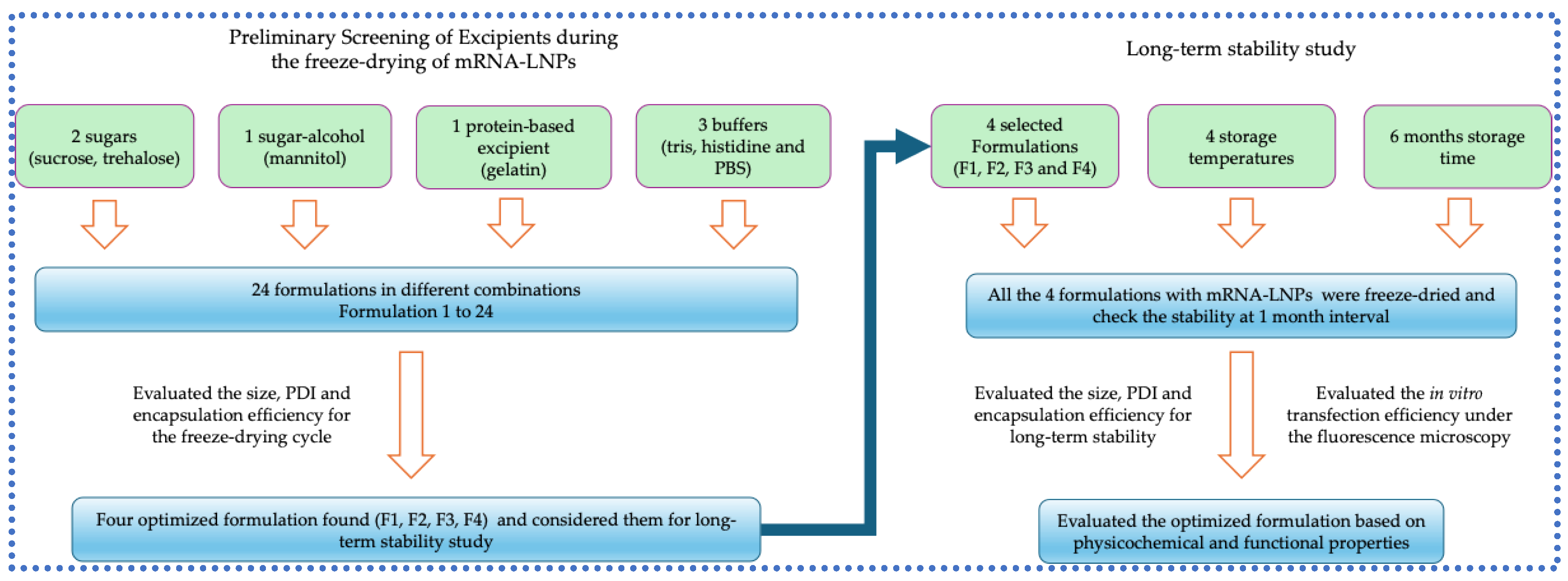
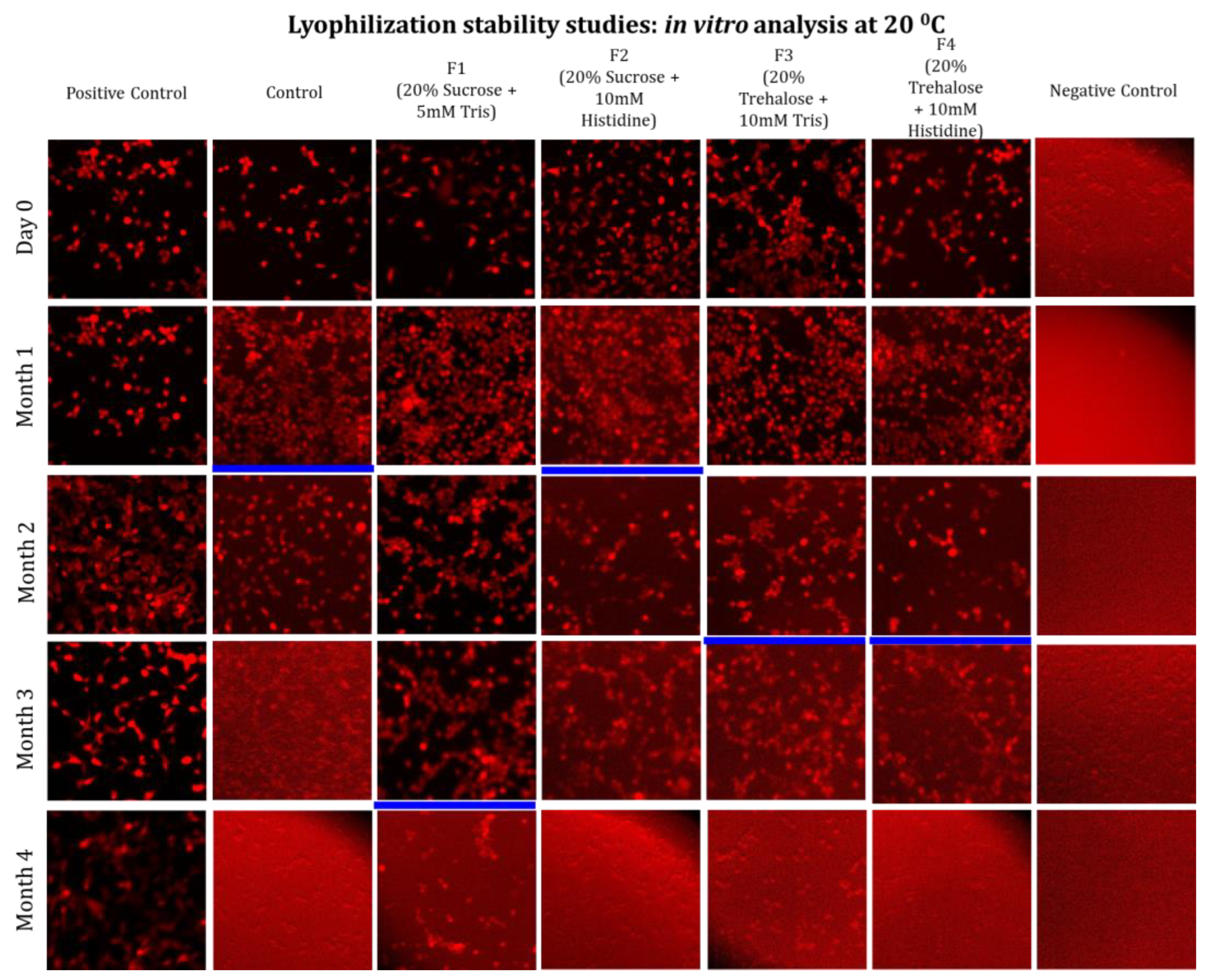
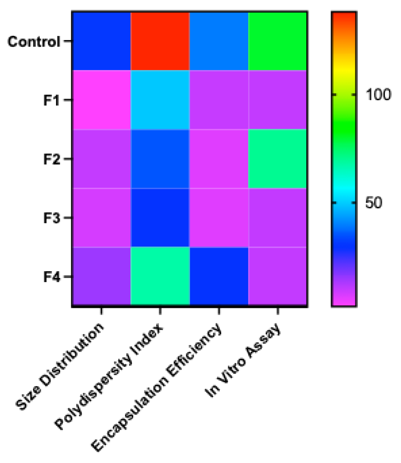
Background: Thermostability remains a key bottleneck for equitable access to mRNA–LNP vaccines, largely due to cold-chain requirements. Objectives and methods: Here, we optimized freeze-drying formulations by screening excipients (sugars, sugar-alcohols, and proteins) and buffers to preserve mRNA–LNP physicochemical (size, polydispersity index -PDI and encapsulation efficiency -EE) and fluorescence intensity-based, functional integrity assay (in vitro transfection) during long-term storage of up to 6 months. Results: In a preliminary screening study, different sugars (sucrose, trehalose), sugar alcohol (mannitol), protein (gelatin) and different buffers (Tris, PBS, histidine) were evaluated. The preliminary result showed that sucrose and trehalose, along with Tris and histidine buffers, had a positive effect on maintaining the physicochemical properties during freeze-drying, while mannitol, gelatin and PBS buffer had a negative effect. Based on these findings, the optimized formulations containing sucrose/Tris, sucrose/histidine, trehalose/Tris and trehalose/histidine were chosen, and a stability study was performed at −80, −20, 4, and 20 °C for six months. Conclusions: Overall, except for the samples maintained at 20 °C, no significant changes in the physicochemical quality of the freeze-dried mRNA-LNPs were observed over six months at −80, −20, and 4 °C. The in vitro stability study demonstrated stability at 4 °C for four months across all formulations, while a formulation with sucrose/Tris maintained satisfactory stability even at 20 °C for the same duration. The main results of this study demonstrate the feasibility of storing mRNA drug products as solid formulations at non-freezing temperatures (≤ 4 °C).

Keywords: 
;  ;  ;  ;  ;  ;  ;  ;  ;  ;  ;  ;  
Subject: 
Engineering  -   Bioengineering

1. Introduction

After a successful COVID-19 vaccination campaign, mRNA vaccines emerged as a next-generation platform for disease prevention and immunization strategy for many previously untreatable diseases, new pathogens, orphan and rare diseases, including delivery of CRISPR-Cas9 systems for gene editing for therapeutic interventions [1,2]. Unlike other vaccine modalities, such as protein subunit vaccines, whole cell-based vaccines, and cell-culture produced viral vaccines, which require complex manufacturing processes and bioprocessing facilities with high capital investment to fully comply with strict safety regulations, mRNA vaccine manufacturing platform is techno-economically feasible and swiftly scalable [3,4,5,6]. However, one of the critical bottlenecks is the poor stability of the core mRNA molecule (therapeutic component) and the encapsulating lipid nanoparticles (LNPs) delivery system [7,8]. This limited thermostability of the mRNA vaccines and their need for ultra-cold storage significantly hinders large-scale distribution and global delivery, particularly in low-resource regions [7,8].
The genetic information coding mRNA molecule is inherently susceptible to degradation through hydrolysis and oxidation [9]. The dominant pathway of mRNA degradation is the hydrolysis in the presence of water. This ultimately affects the base sequence and secondary structure of the mRNA [10]. On the other hand, oxidative damage breaks the mRNA strands, compromising the vaccine efficacy [11]. Also, the presence of nucleases can degrade the mRNA molecule and make it functionally inactive. Vaccine stability is also dependent on the molecular integrity of the mRNA sequence, including the 5′ cap, untranslated regions (UTRs), coding regions, and the poly-A tail, which are critical for successful translation [8,10]. Modifications in these regions can greatly affect the stability of mRNA.
Further, LNPs that encapsulate mRNA serve a two-fold, to aid as a delivery vehicle for the mRNA cargo into target cells and facilitate endosomal escape while also acting as a physical barrier for nucleases and other degradative factors outside the cell [2]. But the LNPs are also prone to stability issues, due to aggregation, fusion or collapse, leading to an increase in particle size or polydispersity index (PDI) of the LNPs [12]. Several factors, including exposure to light, oxygen, metal ion residues, lipid composition, structure, pH, temperature, and buffer, influence this instability [13].
Proper storage conditions are required to maintain the stability of mRNA vaccines. For example, Pfizer’s COVID-19 vaccine needs to be stored at −70 °C and Moderna’s COVID-19 vaccine at −20 °C [3]. To improve the thermostability of mRNA-LNP vaccine, current research focuses on LNP formulations and on advances in formulation strategies [14]. In this context, freeze-drying of mRNA vaccines has the potential to maintain stability at relatively higher temperatures. This is considered an effective process for mRNA vaccine formulation, converting the unstable liquid form into a stable dry powder. This might bypass the strict requirement for storage and transport at ultra-low temperatures [15].
The freeze-drying process comprises several controlled steps. Initially, the mRNA-LNPs are mixed with appropriate excipients (such as cryoprotectants and buffers), and freezing is done at a very low temperature. In the second step, primary drying is performed by applying a vacuum to sublimate water from the mRNA-LNPs formulation. A subsequent secondary drying step gently removes residual water by raising the temperature (up to around 20–40 °C) [1,3,16].
While converting the liquid formulation into a solid formulation by freeze-drying to remove water content seems a plausible approach, the freeze-drying process itself introduces additional in-process stresses that can degrade or destabilize the LNPs and the mRNA therapeutic cargo [17]. The choice of formulation components is vital for a successful freeze-drying cycle. Optimally chosen excipients play a major role in preserving the integrity and prolonging the shelf life of mRNA-LNPs during freeze-drying and after freeze-drying. Certain excipients, mainly sugars and buffers, are used to counteract the process-associated stress, such as excessive moisture [18]. Moisture if not optimally controlled, might lead to the hydrolysis of the mRNA cargo, as observed by several studies [19]. Sugars such as sucrose and maltose play a protective role by stabilizing the LNP structure during freeze-drying and reconstitution [15,20]. Tris- buffer is usually preferred over PBS because the latter induces mRNA leakage during drying [3,15,18]. Optimizing lipid ratios in LNPs is also critical for maintaining the stability of mRNA-LNPs vaccine [1,21].
In this study, we evaluated the effect of different excipients and buffers on the freeze-drying process of mRNA-LNPs. This evaluation guided the selection of optimized formulations for long-term stability at ambient conditions and at 4 ˚C. These optimized formulations were tested in physicochemical (to evaluate mRNA-LNP stability) and in vitro cell-based (HEK-293T) transfection assays to assess transfection efficiency and the integrity of the encapsulated mRNA during storage for up to 6 months under different storage conditions. The results of this study demonstrate the development of an efficient freeze-drying process using improved formulations to achieve long-term stability of mRNA drug product. The findings will ultimately contribute to extending the shelf life of mRNA-LNPs in less stringent cold-chain-free conditions.

2. Materials and Methods

2.1. Preparation of mRNA-LNPs

Capped mCherry-mRNA transcripts were purchased from TriLink Technologies (San Diego, CA, USA). The ionizable lipid ALC-0315 and PEG-lipid ALC-0159 were obtained from Cayman Chemical (Ann Arbor, MI, USA), whereas cholesterol and DSPC were sourced from Millipore Sigma (Burlington, MA, USA). Lipids were resuspended in 100% ethanol at a defined molar ratio of 50:10:38.5:1.5, corresponding to ALC-0315, 1,2-distearoyl-sn-glycero-3-phosphocholine (DSPC), cholesterol, and ALC-0159, respectively.
In parallel, the mRNA (25 µg/mL) was diluted in sodium acetate buffer with a pH 5.0. The lipid-to-mRNA weight ratio was carefully maintained at 10:1 to ensure optimal encapsulation efficiency. mRNA encapsulation into lipid nanoparticles (LNPs) was achieved by manual syringe mixing, where the ethanol-based lipid phase was mixed with the aqueous phase of mRNA solution. Following encapsulation, the resulting mRNA-LNPs solution was subjected to dialysis using Slide-A-Lyzer™ Dialysis Cassettes (10 kDa MWCO; Thermo Fisher Scientific, Waltham, MA, USA). Dialysis was carried out using 200X volume of 5 mM Tris buffer (pH 8.0) at 4 °C for 2 hours to exchange ethanol with the buffer.

2.2. Freeze-Drying

Preliminary formulations were prepared in the early stages of this study, as outlined in Table 2. The excipients used included sucrose, trehalose, and mannitol (ranging from 1% to 20%), gelatin (0.5% to 1%), Tris (5 mM), histidine (10 mM), and PBS (1X) at different concentrations. The optimized formulations identified in the preliminary study were used for the long-term study, as outlined in Table 3. The formulated mRNA-LNPs were prepared, filled into glass vials, and loaded into the Virtis Advantage Pro freeze dryer (SP Scientific, PA, USA).
The encapsulated mRNA-LNPs were mixed with appropriate formulations (excipients and buffer) at a ratio of 1:1 where the final concentration of mRNA was 12.5 µg/mL. Each aliquot (200 µL) was filled into 1 mL sterilized glass vials (WHEATON®, Millville, NJ, USA) and sealed with butyl stoppers. The cycle included freezing at −50 °C for 5 h, primary drying at −30 °C for 5 hours, and secondary drying for 1 hour with gradual temperature increase. The lyophilization cycle parameters were selected based on the glass transition temperature of the respective formulations, as well as prior studies [17,20] that identified optimized conditions for mRNA–LNPs. Vials were stoppered under vacuum without inert gas backfilling.
Table 4. Freeze-drying parameters for mRNA-LNPs.
Freezing Primary Drying Secondary drying
Temperature -50 °C -30 °C 20 °C
Pressure - mTorr 30 mTorr

2.3. Stability Study

All the freeze-dried formulations containing mRNA-LNPs were kept at different time points (0 to 6 months) at different storage conditions (-80, -20, 4, and 20 °C). After specific time points, the freeze-dried cakes were resuspended in RNAse-free water and the size, PDI, EE and transfection efficiency were assessed. After reconstitutions, these formulations were characterized for material properties based on size distribution, polydispersity index and encapsulation efficiency.

2.4. mRNA-LNP Characterization

2.4.1. Encapsulation Efficiency (EE%)

The encapsulation efficiency of mRNA was quantified using the protocol of the Quant-iT RiboGreen RNA Assay Kit (Thermo Fisher Scientific, Waltham, MA, USA). The mRNA-LNPs samples were diluted in 1X TE buffer to measure unencapsulated RNA or in 2% Triton X-100 to measure the total RNA content. To disrupt the LNPs, the plate was incubated with 2% Triton X-100 for 10 min at 37 °C, then RiboGreen dye was added. A standard curve was established using the ribosomal RNA standards supplied with the kit. Fluorescence intensity was then recorded using an Agilent BioTek Synergy HTX MultiMode Microplate Reader (Agilent Technologies, Santa Clara, CA, USA).

2.4.2. Particle Size and Polydispersity Index (PDI)

The particle size and PDI of the mRNA-LNPs were determined by dynamic light scattering (DLS) on a Zetasizer Ultra (Red Label) (Malvern Panalytical, Malvern, UK). Samples were equilibrated to room temperature, with a refractive index of 1.37, and measured in 50 µL disposable cuvettes (Sarstedt Inc., Germany) to determine size and PDI.

2.5. In Vitro Assay for mRNA-LNPs

HEK293 cells were seeded into 96-well plates and cultured for 24 hours to allow cell attachment and growth. Following this, the cells were transfected with freeze-dried mRNA-LNP formulations and the control (without formulations). The transfected cells were then incubated for an additional 24 hours at 37 °C with 5% CO2 to enable mRNA uptake and expression. After the incubation period, all the wells were imaged using an inverted Olympus IX83 confocal microscope to assess fluorescence intensity, indicating successful transfection of the mRNA-LNPs. The images from the microscope were processed and analyzed as described elsewhere [23] using ImageJ FIJI [24], to estimate the timepoint at which the transfection expression was not observed, indicating the lack of functional mRNA in the LNP-excipient sample.

3. Results

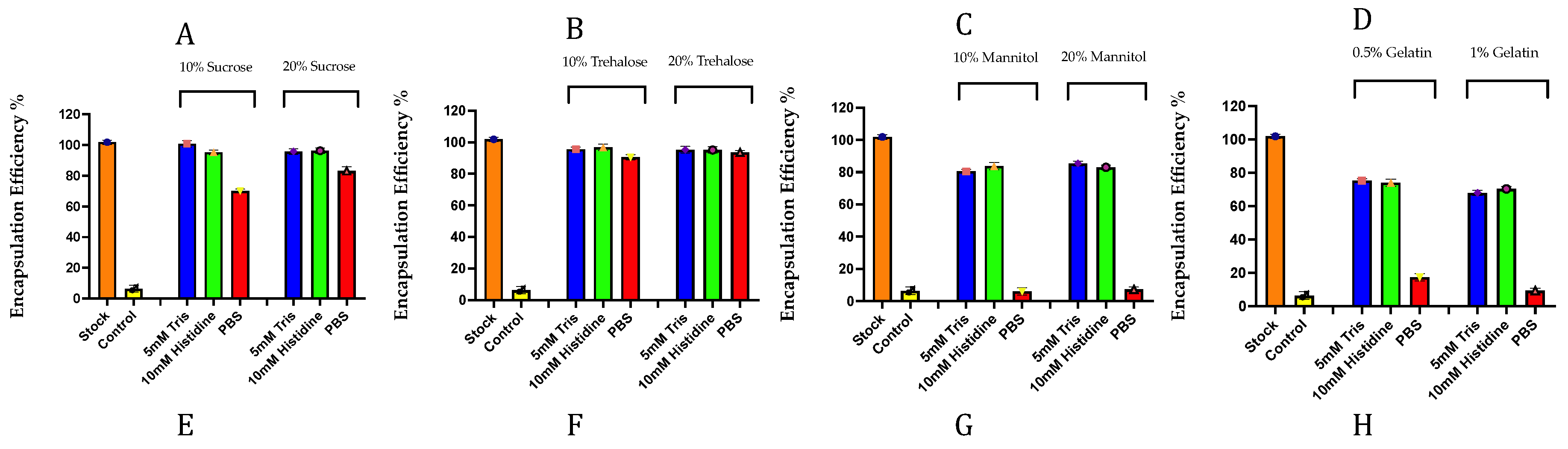
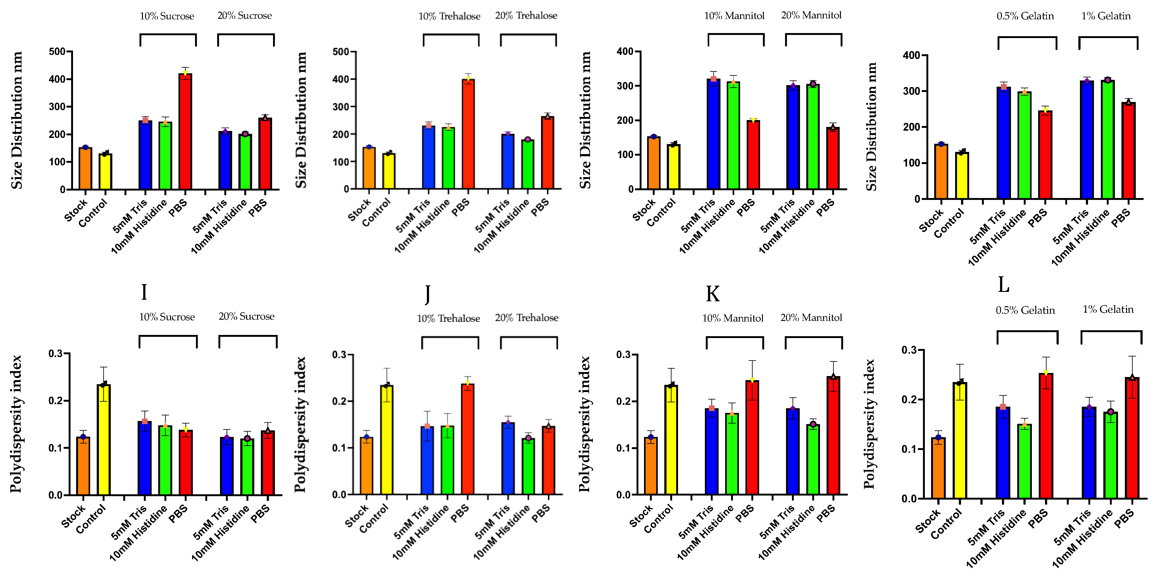
3.1. Preliminary Screening of Excipients During the Freeze-Drying of mRNA-LNPs

A range of excipients, namely two sugars -Sucrose and trehalose, a sugar-alcohol - mannitol and a protein-based lyoprotectant - gelatin, were combined with three different buffers – Tris, Histidine and PBS in different combinations and were assessed for freeze-drying mRNA-LNPs (Table 2). All these formulations were freeze-dried, reconstituted immediately, and the physical parameters, namely size, PDI, and encapsulation efficiency (Figure 3), were evaluated. The goal at this point was to identify the best formulation through physicochemical studies for subsequent long-term storage studies at ambient temperatures.
Among the buffers and molarity conditions tested, 5 mM tris and 10mM histidine are used because these are found effective in other freeze-drying vaccine formulations. This exhibited consistently high encapsulation efficiency at both 10% and 20% sucrose, closely matching the initial stock mRNA-LNPs. In contrast, PBS buffer showed very low efficiency at 10% sucrose but improved slightly at 20% sucrose, still resulting in a 20% loss of encapsulation efficiency compared to freshly prepared mRNA-LNPs (Figure 3A). Excipient formulation of tris and histidine with trehalose showed high encapsulation efficiency at both concentrations (Figure 3B). Sugar-alcohol like mannitol showed around 20% loss in both the Tris and histidine buffer. Another worst-performing combination was mannitol with PBS, which showed a significant decline in encapsulation efficiency (90% loss; Figure 3C). Also, the same trend was observed with gelatin in a Tris-histidine buffer, with a 30% loss in encapsulation efficiency. Like mannitol, gelatin also showed a major drop in encapsulation efficiency with PBS buffer, with a loss of around 80% to 90% (Figure 3D).
Also, the size distribution of lipid nanoparticles varied noticeably with buffer and sugar concentration. In 10% sucrose, Tris, and histidine produced particles around 240 nm, while PBS yielded much larger particles around 400 nm. At 20% sucrose, both Tris and histidine yielded smaller particles around 200 nm, whereas PBS resulted in relatively large particles around 260 nm. Also, trehalose formulations displayed a similar trend in particle size distributions. At 10% trehalose, Tris and histidine generated particles roughly around 220 nm, while PBS produced the largest size at about 400 nm. Increasing trehalose concentration to 20% with Tris and histidine, maintained particle sizes (~200 nm and ~180 nm, respectively), but PBS yielded larger particle sizes (~300 nm). Again, the buffer played a major role with mannitol, where 10 and 20% mannitol with Tris and histidine resulted in particle sizes around 300 nm, while PBS, unlike, yielded particles near 170-200nm. The gelatin with buffer also shows a negative impact on size distribution. With 0.5 and 1% gelatin, Tris and histidine maintain particles of ~300 nm, and PBS yielded sizes near 250-280 nm.
From the PDI data, the results demonstrate that the choice of stabilizer and buffer also affects the homogeneity of the freeze-dried mRNA-LNP sample, reflecting the uniformity of particle size. Samples with sucrose, trehalose, mannitol and gelatin showed lower PDI values (< 0.2), especially when formulated with Tris and histidine buffers, indicating good homogeneity and stability. In contrast, mannitol and gelatin formulations, particularly those containing PBS buffer, exhibited higher PDI values (>0.2). Overall, all these excipients combined with Tris and histidine buffer maintained a more consistent particle distribution.
Overall, the data demonstrated that both buffer type and excipient concentration play a vital role in achieving a standard encapsulation efficiency (Figure 4). Among the buffers, Tris and Histidine looked more promising than PBS. Among the sugars, sugar-alcohols and protein-based lyoprotectant, sugar (sucrose and trehalose) showed superior outcomes in terms of encapsulation efficiency, PDI and size compared to sugar alcohols and proteins. From the above series of physiochemical analysis, the four best formulations were tested further for the next stages of long-term storage studies. The experimental plan is shown in Figure 4, below. Further, though we tested multiple percentages of the lyoprotectant, like 1%, 5%, 10%, 20% (shown in Table 2) for different formulations, we did not test beyond 20% of sugar or sugar alcohols, as high lyoprotectant will lead to excessive viscosity of the injectable formulated product, leading to side reactions like pain during administration. Also, it could contribute to crystallization and other undesirable effects during the lyophilization process optimization. Further, the higher the percentage of the sugars or sugar alcohols, the better the physicochemical stability was achieved at the end of the free-drying cycles. Hence, the next stages of stabilised 20% sugar or sugar alcohol as detailed below.

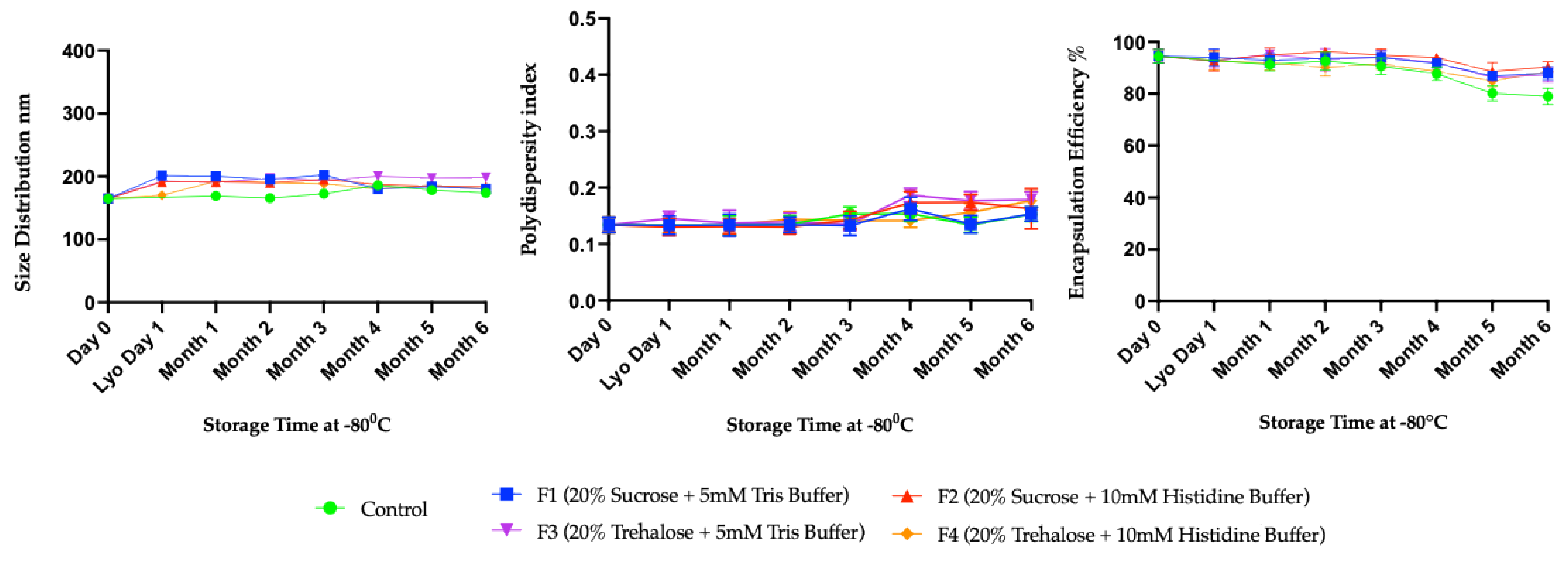
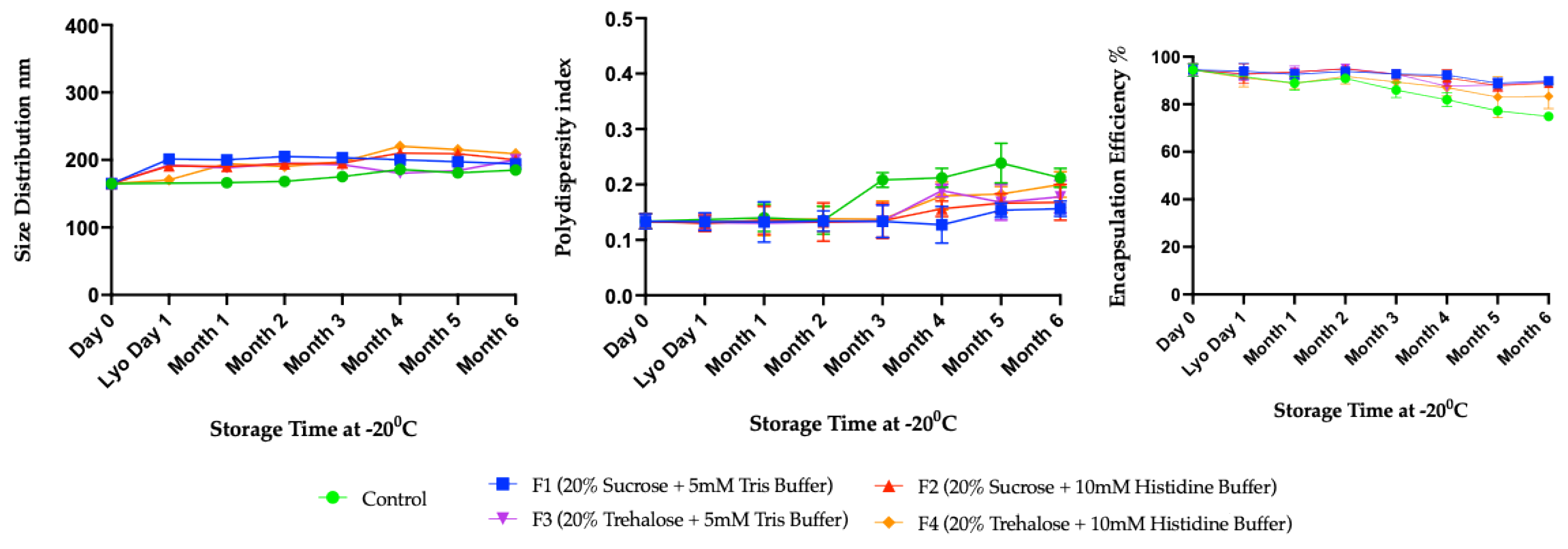
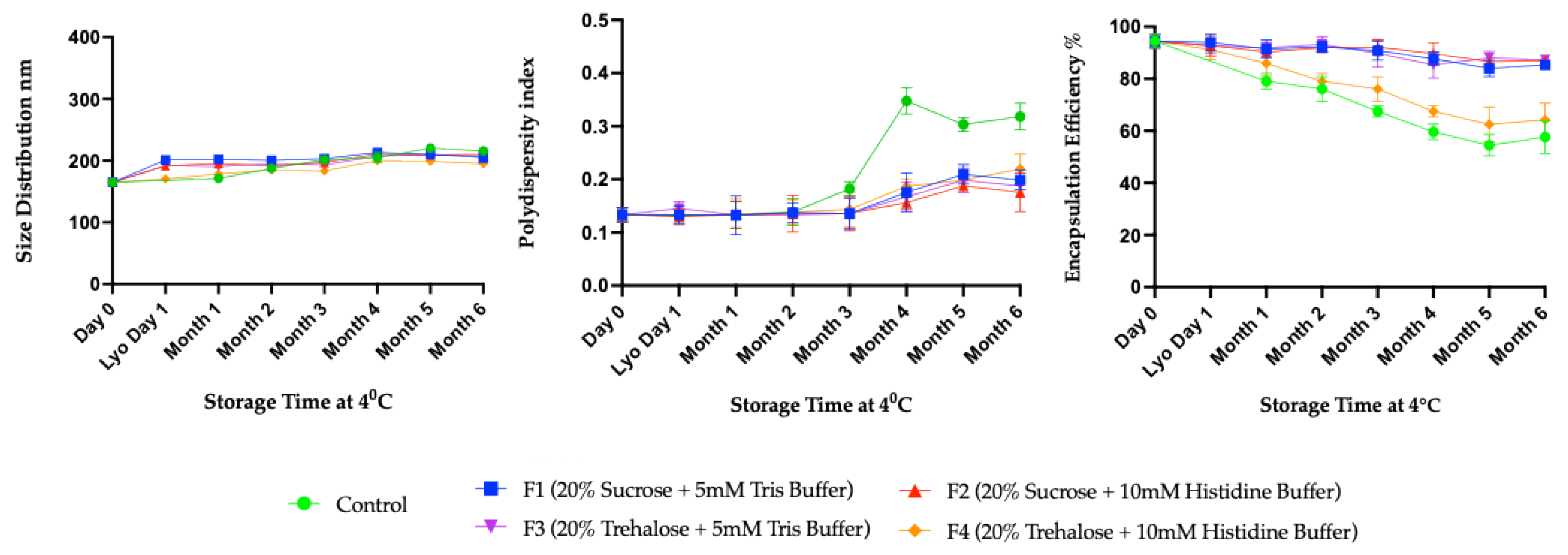
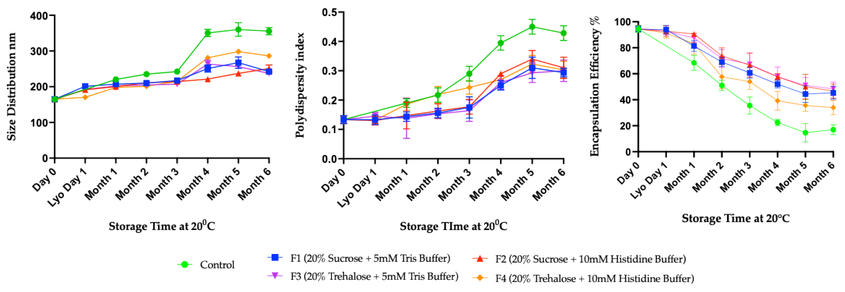
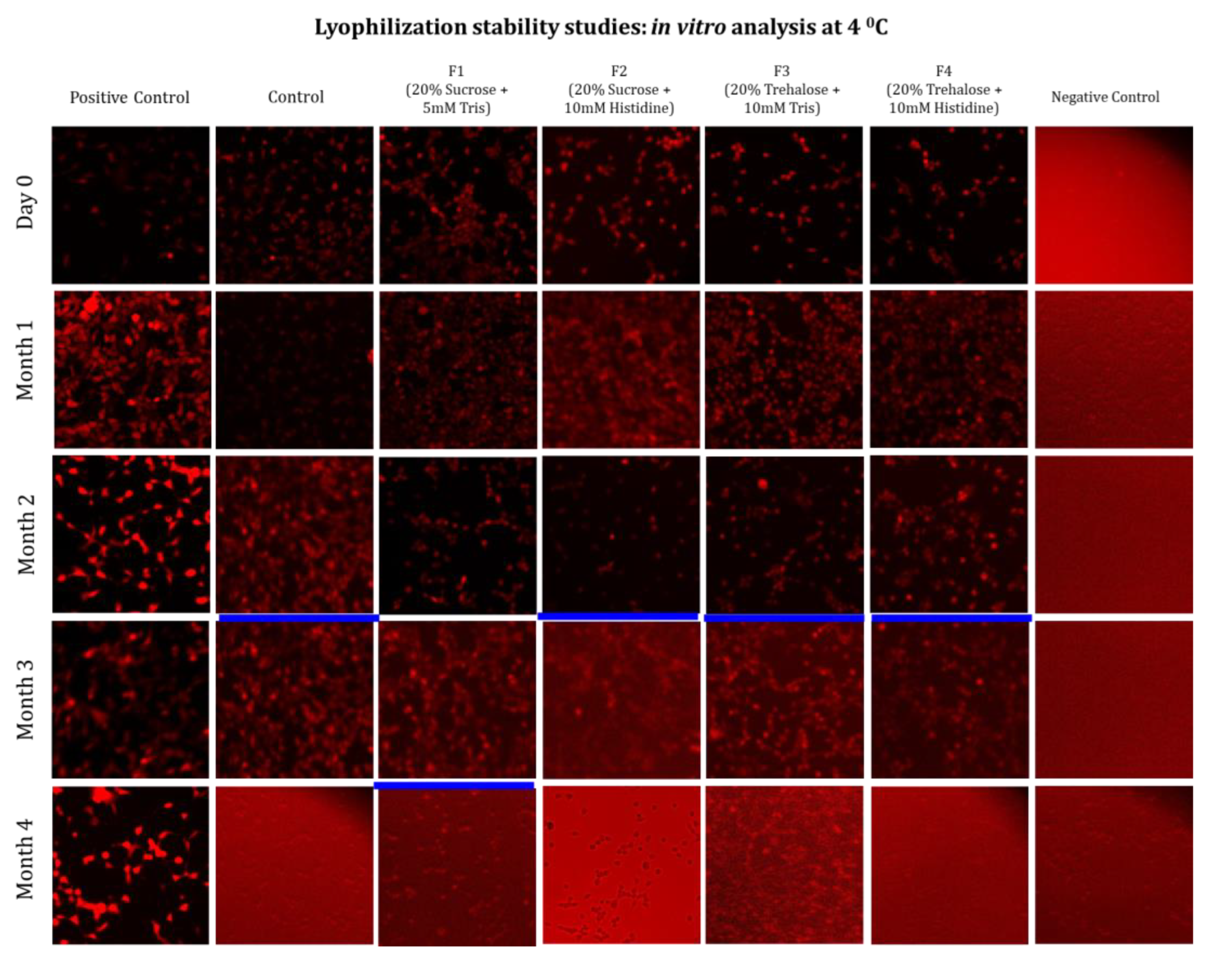
3.2. Long-Term Stability Study

From the preliminary study shown above, the four formulations labelled as F1 (20% sucrose + 5 mM Tris), F2 (20% sucrose + 5 mM histidine), F3 (20% trehalose + 5 mM Tris) and F4 (20% trehalose + 5 mM histidine) were selected for a long-term stability study (Table 3). Also, one liquid control, which was not lyophilized, was kept during the stability study at every temperature and every storage time point as a control. Figure 5, Figure 6, Figure 7 and Figure 8 present the stability characterization using particle size, polydispersity index (PDI), and encapsulation efficiency of mRNA-loaded lipid nanoparticles (mRNA-LNPs) stored under different temperatures (-80 ˚C, -20 ˚C, 4 ˚C, and 20 ˚C).
The freeze-dried mRNA-LNPs formulations stored at −80 °C demonstrated remarkable physical stability, as indicated by particle size measurements across all samples. The mean size distribution remained consistently approximately 200 nm over six months of storage (Figure 5). This indicates that there was no significant aggregation or fusion of nanoparticles over the stability period. Polydispersity index (PDI) analysis further supports the robustness of these formulations. Throughout the six-month storage period, the PDI values for all samples remained low, generally between 0.12 and 0.20, indicating that there was no major deviation in the distribution of the particle-size throughout storage (Figure 5). These findings indicate that lyophilization and storage at −80 °C did not produce significant heterogeneity within the lipid nanoparticle population.
Also, the encapsulation efficiency data confirm that mRNA cargo retention within lipid nanoparticles was maintained during long-term storage (Figure 5). The encapsulation efficiency remained high in all freeze-dried formulations stored at −80 °C, consistently ≥90% over six months. However, a modest decline of about 5% was observed towards the end of the 6-month time points in these freeze-dried formulations. This loss was limited and did not compromise the overall encapsulation performance, compared with the 10-15% loss observed in the liquid control. All these data collectively demonstrate that freeze-dried mRNA-LNPs maintained the key physicochemical properties at −80 °C throughout the study period of more than 6 months.
Figure 5. Long-term stability data using four formulations of mRNA-LNPs at -80 °C. These formulations were characterized at one-month intervals using DLS and the Ribogreen assay to assess size distribution, polydispersity index (PDI), and encapsulation efficiency. All assays were done in biological replicates of n=3.
Preprints 192014 g003
Further, the stability assessment of mRNA-LNPs stored at -20 °C demonstrated that all formulations maintained relatively stable particle size (Figure 6, left) and PDI (Figure 6, middle) over time, although the control showed increases in both size and PDI (Figure 6), due to the lack of excipients. Particle size distributions remained predominantly around 200 nm across all formulations, indicating that the freeze-dried formulations prevented aggregation or fusion of LNPs. Polydispersity index (PDI) analysis also reveals that most formulations maintained robust homogeneity, with PDI values below 0.20 throughout the storage period (Figure 6). In contrast, the control showed an increase in PDI after four months, peaking at 0.25 at six months, indicating substantial heterogeneity and potential particle destabilization. Further, the encapsulation efficiency of control at −20 °C declined more drastically than in ultra-low storage, dropping below 80% after six months (Figure 6). The decline in EE% began after 2 months at -20 ˚C. Formulations containing sucrose or trehalose retained encapsulation efficiency (Figure 6, right) around 90% for up to six months, except the F4 formulation, which had 10 mM histidine instead of 5mM Tris. Overall, these findings confirm that lyophilized mRNA-LNP formulations F1, F2 and F3 were stable at −20 °C during the entire 6 months of observation (Figure 6).
Figure 6. Long-term stability data using four formulations of mRNA-LNPs at -20 °C. These formulations were characterized at one-month intervals by DLS and Ribogreen assay to observe the size distribution, PDI and encapsulation efficiency. All assays were done in biological replicates of n=3.
Preprints 192014 g004
Figure 7 shows mRNA-LNP particles and their physicochemical properties at 4 °C. Here, all formulations initially exhibited similar particle sizes up to 3 months. However, as storage time progressed, the control showed a more pronounced increase in particle size (~230 nm) than the other formulation (Figure 7, left). Except for F4, the other three freeze-dried formulations F1, F2 and F3, maintained constant size distributions throughout the six months. In the PDI graph (Figure 7, middle), all formulations retained low PDI values, indicating a sustained, homogeneous nanoparticle population over time. In contrast, the control’s PDI sharply increased within four months (>0.3), indicating greater size heterogeneity likely due to aggregation or fusion (Figure 7, middle). The PDI data emphasized that freeze-dried formulations with sucrose or trehalose as cryoprotectants, in Tris and histidine buffers, effectively maintained both size stability and particle uniformity during long-term storage. Also, the encapsulation efficiency tended to decrease with storage time, although the three formulations, F1, F2, and F3, maintained their encapsulation efficiencies over the 6 months of the stability study (Figure 7, right). The most pronounced decline was observed in the control group, falling below 60% by six months in the absence of stabilizing excipients. The next highest loss was observed in the F4 formulation containing 20% trehalose and 10 mM histidine. This suggests that mRNA integrity and encapsulation decline over time in the absence of appropriate formulation, whereas inclusion of suitable cryoprotectants and excipients conclusively enhances stability. In summary, these results collectively illustrate that LNP formulations containing sucrose or trehalose with Tris and sucrose with histidine buffer (F1, F2 and F3) were found most effective in maintaining size, dispersity, and encapsulation efficiency during long-term storage at 4 °C. These trends validate the critical role of optimized cryoprotectant and buffer combinations in enhancing the shelf life of mRNA-LNPs.
Figure 7. Long-term stability data using four formulations of mRNA-LNPs at 4 °C. These formulations were characterized at one-month intervals by DLS and Ribogreen assay to observe the size distribution, PDI and encapsulation efficiency. All assays were done in biological replicates of n=3..
Preprints 192014 g005
The results at 20 °C demonstrate a different representation of the stability of freeze-dried mRNA-LNPs over six months (Figure 8). The size and PDI at 20 °C showed more marked increases compared to 4 °C, highlighting that higher temperatures accelerate mRNA-LNPs instability (Figure 8 left and middle). For size distribution, the control exhibited a sharp increase after three months, exceeding 350 nm. But all four formulations (F1–F4) maintained comparatively smaller size over six months. Here, the F1, F2 and F3 retain the particle sizes below 220nm, while the other formulation, F4, exceeds particle sizes above 250 nm. The PDI also followed a similar trend. The control group showed a marked increase in polydispersity from the first month, reaching 0.4 at six months (Figure 8, middle). Such high polydispersity indicated that the particles became more heterogeneous, likely due to aggregation. In contrast, the four formulations kept the PDI below 0.2 for 3 months, indicating more uniformity and homogeneity. After 3 months, all the freeze-dried formulations showed a sharp increase, reaching a PDI of 0.3 over 6 months. This indicates that the freeze-dried formulations could not maintain the homogeneity at this elevated temperature for a long time, likely again due to aggregation or fusion of LNPs. At 20 °C, the EE dropped rapidly in the control, falling below 20% at six months (Figure 8, right). This indicates poor retention of the mRNA payload within the LNPs. Also, major loss was observed in all the freeze-dried formulations, retaining between 40% and 60% efficiency at six months. Overall, the data showed the clear instability of lipid nanoparticles at 20 °C and the ineffectiveness of freeze-dried formulations in maintaining physical integrity and encapsulation efficiency.
Figure 7. Long-term stability study using four formulations of mRNA-LNPs at 20 °C. These formulations were characterized at one-month intervals by DLS and Ribogreen assay to observe the size distribution, PDI and encapsulation efficiency. All assays were done in biological replicates of n=3.
Preprints 192014 g006
In addition to physicochemical studies, key evidence of mRNA integrity under storage conditions, in the presence of excipients, and at different temperatures is provided by in vitro transfection assays. The main goal of the in vitro transfection assays is to evaluate the integrity of mRNA encapsulated within LNPs across excipient combinations and to determine whether changes in the physicochemical properties of mRNA-LNP excipient formulations stored at 4 ˚C and 20 ˚C affect transfection efficiency. Thus, the in vitro study assessing transfection efficiency used HEK293 cells and fluorescence microscopy. The red fluorescence signal corresponded to the expressed mRNA delivered by the LNPs, serving as a qualitative indicator of transfection within the cells. Figure 8 presents representative fluorescence microscopy images of cells transfected with the control (without any formulations) and freeze-dried formulations (F1–F4) up to 4 months of storage at 4 °C. Red fluorescence corresponds to reporter protein expression, reflecting the retained biological activity of each formulation, i.e., mRNA integrity. The stock formulation exhibited a high fluorescence signal for up to two months. Thereafter, the signal intensity progressively weakened, becoming undetectable by the fourth month (Figure 8). Notably, freeze-dried formulations F1, F3, and F4 showed consistently strong fluorescence signals for up to four months at 4 °C. This indicates that these formulations effectively maintained the biological functionality of the mRNA-LNPs during this time frame. In contrast, formulation F2 exhibited a weaker fluorescence signal at 4 months, suggesting major loss of possible structural integrity and reduced transfection efficiency after prolonged storage.
Collectively, this finding highlights the superior performance of F1, F3 and F4, which preserved the functional activity up to four months, thereby demonstrating their suitability for long-term stabilization of the formulation.
Figure 8. Long-term mRNA integrity test by in vitro transfection study using four formulations of mRNA-LNPs at 4 °C. These formulations were characterized at one-month intervals by measuring transfection efficiency using fluorescence microscopy. All assays were performed in biological replicates of N=3, while the image presented above is a representative of one of the biological replicates. The blue line in each of the samples (across each column) represents the time point when transfection efficiency was lost, probably due to the mRNA-LNP integrity being affected.
Preprints 192014 g007
Again, fluorescence microscopy was performed for the freeze-dried formulations stored at 20 °C for 4 months (Figure 9). After 4 months of storage, formulation F1 exhibited a strong fluorescence reported intensity. In contrast, F2, F3, and F4 shows reduced fluorescence intensity after 1 month. Also, the control started to show a very weak fluorescence signal from after 1 month of storage and the signal disappeared after 2 months of storage.
This finding demonstrates that the composition of the sucrose/Tris matrix plays a critical role in maintaining the functional stability during long-term storage at relatively higher temperatures.
To summarize the overall results of the storage stability studies, a heatmap demonstrated that freeze-dried mRNA-LNPs formulated with sucrose or trehalose in combination with Tris and histidine buffers (F1 and F3) provide superior stabilization in both physicochemical attributes and biological activity during long-term refrigerated storage at 4 °C (Figure 10). In the heat map, all formulations with a light shade (pink) indicate that stability is well maintained. All squares with primary colors, red and green shade, mean at least more than 80% loss in stability. For transfection efficiency, it was a ‘yes’ or ‘no’ type of response where all formulations except control and F2 showed In contrast, these results validate the critical role of optimized cryoprotectant and buffer systems in extending the shelf life and functional integrity of mRNA-LNP vaccines.

4. Discussion

Impact of Manual Mixing on Initial mRNA–LNP Physicochemical Properties

This study used manual mixing for mRNA-LNP encapsulation, which, in general, provides slightly larger LNP-mRNA particles, which corroborates well with our reported data here [25]. Also, the addition of excipients to the mRNA-LNP will increase the effective size of the mRNA-LNPs, as previously reported elsewhere. [26]. The observation here suggested that these were well reproduced in this study as well. While it is desirable to use microfluidic or Tee-junction-based encapsulation approaches, the current study employs a manual mixing approach for its simplicity and low sample handling. Based on these, the discussion section below highlights the key general observations and the respective rationales for the experimental observations in the stability studies described above.

Freeze-Drying–Induced Stresses and Their Effects on mRNA–LNP Stability

Freeze-drying subjected lipid nanoparticles to a combination of severe stresses, including ice crystal formation during freezing, dehydration during primary and secondary drying, and physicochemical perturbations during reconstitution. These stresses can induce changes in pH, ionic strength, and molecular organization, ultimately destabilizing LNP structures. In the absence of appropriate protective excipients, such stresses frequently lead to particle aggregation, fusion, and leakage of encapsulated mRNA. The observed increases in particle size and polydispersity index (PDI) across several formulations following lyophilization underscore the sensitivity of mRNA–LNP systems to freeze-drying conditions. These findings are consistent with prior studies reporting enlargement and heterogeneity of LNPs as a consequence of structural stress during dehydration [3,15,17,20]

Role of Excipients in Preserving Particle Size and Polydispersity During Lyophilization

Freeze-drying imposes severe freezing and dehydration stress on lipid nanoparticles. The preliminary screening of mRNA-LNPs formulations highlighted the significant impact of excipients and buffer on the physicochemical properties of lipid nanoparticles during the freeze-drying process. Formulations containing mannitol, gelatin, with Tris, histidine and PBS buffer demonstrated noticeable increases in particle size and polydispersity index (PDI) after freeze-drying (i.e., non-stable formulations). During freeze-drying, mRNA-LNPs are exposed to freeze-drying stresses such as freezing, dehydration, and reconstitution, which bring changes in pH or ionic strength and trigger partial fusion or aggregation of lipid nanoparticles. Ultimately, these stresses disrupt the stability of the LNPs by causing them to cluster together and resulting in a noticeable increase in average particle diameter after reconstitution. This is consistent with previous findings, where freeze-dried LNP formulations showed size enlargement due to structural stress during drying [3,15,17,20].

Structural Rearrangements of Lipid Nanoparticles During Freeze-Drying

Also, during freezing, ice crystallization and water removal cause phase separation and mechanical disruption of the lipid bilayer. This ultimately forces lipids to reorganize and form a bleb-like structure on the nanoparticle surface [16,18,20]. These blebs typically exclude encapsulated mRNA, as their formation occurs after initial assembly and loading steps. In contrast, blebs generated during LNP self-assembly retain the mRNA, maintaining particle stability and functional potency [27]. The lack of nucleic acids within lyophilization-induced blebs leads to reduced transfection efficiency [18,28]. This structural discrepancy underlies the mechanistic link between lyophilization and the changes in the physicochemical properties. Optimizing the freeze-drying process parameters as well as the formulations helps to prevent possible aggregation to maintain the size and PDI. Here, sucrose and trehalose with Tris and histidine buffer are considered optimized formulations because they effectively maintain the size and PDI at the end of the free-drying cycle.

Mechanistic Basis of Encapsulation Efficiency Loss in Excipient-Free Formulations

Also, there was a marked reduction of encapsulation efficiency found in the control without excipients, paralleling results by Muramatsu et al. (2022), who reported that excipient-free liquid formulation suffers severe encapsulation loss in the freeze-drying cycle as well as during storage [29]. When the process is carried out without excipients, the nanoparticles are exposed to mechanical and chemical stresses. The absence of excipients magnifies the risk of irreversible damage (due to the stripping of essential water molecules and mechanical collapse). These stresses disrupt lipid nanoparticles which ultimately leads to the leakage of the encapsulated mRNA [2,3,15,30,31]. In contrast, optimized freeze-dried formulations provide a stabilizing matrix that helps to preserve nanoparticle shape, size, and the integrity of their mRNA payload through each stage of drying and subsequent storage [3,15,32,33,34].

Sugars Are Better Cryoprotectants for mRNA-LNPs Than Sugar Alcohols and Proteins(Gelatin)

Here, in this study, it was resounding that sucrose and trehalose effectively maintained the encapsulation efficiency and particle size distribution in the freeze-drying process by elevating the denaturation temperature of lipids as well as the glass transition temperature [3,17,20]. Sucrose binds more directly to the surface of the LNPs to maintain stability at higher temperatures and lower water content [35]. However, trehalose shows more thermal stability under low-temperature freeze-drying conditions. In contrast, mannitol can lead to increased particle size and lower encapsulation efficiency, likely due to its crystallization behaviour and limited protective interaction with the LNPs [36]. Also, gelatin-based (protein) excipient formulations form larger and less uniform particles with very low encapsulation efficiency, making them less ideal for the freeze-dried formulation.

Buffer-Dependent Modulation of pH, Ionic Strength, and LNP Integrity

Buffers also play a vital role during the freeze-drying of mRNA-LNPs vaccines [28,37]. They regulate the ionic environment as well as pH fluctuations in both the freezing and drying phases. This regulation helps maintain the native shape of the LNPs [14]. In this study, Tris and histidine consistently yielded smaller particle sizes and higher encapsulation efficiency during freeze-drying. Also, a recent study showed that 5 mM Tris maintains the physicochemical properties during the freeze-drying [10,15,38]. Tris and histidine buffers regulate the pH changes under acidic conditions in the presence of Mg²⁺ and Ca²⁺ [1,19,39,40]. Tris also protects the mRNA by scavenging hydroxyl radicals in the freeze-drying process [13]. On the other hand, PBS leads to a larger particle size and lower encapsulation efficiency. The negative effects of PBS buffer align with prior reports highlighting that the high ionic strength of PBS destabilizes the lipid layers [18]. PBS also undergoes substantial pH changes in the freeze-drying process when the temperature shifts from ambient chamber temperature to ultra-low temperature. Additionally, the histidine tested here for stability studies is one of its kind. Although it did not perform well with trehalose, the use of histidine with sucrose is an addition to the existing library of excipients.

Glass Transition and Vitrification Effects in Stabilizing mRNA–LNPs

This superior stability can be attributed to several well-documented mechanisms in the literature. Both sucrose and trehalose provide cryo- and lyoprotectant effects via vitrification mechanism, forming an amorphous glassy matrix. The formation of an anhydrous glassy matrix by cryoprotectants like sucrose or trehalose immobilizes both mRNA and lipid components. This reduction in molecular mobility limits hydrolytic and oxidative reactions. Also, this rigid, vitrified structure subdues nanoparticle aggregation and fusion during storage [16,20,35]. Sucrose and trehalose interact with polar head groups on lipid bilayers and with the phosphate backbone of the RNA. This replaces water molecules through hydrogen bonding during freeze-drying stages. This hydrogen-bond network inhibits direct contact between lipids and ice crystals, reducing mechanical stress and sustaining the native arrangement of LNPs [16,20]. Additionally, these sugars form thin, amorphous coatings that protect encapsulated mRNA from exposure to environmental RNases [3,16,35,44].

Long-Term Stability of Freeze-Dried mRNA–LNPs at Subzero and Refrigerated Temperatures

In the long-term stability study, all freeze-dried formulations stored at −80 °C showed physicochemical stability of the mRNA-LNPs. This confirms that ultra-low temperature storage preserves the mRNA-LNPs’ integrity [29]. Storage at -80 °C is regarded as the gold standard for the long-term preservation of the structural integrity and function of mRNA-LNPs [3,14,41,42]. Also, freeze-dried formulations stored at −20 °C showed promising stability in terms of physiochemical properties and in vitro assays. These results correspond to similar mRNA-LNPs formulations, where standard freezer conditions enable acceptable shelf-life when combined with optimal excipients [43]. At such low temperatures, all chemical degradation processes such as hydrolysis, oxidation, or enzymatic cleavage, are near zero, explaining the stability properties. As a result, mRNA remains highly intact, and the lipid nanoparticle structure is preserved, with minimal aggregation or leakage [13].
The freeze-dried mRNA-LNPs formulations also showed good stability at 4 °C in long-term storage. This reflects comparable data from Muramatsu et al. (2022), who showed that refrigerated lyophilized LNPs retain most physicochemical properties over months [29]. Also, the freeze-dried mRNA-LNPs formulations maintain the biological activity following four months at 4 °C.

Limitations of Ambient Temperature Storage (20 °C)

However, at 20 °C, all samples are found unstable with increased size, PDI and marked loss in encapsulation. This instability was partially prevented by the freeze-dried formulations in the early stage (up to 3 months, in some formulations), but they could not fully prevent degradation or particle aggregation in the long run. These results align with reports from Meulewaeter et al. (2023) and Zhao et al. (2024), who found difficulty in maintaining the stability of mRNA-LNPs at ambient temperature [18,43]. Maintaining optimized freeze-dried mRNA-LNP formulations at 20 °C is difficult because of accelerated degradation compared to storage at 4 °C. At 20 °C, residual moisture and increased molecular mobility within the lyophilized matrix led to enhanced rates of lipid oxidation and hydrolysis. This results in reduced encapsulation efficiency and aggregation or fusion of nanoparticles [3,7,11]. The protective glassy matrix formed by cryoprotectants become less effective at higher temperatures because of increased molecular diffusion and possible crystallization of excipients [19,20]. Therefore, although lyophilization improves stability, long-term storage at 20 °C remains challenging. This requires further formulation optimization or stabilization strategies to ensure longer shelf-life. Based on the tested formulations the in vitro transfection study reveals that only F1 (20% sucrose + 5 mM Tris) retained the biological activity up to 4 months (Matrix, Figure 9). The probable mechanism is that the glassy matrix of sucrose maintained the functional integrity of mRNA-LNPs with a complementary effect by the tris, contributing to stability through pH buffering at this relatively high temperature.

5. Conclusion

In conclusion, this study confirms the critical role of cryoprotectants (sucrose and trehalose) and appropriate buffers (Tris and histidine) in stabilizing mRNA-LNPs during freeze-drying and prolonged storage at wide range of temperatures. While ultra-low temperature and refrigerated temperature maintain LNPs integrity and encapsulation effectively, higher temperature stability remains subtle. These insights improve the understanding of excipient and buffer selection essential for advancing freeze-drying of mRNA-LNPs formulation. Overall, these findings provide a strong foundation for rational formulation design, enabling the development of more robust, storage-stable mRNA-LNP vaccine candidates suitable for global distribution.

Author Contributions

M.F.H.K.: conceptualization, literature review, methodology, experimentation, investigation, data collection/presentation, statistical analysis, writing—original draft preparation, writing—review and editing. A.S.P.: methodology, experimentation, investigation, data collection/presentation, visualization, writing—review and editing. A.A.K.: conceptualization, supervision, funding acquisition, writing—review and editing. All authors have read and agreed to the published version of the manuscript.

Funding

Natural Sciences and Engineering Research Council (NSERC) RGPIN-2021-02691 and Canada Research Chair Grant Number CRC-240394. Canada Biomedical Research Fund, Tri-council Agency, CBRF2-2023-00072.

Data Availability Statement

The original contributions presented in the study are included in the article, further inquiries can be directed to the corresponding author.

Acknowledgments

The authors thank all the members of the Bioprocessing of Viral Vectors and Vaccines team in the Bioengineering Dept. McGill for providing guidance and support with the production and analytical technologies of mRNA vaccine development.

Conflicts of Interest

The authors declare no conflicts of interest.

References

  1. M. Li et al., “Lyophilization process optimization and molecular dynamics simulation of mRNA-LNPs for SARS-CoV-2 vaccine,” NPJ Vaccines, vol. 8, no. 1, p. 153, Oct. 2023. [CrossRef]
  2. M. Youssef, C. Hitti, J. Puppin Chaves Fulber, M. F. H. Khan, A. Sudalaiyadum Perumal, and A. A. Kamen, “Preliminary Evaluation of Formulations for Stability of mRNA-LNPs Through Freeze-Thaw Stresses and Long-Term Storage,” May 2025. [CrossRef]
  3. M. F. H. Khan, F. Baudin, A. S. Perumal, and A. A. Kamen, “Freeze-Drying of mRNA-LNPs Vaccines: A Review,” Vaccines 2025, Vol. 13, Page 853, vol. 13, no. 8, p. 853, Aug. 2025. [CrossRef]
  4. M. F. H. Khan, M. Youssef, S. Nesdoly, and A. A. Kamen, “Development of Robust Freeze-Drying Process for Long-Term Stability of rVSV-SARS-CoV-2 Vaccine,” Viruses, vol. 16, no. 6, p. 942, Jun. 2024. [CrossRef]
  5. G. Zhang, T. Tang, Y. Chen, X. Huang, and T. Liang, “mRNA vaccines in disease prevention and treatment,” Signal Transduct Target Ther, vol. 8, no. 1, p. 365, Sep. 2023. [CrossRef]
  6. N. Pardi, M. J. Hogan, F. W. Porter, and D. Weissman, “mRNA vaccines — a new era in vaccinology,” Nature Reviews Drug Discovery 2018 17:4, vol. 17, no. 4, pp. 261–279, Jan. 2018. [CrossRef]
  7. K. Hashiba et al., “Overcoming thermostability challenges in mRNA–lipid nanoparticle systems with piperidine-based ionizable lipids,” Commun Biol, vol. 7, no. 1, Dec. 2024. [CrossRef]
  8. M. N. Uddin and M. A. Roni, “Challenges of Storage and Stability of mRNA-Based COVID-19 Vaccines,” Vaccines (Basel), vol. 9, no. 9, p. 1033, Sep. 2021. [CrossRef]
  9. C. J. Decker and R. Parker, “Mechanisms of mRNA degradation in eukaryotes,” Trends Biochem Sci, vol. 19, no. 8, pp. 336–340, Aug. 1994. [CrossRef]
  10. L. Schoenmaker et al., “mRNA-lipid nanoparticle COVID-19 vaccines: Structure and stability,” Int J Pharm, vol. 601, p. 120586, May 2021. [CrossRef]
  11. R. E. Young, S. I. Hofbauer, and R. S. Riley, “Overcoming the challenge of long-term storage of mRNA-lipid nanoparticle vaccines,” Molecular Therapy, vol. 30, no. 5, pp. 1792–1793, May 2022. [CrossRef]
  12. H. Zhang and M. Barz, “Investigating the stability of RNA-lipid nanoparticles in biological fluids: Unveiling its crucial role for understanding LNP performance,” Journal of Controlled Release, vol. 381, p. 113559, May 2025. [CrossRef]
  13. E. Oude Blenke et al., “The Storage and In-Use Stability of mRNA Vaccines and Therapeutics: Not A Cold Case,” J Pharm Sci, vol. 112, no. 2, pp. 386–403, Feb. 2023. [CrossRef]
  14. F. Cheng et al., “Research Advances on the Stability of mRNA Vaccines,” Viruses, vol. 15, no. 3, p. 668, Mar. 2023. [CrossRef]
  15. T. Alejo et al., “Comprehensive Optimization of a Freeze-Drying Process Achieving Enhanced Long-Term Stability and In Vivo Performance of Lyophilized mRNA-LNPs,” Int J Mol Sci, vol. 25, no. 19, p. 10603, Oct. 2024. [CrossRef]
  16. Y. Fan et al., “Physicochemical and structural insights into lyophilized mRNA-LNP from lyoprotectant and buffer screenings,” Journal of Controlled Release, vol. 373, pp. 727–737, Sep. 2024. [CrossRef]
  17. T. Wang, T. Yu, W. Li, Q. Liu, T.-C. Sung, and A. Higuchi, “Design and lyophilization of mRNA-encapsulating lipid nanoparticles,” Int J Pharm, vol. 662, p. 124514, Sep. 2024. [CrossRef]
  18. S. Meulewaeter et al., “Continuous freeze-drying of messenger RNA lipid nanoparticles enables storage at higher temperatures,” Journal of Controlled Release, vol. 357, pp. 149–160, May 2023. [CrossRef]
  19. L. Ai et al., “Lyophilized mRNA-lipid nanoparticle vaccines with long-term stability and high antigenicity against SARS-CoV-2,” Cell Discov, vol. 9, no. 1, p. 9, Jan. 2023. [CrossRef]
  20. A. Ruppl, D. Kiesewetter, M. Köll-Weber, T. Lemazurier, R. Süss, and A. Allmendinger, “Formulation screening of lyophilized mRNA-lipid nanoparticles,” Int J Pharm, vol. 671, Feb. 2025. [CrossRef]
  21. X. Jiao et al., “Insights into the formulation of lipid nanoparticles for the optimization of mRNA therapeutics,” Wiley Interdiscip Rev Nanomed Nanobiotechnol, vol. 16, no. 5, p. e1992, Sep. 2024. [CrossRef]
  22. B. Wang et al., “Lyophilized monkeypox mRNA lipid nanoparticle vaccines with long-term stability and robust immune responses in mice,” Hum Vaccin Immunother, vol. 21, no. 1, p. 2477384, Dec. 2025. [CrossRef]
  23. S. Dash et al., “A rapid procedure to generate stably transfected HEK293 suspension cells for recombinant protein manufacturing: Yield improvements, bioreactor production and downstream processing,” Protein Expr Purif, vol. 210, p. 106295, Oct. 2023. [CrossRef]
  24. T. J. Collins, “ImageJ for microscopy,” Biotechniques, vol. 43, no. 1 Suppl, Jul. 2007. [CrossRef]
  25. D. M. Strelkova Petersen, N. Chaudhary, M. L. Arral, R. M. Weiss, and K. A. Whitehead, “The mixing method used to formulate lipid nanoparticles affects mRNA delivery efficacy and organ tropism,” European Journal of Pharmaceutics and Biopharmaceutics, vol. 192, pp. 126–135, Nov. 2023. [CrossRef]
  26. B. J. Heiser et al., “Systematic screening of excipients to stabilize aerosolized lipid nanoparticles for enhanced mRNA delivery,” RSC Pharmaceutics, vol. 2, no. 5, pp. 1139–1154, Sep. 2025. [CrossRef]
  27. J. B. Simonsen, “A perspective on bleb and empty LNP structures,” Journal of Controlled Release, vol. 373, pp. 952–961, Sep. 2024. [CrossRef]
  28. M. I. Henderson, Y. Eygeris, A. Jozic, M. Herrera, and G. Sahay, “Leveraging Biological Buffers for Efficient Messenger RNA Delivery via Lipid Nanoparticles,” Mol Pharm, vol. 19, no. 11, pp. 4275–4285, Nov. 2022. [CrossRef]
  29. H. Muramatsu et al., “Lyophilization provides long-term stability for a lipid nanoparticle-formulated, nucleoside-modified mRNA vaccine,” Molecular Therapy, vol. 30, no. 5, pp. 1941–1951, May 2022. [CrossRef]
  30. M. F. H. Khan, C. E. Wagner, and A. A. Kamen, “Development of Long-Term Stability of Enveloped rVSV Viral Vector Expressing SARS-CoV-2 Antigen Using a DOE-Guided Approach,” Vaccines (Basel), vol. 12, no. 11, Nov. 2024. [CrossRef]
  31. S. Kanti Dey, R. Islam, T. Islam, S. Islam, and N. Hasan, “Molecular Epidemiology of Influenza in Asia,” EASTERN JOURNAL OF MEDICINE, vol. 19, no. 3, pp. 119–125, Jan. 2015, Accessed: Jul. 25, 2025. [Online]. Available: https://dergipark.org.tr/en/pub/ejm/issue/5369/72760.
  32. F. Ahmed, A. Alim, F. Alam, T. Islam, and A. A. Talukder, “Bio-Geo-Chemical Characterization of Bangladeshi Textile Effluents,” Adv Microbiol, vol. 05, no. 05, pp. 317–324, 2015. [CrossRef]
  33. A. K. M. F. Mahmud, K. M. Z. Rahman, S. K. Dey, T. Islam, and A. A. Talukder, “Genome Annotation and Comparative Genomics of ORF Virus,” Adv Microbiol, vol. 04, no. 15, pp. 1117–1131, 2014. [CrossRef]
  34. T. Islam et al., “Optimization of Acetic Acid Production Rate by Thermotolerant <i>Acetobacter</i> spp.,” Adv Microbiol, vol. 07, no. 11, pp. 749–759, 2017. [CrossRef]
  35. X. H. Liu, H. P. Song, L. L. Tao, Z. Zhai, J. X. Huang, and Y. X. Cheng, “Trehalose-loaded LNPs enhance mRNA stability and bridge in vitro in vivo efficacy gap,” NPJ Vaccines, vol. 10, no. 1, pp. 1–12, Dec. 2025. [CrossRef]
  36. A. Torge, P. Grützmacher, F. Mücklich, and M. Schneider, “The influence of mannitol on morphology and disintegration of spray-dried nano-embedded microparticles,” European Journal of Pharmaceutical Sciences, vol. 104, pp. 171–179, Jun. 2017. [CrossRef]
  37. K. Ogawa et al., “Stable and inhalable powder formulation of mRNA-LNPs using pH-modified spray-freeze drying,” Int J Pharm, vol. 665, p. 124632, Nov. 2024. [CrossRef]
  38. “Identifying Critical Quality Attributes for mRNA/LNP.” Accessed: May 26, 2025. [Online]. Available: https://www.biophorum.com/news/an-industry-standard-for-mrna-lnp-analytics/.
  39. H. K. Wayment-Steele et al., “Theoretical basis for stabilizing messenger RNA through secondary structure design,” bioRxiv, Aug. 2021. [CrossRef]
  40. D. Shirane et al., “Development of an Alcohol Dilution–Lyophilization Method for the Preparation of mRNA-LNPs with Improved Storage Stability,” Pharmaceutics, vol. 15, no. 7, p. 1819, Jun. 2023. [CrossRef]
  41. J. Kim, Y. Eygeris, M. Gupta, and G. Sahay, “Self-assembled mRNA vaccines,” Adv Drug Deliv Rev, vol. 170, pp. 83–112, Mar. 2021. [CrossRef]
  42. C. Zeng, C. Zhang, P. G. Walker, and Y. Dong, “Formulation and Delivery Technologies for mRNA Vaccines,” Curr Top Microbiol Immunol, vol. 440, p. 71, 2022. [CrossRef]
  43. P. Zhao et al., “Long-term storage of lipid-like nanoparticles for mRNA delivery,” Bioact Mater, vol. 5, no. 2, pp. 358–363, Jun. 2020. [CrossRef]
  44. B. Kim et al., “Optimization of storage conditions for lipid nanoparticle-formulated self-replicating RNA vaccines,” Journal of Controlled Release, vol. 353, pp. 241–253, Jan. 2023. [CrossRef]
Figure 3. Different freeze-dried formulations for mRNA-LNPs containing different sugars and different buffers were tested to observe encapsulation efficiency, size distribution, and polydispersity index.
Preprints 192014 g001aPreprints 192014 g001b
Figure 4. Schematic describing the experimental design and process workflow.
Preprints 192014 g002
Figure 9. Long-term stability study using four formulations of mRNA-LNPs at 20 °C. These formulations were characterized at one-month intervals by measuring the transfection efficiency under fluorescence microscopy. All assays were performed in biological replicates of N=3, while the image presented above is a representative of one of the biological replicates. The blue line in each of the samples (across each column) represents the time point when transfection efficiency was lost, probably due to the mRNA-LNP integrity being affected.
Preprints 192014 g008
Figure 10. Heatmap illustrating the normalized comparative responses of different formulations at 4 °C across multiple physicochemical and functional parameters at the end of the stability studies, both physicochemical and in vitro assays. Each matrix element corresponds to a formulation–parameter pair, with the colour gradient representing the relative percentage changes of the measured response (cooler colours (pink) indicate lower variation, less than 20% net change compared to the initial conditions, warmer/primary colours (red, green) indicate higher variation, beyond 80% net change).
Preprints 192014 g009
Table 2. Different formulations for the preliminary mRNA-LNPs freeze-drying study.
Buffer Concentration Excipients Concentration Formulations
Tris 5 mM Sucrose 10% 1
Sucrose 20% 2
Trehalose 10% 3
Trehalose 20% 4
Mannitol 10% 5
Mannitol 20% 6
Gelatin 0.5% 7
Gelatin 1% 8
Histidine 10 mM Sucrose 10% 9
Sucrose 20% 10
Trehalose 10% 11
Trehalose 20% 12
Mannitol 10% 13
Mannitol 20% 14
Gelatin 0.5% 15
Gelatin 1% 16
PBS 1 X Sucrose 10% 17
Sucrose 20% 18
Trehalose 10% 19
Trehalose 20% 20
Mannitol 10% 21
Mannitol 20% 22
Gelatin 0.5% 23
Gelatin 1% 24
Table 3. Different formulations for long-term stability.
F1 F2 F3 F4
Sucrose 20% Sucrose 20% Trehalose 20% Trehalose 20%
Tris 5mM Histidine 10 mM Tris 5mM Histidine 10 mM
Disclaimer/Publisher’s Note: The statements, opinions and data contained in all publications are solely those of the individual author(s) and contributor(s) and not of MDPI and/or the editor(s). MDPI and/or the editor(s) disclaim responsibility for any injury to people or property resulting from any ideas, methods, instructions or products referred to in the content.
Copyright: This open access article is published under a Creative Commons CC BY 4.0 license, which permit the free download, distribution, and reuse, provided that the author and preprint are cited in any reuse.
Prerpints.org logo

Preprints.org is a free preprint server supported by MDPI in Basel, Switzerland.

Subscribe

Disclaimer

Terms of Use

Privacy Policy

Privacy Settings

© 2026 MDPI (Basel, Switzerland) unless otherwise stated