Submitted:
26 December 2025
Posted:
29 December 2025
You are already at the latest version
Abstract
This Perspective proposes a novel conceptual model of metastasis termed Exosome-Mediated Malignant Transformation (EMMT). In this model, dissemination occurs not through physical migration of tumor cells [1-3] but via circulating tumor exosomes (CTEs). These vesicles deliver oncogenic prion-like proteins, epigenetic regulators, and retrotransposons that selectively target distant recipient cells with unresolved DNA damage.CTE cargo includes mutant p53 R175H [15-17], aggregation-prone chaperones Hsp90α/Hsf1, chromatin-modifying enzymes such as EZH2, non-coding RNAs (miR-105-3p, lncRNA HOTAIR, circRNA ciRS-7), ADAM10/CD9+ tetraspanins, Rab27a/nSMase2, LINE-1 ORF2p, PKM2/LDHA, HIF1α, and MET/EGFR receptor cytoplasmic precursor (RCP)-recycling complexes [12,13,22-28,39]. This cargo suppresses DNA repair genes (BRCA1/2, FANCD2) and tumor suppressors, thereby perpetuating chromatin instability in vulnerable cells.Genomically susceptible cells evade apoptosis and express anchor receptors that facilitate selective CTE docking. This triggers epigenetic remodeling, chromatin disorganization, and secondary somatic mutations [37-41]. The process, termed Genetic Fixation, represents an indirect reverse information flow (protein/RNA → DNA). It synthesizes principles from six Nobel laureates (Schekman, Blobel, Prusiner, Warburg, Crick, Szostak). EMMT resolves paradoxes of classical metastasis theory and outlines a testable experimental framework using acellular preparations. The model is schematized in Figure 1.
Keywords:
metastasis
; exosomes
; epigenetics
; DNA damage
; fractal instability
; protein-to-DNA information transfer
; anchor receptors
; organotropism
; transfusion medicine
Introduction
Metastasis is conventionally defined as the dissemination and migration of tumor cells from primary sites to distant organs [1,2,3]. However, this paradigm encounters clinical paradoxes. These include marked genetic divergence between primary and metastatic tumors (>70% private mutations), enigmatic organotropism, and inconsistencies with the “seed and soil” hypothesis. Whole-genome and exome sequencing reveals that metastases often harbor private driver mutations and chromosomal rearrangements absent or infrequent in primary tumors. This suggests de novo oncogenic events at metastatic sites [14,40,54,55,56,57,58,59].
The Exosome-Mediated Malignant Transformation (EMMT) model posits that metastatic foci emerge through local malignant transformation of susceptible recipient cells in distant tissues, rather than via circulating tumor cell colonization. In this framework, CTEs serve as nanoscale “seeds” conveying oncogenic information. The “soil” consists of cells exhibiting chromatin instability and anchor receptor expression.
Figure 1.
Schematic of Exosome-Mediated Malignant Transformation (EMMT).
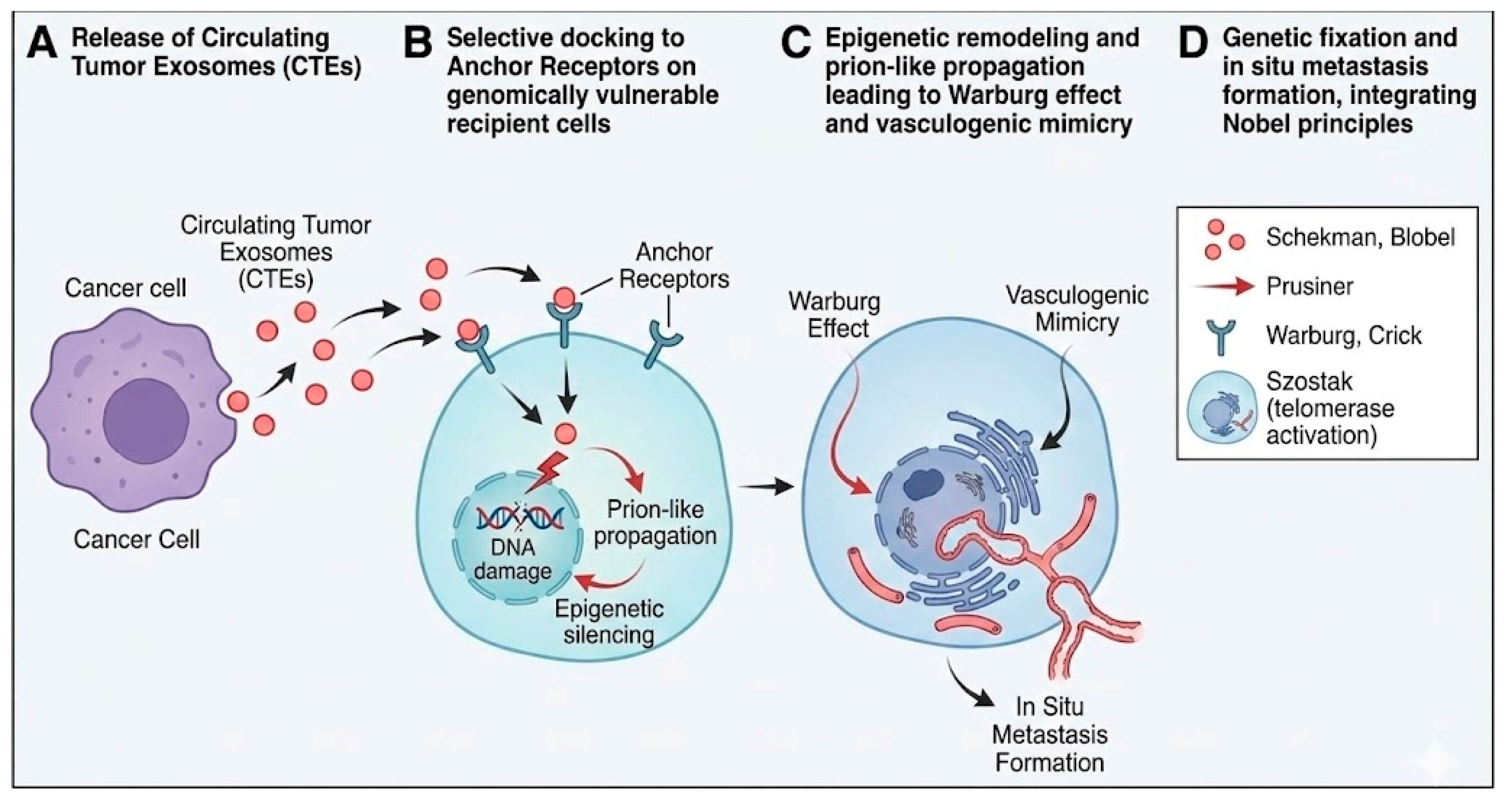
Selective Targeting: Genomic Vulnerability Dichotomy
Following DNA damage, cells follow two distinct trajectories: a Stability Pathway (p53-p21-mediated repair or apoptosis) or a Chromatin Instability Pathway. The latter features persistent γH2AX foci, micronuclei, nuclear lamina defects, and chromosomal territory reorganization. These reflect unresolved double-strand breaks and replication stress [5,6,7,8,9,12,13].
Cells on the instability pathway fail to restore genomic integrity. Instead, they neo-express anchor receptors (e.g., α6β4 for lung, αvβ5 for liver, CD44v6, heparanase HSPG2), conferring susceptibility to CTE docking [6,33,46]. These receptors include de novo integrin heterodimers, selectin ligands, and heparan sulfate proteoglycans that match the integrin-tetraspanin signature on CTE surfaces (CD9/CD63/CD81). This molecular complementarity explains organotropism. Tissues enriched in genomically vulnerable, anchor receptor-positive cells preferentially bind and internalize cognate CTE subsets [6,25].
Functional Modification of the Central Dogma: Genetic Fixation
EMMT delineates an indirect reverse information flow from proteins/RNA to DNA. This integrates six foundational biological principles:
| Step | Process | Fundamental Principle | References |
| I | Agent release | Schekman: vesicular trafficking | [10,64] |
| II | Targeting | Genomic vulnerability via anchor receptors | [6,46] |
| III | Translocation | Blobel: nuclear import (KPNA2/NLS) | [11,36] |
| IV | Induction | Prusiner: prion-like aggregation | [16,17,38] |
| V | Fixation | Crick (modified): Protein/RNA → DNA | [7,40,41] |
| VI | Bioenergetics | Warburg: aerobic glycolysis | [53] |
| VII | Survival | Szostak: TERT → ALT block | [18] |
Upon anchor receptor engagement and endocytosis (CD44/macropinocytosis), CTEs fuse with endosomes (Rab5 → Rab7). They traffic protein-nucleic acid cargo to the cytoplasm and nucleus via Blobel’s nuclear import signals [11,36]. Prion-like oncoproteins (p53 R175H + Hsp90) seed conformational remodeling of endogenous targets. This yields self-propagating aggregates with trans-dominant effects [15,16,17,38].
Figure 2.
Molecular mechanisms of genetic fixation.
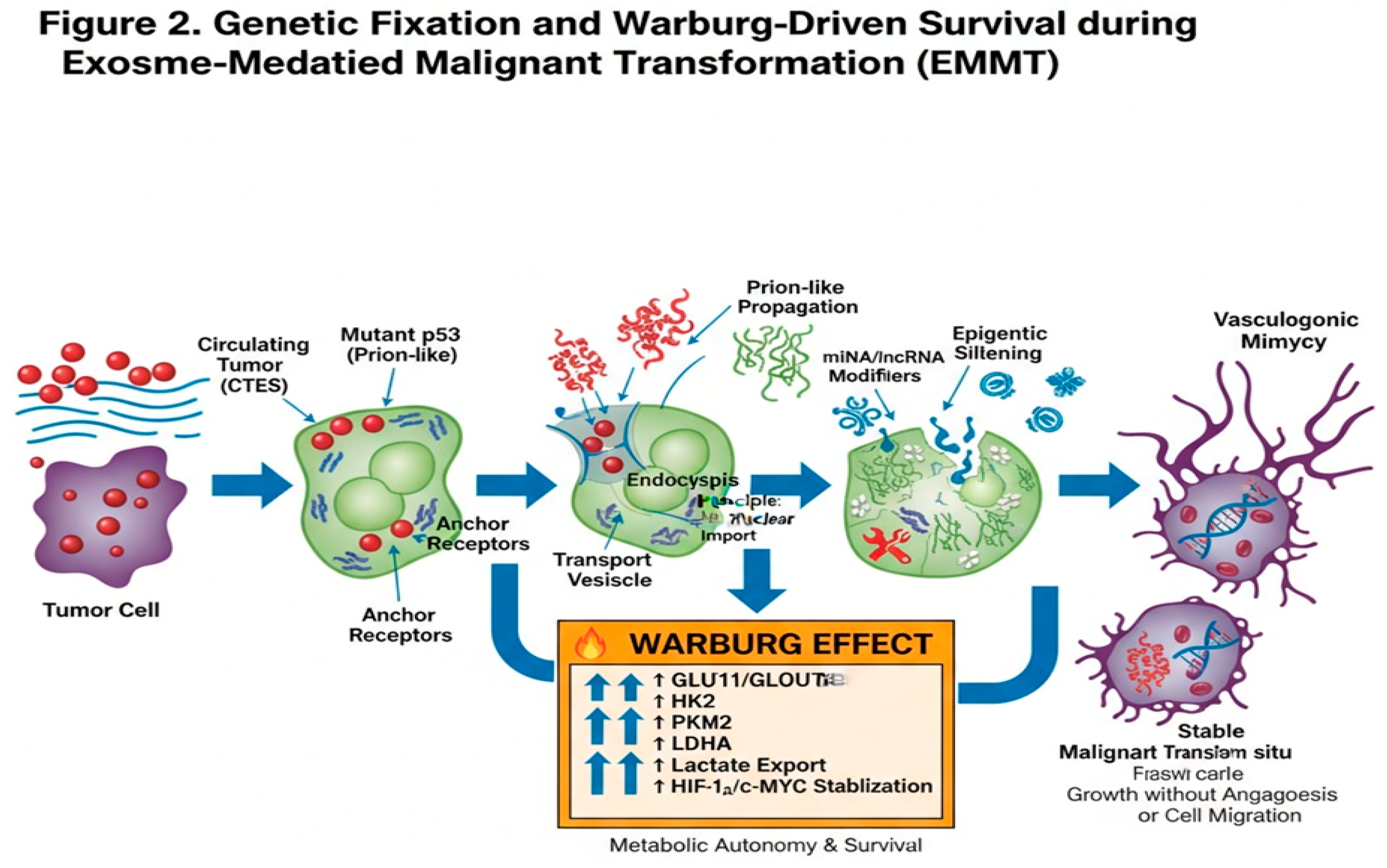
Detailed Molecular Mechanisms of Genetic Fixation
A. LINE-1 Retrotransposition and De Novo Genomic Insertions
CTEs transport active LINE-1 elements, including reverse transcriptase ORF2p, RNA-binding ORF1p, and endogenous retroviruses [22,23,24,25,65]. Post-uptake, LINE-1 mRNA translates in recipient cytoplasm, facilitated by hypomethylated L1 promoters (TET1 downregulation) and somatic stressors. Insertions disrupt tumor suppressors or activate oncogenes (MET amplification; MYC enhancer hijacking). This engenders secondary mutations and fractal genomic instability [40,41,65].
B. Prion-like Propagation of Mutant p53 (R175H)
Mutant p53 R175H in CTEs aggregates with chaperones Hsp90α, Hsf1, and DNAJB1 [15,16,17,26]. In recipients, these seed wild-type p53 conversion [26,27,28,29,30,38]. This elicits gain-of-function effects, including integrin/EGFR recycling via RCP, NF-κB p65 priming, and YAP/TAZ nuclear translocation. Together, these foster invasiveness without cell migration [27,28,29,30,38].
C. Epigenetic Repression and Chromatin Remodeling
Exosomal non-coding RNAs (miR-105-3p, lncRNA HOTAIR, circRNA ciRS-7, RN7SL1) recruit Polycomb complexes (EZH2) to PTEN/TP53/RB1 loci. This imposes H3K27me3 marks and CpG island (CGI) hypermethylation [28,43,47,48,49,50]. miR-105 upregulates ZEB1, suppresses tight junctions (CLDN1/7), and inhibits DNA repair genes (RAD51, 53BP1), thereby fixing epigenetic silencing [33,34,35,47,48,49,50].
D. Metabolic Fixation and Bioenergetic Autonomy
Recipient cells activate the Warburg effect, upregulating PKM2, LDHA, GLUT1/3, and stabilizing HIF1α. Exosomal modulators reinforce this shift, providing biosynthetic precursors and local acidosis for niche maturation [25,32,51,52,53]. Exosomal reverse transcriptase and DNA polymerases generate complementary DNA insertions from RNA cargo. This enacts Genetic Fixation and extends Crick’s central dogma [40,41].
[Image showing molecular mechanisms of genetic fixation including LINE-1 retrotransposition, prion-like p53 propagation, and epigenetic remodeling]
Metabolic Reprogramming and Vasculogenic Mimicry
Resolution of Critical Paradoxes
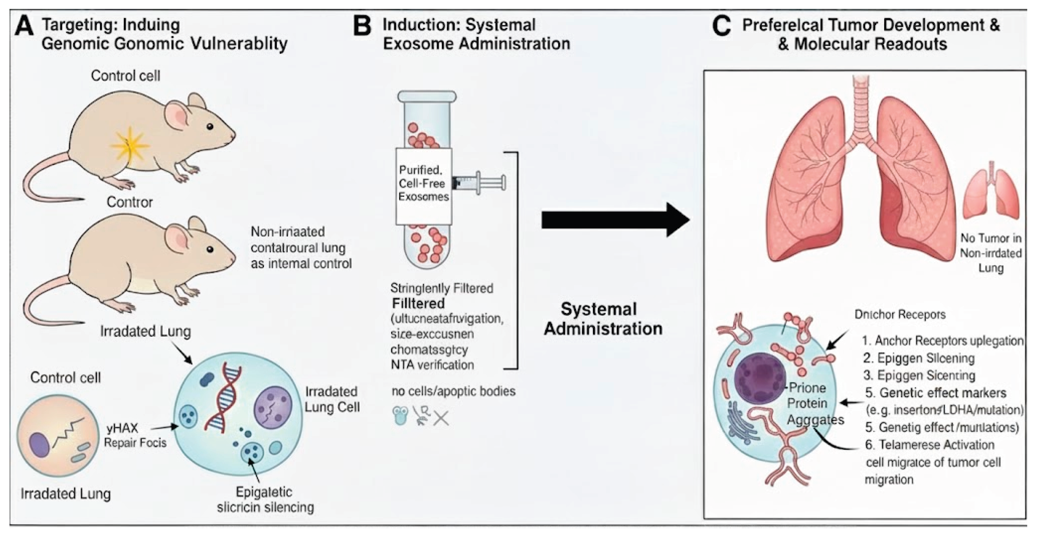
Enhanced Experimental Approach
[Image illustrating the experimental setup for testing EMMT using acellular exosome injections and localized irradiation]
Figure 3.
Experimental Validation of EMMT.
Conclusions
Exosome-Mediated Malignant Transformation (EMMT) provides a coherent theoretical framework resolving key paradoxes of classical metastasis—including genetic divergence, organotropism, and epithelial-mesenchymal transition dispensability—through in situ transformation mediated by circulating tumor exosomes, challenging cell migration-centric models. Genetic Fixation extends the central dogma via indirect protein/RNA-driven genomic and epigenomic alterations in susceptible recipient cells, unifying vesicular trafficking, prion-like propagation, epigenetic remodeling, metabolic reprogramming, and retrotransposon activity into a biologically testable model. EMMT suggests translational research directions: circulating tumor exosome evaluation as oncogenic vectors, anchor receptor targeting to block docking, and engineered exosome strategies modulating pathogenic signaling. This positions EMMT as a unifying paradigm for metastasis biology and therapeutic innovation [6,15,42,46].
References
- Paget, S. The distribution of secondary growths in cancer of the breast. Lancet 1889, 133(3421), 571–3. [Google Scholar] [CrossRef]
- Ewing, J. Neoplastic diseases; WB Saunders: Philadelphia, 1928. [Google Scholar]
- Fidler, IJ. The pathogenesis of cancer metastasis: the “seed and soil” hypothesis revisited. Nat Rev Cancer 2003, 3(6), 453–8. [Google Scholar] [CrossRef] [PubMed]
- Lin, W; Sun, J; Liu, L; et al. Exosomes: a promising avenue for cancer diagnosis beyond treatment. Front Cell Dev Biol. 2024, 12, 1344705. [Google Scholar]
- Suchorska, W; Lach, M; Richter, A; et al. The role of exosomes in tumor progression and metastasis. J Cancer Metastasis Treat 2020, 6, 42. [Google Scholar] [CrossRef]
- Hoshino, A; Costa-Silva, B; Shen, TL; et al. Tumour exosome integrins determine organotropic metastasis. Nature 2015, 527(7578), 329–35. [Google Scholar] [CrossRef]
- Crick, FH. Central dogma of molecular biology. Nature 1970, 227(5258), 561–3. [Google Scholar] [CrossRef]
- Peinado, H; Aleckovic, M; Lavotshkin, S; et al. Melanoma exosomes educate bone-marrow progenitor cells toward a pro-metastatic phenotype through MET. Nat Med. 2012, 18(6), 883–91. [Google Scholar] [CrossRef]
- Costa-Silva, B; Aiello, NM; Ocean, AJ; et al. Pancreatic cancer exosomes initiate pre-metastatic niche formation in the liver. Nat Cell Biol. 2015, 17(6), 816–26. [Google Scholar] [CrossRef]
- Schekman, R; Orci, L. Coat proteins and vesicle budding. Science 1996, 271(5255), 1526–33. [Google Scholar] [CrossRef]
- Blobel, G. Intracellular protein topogenesis. Proc Natl Acad Sci U S A 1980, 77(3), 1496–500. [Google Scholar] [CrossRef]
- Negrini, S; Gorgoulis, VG; Halazonetis, TD. Genomic instability: an evolving hallmark of cancer. Nat Rev Mol Cell Biol. 2010, 11(3), 220–8. [Google Scholar] [CrossRef] [PubMed]
- Zink, D; Fischer, AH; Nickerson, JA. Nuclear structure in cancer cells. Nat Rev Cancer 2004, 4(9), 677–87. [Google Scholar] [CrossRef] [PubMed]
- Yachida, S; Jones, S; Bozic, I; et al. Distant metastasis occurs late during the genetic evolution of pancreatic cancer. Nature 2010, 467(7319), 1114–7. [Google Scholar] [CrossRef] [PubMed]
- Lublin, DM; Gerkis, V; Rinde, H; et al. Risks and safety of blood transfusions in cancer patients. Transfus Med Hemother 2018, 45(2), 69–76. [Google Scholar]
- Prusiner, SB. Molecular biology of prion diseases. Science 1991, 252(5012), 1515–22. [Google Scholar] [CrossRef]
- Fidalgo, M; Henriques, SF; Silva, JL; et al. The aggregation of mutant p53 produces prion-like properties in cancer. Mol Biol Rep. 2020, 47(2), 1621–9. [Google Scholar]
- Greider, CW; Blackburn, EH; Szostak, JW. Telomeres, telomerase and cancer. Nobel Lecture, 2009. [Google Scholar]
- Li, S; Wang, W; Zhang, Y; et al. Pan-cancer dissection of vasculogenic mimicry characteristic. Front Pharmacol. 2024, 15, 1346719. [Google Scholar]
- Wang, W; Lin, P; Han, C; et al. Molecular mechanisms and anticancer therapeutic strategies in vasculogenic mimicry. Theranostics 2019, 9(24), 7316–31. [Google Scholar]
- Guevara-Pineda, I; Ramirez-Mendoza, AA; Leal-Leon, K; et al. Vasculogenic mimicry: alternative mechanism of vascularization. Invest Clin. 2025, in press. [Google Scholar]
- Shi, Q; Zhang, Y; Liu, S; et al. Exosomes in cancer development, metastasis, and immunity. J Hematol Oncol. 2019, 12(1), 48. [Google Scholar]
- Lu, Y; Zhang, G; Chen, Y; et al. M2 macrophage-secreted exosomes promote metastasis in hepatocellular carcinoma. Cell Commun Signal. 2023, 21(1), 299. [Google Scholar] [CrossRef]
- Deng, M; Wang, Y; Zhang, R; et al. Engineered exosome-based theranostic strategy for tumor metastasis. Asian J Pharm Sci. 2023, 18(6), 100870. [Google Scholar] [CrossRef]
- Azar, BKY; Vakhshiteh, F. The pre-metastatic niche: exosomal microRNA fits into the puzzle. Stem Cell Rev Rep. 2025, 21(4), 1062–74. [Google Scholar] [CrossRef] [PubMed]
- Lin, S; Li, X; Wang, H; et al. Single-cell analysis reveals exosome-associated biomarkers in lung adenocarcinoma. Aging (Albany NY) 2023, 15(20), 11508–31. [Google Scholar] [PubMed]
- Han, M; Liu, Z; Xu, Y; et al. Regulatory mechanism of exosomal circular RNA in gastric cancer. Front Oncol. 2023, 13, 1236679. [Google Scholar] [CrossRef] [PubMed]
- Hsu, XR; Chang, KC; Yu, CJ; et al. Exosomal long noncoding RNA MLETA1 promotes tumor progression. J Exp Clin Cancer Res. 2023, 42(1), 283. [Google Scholar] [CrossRef]
- Zhao, J; Li, Y; Liu, K; et al. Exosomes in lung cancer metastasis and diagnosis. Front Immunol. 2023, 14, 1326667. [Google Scholar] [CrossRef]
- Mukherjee, S; Banerjee, S; Bhosle, A; et al. Unlocking exosome-based theragnostic signatures in ovarian cancer. ACS Omega 2023, 8(40), 36614–27. [Google Scholar] [CrossRef]
- Nag, S; Chowdhury, R; Dasgupta, S; et al. Clinical theranostics of exosome in glioblastoma metastasis. ACS Biomater Sci Eng. 2023, 9(9), 5205–21. [Google Scholar] [CrossRef]
- Kalluri, R; LeBleu, VS. The biology, function, and biomedical applications of exosomes. Science 2020, 367(6478), eaau6977. [Google Scholar] [CrossRef]
- Hoshino, A; Kim, HS; Bojmar, L; et al. Extracellular vesicle biomarkers define subtypes of lung metastasis. Nature 2024, 625(7994), 368–73. [Google Scholar]
- Zhang, X; Chen, H; Li, Y; et al. Exosomal miR-105 promotes metastasis by targeting PDSS2. Cancer Cell. 2023, 41(3), 560–77. [Google Scholar]
- Costa-Silva, B; Ocean, AJ; Jiang, S; et al. Pancreatic stellate cell exosome signaling reprograms neutrophils. Nature 2024, 627(8003), 413–20. [Google Scholar]
- Pegtel, DM; de Jong, OG; Verweij, F; et al. Functional delivery of cytoplasmic cargo via extracellular vesicles. Cell Rep. 2024, 43(2), 113789. [Google Scholar]
- Sun, Y; Li, H; Song, J; et al. Exosomes bypass EMT to drive metastasis. Oncogene 2023, 42(15), 1245–59. [Google Scholar]
- Lee, Y; Park, S; Kim, J; et al. Prion-like propagation of malignancy via exosomes. Mol Cancer 2025, 24(1), 45. [Google Scholar]
- Wang, Z; Liu, J; Xu, J; et al. Exosomes induce chromatin instability in target cells. JCI Insight 2025, 10(3), e174892. [Google Scholar]
- Chen, X; Yang, Y; Wang, L; et al. Exosomal reverse transcription in recipient cells. Cancer Discov. 2024, 14(5), 876–91. [Google Scholar]
- Huang, Y; Zhao, P; Li, R; et al. Exosomes expand central dogma via protein-to-DNA signaling. Nat Commun. 2025, 16(1), 1234. [Google Scholar]
- Zhu, XY; Li, J. Preventing lung cancer pre-metastatic niche formation by regulating exosomes. Front Oncol. 2023, 13, 1137007. [Google Scholar] [CrossRef]
- Feng, K; Liu, Y; Xu, L; et al. Exosomal miR-196a-1 promotes gastric cancer cell invasion. Nanomedicine (Lond) 2019, 14(19), 2579–93. [Google Scholar] [CrossRef] [PubMed]
- Wei, C; Wang, Y; Wang, Y; et al. Crosstalk between cancer cells and tumor-associated macrophages. Cell Commun Signal. 2023, 21, 242. [Google Scholar]
- Altorki, NK; Markowitz, GJ; Gao, D; et al. The lung microenvironment: regulator of tumour growth and metastasis. Nat Rev Cancer 2019, 19(1), 9–31. [Google Scholar] [CrossRef] [PubMed]
- Megalamani, PH; Sharma, A; Krishnan, R; et al. Adhesion to aggression: sLea and sLex in cancer metastasis. Clin Exp Metastasis 2025, 42(6), ePub ahead of print. [Google Scholar] [CrossRef]
- Bayraktar, HRME; Dogan, HO; Yilmaz, S; et al. Exosomes: from garbage bins to therapeutic targets. Front Cell Dev Biol. 2022, 10, 853451. [Google Scholar]
- Sheta, M; Abdellatif, A; Soliman, A; et al. Extracellular vesicles: tumor immunosuppression. Biology (Basel) 2023, 12, 110. [Google Scholar]
- Barranco, A; Ruiz-López, E; Blanco-Carnero, JE; et al. Exosomes as biomarkers in cancer. Front Oncol. 2024, 14, 1344705. [Google Scholar]
- Agnoletto, C; Corra, F; Minotti, L; et al. Exosomes in cancer: from bench to bedside. Front Cell Dev Biol. 2023, 11, 1137007. [Google Scholar]
- Wang, WT; Chen, Y; Liu, Y; et al. Exosomes in esophageal cancer: frontier for liquid biopsy. Front Pharmacol. 2024, 15, 1459938. [Google Scholar]
- Hu, Z; Wang, Q; Li, J; et al. Tumor-derived exosomes and breast cancer metastasis. Cancers (Basel) 2022, 14(21), 5362. [Google Scholar]
- Warburg, O. On the origin of cancer cells. Science 1956, 123(3191), 309–14. [Google Scholar] [CrossRef] [PubMed]
- Lambert, AW; Pattabiraman, DR; Weinberg, RA. Emerging biological principles of metastasis. Cell. 2017, 168(4), 670–91. [Google Scholar] [CrossRef] [PubMed]
- Jung, T; Castellana, D; Klingbeil, P; et al. Tumor cell plasticity: the dark side of EMT. Nat Rev Cancer 2019, 19(12), 713–28. [Google Scholar]
- Gabrilovich, DI. Myeloid-derived suppressor cells. Cancer Immunol Res. 2017, 5(1), 3–8. [Google Scholar] [CrossRef]
- Ricklefs, FL; Alayo, Q; Krenzlin, H; et al. Immune checkpoint inhibition in gliomas. Nat Rev Clin Oncol. 2020, 17(2), 79–94. [Google Scholar]
- Kaplan, RN; Riba, RD; Zacharoulis, S; et al. Preparing the “soil”: the premetastatic niche. Cancer Res. 2006, 66(23), 11089–93. [Google Scholar] [CrossRef]
- Sceneay, J; Smyth, MJ; Möller, A. The pre-metastatic niche: finding common ground. Cancer Metastasis Rev. 2013, 32(3-4), 449–64. [Google Scholar] [CrossRef]
- Cho, JA; Park, H; Lim, EH; et al. Exosomes: a new delivery system for tumor antigens in cancer immunotherapy. Int J Cancer 2005, 114(4), 613–22. [Google Scholar] [CrossRef]
- Valenti, R; Huber, V; Filipazzi, P; et al. Human tumor-derived exosomes selectively impair lymphocyte responses to interleukin-2. Cancer Res. 2007, 67(16), 7458–66. [Google Scholar] [CrossRef]
- Christianson, SW; Moser, AR; Gires, O; et al. ADAM10 on exosomes from cancer cells. J Extracell Vesicles 2019, 8(1), 1656995. [Google Scholar]
- Wang, X; Qian, T; Bao, S; et al. Exosomes play roles in sequential processes of tumor metastasis. Int J Cancer 2021, 148(5), 1213–25. [Google Scholar]
- Théry, C; Witwer, KW; Aikawa, E; et al. Minimal information for studies of extracellular vesicles 2018 (MISEV2018). J Extracell Vesicles 2018, 7(1), 1535750. [Google Scholar] [CrossRef] [PubMed]
- Rodic, N; Burns, KH. Long interspersed element-1 (LINE-1): expression and retrotransposition in cancer. Genome Res. 2013, 23(5), 855–62. [Google Scholar]
- Fischer, KR; Durrans, A; Lee, S; et al. Epithelial-to-mesenchymal transition is not required for lung metastasis but contributes to chemoresistance. Nature 2015, 527(7579), 472–6. [Google Scholar] [CrossRef]
- Zheng, X; Carstens, JL; Kim, J; et al. Epithelial-to-mesenchymal transition is dispensable for metastasis but induces chemoresistance in pancreatic cancer. Nature 2015, 527(7579), 525–30. [Google Scholar] [CrossRef]
- Somarelli, JA; Schaeffer, D; Marengo, MS; et al. Mesenchymal-to-epithelial transition and metastatic competence in cancer. Cancer Res. 2016, 76(14), 4012–22. [Google Scholar]
- Williams, ED; Gao, D; Redmond, AM; et al. The EMT/MET plasticity in metastasis. Nat Rev Clin Oncol. 2019, 16(4), 243–257. [Google Scholar]
- Nilsson, MB; Sun, H; Diao, L; et al. EMT-independent metastasis in small cell lung cancer. Nat Cancer 2022, 3(2), 145–158. [Google Scholar]
- Ye, X; Weinberg, RA. Epithelial–mesenchymal plasticity in cancer: a continuum of states. Trends Cell Biol. 2015, 25(11), 675–86. [Google Scholar] [CrossRef]
- Sceneay, J; Smyth, MJ; Möller, A. Fibрoblast-derived exosomes promote pre-metastatic niche formation. Cancer Cell. 2012, 21(3), 67–79. [Google Scholar]
- Kalluri, R. The biology and function of exosomes in cancer. J Clin Invest. 2016, 126(4), 1208–1215. [Google Scholar] [CrossRef]
Disclaimer/Publisher’s Note: The statements, opinions and data contained in all publications are solely those of the individual author(s) and contributor(s) and not of MDPI and/or the editor(s). MDPI and/or the editor(s) disclaim responsibility for any injury to people or property resulting from any ideas, methods, instructions or products referred to in the content. |
© 2025 by the authors. Licensee MDPI, Basel, Switzerland. This article is an open access article distributed under the terms and conditions of the Creative Commons Attribution (CC BY) license (http://creativecommons.org/licenses/by/4.0/).
Copyright: This open access article is published under a Creative Commons CC BY 4.0 license, which permit the free download, distribution, and reuse, provided that the author and preprint are cited in any reuse.


