Preprint
Review

This version is not peer-reviewed.

Phenomenology in Craniofacial Movement Disorders: A Narrative Review

Submitted:

24 December 2025

Posted:

25 December 2025

You are already at the latest version

Abstract

Craniofacial or cranial movement disorders are commonly seen in clinical practice. However, they are usually underdiagnosed or even misdiagnosed, leading to significant impairment in the quality of life of patients affected by these disorders. They are traditionally defined as movement disorders involving the face, jaw, tongue, and palate; but, it is possible to include the eye and ear based on the literature available. The current study aims to generally describe the phenomenology related to these abnormal movements for the general neurologist and practitioner. The most frequent are hemifacial spasm, blepharospasm, and myokymia. Other less commonly discussed are rabbit syndrome, apraxia of the eyelid opening/closing, tardive dyskinesia, chorea, tics, myorhythmia, dystonia, and tremor. Also, there are locations for the development of movement dis-orders, such as the ears (ear dyskinesia and ear myoclonus), chin (geniospasm), and tongue (lingual dystonia and lingual myoclonus) that are rarely discussed. Craniofacial movements are also seen in patients with autoimmune conditions, and the different features of these movements led to new terms like dystonic or dyskinetic stereotypies due to the dynamic change of the phenomenology. Another fact is that up to ten percent of patients with functional movement disorder will have craniofacial movement disorders.

Keywords: 
;  ;  ;  ;  

1. Introduction

Cranial movement disorders are movement disorders that affect the face, eye, jaw, tongue, palate, and ear (Table 1). They are limited to the cranial muscles or manifest as part of a more generalized movement disorder. They can interfere with bulbar functions such as talking, chewing, and swallowing. Noteworthy, there is a significant social embarrassment and discomfort leading to poor quality of life, anxiety, depression, and stigma, in almost half of the patients affected by these disorders [1]. They can originate from the highest (motor cortex) to the lowest (cranial nerve and muscle) levels of the motor system. There is a variable clinical presentation and the lack of diagnostic tests and biomarkers leading to a broad differential diagnosis. The diagnostic work-up should include taking a full past and family history, identification of drug exposure, and recognition of motor and nonmotor signs and symptoms, as well as a full physical examination. Moreover, electrodiagnostic features were scarcely reported in the literature. In this context, the objective of this article is to provide an overview of the current literature regarding the phenomenology of craniofacial movement disorders. The papers for this review were retrieved through a PubMed and Google Scholar search published until December 2024. The following keywords were used: cranial, craniofacial, and movement disorder. Only studies published in English were included.

2. Craniofacial Movement Disorders

2.1. Hemifacial Spasm

Hemifacial spasm is one of the most common craniofacial movement disorders. It usually starts in the lower eyelid and spreads quickly to the rest of the hemifacial muscle and is characterized by intermittent contractions of the hemifacial muscles. Also, it is possible to see the Babinski sign, which is a synkinesis that is the contraction of the ipsilateral frontalis muscle and orbicularis muscle. Platysma contractions can also be observed in hemifacial spasms because this muscle is innervated by the cervical branch of the facial nerve. The spasms can be spontaneous or triggered by facial contractions.
The most common type of hemifacial spasm is idiopathic (62% of the patients), with vascular compression of the seventh cranial nerve by the anterior inferior cerebellar artery [2]. So, neuroimaging involving the arteries of the brain should be performed. However, other etiologies may be found, such as familial, Bell’s palsy, facial nerve injury, brain vascular insult, demyelinating, psychogenic, facial tics, facial dystonia, facial myoclonus, and hemimasticatory spasms.
There are some atypical cases of hemifacial spasm involving only the lower part of facial muscles. Another atypical presentation is the bilateral contraction of “hemi” facial spasm, in which the suspicion of secondary causes should be raised, like schwannoma [3]. These patients will have a pseudonormalization of the Babinski sign, so there will not be asymmetry of the eyebrows.

2.2. Tardive Dyskinesia

It is one of the most common drug-induced movement disorders, and there are multiple phenomenologies. In this context, the oro-buccal-lingual muscles are the most frequent region involved in the face. Orolingual dyskinesia is the most common tardive dyskinesia (77% of the patients) and is characterized by biting (49%), tongue protrusion (21.5%), noises (15.4%), lip pursing (12.3%), and lip puckering (5%). Also, tardive dyskinesia can present with jaw movements side-to-side, involuntary jaw opening, and chewing or lateral jaw movements [4]. Other patients may only have involvement in the muscles of the floor of the mouth.
Lingual movements are a very important part of tardive dyskinesia, but they may be complex. Stereotypic slow movements of the tongue, tongue repetitively moving in several axes, writhing movements of the tongue, lateral inclination of the mouth, and depression of the central part of the mouth can be observed. The central depression of the tongue is caused by the genioglossus muscles.
Anecdotal cases have been reported of cases mimicking tardive dyskinesia. One of the first reports in the American continent of neuroferritinopathy presented with facial stereotype/tic, and the patient was initially diagnosed with tardive dyskinesia and had a complete resolution with tetrabenazine therapy [5]. Deposits of iron in the basal ganglia and thalami, and cavities were found in the basal ganglia on MRI. Another possible misdiagnosis is non-ketotic hyperglycemia has been reported as presenting with isolated lingual protrusion movements [6].

2.3. Rabbit Syndrome

It is a drug-induced movement disorder and is characterized by vertical movements of the central part of the face around the mouth that are usually about four to five hertz, and they have been described as rabbit chewing. A new term recently coined by Rissardo et al. is oral vertical dyskinesia [7]. The differentiation from tardive dyskinesia can be related to tongue movements that are not observed in oral vertical dyskinesia. Also, it is worth mentioning that anticholinergic treat oral vertical dyskinesia, but they can worsen tardive dyskinesia symptoms.

2.4. Chorea

The craniofacial movements of the face involving chorea are more discreet than those with tardive dyskinesia, and the movements can be more easily separated. The classical phenomenology is described in patients with Huntington’s disease with unpredictable muscle involvement. The most common presentation is intermittent widening palpebral fissures and frontalis contractions (76%), other clinical manifestations are brief contractions of procerus (52%), repetitive irregular blinking (16%), brief spasms of orbicularis muscle (8%) [8]. Other findings that may assist in the diagnosis are motor persistence, asking the patient to protrude the tongue, and abnormal eye movements (slow saccades or saccades with increased latency).
McLeod syndrome is a rare, genetic disorder that affects the brain, blood, heart, muscles, and peripheral nerves. It is caused by mutations in the XK gene on the X chromosome. There are many reports of early tongue chorea in this disorder, and at the first appointment, the patient will complain of biting of tongue and lips [9].
A type of choreiform dyskinesia is “edentalous dyskinesia,” which occurs in more than ten percent of edentulous individuals [10].

2.5. Tics And Stereotyped Movements

The most common tic disorder with severe features is Gilles de la Tourette syndrome. So, it is reasonable that most of the knowledge about tics in craniofacial movement disorders would involve this disorders. But, tics can be secondary to medications (antipsychotics, carbamazepine, phenytoin, and psychostimulants), infections (encephalitis), toxins (carbon monoxide), cerebrovascular disorders, and traumatic brain injury [11]. In this context, patients with Tourette syndrome usually will present with craniofacial tics, which usually spread caudally, with variations in distribution, intensity, and frequency.
The most common presentation of oromandibular tics in patients with Tourette syndrome is jaw opening (68.3%), followed by jaw lateral movements (63.4%), jaw closing or clenching (29.3%), and tongue protrusion (12.2%) [12]. They are repetitive, stereotyped, and they have complex phenomenology with possible suppression. Another rare form of tic in patients with Tourette Syndrome is the jaw retraction tic, and some patients also produce some sounds associated with the oromandibular tics.
Isolated upward gaze is the most common tic affecting 70–80% of the patients. Other common presentations are isolated lateral gaze, and eye closure followed by upward-lateral gaze. Ocular tics most commonly affect young patients of the male sex with Tourette syndrome [13]. Ocular tics can also have a dystonic phenomenology with dystonia upward gaze. Also, there are some reports of these tics affecting activities of daily living like driving.
Some patients can have a dystonic facial tic (contraction of the facial and sometimes thoracic muscles) associated with the production of noises. The dystonic tic has a duration of less than 1 second. Vocal (phonic) tics can be observed, and they are produced without using the vocal apparatus, as shown by the asynchronous facial movement and the sound being produced. A similar phenomenology can also be observed in patients with chorea, but in cases of facial tics, there is a potential predictability of the sound being generated and the muscle contraction [14].

2.6. Myorhythmia

It is a controversial topic in the field of movement disorders. But, there is some agreement in the definition of the term oculomasticatory myorhythmia, which is a convergent- divergent nystagmus, concurrent contractions of the masticatory muscles, and supranuclear vertical gaze palsy. And, this sign is considered pathognomonic of Whipple disease. In this context, myorhythmia can also be defined as a tremor-like movement affecting cranial muscles and lingual muscles with the contraction lasting for 100-200 milliseconds, which are repetitive with a frequency of 1 Hz [15]. In this context, some patients may have the movements of the floor of the mouth to amplify their sound, which can last 400–500 milliseconds [16]. Another important thing to mention is that every patient with myorhythmia should be investigated for secondary causes.

2.7. Dystonia

It is one of the most common forms of focal dystonia, and it is characterized by intermittent repetitive, involuntary contractions of the orbital and preseptal orbicularis oculi muscles, resulting in the narrowing of the palpebral fissure or complete closure of the eyelid and lowering of the eyebrow beneath the superior orbital rim (Charcot sign). Blepharospasm usually starts between the fifth and seventh decades of life. The prevalence of blepharospasm is estimated at 12 in Tottori Prefecture in Japan [17], 17 in Rochester in the USA [18], 30 in North England [19], 36 in the Epidemiologic Study of Dystonia in Europe [20], and 133 in Southern Italy [21] cases per million individuals. Also, women are 2.3 times more likely to be affected than men, and usually, they are 7.7 years older [20].
The etiology is still unknown with genetic and environmental factors. There is an increased risk in individuals with a family history of dystonia or postural tremor; and, on the other hand, smoking cigarettes may decrease the risk. Interestingly, benign essential blepharospasm does not predispose to the development of Parkinson’s disease. There are some triggering conditions, such as eye disease (including dry eye), trauma of near the eye, and dental procedures.
The phenomenology of blepharospasm is characterized by alternating dystonic discharges in the pretarsal orbicularis oculi muscles and levator palpebrae superioris muscles, producing brief clonic flickering of the upper eyelids of both eyes. There is a high frequency of ocular symptoms before the onset, like dry eye, corneal irritation, blepharitis, and keratoconjunctivitis [20]. Sensory tricks (geste antagoniste) may reduce or ameliorate the movements by touching the eyebrow or lateral side of the eyelids. The combination of blepharospasm and oromandibular dystonia is called Meige syndrome.
Blink reflex is the essential test to evaluate patients with blepharospasm. There is electrical stimulation of the supraorbital nerve. The R1 component is brief, unilateral, and ipsilateral to the stimulated side, with a latency of around 10 milliseconds. The R2 component is longer in duration and bilateral, with a latency of approximately 30 milliseconds. Common afferent limb of R1 and R2 is the ophthalmic trigeminal division, and common efferent limb is the facial nerve [22].
Blepharospasm should be differentiated from hemifacial spasm due to excessive involuntary spasm of the orbicularis oculi muscle and sensory trick. In the cases of hemifacial spasm, there is a specific distribution of the facial nerve leading to clonic or tonic contractions, and it is most likely unilateral. Noteworthy, some patients may have blepharospasm with apraxia of eyelid opening, and these individuals are even more sensitive to low-intensity light, and sensory tricks are, in most individuals, not so effective.
Apraxia of eyelid opening is the inability to initiate and failure to sustain eyelid elevation in the absence of overt orbicularis oculi muscle contraction. No oculomotor or ocular sympathetic nerve dysfunction or ocular myopathy is present. During episodes of eyelid closure, patients typically elevate their eyebrows in an attempt to open the eyelids. There is involuntary inhibition of the levator palpebrae superioris muscles.
Apraxia of eyelid opening may be difficult to differentiate from blepharospasm, but some of the characteristics exclusive of blepharospasm are spasm of orbicularis oculi muscle, effect of sensory trick, and triggers prompting eyelid closing. On the other hand, features exclusive of apraxia of eyelid opening are spasm of frontalis muscle, elevation of bilateral eyebrow, and ability to keep eyelid opening.
Bilateral apraxia of eyelid opening was already described with top of the basilar syndrome [23]. Other interesting cases have been reported with putaminal [24] and thalamic [25] hemorrhages causing blepharospasm and apraxia of eyelid opening. Other cases reporting isolated blepharospasm were seen with thalamomesencephalic lesion [26] and mesencephalic cyst [27]. Individuals with progressive supranuclear palsy may present severe cases of blepharospasm leading to opening of the eyelids for less than fifteen minutes a day.

2.7.1. Tongue Dystonia

Besides apraxia of the eyelid and blepharospasm, there are uncommon reports of lingual dystonia. It is usually reported in musicians who play wind instruments. Also, these patients may place something on their tongue, like a candy or a tongue depressor, to alleviate the dystonia. Cranial movement disorders, especially dystonia, are commonly found in patients with Wilson’s disease. A rare etiology for tongue dystonia and writhing movements is pantothenate kinase-associated neurodegeneration (PKAN) [28].

2.7.2. Cervical Dystonia

Cervical dystonia, the most common focal dystonia, manifests as involuntary, patterned contractions of neck muscles causing abnormal head postures. It may be idiopathic or secondary to structural lesions. Midbrain, cavernous sinus, or posterior fossa tumors affecting cranial nerves III/IV can mimic dystonia via compensatory head tilt or skew deviation. Sensory tricks (geste antagoniste) and tremulous components are common. Dystonia may fluctuate with posture and voluntary movements, and imaging is essential when clinical features suggest secondary causes, especially in atypical or painful presentations [29].

2.7.3. Trismus

Trismus is not classified as a movement disorder, but it can occur in the context of some movement disorders like dystonia [30]. Phenomenologically, it manifests as involuntary, tonic contraction of jaw-closing muscles, impairing speech, eating, and oral hygiene. It may occur in isolation or alongside cranial or oromandibular dystonia. Unlike temporomandibular joint disorders, trismus lacks mechanical obstruction and often occurs with other cranial dystonias or extrapyramidal signs.

2.8. Tremor

2.8.1. Palatal Tremor

The phenomenology of palatal tremor, previously called palatal myoclonus, is defined by rhythmic movements of the soft palate and sometimes other muscles innervated by cranial or spinal nerves [31]. It occurs due to the dysfunction of the Guillain-Mollaret triangle that involves the red nucleus, inferior olivary nucleus, and dentate nucleus. For the clinical features of essential, psychogenic, and secondary palatal tremor, consider reading Table 2. A variant of palatal tremor is called dancing larynx characterized by complex non-rhythmic movements of the laryngeal structure and common palatal tremor features [32].

2.8.2. Tongue, Jaw, And Head Tremor

It can typically be observed in patients with Parkinson’s disease, but other parkinsonism-plus disorders were also observed with isolated tongue tremor such as progressive supranuclear palsy [33] that was confirmed by pathology studies three years later [34].
Isolated tongue tremor was already noticed after gamma knife radiosurgery for acoustic schwannoma, which was also observed during fluoroscopy and indirect laryngoscopy [35]. Another cause of isolated tongue tremor was ventriculoperitoneal shunt for hydrocephalus following treatment for lower brainstem tumor [36]. Noteworthy, a similar presentation was noticed and led to the diagnosis of brainstem pilocytic astrocytoma [37]. Another possible causes of tongue tremor include stroke [38] or seizures [39] with foci at tongue cortical homunculus area. It is interesting that these lesions led to the development of tremor and not weakness of the tongue.
Tremor of the chin is most commonly seen in Parkinson’s disease, but the jaw tremor was more frequently observed in essential tremor. The phenomonology is different in each individual, such as side-side, up-down, and mixed features without clinical significance [40].
Another common movement disorder is head tremor. The tremor direction may support the clinical diagnosis of some conditions. For example, essential tremor usually has a horizontal tremor (“non-no” tremor) and Parkinson’s disease has a vertical tremor (“yes-yes” tremor) [41]. Also, patients with essential tremor usually have voice tremor and patients with Parkinson’s disease have hypokinetic speech (mono-pitch, mono-loudness, reduced loudness, and rapid rate) [42]. It is worth mentioning that a head tremor should be differentiated from a dystonic head tremor based on the directionality of the head tremor, irregular rhythm, dystonic deviation of the neck, and sensory trick.

2.9. Facial Myokymia and Facial Myoclonus

It is a fine, undulating contractions with visible rippling of the skin commonly involving the muscles of the face (especially around eyelids). Myokymia is related to stressor factors and fatigue, so it is occasionally seen in Guillain-Barre syndrome. Diagnosis can be confirmed with electromyography, revealing grouped discharges or bursts with frequency ranging from 5 to 150 times per second [43].
The most common source of facial myoclonic jerks is the brainstem region, which can lead to abnormal facial, palatal, lingual, and even shoulder movements [44].

2.10. Ear

Among focal dyskinesia, ear dyskinesias are extremely uncommon, and only a few cases have been reported. Movement disorders of the ear include ear dyskinesia, auricular myoclonus, and middle ear myoclonus. Ear dyskinesia, also known as moving ear syndrome, is a rare disorder that causes involuntary contractions of the ear muscles. It is characterized by irregular, jerky, and relatively slow movements involving elevation and posterior rotation of the ear muscles [45]. Auricular myoclonus involves irregular clonic movements of the antitragus and anthelix at a frequency of 70 to 75 per minute. A possible brainstem origin for the condition has been suggested, similarly to palatal myoclonus [46]. Middle ear myoclonus is an umbrella term for conditions that cause rhythmic movement of the tympanic membrane.

2.11. Autoimmune Encephalitis

Patients with anti-NMDA (N-methyl-D-aspartate) receptor encephalitis are difficult to diagnose because they have a mixture of movement disorders like chorea, myorhythmia, cranial dystonia, and stereotypic movements [47]. Some authors consider that they have a phenomenology called dystonic stereotypies. Interestingly, the phenomenology usually changes the pattern with time and with the treatment in these patients.

2.12. Ataxia-Telangiectasia

Patients with ataxia-telangiectasia exhibit a distinct pattern of oculomotor dysfunction, most notably oculomotor apraxia, which presents as difficulty initiating voluntary saccades, often compensated by head thrusts. Eye movements are frequently slow and drifting, with a tendency toward off-foveal gaze and poor fixation stability. Additional features include gaze-evoked nystagmus, jerky and dysmetric smooth pursuits, and delayed, hypometric saccades, reflecting cerebellar and brainstem involvement in the control of eye movements [48].

3. Functional Craniofacial Movement Disorders

A recent study found that females with concurrent headaches or psychiatric disorders are commonly affected by functional craniofacial movement disorders. The most common regions affected were the oromandibular, ocular, lingual, and speech without significant statistical differences in the affected body area [49]. In another study, spasms resembling dystonia were observed in all patients, with the lips being the most frequently affected area (60.7%), followed by the eyelids, perinasal region, and forehead. The predominant presentation involved the downward protrusion of one side of the lower lip accompanied by ipsilateral jaw deviation [50].
One of the findings to differentiate hemifacial spasm from functional hemifacial spasm is that the patient usually has elevation of the contralateral eyebrow, also known as lack of the other Babinski sign (sensitivity 1.00, specificity 0.70). Other features are high-frequency of the spasms, contractions usually sustained (more than three seconds), and downward deviation of the angle of the mouth (sensitivity 0.47, specificity 1.00) [51]. Another possible differentiation is using a tuning fork that assesses sensitivity involvement in the development of the hemifacial spasm. It was observed that patients with functional hemifacial spasms more commonly worse (75%) than improve his motor symptoms with tuning fork application [52].
Functional stereotypies of the mouth may resemble tardive dyskinesia and usually have all the phenomenologies that can be seen in patients with other functional movement disorders, such as suggestibility, distractibility, sudden onset, and transient episodes of improvement. Rabbit syndrome can have a functional presentation that shows changes in frequency with frequency of the limbs during distractibility [53].
In Tourette syndrome, the patient can suppress the tic; but, patients with functional tics usually are not able to suppress it [54]. There are also functional ocular movements that may present with a wide variety of phenomenology, and it is found in 6% of the patients with functional movement disorders. The most common types of ocular movements were oculogyric crisis (54%), opsoclonus (38%), and ocular flutter (8%) [55].
Emerging evidence highlights the substantial impact of psychosocial factors—particularly anxiety, depression, and social stigma—on the phenomenology of craniofacial movement disorders such as blepharospasm, oromandibular dystonia, and functional facial movements [56]. These conditions often affect highly visible regions of the body, contributing to heightened self-consciousness and social withdrawal. Several studies using validated instruments like the Hospital Anxiety and Depression Scale and the Beck Depression Inventory demonstrate a bidirectional relationship: psychosocial distress may exacerbate motor symptoms, while chronic, visible motor dysfunction increases vulnerability to emotional distress [1,57]. For instance, increased symptom severity in blepharospasm has been associated with higher scores on anxiety measures, suggesting that internal emotional states may modulate the expression of involuntary movements [58]. Moreover, stigma—both perceived and enacted—can alter patients’ self-perception, potentially reinforcing maladaptive behaviors and worsening functional outcomes. Psychological interventions, including cognitive-behavioral therapy and mindfulness-based stress reduction, have shown promise in reducing distress and even improving motor symptom burden in small but growing studies [59]. These findings underline the need for integrated treatment approaches that address both the neurological and psychological dimensions of craniofacial movement disorders.

4. Future Directions and Conclusions

Cranial movement disorders are common. Differential diagnosis can be complex in some patients due to the similarities of muscle involvement (Figure 1). Taking a full history, including past medical, family, and exposure to drugs, is important, along with the physical examination. Neuropsychological tests and neuroimaging can be useful in patients with involuntary facial movements and other cranial movement disorders. Treatment should be personalized, in particular botulinum toxin therapy. Further investigations are needed for understanding pathophysiology, diagnosis, and treatment. Diagnostic criteria are mandatory for the development of the field of cranial movement disorder, and some of them lack specific definitions, likely due to the rarity of the condition.

Author Contributions

Conceptualization, J.P.R. and A.L.F.C.; methodology, A.L.F.C.; software, A.L.F.C.; validation, A.L.F.C. and J.P.R.; formal analysis, A.L.F.C.; investigation, A.L.F.C.; resources, A.L.F.C.; data curation, J.P.R.; writing—original draft preparation, J.P.R.; writing—review and editing, J.P.R.; visualization, A.L.F.C.; supervision, A.L.F.C.; project administration, A.L.F.C.; funding acquisition, J.P.R. All authors have read and agreed to the published version of the manuscript.

Funding

This research received no external funding.

Institutional Review Board Statement

Not applicable.

Informed Consent Statement

Not applicable.

Data Availability Statement

All the data are presented in the manuscript.

Conflicts of Interest

The authors declare no conflicts of interest.

References

  1. Yi, M.; Li, J.; Liu, G.; Ou, Z.; Liu, Y.; Li, J.; Chen, Y.; Guo, Y.; Wang, Y.; Zhang, W.; et al. Mental Health and Quality of Life in Patients with Craniofacial Movement Disorders: A Cross-Sectional Study. Front Neurol 2022, 13, 938632. [CrossRef]
  2. Yaltho, T.C.; Jankovic, J. The Many Faces of Hemifacial Spasm: Differential Diagnosis of Unilateral Facial Spasms. Mov Disord 2011, 26, 1582–1592. [CrossRef]
  3. Aghazadeh, K.; Rahmaty, B.; Kouhi, A.; Dabiri, S.; Sohrabpour, S.; Mohammadzadeh, M.; Jafari, N. Bilateral Facial Nerve Schwannoma: A Case Report and Review of the Literature. An International Journal of Otorhinolaryngology Clinics 2021, 13, 47–52.
  4. Anusa, A.M.; Thavarajah, R.; Nayak, D.; Joshua, E.; Rao, U.K.; Ranganathan, K. A Study on Drug-Induced Tardive Dyskinesia: Orofacial Musculature Involvement and Patient’s Awareness. J Orofac Sci 2018, 10, 86–95.
  5. Ondo, W.G.; Adam, O.R.; Jankovic, J.; Chinnery, P.F. Dramatic Response of Facial Stereotype/Tic to Tetrabenazine in the First Reported Cases of Neuroferritinopathy in the United States. Mov Disord 2010, 25, 2470–2472. [CrossRef]
  6. Baizabal-Carvallo, J.F.; Ondo, W.G. Stereotypies as a Manifestation of Acute Hyperglycemia without Ketosis. J Neurol Sci 2012, 315, 176–177. [CrossRef]
  7. Rissardo, J.P.; Kherajani, K.; Vora, N.M.; Yatakarla, V.; Fornari Caprara, A.L.; Ratliff, J.; Caroff, S.N. A Systematic Review of Oral Vertical Dyskinesia (“Rabbit” Syndrome). Medicina (Kaunas) 2024, 60. [CrossRef]
  8. Fekete, R.; Jankovic, J. Upper Facial Chorea in Huntington Disease. J Clin Mov Disord 2014, 1, 7. [CrossRef]
  9. Danek, A.; Tison, F.; Rubio, J.; Oechsner, M.; Kalckreuth, W.; Monaco, A.P. The Chorea of McLeod Syndrome. Mov Disord 2001, 16, 882–889. [CrossRef]
  10. Koller, W.C. Edentulous Orodyskinesia. Ann Neurol 1983, 13, 97–99. [CrossRef]
  11. Evidente, V.G.; Adler, C.H. Hemifacial Spasm and Other Craniofacial Movement Disorders. Mayo Clin Proc 1998, 73, 67–71. [CrossRef]
  12. Baizabal-Carvallo, J.F.; Alonso-Juarez, M.; Jankovic, J. Oromandibular Tics Associated with Tourette Syndrome. J Neurol 2023, 270, 2591–2596. [CrossRef]
  13. Baizabal-Carvallo, J.F.; Jankovic, J. The Clinical Phenomenology and Correlations of Oculogyric Tics. Acta Neurol Belg 2022, 122, 925–930. [CrossRef]
  14. Baizabal-Carvallo, J.F.; Alonso-Juarez, M.; Jankovic, J. Dystonic Motor and Phonic Tics in Tourette Syndrome. J Neurol 2022, 269, 5312–5318. [CrossRef]
  15. Baizabal-Carvallo, J.F.; Cardoso, F.; Jankovic, J. Myorhythmia: Phenomenology, Etiology, and Treatment. Mov Disord 2015, 30, 171–179. [CrossRef]
  16. Leal-Ortega, R.; Baizabal-Carvallo, J.F. Orolingual Myorhythmia, the “Frog Sac” Sign. Acta Neurol Belg 2025. [CrossRef]
  17. Nakashima, K.; Kusumi, M.; Inoue, Y.; Takahashi, K. Prevalence of Focal Dystonias in the Western Area of Tottori Prefecture in Japan. Mov Disord 1995, 10, 440–443. [CrossRef]
  18. Nutt, J.G.; Muenter, M.D.; Aronson, A.; Kurland, L.T.; Melton, L.J. 3rd Epidemiology of Focal and Generalized Dystonia in Rochester, Minnesota. Mov Disord 1988, 3, 188–194. [CrossRef]
  19. Duffey, P.O.; Butler, A.G.; Hawthorne, M.R.; Barnes, M.P. The Epidemiology of the Primary Dystonias in the North of England. Adv Neurol 1998, 78, 121–125.
  20. Epidemiological Study of Dystonia in Europe (ESDE) Collaborative Group A Prevalence Study of Primary Dystonia in Eight European Countries. J Neurol 2000, 247, 787–792. [CrossRef]
  21. Defazio, G.; Abbruzzese, G.; Livrea, P.; Berardelli, A. Epidemiology of Primary Dystonia. Lancet Neurol 2004, 3, 673–678. [CrossRef]
  22. Hallett, M. Blepharospasm: Recent Advances. Neurology 2002, 59, 1306–1312. [CrossRef]
  23. Cimmino, A.T.; Vitali, F.; Iorio, R. Teaching Video NeuroImage: Bilateral Eyelid Opening Apraxia in a Patient With Top of the Basilar Syndrome. Neurology 2023, 100, 734–735. [CrossRef]
  24. Verghese, J.; Milling, C.; Rosenbaum, D.M. Ptosis, Blepharospasm, and Apraxia of Eyelid Opening Secondary to Putaminal Hemorrhage. Neurology 1999, 53, 652. [CrossRef]
  25. Miranda, M.; Millar, A. Blepharospasm Associated with Bilateral Infarcts Confined to the Thalamus: Case Report. Mov Disord 1998, 13, 616–617. [CrossRef]
  26. Kulisevsky, J.; Avila, A.; Roig, C.; Escartín, A. Unilateral Blepharospasm Stemming from a Thalamomesencephalic Lesion. Mov Disord 1993, 8, 239–240. [CrossRef]
  27. Singer, C.; Schatz, N.J.; Bowen, B.; Eidelberg, D.; Kazumata, K.; Sternau, L.; Shulman, L.M.; Weiner, W.J. Asymmetric Predominantly Ipsilateral Blepharospasm and Contralateral Parkinsonism in an Elderly Patient with a Right Mesencephalic Cyst. Mov Disord 1998, 13, 135–139. [CrossRef]
  28. Saeedi, Y.; Kazemi, F.; Habibi, S.A.H.; Tafakhori, A.; Chitsaz, A.; Fasano, A.; Lang, A.E.; Rohani, M. Tongue Protrusion Dystonia in Pantothenate Kinase-Associated Neurodegeneration. Pediatr Neurol 2020, 103, 76–78. [CrossRef]
  29. Stephen, C.D.; Dy-Hollins, M.; Gusmao, C.M.D.; Qahtani, X.A.; Sharma, N. Dystonias: Clinical Recognition and the Role of Additional Diagnostic Testing. Semin Neurol 2023, 43, 17–34. [CrossRef]
  30. Yoshida, K. Coronoidotomy as Treatment for Trismus Due to Jaw-Closing Oromandibular Dystonia. Mov Disord 2006, 21, 1028–1031. [CrossRef]
  31. Chronic Clicking Tinnitus Due to Palatal Tremor: Essential or Secondary?; England, 2021; Vol. 397;
  32. Schrag, A.; Bhatia, K.P.; Quinn, N.P.; Marsden, C.D. The Dancing Larynx--a Variant of Palatal Tremor? Mov Disord 1999, 14, 882–883. [CrossRef]
  33. Shoeibi, A.; Litvan, I. Prominent Tongue and Jaw Tremor in a Patient with Probable Progressive Supranuclear Palsy. Mov Disord Clin Pract 2018, 5, 99–100. [CrossRef]
  34. Olfati, N.; Dickson, D.W.; Shoeibi, A.; Haubenberger, D.; Litvan, I. Isolated Jaw and Tongue Tremor in a Definite Case of Progressive Supranuclear Palsy. Mov Disord Clin Pract 2023, 10, 348–349. [CrossRef]
  35. Chung, S.J.; Im, J.-H.; Lee, J.-H.; Cho, A.-H.; Kwon, M.; Lee, M.C. Isolated Tongue Tremor after Gamma Knife Radiosurgery for Acoustic Schwannoma. Mov Disord 2005, 20, 108–111. [CrossRef]
  36. Kim, S.J.; Lee, W.Y.; Kim, B.J.; Kim, J.-Y.; Hong, S.B.; Tae, W.S.; Seo, D.-W. Isolated Tongue Tremor after Removal of Cerebellar Pilocytic Astrocytoma: Functional Analysis with SPECT Study. Mov Disord 2007, 22, 1825–1828. [CrossRef]
  37. Saka, E.; Ozkaynak, S.; Tuncer, R. Tongue Tremor in Brainstem Pilocytic Astrocytoma. J Clin Neurosci 2006, 13, 503–506. [CrossRef]
  38. Thakolwiboon, S.; Ruthirago, D.; Laengvejkal, P.; Wilms, H. Mystery Case: Symptomatic Isolated Tongue Tremor of Cortical Origin Due to Stroke. Neurology 2020, 94, 591–592. [CrossRef]
  39. Salari, M.; Rezaei, K.; Mirdehghan, A.; Behzadi, A.; Etemadifar, M. Focal Epilepsy Presenting as Tongue Tremor: A Case Report. Clin Case Rep 2022, 10, e05478. [CrossRef]
  40. Louis, E.D.; Rios, E.; Applegate, L.M.; Hernandez, N.C.; Andrews, H.F. Jaw Tremor: Prevalence and Clinical Correlates in Three Essential Tremor Case Samples. Mov Disord 2006, 21, 1872–1878. [CrossRef]
  41. Robakis, D.; Louis, E.D. Head Tremor in Essential Tremor: “Yes-Yes”, “No-No”, or “Round and Round”? Parkinsonism Relat Disord 2016, 22, 98–101. [CrossRef]
  42. Sulica, L.; Louis, E.D. Clinical Characteristics of Essential Voice Tremor: A Study of 34 Cases. Laryngoscope 2010, 120, 516–528. [CrossRef]
  43. Lynch, M.C.; Cohen, J.A. A Primer on Electrophysiologic Studies in Myopathy. Rheum Dis Clin North Am 2011, 37, 253–268, vii. [CrossRef]
  44. Gobernado, J.M.; Galarreta, M.; De Blas, G.; Jimenez-Escrig, A.; Hernandez, A. Isolated Continuous Rhythmic Lingual Myoclonus. Mov Disord 1992, 7, 367–369. [CrossRef]
  45. Carluer, L.; Schupp, C.; Defer, G.-L. Ear Dyskinesia. J Neurol Neurosurg Psychiatry 2006, 77, 802–803. [CrossRef]
  46. Kirk, A.; Heilman, K.M. Auricular Myoclonus. Can J Neurol Sci 1991, 18, 503–504. [CrossRef]
  47. Rizvi, I.; Garg, R.K.; Malhotra, H.S.; Kumar, N.; Uniyal, R. Severe Lingual and Orofacial Dyskinesias in Anti-NMDA Receptor Encephalitis: A Case Report and Review of Indian Literature. Neurol India 2020, 68, 225–229. [CrossRef]
  48. Dineen, R.A.; Whitehouse, W.P. Ataxia Telangiectasia. In Essentials of Cerebellum and Cerebellar Disorders: A Primer For Graduate Students; Gruol, D.L., Koibuchi, N., Manto, M., Molinari, M., Schmahmann, J.D., Shen, Y., Eds.; Springer International Publishing: Cham, 2023; pp. 621–630 ISBN 978-3-031-15070-8.
  49. Mishra, A.; Pandey, S. Cranial Functional Movement Disorders: A Case Series with Literature Review. Tremor Other Hyperkinet Mov (N Y) 2020, 10, 33. [CrossRef]
  50. Fasano, A.; Valadas, A.; Bhatia, K.P.; Prashanth, L.K.; Lang, A.E.; Munhoz, R.P.; Morgante, F.; Tarsy, D.; Duker, A.P.; Girlanda, P.; et al. Psychogenic Facial Movement Disorders: Clinical Features and Associated Conditions. Mov Disord 2012, 27, 1544–1551. [CrossRef]
  51. Baizabal-Carvallo, J.F.; Jankovic, J. Distinguishing Features of Psychogenic (Functional) versus Organic Hemifacial Spasm. J Neurol 2017, 264, 359–363. [CrossRef]
  52. Baizabal-Carvallo, J.F.; Jankovic, J. Examiner Manoeuvres “sensory Tricks” in Functional (Psychogenic) Movement Disorders. J Neurol Neurosurg Psychiatry 2017, 88, 453–455. [CrossRef]
  53. Aniello, M.S.; Altomare, S.; Difazio, P.; Giorelli, M. Functional Rabbit Syndrome: A Case Report. Tremor Other Hyperkinet Mov (N Y) 2021, 11, 56. [CrossRef]
  54. Baizabal-Carvallo, J.F.; Jankovic, J. The Clinical Features of Psychogenic Movement Disorders Resembling Tics. J Neurol Neurosurg Psychiatry 2014, 85, 573–575. [CrossRef]
  55. Baizabal-Carvallo, J.F.; Jankovic, J. Functional (Psychogenic) Saccadic Oscillations and Oculogyric Crises. Lancet Neurol 2016, 15, 791. [CrossRef]
  56. McLoughlin, C.; McWhirter, L.; Pisegna, K.; Tijssen, M.A.J.; Tak, L.M.; Carson, A.; Stone, J. Stigma in Functional Neurological Disorder (FND) - A Systematic Review. Clin Psychol Rev 2024, 112, 102460. [CrossRef]
  57. Baydar, C.; Yevgi, R.; Karataş, Ö. Assessment of Depression and Anxiety After Botulinum Toxin Injection Treatment in Patients with Hemifacial Spasm and Blepharospasm. IAMR 2024, 16, 1–10. [CrossRef]
  58. Dong, H.; Fan, S.; Luo, Y.; Peng, B. Botulinum Toxin Relieves Anxiety and Depression in Patients with Hemifacial Spasm and Blepharospasm. Neuropsychiatr Dis Treat 2019, 15, 33–36. [CrossRef]
  59. Berardelli, I.; Pasquini, M.; Roselli, V.; Biondi, M.; Berardelli, A.; Fabbrini, G. Cognitive Behavioral Therapy in Movement Disorders: A Review. Mov Disord Clin Pract 2015, 2, 107–115. [CrossRef]
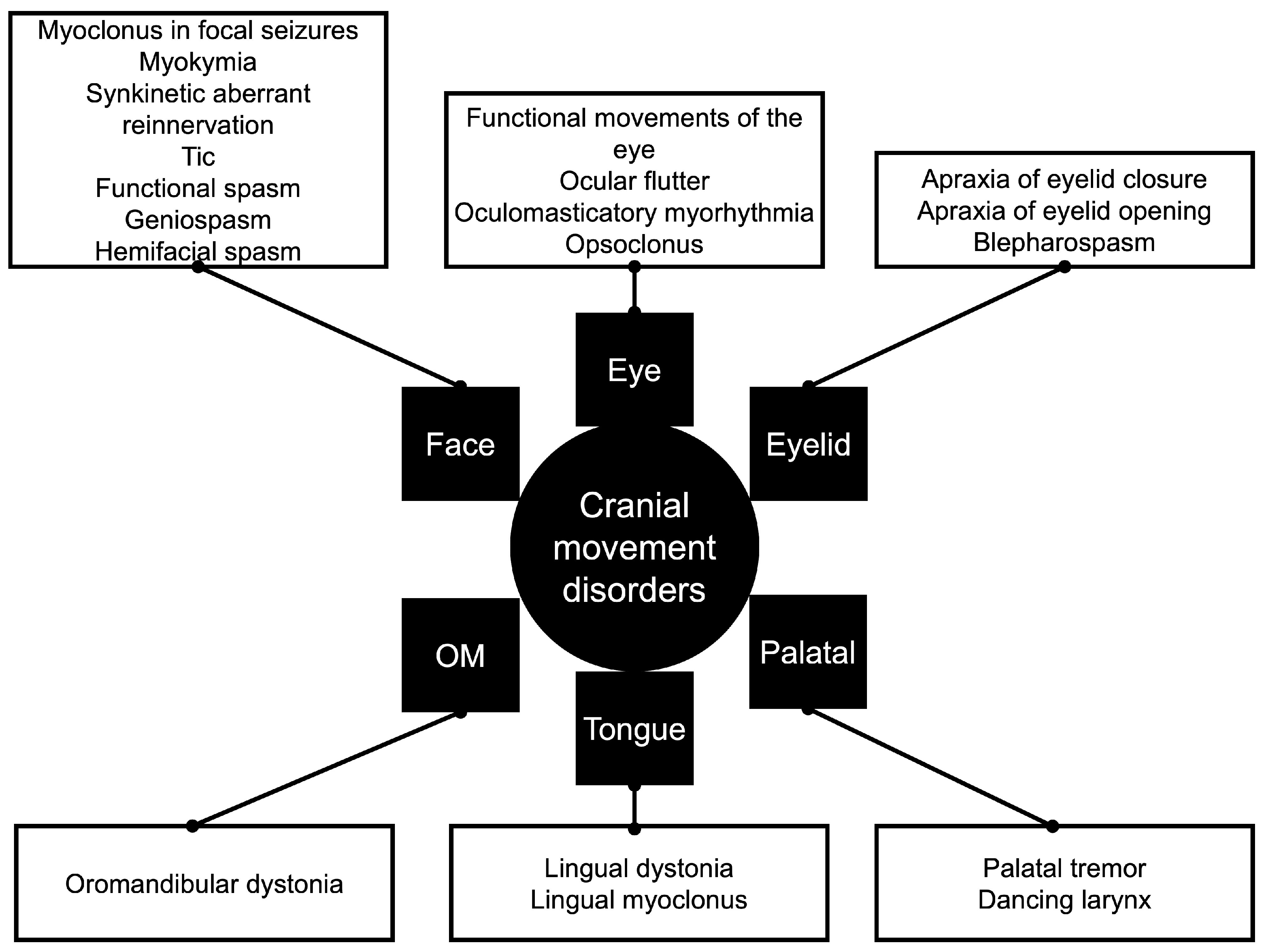
Figure 1. Cranial movement disorders. OM, oromandibular.
Preprints 191373 g001
Table 1. Summary Of Craniofacial Movement Disorders.
Movement Disorder Note
Apraxia of eyelid closure Impaired voluntary eyelid closure despite normal reflex blinking.
Apraxia of eyelid opening Difficulty initiating voluntary eyelid elevation despite normal levator muscle function.
Blepharospasm It is a focal dystonia involving involuntary, bilateral eyelid contractions caused by orbicularis oculi hyperactivity.
Facial myoclonus in focal seizures Jerking movements of facial muscles, resulting from abnormal electrical activity in the contralateral motor cortex or brainstem regions.
Facial myokymia/ neuromyotonia/ superior oblique myokymia and facial fasciculation Continuous, fine, rippling muscle contractions involving the facial muscles, often without voluntary control. Unlike simple fasciculations, myokymia produces persistent wave-like movements that may spread across muscle groups. Neuromyotonia is more severe than myokymia and involves delayed relaxation and stiffness of the affected muscles.
Facial nerve palsy with synkinetic aberrant reinnervation Improper regeneration of facial nerve fibers, causing involuntary muscle contractions and uncoordinated facial movements, often seen as simultaneous eye and mouth muscle activity.
Facial tic Repetitive muscle movement or vocalization affecting the face, often linked to conditions like Tourette syndrome, exacerbated by stress or anxiety.
Functional facial spasm Episodic facial muscle contractions without underlying neurological pathology, often triggered by stress or emotional factors. Unlike dystonias, functional facial spasms may resolve with relaxation or psychological interventions, and they lack the persistent or repetitive nature seen in other movement disorders.
Functional movement disorders of the eye Convergence spasm, convergence paralysis, gaze limitation, functional and voluntary nystagmus and oscillopsia.
Geniospasm Involuntary contraction of the mentalis muscle, causing chin tightening or protrusion.
Hemifacial spasm Unilateral contractions of facial muscles, often due to irritation of the facial nerve, typically resulting from vascular compression or nerve lesions.
Hemimasticatory spasm Sustained contractions of unilateral chewing muscles (masseter, temporalis), causing jaw clenching and difficulty opening the mouth.
Lingual dystonia Sustained contractions of the tongue muscles, leading to abnormal tongue postures, difficulty speaking or swallowing.
Lingual myoclonus Jerking movements of the tongue, often due to brainstem or basal ganglia lesions.
Myotonia Delayed muscle relaxation in the face after contraction, causing stiffness or difficulty with facial expressions. It is often associated with myotonic dystrophy.
Ocular flutter Rapid, uncontrolled, repetitive horizontal eye movements.
Oculomasticatory myorhythmia Convergent-divergent nystagmus, concurrent contractions of the masticatory muscles.
Opsoclonus Rapid, multidirectional eye movements, without slow-phase.
Oromandibular dystonia Contractions of the jaw, lips, and tongue muscles, leading to difficulty speaking, chewing, or swallowing
Palatal Tremor Rhythmic, involuntary contractions of the soft palate muscles
Table 2. Clinical Features Of Essential, Psychogenic, And Secondary Palatal Tremor.
Variable Essential palatal tremor Psychogenic palatal tremor Secondary palatal tremor
Etiology Unknown Psychiatric disorder Brainstem, cerebellar, and basal ganglia
Muscle involved Tensor veli palatini Levator veli palatini and tensor veli palatini Levator veli palatini
Nerve Innervated Trigeminal nerve Trigeminal, glossopharyngeal, and vagus cranial nerves Glossopharyngeal and vagus cranial nerves
Side Bilateral Variable, mainly bilateral Monolateral
Voluntary control Rarely Yes No
Ear clicks (tensor veli palatini) Frequent Frequent Absent
Persistence during sleep Absence Absence Presence
Olivary autonomous oscillator Unknown No Evidence
Course Disappear spontaneously Variable Continuous
Disclaimer/Publisher’s Note: The statements, opinions and data contained in all publications are solely those of the individual author(s) and contributor(s) and not of MDPI and/or the editor(s). MDPI and/or the editor(s) disclaim responsibility for any injury to people or property resulting from any ideas, methods, instructions or products referred to in the content.
Copyright: This open access article is published under a Creative Commons CC BY 4.0 license, which permit the free download, distribution, and reuse, provided that the author and preprint are cited in any reuse.
Prerpints.org logo

Preprints.org is a free preprint server supported by MDPI in Basel, Switzerland.

Subscribe

Disclaimer

Terms of Use

Privacy Policy

Privacy Settings

© 2026 MDPI (Basel, Switzerland) unless otherwise stated