Preprint
Article

This version is not peer-reviewed.

Diagnostic Value of 99mTc-MIBI SPECT/CT Versus Planar Scintigraphy in Primary Hyperparathyroidism: Association with PTH and Calcium Levels

Submitted:

17 December 2025

Posted:

23 December 2025

You are already at the latest version

Abstract

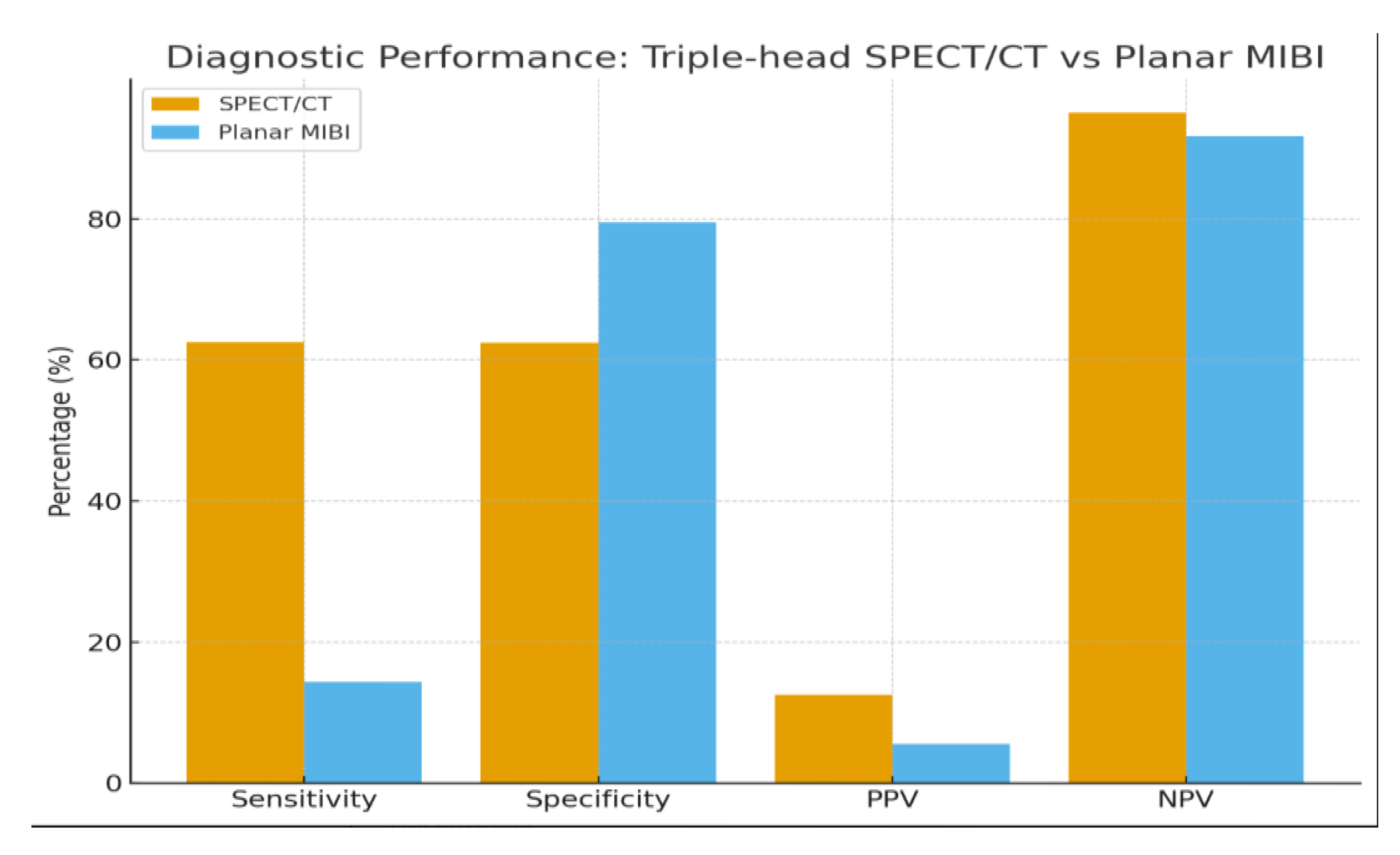
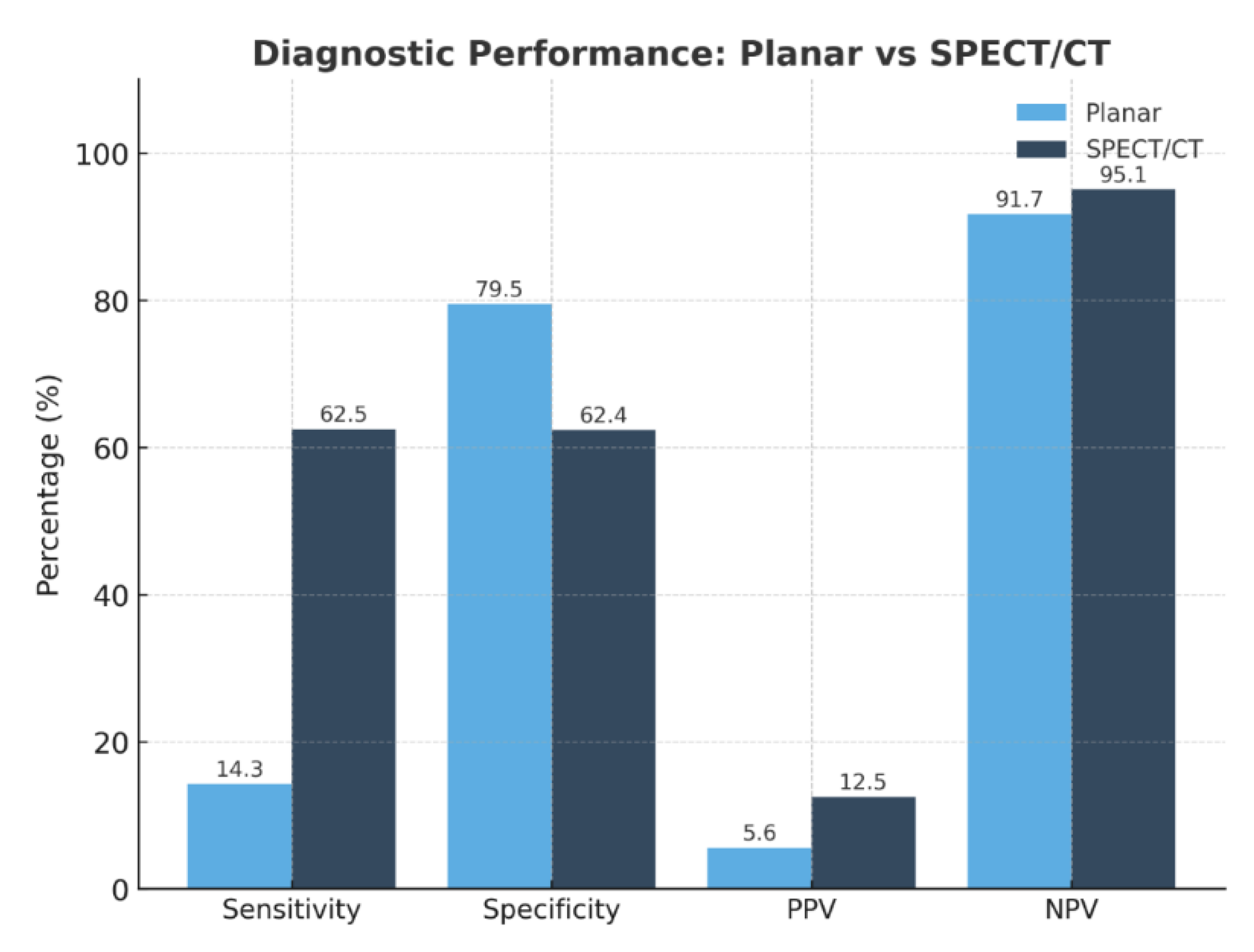
Background/Objectives: Accurate preoperative localization of hyperfunctioning parathyroid tissue is essential for minimally invasive parathyroidectomy. Conventional planar dual-phase 99mTc-sestamibi scintigraphy is widely used but shows reduced diagnostic accuracy in patients with thyroid nodules or ectopic glands. Hybrid triple-head SPECT/CT integrates functional and anatomical imaging and may improve lesion detection. This study evaluated the diagnostic performance of triple-head SPECT/CT compared with planar scintigraphy and explored correlations between biochemical markers and imaging positivity. Methods: A retrospective single-center study included 90 adults referred for suspected primary hyperparathyroidism between January 2021 and August 2025. Demographic data, laboratory parameters (PTH, total and ionized calcium, 25-hydroxyvitamin D), and imaging results were collected. Diagnostic accuracy was assessed in patients with surgical confirmation or robust clinical verification. Correlations between biochemical markers and imaging positivity were analyzed using Pearson correlation coefficients. Results: SPECT/CT demonstrated significantly higher sensitivity than planar scintigraphy (62.5% vs. 14.3%) and an excellent negative predictive value (95.1%), whereas planar imaging showed slightly higher specificity (79.5%). Ionized calcium correlated significantly with SPECT/CT positivity (r = 0.39; p = 0.009), while PTH and 25-hydroxyvitamin D showed no significant association. SPECT/CT accurately localized ectopic parathyroid glands and lesions in patients with coexisting thyroid nodularity, overcoming limitations of planar imaging. Several lesions undetected by planar scintigraphy were identified on SPECT/CT, supporting its role in anatomically complex or subtle cases. Conclusions: Triple-head SPECT/CT provided superior diagnostic performance over planar scintigraphy for preoperative localization in primary hyperparathyroidism, particularly in patients with thyroid nodularity or ectopic glands. Ionized calcium may serve as a complementary predictor of lesion detectability. These findings support hybrid SPECT/CT as the preferred first-line imaging modality, facilitating targeted minimally invasive surgery and optimizing surgical planning.

Keywords: 
;  ;  ;  

1. Introduction

Primary hyperparathyroidism (PHPT) is one of the most common endocrine disorders and represents the leading cause of chronic hypercalcemia in outpatient practice [1,2]. Most cases are caused by a single parathyroid adenoma, but multiglandular disease, ectopic glands, or atypical anatomy are not uncommon [3,4]. A safe and effective surgical treatment for PHPT relies on the surgeon correctly locating all abnormal parathyroid glands [5]. Accurate preoperative localization of hyperfunctioning parathyroid tissue is essential for successful minimally invasive parathyroidectomy (MIP), as it allows surgeons to reduce operative time, avoid unnecessary neck exploration, and minimize the risk of persistent or recurrent disease [6,7]. For many years, dual-phase planar 99mTc-sestamibi scintigraphy has been the standard imaging technique for preoperative localization in PHPT [8,9]. Its widespread use reflects its availability, simplicity, and established diagnostic protocols [10]. However, planar imaging has important limitations. Its sensitivity decreases in patients with small adenomas, multiglandular disease, cystic degeneration, or concurrent thyroid nodules. These factors increase the likelihood of false-negative or equivocal results, which can complicate surgical planning and reduce the success of minimally invasive approaches [11]. Hybrid imaging with single-photon emission computed tomography fused with computed tomography (SPECT/CT) addresses some of these limitations by combining functional and anatomical information [11,12,13,14]. SPECT/CT allows three-dimensional visualization of lesions, better anatomical correlation, and attenuation-corrected evaluation of tracer uptake. This technique has been shown to improve lesion localization compared with planar imaging, particularly in patients with ectopic glands, complex neck anatomy, or coexisting thyroid pathology [15,16,17]. Despite its advantages, reported sensitivity and specificity vary across studies due to differences in imaging protocols, patient populations, and reference standards [18]. While emerging modalities such as 18F-fluorocholine PET/CT or 4D-CT show excellent sensitivity, their high cost and limited availability restrict their use in routine clinical practice [18,19]. Biochemical markers, including intact parathyroid hormone (PTH), total and ionized calcium, and 25-hydroxyvitamin D, are central to the diagnosis of PHPT and may influence radiotracer uptake [20]. It is hypothesized that higher calcium levels or elevated PTH may correspond to larger or more metabolically active adenomas, which could improve lesion detectability [20,21,22]. However, evidence on the predictive value of these biochemical parameters for imaging success remains inconsistent, and few studies have evaluated their association with planar versus SPECT/CT imaging in real-world, unselected patient populations. The University Clinical Center of Kosovo (QKUK), as the national referral center, provides care for all patients with suspected PHPT, regardless of disease complexity or thyroid status. This offers a unique opportunity to evaluate imaging performance in a consecutive, real-world cohort that reflects everyday clinical practice. The present study therefore aimed to directly compare planar ⁹⁹mTc-sestamibi scintigraphy with triple-head SPECT/CT for preoperative localization of hyperfunctioning parathyroid tissue in PHPT and to investigate whether biochemical markers can predict imaging positivity. These findings may help optimize first-line imaging strategies and guide surgical planning in routine clinical practice.

2. Materials and Methods

2.1. Study Design and Setting

This retrospective observational cohort study was conducted at the Department of Nuclear Medicine, University Clinical Center of Kosovo (UCKK), Prishtina. All consecutive adult patients referred for suspected parathyroid disease or hyperparathyroidism between January 2021 and August 2025 were included. Clinical data were obtained from the institutional electronic registry and comprised patient characteristics, biochemical values (intact PTH, total calcium, ionized calcium, and 25-OH vitamin D), and—when available—ultrasound and DEXA findings. Nuclear medicine data included structured reports from dual-phase planar 99mTc-sestamibi scintigraphy and hybrid SPECT/CT examinations acquired within the same diagnostic workflow [23,24].

2.2. Radiopharmaceutical Preparation and Radiolabeling Procedure

Radiopharmaceutical preparation was performed using the commercially available MIBI kit (Monrol), designed for myocardial, parathyroid, and breast imaging following radiolabeling with technetium-99m. Each vial contained a standardized formulation composed of 1.0 mg methoxyisobutylisonitrile (MIBI), 0.075 mg stannous chloride dihydrate, 1.0 mg L-cysteine hydrochloride monohydrate, 2.6 mg sodium citrate dihydrate, and 20 mg D-mannitol, ensuring optimal chelation efficiency and stability of the final complex. Radiolabeling was achieved by adding 5 mL of sterile sodium pertechnetate (99mTcO₄⁻) solution to the lyophilized vial, with a maximum allowable activity of up to 400 mCi (14.8 GBq). The mixture was gently agitated for approximately one minute and subsequently heated in a boiling water bath for 12 minutes to facilitate complete complexation. After controlled cooling to room temperature, the final 99mTc-MIBI preparation demonstrated high radiochemical stability for at least six hours’ post-reconstitution, permitting reliable administration and image acquisition within routine clinical workflow.

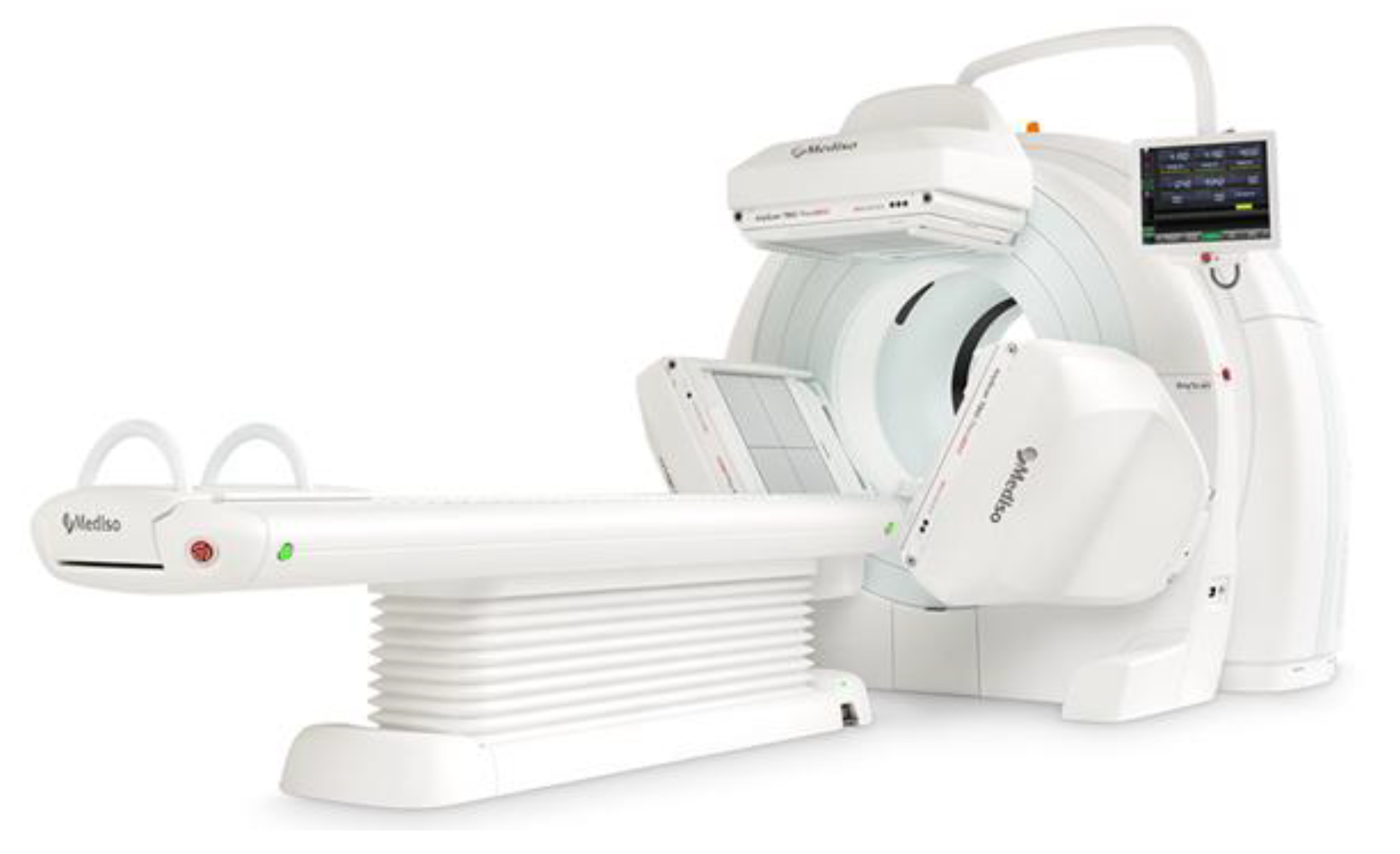
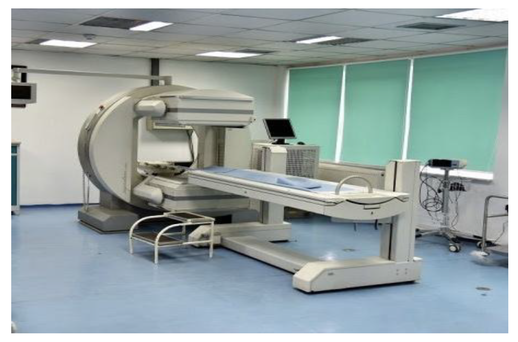
2.3. Imaging Equipment

Hybrid SPECT/CT examinations were performed on the AnyScan TRIO SPECT/CT system (Mediso Medical Imaging Systems, Budapest, Hungary), equipped with a triple-detector SPECT configuration to enable high-resolution functional imaging and precise anatomical localization of parathyroid lesions, including ectopic glands (Figure 1 and Figure 2). The system incorporates the TheraMax platform with the MaX/123-15.9 high-performance detection module, optimized to enhance photon sensitivity, improve lesion contrast, and ensure robust diagnostic accuracy in nuclear medicine applications. For comparison, planar dual-phase 99mTc-sestamibi scintigraphy was performed using a Siemens dual-head gamma camera (Siemens Healthcare, Erlangen, Germany), installed in 2009, with image acquisition parameters standardized across the study population (Figure 1 and Figure 2).

2.4. Imaging Protocols

Planar dual-phase 99mTc-sestamibi scintigraphy consisted of early and delayed anterior acquisitions of the neck and upper mediastinum following intravenous tracer administration. Imaging matrices and acquisition parameters were standardized for all patients to ensure comparability across the cohort. Hybrid SPECT/CT was performed sequentially after planar imaging, using low-dose CT for anatomical correlation and attenuation correction, followed by iterative reconstruction of SPECT data. Interpretation criteria required focal radiotracer persistence or increased uptake relative to surrounding thyroid and background structures on both early and delayed phases, in either orthotopic or ectopic parathyroid locations. CT information supported precise lesion localization and discrimination from thyroid nodules or non-specific uptake [23,24]

2.5. Endpoints and Statistical Analysis

The primary endpoint was modality-specific detectability, categorized as positive or negative for each imaging technique. Diagnostic accuracy metrics (true positive, false positive, true negative, false negative classifications), including sensitivity, specificity, positive predictive value (PPV), and negative predictive value (NPV), were calculated in the subset of patients with histopathological confirmation or robust clinico-biochemical validation. Associations between biochemical parameters and imaging positivity were evaluated using Pearson correlation coefficients, with statistical significance set at α=0.05.

3. Results

3.1. Cohort Characteristics

A total of 90 consecutive patients were included in the analysis. The mean age was 56.7 ± 12.4 years (range 23–82), and the majority were female (83.3%). Biochemical measurements showed considerable variability, with a mean PTH level of 338.8 ± 512.6 pg/mL (range 1–3144), mean total serum calcium of 2.56 ± 0.42 mmol/L, mean ionized calcium of 1.35 ± 0.21 mmol/L, and mean 25-hydroxyvitamin D of 20.3 ± 11.5 ng/mL. Thyroid ultrasonography revealed coexisting thyroid nodularity in a substantial proportion of patients, reflecting the high prevalence of multinodular goiter in the region. Baseline demographic and biochemical characteristics of the study population are summarized in Table 1.

3.2. Diagnostic Performance

Within the reference-confirmed cohort, hybrid SPECT/CT demonstrated substantially higher diagnostic performance than planar scintigraphy, particularly with respect to sensitivity and negative predictive value (Table 2). The enhanced performance of hybrid imaging is attributable to its superior anatomical detail, improved spatial resolution, and attenuation correction, which collectively increase reader confidence and reduce equivocal interpretations. The added value of SPECT/CT was especially notable in patients with ectopic parathyroid tissue or concomitant thyroid nodularity, clinical scenarios in which planar imaging often cannot reliably differentiate parathyroid from thyroid activity. Among the 90 patients with positive scintigraphic studies, 70 were surgically confirmed to harbor a parathyroid adenoma or ectopic gland. Conversely, four patients demonstrated positive scintigraphic findings but negative results on ultrasound-guided fine-needle aspiration, representing false-positive outcomes likely related to adjacent thyroid uptake or interpretive difficulties in nodular goiter (Figure 3 and Figure 4). In addition, several lesions not visualized on planar imaging were correctly detected by SPECT/CT, further underscoring its importance in identifying subtle tracer retention or deep mediastinal disease. Collectively, these results support the adoption of SPECT/CT as the preferred preoperative imaging modality, particularly when minimally invasive parathyroidectomy is anticipated.

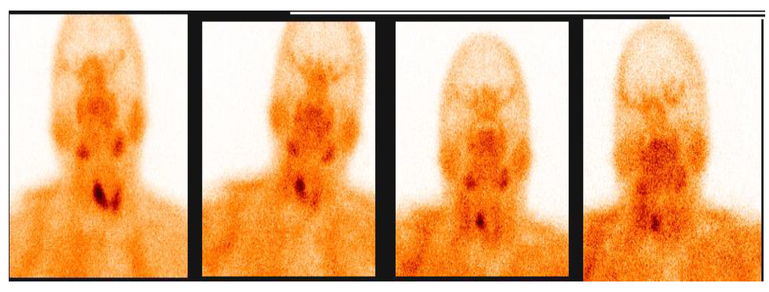
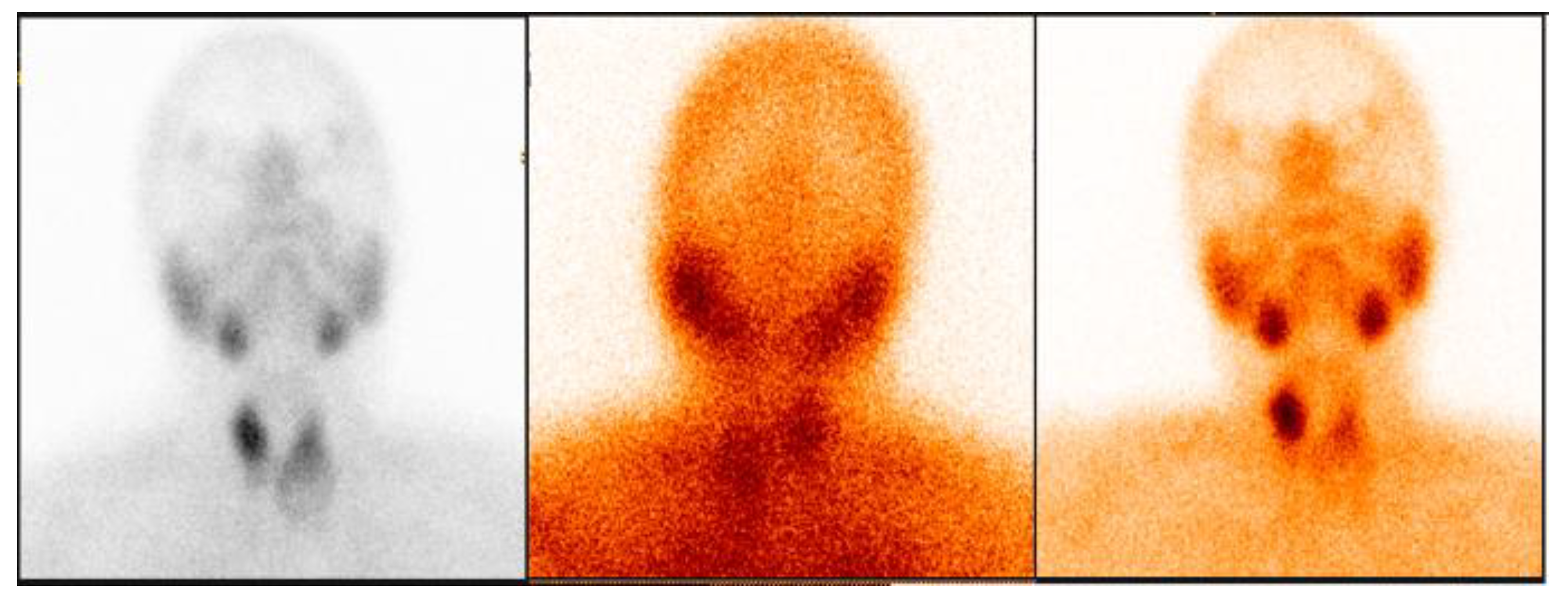
3.2.1. Representative Clinical Case 1

A 71-year-old female with biochemically confirmed primary hyperparathyroidism (intact PTH 154 pg/mL, hypercalcemia, vitamin D insufficiency) underwent dual-phase planar 99mTc-sestamibi scintigraphy. Early imaging revealed focal radiotracer uptake at the inferior pole of the right thyroid lobe, with persistent activity on delayed images suggestive of a right inferior parathyroid adenoma (Figure 3). However, interpretation was limited by coexisting thyroid nodularity. Subsequent hybrid SPECT/CT, acquired approximately 90 minutes after tracer administration, localized a well-defined extracapsular lesion inferior to the right thyroid lobe (Figure 4). CT co-registration clearly distinguished the parathyroid adenoma from adjacent thyroid nodules, providing definitive anatomical localization. The triple-detector system’s improved photon-detection efficiency and angular sampling resulted in enhanced lesion conspicuity, particularly in the setting of subtle tracer retention.

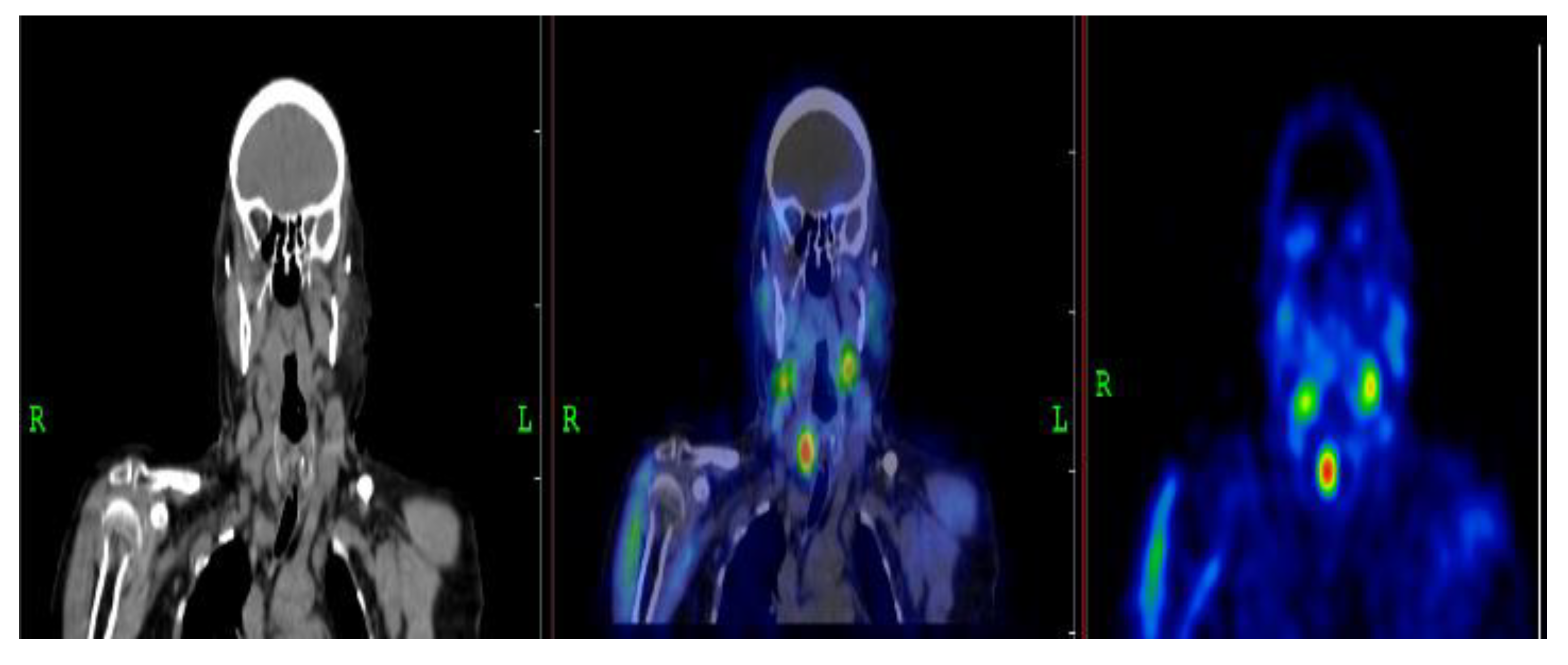
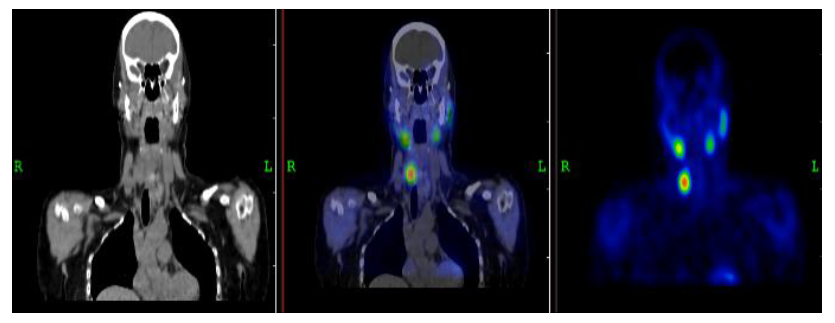
3.2.2. Representative Clinical Case 2

A 62-year-old male with biochemical evidence of primary hyperparathyroidism (PTH 215 pg/mL, hypercalcemia, low–normal vitamin D) exhibited persistent focal tracer retention inferior to the right thyroid lobe on dual-phase planar imaging. Hybrid triple-detector SPECT/CT performed 90 minutes post-injection precisely localized the focus and confirmed an extracapsular right inferior parathyroid adenoma (Figure 5 and Figure 6). The hybrid dataset effectively differentiated the lesion from heterogeneous thyroid parenchyma and reduced interpretive uncertainty. This case further demonstrates the incremental diagnostic contribution of SPECT/CT, particularly in patients with mild biochemical abnormalities or complex cervical anatomy. Early and delayed planar 99mTc-sestamibi imaging in a 62-year-old male with biochemical evidence of primary hyperparathyroidism revealed a well-defined focus of persistent radiotracer uptake inferior to the right thyroid lobe, consistent with a hyperfunctioning parathyroid lesion. A triple-detector SPECT/CT acquisition performed 90 minutes after tracer administration provided precise anatomical localization, confirming a right-sided parathyroid adenoma adjacent to the lower pole of the thyroid gland. The hybrid dataset clearly distinguished the adenoma from surrounding thyroid tissue and substantially reduced the interpretive limitations inherent to planar imaging. This case highlights the incremental diagnostic utility of SPECT/CT in preoperative localization and individualized surgical planning. (Images courtesy of the Department of Nuclear Medicine, Prishtina, Kosovo.)

3.3. Diagnostic Performance

In the reference-standard subset, triple-head SPECT/CT demonstrated substantially higher sensitivity than planar 99mTc-MIBI scintigraphy (62.5% vs. 14.3%), confirming its superior ability to detect hyperfunctioning parathyroid tissue. SPECT/CT also achieved a high negative predictive value (NPV) of 95.1%, indicating strong reliability in ruling out disease when findings were negative. In contrast, planar imaging showed markedly reduced detectability and contributed disproportionately to false-negative results. Specificity and positive predictive value (PPV) for SPECT/CT appeared modest when calculated across the entire cohort (62.4% and 12.5%, respectively). However, these metrics must be interpreted within the clinical context: nearly all patients had biochemically and/or surgically confirmed primary hyperparathyroidism, resulting in a very small number of truly disease-negative individuals (<10%). This high pretest probability artificially lowers specificity estimates and increases the proportion of scans categorized as false positives. Particularly, several cases labeled as “false-positive” occurred in patients who did not undergo surgery, reflecting a lack of definitive verification rather than true imaging inaccuracy. These patterns are consistent with previously reported diagnostic behavior of SPECT/CT in high-prevalence referral populations. Table 2 summarizes performance indices for both modalities.
Diagnostic performance of triple-detector SPECT/CT and dual-phase planar 99mTc-MIBI scintigraphy in patients with suspected primary hyperparathyroidism at a single national center. SPECT/CT exhibited markedly higher sensitivity (62.5%) and negative predictive value (95.1%) compared with planar scintigraphy (14.3% and 91.7%, respectively), reflecting superior detection of hyperfunctioning parathyroid lesions, including ectopic glands and cases with thyroid nodularity. Although planar scintigraphy demonstrated higher specificity (79.5% vs. 62.4%), this finding should be interpreted cautiously due to the low number of true negative cases and high disease prevalence. Positive predictive values were low for both modalities, owing to the limited number of histologically confirmed negative findings (Figure 7).

3.4. Biochemical Correlations

Ionized calcium exhibited the strongest and most clinically significant association with SPECT/CT detectability, reflecting its direct influence on cellular metabolic activity and mitochondrial density in oxyphil-rich parathyroid adenomas. This mechanistic link showed biochemical severity particularly hypercalcemia as the primary determinant of radiotracer retention and functional visibility on sestamibi imaging, whereas isolated elevations in PTH do not reliably indicate the cellular energetic status required for tracer uptake. In contrast, planar scintigraphy demonstrated no consistent biochemical correlations, highlighting its susceptibility to false-negative results in mild disease or in the presence of nodular thyroid pathology. These observations support a calcium-guided approach to selecting hybrid imaging, establishing SPECT/CT as the preferred first-line modality for precise preoperative localization in primary hyperparathyroidism.SPECT/CT also showed superior diagnostic performance, particularly for the detection of ectopic parathyroid glands (Figure 8), reinforcing its clinical utility as a first-line localization strategy in contexts where surgical planning relies on accurate preoperative mapping. Full statistical correlation outputs are provided in Table 3 and Table 4.

4. Discussion

In this consecutive, real-world cohort of patients with suspected primary hyperparathyroidism (PHPT), hybrid 99mTc-sestamibi SPECT/CT provided substantially improved lesion localization compared with conventional planar scintigraphy [25,26,27].. The benefit was especially evident in patients with coexisting thyroid nodularity or ectopic parathyroid glands [28,29,30]. In these anatomically complex cases, CT co-registration enhances lesion delineation, reduces interpretive uncertainty, and allows for more precise preoperative localization [31]. This level of accuracy has clear clinical relevance, as it can guide targeted minimally invasive surgery, reduce operative time, limit the extent of dissection, and lower the risk of persistent or recurrent disease. By combining functional and anatomical information, SPECT/CT overcomes limitations of planar scintigraphy, which can be confounded by overlapping structures or variable tracer uptake in thyroid tissue [32,33]. These observations are consistent with previous reports supporting the superiority of hybrid imaging in challenging anatomical or functional scenarios and reinforce its role as a first-line modality for preoperative localization [34,35,36]. Our biochemical analyses further support these imaging results. Ionized calcium levels demonstrated a strong and statistically significant association with SPECT/CT positivity, reflecting the underlying mechanism of sestamibi uptake in mitochondrial-rich oxyphil cells within hyperfunctioning parathyroid tissue [37,38]. This indicates that functional activity, rather than circulating hormone concentration alone, primarily determines detectability on imaging. In contrast, PTH showed only a weak and non-significant relationship, showing its limited predictive value when interpreted alone, likely due to confounding effects of renal function, vitamin D status, and compensatory glandular activity [39]. Similarly, 25-hydroxyvitamin D levels did not correlate with SPECT/CT positivity, in agreement with previous studies showing that vitamin D deficiency is common across PHPT severity and does not independently influence sestamibi uptake [40,41]. Importantly, none of the biochemical markers correlated with planar scintigraphy positivity, further demonstrating the limited sensitivity of planar imaging, particularly in mild hypercalcemia or in patients with thyroid nodularity [38,42]
Collectively, these findings support the reliability of SPECT/CT as a functional anatomical tool, with ionized calcium providing complementary predictive information [43]. These results are consistent with current European and international guidelines, which recommend prioritizing hybrid functional–anatomical imaging for patients with high pretest probability of PHPT or discordant ultrasound results [44].Ectopic gland localization remains a key indication for SPECT/CT, as it allows accurate anatomical mapping and facilitates minimally invasive surgical approaches [45,46]. Accurate preoperative identification of both typical and ectopic lesions can reduce the extent of surgical exploration, decrease operative complications, and improve postoperative outcomes [47]. Although SPECT/CT significantly improved sensitivity in our cohort, a modest reduction in specificity was observed, consistent with reports of false-positive uptake in multinodular goiter or cystic/dystrophic thyroid lesions [48,49]. These findings show the need to interpret SPECT/CT results carefully in the context of coexisting thyroid pathology. Emerging evidence suggests that 18F-fluorocholine PET/CT may offer additional value in particularly complex or discordant cases, although its routine use is limited by accessibility and cost considerations [50]. Despite the slight decrease in specificity, the very high negative predictive value observed in this study (95.1%) confirms SPECT/CT as a reliable method to exclude hyperfunctioning parathyroid tissue and avoid unnecessary bilateral neck exploration [51,52]. The use of triple-detector SPECT/CT in this study provides further advantages, including improved spatial resolution and shorter acquisition times compared with earlier dual-detector systems [53,54]. This technological improvement is particularly relevant for centers incorporating hybrid imaging into routine PHPT assessment, allowing precise lesion detection without compromising workflow efficiency [46,55,56]. Overall, these findings show the value of hybrid SPECT/CT as the preferred preoperative localization strategy in PHPT. Combining biochemical assessment with advanced functional–anatomical imaging supports optimal surgical planning, reduces operative morbidity, and improves patient outcomes. Future research should focus on larger, multicenter studies to validate these findings and establish standardized imaging protocols for PHPT. Combining SPECT/CT with advanced biochemical or molecular markers may further refine patient selection and improve preoperative planning. Additionally, cost-effectiveness analyses comparing hybrid SPECT/CT with other emerging imaging techniques would help guide clinical decision-making and resource allocation. Such work will contribute to optimizing preoperative localization, facilitating minimally invasive surgery, and improving outcomes for patients with primary hyperparathyroidism.

5. Conclusions

In this study, hybrid triple-detector 99mTc-sestamibi SPECT/CT demonstrated superior preoperative localization of hyperfunctioning parathyroid tissue compared with conventional planar scintigraphy, particularly in patients with ectopic glands or coexisting thyroid nodularity. The combination of functional and anatomical imaging resulted in higher sensitivity and negative predictive value, which facilitated precise surgical planning, minimized unnecessary dissection, and supported minimally invasive parathyroidectomy. Ionized calcium levels, rather than PTH or vitamin D alone, showed the strongest association with SPECT/CT detectability, reflecting the mechanistic link between hypercalcemia and radiotracer uptake. These findings reinforced the role of hybrid SPECT/CT as the preferred first-line imaging modality for PHPT evaluation and underscored the need for multicenter studies and cost-effectiveness analyses to further optimize preoperative imaging strategies and patient management.

Author Contributions

Conceptualization, I,B. and A.J.; methodology, I,B.; software, A.J.; validation, S.D.E, and I,B.; formal analysis, I,B.; investigation, A.J..; resources, S.D.E and S.S.; data curation, I.B.; writing—original draft preparation, A.J writing—review and editing, I,B. and A.J.; visualization, S.D.E.; supervision. All authors have read and agreed to the published version of the manuscript.

Funding

This research received no external funding.

Informed Consent Statement

The study used only pre-existing medical data; therefore, patient consent was not required.

Data Availability Statement

All data supporting the findings of this study are contained within the article. No additional datasets were generated or analyzed.

Conflicts of Interest

The authors declare no conflicts of interest.

References

  1. Palermo, A.; Tabacco, G.; Makras, P.; Zavatta, G.; Trimboli, P.; Castellano, E.; Anastasilakis, A.D. Primary Hyperparathyroidism: From Guidelines to Outpatient Clinic. Rev. Endocr. Metab. Disord. 2024, 25, 875–896. [Google Scholar] [CrossRef]
  2. Bilezikian, J.P.; Cusano, N.E.; Khan, A.A.; Liu, J.M.; Marcocci, C.; Bandeira, F. Primary Hyperparathyroidism. Nat. Rev. Dis. Primers 2016, 2, 16033. [Google Scholar] [CrossRef] [PubMed]
  3. DeLellis, R.A.; Mazzaglia, P.; Mangray, S. Primary Hyperparathyroidism: A Current Perspective. Arch. Pathol. Lab. Med. 2008, 132, 1251–1262. [Google Scholar] [CrossRef]
  4. Kowalski, G.J.; Buła, G.; Żądło, D.; Gawrychowska, A.; Gawrychowski, J. Primary Hyperparathyroidism. Endokrynol. Pol. 2020, 71, 260–270. [Google Scholar] [CrossRef] [PubMed]
  5. Hindié, E.; Schwartz, P.; Avram, A.M.; Imperiale, A.; Sebag, F.; Taïeb, D. Primary Hyperparathyroidism: Defining the Appropriate Preoperative Imaging Algorithm. J. Nucl. Med. 2021, 62, 3S–12S. [Google Scholar] [CrossRef]
  6. Mariani, G.; Gulec, S.A.; Rubello, D.; Boni, G.; Puccini, M.; Pelizzo, M.R.; Giuliano, A.E. Preoperative Localization and Radioguided Parathyroid Surgery. J. Nucl. Med. 2003, 44, 1443–1458. [Google Scholar] [PubMed]
  7. Kunstman, J.W.; Kirsch, J.D.; Mahajan, A.; Udelsman, R. Parathyroid Localization and Implications for Clinical Management. J. Clin. Endocrinol. Metab. 2013, 98, 902–912. [Google Scholar] [CrossRef]
  8. Özçevik, H.; Tamam, M.Ö.; Tatoğlu, M.T.; Mülazımoğlu, M. Comparison of Dual-Phase Tc-99m-Sestamibi Planar Imaging and SPECT/CT in Hyperparathyroidism. Mol. Imaging Radionucl. Ther. 2022, 31, 191–197. [Google Scholar] [CrossRef]
  9. Yang, J.; Lu, X.; Zhou, P.; Gao, Z.; Ding, C.; Weng, W.; Su, X. Comparison of Four-Dimensional CT and Sestamibi SPECT/CT in the Localization Management of Primary Hyperparathyroidism. Cancer Imaging 2025, 25, 90. [Google Scholar] [CrossRef]
  10. Wang, Y.; Liu, Y.; Li, N.; Xu, K.; Zhang, W. Quantitative Application of Dual-Phase 99mTc-Sestamibi SPECT/CT for Parathyroid Lesions: Identification of Optimal Timing in Secondary Hyperparathyroidism. EJNMMI Phys. 2023, 10, 29. [Google Scholar] [CrossRef]
  11. Lavely, W.C.; Goetze, S.; Friedman, K.P.; Leal, J.P.; Zhang, Z.; Garret-Mayer, E.; Ziessman, H.A. Comparison of SPECT/CT, SPECT, and Planar Imaging Using 99mTc-Sestamibi. J. Nucl. Med. 2007, 48, 1084–1089. [Google Scholar] [CrossRef] [PubMed]
  12. Papathanassiou, D.; Bruna-Muraille, C.; Jouannaud, C.; Gagneux-Lemoussu, L.; Eschard, J.P.; Liehn, J.C. SPECT/CT in Bone Diseases. Joint Bone Spine 2009, 76, 474–480. [Google Scholar] [CrossRef] [PubMed]
  13. El Yaman, A.; Alwan, M.; El Ghazawi, A.; Al-Mallah, M.H.; Al Rifai, M. Advances in Nuclear Hybrid Imaging in Cardiovascular Diseases. Prog. Cardiovasc. Dis. 2025, in press. [Google Scholar] [CrossRef] [PubMed]
  14. Prakash, D.; Kotian, R.P. Hybrid Imaging: Physics, Principles, and Quality Control. In Fundamentals of X-ray Imaging, 2nd ed.; Springer Nature Singapore: Singapore, 2025; pp. 417–506. [Google Scholar] [CrossRef]
  15. Even-Sapir, E.; Keidar, Z.; Bar-Shalom, R. Hybrid Imaging Improving Diagnostic Accuracy. In Semin. Nucl. Med.; WB Saunders, July 2009; Vol. 39, pp. 264–275. [Google Scholar]
  16. Mariani, G.; Bruselli, L.; Kuwert, T.; Kim, E.E.; Flotats, A.; Israel, O.; Watanabe, N. Clinical Uses of SPECT/CT—A Review. Eur. J. Nucl. Med. Mol. Imaging 2010, 37, 1959–1985. [Google Scholar] [CrossRef]
  17. Spanu, A.; Schillaci, O.; Piras, B.; Madeddu, G. SPECT/CT in Hyperparathyroidism. Clin. Transl. Imaging 2014, 2, 537–555. [Google Scholar] [CrossRef]
  18. Scerrino, G.; Paladino, N.C.; Graceffa, G.; Melfa, G.; Orlando, G.; Di Vuolo, R.; Lo Casto, A. 18F-Fluorocholine PET/CT vs 4D-CT for Parathyroid Identification. J. Clin. Med. 2025, 14, 5468. [Google Scholar] [CrossRef]
  19. Latge, A.; Riehm, S.; Vix, M.; Bani, J.; Ignat, M.; Pretet, V.; Imperiale, A. 18F-Fluorocholine PET and 4D-CT in Persistent Primary Hyperparathyroidism. Diagnostics 2021, 11, 2384. [Google Scholar] [CrossRef] [PubMed]
  20. Al-Hraishawi, H.K. Comparison of Metabolic and Characteristic Features of PHPT Patients with Different PTH Levels. Master’s Thesis, Rutgers University, USA, 2016. [Google Scholar]
  21. Bhan, A.; Athimulam, S.; Kumari, P.; Pal, R.; Bhadada, S.K.; Cook, B.C.; Rao, S.D. Large Parathyroid Adenomas: Reconciling Adenoma Size and Phenotype. Front. Endocrinol. 2023, 14, 1009516. [Google Scholar] [CrossRef]
  22. Kazakou, P.; Vrachnis, D.; Paschou, S.A.; Nastos, K.; Sarlani, H.; Kantreva, K.; Saltiki, K. Giant Parathyroid Adenoma with Carcinoma-like Presentation: Case Report. Hormones 2025, 1–6. [Google Scholar] [CrossRef]
  23. Lee, S.H.; Shin, E.; Ha, S.; Oh, J.S.; Song, D.E.; Ryu, J.S. Dual- vs. Single-Phase SPECT/CT for Parathyroid Lesion Localization. Medicine 2020, 99, e19989. [Google Scholar] [CrossRef]
  24. Giovanella, L.; Hindie, E.; Huellner, M.W.; Talbot, J.N.; Verburg, F.A. Summary of the New EANM Guidelines for Parathyroid Imaging. Q. J. Nucl. Med. Mol. Imaging 2022, 66, 93–103. [Google Scholar] [CrossRef]
  25. Lavely, W.C.; Goetze, S.; Friedman, K.P.; Leal, J.P.; Zhang, Z.; Garrett-Mayer, E.; Ziessman, H.A. Tc-99m Sestamibi Planar, SPECT, and SPECT/CT Comparison. J. Nucl. Med. 2007, 48, 1084–1089. [Google Scholar] [CrossRef] [PubMed]
  26. Jacene, H.A.; Goetze, S.; Patel, H.; Wahl, R.L.; Ziessman, H.A. Advantages of Hybrid SPECT/CT. Open Med. Imaging J. 2008, 2, 67–79. [Google Scholar] [CrossRef]
  27. Durma, A.D.; Saracyn, M.; Kołodziej, M.; Jóźwik-Plebanek, K.; Kamiński, G. Use of [11C]C-Methionine in Endocrine Diagnostics. Pharmaceuticals 2025, 18, 229. [Google Scholar] [CrossRef]
  28. Wong, K.K.; Fig, L.M.; Gross, M.D.; Dwamena, B.A. Parathyroid Adenoma Localization with Sestamibi SPECT/CT: Meta-analysis. Nucl. Med. Commun. 2015, 36, 363–375. [Google Scholar] [CrossRef]
  29. Gayed, I.W.; Kim, E.E.; Broussard, W.F.; Evans, D.; Lee, J.; Broemeling, L.D.; Podoloff, D.A. Value of SPECT/CT Over SPECT in Parathyroid Imaging. J. Nucl. Med. 2005, 46, 248–252. [Google Scholar]
  30. Morris, M.A.; Saboury, B.; Ahlman, M.; Malayeri, A.A.; Jones, E.C.; Chen, C.C.; Millo, C. Parathyroid Imaging: Past, Present, and Future. Front. Endocrinol. 2022, 12, 760419. [Google Scholar] [CrossRef]
  31. Leitha, T.; Staudenherz, A. Hybrid PET/CT and SPECT/CT Imaging. In Computed Tomography—Clinical Applications; InTech, 2012; pp. 269–294. [Google Scholar]
  32. Israel, O.; Goldsmith, S.J. (Eds.) Hybrid SPECT/CT Imaging in Clinical Practice; CRC Press: Boca Raton, FL, USA, 2016. [Google Scholar]
  33. Pata, G.; Casella, C.; Magri, G.C.; Lucchini, S.; Panarotto, M.B.; Crea, N.; Salerni, B. Financial and Clinical Implications of Low-Energy CT with 99mTc-MIBI SPECT. Ann. Surg. Oncol. 2011, 18, 2555–2563. [Google Scholar] [CrossRef] [PubMed]
  34. Tay, D.; Das, J.P.; Yeh, R. Preoperative Localization for PHPT: A Clinical Review. Biomedicines 2021, 9, 390. [Google Scholar] [CrossRef]
  35. Sandqvist, P. Preoperative Localisation of Parathyroid Adenoma Using Tc-99m-Sestamibi SPECT/CT. Thesis, Karolinska Institutet, Sweden, 2022. [Google Scholar]
  36. Hindie, E.; Ureña-Torres, P.A.; Taïeb, D. Parathyroid Imaging in Renal Hyperparathyroidism. In Parathyroid Glands in Chronic Kidney Disease; Springer: Cham, Switzerland, 2020; pp. 35–49. [Google Scholar] [CrossRef]
  37. Hoang, T.D.; Jani, A.G.; Mai, V.Q.; Tuamokumo, F.O.; Shakir, M.K. Associations of Biochemical Markers with Parathyroid Scans. Endocr. Pract. 2019, 25, 16–22. [Google Scholar] [CrossRef] [PubMed]
  38. Swanson, T.W.; Chan, S.K.; Jones, S.J.; Bugis, S.; Irvine, R.; Belzberg, A.; Wiseman, S.M. Determinants of Sestamibi SPECT Sensitivity. Am. J. Surg. 2010, 199, 614–620. [Google Scholar] [CrossRef]
  39. Niculescu, D.A.; Deacu, L.G.; Caragheorgheopol, A.; Popescu, N.; Ghemigian, A.; Procopiuc, C.; Poiana, C. Effects of Vitamin D, Renal Function and Age on PTH. Front. Endocrinol. 2021, 12, 657991. [Google Scholar] [CrossRef]
  40. Jarvis, P.; Downs, J.; Skene, T.; Evans, A.; Richardson, T.; Parekh, A. Factors Associated with Successful Parathyroid Localization. Int. J. Endocrinol. 2025, 2025, 3922886. [Google Scholar] [CrossRef]
  41. Sencar, M.E.; Sakiz, D.; Unsal, I.O.; Hepsen, S.; Calapkulu, M.; Gumus, P.; Cakal, E. Vitamin D Does Not Affect Parathyroid Imaging Sensitivity. Sci. Rep. 2019, 9, 12035. [Google Scholar] [CrossRef]
  42. Mshelia, D.S.; Hatutale, A.N.; Mokgoro, N.P.; Nchabaleng, M.E.; Buscombe, J.R.; Sathekge, M.M. Serum Calcium and Dual-Phase Sestamibi Sensitivity. Clin. Physiol. Funct. Imaging 2012, 32, 19–24. [Google Scholar] [CrossRef]
  43. Silov, G.; Erdogan Ozbodur, S. Relationship between SPECT/CT Parameters and Functional Markers. Diagnostics 2023, 13, 3182. [Google Scholar] [CrossRef] [PubMed]
  44. Petranović Ovčariček, P.; Giovanella, L.; Carrió Gasset, I.; Hindié, E.; Huellner, M.W.; Luster, M.; Verburg, F.A. EANM Practice Guidelines for Parathyroid Imaging. Eur. J. Nucl. Med. Mol. Imaging 2021, 48, 2801–2822. [Google Scholar] [CrossRef] [PubMed]
  45. Krausz, Y.; Bettman, L.; Guralnik, L.; Yosilevsky, G.; Keidar, Z.; Bar-Shalom, R.; Israel, O. Tc-99m MIBI SPECT/CT in Primary Hyperparathyroidism. World J. Surg. 2006, 30, 76–83. [Google Scholar] [CrossRef]
  46. Taubman, M.L.; Goldfarb, M.; Lew, J.I. Role of SPECT and SPECT/CT in Parathyroid Surgery. Int. J. Mol. Imaging 2011, 2011, 141593. [Google Scholar] [CrossRef] [PubMed]
  47. Patel, S.G.; Saunders, N.D.; Jamshed, S.; Weber, C.J.; Sharma, J. Multimodal Localization Improves Reoperative Parathyroidectomy Outcomes. Am. Surg. 2019, 85, 939–943. [Google Scholar] [CrossRef]
  48. Nandedkar, S.S.; Dixit, M.; Malukani, K.; Varma, A.V.; Gambhir, S. Evaluation of Thyroid Lesions with FNAC Using Bethesda System. Int. J. Appl. Basic Med. Res. 2018, 8, 76–82. [Google Scholar] [CrossRef]
  49. Nockel, P.; Millo, C.; Keutgen, X.; Klubo-Gwiezdzinska, J.; Shell, J.; Patel, D.; Kebebew, E. Incidental Thyroid Uptake on 68Ga-DOTATATE PET/CT. Thyroid 2016, 26, 831–835. [Google Scholar] [CrossRef] [PubMed]
  50. Garnier, S.; Mahéo, C.; Potard, G.; Cavarec, M.B.; Roudaut, N.; Thuillier, P.; Leclere, J.C. Value of 18F-Fluorocholine PET-CT in Localizing Parathyroid Adenoma. Sci. Rep. 2025, 15, 10018. [Google Scholar] [CrossRef] [PubMed]
  51. Lavely, W.; Goetze, S.; Friedman, K.; Leal, J.; Zhang, Z.; Garrett-Mayer, E.; Ziessman, H. Dual-Phase Tc-99m Sestamibi Planar and SPECT/CT for Parathyroid Localization. Unpublished Work. 2006; submitted. [Google Scholar]
  52. Smith, J.R.; Oates, M.E. Radionuclide imaging of the parathyroid glands: Patterns, pearls, and pitfalls. Radiographics 2004, 24, 1101–1115. [Google Scholar] [CrossRef]
  53. Krizsan, A.K.; Kukuts, K.; Al-Muhanna, W.; Szoboszlai, Z.; Balazs, L.; Szabo, B.; Forgacs, A.; et al. Performance evaluation of a novel multipinhole collimator on a triple–NaI-detector SPECT/CT system for dedicated myocardial imaging. EJNMMI Physics 2023, 10, 24. [Google Scholar] [CrossRef] [PubMed]
  54. Willowson, K.P.; Bailey, D.L. Evolving SPECT/CT technology. Br. J. Radiol. 2024, tqae200. [Google Scholar] [CrossRef]
  55. Kaseb, A.; Benider, H.; Treglia, G.; Cusumano, C.; Bessac, D.; Trimboli, P.; Imperiale, A.; et al. Refining the role of presurgical PET/4D-CT in a large series of patients with primary hyperparathyroidism undergoing [18F]fluorocholine PET/CT. Eur. J. Clin. Investig. 2025, 55, e14336. [Google Scholar] [CrossRef]
  56. Bouchareb, Y.; AlSaadi, A.; Zabah, J.; Jain, A.; Al-Jabri, A.; Phiri, P.; Sirasanagandla, S.R.; et al. Technological advances in SPECT and SPECT/CT imaging. Diagnostics 2024, 14, 1431. [Google Scholar] [CrossRef]
Figure 1. AnyScan TRIO SPECT/CT with triple SPECT detectors Mediso (courtesy Mediso company).
Preprints 190180 g001
Figure 2. Siemens gamma camera dual head E-cam (courtesy from Nuclear Medicine Department –Prishtina-Kosovo).
Preprints 190180 g002
Figure 3. Parathyroid Scintigraphy Imaging planar (courtesy from Department of Nuclear Medicine-Kosovo).
Preprints 190180 g003
Figure 4. SPECT CT with triple detectors (courtesy from Department of Nuclear Medicine-Kosovo).
Preprints 190180 g004
Figure 5. Parathyroid Scintigraphy Imaging planar (courtesy from Department of Nuclear Medicine-Kosovo).
Preprints 190180 g005
Figure 6. SPECT CT with triple detectors (courtesy from Department of Nuclear Medicine-Kosovo).
Preprints 190180 g006
Figure 7. Diagnostic performance of triple-head SPECT/CT versus planar 99mTc-MIBI scintigraphy.
Preprints 190180 g007
Figure 8. Diagnostic performance comparison between planar scintigraphy and SPECT/CT in the full cohort.
Preprints 190180 g008
Table 1. .
Table 1. .
Variable Mean ± SD Range Units
Age 56.7 ± 13.0 23–82 Years
Female sex 75 (83.3%)
PTH 336.1 ± 564.3 1.98–3144 pg/mL
Total calcium 2.55 ± 1.46 mmol/L
Ionized calcium 1.35 ± 0.21 mmol/L
25-OH Vitamin D 20.7 ± 8.8 ng/mL
*PTH statistics exclude qualitative values reported above the analytical measurement range (“punktat”).
Table 2. Diagnostic performance of planar 99mTc-MIBI and SPECT/CT (reference-standard subset).
Modality TP FP TN FN Sensitivity (%) Specificity (%) PPV (%) NPV (%)
Planar 99mTc-MIBI 1 17 66 6 14.3 79.5 5.6 91.7
SPECT/CT 5 35 58 3 62.5 62.4 12.5 95.1
Table 3. Correlation between biochemical parameters and SPECT/CT positivity (Pearson r, p-value).
Parameters r p-value
PTH +0.18 0.099
Total calcium +0.26 0.054
Ionized calcium +0.39 0.009
25-OH Vitamin D +0.12 0.428
Table 4. Correlation between biochemical parameters and planar positivity (Pearson r, p-value).
Parameters r p-value
PTH +0.09 0.423
Total calcium +0.03 0.834
Ionized calcium +0.27 0.083
25-OH Vitamin D −0.09 0.524
Disclaimer/Publisher’s Note: The statements, opinions and data contained in all publications are solely those of the individual author(s) and contributor(s) and not of MDPI and/or the editor(s). MDPI and/or the editor(s) disclaim responsibility for any injury to people or property resulting from any ideas, methods, instructions or products referred to in the content.
Copyright: This open access article is published under a Creative Commons CC BY 4.0 license, which permit the free download, distribution, and reuse, provided that the author and preprint are cited in any reuse.
Prerpints.org logo

Preprints.org is a free preprint server supported by MDPI in Basel, Switzerland.

Subscribe

Disclaimer

Terms of Use

Privacy Policy

Privacy Settings

© 2025 MDPI (Basel, Switzerland) unless otherwise stated