
Submitted:
12 December 2025
Posted:
15 December 2025
You are already at the latest version
Abstract
Neuromechanobiology has emerged as a multidisciplinary field at the interface of neu-roscience and mechanobiology, aiming to elucidate how mechanical forces influence the development, organization, and function of the nervous system. This review offers a comprehensive overview of the historical evolution of the discipline, its molecular and biophysical foundations, and the experimental strategies employed to investigate it. Recent advances have revealed the pivotal roles of substrate stiffness, mechanical sig-naling, and force transduction in neural stem proliferation, axon guidance, synapse formation, and neural circuit maturation. All these effects originate at the molecular level and extend to the mesoscopic scale. Disrupted mechanotransduction has been increas-ingly implicated in neurodevelopmental disorders and neurodegenerative diseases, underscoring its clinical relevance. Key unresolved questions and future directions are also highlighted, with emphasis on the need for integrative approaches to decipher the complex interplay between mechanical forces and neural function.
Keywords:
neuromechanobiology
; mechanical forces
; mechanotransduction
; brain development
; neural migration and regional specification
; axon guidance
; synaptic activity
1. Introduction
Mechanobiology is an interdisciplinary field that explores how mechanical cues influence biological processes such as cell proliferation, differentiation, migration, and function [1,2,3,4]. In the nervous system, mechanical signals are essential throughout development, from the earliest stages of neural tube formation to the ongoing regulation of neuronal function and plasticity, aging, and repair [5,6,7,8,9,10]. During embryonic development, mechanical forces sculpt the structural organization of the brain [10,11,12]. In fact, biophysical factors (e.g., hydrostatic pressure), gradients of extracellular matrix (ECM) stiffness, and cell-generated tension drive neural tube closure, cortical folding, and axonal pathfinding [13,14,15,16,17]. For example, when considering differential brain stiffness and neurite migration, some opposing reports described that neurons cultured on stiffer surfaces tend to develop shorter or longer axons ([15,18]see also [19]). However, the forces generated by the growth cone regulate axon development and fasciculation [20]. Indeed, neural progenitor cells are especially sensitive to extracellular cell mechanics, which influence lineage commitment and the spatial arrangement of neural circuits [21,22,23,24,25]. The ability of neurons and glial cells to sense and respond to their mechanical environment is mediated by a range of mechanotransduction pathways, including integrins, cytoskeletal linkers, and mechanically activated ion channels [22,26,27,28,29,30,31,32]. In mature nervous tissue, mechanobiological feedback continues to play a central role. Synaptic activity, dendritic spine morphology, and glial modulation are all affected by mechanical stimuli [33,34,35,36,37].
On the other hand, mechanical signals can regulate neurotransmission and synaptic activity through cytoskeletal remodeling and changes in ion channel conductance [38]. For example, it has been described that stiff substrates increase neuronal activity [39,40,41]. In parallel, astrocytes and microglia, which maintain homeostasis and respond to injury, also rely on mechanosensing to regulate their functions [42,43,44,45,46,47].
Disruptions in tissue mechanics are increasingly recognized as contributing factors in neurodegenerative diseases. In conditions such as Alzheimer’s disease (AD) or multiple sclerosis (MS), changes in tissue stiffness and ECM composition can impair cellular communication and mechanosensitive signaling [48,49,50,51]. These alterations may exacerbate inflammation, synaptic loss, and neuronal death. In fact, aging itself is associated with shifts in brain viscoelasticity, potentially predisposing neurons to mechanical stress and degeneration [52,53].
Mechanobiology and mechanosensing are also deeply involved in the progression of brain tumors, particularly glioblastoma multiforme (GBM) [54,55,56,57,58]. GBM cells exploit mechanical signals in their microenvironment to enhance invasion, proliferation, and resistance to therapy (e.g., [55]). Tumor progression is accompanied by ECM stiffening, which activates signaling pathways such as Rho GTPases, focal adhesion kinase (FAK), and Yes-associated protein and transcriptional coactivator with PDZ-binding motif (YAP/TAZ) [57,59,60,61]. These pathways regulate cytoskeletal tension, nuclear deformation, and gene expression, promoting aggressive behavior and stem-like properties in tumor cells [1,57]. Mechanosensitive ion channels like Piezo1 are upregulated in GBM and contribute to calcium influx and downstream oncogenic signaling [62]. The tumor’s ability to remodel its microenvironment through both biochemical and mechanical means enables it to infiltrate healthy brain tissue and evade treatment. Indeed, advanced models using stiffness-tunable hydrogels and organotypic cultures have been developed to study GBM mechanobiology and test potential therapies in physiologically relevant conditions [63,64,65,66].
Altogether, mechanobiology provides a powerful framework for understanding the development, function, and dysfunction of the nervous system. It opens new avenues for diagnostics, regenerative strategies, and targeted treatments that consider not only biochemical cues but also the mechanical forces shaping brain health and disease. In this review, we aim to provide a comprehensive overview of the emerging field of Neuromechanobiology. We briefly examine its historical development, the molecular and biophysical principles underlying the organization of the nervous system, and the most significant findings from recent years, with particular emphasis on the development and maturation of the nervous system.
2. Neuromechanobiology: A Brief Historical Perspective
Mechanobiology has become an increasingly important discipline within Neuroscience. Around 1993, the term mechanobiology appeared in the title of a scientific publication [67]. In contrast, the term Neuromechanobiology first appeared in PubMed in a 2021 review [68] and previously in some book chapter (e.g.,[69]), although studies on mechanobiology, mechanosensing, and mechanotransduction related to the nervous system, at all scales from subcellular to organ level, have been conducted for several decades. The field’s roots date back over a century, with initial anatomical observations of mechanosensitive structures such as the Golgi tendon organ, identified in the late 19th century, and early theoretical contributions like D’Arcy Thompson’s seminal work On Growth and Form, which posited that physical forces shape biological forms [70]. These early milestones laid a conceptual foundation that bridged physics, biology, and neuroanatomy. By the mid-20th century, experimental investigations characterized sensory neurons’ ability to transduce mechanical stimuli. Studies on muscle spindles [71,72], proprioceptors [73,74], and nociceptors [75,76,77] revealed that neurons respond not only to chemical but also to mechanical cues, establishing the concept of mechanoreception as a fundamental sensory modality [78]. A landmark advancement came when Hudspeth and Corey demonstrated that mechanical deflection of hair cell bundles in the inner ear caused rapid ionic currents through mechanically gated ion channels, providing direct evidence of mechanical transduction at the molecular level (see [79,80] for reviews).
Next decades saw rapid progress in identifying molecular components of mechanosensation, including mechanically activated potassium channels such as TREK and TRAAK [81,82,83], alongside advances in understanding the cytoskeleton’s role in force transmission through models like the tensegrity framework (e.g.,[84]). These studies highlighted how mechanical forces integrate with biochemical signaling pathways to regulate neural cell architecture and function. Developmental research further revealed that mechanosensitivity in sensory neurons emerges in temporally distinct waves, with mechanoreceptors and proprioceptors acquiring mature mechanotransduction earlier than nociceptors, which depend on target-derived growth factors for maturation [85,86,87]. This underscores the complexity and dynamic nature of mechanobiological processes in nervous system development. In the 21st century, technological innovations such as atomic force microscopy, traction force microscopy, and microfabricated substrates have enabled precise measurement and manipulation of mechanical forces at cellular and subcellular scales (e.g.,[7,88,89]). These tools have expanded mechanobiology’s relevance, elucidating its roles in neuronal plasticity, injury response, and pathologies, including neurodegenerative diseases and brain tumors. Understanding mechanobiological mechanisms promises new therapeutic strategies targeting mechanical signaling pathways in the nervous system [90]. Thus, Neuromechanobiology stands as a vital interdisciplinary frontier that bridges biomechanics, molecular neuroscience, and clinical neurology, providing fresh perspectives on how physical forces shape neural health and disease [69].
3. Decoding the Molecular Symphony of Mechanotransduction in the Nervous System: From Mechanical Signals, Ion Channels to Cytoskeletal Dynamics
3.1. Mechanics of the Brain
The brain is an exceptionally soft and complex tissue, often regarded as the softest in the human body [91] (Figure 1). Protected from most external mechanical stimuli by the skull, its mechanical properties rely on the large proportion of proteoglycan-bound water and lipids, and on the relatively low content of fibrous proteins (e.g., collagen and fibronectin) compared to other tissues [25,68,91,92]. Although some data indicate no relevant changes in the stiffness of the mouse brain during postnatal development (e.g.,[93]) or a decrease in stiffness with aging [94], it is generally assumed that, although brain stiffness is different between mice and higher mammals, including humans, stiffness increases with age, starting from development [95,96,97,98,99]. For example, during development, brain cortical stiffness increases from approximately 0.1–1 kPa in early gestation to around 0.5–2 kPa at birth, while adult brain cortex stiffness typically ranges between 2 and 5 kPa, depending on the region and measurement method, with white matter being stiffer than grey matter [100,101,102,103]. Regarding its mechanical anisotropy, the brain has been described as isotropic, anisotropic, or as exhibiting a mixture of both properties, depending on the study (see [97] for details). At very small strains, brain tissue behaves as a linear viscoelastic material, with both elastic and viscous moduli increasing with frequency in rheological measurements [104]. Beyond this range, the response becomes nonlinear, with stiffness depending on strain and loading type, exhibiting strain-stiffening behavior. Small deformations produce a soft elastic response, whereas larger elastic deformations lead to a pronounced increase in stiffness, characteristic of nonlinear elastic networks [97,105,106]. At high strain levels, this increased stiffness may coincide with thresholds for tissue damage, contributing to its susceptibility to injury [107,108,109].
3.2. Mechanosensing in Brain Cells
Neurons and glial cells are continuously exposed to a variety of mechanical signals. They actively sense and respond to mechanical cues in their environment, such as stiffness, viscosity, and spatial constraints. At the same time, they experience forces arising from the activity of neighboring cells, fluid dynamics (e.g., Cerebrospinal fluid, (CSF)), or larger-scale tissue deformations (e.g.,[36,111,117,118,119,120,121]). Key players in neural mechanosensing are mechanosensitive ion channels (MSICs), which act as primary molecular transducers. Mechanosensitive channels identified in the nervous system comprise epithelial sodium channels (ENaC)/Degenerins (DEG) (which are not present in mammals), transient receptor potential (TRP) channels, the TWIK-related potassium channels, belonging to the two-pore domain potassium (K2P) family, as well as Piezo channels (Table 1, see [122,123]). The Piezo family, particularly Piezo1 and Piezo2, is a critical component for detecting mechanical forces such as membrane stretch, shear stress, and pressure in neurons and glial cells [43,124,125,126,127,128,129,130]. Cryo-EM studies and liposome reconstitution have successfully determined the high-resolution 3D structures of the Piezo channels (e.g.,[131,132]) (Figure 2). Each functional Piezo channel is a trimer, with three identical subunits arranged around a central ion-conducting pore [132,133,134]. Every subunit contains 38 transmembrane helices, organized into a series of curved “blade” domains that extend radially from the pore like the arms of a propeller. This distinctive curvature is not merely a structural curiosity: it is thought to bend the surrounding lipid bilayer, creating a dome-shaped membrane footprint that is exquisitely sensitive to changes in tension. When mechanical forces such as membrane stretch, shear stress, or indentation distort this dome, the conformational rearrangements in the blade domains are transmitted inward toward the central pore, triggering its opening [128,135,136,137]. Once activated, both Piezo channels function as non-selective cation channels, permitting the influx of Na⁺ and Ca²⁺ and, to a lesser extent, K⁺ [128,136] (Figure 2). This model is the membrane curvature-dependent gating model [132,137]. By integrating structural analysis, electrophysiological experiments, and molecular dynamics simulations, researchers have recently proposed four distinct conformational states of Piezo: a curved-closed state, an intermediate state, a putative open state, and a flattened-inactivated state (see [133] and [138] for details). The resulting depolarization can directly trigger action potentials in excitable cells or activate downstream calcium-dependent signaling cascades in non-excitable tissues (see [139] for a recent review). Piezo1 activation and inactivation are rapid and involve distinct mechanisms that ensure proper channel opening, mediated through interactions with accessory proteins [140,141,142], membrane lipids [143], pore lipids [132], and the cytoskeleton [144]. For example, although described in fibroblasts, a MyoD family inhibitor protein (MDFIC) has recently been identified as an auxiliary subunit of Piezo channels, capable of slowing channel inactivation and reducing mechanosensitivity [140]. Additionally, an interaction between Piezo1 and E-cadherin has been reported [144], highlighting the crucial role of actin cytoskeleton integrity in Piezo1 gating [145]. Importantly, these observations indicate that Piezo1 mechanosensitivity is not solely determined by lipid bilayer tension but is also critically modulated by direct coupling to the actin cytoskeleton via the cadherin-β-catenin complex and accessory proteins. This physical tethering allows the channel to detect and transduce long-range mechanical forces transmitted through cell-cell adhesions, supporting a force-from-filament model [123] that complements the classical force-from-lipids paradigm [123], which is also modulated by MDFIC. Although additional mechanisms are likely to be discovered, such dual or currently undescribed gating processes provide Piezo channels with the versatility to integrate multiple mechanical inputs, enhancing their functional adaptability across diverse cellular contexts [144]. Nevertheless, the existence of similar regulatory mechanisms in neural tissue remains to be determined (Table 2).
As indicated, increasing evidence indicates that Piezo1 is expressed across multiple cellular populations of the nervous system, including dorsal root ganglion neurons [149,150], Schwann cells [151,152], reactive astrocytes [126], neural stem and progenitor cells [153,154], oligodendrocytes [155,156], and microglia [157,158]. Its activity has been implicated in diverse physiological and pathophysiological processes, underscoring its significance in both healthy and diseased nervous systems. In contrast, Piezo2 is highly enriched in somatosensory neurons [159], especially those in dorsal root ganglia (DRG) [160,161], Schwann cells [151] and specialized skin mechanoreceptors such as Merkel cells [162,163,164], where it converts light touch, vibration, and proprioceptive cues into patterned neural activity transmitted to the nervous system [130,165] (See Table 1 for comparison with other mechanosensing ion channels).
Complementary to Piezo channels, mechanically activated ion channels include members of the K2P family. Within this group, potassium channel subfamily K member 2 (TREK1 or KCNK2) was the first mammalian ion channel identified as mechanosensitive [174]. Later, two additional K2P channels with similar properties were characterized: potassium channel subfamily K member 10 (TREK2 or KCNK10) [175] and TWIK-related arachidonic acid-stimulated K⁺ channel or potassium channel subfamily K member 4 (TRAAK or KCNK4) [167]. Activation of K2P channels reduces neuronal excitability in sensory pathways by hyperpolarizing the membrane, and their opening can be triggered by asymmetric tension generated through membrane curvature [166] (see also [170] for review). Another group of channels: The TRP channels form a large family with several subfamilies: TRPA (Ankyrin), TRPC (Canonical), TRPM (Melastatin), TRPML (Mucolipin), TRPN (NO-mechano-potential, NOMP), TRPP (Polycystin), and TRPV (Vanilloid). Members such as TRPA1, TRPC1, TRPV2, and TRPV4 act as polymodal mechanosensors, capable of detecting diverse stimuli ranging from osmotic swelling to substrate stiffness, while often integrating both chemical and mechanical signals. Diverse stimuli activate them and are permeable to a range of cations, including Ca2+, Mg2+, Na+, and K+ (e.g., [176,177,178,179,180] see also [171]). Additionally, Transmembrane protein 150C (TMEM150C or Tentonin 3), a member of the large “Transmembrane proteins” (TMEM) family, has been suggested to mediate slowly inactivating mechanosensitive currents in proprioceptive neurons of the DRG in mice [181]. However, heterologous expression of TMEM150C alone does not produce mechanically activated currents in cells lacking Piezo1, indicating that it may not act as a pore-forming channel by itself [182]. Further evidence supports the idea that TMEM150C functions more broadly as a general regulator rather than as a stand-alone mechanosensitive channel [183] . A comprehensive review of the functional properties of the channel members from the TMEM family can be found here [184]. To this point, all the mechanotransduction mechanisms described in these paragraphs are ionotropic, operating rapidly through the activation of ion channels. Summary of the channels and their described regulators can be found in Table 1 and Table 2.
In contrast, a slower mechanism also plays a role - the metabotropic mechanotransduction, which relies on G-protein-coupled receptors (GPCRs) that respond to mechanical stimuli like shear stress [186,187]. Several systems, including the sensory system, exhibit GPCR-mediated mechanotransduction [28]. For example, Latrophilin-2 (LPHN2/ADGRL2), expressed in utricular hair cells, is responsible for equilibrium [188] in parallel to GPR133 [189], GPR68 that is present in DRG neurons [190], as well in vascular tissue [191], or GPR126 that plays a crucial role in peripheral nervous system development, particularly Schwann cell myelination [192,193,194]. Despite increasing evidence of their significance, the role of mechanosensitive GPCRs in the nervous system remains largely unexplored, although relevant functions have been described in GBM progression (e.g., GPR133, ADRGE5, or GPR56/ADGRG1) [195,196,197,198].
3.3. Integrating Mechanical Signals in Neural Cells
The cytoskeleton acts as a crucial intracellular conduit for mechanical signals; actin filaments [199], microtubules [200,201] and intermediate filaments [202,203,204] form dynamic networks that transmit extracellular forces from focal adhesion complexes inward, modulating mechanosensitive signaling [205]. Integrins, transmembrane receptors that link ECM components like laminins, fibronectin, and collagen to the actin cytoskeleton, serve as mechanotransducers by activating FAK and Src-family kinases, thereby initiating signaling pathways such as RhoA/ROCK and MAPK cascades. These pathways regulate cytoskeletal remodeling, cell adhesion, and gene transcription, which are vital for processes including neurite outgrowth, synaptic plasticity, or myelination (e.g., [69,206,207,208,209]). Beyond ligand binding, integrins can also be triggered by variations in membrane tension induced by mechanical stress, as well as by actomyosin-dependent cytoskeletal forces conveyed through adaptor proteins [210,211]. In addition, integrin-mediated signaling is also known to depend on inside-out activation [205]. Thus, integrins can regulate adhesion and cytoskeletal dynamics bidirectionally, enabling precise cellular responses to extracellular signals (see [211] for review). In this scenario emerged the motor–clutch model originally developed to describe how cells convert cytoskeletal dynamics into mechanical forces during migration and adhesion [212]. In this model, based on the role of focal adhesion, the “motor” corresponds to myosin II activity [213] that generates retrograde actin flow in the leading edge of the cell, while the “clutch” represents dynamic adhesion molecules (e.g., integrins and associated proteins) that transiently engage the actin cytoskeleton to the extracellular matrix [214]. This interplay determines whether actin polymerization results in forward protrusion, retrograde flow, or traction force generation, thereby providing a mechanistic framework for understanding cell motility and force transduction in different substrates [215]. On stiff substrates, rapid tension build-up causes detachment between adhesions and actin filaments, a phenomenon known as frictional slipping. In contrast, on compliant substrates, slower tension development prolongs the engagement of the clutch with the actin network, thereby increasing the cumulative tension until a load-and-fail event occurs [216]. Consequently, the maximum traction force is achieved when the lifetime of a new clutch engagement matches the overall cycle time of the molecular clutch system, corresponding to the optimal balance between force generation and detachment (see [217] for a review of recent advances in the motor-clutch modeling). This model and its modifications [216] have been instrumental in explaining mechanosensing phenomena such as durotaxis [218], negative durotaxis [219,220,221], curvotaxis [222], growth cone motility [214,223], and substrate-dependent migration [216]. However, the model is not considered by the scientific community to be a universal explanation of cell mechanics. Its classical formulation is often viewed as a simplified approximation, as it does not fully incorporate biochemical signaling pathways, membrane mechanics, or the stochastic variability observed in dynamic adhesions [224]. Moreover, certain cell types, such as rapidly migrating immune cells (e.g., neutrophils), among others, exhibit motility behaviors that deviate from motor-clutch predictions, suggesting the involvement of additional or alternative mechanisms [225]. To address these discrepancies, several extensions have been proposed, including stochastic and feedback-based clutch models, which better account for the diversity of cellular responses across mechanical environments (e.g., viscoelastic substrates) [215]. Moreover, the contribution of the microtubule cytoskeleton is not examined in detail, despite its well-established role in regulating GBM tumor cell motility and other cellular processes across different cell types (see below) [226].
In Neuroscience, the motor–clutch model has been especially influential in explaining the biomechanics of the neuronal growth cone [214,227]. Growth cone motility relies on actin-dynamics and substrate adhesions, and the motor–clutch framework has successfully explained how axons sense substrate stiffness, regulate protrusion–retraction cycles, and integrate mechanical with chemical guidance cues [214,223,228]. Beyond neurons, extensions of this model have also been applied to glial cell migration, including astrocytes and progenitor cells during nervous system development or repair [24,229,230]. However, since the work published by F. Bradke’s laboratory, it has become clear that the migratory behavior of axonal growth cones strongly differs between 2D and 3D substrates (see Figure 3) [231]. In 3D environments, hippocampal growth cones extend axons independently of substrate pulling forces without requiring adhesions [209]. In this context, the 3D amoeboid-like mode of migration aligns with the predictions of the motor-clutch model in durotaxis. Under such conditions, more closely resembling the in vivo situation, the cytoskeletal organization and functional roles within the growth cone differ markedly from those observed in 2D. Specifically, both microtubule and actin cytoskeleton dynamics exhibit distinct features in 3D compared to 2D. Although actin retrograde flow and microtubule growth speed were found to be similar in both settings, neurons cultured in 3D hydrogels displayed microtubule ends protruding closer to the leading edge of growth cones, in contrast to the more restricted localization observed in 2D [231]. Along similar lines, a recent study from R. Mayor’s group, employing Walker 256 carcinosarcoma cells in microchannel confinement models, described a migration paradigm independent of focal adhesions, termed frictiotaxis. In this model, durotactic migration relies on an asymmetric distribution of myosin and actomyosin retrograde flow. The authors proposed that the mechanism driving this focal adhesion-independent durotaxis is that stiffer substrates provide greater friction [232]. In fact, increasing levels of confinement favor adhesion-independent migration in many healthy cell types (e.g., dendritic cells, neutrophils, lymphocytes, or Schwann cells [233,234,235,236]. Nevertheless, although the mechanisms described in both studies may share parallels with growth cone dynamics in 3D, frictiotaxis has not yet been demonstrated in axonal growth.
Thus, these observations point to microtubule dynamics as an additional player in mechanotransduction processes. Most of these studies have focused on models of traumatic brain injury [238], axonal lesion [239,240], or neurodegenerative disease (e.g., [48]). In summary, current knowledge indicates that crucial elements of microtubule dynamics, such as the microtubule-associated protein Tau, play an important role (see, for example, [241] for review). In this context, it has been reported that mechanical force alone can induce Tau mislocalization to dendritic spines and a loss of synaptic function in in vitro neuronal cultures with random cell organization [242,243]. In fact, this mislocalization of Tau leads to postsynaptic deficits, partly due to the internalization of α-amino-3-hydroxy-5-methyl-4-isoxazolepropionic acid (AMPA) receptors in the affected dendritic spines, impairing synaptic function [242,243]. In parallel, the negative effects of pathogenic Tau on the actin cytoskeleton and microtubules cause a toxic destabilization of the lamin nucleoskeleton and formation of nuclear invaginations and blebs [244,245,246]. These structural changes often precede apoptosis, gene dysfunction, or premature cellular aging [247,248]. De facto, anti-Tau strategies, in addition to targeting its aggregation or intracellular transport [249], have recently shifted their focus toward its nuclear effects to correct changes in genetic expression and cell death. In this context, a recent study reported a large-scale screening of small molecules designed to suppress Tau-mediated nuclear pathologies in human frontotemporal dementia (FTD) neurons. From a library of 20,000 small molecules, the screen identified more than 20 compounds that corrected the phenotype in a dose-dependent manner [250]. In this context, and as a link between the cooperative activity of the actomyosin axis and microtubules, several studies have also focused on the molecular connections between these two cytoskeletal components. One of the most extensively studied molecules is Spectrin. A recent study using expansion microscopy revealed that, in fibroblasts, distinct Spectrin meshwork densities coexist inside cells. Through biophysical measurements and computational modeling, it was shown that the non-polarized Spectrin network, with the intervention of actomyosin, can dynamically transition into polarized clusters fenced by actin stress fibers, resembling the periodic arrays observed in neurons [251]. In this regard, Swaim et al., demonstrated that mechanotransduction signaling, regulated by RhoA and Spectrin, controls the distribution of axonal microtubule polymers in response to forces exerted on the axon by muscle contractility [252]. Regarding the third fundamental component of the cytoskeleton - the intermediate filaments - several studies have linked their mechanical properties to their involvement in mechanotransduction processes [203,253]. However, studies focused on their function in Neuromechanobiology require further and more in-depth investigation, as most of them have been conducted on Vimentin or Keratin present in other cell types.
3.4. Hippo Signaling at the Crossroads of Cytoskeletal Dynamics and Neuronal Mechanotransduction
The Hippo signaling pathway, from the Drosophila gene hippo, whose loss-of-function mutations caused tissue overgrowth resembling an organ “as big as a hippopotamus” [254], is a highly conserved kinase cascade that orchestrates aspects of neuronal development and physiology by precisely regulating the transcriptional coactivators YAP and TAZ [255]. In neurons, the pathway is initiated by Mammalian Sterile 20-like protein kinase 1/2 (MST1/2) kinases, which, upon autophosphorylation at Thr183/Thr180, associate with the scaffolding protein Salvador homolog 1 (SAV1), facilitating recruitment and phosphorylation of the Large Tumor Suppressor 1/2 (LATS1/2) kinases at Thr1079/Thr1041. Activated LATS1/2 subsequently phosphorylate YAP at Ser61, Ser109, Ser127, Ser164, Ser381, and TAZ at Ser66, Ser89, Ser117, Ser311, generating binding sites for 14-3-3 proteins and promoting cytoplasmic sequestration, ubiquitin-mediated degradation via β-TrCP E3 ligase, or lysosomal turnover. Dephosphorylation processes mediated by intracellular phosphatases such as PP2A and PTPN14 dynamically counteract these phosphorylations, allowing context-dependent nuclear translocation of YAP/TAZ. Mechanical cues sensed through integrin-mediated adhesion, focal adhesion proteins (vinculin, talin, paxillin), and actomyosin contractility regulated by RhoA-ROCK signaling inhibit Hippo kinase activity, thereby stabilizing nuclear YAP/TAZ (at least during embryo development) [256,257] (see Figure 4 for more details of Hippo signaling). Once in the nucleus, YAP/TAZ associates with TEAD1-4 (in tumor cells [258]) or SMAD2/3 (in fibroblasts [259]), or with the miR-29a promoter to induce expression of genes governing neurite outgrowth (e.g., GAP43 and NF-200) in Neuroblastoma cell lines [260].
In addition to transcriptional regulation, YAP/TAZ directly interfaces with the actin cytoskeleton, binding to F-Actin, Angiomotin (AMOT), and angiomotin-like proteins (e.g., AMOTL1 and AMOTL2), which both sense cytoskeletal tension and feed back to modulate YAP/TAZ localization [262,263,264]. Post-translational modifications such as acetylation of Lys494/Lys497 by the histone acetyltransferases p300 and CBP enhance nuclear retention [265], while SUMOylation and methylation [266] fine-tune transcriptional activity in a context-dependent manner (see [267] for a recent review). Hippo/YAP signaling is integrated with other key pathways, including Wnt/β-catenin [268,269], Notch [270], mTOR [271], MAPK [272], or Sonic hedgehog (SHH) [273]. Although these interactions have been described mainly in developmental processes and tumorigenesis, they are very likely also to occur in the normal and/or pathological nervous system (see below). In fact, during early cortical development, overactivation of YAP has been shown to cause excessive ectopic production of radial glial cells and the formation of heterotopias [274,275] or enlarged neocortex with abnormal folding [276] as also occurs in the developing cerebellum [277]. Furthermore, inactivation of LATS1 and LATS2, the main negative regulators of YAP/TAZ activity, leads neural progenitor cells to transiently over-proliferate, followed by rapid and extensive cell death [278].
4. Mechanotransduction in Neurons and Glial Cells
In the following paragraphs, a general overview of the most relevant findings in the field of developmental Neuromechanobiology is provided. In this context, recent data on the roles of mechanosensing and mechanotransduction in governing the physiology of neural progenitors, their dynamic behavior during the process of neural tube formation, and their subsequent functional development in the cerebral cortex as a model system are examined.
4.1. Neural Stem Cell Proliferation and Cortical Lamination
Neural stem cells (NSCs) integrate multiple signals to determine cell fate. In fact, mechanical properties such as stiffness, elasticity, and topography of the ECM have been shown to influence NSC proliferation and lineage commitment, with softer matrices generally favoring NPC self-renewal and stiffer environments promoting neuronal and glial differentiation [279,280]. This is a dynamic process that is modulated during development, as demonstrated by Ryu et al. [281]. These extracellular signals are integrated through intracellular mechanotransduction pathways in which YAP and TAZ act as central effectors. YAP/TAZ nuclear localization is enhanced in response to increased matrix stiffness and cell-ECM adhesion, thereby promoting transcriptional programs that sustain proliferation. Conversely, activation of the Hippo pathway leads to YAP phosphorylation and cytoplasmic retention, ultimately limiting NSC expansion [282,283]. Thus, the interplay between ECM biomechanics and Hippo/YAP signaling constitutes a critical regulatory axis in NSC biology. Multiple studies carried out in pluripotent cells from different species, including humans, have shown their roles in maintaining NSCs’ self-renewal. In the developing chick neural tube, overactivation of YAP markedly increased the pool of neural progenitors in a TEAD-dependent manner [284]. Conversely, YAP/TAZ is expressed by neural progenitor cells and is required for maintaining the proliferative potential and structural organization, and its ablation during neocortical development reduces the number of cortical projection neurons in the upper cortical layer and causes the loss of ependymal cells, resulting in hydrocephaly [275,285]. Similar results were determined in the developing cerebellum [277].
Thus, in an attempt to better understand the role of ECM stiffness in NSC renewal and differentiation, several in vitro approaches have been developed. For instance, Leipzig et al. reported that NSCs cultured on methacrylamide chitosan (MAC) hydrogels displayed maximal proliferation at ~3.5 kPa, with neuronal differentiation favored on very soft substrates (<1 kPa) and oligodendrocytic outcomes enhanced on stiffer matrices (>7 kPa) (Figure 5) [279]. Similar findings were observed in 3D alginate hydrogels, where neuronal marker expression (β-tubulin III (TUJ1)) increased on soft gels resembling brain tissue [286]. More recently, NSCs cultured in hyaluronic acid hydrogels with tunable elasticity (17–250 Pa) showed distinct behaviors depending on stiffness: softer matrices supported long-term growth and neuronal differentiation, whereas stiffer ones promoted early adhesion and later glial commitment (Liu et al., 2022). Variable-modulus polymer networks further confirmed that neurogenesis is maximized on substrates of 100-500 Pa, while glial cells dominate at 1,000-10,000 Pa (Figure 5) [280]. Beyond stiffness alone, the combination of mechanical cues and topographical features can further guide NSC fate, as demonstrated by PAA-ACA platforms where increased rigidity, together with microgrids, enhanced neuronal differentiation and neurite maturation [287]. Consistently, 3D graphene scaffolds revealed that rigid substrates (~64 kPa) bias NSCs toward astroglial fates and suppress neuronal differentiation compared with softer matrices (~30 kPa) [288].
An important line of experimental research has focused not only on identifying biochemical factors but also on elucidating the biomechanical mechanisms involved in neurulation, or neural tube formation, and neural crest formation using several models. Readers interested in a comprehensive and up-to-date overview of the molecular mechanisms governing neural tube development may consult these reviews [289,290]. The complex dynamics of mechanical forces during mammalian neurulation were described by Gadea et al. Using laser ablation at a single point of the closing neural tube, Gadea and colleagues demonstrated that this was enough to reopen a section of it longitudinally [291]. Notably, following this reopening, tissues located far from the ablation site exhibited expansions or compressions, indicating a mechanical connection between these distant tissues and the closing neural tube mediated by the actin cytoskeleton [291]. Regarding neural crest induction (also referred to as competence), beyond the well-known biochemical factors such as BMP, Wnt, and FGF, hydrostatic pressure has emerged as an important regulator of neural crest competence. In an elegant study, R. Mayor’s laboratory demonstrated this by manipulating hydrostatic pressure in vivo. They showed that an increase in pressure in the blastocoel inhibits YAP signaling and, consequently, disrupts Wnt activation in the responding tissue - both processes being essential for proper neural crest induction [292]. In fact, these phenotypes are rescued by increased nuclear -catenin but not by its cytoplasmic accumulation. The study shows that increased hydrostatic pressure during blastocoel expansion leads to higher cell density, resulting in cytoplasmic retention of YAP and a loss of competence. Since the vitelline membrane restricts embryo growth, the rise in pressure forces cell compaction rather than volume expansion [292]. In fact, it was shown that high confluence of mammalian cells leads to the activation of LATS and the phosphorylation and inactivation of YAP [293]. Moreover, and importantly, the intrinsic degree of fluidity between regions of the hindbrain and the spinal cord accounts for the differences in folding and expansion observed at the dorso-caudal level in chicken embryos during development [294]. Finally, one of the aspects that has long been used as a model is the migration of neural crest cells. With this process in mind, it has been determined that, in addition to the chemotaxis described in various studies (see [295] for review), neural crest cells also migrate influenced by the stiffness of the surrounding substrate [296,297]. In this regard, it has been shown that these cells remodel the ECM and its stiffness to find the environment most suitable for their migration [296,297].
4.2. Modeling Neural Stem Cell Proliferation and Neural Tube Closure in Brain Organoids
Cerebral organoids, first pioneered by Lancaster and colleagues in 2013 [298], have become transformative tools for modeling early human brain development [299,300,301]. Two major approaches exist: unguided protocols, which rely on the intrinsic self-organization of pluripotent stem cells to generate heterogeneous brain-like regions [302,303], and guided protocols, which use patterning cues such as dual-SMAD inhibition, Wnt, or SHH modulation to direct differentiation toward specific neural lineages [302,304,305]. The use of organoids has increased greatly in recent years. In fact, several studies have generated neural tube-like structures in vitro from mouse and human pluripotent stem cells [306,307,308]. Beyond lineage specification, recent work has highlighted how physical forces, matrix stiffness, and actomyosin contractility shape tissue morphogenesis and neural tube-like patterning in organoids [309]. Together, these insights underscore that brain organoids are not only biochemical but also biophysical systems, where mechanical forces orchestrate morphogenesis, symmetry breaking, dorso-ventral polarization, and ultimately the fidelity of neurodevelopmental modeling in vitro. For example, a study identifies NUAK2 (a serine/threonine protein kinase that belongs to the AMPK-related kinase family) as a critical regulator of human neural tube closure. Researchers reported a consanguineous family with recurrent cases of anencephaly, in which exome sequencing uncovered a homozygous loss-of-function mutation in NUAK2. Using human induced pluripotent stem cell-derived neural progenitors and cerebral unguided organoids, the authors demonstrated that NUAK2 deficiency disrupts Hippo/YAP signaling, leading to aberrant cytoplasmic retention of YAP and defective apical actomyosin contractility. These defects impair the apical constriction and elongation of neuroepithelial cells, processes essential for neurulation. Collectively, the work provides the first genetic and mechanistic evidence linking NUAK2 activity to neural tube morphogenesis in humans, underscoring how alterations in mechanobiological pathways can directly cause severe developmental disorders such as anencephaly [310]. The application of organoids in Neuromechanobiology research has expanded to encompass a wide range of purposes.
5. From Mechanical Signals to Brain Architecture (i): Mechanotransduction in Cortical Gyrification and Brain Regionalization
Cortical gyrification, defined as the folding of the cerebral cortex into sulci and gyri, first appears in gyrencephalic species (such as carnivores and large rodents) and becomes progressively more pronounced in primates, reaching its highest complexity in the human brain [311]. Cortical development in rodents (agyrencephalic) and humans (gyrencephalic) brains begins with NSCs in the lower ventricular zone, but the diversity and dynamics of these progenitors differ significantly across species. In rodents, apical radial glia (aRG) are the principal progenitors, dividing at the ventricular surface to generate neurons directly or through intermediate progenitors (IPs) located in the subventricular zone. These IPs typically undergo a limited number of divisions before differentiation, supporting the rapid and relatively simple lamination of the rodent cortex [17]. In humans, while aRGs and IPs are also present, the expansion of the outer subventricular zone introduces an additional population of progenitors, known as outer radial glia (oRG) [312]. These cells are capable of extensive self-renewal and neurogenic divisions, greatly amplifying neuronal output and enabling the dramatic expansion of cortical surface area between gyri and sulci. Thus, this region-specific and prolonged activity of oRGs in humans, compared with their scarcity in rodents, underlie the emergence of cortical gyrification and the evolutionary increase in neuronal diversity and connectivity [313]. Thus, the human neocortex exhibits a folded geometry that enhances network capacity by increasing surface area within the cranial vault. Indeed, gyrification is driven not only by genetic programs controlling progenitor proliferation [313,314,315]. Classic morphoelastic models (e.g., constrained cortical expansion (CCE)) show that modest differences in tangential growth of the cortical plate versus subplate/white matter can trigger buckling instabilities that seed gyri and sulci [315,316,317]. These growth fields are themselves sculpted by progenitor dynamics, ECM composition, axonal tethering, and mechanotransduction. The synthesis below foregrounds those mechanical threads that tie genes to geometry [317,318]. In addition, the classic “tension-based morphogenesis or tension-induced growth (TIG)” hypothesis posits that longitudinal tension along axons/dendrites helps compact wiring and could shape folds [319]. While axons do bear tension, measurements and modern models indicate that axonal tension alone does not set fold topology [316]; rather, it likely acts with, or is downstream of, differential growth to bias patterns and stabilize formed folds. The current consensus is a hybrid view: growth-driven buckling provides the primary instability; axonal and glial process tensions, plus regional material properties, fine-tune placement and maintenance.
5.1. Brain Regionalization
Brain regionalization is again a multi-layered process integrating transcriptional programs, signaling gradients, and biomechanical inputs [320,321,322]. While classical models have emphasized morphogens in cortical arealization [320,322,323,324,325], physical properties of neural tissues are equally critical in shaping regional boundaries and guiding neuronal trajectories [10,121,326]. Quantitative measurements of cortical mechanics illustrate striking regional heterogeneity. Magnetic resonance elastography (MRE) has shown that the human occipital cortex exhibits mean stiffness values of ~3.12 ± 0.26 kPa, significantly higher than the lateral occipital cortex (~1.99 ± 0.18 kPa) [103]. Similarly, in high-resolution MRE, the superior parietal cortex demonstrates a mean shear stiffness of ~236 ± 35 Pa, compared to ~192 ± 12 Pa in the postcentral gyrus and ~214 ± 23 Pa in the occipital cortex [327]. Developmental studies further reveal dynamic changes: from childhood to adulthood (ages 5-35), both stiffness and damping ratios evolve differently across cortical regions. Notably, the occipital lobe shows a 30.6% increase in damping ratio, compared to 20.1% in the parietal lobe [328]. This steeper rise suggests that occipital regions undergo greater maturation in their viscoelastic properties, potentially aligning with the establishment of their specialized circuitry. These mechanical distinctions are not trivial. A stiffer occipital cortex may enhance radial glial scaffolding and promote laminar precision needed for visual circuits, whereas parietal regions, characterized by greater compliance, may allow broader dendritic arborization and multisensory integration. The interregional mechanical properties of the cortex may plausibly arise from molecular and biophysical factors that have been progressively integrated into its developmental program over the course of evolution. Nevertheless, a particularly relevant and still fully underexplored aspect concerns the partitioning between the outer telencephalic domain, or pallium, and the inner domain, the subpallium.
5.2. Pallial–Subpallial Domains: Mechanics and Migration
To the general knowledge and from a biophysical view, the pallium–subpallium boundary illustrates how biomechanics reinforce molecular segregation. In embryonic mouse brains (E12.5–E14.5), atomic force microscopy reveals that the pallium exhibits higher apical stiffness (~1487 Pa at E12.5, decreasing to ~1327 Pa at E14.5), compared to the ganglionic eminence (GE) (~710 Pa at E12.5, dropping to ~541 Pa at E14.5) [110,329]. These differences have been correlated with neural progenitor density (r = 0.99 in the pallium; r = 0.93 in the GE), underscoring a link between cellular packing, stiffness, and domain identity. The stiffer pallium favors radial migration and cortical lamination, while the softer GE facilitates tangential interneuron migration into the pallium, a hallmark of subpallial contribution to cortical inhibitory networks [330,331]. Mechanical inputs at this boundary also interact with ECM dynamics. Laminin-rich basal lamina and actomyosin-generated contractility create anisotropic forces that restrict progenitors to their respective compartments. In fact, stiffness differences in dorsal vs ventral developing neocortex have also been described during early mouse development (e.g., [110,111,329]). With respect to humans, a study by Hiscox et al. reported an atlas of the viscoelastic properties of the adult human brain, which will undoubtedly be of great relevance for future investigations in both normal and pathological conditions [103]. In addition, data from developmental studies reported that the brainstem was found to be approximately 2-3 times stiffer than both gray and white matter from corona radiata and thalamus (from 5 to 22 months [100]), with similar results from adolescent vs adult [332]. These differences reflect the progressive changes (from molecules to mesoscopic scales) in the brain associated with evolutionary and adaptive processes in response to various stimuli and functions that have been specifically enhanced across different species [333].
6. From Mechanical Signals to Brain Architecture (ii): Mechanosensing During Neuronal Functional Maturation
6.1. Environment, Axonal Growth, and Development of Neuronal Activity
As discussed in the previous sections, the growth cone presents a cytoskeletal structure that is clearly designed to explore the extracellular environment, guided by both chemical and mechanical cues [334]. During axon elongation, different types of neurons responded differently to substrate stiffness. Softer substrates were shown to promote better neurite growth and branching in spinal cord and hippocampal neurons (e.g.,[119,335]), but not in cortical [336] or DRG neurons [337]. In this context, and specifically during injury processes, the Piezo1 receptors translocate to the growth cone and mediate the inhibition of its dynamics, thereby preventing axonal regrowth [338]. Therefore, it is reasonable to consider that growth cones act as functional “hubs” for the integration of epigenetic, molecular, and biophysical information [334]. On the other hand, in most studies, the parameters used to model extracellular stiffness or other cues perceived by the growth cone vary considerably. In many cases, these studies present only a single “snapshot” of a dynamic process. However, it has been demonstrated in vivo that changes in cellular proliferation and fluid dynamics can modify and modulate the behavior of growing axons [339], and both the measurement techniques and methodological approaches employed differ across numerous studies (see [89] or [32]). On the other hand, the behavior of the growth cone is different in 2D vs 3D substrates, as well as its functional activity as described above.
Concerning neural activity linked to membrane depolarization and action potential generation, several studies over the past decades have provided relevant insights. In this regard, one study using induced pluripotent stem cells, rodent neurons, and postmortem human fetal brain neurons illustrated, despite the inherent limitations of cell culture, the remarkable speed with which rodent neurons can develop spontaneous activity. While human-derived cultures exhibited neuronal activity (measured by Ca2+ imaging) at day 20 [340], rodent primary cultures showed it already from days 4-7 (measured by Ca2+ imaging or multielectrode arrays (MEA) [340,341,342,343]. Additionally, this process leads to much earlier synchronization of the culture in the case of rodent neurons. Given these data and from a mechanobiology point of view and considering factors such as the stiffness or the topography of the substrate where they are cultured, we can make some comments. i) Although obviously from a biological point of view, the intrinsic development and maturation of rodent neurons is faster than that of human neurons; ii) It is clearly demonstrated that different neuronal domains showed different rheological properties, the soma behaves like a soft elastic solid, whereas neurites are stiffer and more viscous [344]; iii) concerning topography, topographically guided cultures displayed more diverse spatiotemporal activity patterns compared to flat substrates. Functional modules emerged that reflected the underlying geometry (e.g., microchannels), and wave-like activity propagation was shaped by the patterned surfaces. On flat surfaces without topography, activity was more globally coherent and highly synchronized (e.g., [345]); iv) from ECM molecules it is known that substrates enriched with laminin generate a significant increase in activity depending on their stiffness, compared to substrates enriched with fibronectin [346]; and v) various studies show that neurons growing on compatible but rigid substrates exhibit higher activity due to an increase in voltage-gated calcium channels [40]. In addition to these aspects, we must keep in mind that cell density plays a fundamental role, since higher cell density leads to faster neuronal interconnection and, consequently, quicker activity and synchronization (e.g., [347]). Although substrate stiffness has been reported to play a role in neuronal activity at the level of firing rate, a recent study combining atomic force microscopy (AFM) and MEA measurements demonstrated that no strong correlations between soma and neurite stiffness and mean neuronal firing rate were observed in this preparation [35]. This technically relevant study also showed that although mechanical stiffness of neuronal compartments does not correlate with activity, transient mechanical stimuli on neurons can evoke action potentials and modulate firing rates, indicating that neuronal networks can differentially respond to specific mechanical cues [35], corroborating previous data [36,117]. In this regard, a recent study from the laboratory of R. Luttge demonstrated, using microfabrication and substrate deformation techniques, the activation of neurons in response to deformation processes. These results are in line with previous observations and confirm that topographical modifications of the substrate can modulate neuronal activity [348]. However, a recent study using patch clamp electrophysiology, calcium imaging, and immunofluorescence, reported that hippocampal neurons cultured on stiffer substrates showed a delay in voltage-gated ion channel activity, spontaneous and evoked action potentials, and synapse formation [349].
7. Can 3D Cultures and Lab-on-Chip Devices Accurately Model Mechanotransduction in Neuronal Activity?
While 2D factors such as substrate deformability, stiffness, the presence of fluids, and cell density have been analyzed as modulators of neuronal activity (see above), to date, and particularly when considering the differences between 2D and 3D systems, no studies have provided a detailed and dynamic analysis of the changes that occur in the nervous system within 3D contexts as activity patterns evolve. Unfortunately, no established method exists to provide brain organoids with long-term scaffolding that adapts as they grow, as happens in vivo. As organoids expand, scaffolds must dynamically accommodate this growth without limiting development or compromising structural integrity.
In an effort to develop new models, several studies have examined developmental trajectories using organoids derived from distinct brain regions through differential hiPSC differentiation, exploring how maturation unfolds and how these processes can be adapted or modified in model systems (e.g.,[350,351,352,353]). Nevertheless, it is important to acknowledge that the current development of brain organoids still faces significant limitations as indicated (e.g.,[354,355,356]), including the absence of functional vascularization (e.g.,[357]), the low degree of myelination achieved [358], and the intrinsic temporal constraints of functional organoid maturation (e.g., [359,360]). Nevertheless, Trujillo et al. provided a detailed characterization of the development and evolution of neuronal-derived brain waves in cortical organoids [304]. They found that at around 2 months, organoids exhibited sporadic, isolated spikes with little network organization. By 3-4 months, network bursts appeared, showing synchronized firing across electrodes. Between 6-9 months, complex oscillatory patterns with nested oscillations emerged, resembling delta and theta rhythms observed in preterm human electroencephalogram (EEG) [304]. These waves are typically indicative of high network synchronicity, where neurons fire in a coordinated manner and their presence demonstrates that organoid neuronal networks progressively mature in vitro, modeling aspects of early human cortical development [304]. These observations have also been described from mouse-derived organoids [361]. However, gamma oscillations, reflecting a more complex and mature network state, can only be detected in assembloids of interconnected dorsal and ventral forebrain neurons. The resulting network displays enhanced gamma rhythms and structured bursting activity, not observable in non-connected organoids ([362,363,364]see [365] for a recent review). With respect to whether we can model the effect of mechanical forces on neuronal activity in cortical organoids, a manuscript by Beltrán et al. focused on applying uniaxial compression to human cerebral organoids to mimic traumatic brain injury (TBI) conditions [366]. Using a custom-designed mechanical stimulation system, the researchers applied controlled mechanical forces to the organoids and analyzed their neural responses, demonstrating that as the loading strain rate increased for each sample, the mean Ca2+ intensity also increased over time in affected neurons [366]. Unfortunately, no functional analysis has been performed to evaluate the effects of mechanical stimulation on neuronal activity in the study. Thus, while several studies have demonstrated that mechanical cues can be applied to organoids (e.g.,[367]), the impact of such stimuli on network activity, firing patterns, or synaptic function remains largely unexplored, currently being focused on issues of proliferation or differentiation. As indicated, the spatial organization and the properties of the ECM are also crucial for essential developmental and maturational processes. Among other issues, to effectively incorporate exogenous ECM, a comprehensive map of the developing brain’s “matrisome” is required. This knowledge, with advanced biofabrication approaches with ECMs featuring tunable mechanical and biochemical properties, could enable precise control over organoid geometry and the spatial-temporal presentation of extracellular cues, ultimately guiding neural organoid development and functionality.
8. Conclusions
Mechanical forces are increasingly recognized as fundamental regulators of brain function and pathology, yet traditional neuronal culture models frequently overlook them as key modulators of cellular and network activity. In this context, there is an urgent need for innovative approaches that, guided by consensus and standardized criteria, can provide the research community with robust tools to explore these phenomena. Such strategies are especially compelling, as mechanical cues have the power to shape neuronal development and functionality under both physiological and pathological conditions, revealing mechanisms that remain hidden in conventional experimental setups. The challenges are significant, yet technological progress is advancing even faster. However, reaching these breakthroughs requires acknowledging that much still remains to be explored at the very foundations of Neuromechanobiology.
Author Contributions
Conceptualization, J.A.d.R.; writing - original draft preparation, K.Z., M.R-V., and J.A.d.R.; writing - review and editing, K.Z., M.R-V., and J.A.d.R.; supervision, J.A.d.R.; funding acquisition, J.A.d.R. All authors have read and agreed to the published version of the manuscript.
Funding
This research was supported by PRPCDEVTAU (PID2021-123714OB-I00), ALTERNED (PLEC2022-009401), and THRIVE PID2024-162521OBI00, funded by MICIU/AEI/10.13039/501100011033, by “ERDF A way of making Europe,” the CERCA Programme, and by the Commission for Universities and Research of the Department of Innovation, Universities, and Enterprise of the Generalitat de Catalunya (SGR2021-00453). KZ was supported by an FPI scholarship (PRE2021-098014 associated to CEX2018-000789-S-21-4). M.R-V. was supported by the FPU program from the Spanish Ministry of Science and Universities (FPU23/03629).
Acknowledgments
Most figures were designed and created using BioRender.com (https://www.biorender.com/). The authors sincerely thank all the members of the Molecular and Cellular Neurobiotechnology lab for their helpful comments and wholehearted efforts. During the preparation of this manuscript, the authors used ChatGPT4o in order to check grammar and improve readability. After using this tool, the authors reviewed and edited the content as needed and take full responsibility for the content of this publication.
Conflicts of Interest
The authors declare no conflicts of interest.
Abbreviations
The following abbreviations are used in this manuscript:
| ECM | Extracellular matrix |
| AD | Alzheimer’s disease |
| MS | Multiple sclerosis |
| GBM | Glioblastoma multiforme |
| FAK | Focal adhesion kinase |
| YAP | Yes-associated protein |
| TAZ | Transcriptional coactivator with PDZ-binding motif |
| CSF | Cerebrospinal fluid |
| MSICs | Mechanosensitive ion channels |
| ENaC | Epithelial sodium channels |
| DEG | Degenerins |
| TRP | Transient receptor potential |
| K2P | Two-pore domain potassium channel |
| MDFIC | MyoD family inhibitor protein |
| DRG | Dorsal root ganglia |
| KCNK2 | Potassium channel subfamily K member 2 |
| KCNK10 | Potassium channel subfamily K member 10 |
| TRAAK | TWIK-related arachidonic acid-stimulated K⁺ channel |
| KCNK4 | Potassium channel subfamily K member 4 |
| TRPA | Transient receptor potential Ankyrin |
| TRPC | Transient receptor potential Canonical |
| TRPM | Transient receptor potential Melastatin |
| TRPML | Transient receptor potential Mucolipin |
| TRPN | Transient receptor potential NO-mechano-potential, NOMP |
| TRPP | Transient receptor potential Polycystin |
| TRPV | Transient receptor potential Vanilloid |
| TMEM150C | Transmembrane protein 150C |
| TMEM | Transmembrane protein |
| GPCRs | G-protein-coupled receptors |
| LPHN2 | Latrophilin-2 |
| AMPA | A -amino-3-hydroxy-5-methyl-4-isoxazolepropionic acid |
| FTD | Frontotemporal dementia |
| MST1/2 | Mammalian Sterile 20-like protein kinase 1/2 |
| SAV1 | Salvador homolog 1 |
| LATS1/2 | Large Tumor Suppressor 1/2 |
| AMOT | Angiomotin |
| SHH | Sonic hedgehog |
| NSCs | Neural stem cells |
| MAC | Methacrylamide chitosan |
| TUJ1 | Type III β-tubulin isoform |
| NPC | Neural progenitor cell |
| aRG | Apical radial glia |
| IPs | Intermediate progenitors |
| oRG | Outer radial glia |
| CCE | Constrained cortical expansion |
| TIG | Tension-induced growth |
| MRE | Magnetic resonance elastography |
| GE | Ganglionic eminence |
| MEA | Microelectrode array |
| AFM | Atomic force microscopy |
| EEG | Electroencephalogram |
| TBI | Traumatic brain injury |
References
- Nagatomi, J.; Ebong, E.E. Mechanobiology handbook, Second edition; CRC Press, Taylor & Francis: Boca Raton, 2019; p. xxii, 681 pages. [Google Scholar]
- Procès, A.; Luciano, M.; Kalukula, Y.; Ris, L.; Gabriele, S. Multiscale Mechanobiology in Brain Physiology and Diseases. Front. Cell Dev. Biol. 2022, 10, 823857. [Google Scholar] [CrossRef]
- Dawson, L.W.; Cronin, N.M.; DeMali, K.A. Mechanotransduction: Forcing a change in metabolism. Curr. Opin. Cell Biol. 2023, 84, 102219–102219. [Google Scholar] [CrossRef] [PubMed]
- Bakhshandeh, B.; Sorboni, S.G.; Ranjbar, N.; Deyhimfar, R.; Abtahi, M.S.; Izady, M.; Kazemi, N.; Noori, A.; Pennisi, C.P. Mechanotransduction in tissue engineering: Insights into the interaction of stem cells with biomechanical cues. Exp. Cell Res. 2023, 431, 113766. [Google Scholar] [CrossRef] [PubMed]
- Hall, C.M.; Moeendarbary, E.; Sheridan, G.K. Mechanobiology of the brain in ageing and Alzheimer’s disease. Eur J Neurosci 2021, 53(12), 3851–3878. [Google Scholar] [CrossRef] [PubMed]
- Abuwarda, H.; Pathak, M.M. Mechanobiology of neural development. Curr Opin Cell Biol 2020, 66, 104–111. [Google Scholar] [CrossRef]
- Ransanz, L.C.; Van Altena, P.F.J.; Heine, V.M.; Accardo, A. Engineered cell culture microenvironments for mechanobiology studies of brain neural cells. Front. Bioeng. Biotechnol. 2022, 10, 1096054. [Google Scholar] [CrossRef]
- Carvalho, E.D.; Morais, M.R.; Ferreira, H.P.; Silva, M.M.; Guimarães, S.C.; Pêgo, A.P. A paradigm shift: Bioengineering meets mechanobiology towards overcoming remyelination failure. Biomaterials 2022, 283, 121427. [Google Scholar] [CrossRef]
- Minegishi, T.; Inagaki, N. Forces to Drive Neuronal Migration Steps. Front. Cell Dev. Biol. 2020, 8. [Google Scholar] [CrossRef]
- Javier-Torrent, M.; Zimmer-Bensch, G.; Nguyen, L. Mechanical Forces Orchestrate Brain Development. Trends Neurosci. 2021, 44, 110–121. [Google Scholar] [CrossRef]
- Moon, L.D.; Xiong, F. Mechanics of neural tube morphogenesis. Semin. Cell Dev. Biol. 2022, 130, 56–69. [Google Scholar] [CrossRef]
- Van Essen, D.C. Biomechanical models and mechanisms of cellular morphogenesis and cerebral cortical expansion and folding. Semin Cell Dev Biol 2023, 140, 90–104. [Google Scholar] [CrossRef] [PubMed]
- Mutalik, S.P.; Ghose, A. Axonal cytomechanics in neuronal development. J. Biosci. 2020, 45, 1–17. [Google Scholar] [CrossRef]
- McCormick, L.E.; Gupton, S.L. Mechanistic advances in axon pathfinding. Curr. Opin. Cell Biol. 2020, 63, 11–19. [Google Scholar] [CrossRef] [PubMed]
- Koser, D.E. Mechanosensing is critical for axon growth in the developing brain. Nat Neurosci 2016, 19(12), 1592–1598. [Google Scholar] [CrossRef]
- Schaeffer, J.; Weber, I.P.; Thompson, A.J.; Keynes, R.J.; Franze, K. Axons in the Chick Embryo Follow Soft Pathways Through Developing Somite Segments. Front. Cell Dev. Biol. 2022, 10, 917589. [Google Scholar] [CrossRef]
- Llinares-Benadero, C.; Borrell, V. Deconstructing cortical folding: genetic, cellular and mechanical determinants. Nat. Rev. Neurosci. 2019, 20, 161–176. [Google Scholar] [CrossRef]
- Martínez, G.F.; Fagetti, J.; Vierci, G.; Brauer, M.M.; Unsain, N.; Richeri, A. Extracellular matrix stiffness negatively affects axon elongation, growth cone area and F-actin levels in a collagen type I 3D culture. J. Tissue Eng. Regen. Med. 2021, 16, 151–162. [Google Scholar] [CrossRef]
- Oliveri, H.; Franze, K.; Goriely, A. Theory for Durotactic Axon Guidance. Phys. Rev. Lett. 2021, 126, 118101. [Google Scholar] [CrossRef]
- Šmít, D.; Fouquet, C.; Pincet, F.; Zapotocky, M.; Trembleau, A. Axon tension regulates fasciculation/defasciculation through the control of axon shaft zippering. eLife 2017, 6. [Google Scholar] [CrossRef]
- Minegishi, T.; Hasebe, H.; Aoyama, T.; Naruse, K.; Takahashi, Y.; Inagaki, N. Mechanical signaling through membrane tension induces somal translocation during neuronal migration. EMBO J. 2024, 44, 767–780. [Google Scholar] [CrossRef]
- Nakazawa, N. PIEZO1-dependent mode switch of neuronal migration in heterogeneous microenvironments in the developing brain. Cell Rep 2025, 44(3), 115405. [Google Scholar] [CrossRef] [PubMed]
- Keung, A.J.; de Juan-Pardo, E.M.; Schaffer, D.V.; Kumar, S. Rho GTPases Mediate the Mechanosensitive Lineage Commitment of Neural Stem Cells. STEM CELLS 2011, 29, 1886–1897. [Google Scholar] [CrossRef] [PubMed]
- Qiao, E. Substrate stress relaxation regulates neural stem cell fate commitment. Proc Natl Acad Sci U S A 2024, 121(28), p. e2317711121. [Google Scholar] [CrossRef] [PubMed]
- Gentile, A.; Stavropoulou, K.; Long, K.R. ECM Mechanics in Central Nervous System Morphogenesis. Dev. Neurosci. 2025, 1–16. [Google Scholar] [CrossRef]
- Qiao, E.; Baek, J.; Fulmore, C.; Song, M.; Kim, T.-S.; Kumar, S.; Schaffer, D.V. Spectrin mediates 3D-specific matrix stress-relaxation response in neural stem cell lineage commitment. Sci. Adv. 2024, 10, eadk8232. [Google Scholar] [CrossRef]
- Kim, H.Y.; Kang, B.; Lee, P.R.; Kim, K.; Hong, G.-S. Expression patterns of Piezo1 in the developing mouse forebrain. Anat. Embryol. 2024, 229, 759–773. [Google Scholar] [CrossRef]
- Coutiño, B.C.; Mayor, R. Neural crest mechanosensors: Seeing old proteins in a new light. Dev. Cell 2022, 57, 1792–1801. [Google Scholar] [CrossRef]
- Shellard, A.; Mayor, R. Integrating chemical and mechanical signals in neural crest cell migration. Curr. Opin. Genet. Dev. 2019, 57, 16–24. [Google Scholar] [CrossRef]
- Tortorella, I.; Argentati, C.; Emiliani, C.; Morena, F.; Martino, S. Biochemical Pathways of Cellular Mechanosensing/Mechanotransduction and Their Role in Neurodegenerative Diseases Pathogenesis. Cells 2022, 11, 3093. [Google Scholar] [CrossRef]
- Tyler, W.J. The mechanobiology of brain function. Nat Rev Neurosci 2012, 13(12), 867–78. [Google Scholar] [CrossRef]
- Pillai, E.K.; Franze, K. Mechanics in the nervous system: From development to disease. Neuron 2023, 112, 342–361. [Google Scholar] [CrossRef]
- Kilinc, D. The Emerging Role of Mechanics in Synapse Formation and Plasticity. Front. Cell. Neurosci. 2018, 12, 483. [Google Scholar] [CrossRef]
- Jaudon, F.; Cingolani, L.A. Unlocking mechanosensitivity: integrins in neural adaptation. Trends Cell Biol. 2024, 34, 1029–1043. [Google Scholar] [CrossRef] [PubMed]
- Kasuba, K.C.; Buccino, A.P.; Bartram, J.; Gaub, B.M.; Fauser, F.J.; Ronchi, S.; Kumar, S.S.; Geissler, S.; Nava, M.M.; Hierlemann, A.; et al. Mechanical stimulation and electrophysiological monitoring at subcellular resolution reveals differential mechanosensation of neurons within networks. Nat. Nanotechnol. 2024, 19, 825–833. [Google Scholar] [CrossRef] [PubMed]
- Gaub, B.M.; Kasuba, K.C.; Mace, E.; Strittmatter, T.; Laskowski, P.R.; Geissler, S.A.; Hierlemann, A.; Fussenegger, M.; Roska, B.; Müller, D.J. Neurons differentiate magnitude and location of mechanical stimuli. Proc. Natl. Acad. Sci. 2019, 117, 848–856. [Google Scholar] [CrossRef] [PubMed]
- Smith, B.A.; Roy, H.; De Koninck, P.; Grütter, P.; De Koninck, Y. Dendritic Spine Viscoelasticity and Soft-Glassy Nature: Balancing Dynamic Remodeling with Structural Stability. Biophys. J. 2007, 92, 1419–1430. [Google Scholar] [CrossRef]
- Ucar, H.; Watanabe, S.; Noguchi, J.; Morimoto, Y.; Iino, Y.; Yagishita, S.; Takahashi, N.; Kasai, H. Mechanical actions of dendritic-spine enlargement on presynaptic exocytosis. Nature 2021, 600, 686–689. [Google Scholar] [CrossRef]
- Yu, Y.; Liu, S.; Wu, X.; Yu, Z.; Xu, Y.; Zhao, W.-J.; Zavodnik, I.; Zheng, J.; Li, C.; Zhao, H. Mechanism of Stiff Substrates up-Regulate Cultured Neuronal Network Activity. ACS Biomater. Sci. Eng. 2019, 5, 3475–3482. [Google Scholar] [CrossRef]
- Zhang, Q.-Y.; Zhang, Y.-Y.; Xie, J.; Li, C.-X.; Chen, W.-Y.; Liu, B.-L.; Wu, X.-A.; Li, S.-N.; Huo, B.; Jiang, L.-H.; et al. Stiff substrates enhance cultured neuronal network activity. Sci. Rep. 2014, 4, srep06215. [Google Scholar] [CrossRef]
- Patz, S.; Fovargue, D.; Schregel, K.; Nazari, N.; Palotai, M.; Barbone, P.E.; Fabry, B.; Hammers, A.; Holm, S.; Kozerke, S.; et al. Imaging localized neuronal activity at fast time scales through biomechanics. Sci. Adv. 2019, 5, eaav3816. [Google Scholar] [CrossRef]
- C.B., J. Mechanobiological Modulation of In Vitro Astrocyte Reactivity Using Variable Gel Stiffness. ACS Biomater Sci Eng 2024, 10(7), 4279–4296. [Google Scholar]
- Chi, S.; Cui, Y.; Wang, H.; Jiang, J.; Zhang, T.; Sun, S.; Zhou, Z.; Zhong, Y.; Xiao, B. Astrocytic Piezo1-mediated mechanotransduction determines adult neurogenesis and cognitive functions. Neuron 2022, 110, 2984–2999.e8. [Google Scholar] [CrossRef] [PubMed]
- Chen, K.H.; Qiu, Z. Sensational astrocytes: Mechanotransduction in adult brain function. Neuron 2022, 110, 2891–2893. [Google Scholar] [CrossRef] [PubMed]
- Turovsky, E.A.; Braga, A.; Yu, Y.; Esteras, N.; Korsak, A.; Theparambil, S.M.; Hadjihambi, A.; Hosford, P.S.; Teschemacher, A.G.; Marina, N.; et al. Mechanosensory Signaling in Astrocytes. J. Neurosci. 2020, 40, 9364–9371. [Google Scholar] [CrossRef] [PubMed]
- Jäntti, H.; Sitnikova, V.; Ishchenko, Y.; Shakirzyanova, A.; Giudice, L.; Ugidos, I.F.; Gómez-Budia, M.; Korvenlaita, N.; Ohtonen, S.; Belaya, I.; et al. Microglial amyloid beta clearance is driven by PIEZO1 channels. J. Neuroinflammation 2022, 19, 1–22. [Google Scholar] [CrossRef]
- Fang, X.; Jia, H.; Pan, S.; Liu, Q.; Wang, Q.; Feng, Y.; Ding, W.; Luo, T. Matrix Stiffness Regulates Interleukin-10 Secretion in Human Microglia (HMC3) via YAP-Mediated Mechanotransduction. Cell Biochem. Funct. 2025, 43, e70061. [Google Scholar] [CrossRef]
- Donnaloja, F.; Limonta, E.; Mancosu, C.; Morandi, F.; Boeri, L.; Albani, D.; Raimondi, M.T. Unravelling the mechanotransduction pathways in Alzheimer’s disease. J. Biol. Eng. 2023, 17, 1–15. [Google Scholar] [CrossRef]
- Bryniarska-Kubiak, N.; Kubiak, A.; Basta-Kaim, A. Mechanotransductive Receptor Piezo1 as a Promising Target in the Treatment of Neurological Diseases. Curr. Neuropharmacol. 2023, 21, 2030–2035. [Google Scholar] [CrossRef]
- Bauer, N.G.; Ffrench-Constant, C. Physical forces in myelination and repair: a question of balance? J. Biol. 2009, 8, 78–4. [Google Scholar] [CrossRef]
- Sitnikova, V.; Nurkhametova, D.; Braidotti, N.; Ciubotaru, C.D.; Giudice, L.; Impola, U.; Kärkkäinen, S.; Kalapudas, J.; Penttilä, E.; Löppönen, H.; et al. Increased activity of Piezo1 channel in red blood cells is associated with Alzheimer's disease-related dementia. Alzheimer's Dement. 2025, 21, e70368. [Google Scholar] [CrossRef]
- Twohy, K.E.; Kramer, M.K.; Diano, A.M.; Bailey, O.M.; Delgorio, P.L.; McIlvain, G.; McGarry, M.D.J.; Martens, C.R.; Schwarb, H.; Hiscox, L.V.; et al. Mechanical Properties of the Cortex in Older Adults and Relationships With Personality Traits. Hum. Brain Mapp. 2025, 46, e70147. [Google Scholar] [CrossRef]
- Pavuluri, K.; Huston, J.; Ehman, R.L.; Manduca, A.; Vemuri, P.; Jack, C.R.; Senjem, M.L.; Murphy, M.C. Brain mechanical properties predict longitudinal cognitive change in aging and Alzheimer's disease. Neurobiol. Aging 2025, 147, 203–212. [Google Scholar] [CrossRef] [PubMed]
- Kondapaneni, R.V.; Gurung, S.K.; Nakod, P.S.; Goodarzi, K.; Yakati, V.; Lenart, N.A.; Rao, S.S. Glioblastoma mechanobiology at multiple length scales. Mater. Sci. Eng. C 2024, 160, 213860. [Google Scholar] [CrossRef]
- Sinha, S.; Fleck, M.; Ayushman, M.; Tong, X.; Mikos, G.; Jones, S.; Soto, L.; Yang, F. Matrix Stiffness Regulates GBM Migration and Chemoradiotherapy Responses via Chromatin Condensation in 3D Viscoelastic Matrices. ACS Appl. Mater. Interfaces 2025, 17, 10342–10359. [Google Scholar] [CrossRef] [PubMed]
- Pontes, B.; Mendes, F.A. Mechanical Properties of Glioblastoma: Perspectives for YAP/TAZ Signaling Pathway and Beyond. Diseases 2023, 11, 86. [Google Scholar] [CrossRef] [PubMed]
- Khoonkari, M.; Liang, D.; Kamperman, M.; Kruyt, F.A.E.; van Rijn, P. Physics of Brain Cancer: Multiscale Alterations of Glioblastoma Cells under Extracellular Matrix Stiffening. Pharmaceutics 2022, 14, 1031. [Google Scholar] [CrossRef]
- Breves, S.L.; Di Giammartino, D.C.; Nicholson, J.; Cirigliano, S.; Mahmood, S.R.; Lee, U.J.; Martinez-Fundichely, A.; Jungverdorben, J.; Singhania, R.; Rajkumar, S.; et al. Three-dimensional regulatory hubs support oncogenic programs in glioblastoma. Mol. Cell 2025, 85, 1330–1348.e6. [Google Scholar] [CrossRef]
- Turpo-Peqqueña, A.G.; Luna-Prado, S.; Valencia-Arce, R.J.; Del-Carpio-Carrazco, F.L.; Gómez, B. A Theoretical Study on the Efficacy and Mechanism of Combined YAP-1 and PARP-1 Inhibitors in the Treatment of Glioblastoma Multiforme Using Peruvian Maca Lepidium meyenii. Curr. Issues Mol. Biol. 2025, 47, 40. [Google Scholar] [CrossRef]
- Wolf, K.J.; Chen, J.; Coombes, J.D.; Aghi, M.K.; Kumar, S. Dissecting and rebuilding the glioblastoma microenvironment with engineered materials. Nat. Rev. Mater. 2019, 4, 651–668. [Google Scholar] [CrossRef]
- Grundy, T.J.; De Leon, E.; Griffin, K.R.; Stringer, B.W.; Day, B.W.; Fabry, B.; Cooper-White, J.; O’nEill, G.M. Differential response of patient-derived primary glioblastoma cells to environmental stiffness. Sci. Rep. 2016, 6, 23353–23353. [Google Scholar] [CrossRef]
- Fu, W.; Hou, X.; Ding, L.; Wei, J.; Hou, W. Piezo1-related physiological and pathological processes in glioblastoma. Front. Cell Dev. Biol. 2025, 13, 1536320. [Google Scholar] [CrossRef] [PubMed]
- Kim, J.; Chang, H.; Yeun, J.; Baek, J.; Im, S.G. Enhanced Glioblastoma-Targeted Aptamer Discovery by 3D Cell-SELEX with Hyaluronic Acid Hydrogel. ACS Biomater. Sci. Eng. 2025, 11, 4806–4817. [Google Scholar] [CrossRef] [PubMed]
- Payan, B.A. Hydrogel microdroplet based glioblastoma drug screening platform. bioRxiv 2025. [Google Scholar] [CrossRef] [PubMed]
- Wanigasekara, J.; Cullen, P.J.; Bourke, P.; Tiwari, B.; Curtin, J.F. Advances in 3D culture systems for therapeutic discovery and development in brain cancer. Drug Discov. Today 2022, 28, 103426. [Google Scholar] [CrossRef]
- Zhang, J.; Chen, C.; Li, A.; Jing, W.; Sun, P.; Huang, X.; Liu, Y.; Zhang, S.; Du, W.; Zhang, R.; et al. Immunostimulant hydrogel for the inhibition of malignant glioma relapse post-resection. Nat. Nanotechnol. 2021, 16, 538–548. [Google Scholar] [CrossRef]
- van Der Meulen, M.; Beaupré, G.; Carter, D. Mechanobiologic influences in long bone cross-sectional growth. Bone 1993, 14, 635–642. [Google Scholar] [CrossRef]
- Motz, C.T.; Kabat, V.; Saxena, T.; Bellamkonda, R.V.; Zhu, C. Neuromechanobiology: An Expanding Field Driven by the Force of Greater Focus. Adv. Heal. Mater. 2021, 10, 2100102. [Google Scholar] [CrossRef]
- Tyler, W.J. Neuromechanobiology, in Mechanobiology in Health and Disease; 2018; pp. 327–348. [Google Scholar]
- Thompson, D.A.W. On growth and form; University press: Cambridge Eng., 1917; p. xv, 793 p. [Google Scholar]
- Crowe, A. A mechanical model of the mammalian muscle spindle. J. Theor. Biol. 1968, 21, 21–41. [Google Scholar] [CrossRef]
- Hagbarth, K.E.; Vallbo, A.B. Afferent response to mechanical stimulation of muscle receptors in man. Acta Soc Med Ups 1967, 72(1), 102–4. [Google Scholar]
- Hustert, R.; Bräunig, P.; Pflüger, H.J. Distribution and specific central projections of mechanoreceptors in the thorax and proximal leg joints of locusts. I. Morphology, location and innervation of internal proprioceptors of pro- and metathorax and their central projections. Cell Tissue Res. 1981, 216, 57–77. [Google Scholar] [CrossRef]
- Nordin, M.; Hagbarth, K. Mechanoreceptive units in the human infra-orbital nerve. Acta Physiol. Scand. 1989, 135, 149–161. [Google Scholar] [CrossRef] [PubMed]
- Honda, C.N.; Mense, S.; Perl, E.R. Neurons in ventrobasal region of cat thalamus selectively responsive to noxious mechanical stimulation. J. Neurophysiol. 1983, 49, 662–673. [Google Scholar] [CrossRef] [PubMed]
- Wiesenfeld-Hallin, Z.; Hallin, R.G. The influence of the sympathetic system on mechanoreception and nociception. A review. Hum. Neurobiol. 1984, 3, 41–6. [Google Scholar] [PubMed]
- McLain, R.F. Mechanoreceptor endings in human cervical facet joints. Iowa Orthop. J. 1993, 13, 149–54. [Google Scholar] [CrossRef]
- Sachs, F. Mechanical transduction in biological systems. Crit. Rev. Biomed. Eng. 1988, 16, 141–69. [Google Scholar]
- Pickles, J.O.; Corey, D.P. Mechanoelectrical transduction by hair cells. Trends Neurosci. 1992, 15, 254–259. [Google Scholar] [CrossRef]
- Hudspeth, A.J. Models for mechanoelectrical transduction by hair cells. Prog. Clin. Biol. Res. 1985, 176, 193–205. [Google Scholar]
- Patel, A.J.; Honoré, E. Properties and modulation of mammalian 2P domain K+ channels. Trends Neurosci. 2001, 24, 339–346. [Google Scholar] [CrossRef]
- Lesage, F.; Lazdunski, M. Molecular and functional properties of two-pore-domain potassium channels. Am. J. Physiol. Physiol. 2000, 279, F793–F801. [Google Scholar] [CrossRef]
- Patel, A.J.; Lazdunski, M.; Honoré, E. Lipid and mechano-gated 2P domain K+ channels. Curr. Opin. Cell Biol. 2001, 13, 422–428. [Google Scholar] [CrossRef]
- Ingber, D.E. Tensegrity-based mechanosensing from macro to micro. Prog. Biophys. Mol. Biol. 2008, 97, 163–179. [Google Scholar] [CrossRef] [PubMed]
- Woo, S.-H.; Lukacs, V.; de Nooij, J.C.; Zaytseva, D.; Criddle, C.R.; Francisco, A.; Jessell, T.M.; A Wilkinson, K.; Patapoutian, A. Piezo2 is the principal mechanotransduction channel for proprioception. Nat. Neurosci. 2015, 18, 1756–1762. [Google Scholar] [CrossRef] [PubMed]
- Fleming, M.S.; Li, J.J.; Ramos, D.; Li, T.; Talmage, D.A.; Abe, S.-I.; Arber, S.; Luo, W. A RET-ER81-NRG1 Signaling Pathway Drives the Development of Pacinian Corpuscles. J. Neurosci. 2016, 36, 10337–10355. [Google Scholar] [CrossRef] [PubMed]
- Bai, L.; Lehnert, B.P.; Liu, J.; Neubarth, N.L.; Dickendesher, T.L.; Nwe, P.H.; Cassidy, C.; Woodbury, C.J.; Ginty, D.D. Genetic Identification of an Expansive Mechanoreceptor Sensitive to Skin Stroking. Cell 2015, 163, 1783–1795. [Google Scholar] [CrossRef]
- Roca-Cusachs, P.; Conte, V.; Trepat, X. Quantifying forces in cell biology. Nat. Cell Biol. 2017, 19, 742–751. [Google Scholar] [CrossRef]
- Athamneh, A.I.M.; Suter, D.M. Quantifying mechanical force in axonal growth and guidance. Front. Cell. Neurosci. 2015, 9, 359. [Google Scholar] [CrossRef]
- Gomes, L.; Pardo-Pastor, C.; Rosenblatt, J.; Pouliopoulos, A.N. Mechanotransduction as a therapeutic target for brain tumours. EBioMedicine 2025, 117, 105808. [Google Scholar] [CrossRef]
- Guimarães, C.F.; Gasperini, L.; Marques, A.P.; Reis, R.L. The stiffness of living tissues and its implications for tissue engineering. Nat. Rev. Mater. 2020, 5, 351–370. [Google Scholar] [CrossRef]
- Whittall, K.P. In vivo measurement of T2 distributions and water contents in normal human brain. Magn Reson Med 1997, 37(1), 34–43. [Google Scholar] [CrossRef]
- Eberle, D.; Fodelianaki, G.; Kurth, T.; Jagielska, A.; Möllmert, S.; Ulbricht, E.; Wagner, K.; Taubenberger, A.V.; Träber, N.; Escolano, J.-C.; et al. Acquired demyelination but not genetic developmental defects in myelination leads to brain tissue stiffness changes. Brain Multiphysics 2020, 1. [Google Scholar] [CrossRef]
- Coelho, A.; Sousa, N. Magnetic resonance elastography of the ageing brain in normal and demented populations: A systematic review. Hum. Brain Mapp. 2022, 43, 4207–4218. [Google Scholar] [CrossRef]
- Antonovaite, N.; Hulshof, L.A.; Hol, E.M.; Wadman, W.J.; Iannuzzi, D. Viscoelastic mapping of mouse brain tissue: Relation to structure and age. J. Mech. Behav. Biomed. Mater. 2021, 113, 104159. [Google Scholar] [CrossRef] [PubMed]
- MacManus, D.B.; Pierrat, B.; Murphy, J.G.; Gilchrist, M.D. Region and species dependent mechanical properties of adolescent and young adult brain tissue. Sci. Rep. 2017, 7, 1–12. [Google Scholar] [CrossRef] [PubMed]
- Budday, S.; Ovaert, T.C.; Holzapfel, G.A.; Steinmann, P.; Kuhl, E. Fifty Shades of Brain: A Review on the Mechanical Testing and Modeling of Brain Tissue. Arch. Comput. Methods Eng. 2020, 27, 1187–1230. [Google Scholar] [CrossRef]
- Takamura, T.; Motosugi, U.; Sasaki, Y.; Kakegawa, T.; Sato, K.; Glaser, K.J.; Ehman, R.L.; Onishi, H. Influence of Age on Global and Regional Brain Stiffness in Young and Middle-Aged Adults. J. Magn. Reson. Imaging 2019, 51, 727–733. [Google Scholar] [CrossRef] [PubMed]
- Elkin, B.S.; Ilankovan, A.; Morrison, B. Age-Dependent Regional Mechanical Properties of the Rat Hippocampus and Cortex. J. Biomech. Eng. 2009, 132, 011010. [Google Scholar] [CrossRef]
- Chatelin, S.; Vappou, J.; Roth, S.; Raul, J.; Willinger, R. Towards child versus adult brain mechanical properties. J. Mech. Behav. Biomed. Mater. 2012, 6, 166–173. [Google Scholar] [CrossRef]
- Muthupillai, R.; Lomas, D.J.; Rossman, P.J.; Greenleaf, J.F.; Manduca, A.; Ehman, R.L. Magnetic Resonance Elastography by Direct Visualization of Propagating Acoustic Strain Waves. Science 1995, 269, 1854–1857. [Google Scholar] [CrossRef]
- Budday, S.; Sommer, G.; Birkl, C.; Langkammer, C.; Haybaeck, J.; Kohnert, J.; Bauer, M.; Paulsen, F.; Steinmann, P.; Kuhl, E.; et al. Mechanical characterization of human brain tissue. Acta Biomater. 2017, 48, 319–340. [Google Scholar] [CrossRef]
- Hiscox, L.V.; McGarry, M.D.J.; Schwarb, H.; Van Houten, E.E.W.; Pohlig, R.T.; Roberts, N.; Huesmann, G.R.; Burzynska, A.Z.; Sutton, B.P.; Hillman, C.H.; et al. Standard-space atlas of the viscoelastic properties of the human brain. Hum. Brain Mapp. 2020, 41, 5282–5300. [Google Scholar] [CrossRef]
- Bilston, L.E.; Liu, Z.; Phan-Thien, N. Linear Viscoelastic Properties of Bovine Brain Tissue in Shear. Biorheology 1997, 34, 377–385. [Google Scholar] [CrossRef] [PubMed]
- Budday, S.; Nay, R.; de Rooij, R.; Steinmann, P.; Wyrobek, T.; Ovaert, T.C.; Kuhl, E. Mechanical properties of gray and white matter brain tissue by indentation. J. Mech. Behav. Biomed. Mater. 2015, 46, 318–330. [Google Scholar] [CrossRef] [PubMed]
- Zhang, C.; Liu, C.; Zhao, H. Mechanical properties of brain tissue based on microstructure. J. Mech. Behav. Biomed. Mater. 2022, 126, 104924. [Google Scholar] [CrossRef] [PubMed]
- McINTOSH, T.K.; Juhler, M.; Wieloch, T. Novel Pharmacologic Strategies in the Treatment of Experimental Traumatic Brain Injury: 1998. J. Neurotrauma 1998, 15, 731–769. [Google Scholar] [CrossRef]
- Shao, X.; Liu, Z.; Mao, S.; Han, L. Unraveling the Mechanobiology Underlying Traumatic Brain Injury with Advanced Technologies and Biomaterials. Adv. Heal. Mater. 2022, 11, e2200760. [Google Scholar] [CrossRef]
- De Kegel, D. Investigation of tissue level tolerance for cerebral contusion in a controlled cortical impact porcine model. Traffic Inj Prev 2021, 22(8), 616–622. [Google Scholar] [CrossRef]
- Iwashita, M.; Kataoka, N.; Toida, K.; Kosodo, Y. Systematic profiling of spatiotemporal tissue and cellular stiffness in the developing brain. Development 2014, 141, 3793–3798. [Google Scholar] [CrossRef]
- López-Mengual, A.; Segura-Feliu, M.; Sunyer, R.; Sanz-Fraile, H.; Otero, J.; Mesquida-Veny, F.; Gil, V.; Hervera, A.; Ferrer, I.; Soriano, J.; et al. Involvement of Mechanical Cues in the Migration of Cajal-Retzius Cells in the Marginal Zone During Neocortical Development. Front. Cell Dev. Biol. 2022, 10, 886110. [Google Scholar] [CrossRef]
- Efremov, Y.M.; Zurina, I.M.; Presniakova, V.S.; Kosheleva, N.V.; Butnaru, D.V.; Svistunov, A.A.; Rochev, Y.A.; Timashev, P.S. Mechanical properties of cell sheets and spheroids: the link between single cells and complex tissues. Biophys. Rev. 2021, 13, 541–561. [Google Scholar] [CrossRef]
- Johnston, I.D.; McCluskey, D.K.; Tan, C.K.L.; Tracey, M.C. Mechanical characterization of bulk Sylgard 184 for microfluidics and microengineering. J. Micromechanics Microengineering 2014, 24. [Google Scholar] [CrossRef]
- Yue, K.; Trujillo-de Santiago, G.; Alvarez, M.M.; Tamayol, A.; Annabi, N.; Khademhosseini, A. Synthesis, properties, and biomedical applications of gelatin methacryloyl (GelMA) hydrogels. Biomaterials 2015, 73, 254–271. [Google Scholar] [CrossRef] [PubMed]
- Sun, J.-Y.; Zhao, X.; Illeperuma, W.R.K.; Chaudhuri, O.; Oh, K.H.; Mooney, D.J.; Vlassak, J.J.; Suo, Z. Highly stretchable and tough hydrogels. Nature 2012, 489, 133–136. [Google Scholar] [CrossRef] [PubMed]
- Bertucci, T.B.; Dai, G. Biomaterial Engineering for Controlling Pluripotent Stem Cell Fate. Stem Cells Int. 2018, 2018, 1–12. [Google Scholar] [CrossRef] [PubMed]
- Franze, K.; Gerdelmann, J.; Weick, M.; Betz, T.; Pawlizak, S.; Lakadamyali, M.; Bayer, J.; Rillich, K.; Gögler, M.; Lu, Y.-B.; et al. Neurite Branch Retraction Is Caused by a Threshold-Dependent Mechanical Impact. Biophys. J. 2009, 97, 1883–1890. [Google Scholar] [CrossRef]
- Lindqvist, N.; Liu, Q.; Zajadacz, J.; Franze, K.; Reichenbach, A. Retinal Glial (Müller) Cells: Sensing and Responding to Tissue Stretch. Investig. Opthalmology Vis. Sci. 2010, 51, 1683–1690. [Google Scholar] [CrossRef]
- Flanagan, L.A.; Ju, Y.-E.; Marg, B.; Osterfield, M.; Janmey, P.A. Neurite branching on deformable substrates. NeuroReport 2002, 13, 2411–2415. [Google Scholar] [CrossRef]
- De Vincentiis, S. Low Forces Push the Maturation of Neural Precursors into Neurons. Small 2023, 19(30), p. e2205871. [Google Scholar] [CrossRef]
- Tallinen, T.; Chung, J.Y.; Rousseau, F.; Girard, N.; Lefèvre, J.; Mahadevan, L. On the growth and form of cortical convolutions. Nat. Phys. 2016, 12, 588–593. [Google Scholar] [CrossRef]
- Nicoletti, M.; Chiodo, L.; Loppini, A. Biophysics and Modeling of Mechanotransduction in Neurons: A Review. Mathematics 2021, 9, 323. [Google Scholar] [CrossRef]
- Aacute; rnadóttir, J.; Chalfie, M. Eukaryotic Mechanosensitive Channels. Annu. Rev. Biophys. 2010, 39, 111–137. [Google Scholar] [CrossRef]
- Coste, B.; Mathur, J.; Schmidt, M.; Earley, T.J.; Ranade, S.; Petrus, M.J.; Dubin, A.E.; Patapoutian, A. Piezo1 and Piezo2 Are Essential Components of Distinct Mechanically Activated Cation Channels. Science 2010, 330, 55–60. [Google Scholar] [CrossRef] [PubMed]
- Raha, A.; Wu, Y.; Zhong, L.; Raveenthiran, J.; Hong, M.; Taiyab, A.; Wang, L.; Wang, B.; Geng, F. Exploring Piezo1, Piezo2, and TMEM150C in human brain tissues and their correlation with brain biomechanical characteristics. Mol. Brain 2023, 16, 1–11. [Google Scholar] [CrossRef] [PubMed]
- Velasco-Estevez, M.; Rolle, S.O.; Mampay, M.; Dev, K.K.; Sheridan, G.K. Piezo1 regulates calcium oscillations and cytokine release from astrocytes. Glia 2019, 68, 145–160. [Google Scholar] [CrossRef] [PubMed]
- Zheng, Q.; Liu, H.; Yu, W.; Dong, Y.; Zhou, L.; Deng, W.; Hua, F. Mechanical properties of the brain: Focus on the essential role of Piezo1-mediated mechanotransduction in the CNS. Brain Behav. 2023, 13, e3136. [Google Scholar] [CrossRef]
- Zong, B.; Yu, F.; Zhang, X.; Pang, Y.; Zhao, W.; Sun, P.; Li, L. Mechanosensitive Piezo1 channel in physiology and pathophysiology of the central nervous system. Ageing Res. Rev. 2023, 90, 102026. [Google Scholar] [CrossRef]
- Cheng, Z.; Wu, Z.; Wu, M.; Xie, L.; Chen, Q. Piezo2 in Mechanosensory Biology: From Physiological Homeostasis to Disease-Promoting Mechanisms. Cell Prolif. 2025, e70112. [Google Scholar] [CrossRef]
- Delmas, P.; Parpaite, T.; Coste, B. PIEZO channels and newcomers in the mammalian mechanosensitive ion channel family. Neuron 2022, 110, 2713–2727. [Google Scholar] [CrossRef]
- Ge, J.; Li, W.; Zhao, Q.; Li, N.; Chen, M.; Zhi, P.; Li, R.; Gao, N.; Xiao, B.; Yang, M. Architecture of the mammalian mechanosensitive Piezo1 channel. Nature 2015, 527, 64–69. [Google Scholar] [CrossRef]
- Shan, Y. Structure of human PIEZO1 and its slow-inactivating channelopathy mutants. Elife 2025, 13. [Google Scholar] [CrossRef]
- Mulhall, E.M.; Gharpure, A.; Lee, R.M.; Dubin, A.E.; Aaron, J.S.; Marshall, K.L.; Spencer, K.R.; Reiche, M.A.; Henderson, S.C.; Chew, T.-L.; et al. Direct observation of the conformational states of PIEZO1. Nature 2023, 620, 1117–1125. [Google Scholar] [CrossRef]
- Wang, L.; Zhou, H.; Zhang, M.; Liu, W.; Deng, T.; Zhao, Q.; Li, Y.; Lei, J.; Li, X.; Xiao, B. Structure and mechanogating of the mammalian tactile channel PIEZO2. Nature 2019, 573, 225–229. [Google Scholar] [CrossRef] [PubMed]
- Xiao, B. Mechanisms of mechanotransduction and physiological roles of PIEZO channels. Nat. Rev. Mol. Cell Biol. 2024, 25, 886–903. [Google Scholar] [CrossRef] [PubMed]
- Lewis, A.H.; Grandl, J. Mechanical sensitivity of Piezo1 ion channels can be tuned by cellular membrane tension. eLife 2015, 4. [Google Scholar] [CrossRef] [PubMed]
- Yang, X.; Lin, C.; Chen, X.; Li, S.; Li, X.; Xiao, B. Structure deformation and curvature sensing of PIEZO1 in lipid membranes. Nature 2022, 604, 377–383. [Google Scholar] [CrossRef]
- Liu, S.; Yang, X.; Chen, X.; Zhang, X.; Jiang, J.; Yuan, J.; Liu, W.; Wang, L.; Zhou, H.; Wu, K.; et al. An intermediate open structure reveals the gating transition of the mechanically activated PIEZO1 channel. Neuron 2024, 113, 590–604.e6. [Google Scholar] [CrossRef]
- Wang, J.; Jing, F.; Zhao, Y.; You, Z.; Zhang, A.; Qin, S. Piezo1: structural pharmacology and mechanotransduction mechanisms. Trends Pharmacol. Sci. 2025, 46, 752–770. [Google Scholar] [CrossRef]
- Zhou, Z.; Ma, X.; Lin, Y.; Cheng, D.; Bavi, N.; Secker, G.A.; Li, J.V.; Janbandhu, V.; Sutton, D.L.; Scott, H.S.; et al. MyoD-family inhibitor proteins act as auxiliary subunits of Piezo channels. Science 2023, 381, 799–804. [Google Scholar] [CrossRef]
- Chen, Z.; Zhang, H.; Li, W.; Ling, Q.; Cong, W.; Deng, Y.; Chen, K.; Li, S.; Cui, C.; Wang, W.; et al. GPR146 Facilitates Blood Pressure Elevation and Vascular Remodeling via PIEZO1. Circ. Res. 2025, 137, 605–624. [Google Scholar] [CrossRef]
- Ma, M.; Li, J.; Li, X.; Jing, M.; Wang, L.; Jiang, Y.; Yang, Z.; He, J.; Wang, M.; Liu, H.; et al. Piezo1/ITGB1 Synergizes With Ca2+/YAP Signaling to Propel Bladder Carcinoma Progression via a Stiffness-Dependent Positive Feedback Loop. Cancer Med. 2025, 14, e71059. [Google Scholar] [CrossRef]
- Buyan, A.; Cox, C.D.; Barnoud, J.; Li, J.; Chan, H.S.; Martinac, B.; Marrink, S.J.; Corry, B. Piezo1 Forms Specific, Functionally Important Interactions with Phosphoinositides and Cholesterol. Biophys. J. 2020, 119, 1683–1697. [Google Scholar] [CrossRef]
- Wang, J.; Jiang, J.; Yang, X.; Zhou, G.; Wang, L.; Xiao, B. Tethering Piezo channels to the actin cytoskeleton for mechanogating via the cadherin-β-catenin mechanotransduction complex. Cell Rep. 2022, 38, 110342. [Google Scholar] [CrossRef] [PubMed]
- Wang, H.J.; Wang, Y.; Mirjavadi, S.S.; Andersen, T.; Moldovan, L.; Vatankhah, P.; Russell, B.; Jin, J.; Zhou, Z.; Li, Q.; et al. Microscale geometrical modulation of PIEZO1 mediated mechanosensing through cytoskeletal redistribution. Nat. Commun. 2024, 15, 1–17. [Google Scholar] [CrossRef] [PubMed]
- Dienes, B.; Bazsó, T.; Szabó, L.; Csernoch, L. The Role of the Piezo1 Mechanosensitive Channel in the Musculoskeletal System. Int. J. Mol. Sci. 2023, 24, 6513. [Google Scholar] [CrossRef] [PubMed]
- Jin, C.; Su, S.; Yu, S.; Zhang, Y.; Chen, K.; Xiang, M.; Ma, H. Essential Roles of PIEZO1 in Mammalian Cardiovascular System: From Development to Diseases. Cells 2024, 13, 1422. [Google Scholar] [CrossRef]
- Saotome, K.; Murthy, S.E.; Kefauver, J.M.; Whitwam, T.; Patapoutian, A.; Ward, A.B. Structure of the mechanically activated ion channel Piezo1. Nature 2017, 554, 481–486. [Google Scholar] [CrossRef]
- Shin, S.M.; Itson-Zoske, B.; Fan, F.; Gani, U.; Rahman, M.; Hogan, Q.H.; Yu, H. Peripheral sensory neurons and non-neuronal cells express functional Piezo1 channels. Mol. Pain 2023, 19. [Google Scholar] [CrossRef]
- Wang, J.; La, J.-H.; Hamill, O.P. PIEZO1 Is Selectively Expressed in Small Diameter Mouse DRG Neurons Distinct From Neurons Strongly Expressing TRPV1. Front. Mol. Neurosci. 2019, 12, 178. [Google Scholar] [CrossRef]
- Acheta, J.; Bhatia, U.; Haley, J.; Hong, J.; Rich, K.; Close, R.; Bechler, M.E.; Belin, S.; Poitelon, Y. Piezo channels contribute to the regulation of myelination in Schwann cells. Glia 2022, 70, 2276–2289. [Google Scholar] [CrossRef]
- Tricaud, N. Myelinating Schwann Cell Polarity and Mechanically-Driven Myelin Sheath Elongation. Front. Cell. Neurosci. 2018, 11, 414. [Google Scholar] [CrossRef]
- Pathak, M.M.; Nourse, J.L.; Tran, T.; Hwe, J.; Arulmoli, J.; Dai Trang, T.L.; Bernardis, E.; Flanagan, L.A.; Tombola, F. Stretch-activated ion channel Piezo1 directs lineage choice in human neural stem cells. Proc. Natl. Acad. Sci. USA 2014, 111, 16148–16153. [Google Scholar] [CrossRef]
- Blumenthal, N.R.; Hermanson, O.; Heimrich, B.; Shastri, V.P. Stochastic nanoroughness modulates neuron–astrocyte interactions and function via mechanosensing cation channels. Proc. Natl. Acad. Sci. 2014, 111, 16124–16129. [Google Scholar] [CrossRef] [PubMed]
- Segel, M.; Neumann, B.; Hill, M.F.E.; Weber, I.P.; Viscomi, C.; Zhao, C.; Young, A.; Agley, C.C.; Thompson, A.J.; Gonzalez, G.A.; et al. Niche stiffness underlies the ageing of central nervous system progenitor cells. Nature 2019, 573, 130–134. [Google Scholar] [CrossRef] [PubMed]
- Velasco-Estevez, M.; Koch, N.; Klejbor, I.; Caratis, F.; Rutkowska, A. Mechanoreceptor Piezo1 Is Downregulated in Multiple Sclerosis Brain and Is Involved in the Maturation and Migration of Oligodendrocytes in vitro. Front. Cell. Neurosci. 2022, 16, 914985. [Google Scholar] [CrossRef] [PubMed]
- Liu, H.; Bian, W.; Yang, D.; Yang, M.; Luo, H. Inhibiting the Piezo1 channel protects microglia from acute hyperglycaemia damage through the JNK1 and mTOR signalling pathways. Life Sci. 2021, 264, 118667. [Google Scholar] [CrossRef]
- Hu, J. Microglial Piezo1 senses Abeta fibril stiffness to restrict Alzheimer’s disease. Neuron 2023, 111(1), 15–29 e8. [Google Scholar] [CrossRef]
- Servin-Vences, M.R.; Lam, R.M.; Koolen, A.; Wang, Y.; Saade, D.N.; Loud, M.; Kacmaz, H.; Frausto, S.; Zhang, Y.; Beyder, A.; et al. PIEZO2 in somatosensory neurons controls gastrointestinal transit. Cell 2023, 186, 3386–3399.e15. [Google Scholar] [CrossRef]
- Ray, K. A prime role for PIEZO2 in DRG neurons in mechanosensation in the gut. Nat. Rev. Gastroenterol. Hepatol. 2023, 20, 693–693. [Google Scholar] [CrossRef]
- Jia, Z.; Ikeda, R.; Ling, J.; Viatchenko-Karpinski, V.; Gu, J.G. Regulation of Piezo2 Mechanotransduction by Static Plasma Membrane Tension in Primary Afferent Neurons. J. Biol. Chem. 2016, 291, 9087–9104. [Google Scholar] [CrossRef]
- Woo, S.-H.; Ranade, S.; Weyer, A.D.; Dubin, A.E.; Baba, Y.; Qiu, Z.; Petrus, M.; Miyamoto, T.; Reddy, K.; Lumpkin, E.A.; et al. Piezo2 is required for Merkel-cell mechanotransduction. Nature 2014, 509, 622–626. [Google Scholar] [CrossRef]
- Vásquez, V.; Scherrer, G.; Goodman, M.B. Sensory Biology: It Takes Piezo2 to Tango. Curr. Biol. 2014, 24, R566–R569. [Google Scholar] [CrossRef]
- Ranade, S.S.; Woo, S.-H.; Dubin, A.E.; Moshourab, R.A.; Wetzel, C.; Petrus, M.; Mathur, J.; Bégay, V.; Coste, B.; Mainquist, J.; et al. Piezo2 is the major transducer of mechanical forces for touch sensation in mice. Nature 2014, 516, 121–125. [Google Scholar] [CrossRef] [PubMed]
- Feng, J.; Luo, J.; Yang, P.; Du, J.; Kim, B.S.; Hu, H. Piezo2 channel–Merkel cell signaling modulates the conversion of touch to itch. Science 2018, 360, 530–533. [Google Scholar] [CrossRef] [PubMed]
- Ranade, S.S.; Syeda, R.; Patapoutian, A. Mechanically Activated Ion Channels. Neuron 2015, 87(6), 1162–1179. [Google Scholar] [CrossRef] [PubMed]
- Maingret, F.; Fosset, M.; Lesage, F.; Lazdunski, M.; Honoré, E. TRAAK Is a Mammalian Neuronal Mechano-gated K+Channel. J. Biol. Chem. 1999, 274, 1381–1387. [Google Scholar] [CrossRef]
- Medhurst, A.D.; Rennie, G.; Chapman, C.G.; Meadows, H.; Duckworth, M.D.; Kelsell, R.E.; Gloger, I.I.; Pangalos, M.N. Distribution analysis of human two pore domain potassium channels in tissues of the central nervous system and periphery. Mol. Brain Res. 2001, 86, 101–114. [Google Scholar] [CrossRef]
- Luo, Y.; Huang, L.; Liao, P.; Jiang, R. Contribution of Neuronal and Glial Two-Pore-Domain Potassium Channels in Health and Neurological Disorders. Neural Plast. 2021, 2021, 1–12. [Google Scholar] [CrossRef]
- Natale, A.M.; Deal, P.E.; Minor, D.L. Structural Insights into the Mechanisms and Pharmacology of K2P Potassium Channels. J. Mol. Biol. 2021, 433, 166995–166995. [Google Scholar] [CrossRef]
- Diver, M.M.; King, J.V.L.; Julius, D.; Cheng, Y. Sensory TRP Channels in Three Dimensions. Annu. Rev. Biochem. 2022, 91, 629–649. [Google Scholar] [CrossRef]
- Liedtke, W.B.; Heller, S. TRP ion channel function in sensory transduction and cellular signaling cascades. In Frontiers in neuroscience; CRC/Taylor & Francis: Boca Raton, FL, 2007. [Google Scholar]
- Izquierdo, C.; Martín-Martínez, M.; Gómez-Monterrey, I.; González-Muñiz, R. TRPM8 Channels: Advances in Structural Studies and Pharmacological Modulation. Int. J. Mol. Sci. 2021, 22, 8502. [Google Scholar] [CrossRef]
- Maingret, F.; Patel, A.J.; Lesage, F.; Lazdunski, M.; Honoré, E. Mechano- or Acid Stimulation, Two Interactive Modes of Activation of the TREK-1 Potassium Channel. J. Biol. Chem. 1999, 274, 26691–26696. [Google Scholar] [CrossRef]
- Bang, H.; Kim, Y.; Kim, D. TREK-2, a New Member of the Mechanosensitive Tandem-pore K+ Channel Family. J. Biol. Chem. 2000, 275, 17412–17419. [Google Scholar] [CrossRef] [PubMed]
- Servin-Vences, M.R.; Moroni, M.; Lewin, G.R.; Poole, K. Direct measurement of TRPV4 and PIEZO1 activity reveals multiple mechanotransduction pathways in chondrocytes. eLife 2017, 6. [Google Scholar] [CrossRef] [PubMed]
- Sianati, S.; Schroeter, L.; Richardson, J.; Tay, A.; Lamandé, S.R.; Poole, K. Modulating the Mechanical Activation of TRPV4 at the Cell-Substrate Interface. Front. Bioeng. Biotechnol. 2021, 8. [Google Scholar] [CrossRef] [PubMed]
- Kwan, K.Y.; Glazer, J.M.; Corey, D.P.; Rice, F.L.; Stucky, C.L. TRPA1 Modulates Mechanotransduction in Cutaneous Sensory Neurons. J. Neurosci. 2009, 29, 4808–4819. [Google Scholar] [CrossRef]
- Jo, A.O.; Lakk, M.; Rudzitis, C.N.; Križaj, D. TRPV4 and TRPC1 channels mediate the response to tensile strain in mouse Müller cells. Cell Calcium 2022, 104, 102588–102588. [Google Scholar] [CrossRef]
- Sugio, S.; Nagasawa, M.; Kojima, I.; Ishizaki, Y.; Shibasaki, K. Transient receptor potential vanilloid 2 activation by focal mechanical stimulation requires interaction with the actin cytoskeleton and enhances growth cone motility. FASEB J. 2016, 31, 1368–1381. [Google Scholar] [CrossRef]
- Hong, G.-S.; Lee, B.; Oh, U. Evidence for Mechanosensitive Channel Activity of Tentonin 3/TMEM150C. Neuron 2017, 94, 271–273.e2. [Google Scholar] [CrossRef]
- Ojeda-Alonso, J.; Bégay, V.; Garcia-Contreras, J.A.; Campos-Pérez, A.F.; Purfürst, B.; Lewin, G.R. Lack of evidence for participation of TMEM150C in sensory mechanotransduction. J. Gen. Physiol. 2022, 154. [Google Scholar] [CrossRef]
- Anderson, E.O.; Schneider, E.R.; Matson, J.D.; Gracheva, E.O.; Bagriantsev, S.N. TMEM150C/Tentonin3 Is a Regulator of Mechano-gated Ion Channels. Cell Rep. 2018, 23, 701–708. [Google Scholar] [CrossRef]
- Kang, H.; Lee, C.J. Transmembrane proteins with unknown function (TMEMs) as ion channels: electrophysiological properties, structure, and pathophysiological roles. Exp. Mol. Med. 2024, 56, 850–860. [Google Scholar] [CrossRef]
- Qi, Y.; Andolfi, L.; Frattini, F.; Mayer, F.; Lazzarino, M.; Hu, J. Membrane stiffening by STOML3 facilitates mechanosensation in sensory neurons. Nat. Commun. 2015, 6, 8512. [Google Scholar] [CrossRef]
- Lam, R.M.; Chesler, A.T. Shear elegance: A novel screen uncovers a mechanosensitive GPCR. J. Gen. Physiol. 2018, 150, 907–910. [Google Scholar] [CrossRef] [PubMed]
- Lin, H.-H.; Ng, K.-F.; Chen, T.-C.; Tseng, W.-Y. Ligands and Beyond: Mechanosensitive Adhesion GPCRs. Pharmaceuticals 2022, 15, 219. [Google Scholar] [CrossRef]
- Yang, Z.; Zhou, S.-H.; Zhang, Q.-Y.; Song, Z.-C.; Liu, W.-W.; Sun, Y.; Wang, M.-W.; Fu, X.-L.; Zhu, K.-K.; Guan, Y.; et al. A force-sensitive adhesion GPCR is required for equilibrioception. Cell Res. 2025, 35, 243–264. [Google Scholar] [CrossRef]
- Yang, Z. Force sensing GPR133 is essential for 1 normal balance and modulates vestibular hair cell membrane 2 excitability via Gi signaling and CNGA3 coupling. bioRxiv 2023. [Google Scholar]
- Khan, Z.; Jung, M.; Crow, M.; Mohindra, R.; Maiya, V.; Kaminker, J.S.; Hackos, D.H.; Chandler, G.S.; McCarthy, M.I.; Bhangale, T. Whole genome sequencing across clinical trials identifies rare coding variants in GPR68 associated with chemotherapy-induced peripheral neuropathy. Genome Med. 2023, 15, 1–15. [Google Scholar] [CrossRef] [PubMed]
- Xu, J.; Mathur, J.; Vessières, E.; Hammack, S.; Nonomura, K.; Favre, J.; Grimaud, L.; Petrus, M.; Francisco, A.; Li, J.; et al. GPR68 Senses Flow and Is Essential for Vascular Physiology. Cell 2018, 173, 762–775.e16. [Google Scholar] [CrossRef]
- Monk, K.R.; Oshima, K.; Jörs, S.; Heller, S.; Talbot, W.S. Gpr126 is essential for peripheral nerve development and myelination in mammals. Development 2011, 138, 2673–2680. [Google Scholar] [CrossRef]
- Mogha, A.; Benesh, A.E.; Patra, C.; Engel, F.B.; Schöneberg, T.; Liebscher, I.; Monk, K.R. Gpr126 Functions in Schwann Cells to Control Differentiation and Myelination via G-Protein Activation. J. Neurosci. 2013, 33, 17976–17985. [Google Scholar] [CrossRef]
- Kuffer, A. The prion protein is an agonistic ligand of the G protein-coupled receptor Adgrg6. Nature 2016, 536(7617), 464–8. [Google Scholar] [CrossRef]
- Bayin, N.S.; Frenster, J.D.; Kane, J.R.; Rubenstein, J.; Modrek, A.S.; Baitalmal, R.; Dolgalev, I.; Rudzenski, K.; Scarabottolo, L.; Crespi, D.; et al. GPR133 (ADGRD1), an adhesion G-protein-coupled receptor, is necessary for glioblastoma growth. Oncogenesis 2016, 5, e263–e263. [Google Scholar] [CrossRef] [PubMed]
- Slepak, T.I.; Guyot, M.; Walters, W.; Eichberg, D.G.; Ivan, M.E. Dual role of the adhesion G-protein coupled receptor ADRGE5/CD97 in glioblastoma invasion and proliferation. J. Biol. Chem. 2023, 299, 105105. [Google Scholar] [CrossRef] [PubMed]
- Moreno, M.; Pedrosa, L.; Paré, L.; Pineda, E.; Bejarano, L.; Martínez, J.; Balasubramaniyan, V.; Ezhilarasan, R.; Kallarackal, N.; Kim, S.-H.; et al. GPR56/ADGRG1 Inhibits Mesenchymal Differentiation and Radioresistance in Glioblastoma. Cell Rep. 2017, 21, 2183–2197. [Google Scholar] [CrossRef] [PubMed]
- Shashidhar, S.; Lorente, G.; Nagavarapu, U.; Nelson, A.; Kuo, J.; Cummins, J.; Nikolich, K.; Urfer, R.; Foehr, E.D. GPR56 is a GPCR that is overexpressed in gliomas and functions in tumor cell adhesion. Oncogene 2005, 24, 1673–1682. [Google Scholar] [CrossRef]
- Chaudhuri, O.; Parekh, S.H.; Fletcher, D.A. Reversible stress softening of actin networks. Nature 2007, 445, 295–298. [Google Scholar] [CrossRef]
- Seetharaman, S.; Vianay, B.; Roca, V.; Farrugia, A.J.; De Pascalis, C.; Boëda, B.; Dingli, F.; Loew, D.; Vassilopoulos, S.; Bershadsky, A.; et al. Microtubules tune mechanosensitive cell responses. Nat. Mater. 2021, 21, 366–377. [Google Scholar] [CrossRef]
- Seetharaman, S.; Etienne-Manneville, S. Microtubules at focal adhesions – a double-edged sword. J. Cell Sci. 2019, 132. [Google Scholar] [CrossRef]
- Pogoda, K.; A Janmey, P. Transmit and protect: The mechanical functions of intermediate filaments. Curr. Opin. Cell Biol. 2023, 85, 102281–102281. [Google Scholar] [CrossRef]
- Ndiaye, A.-B.; Koenderink, G.H.; Shemesh, M. Intermediate Filaments in Cellular Mechanoresponsiveness: Mediating Cytoskeletal Crosstalk From Membrane to Nucleus and Back. Front. Cell Dev. Biol. 2022, 10, 882037. [Google Scholar] [CrossRef]
- Laly, A.C.; Sliogeryte, K.; Pundel, O.J.; Ross, R.; Keeling, M.C.; Avisetti, D.; Waseem, A.; Gavara, N.; Connelly, J.T. The keratin network of intermediate filaments regulates keratinocyte rigidity sensing and nuclear mechanotransduction. Sci. Adv. 2021, 7, eabd6187. [Google Scholar] [CrossRef]
- Mohammed, D.; Versaevel, M.; Bruyère, C.; Alaimo, L.; Luciano, M.; Vercruysse, E.; Procès, A.; Gabriele, S. Innovative Tools for Mechanobiology: Unraveling Outside-In and Inside-Out Mechanotransduction. Front. Bioeng. Biotechnol. 2019, 7, 162. [Google Scholar] [CrossRef]
- Xiong, W.-C.; Mei, L. Roles of FAK family kinases in nervous system. Front. Biosci. 2003, 8, s676–682. [Google Scholar] [CrossRef] [PubMed]
- Fujita, Y.; Yamashita, T. Axon growth inhibition by RhoA/ROCK in the central nervous system. Front. Neurosci. 2014, 8, 338–338. [Google Scholar] [CrossRef] [PubMed]
- Martín-Cámara, O.; Cores, Á.; López-Alvarado, P.; Menéndez, J.C. Emerging targets in drug discovery against neurodegenerative diseases: Control of synapsis disfunction by the RhoA/ROCK pathway. Eur. J. Med. Chem. 2021, 225, 113742. [Google Scholar] [CrossRef] [PubMed]
- Geiger, B.; Spatz, J.P.; Bershadsky, A.D. Environmental sensing through focal adhesions. Nat. Rev. Mol. Cell Biol. 2009, 10, 21–33. [Google Scholar] [CrossRef]
- Sun, Z.; Guo, S.S.; Fassler, R. Integrin-mediated mechanotransduction. J Cell Biol 2016, 215(4), 445–456. [Google Scholar] [CrossRef]
- Sun, Z.; Costell, M.; Fässler, R. Integrin activation by talin, kindlin and mechanical forces. Nat. Cell Biol. 2019, 21, 25–31. [Google Scholar] [CrossRef]
- Bidone, T.C.; Odde, D.J. Multiscale models of integrins and cellular adhesions. Curr. Opin. Struct. Biol. 2023, 80, 102576–102576. [Google Scholar] [CrossRef]
- Bangasser, B.L.; Rosenfeld, S.S.; Odde, D.J. Determinants of Maximal Force Transmission in a Motor-Clutch Model of Cell Traction in a Compliant Microenvironment. Biophys. J. 2013, 105, 581–592. [Google Scholar] [CrossRef]
- Chan, C.E.; Odde, D.J. Traction Dynamics of Filopodia on Compliant Substrates. Science 2008, 322, 1687–1691. [Google Scholar] [CrossRef]
- Bangasser, B.L.; Shamsan, G.A.; Chan, C.E.; Opoku, K.N.; Tüzel, E.; Schlichtmann, B.W.; Kasim, J.A.; Fuller, B.J.; McCullough, B.R.; Rosenfeld, S.S.; et al. Shifting the optimal stiffness for cell migration. Nat. Commun. 2017, 8, 15313. [Google Scholar] [CrossRef] [PubMed]
- Panzetta, V.; De Clemente, C.; Russo, M.; Fusco, S.; Netti, P.A. Insight to motor clutch model for sensing of ECM residual strain. Mechanobiol. Med. 2023, 1. [Google Scholar] [CrossRef] [PubMed]
- Xu, Z.; Xu, F.; Cheng, B. The motor-clutch model in mechanobiology and mechanomedicine. Mechanobiol. Med. 2024, 2. [Google Scholar] [CrossRef] [PubMed]
- DiMilla, P.; Barbee, K.; Lauffenburger, D. Mathematical model for the effects of adhesion and mechanics on cell migration speed. Biophys. J. 1991, 60, 15–37. [Google Scholar] [CrossRef]
- Isomursu, A.; Park, K.-Y.; Hou, J.; Cheng, B.; Mathieu, M.; Shamsan, G.A.; Fuller, B.; Kasim, J.; Mahmoodi, M.M.; Lu, T.J.; et al. Directed cell migration towards softer environments. Nat. Mater. 2022, 21, 1081–1090. [Google Scholar] [CrossRef]
- Mathieu, M.; Isomursu, A.; Ivaska, J. Positive and negative durotaxis – mechanisms and emerging concepts. J. Cell Sci. 2024, 137. [Google Scholar] [CrossRef]
- Ji, C.; Huang, Y. Durotaxis and negative durotaxis: where should cells go? Commun. Biol. 2023, 6, 1–7. [Google Scholar] [CrossRef]
- Pieuchot, L.; Marteau, J.; Guignandon, A.; Dos Santos, T.; Brigaud, I.; Chauvy, P.-F.; Cloatre, T.; Ponche, A.; Petithory, T.; Rougerie, P.; et al. Curvotaxis directs cell migration through cell-scale curvature landscapes. Nat. Commun. 2018, 9, 1–13. [Google Scholar] [CrossRef]
- Cifuentes, L.P.; Athamneh, A.I.M.; Efremov, Y.; Raman, A.; Kim, T.; Suter, D.M. A modified motor-clutch model reveals that neuronal growth cones respond faster to soft substrates. Mol. Biol. Cell 2024, 35. [Google Scholar] [CrossRef]
- Elosegui-Artola, A.; Trepat, X.; Roca-Cusachs, P. Control of Mechanotransduction by Molecular Clutch Dynamics. Trends Cell Biol. 2018, 28, 356–367. [Google Scholar] [CrossRef]
- Maiuri, P.; Rupprecht, J.-F.; Wieser, S.; Ruprecht, V.; Bénichou, O.; Carpi, N.; Coppey, M.; De Beco, S.; Gov, N.; Heisenberg, C.-P.; et al. Actin Flows Mediate a Universal Coupling between Cell Speed and Cell Persistence. Cell 2015, 161, 374–386. [Google Scholar] [CrossRef] [PubMed]
- Prahl, L.S.; Bangasser, P.F.; Stopfer, L.E.; Hemmat, M.; White, F.M.; Rosenfeld, S.S.; Odde, D.J. Microtubule-Based Control of Motor-Clutch System Mechanics in Glioma Cell Migration. Cell Rep. 2018, 25, 2591–2604.e8. [Google Scholar] [CrossRef] [PubMed]
- Mitchison, T.; Kirschner, M. Cytoskeletal dynamics and nerve growth. Neuron 1988, 1, 761–772. [Google Scholar] [CrossRef]
- Craig, E.M.; Van Goor, D.; Forscher, P.; Mogilner, A. Membrane Tension, Myosin Force, and Actin Turnover Maintain Actin Treadmill in the Nerve Growth Cone. Biophys. J. 2012, 102, 1503–1513. [Google Scholar] [CrossRef] [PubMed]
- Jansen, K.A.; Bacabac, R.G.; Piechocka, I.K.; Koenderink, G.H. Cells Actively Stiffen Fibrin Networks by Generating Contractile Stress. Biophys. J. 2013, 105, 2240–2251. [Google Scholar] [CrossRef]
- Chen, S.; Liu, A.; Wu, C.; Chen, Y.; Liu, C.; Zhang, Y.; Wu, K.; Wei, D.; Sun, J.; Zhou, L.; et al. Static–Dynamic Profited Viscoelastic Hydrogels for Motor-Clutch-Regulated Neurogenesis. ACS Appl. Mater. Interfaces 2021, 13, 24463–24476. [Google Scholar] [CrossRef]
- Santos, T.E.; Schaffran, B.; Broguière, N.; Meyn, L.; Zenobi-Wong, M.; Bradke, F. Axon Growth of CNS Neurons in Three Dimensions Is Amoeboid and Independent of Adhesions. Cell Rep. 2020, 32, 107907. [Google Scholar] [CrossRef]
- Shellard, A.; Weißenbruch, K.; Hampshire, P.A.E.; Stillman, N.R.; Dix, C.L.; Thorogate, R.; Imbert, A.; Charras, G.; Alert, R.; Mayor, R. Frictiotaxis underlies focal adhesion-independent durotaxis. Nat. Commun. 2025, 16, 1–15. [Google Scholar] [CrossRef]
- Lämmermann, T.; Bader, B.L.; Monkley, S.J.; Worbs, T.; Wedlich-Söldner, R.; Hirsch, K.; Keller, M.; Förster, R.; Critchley, D.R.; Fässler, R.; et al. Rapid leukocyte migration by integrin-independent flowing and squeezing. Nature 2008, 453, 51–55. [Google Scholar] [CrossRef]
- Friedl, P.; Entschladen, F.; Conrad, C.; Niggemann, B.; Zänker, K.S. CD4+ T lymphocytes migrating in three-dimensional collagen lattices lack focal adhesions and utilize β1 integrin-independent strategies for polarization, interaction with collagen fibers and locomotion. Eur. J. Immunol. 1998, 28, 2331–2343. [Google Scholar] [CrossRef]
- Cattin, A.-L.; Burden, J.J.; Van Emmenis, L.; Mackenzie, F.E.; Hoving, J.J.; Calavia, N.G.; Guo, Y.; McLaughlin, M.; Rosenberg, L.H.; Quereda, V.; et al. Macrophage-Induced Blood Vessels Guide Schwann Cell-Mediated Regeneration of Peripheral Nerves. Cell 2015, 162, 1127–1139. [Google Scholar] [CrossRef] [PubMed]
- Paluch, E.K.; Aspalter, I.M.; Sixt, M. Focal Adhesion–Independent Cell Migration. Annu. Rev. Cell Dev. Biol. 2016, 32, 469–490. [Google Scholar] [CrossRef] [PubMed]
- Bergert, M.; Erzberger, A.; Desai, R.A.; Aspalter, I.M.; Oates, A.C.; Charras, G.; Salbreux, G.; Paluch, E.K. Force transmission during adhesion-independent migration. Nat. Cell Biol. 2015, 17, 524–529. [Google Scholar] [CrossRef] [PubMed]
- Gu, Y.; Jukkola, P.; Wang, Q.; Esparza, T.; Zhao, Y.; Brody, D.; Gu, C. Polarity of varicosity initiation in central neuron mechanosensation. J. Cell Biol. 2017, 216, 2179–2199. [Google Scholar] [CrossRef]
- Ahmadzadeh, H.; Smith, D.H.; Shenoy, V.B. Mechanical Effects of Dynamic Binding between Tau Proteins on Microtubules during Axonal Injury. Biophys. J. 2015, 109, 2328–2337. [Google Scholar] [CrossRef]
- Ahmadzadeh, H.; Smith, D.H.; Shenoy, V.B. Viscoelasticity of Tau Proteins Leads to Strain Rate-Dependent Breaking of Microtubules during Axonal Stretch Injury: Predictions from a Mathematical Model. Biophys. J. 2014, 106, 1123–1133. [Google Scholar] [CrossRef]
- Sotiropoulos, I.; Galas, M.-C.; Silva, J.M.; Skoulakis, E.; Wegmann, S.; Maina, M.B.; Blum, D.; Sayas, C.L.; Mandelkow, E.-M.; Mandelkow, E.; et al. Atypical, non-standard functions of the microtubule associated Tau protein. Acta Neuropathol. Commun. 2017, 5, 1–11. [Google Scholar] [CrossRef]
- Braun, N.J.; Yao, K.R.; Alford, P.W.; Liao, D. Mechanical injuries of neurons induce tau mislocalization to dendritic spines and tau-dependent synaptic dysfunction. Proc. Natl. Acad. Sci. 2020, 117, 29069–29079. [Google Scholar] [CrossRef]
- Hoover, B.R.; Reed, M.N.; Su, J.; Penrod, R.D.; Kotilinek, L.A.; Grant, M.K.; Pitstick, R.; Carlson, G.A.; Lanier, L.M.; Yuan, L.-L.; et al. Tau Mislocalization to Dendritic Spines Mediates Synaptic Dysfunction Independently of Neurodegeneration. Neuron 2010, 68, 1067–1081. [Google Scholar] [CrossRef]
- Sohn, C.; Ma, J.; Ray, W.J.; Frost, B. Pathogenic tau decreases nuclear tension in cultured neurons. Front. Aging 2023, 4, 1058968. [Google Scholar] [CrossRef]
- Chinnathambi, S. Nuclear Tau accumulation in Alzheimer’s disease. Adv Protein Chem Struct Biol 2025, 143, 323–337. [Google Scholar] [PubMed]
- Essepian, N. Tau Oligomerization Drives Neurodegeneration via Nuclear Membrane Invagination and Lamin B Receptor Binding in Alzheimer’s disease. bioRxiv 2025. [Google Scholar] [CrossRef] [PubMed]
- Frost, B.; Bardai, F.H.; Feany, M.B. Lamin Dysfunction Mediates Neurodegeneration in Tauopathies. Curr. Biol. 2016, 26, 129–136. [Google Scholar] [CrossRef] [PubMed]
- Paonessa, F.; Evans, L.D.; Solanki, R.; Larrieu, D.; Wray, S.; Hardy, J.; Jackson, S.P.; Livesey, F.J. Microtubules Deform the Nuclear Membrane and Disrupt Nucleocytoplasmic Transport in Tau-Mediated Frontotemporal Dementia. Cell Rep. 2019, 26, 582–593.e5. [Google Scholar] [CrossRef]
- Congdon, E.E.; Ji, C.; Tetlow, A.M.; Jiang, Y.; Sigurdsson, E.M. Tau-targeting therapies for Alzheimer disease: current status and future directions. Nat. Rev. Neurol. 2023, 19, 715–736. [Google Scholar] [CrossRef]
- Maxwell, H.; Smith, J.; Campbell, T.; Clark, J.; La Rocca, R.; Paonessa, F.; Sitnikov, S.; Evans, M.; Maycox, P.; Livesey, F.J. A phenotypic screen for novel small molecules that correct tau-mediated pathologies in human frontotemporal dementia neurons. Alzheimer's Dement. 2025, 21, e70620. [Google Scholar] [CrossRef]
- Ghisleni, A.; Bonilla-Quintana, M.; Crestani, M.; Lavagnino, Z.; Galli, C.; Rangamani, P.; Gauthier, N.C. Mechanically induced topological transition of spectrin regulates its distribution in the mammalian cell cortex. Nat. Commun. 2024, 15, 1–21. [Google Scholar] [CrossRef]
- Swaim, G.L. Axonal Mechanotransduction Drives Cytoskeletal Responses to Physiological Mechanical Forces. bioRxiv 2025. [Google Scholar] [CrossRef]
- Sanghvi-Shah, R.; Weber, G.F. Intermediate Filaments at the Junction of Mechanotransduction, Migration, and Development. Front. Cell Dev. Biol. 2017, 5, 81–81. [Google Scholar] [CrossRef]
- Pantalacci, S.; Tapon, N.; Léopold, P. The Salvador partner Hippo promotes apoptosis and cell-cycle exit in Drosophila. Nat. Cell Biol. 2003, 5, 921–927. [Google Scholar] [CrossRef]
- Fu, M.; Hu, Y.; Lan, T.; Guan, K.-L.; Luo, T.; Luo, M. The Hippo signalling pathway and its implications in human health and diseases. Signal Transduct. Target. Ther. 2022, 7, 376. [Google Scholar] [CrossRef] [PubMed]
- Kono, K.; Tamashiro, D.A.A.; Alarcon, V.B. Inhibition of RHO–ROCK signaling enhances ICM and suppresses TE characteristics through activation of Hippo signaling in the mouse blastocyst. Dev. Biol. 2014, 394, 142–155. [Google Scholar] [CrossRef] [PubMed]
- Mihajlović, A.I.; Bruce, A.W. Rho-associated protein kinase regulates subcellular localisation of Angiomotin and Hippo-signalling during preimplantation mouse embryo development. Reprod. Biomed. Online 2016, 33, 381–390. [Google Scholar] [CrossRef] [PubMed]
- Lin, K.C.; Moroishi, T.; Meng, Z.; Jeong, H.-S.; Plouffe, S.W.; Sekido, Y.; Han, J.; Park, H.W.; Guan, K.-L. Regulation of Hippo pathway transcription factor TEAD by p38 MAPK-induced cytoplasmic translocation. Nat. Cell Biol. 2017, 19, 996–1002. [Google Scholar] [CrossRef]
- Qin, Z.; Xia, W.; Fisher, G.J.; Voorhees, J.J.; Quan, T. YAP/TAZ regulates TGF-β/Smad3 signaling by induction of Smad7 via AP-1 in human skin dermal fibroblasts. Cell Commun. Signal. 2018, 16, 1–12. [Google Scholar] [CrossRef]
- Tan, C.; Yu, C.; Song, Z.; Zou, H.; Xu, X.; Liu, J. Expression of MicroRNA-29a Regulated by Yes-Associated Protein Modulates the Neurite Outgrowth in N2a Cells. BioMed Res. Int. 2017, 2017, 1–13. [Google Scholar] [CrossRef]
- Sampayo, R.G.; Sakamoto, M.; Wang, M.; Kumar, S.; Schaffer, D.V. Mechanosensitive stem cell fate choice is instructed by dynamic fluctuations in activation of Rho GTPases. Proc. Natl. Acad. Sci. 2023, 120. [Google Scholar] [CrossRef]
- Mana-Capelli, S.; Paramasivam, M.; Dutta, S.; McCollum, D. Angiomotins link F-actin architecture to Hippo pathway signaling. Mol. Biol. Cell 2014, 25, 1676–1685. [Google Scholar] [CrossRef]
- Dasgupta, I.; McCollum, D. Control of cellular responses to mechanical cues through YAP/TAZ regulation. J. Biol. Chem. 2019, 294, 17693–17706. [Google Scholar] [CrossRef]
- Zaltsman, Y.; Masuko, S.; Bensen, J.J.; Kiessling, L.L. Angiomotin Regulates YAP Localization during Neural Differentiation of Human Pluripotent Stem Cells. Stem Cell Rep. 2019, 12, 869–877. [Google Scholar] [CrossRef]
- Hata, S.; Hirayama, J.; Kajiho, H.; Nakagawa, K.; Hata, Y.; Katada, T.; Furutani-Seiki, M.; Nishina, H. A Novel Acetylation Cycle of Transcription Co-activator Yes-associated Protein That Is Downstream of Hippo Pathway Is Triggered in Response to SN2 Alkylating Agents. J. Biol. Chem. 2012, 287, 22089–22098. [Google Scholar] [CrossRef] [PubMed]
- Yu, J.; Yu, B.; Peng, Z.; Zhang, J.; Sun, J.; Yang, B.; Xu, L.; Luo, D. PRMT1-mediated arginine methylation promotes YAP activation and hepatocellular carcinoma proliferation. FEBS Open Bio 2024, 14, 2104–2112. [Google Scholar] [CrossRef] [PubMed]
- Zhang, Z.; He, P.; Yang, L.; Gong, J.; Qin, R.; Wang, M. Posttranslational modifications of YAP/TAZ: molecular mechanisms and therapeutic opportunities. Cell. Mol. Biol. Lett. 2025, 30, 83. [Google Scholar] [CrossRef] [PubMed]
- Azzolin, L.; Panciera, T.; Soligo, S.; Enzo, E.; Bicciato, S.; Dupont, S.; Bresolin, S.; Frasson, C.; Basso, G.; Guzzardo, V.; et al. YAP/TAZ Incorporation in the β-Catenin Destruction Complex Orchestrates the Wnt Response. Cell 2014, 158, 157–170. [Google Scholar] [CrossRef]
- Konsavage, W.M.; Yochum, G.S. Intersection of Hippo/YAP and Wnt/β-catenin signaling pathways. Acta Biochim. et Biophys. Sin. 2012, 45, 71–79. [Google Scholar] [CrossRef]
- Lu, X.; Peng, B.; Chen, G.; Pes, M.G.; Ribback, S.; Ament, C.; Xu, H.; Pal, R.; Rodrigues, P.M.; Banales, J.M.; et al. YAP Accelerates Notch-Driven Cholangiocarcinogenesis via mTORC1 in Mice. Am. J. Pathol. 2021, 191, 1651–1667. [Google Scholar] [CrossRef]
- Hansen, C.G.; Ng, Y.L.D.; Lam, W.-L.M.; Plouffe, S.W.; Guan, K.-L. The Hippo pathway effectors YAP and TAZ promote cell growth by modulating amino acid signaling to mTORC1. Cell Res. 2015, 25, 1299–1313. [Google Scholar] [CrossRef]
- Feng, R.; Gong, J.; Wu, L.; Wang, L.; Zhang, B.; Liang, G.; Zheng, H.; Xiao, H. MAPK and Hippo signaling pathways crosstalk via the RAF-1/MST-2 interaction in malignant melanoma. Oncol. Rep. 2017, 38, 1199–1205. [Google Scholar] [CrossRef]
- Lin, Y.-T.; Ding, J.-Y.; Li, M.-Y.; Yeh, T.-S.; Wang, T.-W.; Yu, J.-Y. YAP regulates neuronal differentiation through Sonic hedgehog signaling pathway. Exp. Cell Res. 2012, 318, 1877–1888. [Google Scholar] [CrossRef]
- Liu, W.A.; Chen, S.; Li, Z.; Lee, C.H.; Mirzaa, G.; Dobyns, W.B.; Ross, M.E.; Zhang, J.; Shi, S.-H. PARD3 dysfunction in conjunction with dynamic HIPPO signaling drives cortical enlargement with massive heterotopia. Genes Dev. 2018, 32, 763–780. [Google Scholar] [CrossRef]
- Lavado, A.; Gangwar, R.; Paré, J.; Wan, S.; Fan, Y.; Cao, X. YAP/TAZ maintain the proliferative capacity and structural organization of radial glial cells during brain development. Dev. Biol. 2021, 480, 39–49. [Google Scholar] [CrossRef]
- Shao, W.; Yang, J.; He, M.; Yu, X.-Y.; Lee, C.H.; Yang, Z.; Joyner, A.L.; Anderson, K.V.; Zhang, J.; Tsou, M.-F.B.; et al. Centrosome anchoring regulates progenitor properties and cortical formation. Nature 2020, 580, 106–112. [Google Scholar] [CrossRef] [PubMed]
- Hughes, L.J.; Park, R.; Lee, M.J.; Terry, B.K.; Lee, D.J.; Kim, H.; Cho, S.-H.; Kim, S. Yap/Taz are required for establishing the cerebellar radial glia scaffold and proper foliation. Dev. Biol. 2020, 457, 150–162. [Google Scholar] [CrossRef] [PubMed]
- Lavado, A.; Park, J.Y.; Paré, J.; Finkelstein, D.; Pan, H.; Xu, B.; Fan, Y.; Kumar, R.P.; Neale, G.; Kwak, Y.D.; et al. The Hippo Pathway Prevents YAP/TAZ-Driven Hypertranscription and Controls Neural Progenitor Number. Dev. Cell 2018, 47, 576–591.e8. [Google Scholar] [CrossRef] [PubMed]
- Leipzig, N.D.; Shoichet, M.S. The effect of substrate stiffness on adult neural stem cell behavior. Biomaterials 2009, 30, 6867–6878. [Google Scholar] [CrossRef]
- Musah, S.; Wrighton, P.J.; Zaltsman, Y.; Zhong, X.; Zorn, S.; Parlato, M.B.; Hsiao, C.; Palecek, S.P.; Chang, Q.; Murphy, W.L.; et al. Substratum-induced differentiation of human pluripotent stem cells reveals the coactivator YAP is a potent regulator of neuronal specification. Proc. Natl. Acad. Sci. 2014, 111, 13805–13810. [Google Scholar] [CrossRef]
- Ryu, Y.; Iwashita, M.; Lee, W.; Uchimura, K.; Kosodo, Y. A Shift in Tissue Stiffness During Hippocampal Maturation Correlates to the Pattern of Neurogenesis and Composition of the Extracellular Matrix. Front. Aging Neurosci. 2021, 13, 709620. [Google Scholar] [CrossRef]
- Dupont, S. Role of YAP/TAZ in mechanotransduction. 2011. [Google Scholar] [CrossRef]
- Han, D.; Byun, S.-H.; Park, S.; Kim, J.; Kim, I.; Ha, S.; Kwon, M.; Yoon, K. YAP/TAZ enhance mammalian embryonic neural stem cell characteristics in a Tead-dependent manner. Biochem. Biophys. Res. Commun. 2015, 458, 110–116. [Google Scholar] [CrossRef]
- Cao, X.; Pfaff, S.L.; Gage, F.H. YAP regulates neural progenitor cell number via the TEA domain transcription factor. Genes Dev. 2008, 22, 3320–3334. [Google Scholar] [CrossRef]
- Su, Y.; Hung, T.; Wang, T.; Lee, Y.; Wang, T.; Yu, J. YAP maintains the production of intermediate progenitor cells and upper-layer projection neurons in the mouse cerebral cortex. Dev. Dyn. 2021, 251, 846–863. [Google Scholar] [CrossRef] [PubMed]
- Banerjee, A.; Arha, M.; Choudhary, S.; Ashton, R.S.; Bhatia, S.R.; Schaffer, D.V.; Kane, R.S. The influence of hydrogel modulus on the proliferation and differentiation of encapsulated neural stem cells. Biomaterials 2009, 30, 4695–4699. [Google Scholar] [CrossRef] [PubMed]
- Mattiassi, S.; Conner, A.A.; Feng, F.; Goh, E.L.K.; Yim, E.K.F. The Combined Effects of Topography and Stiffness on Neuronal Differentiation and Maturation Using a Hydrogel Platform. Cells 2023, 12, 934. [Google Scholar] [CrossRef] [PubMed]
- Li, N.; Zhang, Q.; Gao, S.; Song, Q.; Huang, R.; Wang, L.; Liu, L.; Dai, J.; Tang, M.; Cheng, G. Three-dimensional graphene foam as a biocompatible and conductive scaffold for neural stem cells. Sci. Rep. 2013, 3, 1604. [Google Scholar] [CrossRef]
- Karzbrun, E.; Khankhel, A.H.; Megale, H.C.; Glasauer, S.M.K.; Wyle, Y.; Britton, G.; Warmflash, A.; Kosik, K.S.; Siggia, E.D.; Shraiman, B.I.; et al. Human neural tube morphogenesis in vitro by geometric constraints. Nature 2021, 599, 268–272. [Google Scholar] [CrossRef]
- Saade, M.; Martí, E. Early spinal cord development: from neural tube formation to neurogenesis. Nat. Rev. Neurosci. 2025, 26, 195–213. [Google Scholar] [CrossRef]
- Galea, G.L. Biomechanical coupling facilitates spinal neural tube closure in mouse embryos. Proc Natl Acad Sci U S A 2017, 114(26), E5177–E5186. [Google Scholar] [CrossRef]
- Alasaadi, D.N.; Alvizi, L.; Hartmann, J.; Stillman, N.; Moghe, P.; Hiiragi, T.; Mayor, R. Competence for neural crest induction is controlled by hydrostatic pressure through Yap. Nat. Cell Biol. 2024, 26, 530–541. [Google Scholar] [CrossRef]
- Gumbiner, B.M.; Kim, N.-G. The Hippo-YAP signaling pathway and contact inhibition of growth. J. Cell Sci. 2014, 127, 709–717. [Google Scholar] [CrossRef]
- McLaren, S.B.; Xue, S.-L.; Ding, S.; Winkel, A.K.; Baldwin, O.; Dwarakacherla, S.; Franze, K.; Hannezo, E.; Xiong, F. Differential tissue deformability underlies fluid pressure-driven shape divergence of the avian embryonic brain and spinal cord. Dev. Cell 2025, 60, 2237–2247.e4. [Google Scholar] [CrossRef]
- Szabó, A.; Mayor, R. Mechanisms of Neural Crest Migration. Annu. Rev. Genet. 2018, 52, 43–63. [Google Scholar] [CrossRef] [PubMed]
- Barriga, E.H.; Franze, K.; Charras, G.; Mayor, R. Tissue stiffening coordinates morphogenesis by triggering collective cell migration in vivo. Nature 2018, 554, 523–527. [Google Scholar] [CrossRef] [PubMed]
- Scarpa, E.; Szabó, A.; Bibonne, A.; Theveneau, E.; Parsons, M.; Mayor, R. Cadherin Switch during EMT in Neural Crest Cells Leads to Contact Inhibition of Locomotion via Repolarization of Forces. Dev. Cell 2015, 34, 421–434. [Google Scholar] [CrossRef] [PubMed]
- Lancaster, M.A.; Renner, M.; Martin, C.-A.; Wenzel, D.; Bicknell, L.S.; Hurles, M.E.; Homfray, T.; Penninger, J.M.; Jackson, A.P.; Knoblich, J.A. Cerebral organoids model human brain development and microcephaly. Nature 2013, 501, 373–379. [Google Scholar] [CrossRef]
- Eichmüller, O.L.; Knoblich, J.A. Human cerebral organoids — a new tool for clinical neurology research. Nat. Rev. Neurol. 2022, 18, 661–680. [Google Scholar] [CrossRef]
- Zhao, Z. Organoids. Nat Rev Methods Primers 2022, 2. [Google Scholar] [CrossRef]
- Birtele, M.; Lancaster, M.; Quadrato, G. Modelling human brain development and disease with organoids. Nat. Rev. Mol. Cell Biol. 2024, 26, 389–412. [Google Scholar] [CrossRef]
- Pașca, S.P.; Arlotta, P.; Bateup, H.S.; Camp, J.G.; Cappello, S.; Gage, F.H.; Knoblich, J.A.; Kriegstein, A.R.; Lancaster, M.A.; Ming, G.-L.; et al. A nomenclature consensus for nervous system organoids and assembloids. Nature 2022, 609, 907–910. [Google Scholar] [CrossRef]
- Luo, C.; Lancaster, M.A.; Castanon, R.; Nery, J.R.; Knoblich, J.A.; Ecker, J.R. Cerebral Organoids Recapitulate Epigenomic Signatures of the Human Fetal Brain. Cell Rep. 2016, 17, 3369–3384. [Google Scholar] [CrossRef]
- Trujillo, C.A.; Gao, R.; Negraes, P.D.; Gu, J.; Buchanan, J.; Preissl, S.; Wang, A.; Wu, W.; Haddad, G.G.; Chaim, I.A.; et al. Complex Oscillatory Waves Emerging from Cortical Organoids Model Early Human Brain Network Development. Cell Stem Cell 2019, 25, 558–569.e7. [Google Scholar] [CrossRef]
- Yoon, S.-J.; Elahi, L.S.; Pașca, A.M.; Marton, R.M.; Gordon, A.; Revah, O.; Miura, Y.; Walczak, E.M.; Holdgate, G.M.; Fan, H.C.; et al. Reliability of human cortical organoid generation. Nat. Methods 2019, 16, 75–78. [Google Scholar] [CrossRef] [PubMed]
- Ishihara, K. Reconstitution of a Patterned Neural Tube from Single Mouse Embryonic Stem Cells. Methods Mol Biol 2017, 1597, 43–55. [Google Scholar] [PubMed]
- Meinhardt, A.; Eberle, D.; Tazaki, A.; Ranga, A.; Niesche, M.; Wilsch-Bräuninger, M.; Stec, A.; Schackert, G.; Lutolf, M.; Tanaka, E.M. 3D Reconstitution of the Patterned Neural Tube from Embryonic Stem Cells. Stem Cell Rep. 2014, 3, 987–999. [Google Scholar] [CrossRef] [PubMed]
- Tang, C. Production of Neuroepithelial Organoids from Human-Induced Pluripotent Stem Cells for Mimicking Early Neural Tube Development. Methods Mol Biol 2025, 2951, 111–123. [Google Scholar]
- Fattah, A.R.A.; Daza, B.; Rustandi, G.; Berrocal-Rubio, M.Á.; Gorissen, B.; Poovathingal, S.; Davie, K.; Barrasa-Fano, J.; Cóndor, M.; Cao, X.; et al. Actuation enhances patterning in human neural tube organoids. Nat. Commun. 2021, 12, 1–13. [Google Scholar] [CrossRef]
- Bonnard, C.; Navaratnam, N.; Ghosh, K.; Chan, P.W.; Tan, T.T.; Pomp, O.; Ng, A.Y.J.; Tohari, S.; Changede, R.; Carling, D.; et al. A loss-of-function NUAK2 mutation in humans causes anencephaly due to impaired Hippo-YAP signaling. J. Exp. Med. 2020, 217. [Google Scholar] [CrossRef]
- Moffat, A.; Schuurmans, C. The Control of Cortical Folding: Multiple Mechanisms, Multiple Models. Neurosci. 2023, 30, 704–722. [Google Scholar] [CrossRef]
- Del-Valle-Anton, L.; Amin, S.; Cimino, D.; Neuhaus, F.; Dvoretskova, E.; Fernández, V.; Babal, Y.K.; Garcia-Frigola, C.; Prieto-Colomina, A.; Murcia-Ramón, R.; et al. Multiple parallel cell lineages in the developing mammalian cerebral cortex. Sci. Adv. 2024, 10, eadn9998. [Google Scholar] [CrossRef]
- Fernández, V.; Borrell, V. Developmental mechanisms of gyrification. Curr. Opin. Neurobiol. 2023, 80, 102711. [Google Scholar] [CrossRef]
- Akula, S.K.; Exposito-Alonso, D.; Walsh, C.A. Shaping the brain: The emergence of cortical structure and folding. Dev. Cell 2023, 58, 2836–2849. [Google Scholar] [CrossRef]
- Garcia, K.E.; Kroenke, C.D.; Bayly, P.V. Mechanical stress connects cortical folding to fiber organization in the developing brain. Trends Neurosci. 2025, 48, 395–402. [Google Scholar] [CrossRef] [PubMed]
- Xu, G.; Knutsen, A.K.; Dikranian, K.; Kroenke, C.D.; Bayly, P.V.; Taber, L.A. Axons Pull on the Brain, But Tension Does Not Drive Cortical Folding. J. Biomech. Eng. 2010, 132, 071013–071013. [Google Scholar] [CrossRef] [PubMed]
- Tallinen, T.; Chung, J.Y.; Biggins, J.S.; Mahadevan, L. Gyrification from constrained cortical expansion. Proc. Natl. Acad. Sci. 2014, 111, 12667–12672. [Google Scholar] [CrossRef] [PubMed]
- Ronan, L.; Voets, N.; Rua, C.; Alexander-Bloch, A.; Hough, M.; Mackay, C.; Crow, T.J.; James, A.; Giedd, J.N.; Fletcher, P.C. Differential Tangential Expansion as a Mechanism for Cortical Gyrification. Cereb. Cortex 2013, 24, 2219–2228. [Google Scholar] [CrossRef]
- Bayly, P.V.; Okamoto, R.J.; Xu, G.; Shi, Y.; A Taber, L. A cortical folding model incorporating stress-dependent growth explains gyral wavelengths and stress patterns in the developing brain. Phys. Biol. 2013, 10, 016005–016005. [Google Scholar] [CrossRef] [PubMed]
- Bianchi, L.M. The Developing Brain and its Connections; CRC Press, 2022. [Google Scholar]
- Developmental Neurobiology, Fourth Edition ed.; Kluwer Academic.
- Bianchi, L. Developmental Neurobiology; Garland Science, 2017. [Google Scholar]
- O’lEary, D.D.; Sahara, S. Genetic regulation of arealization of the neocortex. Curr. Opin. Neurobiol. 2008, 18, 90–100. [Google Scholar] [CrossRef]
- Puelles, L. Pallial and subpallial derivatives in the embryonic chick and mouse telencephalon, traced by the expression of the genes Dlx-2, Emx-1, Nkx-2.1, Pax-6, and Tbr-1. J Comp Neurol 2000, 424(3), 409–38. [Google Scholar] [CrossRef]
- Romero, C.d.J.; Borrell, V. Genetic maps and patterns of cerebral cortex folding. Curr. Opin. Cell Biol. 2017, 49, 31–37. [Google Scholar] [CrossRef]
- Akaike, M.; Hatakeyama, J.; Nakashima, Y.; Shimamura, K. Measuring intraventricular pressure in developing mouse embryos: Uncovering a repetitive mechanical cue for brain development. Dev. Growth Differ. 2025, 67, 230–244. [Google Scholar] [CrossRef]
- Ingeberg, M.B.; Van Houten, E.; Zwanenburg, J.J.M. Estimating the viscoelastic properties of the human brain at 7 T MRI using intrinsic MRE and nonlinear inversion. Hum. Brain Mapp. 2023, 44, 6575–6591. [Google Scholar] [CrossRef]
- McIlvain, G.; Schneider, J.M.; A Matyi, M.; McGarry, M.D.; Qi, Z.; Spielberg, J.M.; Johnson, C.L. Mapping brain mechanical property maturation from childhood to adulthood. NeuroImage 2022, 263, 119590–119590. [Google Scholar] [CrossRef]
- Nagasaka, A.; Miyata, T. Comparison of the Mechanical Properties Between the Convex and Concave Inner/Apical Surfaces of the Developing Cerebrum. Front. Cell Dev. Biol. 2021, 9. [Google Scholar] [CrossRef] [PubMed]
- Tan, X.; Shi, S. Neocortical neurogenesis and neuronal migration. Wiley Interdiscip. Rev. Dev. Biol. 2012, 2, 443–459. [Google Scholar] [CrossRef] [PubMed]
- López-Bendito, G.; Cautinat, A.; Sánchez, J.A.; Bielle, F.; Flames, N.; Garratt, A.N.; Talmage, D.A.; Role, L.W.; Charnay, P.; Marín, O.; et al. Tangential Neuronal Migration Controls Axon Guidance: A Role for Neuregulin-1 in Thalamocortical Axon Navigation. Cell 2006, 125, 127–142. [Google Scholar] [CrossRef] [PubMed]
- McIlvain, G.; Schwarb, H.; Cohen, N.J.; Telzer, E.H.; Johnson, C.L. Mechanical properties of the in vivo adolescent human brain. Dev. Cogn. Neurosci. 2018, 34, 27–33. [Google Scholar] [CrossRef]
- Rakic, P. Evolution of the neocortex: a perspective from developmental biology. Nat. Rev. Neurosci. 2009, 10, 724–735. [Google Scholar] [CrossRef]
- Lowery, L.A. D. Van Vactor, The trip of the tip: understanding the growth cone machinery. Nat Rev Mol Cell Biol 2009, 10(5), 332–43. [Google Scholar] [CrossRef]
- Jiang, F.X.; Yurke, B.; Firestein, B.L.; Langrana, N.A. Neurite Outgrowth on a DNA Crosslinked Hydrogel with Tunable Stiffnesses. Ann. Biomed. Eng. 2008, 36, 1565–1579. [Google Scholar] [CrossRef]
- Norman, L.L.; Aranda-Espinoza, H. Cortical Neuron Outgrowth is Insensitive to Substrate Stiffness. Cell. Mol. Bioeng. 2010, 3, 398–414. [Google Scholar] [CrossRef]
- Koch, D.; Rosoff, W.J.; Jiang, J.; Geller, H.M.; Urbach, J.S. Strength in the Periphery: Growth Cone Biomechanics and Substrate Rigidity Response in Peripheral and Central Nervous System Neurons. Biophys. J. 2012, 102, 452–460. [Google Scholar] [CrossRef]
- Song, Y.; Li, D.; Farrelly, O.; Miles, L.; Li, F.; Kim, S.E.; Lo, T.Y.; Wang, F.; Li, T.; Thompson-Peer, K.L.; et al. The Mechanosensitive Ion Channel Piezo Inhibits Axon Regeneration. Neuron 2019, 102, 373–389.e6. [Google Scholar] [CrossRef]
- Thompson, A.J.; Pillai, E.K.; Dimov, I.B.; Foster, S.K.; E Holt, C.; Franze, K. Rapid changes in tissue mechanics regulate cell behaviour in the developing embryonic brain. eLife 2019, 8. [Google Scholar] [CrossRef] [PubMed]
- Estévez-Priego, E.; Moreno-Fina, M.; Monni, E.; Kokaia, Z.; Soriano, J.; Tornero, D. Long-term calcium imaging reveals functional development in hiPSC-derived cultures comparable to human but not rat primary cultures. Stem Cell Rep. 2022, 18, 205–219. [Google Scholar] [CrossRef] [PubMed]
- A Wagenaar, D.; Pine, J.; Potter, S.M. An extremely rich repertoire of bursting patterns during the development of cortical cultures. BMC Neurosci. 2006, 7, 11–11. [Google Scholar] [CrossRef]
- Charlesworth, P.; Cotterill, E.; Morton, A.; Grant, S.G.; Eglen, S.J. Quantitative differences in developmental profiles of spontaneous activity in cortical and hippocampal cultures. Neural Dev. 2015, 10, 1. [Google Scholar] [CrossRef] [PubMed]
- Wagenaar, D.A.; Madhavan, R.; Pine, J.; Potter, S.M. Controlling Bursting in Cortical Cultures with Closed-Loop Multi-Electrode Stimulation. J. Neurosci. 2005, 25, 680–688. [Google Scholar] [CrossRef]
- Grevesse, T.; Dabiri, B.E.; Parker, K.K.; Gabriele, S. Opposite rheological properties of neuronal microcompartments predict axonal vulnerability in brain injury. Sci. Rep. 2015, 5, srep09475–9475. [Google Scholar] [CrossRef]
- Montalà-Flaquer, M.; López-León, C.F.; Tornero, D.; Houben, A.M.; Fardet, T.; Monceau, P.; Bottani, S.; Soriano, J. Rich dynamics and functional organization on topographically designed neuronal networks in vitro. iScience 2022, 25. [Google Scholar] [CrossRef]
- Wen, Y.-Q.; Gao, X.; Wang, A.; Yang, Y.; Liu, S.; Yu, Z.; Song, G.-B.; Zhao, H.-C. Substrate stiffness affects neural network activity in an extracellular matrix proteins dependent manner. Colloids Surfaces B: Biointerfaces 2018, 170, 729–735. [Google Scholar] [CrossRef]
- Biffi, E.; Regalia, G.; Menegon, A.; Ferrigno, G.; Pedrocchi, A. The Influence of Neuronal Density and Maturation on Network Activity of Hippocampal Cell Cultures: A Methodological Study. PLOS ONE 2013, 8, e83899. [Google Scholar] [CrossRef]
- Akcay, G.; Luttge, R. Mechanodynamic brain on chip for studying human stem cell derived neuronal networks. Sci. Rep. 2025, 15, 29631. [Google Scholar] [CrossRef]
- Kreysing, E. 2025.
- Xiang, Y.; Tanaka, Y.; Patterson, B.; Kang, Y.-J.; Govindaiah, G.; Roselaar, N.; Cakir, B.; Kim, K.-Y.; Lombroso, A.P.; Hwang, S.-M.; et al. Fusion of Regionally Specified hPSC-Derived Organoids Models Human Brain Development and Interneuron Migration. Cell Stem Cell 2017, 21, 383–398.e7. [Google Scholar] [CrossRef] [PubMed]
- Zhang, Z.; O'LAughlin, R.; Song, H.; Ming, G.-L. Patterning of brain organoids derived from human pluripotent stem cells. Curr. Opin. Neurobiol. 2022, 74, 102536–102536. [Google Scholar] [CrossRef] [PubMed]
- Birey, F.; Andersen, J.; Makinson, C.D.; Islam, S.; Wei, W.; Huber, N.; Fan, H.C.; Metzler, K.R.C.; Panagiotakos, G.; Thom, N.; et al. Assembly of functionally integrated human forebrain spheroids. Nature 2017, 545, 54–59. [Google Scholar] [CrossRef]
- Sloan, S.A.; Andersen, J.; Pașca, A.M.; Birey, F.; Pașca, S.P. Generation and assembly of human brain region–specific three-dimensional cultures. Nat. Protoc. 2018, 13, 2062–2085. [Google Scholar] [CrossRef]
- Castiglione, H.; Vigneron, P.-A.; Baquerre, C.; Yates, F.; Rontard, J.; Honegger, T. Human Brain Organoids-on-Chip: Advances, Challenges, and Perspectives for Preclinical Applications. Pharmaceutics 2022, 14, 2301. [Google Scholar] [CrossRef]
- Qian, X.; Song, H.; Ming, G.-L. Brain organoids: advances, applications and challenges. Development 2019, 146. [Google Scholar] [CrossRef]
- Sun, Y.; Ikeuchi, Y.; Guo, F.; Hyun, I.; Ming, G.-L.; Fu, J. Bioengineering innovations for neural organoids with enhanced fidelity and function. Cell Stem Cell 2025, 32, 689–709. [Google Scholar] [CrossRef]
- Gao, Q.; Wang, J.; Zhang, H.; Wang, J.; Jing, Y.; Su, J. Organoid Vascularization: Strategies and Applications. Adv. Heal. Mater. 2025, 14, e2500301. [Google Scholar] [CrossRef]
- Eremeev, A.V.; Lebedeva, O.S.; Bogomiakova, M.E.; Lagarkova, M.A.; Bogomazova, A.N. Cerebral Organoids—Challenges to Establish a Brain Prototype. Cells 2021, 10, 1790. [Google Scholar] [CrossRef] [PubMed]
- Logan, S.; Arzua, T.; Yan, Y.; Jiang, C.; Liu, X.; Yu, L.-K.; Liu, Q.-S.; Bai, X. Dynamic Characterization of Structural, Molecular, and Electrophysiological Phenotypes of Human-Induced Pluripotent Stem Cell-Derived Cerebral Organoids, and Comparison with Fetal and Adult Gene Profiles. Cells 2020, 9, 1301. [Google Scholar] [CrossRef] [PubMed]
- Paşca, A.M.; Sloan, S.A.; Clarke, L.E.; Tian, Y.; Makinson, C.D.; Huber, N.; Kim, C.H.; Park, J.-Y.; O'Rourke, N.A.; Nguyen, K.D.; et al. Functional cortical neurons and astrocytes from human pluripotent stem cells in 3D culture. Nat. Methods 2015, 12, 671–678. [Google Scholar] [CrossRef] [PubMed]
- Ciarpella, F.; Zamfir, R.G.; Campanelli, A.; Ren, E.; Pedrotti, G.; Bottani, E.; Borioli, A.; Caron, D.; Di Chio, M.; Dolci, S.; et al. Murine cerebral organoids develop network of functional neurons and hippocampal brain region identity. iScience 2021, 24, 103438. [Google Scholar] [CrossRef]
- Hernandez, S. Self-Organizing Neural Networks in Organoids Reveal Principles of Forebrain Circuit Assembly. bioRxiv 2025. [Google Scholar] [CrossRef]
- Revah, O.; Gore, F.; Kelley, K.W.; Andersen, J.; Sakai, N.; Chen, X.; Li, M.-Y.; Birey, F.; Yang, X.; Saw, N.L.; et al. Maturation and circuit integration of transplanted human cortical organoids. Nature 2022, 610, 319–326. [Google Scholar] [CrossRef]
- Paulsen, B.; Velasco, S.; Kedaigle, A.J.; Pigoni, M.; Quadrato, G.; Deo, A.J.; Adiconis, X.; Uzquiano, A.; Sartore, R.; Yang, S.M.; et al. Autism genes converge on asynchronous development of shared neuron classes. Nature 2022, 602, 268–273. [Google Scholar] [CrossRef]
- Mancinelli, S.; Bariselli, S.; Lodato, S. The emergence of electrical activity in human brain organoids. Stem Cell Rep. 2025, 20, 102632. [Google Scholar] [CrossRef]
- Beltrán, S.M.; Bobo, J.; Habib, A.; Kodavali, C.V.; Edwards, L.; Mamindla, P.; Taylor, R.E.; LeDuc, P.R.; Zinn, P.O. Characterization of neural mechanotransduction response in human traumatic brain injury organoid model. Sci. Rep. 2023, 13, 1–11. [Google Scholar] [CrossRef]
- Fattah, A.R.A.; Kolaitis, N.; Van Daele, K.; Daza, B.; Rustandi, A.G.; Ranga, A. Targeted mechanical stimulation via magnetic nanoparticles guides in vitro tissue development. Nat. Commun. 2023, 14, 1–14. [Google Scholar] [CrossRef]
Figure 1.
Scheme illustrating the values of the elastic modulus in Pa for some tissues as well as for various currently used biomaterials. The values shown were obtained from the following studies. Developing brain [110,111] other tissues [91,112], PDMS [113], GelMa [114], Alginate [115], MatrigelTM [111], other biomaterials [116].
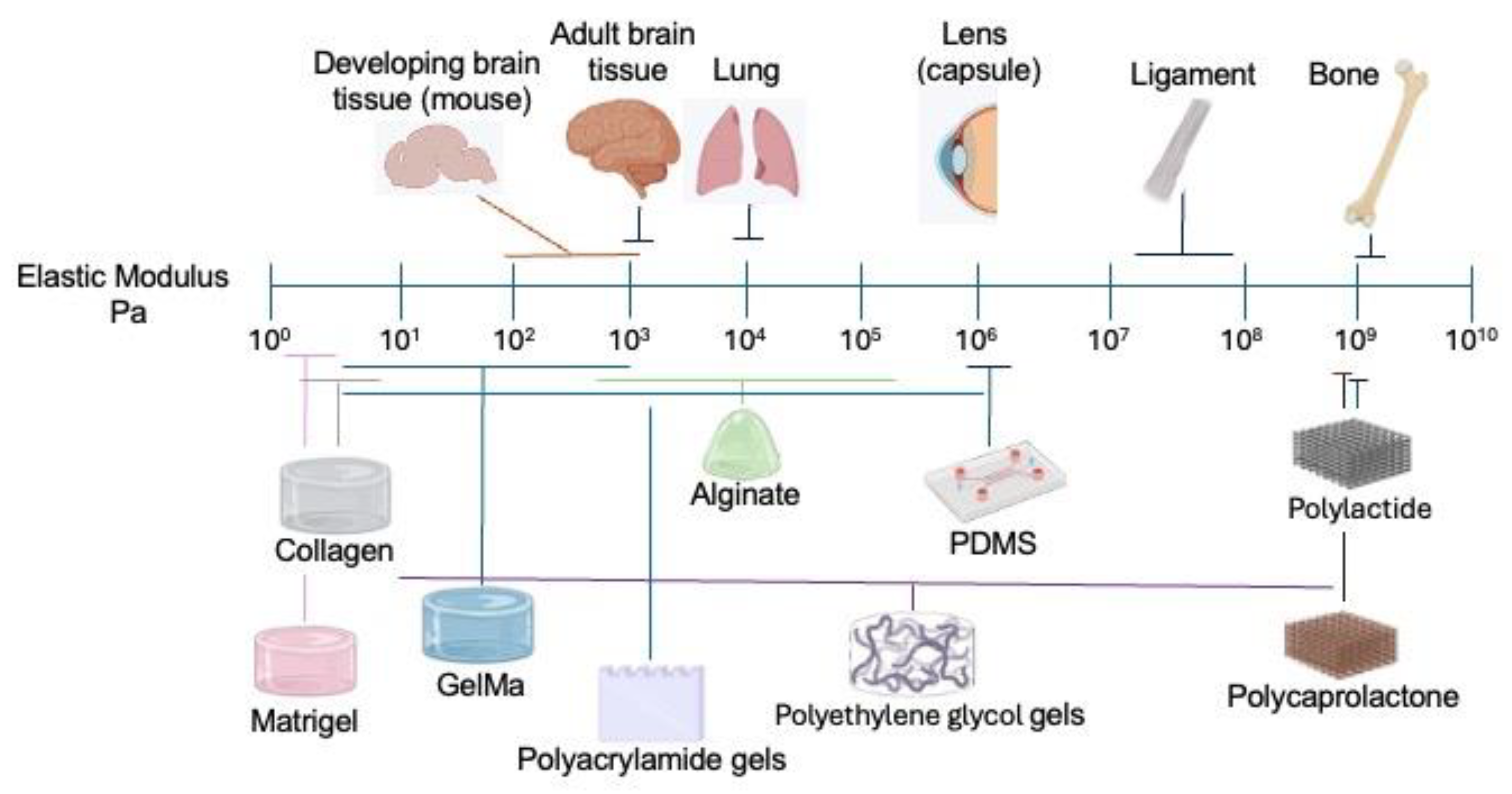
Figure 2.
A-B) The structure of Piezo1 is from PDB https://doi.org/10.2210/pdb6LQI/pdb structure ID: 6LQI (upper view) (A) and https://doi.org/10.2210/pdb6BPZ/pdb structure ID: 6BPZ (side view) (B). The most relevant domains of Piezo1 are indicated. C) Schematic representation showing the arrangement of the main domains of Piezo1 in an open conformation, as well as the described interactions with the actin cytoskeleton via E-cadherin and the interaction with MDFIC, as described in several studies. As indicated in the text, the opening of Piezo channels depends not only on forces derived from changes in plasma membrane tension; Piezo channels can also modulate their conformational states in response to mechanical forces originating from the intracellular cytoskeleton, which are modulated by accessory proteins. Abbreviations: CED, C-terminal extracellular domain; CTD, intracellular C-terminal domain; I/OH, inner and outer helices; PR (a–c), Piezo transmembrane repeats A–C. The illustration was created using PDB data and designed in Biorender (https://BioRender.com/ki8ia0h) modified from [146,147,148].
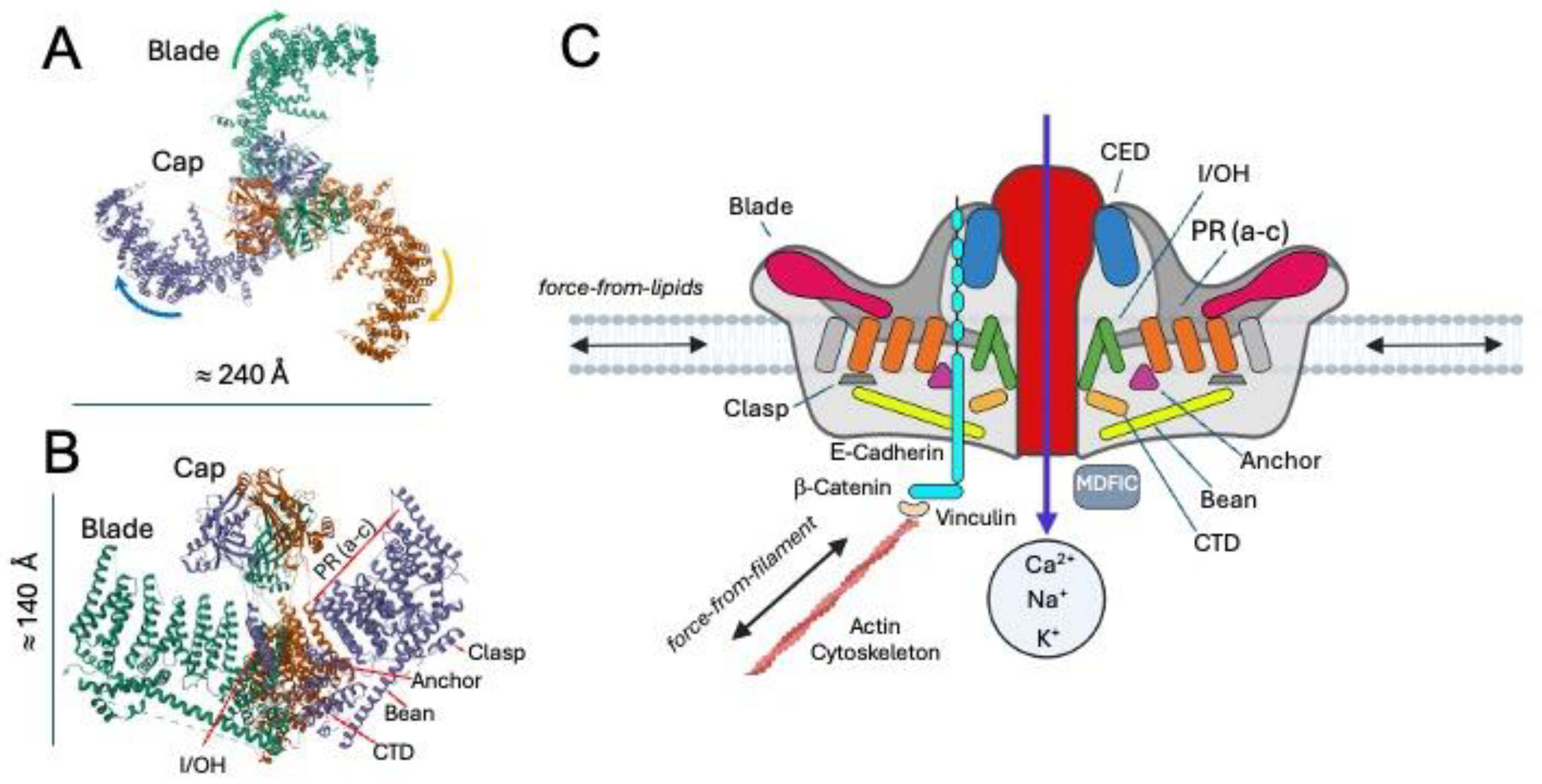
Figure 3.
A) The axonal growth cone is classically divided into three main domains: the central domain (not shown), the transitional zone (Actomyosin (ARC) domain), and the peripheral domain. Actin filaments are most abundant in the peripheral domain, where they form dense branched networks in lamellipodia and parallel bundles in filopodia, providing structural support and driving protrusive activity. Myosin II is enriched in the transitional zone, where it generates contractile forces that regulate actin retrograde flow and mediate the coupling between actin filaments and microtubules extending from the central domain. B) In the motor–clutch model, the axonal growth cone’s dynamics are explained by the interplay between actin polymerization, molecular motors, and substrate adhesion. Actin filaments in the peripheral domain polymerize at the leading edge, pushing the membrane forward, while retrograde flow driven by myosin II motors pulls the actin network rearward. Transmembrane adhesion molecules (e.g., integrins), often linked to the extracellular matrix, act as “clutches” with the help of an adaptor protein (e.g., talin or vinculin) that can engage or disengage with the retrograding actin filaments. But also, a dynamic equilibrium is established between integrin-ECM binding. In the classical model, when clutches engage, they transmit traction forces to the substrate, slowing retrograde flow and allowing forward protrusion of the growth cone. When they disengage, retrograde flow accelerates, reducing traction and leading to retraction. The clutch model predicts the existence of an optimal stiffness/ECM density in which cells’ force and migration are maximal. C) In two-dimensional (2D) cultures, neuronal growth cones tend to be large, flattened, and rich in filopodia and lamellipodia, expanding across the substrate and forming distinct peripheral and central domains; for example, sensory neuron growth cones on 2D collagen often exceed 200 µm² in projected area, with abundant protrusive structures. By contrast, in three-dimensional (3D) matrices like collagen gels, growth cones are significantly smaller - often around a third the size - with fewer lamellipodia and filopodia, and exhibit morphologies more akin to those seen in vivo. Moreover, whereas 2D growth cones typically maintain discrete actin- and microtubule-defined domains, in 3D the domain segregation becomes blurred: growth cones lack distinct partitions, displaying an actin-rich front with filopodia but without actin arcs or lamellipodia, and microtubule tips extend much closer to the leading edge. The illustration was modified from [217,223,231,237].
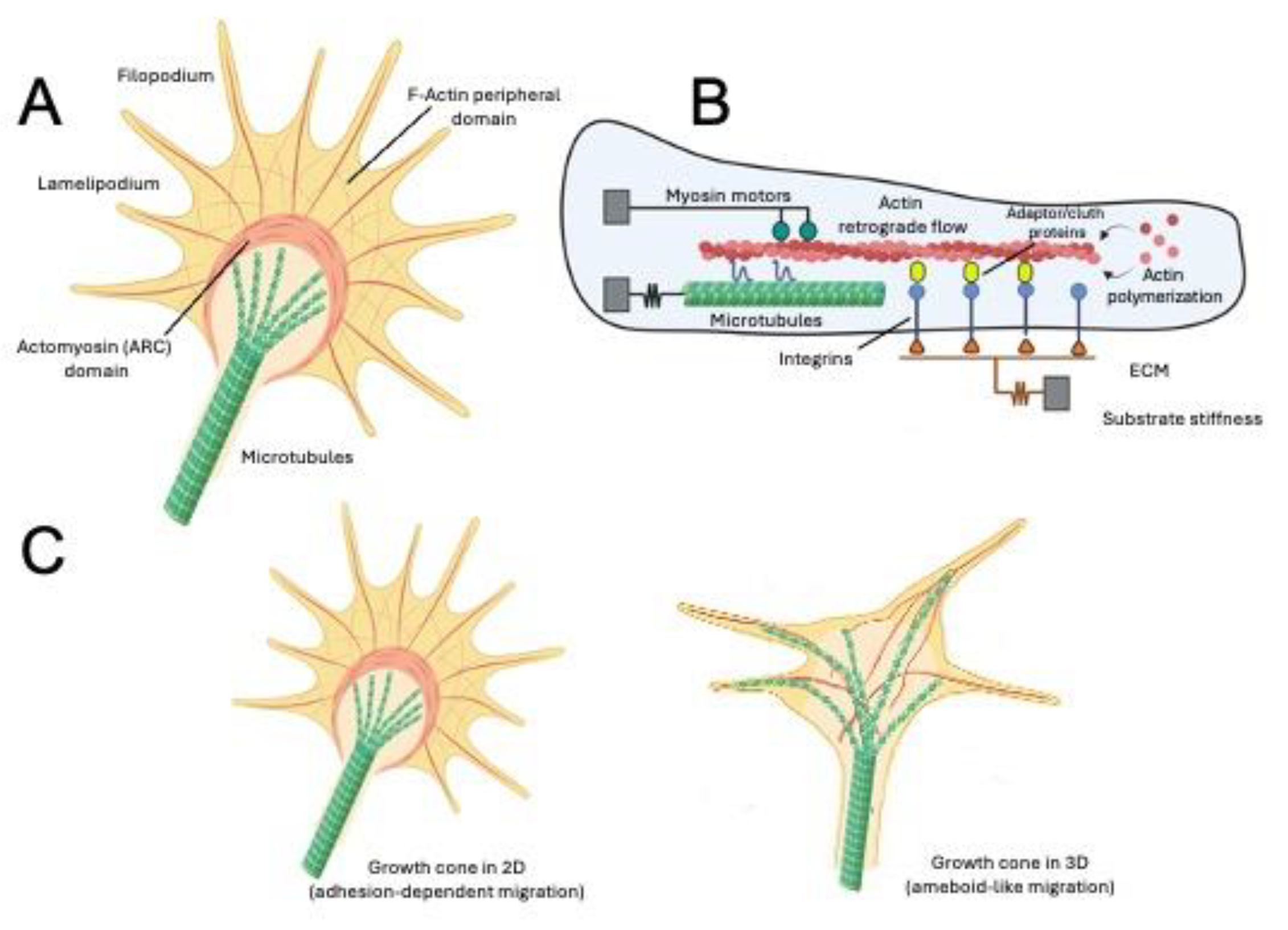
Figure 4.
A graphical summary of Hippo signaling. Although several components have been identified across different physiological contexts, in neurons, the extracellular stress, such as mechanical strain or ATP release, is sensed by integrins and purinergic GPCRs (P1 and P2Y receptors), converging on cytoskeletal and stress-response pathways. A) Chronic extracellular stress or excessive purinergic signaling favors activation of MST1/2 through phosphorylation, which in turn phosphorylates and activates LATS1/2. Active, phosphorylated LATS1/2 phosphorylate YAP/TAZ, retaining them in the cytoplasm and, after additional phosphorylation events, lead to polyubiquitination and proteasome-mediated degradation. The phosphatase PP2A counteracts this process by dephosphorylating MST1/2, LATS1/2, or YAP/TAZ depending on the context, serving as a regulatory brake that can buffer stress-induced Hippo activation. B) In conditions of moderate stress, Integrin engagement increases actin tension and increases RhoA balance in its GTP-bound state, which stabilize actomyosin cytoskeleton and indirectly suppresses Hippo kinase activity. Under these conditions, LATS1/2 remain largely unphosphorylated, allowing YAP/TAZ to remain dephosphorylated, translocate into the nucleus, and activate survival and plasticity gene programs. In fact, substrate stiffness modulates the frequency of Rho GTPase activation and generation of protrusions at least in adult neural stem cells [261]. Through this phosphorylation–dephosphorylation balance, integrins, purinergic GPCRs, and PP2A jointly determine whether extracellular stress drives adaptive neuronal remodeling or degenerative outcomes. Created in BioRender, https://BioRender.com/1l91fz6.
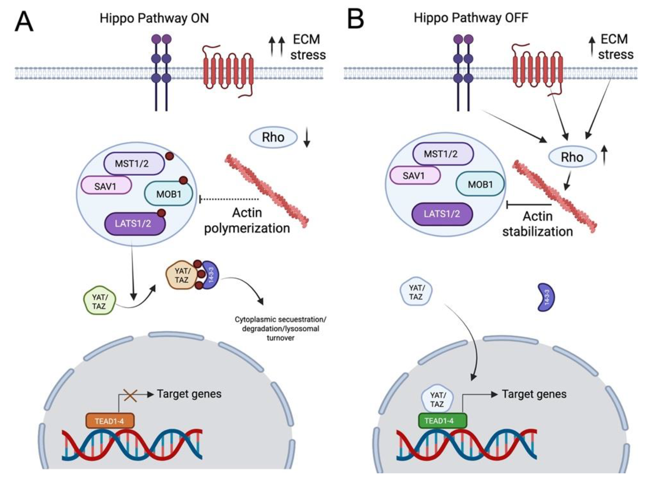
Figure 5.
Influence of matrix stiffness on human neural progenitor cell (NPC) fate. Substrate stiffness can govern NPC lineage specification by modulating mechanosensitive signaling. Soft matrices (0.5 - 1 kPa) that mimic the compliant embryonic developing cortex, such as low-crosslink PEG-diacrylate (~0.7 kPa), dilute (0.5%) hyaluronic acid or Matrigel (~0.3 - 0.5 kPa), or low-percentage collagen I (~0.4 kPa), maintain NPCs’ proliferative capacity and favor neuronal differentiation. The limited integrin tension in soft matrices keeps RhoA/ROCK activity low and YAP/TAZ cytoplasmic, allowing basal Wnt/β-catenin and TRPC1 activity to promote neurogenesis (β-III-tubulin⁺/MAP2⁺ neurons). Intermediate stiffness (2 - 5 kPa) - recapitulated by PEG-DA, GelMA, collagen/alginate, or fibrin hydrogels - matches adult brain parenchyma mechanics (≈ 2 - 4 kPa) and induces balanced mechanotransduction: partial YAP/TAZ nuclear entry with TRPV4-mediated Ca²⁺ influx activates the JAK/STAT3 astroglial pathway, while TRPC1-Wnt/Akt signaling maintains neuronal output. NPCs thus yield mixed neuronal and glial populations. Furthermore, the exact balance can be modulated by external stimuli, e.g., adding stretch favors glia, while microgrids or electrical stimulation favors neurons. Stiff matrices (> 5 kPa) such as high-crosslink PEG-DA, polyacrylamide (5 - 10 kPa), or dense collagen gels, mimic reactive-glial scar tissue (≈ 6 - 12 kPa). Strong focal-adhesion formation drives high RhoA/ROCK activity, nuclear YAP/TAZ, and maximal TRPV4-dependent JAK/STAT3 as well as Shh/Gli1 (oligodendroglial) activation, while TRPC1/Wnt signaling is suppressed, promoting GFAP⁺ astrocytic and OLIG2⁺/MBP⁺ oligodendroglial differentiation. Neuronal markers are reduced, and the cells display a more flattened, stress-fiber-rich morphology. Created with Biorender, https://BioRender.com/koq15cs.
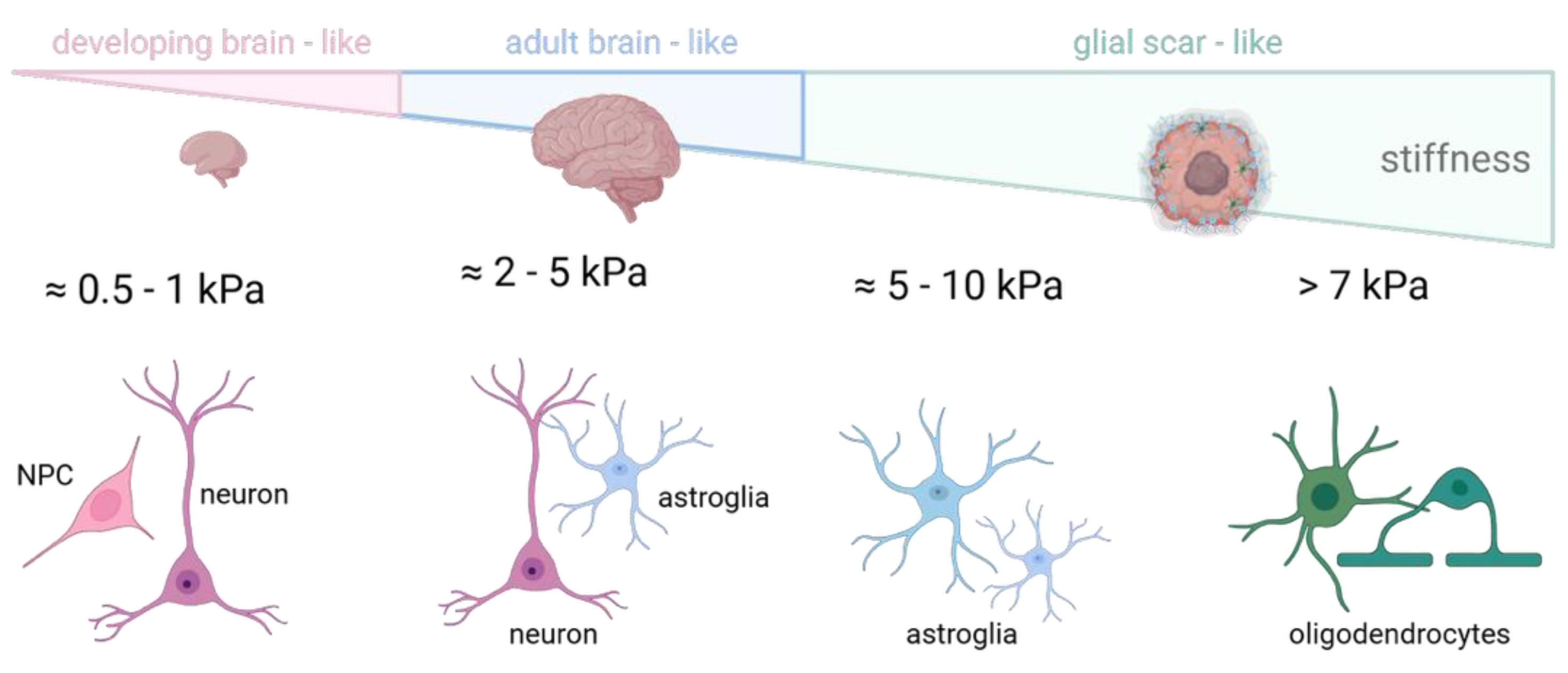

Table 1.
Ionotropic mechanosensors in the nervous system. Comparative summary of main families of mechanosensitive channels, properties, functions and cell types where they are present in the nervous system.
Table 1.
Ionotropic mechanosensors in the nervous system. Comparative summary of main families of mechanosensitive channels, properties, functions and cell types where they are present in the nervous system.
|
Channel Family |
Mechanical Stimuli they Transduce |
Ion Permeability |
Nervous Cell Type where Present |
Described Physiological Function |
|
Piezo; Includes Piezo 1 and Piezo 2 |
Membrane stretch, shear stress and pressure [43,124,125,126,127,128,129,130]. | Mainly Na+ and Ca2+, also K+ [128,136]. |
Piezo 1: NPCs/NSCs, DRG neurons, reactive astrocytes, Schwann cells, oligodendrocytes and microglia. Piezo 2: Somatosensory DRG neurons, Schwann cells, Merkel cells [126,149,150,151,152,153,154,155,156,157,158,159,160,161,162,163,164,165]. |
Wide range of physiological processes, from Piezo 1 sensing Aß fibrils in microglia [158] to Piezo 2 control of gastrointestinal transit in somatosensory neurons [159]. |
|
K2P; Includes TREK1, TREK2 and TRAAK |
Membrane curvature generated by asymmetric tension [166]. | K+ [167]. | Widespread in CNS Neurons but also on glial cells. Expressed in the PNS’s sensory neurons [168,169]. | Maintenance and regulation of resting membrane potential. Modulation of sensory pathways by reducing neuronal excitability [170]. |
|
TRP; Subfamilies include TRP A/C/M/MC/N/P/V |
Polymodal, from osmotic swelling to substrate stiffness [171]. | Ca2+, Mg2+, Na+ and K+ [171]. | Present in neurons of the sensory and nociceptive pathways, mostly in PNS sensory neurons [172]. | Transducers of sensory pathways of pain and heat, through mechanical and chemical sensing. E.g. TRPM8 is activated by temperature, cooling compounds and pressure [172,173]. |
Table 2.
Regulators of mechanosensing channels. Main described mechanosensory regulators and a summary of their function and relevance.
Table 2.
Regulators of mechanosensing channels. Main described mechanosensory regulators and a summary of their function and relevance.
|
Mechanosensory Regulator |
Described Physiological Function |
| MDFIC | Regulates activation of Piezo channels in different cell types [140]. |
| E-Cadherin | Mediates interaction of Piezo channels with the actin cytoskeleton [144,145] . |
| TMEM150C | Mediates slowly mechanosensitive currents in proprioceptive DRG neurons, acts as a general regulator [183]. |
| STOML3 | Scaffolding protein that modulates the activity of mechanosensitive ion channels (e.g. Piezo 1 and 2) by altering membrane mechanics. Essential for normal touch sensation in mammals [185]. |
Disclaimer/Publisher’s Note: The statements, opinions and data contained in all publications are solely those of the individual author(s) and contributor(s) and not of MDPI and/or the editor(s). MDPI and/or the editor(s) disclaim responsibility for any injury to people or property resulting from any ideas, methods, instructions or products referred to in the content. |
© 2025 by the authors. Licensee MDPI, Basel, Switzerland. This article is an open access article distributed under the terms and conditions of the Creative Commons Attribution (CC BY) license (http://creativecommons.org/licenses/by/4.0/).
Copyright: This open access article is published under a Creative Commons CC BY 4.0 license, which permit the free download, distribution, and reuse, provided that the author and preprint are cited in any reuse.


