
Submitted:
11 December 2025
Posted:
12 December 2025
You are already at the latest version
Abstract
Flexible biosensors offer rapid and low-cost diagnostics but are often limited by the mechanical and electrochemical instability of polymer-based designs in biological media. Here, we introduce a metallic flexible sensing platform that exploits the intrinsic deformability of superelastic nickel–titanium (NiTi) for label-free impedimetric detection. Mechanical bending of NiTi wires spontaneously generates martensitic-phase microcracks whose metal–gap–metal geometry forms the active transduction sites, where functional interfacial layers and captured analytes modulate the local dielectric environment and governing the impedance response. Functionalization with thiolated monolayers and Escherichia coli-specific antibodies enables these microdomains to modulate interfacial charge transfer in response to analyte binding, creating a direct coupling between mechanical deformation and resulting impedance signal. The γ-bent NiTi sensors achieved stable and quantitative detection of E. coli ATCC 25922 in sterile human urine, with a detection limit of 53 CFU mL⁻¹ within 45 minutes, without redox mediators, external labels, or amplification steps. This work establishes the first use of self-healing martensitic microcracks in a superelastic alloy as functional transduction elements, defining a new class of metallic flexible biosensors that integrate mechanical robustness, analytical reliability, and scalability for point-of-care biosensing.
Keywords:
flexible Nickel–Titanium (NiTi) biosensor
; label-free impedimetric sensing
; martensitic microcracks
; point-of-care biosensing
; self-healing interfaces
1. Introduction
Flexible biosensors play a central role in rapid, low-cost, and point-of-care diagnostics, enabling real-time biochemical monitoring in wearable and disposable platforms [1,2]. Among various designs, polymer-based substrates such as hydrogels, elastomers, and conductive composites have dominated recent developments owing to their softness and adaptability to biological interfaces [3,4]. However, the long-term performance of these soft matrices is often limited by mechanical fatigue, swelling or dehydration and chemical aging under repeated operation, which can lead to signal drift and poor reproducibility. These limitations motivate the development of flexible transducers that combine conformal contact with improved structural and chemical robustness beyond conventional polymer architectures [5].
To overcome these limitations, several approaches have integrated metallic nanostructures or conductive films onto soft polymer backings, allowing the substrate to absorb mechanical strain while the metallic layer provides electrochemical activity [2,6]. At the same time, the flexibility of such systems still depends on the integrity of the polymer support, and repeated bending or stretching can cause cracking, delamination or loss of adhesion in the conductive layer [7]. An alternative strategy is to employ intrinsically flexible metals whose mechanical compliance originates from solid-state phase transformations rather than external soft supports.
Nickel–titanium (NiTi) shape-memory alloys combine high fatigue resistance, corrosion resistance and good biocompatibility, and are widely used in devices such as stents, filters and orthodontic wires [8,9]. Their superelastic behavior is governed by stress-induced martensitic transformation and allows several percent of reversible strain without permanent plastic deformation, during which the alloy naturally forms reversible, self-healing microcracks that open under stress and close upon unloading [10]. NiTi has also been used directly as an electrochemically active substrate, for example by forming Ni–Ti–O or Ni(OH)\₂/TiO₂ oxide layers in situ for non-enzymatic glucose sensing, showing that mechanically robust NiTi wires can support stable redox-active interfaces [11,12]. From an environmental perspective, metallic systems such as NiTi are compatible with strategies for green flexible electronics and metal recovery, and can reduce the use of disposable plastic substrates [13,14]. Despite these advantages, the superelastic microstructure of NiTi has not been used as an active transduction element in biosensing.
Urinary tract infections (UTIs) are among the most common bacterial infections worldwide, and Escherichia coli (E. coli) is responsible for the majority of uncomplicated cases and was selected as a model pathogen and clinically relevant target for rapid screening assays. In routine clinical microbiology, significant bacteriuria is traditionally defined as ≥10⁵ CFU mL⁻¹ in midstream urine, although lower diagnostic thresholds (10³–10⁴ CFU mL⁻¹) are accepted in acute cystitis in women and catheter-associated infections [15]. Culture-based identification typically requires 18–48 hours, and although various optical, electrochemical and lateral-flow assays have been proposed, many still rely on labels, redox mediators or multi-step liquid handling, underscoring the need for simple label-free sensors capable of detecting E. coli directly in urine with clinically relevant limits of detection [16].
Here we introduce a NiTi wire microcrack transducer that exploits metallic flexibility for label-free impedimetric sensing of E. coli in urine (Figure 1). When a superelastic NiTi wire is bent into a γ-shaped configuration, stress-induced martensitic transformation generates a network of reversible microcracks that behave as confined cavities hosting a dielectric interior rather than structural defects [10]. In this architecture, bare γ-bent NiTi behaves essentially as a metallic conductor (trace 1), whereas surface functionalization fills these cavities with functional interfacial layers that establish a dielectric environment (trace 2) and subsequent E. coli binding further modulates this environment to define the impedance response (trace 3).
As a proof of concept of this mechanically driven dielectric sensing mechanism, γ-bent NiTi wire sensors achieved a detection limit of 53 CFU mL⁻¹ for E. coli in sterile human urine, with a linear range of 10³–10⁷ CFU mL⁻¹ and a total assay time of 45 minutes. To our knowledge, this is the first demonstration of superelastic NiTi microcracks functioning as self-healing metal-gap-metal transduction cavities for biosensing. These results establish a new class of metallic flexible biosensors that combine mechanical robustness, simple fabrication, and clinically relevant analytical performance for point-of-care applications.
2. Materials and Methods
2.1. Materials
Superelastic NiTi wires (100 µm diameter) were obtained in straight form and cut into 4 cm segments. Hydrofluoric and nitric acid solutions for Kroll etching, sodium hydroxide for hydroxylation, ethanol, phosphate-buffered saline (PBS, pH 7.4), 11-mercaptoundecanoic acid (MUA), EDC, Sulfo-NHS, bovine serum albumin (BSA), and all other analytical-grade reagents were used as received. E. coli ATCC 25922, Proteus mirabilis, and Klebsiella pneumoniae strains were supplied by the clinical microbiology laboratory. Sterile human urine (negative control matrix) was used for all electrochemical tests. Detailed reagent information is provided in the Supplementary Material.
2.2. Preparation of NiTi Microcrack Transducers
Straight superelastic NiTi wires were cleaned in ethanol, etched with Kroll’s reagent for 2 min, rinsed with deionized water, and hydroxylated in 10 M NaOH at 60 °C for 30 min. After chemical treatment, each wire was bent into a γ-shaped configuration using a fixed holder (Figure S4B). This deformation generated stress-induced self-healing microcracks associated with martensitic transformation, whose internal gaps served as confined cavities that hosted the functional layers and the subsequently captured analytes. The γ-shaped constraint was maintained during all following functionalization and measurements to preserve the microcrack architecture.
2.3. Surface Modification
Wires were incubated overnight in an ethanolic 11-mercaptoundecanoic acid (MUA) solution to form thiolated self-assembled monolayers via Ni–S and Ti–O–S bonding. Carboxyl groups were activated in EDC/Sulfo-NHS solution for 60 min and briefly rinsed with PBS. E. coli-specific antibodies were incubated with the activated surface for 60 min, yielding covalently linked amide bonds. BSA blocking was applied solely for AFM studies to visualize protein retention and was not included in biosensing experiments.
A schematic of all modification steps is provided in Figure S4.
2.4. Impedance Measurements
Impedance measurements were performed using a two-electrode configuration, where the γ-bent NiTi wire served as both working and counter electrodes. Measurements were conducted with a portable impedance analyzer (AIM-4300) over the 0.01–10 MHz frequency range.
After antibody immobilization, the wire was placed in 150 µL sterile human urine to stabilize the baseline for 45 minutes. Bacterial suspensions (10²–10⁷ CFU/mL) were then added without any washing step to avoid disturbing the stress-induced microcrack structure. Impedance spectra were collected at 15, 30, and 45 minutes.
ΔRct values were calculated from the real-axis intercepts of fitted Nyquist plots using a parabolic model in Origin. ΔRct was obtained from the change in the real-axis intercept between baseline and post-incubation spectra. The percentage change in charge-transfer resistance (ΔRct (%)) was calculated using:
where Rct,before denotes the baseline resistance prior to bacterial exposure, and Rct,after corresponds to the value measured after incubation. All measurements were performed in triplicate.
2.5. Selectivity and Repeatability Tests
Selectivity was evaluated by comparing impedance responses from mixtures containing only non-target bacteria (K. pneumoniae + P. mirabilis, 10⁵ CFU/mL each) and mixtures additionally containing E. coli at the same concentration. Repeatability was assessed from triplicate measurements at each bacterial concentration (n = 3 per level), and the corresponding statistical metrics (mean, SD, SE, %RSD) are summarized in Table S1.
2.6. Characterization and Analysis
Surface chemistry and morphology were assessed using FTIR spectroscopy, AFM, and SEM. FTIR confirmed SAM formation through characteristic COO⁻, CH₂, and Ti–O–S bands. AFM quantified roughness changes across modification steps. SEM visualized E. coli entrapment within stress-induced microcracks. Detailed instrumentation parameters are provided in the Supplementary Material.
3. Results
The impedimetric behavior of the superelastic NiTi sensor stems from stress-induced martensitic transformation, which produces a heterogeneous surface morphology composed of non-cracked austenitic regions passivated by a native TiO₂ layer and a network of self-healing microcracks. This configuration enables the alloy to withstand substantial deformation without permanent damage and forms the basis for its sensing functionality [17,18].
In the undeformed austenitic state, the NiTi surface exhibits homogeneous metallic character with negligible dielectric heterogeneity. Hence, contributions from microcrack-based capacitive elements are effectively absent under these conditions. Upon alkaline hydroxylation and mechanical deformation, however, the surface becomes chemically and structurally heterogeneous, with hydroxylated TiO₂ regions and exposed Ni-rich domains within microcracks. These domains provide reactive sites for thiol-based surface functionalization. Self-assembled monolayers of 11-mercaptoundecanoic acid (MUA) were chemisorbed onto the surface, forming Ti–O–S linkages at hydroxylated sites [19,20], and Ni–S bonds at metallic Ni sites [21,22] (Figure S1). The terminal carboxylic acid (–COOH) groups of MUA remained available for subsequent EDC/NHS crosslinking and antibody immobilization. Once embedded within the microcrack network, the MUA layer acts as a confined dielectric interface between conductive NiTi crack walls, effectively behaving as a thin parallel-plate capacitor. Exposure to analytes then modulated the local dielectric environment, producing measurable impedance changes driven by interfacial capacitance.
To aid interpretation of this behavior, a simplified equivalent circuit is included in the inset of Figure 3C. While prior studies have examined the electrochemical behavior of NiTi under mechanical loading and stress-induced martensitic reorientation [23], the present illustration contain functional layers and captured analytes, which introduce additional dielectric components. The capacitive terms associated with these confined regions (C_functional and C_analyte) dominate the high-frequency response, where impedance is primarily governed by dielectric modulation. Resistive pathways formed by stress-induced microcracks (R_microcrack) describe heterogeneous conduction across transformed domains, while the solution resistance (R_solution) represents the bulk electrolyte. Together, these elements provide a qualitative description of the shift from low-impedance metallic behavior in the austenitic state to a higher-impedance dielectric–metal interface after martensitic transformation and biofunctionalization.
3.1. Morphological and Electrochemical Characterization of the NiTi Biosensor
Scanning electron microscopy (SEM) was employed to visualize the interaction of E. coli with stress-induced microcracks on the NiTi sensor surface. Representative images at 10³ and 10⁷ CFU/mL (Figure 2A–B) show bacterial colonies entrapped within martensitic microcracks, highlighted in red circles. Increasing bacterial density was evident at higher concentrations, consistent with the concentration-dependent electrical response of the sensor.
Electrochemical impedance spectroscopy (EIS) was then used to monitor the dynamic binding behavior of the biosensor across a wide frequency range (0.01–10 MHz). The impedimetric response was evaluated through Nyquist plots, where the real part of impedance (Z′) is plotted on the x-axis and the imaginary part (Z″) on the y-axis [24]. Time-resolved Nyquist plots (Figure 2C–D) showed gradual increases in impedance at 10³ CFU/mL over 15–45 minutes, indicative of progressive antibody–antigen binding. In contrast, exposure to 10⁷ CFU/mL produced a rapid rise within the first 15 minutes, followed by signal saturation. Frequency-domain spectra (Figure 2E–F) exhibited pseudocapacitive behavior, with phase angles approaching −80° at high frequencies [23].
3.2. Analytical Performance of the NiTi Biosensor
EIS was used to monitor each step of the surface modification process (Figure 3A). Distinct impedance changes were observed following NaOH activation, MUA functionalization, EDC/NHS crosslinking, and antibody immobilization, confirming successful functionalization. Surface roughness changes accompanying these steps were also verified by AFM (Figure S2). After the final modification, the sensor was incubated in sterile human urine for 45 minutes, yielding a stable baseline spectrum suitable for subsequent bacterial detection. Baseline stability was further examined through repeated measurements in sterile urine. The thinner 100 μm sensor reached a stable baseline, whereas the 250 μm wire showed variable profiles, highlighting the effect of wire thickness on stability (Figure S3).
Following surface preparation, sensors were exposed to increasing concentrations of E. coli (10²–10⁷ CFU/mL) for 45 minutes. The Nyquist plots showed progressively larger semicircle diameters with increasing concentration (Figure 3B). The corresponding calibration curve (Figure 3C) revealed two linear regions, with a strong correlation in the 10³–10⁷ CFU/mL range (R² = 0.9376). The limit of detection (LOD) was estimated at 53 CFU/mL from the graphical intersection of two fitted segments, in accordance with established methods [25].
Time-resolved experiments at 10³ and 10⁷ CFU/mL confirmed the concentration-dependent binding kinetics described above (Figure 2C–F), with ΔRct increasing gradually at low loads and saturating rapidly at high loads.
Selectivity was further assessed by comparing E. coli with non-target bacteria. While the mixture of Klebsiella pneumoniae and Proteus mirabilis (KP + PM) produced a negligible signal (ΔRct = 8.75% ± 1.77), the inclusion of E. coli (KP + PM + EC) yielded a pronounced increase (ΔRct = 434.72% ± 44.69) (Figure 3D). Error bars represent the standard error of the mean (n = 3), and statistical analysis confirmed the difference to be significant (p < 0.001, one-way ANOVA).
4. Discussion
This study introduces a fundamentally new sensing architecture by transforming superelastic NiTi wires into label-free impedimetric biosensors. By exploiting stress-induced martensitic microcracks as confined cavities that host the functional interfacial layers, the system achieves molecular recognition purely through impedance changes, without faradaic reactions or diffusion limited processes. Notably, no prior biosensor has harnessed the intrinsic microstructural changes of NiTi in this way; for the first time, its stress induced microcracks are repurposed as confined transduction cavities whose dielectric interiors form the active sensing domain, eliminating the need for engineered nanostructures or external capacitive layers.
The sensing principle relies on self-healing microcracks generated during stress-induced martensitic transformation [10,26,27,28]. These cracks open under stress and close upon unloading, forming reversible confined cavities that can host functional layers and captured analytes [29]. While microcracks provide the physical transduction framework, selective molecular recognition arises from stepwise surface functionalization with thiolated SAMs, EDC/NHS chemistry, and antibody immobilization [30,31]. AFM measurements confirmed progressive increase in surface roughness across these steps (Figure S2), supporting the formation of a heterogeneous, biofunctional interface.
Distinct from conventional impedimetric platforms, this system exhibited a strong pseudocapacitive component at higher frequencies within the 0.01–10 MHz window, particularly under elevated bacterial loads, where phase angles exceeded −80° (Figure 3)[23]. Previous NiTi electrochemical models primarily described passive film resistance and charge transfer kinetics [23,27,28,32,33]. In contrast, the present findings show that high-frequency dielectric behavior arises when stress-induced microcrack cavities are functionalized and their interiors are filled with biomolecular layers. This behavior links the microcrack architecture directly to dielectric modulation, providing a previously unreported mechanism for signal generation in NiTi-based systems.
Stepwise modification of the NiTi surface produced predictable shifts in charge transfer resistance, consistent with changes in surface chemistry (Figure 3A). The initial increase in Rct after NaOH treatment reflected the formation of a resistive hydroxide layer [34,35]. Subsequent MUA functionalization, EDC/NHS crosslinking, and antibody immobilization progressively decreased Rct as molecular order improved, establishing a stable bio-recognition interface. FTIR spectra (Figure S1) further confirmed thiol anchoring by the disappearance of the –SH stretching band [36].
Superelastic deformation in the γ-bent configuration enhanced this chemistry by generating martensitic microcracks that exposed nickel-rich sites for Ni–S bonding [21,28,36,37] and hydroxylated TiO2 regions for Ti–O–S linkages [19,20] (Figure S4). This spatial heterogeneity increased the density of binding sites and promoted uniform antibody coverage. Microcracks acted as thin capacitor-like gaps, where high permittivity (ε) and narrow dielectric spacing (d) raise interfacial capacitance according to C = εA/d. These dielectric changes, combined with shifts in charge transfer resistance, underpin the overall impedimetric sensing response.
Sensor stability was influenced by wire diameter and mechanical design. Supplementary data (Figure S3) showed that 100 μm NiTi sensors produced consistent Nyquist profiles that stabilized within minutes, whereas 250 μm wires displayed inconsistent signals without a stable baseline. A similar diameter dependence has been reported in independent studies on NiTi strain sensors, where 100–150 μm wires exhibit clear cyclic resistance changes, whereas 200–250 μm wires respond mainly in the first cycle and are deemed unsuitable for repeated sensing [38]. Taken together, these observations support an explanation based on crack morphology: under the imposed bent geometry, thinner wires preferentially form narrower, more homogeneous microcrack networks, whereas thicker wires develop more dispersed and leaky domains [39,40]. The γ-bent design concentrates stress along a confined region of the wire, promoting a dense and reproducible microcrack pattern [10,28] and thereby enhancing the uniformity of sensor response. This mechanically engineered architecture demonstrates how controlled deformation can be used as a design parameter to optimize biosensor performance. Given that NiTi maintains superelasticity down to ~100 nm [41,42,43], further miniaturization is expected to enhance sensitivity and integration into portable devices.
Analytically, the sensor achieved a detection limit of 53 CFU/mL with a broad dynamic range extending to 10⁷ CFU/mL. ΔRct values increased systematically with concentration, with saturation observed beyond 10⁶ CFU/mL. Repeatability analysis (Table S1) confirmed consistent responses across independent measurements. Notably, no Warburg-type diffusion elements were observed even at high concentrations, indicating that signal modulation is governed by interfacial dielectric changes rather than mass transport [44,45]. Selectivity tests demonstrating that gram-negative bacteria (K. pneumoniae and P. mirabilis) induced negligible impedance changes, whereas samples containing E. coli produced pronounced ΔRct shifts (Figure 3D). These findings confirm that selective recognition arises from antibody immobilization, while the functionalized and analyte-filled microcrack environments drive the dielectric modulation underlying the impedance response. Although this work focused on E. coli, the modular surface chemistry can be readily adapted to other biorecognition elements, extending the platform to proteins, toxins, or viral targets.
To contextualize these findings, Table 1 compares the present NiTi platform with representative flexible biosensors reported in recent literature, categorized by substrate type and mechanical flexibility origin. The comparison highlights that polymer-based systems often depend on engineered nanostructures or composite layers to achieve flexibility, whereas the current work uniquely exploits the intrinsic solid-state transformation of a metallic alloy. The NiTi architecture therefore bridges the gap between polymeric flexibility and metallic robustness, introducing a recyclable, corrosion-resistant, and mechanically adaptive alternative for flexible biosensing.
The use of stress-induced microcracks in NiTi may appear less precise than lithographically defined microchannels, yet these cracks form self-healing confined cavities governed by reversible martensitic transformation. Unlike etched microstructures, which remain fixed, the martensitic microcracks in NiTi form reversible gaps: they open during mechanical deformation and, although they partially close when the load is removed, they leave behind confined cavities that subsequently accommodate functional and analyte-filled dielectric environments that modulate impedance. Importantly, Ni-rich regions exposed within the cracks enable direct chemisorption of thiolated ligands, a dual functionalization mechanism not achievable on smooth or patterned surfaces. Whereas lithographic systems demand multistep fabrication to generate stable binding sites, NiTi spontaneously forms adaptive sensing domains through its native phase transformation. By exploiting this controlled disorder, the platform transforms a traditionally passive alloy into a chemomechanically responsive transducer, and establishing a blueprint for flexible biosensor design.
5. Conclusions
This work demonstrates that superelastic NiTi can serve as a mechanically flexible, low-cost and label-free impedimetric transducer. Stress-induced microcracks form self-healing confined cavities whose dielectric interiors generate measurable impedance changes after functionalization and analyte binding. The platform enabled rapid detection of E. coli in urine (53 CFU mL⁻¹ in 45 minutes) using simple bending and standard wet-chemistry processing. These features highlight the potential of NiTi microcrack architectures for developing simple, robust, affordable and portable point-of-care biosensors.
6. Patents
An international patent application (PCT/TR2024/050849) related to the findings of this study was filed on 18 July 2024.
Supplementary Materials
The following supporting information can be downloaded at the website of this paper posted on Preprints.org. The Supplementary Information contains extended Materials and Methods, including detailed procedures for NiTi surface preparation, γ-bent sensor fabrication, functionalization steps (hydroxylation, SAM formation, EDC/Sulfo-NHS crosslinking, antibody immobilization, and BSA treatment), AFM/FTIR/SEM characterization protocols, the full EIS measurement setup, and additional experimental descriptions. It also includes: Figure S1: FTIR spectra of bare, NaOH-treated, and MUA-functionalized NiTi surfaces; Figure S2: AFM images showing surface morphology changes during sequential modification steps; Figure S3: Time -resolved Nyquist plots of γ-bent NiTi electrodes in sterile human urine (negative controls); Figure S4: Schematic of the surface functionalization workflow and photograph of the γ-bent NiTi sensor setup Table S1: Calibration repeatability results including mean, SD, SE, and %RSD values.
Author Contributions
Conceptualization, M.C.S.; methodology, G.O.T. and M.C.S.; software, —; validation, G.O.T. and M.C.S.; formal analysis, G.O.T.; investigation, G.O.T.; resources, M.C.S.; data curation, G.O.T.; writing—original draft preparation, G.O.T.; writing—review and editing, G.O.T. and M.C.S.; visualization, G.O.T.; supervision, M.C.S.; project administration, M.C.S. All authors have read and agreed to the published version of the manuscript.
Funding
This research received no external funding.
Institutional Review Board Statement
Not applicable. This study did not involve humans, human-derived materials requiring ethical approval, or live animals.
Informed Consent Statement
Not applicable.
Data Availability Statement
The data presented in this study are available from the corresponding author upon request.
Acknowledgments
The authors thank the Central Laboratory of Erciyes University Hospitals for supplying the bacterial strains used in this study. The authors also acknowledge the Environmental Engineering Laboratory of Erciyes University for their technical support. The superelastic NiTi wires were purchased from Aksöz R&D Co. (Pamukkale, Türkiye). The authors have reviewed and edited the output and take full responsibility for the content of this publication.
Conflicts of Interest
The authors declare no conflicts of interest.
References
- Liu, T.; Liu, L.; Gou, G. Y.; Fang, Z.; Sun, J.; Chen, J.; Cheng, J.; Han, M.; Ma, T.; Liu, C.; Xue, N. Recent Advancements in Physiological, Biochemical, and Multimodal Sensors Based on Flexible Substrates: Strategies, Technologies, and Integrations. ACS Applied Materials & Interfaces CrossRef. 2023, 15(18), 21721–21745. [Google Scholar] [CrossRef] [PubMed]
- Ferreira, R. G.; Silva, A. P.; Nunes-Pereira, J. Current on-skin flexible sensors, materials, manufacturing approaches, and study trends for health monitoring: a review. ACS sensors CrossRef. 2024, 9(3), pp. 1104–1133. [Google Scholar] [CrossRef] [PubMed]
- Lawaniya, S. D.; Awasthi, A.; Kuznetsova, I.; Kolesov, V.; Awasthi, K. Polymer Hybrid based Nanomaterials for Next-Generation Sensing Technologies. Sensors and Actuators B: Chemical CrossRef. 2025, 138162. [Google Scholar] [CrossRef]
- Zhou, P.; Zhang, Z.; Mo, F.; Wang, Y. A review of functional hydrogels for flexible chemical sensors. Advanced Sensor Research CrossRef. 2024, 3(3), 2300021. [Google Scholar] [CrossRef]
- Xu, M.; Obodo, D.; Yadavalli, V. K. The design, fabrication, and applications of flexible biosensing devices. Biosensors and Bioelectronics CrossRef. 2019, 124, 96–114. [Google Scholar] [CrossRef]
- Gao, F.; Liu, C.; Zhang, L.; Liu, T.; Wang, Z.; Song, Z.; Cai, H.; Fang, Z.; Chen, J.; Wang, J.; Han, M.; Wang, J.; Lin, K.; Wang, R.; Li, M.; Mei, Q.; Ma, X.; Liang, S.; Gou, G.; Xue, N. Wearable and flexible electrochemical sensors for sweat analysis: a review. Microsystems & Nanoengineering CrossRef. 2023, 9(1), pp. 1–21. [Google Scholar] [CrossRef]
- Han, X.; Lin, X.; Sun, Y.; Huang, L.; Huo, F.; Xie, R. Advancements in Flexible Electronics Fabrication: Film Formation, Patterning, and Interface Optimization for Cutting-Edge Healthcare Monitoring Devices. ACS Applied Materials & Interfaces CrossRef. 2024, 16(41), 54976–55010. [Google Scholar]
- Duerig, T.; Pelton, A.; Stöckel, D. An overview of nitinol medical applications. Materials Science and Engineering A CrossRef. 1999, 273-275, 149–160. [Google Scholar] [CrossRef]
- Patel, S. K.; Behera, B.; Swain, B.; Roshan, R.; Sahoo, D.; Behera, A. A review on NiTi alloys for biomedical applications and their biocompatibility. Materials today: proceedings CrossRef. 2020, 33, pp. 5548–5551. [Google Scholar] [CrossRef]
- Gollerthan, S.; Young, M. L.; Neuking, K.; Ramamurty, U.; Eggeler, G. Direct physical evidence for the back-transformation of stress-induced martensite in the vicinity of cracks in pseudoelastic NiTi shape memory alloys. Acta materialia CrossRef. 2009, 57(19), 5892–5897. [Google Scholar] [CrossRef]
- Gao, A.; Zhang, X.; Peng, X.; Wu, H.; Bai, L.; Jin, W.; Wu, G.; Hang, R.; Chu, P. K. In situ synthesis of Ni (OH) 2/TiO2 composite film on NiTi alloy for non-enzymatic glucose sensing. Sensors and Actuators B: Chemical CrossRef. 2016, 232, 150–157. [Google Scholar] [CrossRef]
- Hang, R.; Liu, Y.; Gao, A.; Bai, L.; Huang, X.; Zhang, X.; Lin, N.; Tang, B.; Chu, P. K. Highly ordered Ni–Ti–O nanotubes for non-enzymatic glucose detection. Materials Science and Engineering: C CrossRef. 2015, 51, 37–42. [Google Scholar] [CrossRef] [PubMed]
- Hui, Z.; Zhang, L.; Ren, G.; Sun, G.; Yu, H. D.; Huang, W. Green flexible electronics: natural materials, fabrication, and applications. Advanced Materials CrossRef. 2023, 35(28), 2211202. [Google Scholar] [CrossRef] [PubMed]
- Cui, F.; Wang, G.; Yu, D.; Gan, X.; Tian, Q.; Guo, X. Towards “zero waste” extraction of nickel from scrap nickel-based superalloy using magnesium. Journal of Cleaner Production CrossRef. 2020, 262, 121275. [Google Scholar] [CrossRef]
- Campuzano, S.; Yáñez-Sedeño, P.; Pingarrón, J. M. Molecular biosensors for electrochemical detection of infectious pathogens in liquid biopsies: current trends and challenges. Sensors CrossRef. 2017, 17(11), 2533, pp. 1–21. [Google Scholar] [CrossRef]
- Dospinescu, V. M.; Tiele, A.; Covington, J. A. Sniffing out urinary tract infection—Diagnosis based on volatile organic compounds and smell profile. Biosensors CrossRef. 2020, 10(8)(83), 2–28. [Google Scholar] [CrossRef]
- Otsuka, K.; Ren, X. Recent developments in the research of shape memory alloys. Intermetallics CrossRef. 1999, 7(5), 511–528. [Google Scholar] [CrossRef]
- Gall, K.; Sehitoglu, H. The role of texture in tension–compression asymmetry in polycrystalline NiTi. International Journal of Plasticity CrossRef. 1999, 15(1), 69–92. [Google Scholar] [CrossRef]
- Chen, B.; Li, L.; Liu, L.; Cao, J. Molecular simulation of adsorption properties of thiol-functionalized titanium dioxide (TiO2) nanostructure for heavy metal ions removal from aqueous solution. Journal of Molecular Liquids CrossRef. 2022, 346, 118281. [Google Scholar]
- Sánchez-Bodón, J.; Andrade del Olmo, J.; Alonso, J. M.; Moreno-Benítez, I.; Vilas-Vilela, J. L.; Pérez-Álvarez, L. Bioactive coatings on titanium: a review on hydroxylation, self-assembled monolayers (SAMs) and surface modification strategies. Polymers CrossRef. 2021, 14(1)(165), 1–30. [Google Scholar] [CrossRef]
- Fontanesi, C.; Tassinari, F.; Parenti, F.; Cohen, H.; Mondal, P. C.; Kiran, V.; Giglia, A.; Pasquali, L.; Naaman, R. New one-step thiol functionalization procedure for Ni by self-assembled monolayers. Langmuir CrossRef. 2015, 31(11), 3546–3552. [Google Scholar] [CrossRef] [PubMed]
- Yeyin, T.; Xiaoya, L.; Peiming, H.; Shusen, P.; Yongcun, M. Protection of nickel by self-assembled monolayers prepared in an aqueous self-emulsifying solution of a novel amphipathic organothiol. RSC advances CrossRef. 2023, 13(7), 4331–4339. [Google Scholar] [CrossRef] [PubMed]
- Mohajeri, M.; Case, R.; Haghgouyan, B.; Lagoudas, D. C.; Castaneda, H. Loading influence on the corrosion assessment during stress-induced martensite reorientation in nickel-titanium SMA. Smart Materials and Structures CrossRef. 2020, 29(3), 035013. Pp. 1–12. [Google Scholar] [CrossRef]
- Instruments, Gamry. Complex impedance in Corrosion. In Basics of electrochemical impedance spectroscop; 2007; Volume 1, pp. 1–30. [Google Scholar]
- Buck, R. P.; Lindner, E. Recommendations for nomenclature of ionselective electrodes (IUPAC Recommendations 1994). Pure and Applied Chemistry CrossRef. 1994, 66(12), 2527–2536. [Google Scholar] [CrossRef]
- Wu, Y.; Ojha, A.; Patriarca, L.; Sehitoglu, H. Fatigue crack growth fundamentals in shape memory alloys. Shape Memory and Superelasticity CrossRef. 2015, 1(1), 18–40. [Google Scholar] [CrossRef]
- Perumal, G.; Selvam, K. T.; Swayne, M.; McCarthy, E.; Babu, A.; Dzhurinskiy, D.; Brabazon, D. Exploring the role of volume energy density in altering microstructure and corrosion behavior of nitinol alloys produced by laser powder bed fusion. Scientific Reports CrossRef. 2025, 15(1)(2055), 1–14. [Google Scholar] [CrossRef]
- Racek, J.; Šittner, P.; Heller, L.; Pilch, J.; Petrenec, M.; Sedlák, P. Corrosion of NiTi wires with cracked oxide layer. Journal of materials engineering and performance CrossRef. 2014, 23(7), pp. 2659–2668. [Google Scholar] [CrossRef]
- Robertson, S. W.; Mehta, A.; Pelton, A. R.; Ritchie, R. O. Evolution of crack-tip transformation zones in superelastic Nitinol subjected to in situ fatigue: A fracture mechanics and synchrotron X-ray microdiffraction analysis. Acta Materialia CrossRef. 2007, 55(18), 6198–6207. [Google Scholar] [CrossRef]
- Soysaldı, F.; Dincyurek Ekici, D.; Soylu, M. Ç.; Mutlugun, E. Electrochemical and Optical Multi-Detection of Escherichia coli Through Magneto-Optic Nanoparticles: A Pencil-on-Paper Biosensor. Biosensors CrossRef. 2024, 14(12)(603), 1–17. [Google Scholar] [CrossRef]
- Keser, K.; Soylu, M. Ç. Simple, Specific, and Ultra-Sensitive Arsenic Detection in Real Drinking Water Samples Using an Impedimetric Green Sensor with Dimercaprol-Doped Solid Electrolyte. Electroanalysis CrossRef. 2025, 37(1), e202400253. [Google Scholar] [CrossRef]
- Zhang, S.; Pang, X.; Wang, Y.; Gao, K. Corrosion behavior of steel with different microstructures under various elastic loading conditions. Corrosion science CrossRef. 2013, 75, 293–299. [Google Scholar] [CrossRef]
- Zhao, X.; Liu, Y.; Jia, C.; Chang, H.; Zhang, W.; Bai, Y.; Li, S.; Zhang, L. C.; Yuan, W. Corrosion behavior of laser powder bed fusion additive manufacturing produced TiNi alloy by micro-arc oxidation. npj Materials Degradation CrossRef. 2024, 8(1)(13), 1–13. [Google Scholar] [CrossRef]
- Sun, T.; Wang, M.; Lee, W. C. Surface characteristics, properties and in vitro biological assessment of a NiTi shape memory alloy after high temperature heat treatment or surface H2O2-oxidation: a comparative study. Materials Chemistry and Physics CrossRef. 2011, 130(1-2), 45–58. [Google Scholar] [CrossRef]
- Chu, C. L.; Hu, T.; Zhou, J.; Pu, Y. P.; Yin, L. H.; Dong, Y. S.; Lin, P. H.; Chung, J. C. Y.; Chu, P. K. Effects of H2O2 pretreatment on surface characteristics and bioactivity of NaOH-treated NiTi shape memory alloy. Transactions of Nonferrous Metals Society of China CrossRef. 2006, 16(6), 1295–1300. [Google Scholar] [CrossRef]
- Love, J. C.; Estroff, L. A.; Kriebel, J. K.; Nuzzo, R. G.; Whitesides, G. M. Self-assembled monolayers of thiolates on metals as a form of nanotechnology. Chemical reviews CrossRef. 2005, 105(4), 1103–1170. [Google Scholar] [CrossRef]
- Mani, G.; Porter, D.; Grove, K.; Collins, S.; Ornberg, A.; Shulfer, R. Surface finishing of N itinol for implantable medical devices: A review. Journal of Biomedical Materials Research Part B: Applied Biomaterials CrossRef. 2022, 110(12), 2763–2778. [Google Scholar] [CrossRef]
- Sławski, S.; Kciuk, M.; Klein, W. Change in electrical resistance of SMA (NiTi) wires during cyclic stretching. Sensors CrossRef. 2022, 22(9)(3584), 1–19. [Google Scholar] [CrossRef]
- Perinetti, G.; Contardo, L.; Ceschi, M.; Antoniolli, F.; Franchi, L.; Baccetti, T.; Di Lenarda, R. Surface corrosion and fracture resistance of two nickel–titanium-based archwires induced by fluoride, pH, and thermocycling. An in vitro comparative study. The European Journal of Orthodontics CrossRef. 2012, 34(1), 1–9. [Google Scholar] [CrossRef]
- Ghazal, A. R. A.; Hajeer, M. Y.; Al-Sabbagh, R.; Alghoraibi, I.; Aldiry, A. An evaluation of two types of nickel-titanium wires in terms of micromorphology and nickel ions’ release following oral environment exposure. In Progress in orthodontics; CrossRef; PubMed, 2015; Volume 16(1), 9, pp. 1–8. [Google Scholar]
- Waitz, T.; Spišák, D.; Hafner, J.; Karnthaler, H. P. Size-dependent martensitic transformation path causing atomic-scale twinning of nanocrystalline NiTi shape memory alloys. Europhysics Letters CrossRef. 2005, 71(1)(98), 98–103. [Google Scholar] [CrossRef]
- Fu, Y. Q.; Zhang, S.; Wu, M. J.; Huang, W. M.; Du, H. J.; Luo, J. K.; Flewitt, A. J.; Milne, W. I. On the lower thickness boundary of sputtered TiNi films for shape memory application. Thin Solid Films CrossRef. 2006, 515(1), pp. 80–86. [Google Scholar] [CrossRef]
- Waitz, T.; Antretter, T.; Fischer, F. D.; Karnthaler, H. P. Size effects on martensitic phase transformations in nanocrystalline NiTi shape memory alloys. Materials Science and Technology CrossRef. 2008, 24(8), 934–940. [Google Scholar] [CrossRef]
- Shoute, L. C.; Abdelrasoul, G. N.; Ma, Y.; Duarte, P. A.; Edwards, C.; Zhuo, R.; Zeng, J.; Feng, Y.; Charlton, C. L.; Kanji, J. N.; Babiuk, S.; Chen, J. Label-free impedimetric immunosensor for point-of-care detection of COVID-19 antibodies. Microsystems & Nanoengineering CrossRef. 2023, 9(1)(3), 1–16. [Google Scholar]
- Bard, A. J.; Faulkner, L. R.; White, H. S. Electrochemical methods: fundamentals and applications.; John Wiley & Sons, 2022. [Google Scholar]
- Zhang, H.; Hu, H.; Li, Y.; Wang, J.; Ma, L. A ferrocene-based hydrogel as flexible electrochemical biosensor for oxidative stress detection and antioxidation treatment. Biosensors and Bioelectronics CrossRef. 2024, 248, 115997. [Google Scholar] [CrossRef] [PubMed]
- Yoon, J.; Lee, S. N.; Shin, M. K.; Kim, H. W.; Choi, H. K.; Lee, T.; Choi, J. W. Flexible electrochemical glucose biosensor based on GOx/gold/MoS2/gold nanofilm on the polymer electrode. Biosensors and Bioelectronics CrossRef. 2019, 140(111343), 1–7. [Google Scholar] [CrossRef]
- You, Z.; Qiu, Q.; Chen, H.; Feng, Y.; Wang, X.; Wang, Y.; Ying, Y. Laser-induced noble metal nanoparticle-graphene composites enabled flexible biosensor for pathogen detection. Biosensors and Bioelectronics CrossRef. 2020, 150, 111896. [Google Scholar] [CrossRef]
- Xu, M.; Yadavalli, V. K. Flexible biosensors for the impedimetric detection of protein targets using silk-conductive polymer biocomposites. Acs Sensors CrossRef. 2019, 4(4), 1040–1047. [Google Scholar] [CrossRef]
- Lamanna, L.; Rizzi, F.; Bhethanabotla, V. R.; De Vittorio, M. Conformable surface acoustic wave biosensor for E-coli fabricated on PEN plastic film. Biosensors and Bioelectronics CrossRef. 2020, 163(112164), 1–10. [Google Scholar] [CrossRef]
- Tian, B.; Zhang, J.; Zhou, S.; Liu, C.; Wang, Q.; Liu, Y.; Lu, M.; Sun, G.; Wang, C.; Gu, B. Silica interlayer-protected colorimetric-fluorescent dual-signal microbial tag mediate lateral flow immunoassay for flexible and ultrasensitive detection of sepsis biomarkers. Chemical Engineering Journal CrossRef. 2025, 163557. [Google Scholar] [CrossRef]
- Jampasa, S.; Sangthong, N.; Ozer, T.; Traiopop, S.; Ngamrojanavanich, N.; Panphut, W.; Chailapakul, O.; Kaneta, T.; Wonsawat, W. Silver-decorated nanobeads for high-performance and flexible analysis of Shigella dysenteriae utilizing a paper-based aptasensor. Bioelectrochemistry CrossRef. 2025, 109140. [Google Scholar] [CrossRef]
- Wang, R.; Zhai, Q.; An, T.; Gong, S.; Cheng, W. Stretchable gold fiber-based wearable textile electrochemical biosensor for lactate monitoring in sweat. Talanta CrossRef. 2021, 222(121484), 1–8. [Google Scholar] [CrossRef]
- Lim, T.; Zhang, H. Multilayer carbon nanotube/gold nanoparticle composites on gallium-based liquid metals for electrochemical biosensing. ACS Applied Nano Materials CrossRef. 2021, 4(11), 12690–12701. [Google Scholar] [CrossRef]
- Pan, T. M.; Chen, C. H.; Weng, W. C. Rapid and label-free detection of BNP and NT-proBNP in human serum using Ti-doped MoTex film-based extended-gate FET biosensors for heart failure diagnosis. Sensors and Actuators B: Chemical CrossRef. 2025, 138922. [Google Scholar] [CrossRef]
- Gaikwad, P. N.; Desai, T. R.; Ghosh, S.; Gurnani, C. Flexible Nanostructured NiS-Based Electrochemical Biosensor for Simultaneous Detection of DNA Nucleobases. ACS omega CrossRef. 2024, 10(3), 2561–2574. [Google Scholar] [CrossRef]
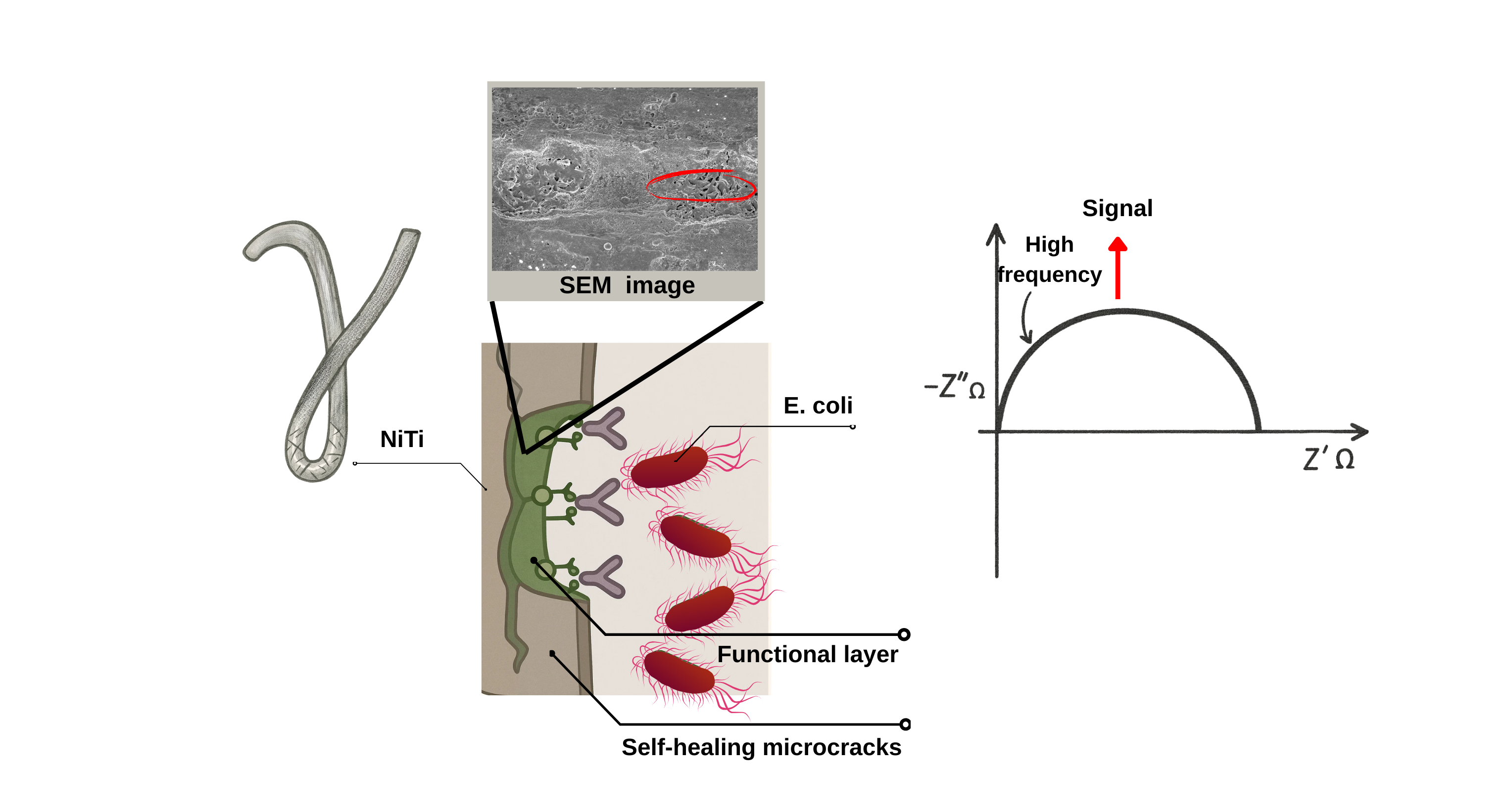
Figure 1.
Schematic illustration of the NiTi microcrack-based biosensor. The γ-bent superelastic NiTi wire is immersed in urine and connected to a portable impedance analyzer; stress-induced martensitic microcracks along the wire surface are functionalized with a thiolated monolayer and E. coli–specific antibodies to capture bacteria. Insets show the SEM image of E. coli bound within the microcrack network (top) and Nyquist responses of (1) bare NiTi, (2) functionalized surface, and (3) after incubation with 10³ CFU/mL E. coli (right).
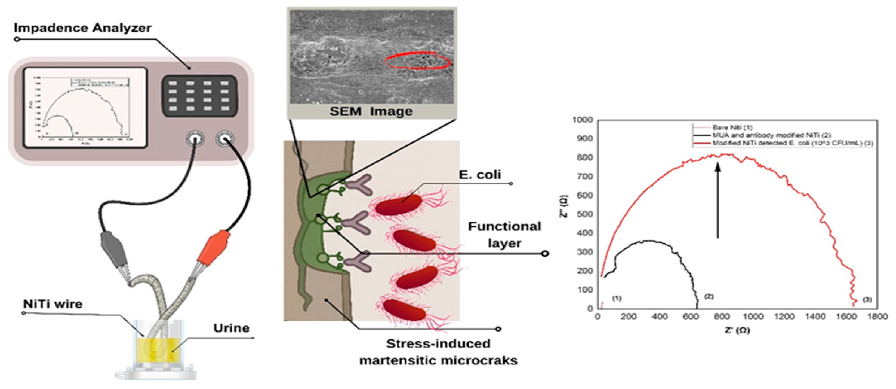
Figure 2.
Morphological and electrochemical responses of the NiTi biosensor to E. coli in urine. (A–B) SEM images of sensor surfaces after exposure to 10³ CFU/mL (A) and 10⁷ CFU/mL (B), showing bacterial colonies entrapped within martensitic microcracks (red circles). (C–D) Time-resolved Nyquist plots for 10³ CFU/mL (C) and 10⁷ CFU/mL (D),concentration-dependent increases in impedance. (E–F) Frequency-domain spectra at 10³ CFU/mL (E) and 10⁷ CFU/mL (F).
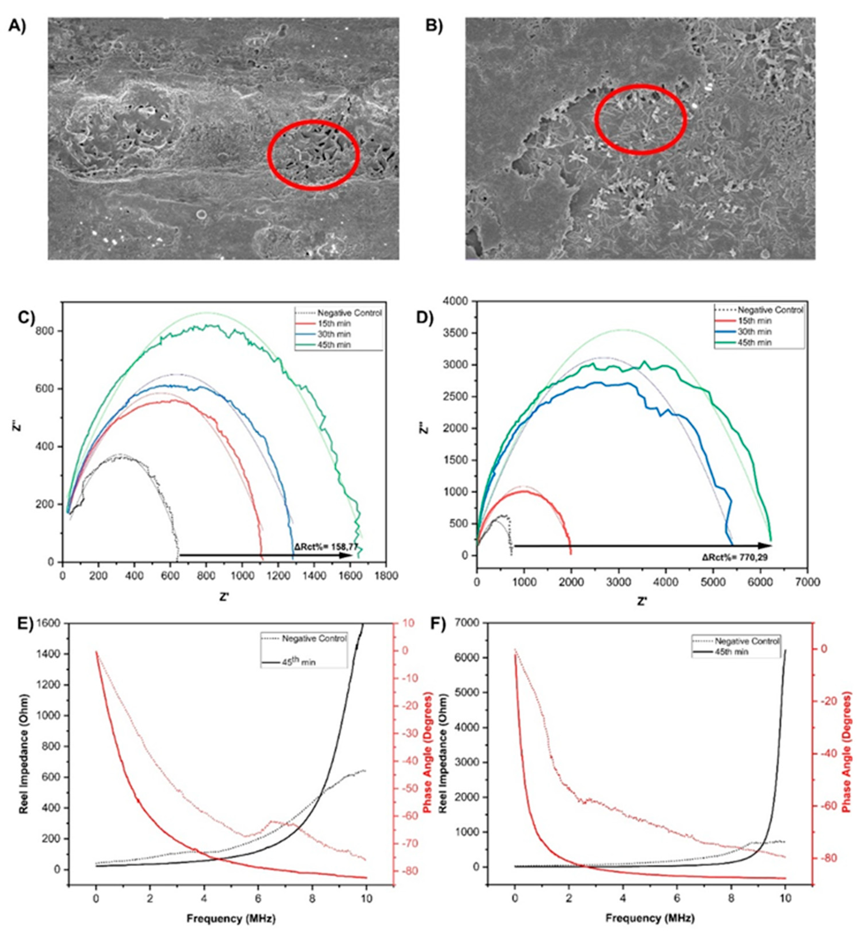
Figure 3.
Electrochemical characterization of the NiTi biosensor. (A) Nyquist plots of a single NiTi wire recorded after sequential surface modification (B) Nyquist plots after 45 min incubation with E. coli at concentrations ranging from 10² to 10⁷ CFU/mL. (C) Calibration curve of ΔRct (%) versus log[E. coli] concentration (n = 3). The inset shows the equivalent circuit model proposed to describe the system (D) Selectivity test showing average ΔRct (%) responses for a non-target bacterial mixture (performed at 10⁵ CFU/mL, n = 3).
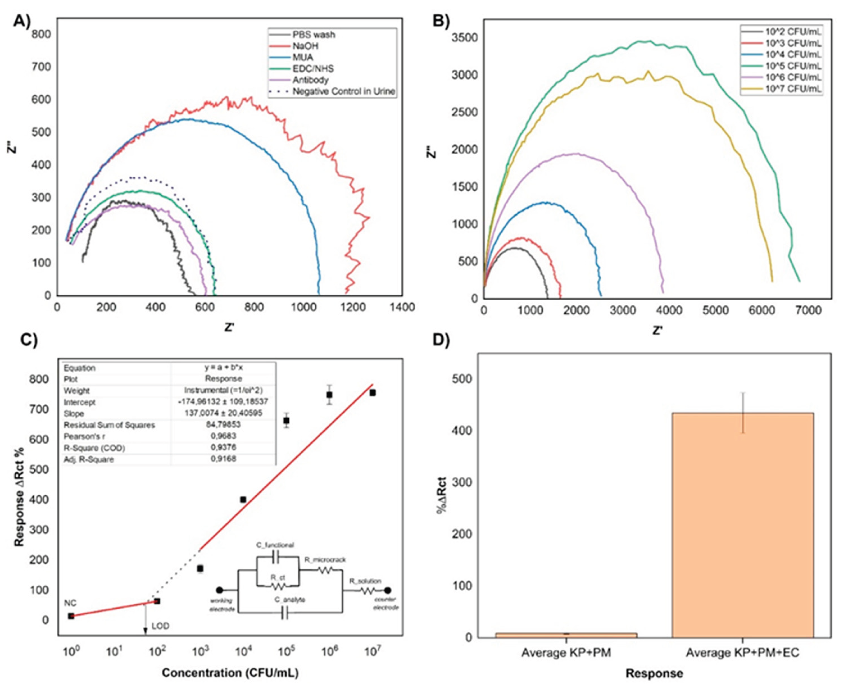

Table 1.
Comparison of representative flexible biosensors categorized by substrate type (polymeric, metallic, or hybrid) and mechanical flexibility origin.
Table 1.
Comparison of representative flexible biosensors categorized by substrate type (polymeric, metallic, or hybrid) and mechanical flexibility origin.
| Sensor Material | Flexibility Type | LOD | Linear Range | Detection Time (min) | Matrix | POCT Suitability | Ref |
| Ferrocene-based PVA/SA/PEI-Fc hydrogel | Polymeric (Hydrogel) | Not reported | Real time | 0-120 µM | PBS; cell culture media (L929, RAW 264.7) | Yes | [46] |
| PDMS/PET–Au/MoS₂–GOx nanofilm | Polymeric (PET) | 0.10 nM | 10–500 nM | 30 min | Glucose in PBS | Yes (Enzymatic) | [47] |
| AuNPs-LIG interdigitated electrode (flexible) | Polyimide substrate | 10² CFU/mL | 10²–10⁸ CFU/mL | 30 min | E. coli in PBS | No (Not target) | [48] |
| Silk fibroin flexible substrate+ PEDOT:PSS+anti-VEGF | Polymeric (organic) | 22-25 pg/mL | 1 pg/mL–10 ng/mL | 15 min | PBS, serum, artificial urine | Yes | [49] |
| AlN thin film on PEN substrate (SAW) | Polymeric (PEN) | 6.5x10⁵ CFU/mL | 106-108 CFU/mL | 90 min | E. coli in water | Moderate (High LOD) | [50] |
| AuNP-SiO₂-QDs nanocomposite on LFIA strip | Polymeric LFIA substrate | IL-6: 4.7 pg /mL; PCT: 29.5 pg /mL | 0.1–100 ng /mL | 15 min | Human plasma (clinical) | Yes (clinically validated) | [51] |
| Ag-decorated magnetic nanobeads on paper-based aptasensor | Paper substrate | DPV: 90; EIS: 8.09 CFU/mL | DPV: 10²–10⁸; EIS: 10¹–10⁹ CFU/mL | 40 min | Food | Yes (on-site NFC-integrated) | [52] |
| Stretchable gold fiber textile | Elastomeric textile | 0.137 mM | 0-30 mM | Real time | Lactate in artificial sweat | Yes (Enzymatic) | [53] |
| CNT/PDDA + AuNP multilayer on Ga-based liquid metal | Intrinsically soft liquid metal core (ultrasoft) | 23 nM | 25 nM–1 µM | Real time | PBS / ACSF (in vitro neurochemical buffer) | Not target | [54] |
| Ti-doped MoTeₓ film EGFET | Thin-film hybrid | 5x10⁻³ pg/mL | 10-2-105 pg/mL | Real time | BNP in serum | Yes (clinically validated) | [55] |
| NiS/ Ni-foam | Metallic foam | 159 μM (A), 147.6μM (G), 16.8 μM (T), 45.9 μM (C) | 200–1000 μM (A,G); 50–500 μM (T,C) | Real time | Calf thymus DNA | Yes (portable) | [56] |
| Superelastic NiTi alloy | Intrinsic solid-state microcracks | 53 CFU/mL | 103-107 CFU/mL | 45 min | E. coli in sterile human urine | Yes (preliminary) | This work |
Disclaimer/Publisher’s Note: The statements, opinions and data contained in all publications are solely those of the individual author(s) and contributor(s) and not of MDPI and/or the editor(s). MDPI and/or the editor(s) disclaim responsibility for any injury to people or property resulting from any ideas, methods, instructions or products referred to in the content. |
© 2025 by the authors. Licensee MDPI, Basel, Switzerland. This article is an open access article distributed under the terms and conditions of the Creative Commons Attribution (CC BY) license (http://creativecommons.org/licenses/by/4.0/).
Copyright: This open access article is published under a Creative Commons CC BY 4.0 license, which permit the free download, distribution, and reuse, provided that the author and preprint are cited in any reuse.


