Submitted:
09 December 2025
Posted:
09 December 2025
You are already at the latest version
Abstract
Background: Psoriasis and psoriatic arthritis (PsA) occasionally coexist with antinu-clear antibody (ANA) positivity, cutaneous lupus erythematosus (CLE), or systemic lupus erythematosus (SLE), creating one of the most challenging therapeutic overlap scenarios in immunodermatology. Divergent immune pathways—IL-23/Th17-driven psoriatic inflammation versus type I interferon–mediated autoimmunity—generate unique vulnerabilities when systemic treatments are used. Objectives: To synthesize treatment outcomes, lupus-related safety signals, and mechanistic insights across systemic therapies in patients with psoriasis or PsA who also exhibit ANA positivity, CLE, or SLE. Methods: A systematic review following PRISMA 2020 guidelines was performed across PubMed/MEDLINE, Embase, Cochrane, Scopus, and ClinicalTrials.gov. Thir-ty-three eligible reports (29 unique clinical studies; 1,429 patients) were included and organized into six prespecified overlap subgroups. Mechanistic and translational studies—including ustekinumab and deucravacitinib SLE trial data and IL-17 inhibi-tor–induced CLE reports—were incorporated for contextual interpretation. Results: IL-23 inhibitors demonstrated the most favorable cross-disease safety, show-ing no signal for CLE worsening, SLE flares, or drug-induced autoimmunity. IL-17 in-hibitors maintained excellent psoriatic efficacy but showed a consistent association with de novo or exacerbated CLE. TNF-α inhibitors carried the highest risk for ANA seroconversion, dsDNA induction, drug-induced lupus, and lupus flares. Ustekinumab exhibited a stable safety profile across lupus-spectrum disease despite mixed efficacy in formal SLE trials. TYK2 inhibition provided dual modulation of IL-23 and type I in-terferon pathways and showed emerging utility in psoriasis/PsA with CLE or SLE. Apremilast, methotrexate, and mycophenolate mofetil remained reliable non-biologic options. Phototherapy required caution in ANA-positive or lupus-susceptible popula-tions. Conclusions: IL-23 inhibition and TYK2 inhibition appear to offer the most balanced combination of efficacy and safety for psoriatic disease complicated by lupus-spectrum autoimmunity. IL-17 inhibitors and TNF-α inhibitors warrant caution or avoidance in CLE- or SLE-prone patients. Personalized treatment should integrate psoriatic versus lupus disease dominance, ANA/ENA profile, CLE subtype, and mechanistic risk. Pro-spective, biomarker-driven studies are needed to guide therapy in this increasingly recognized overlap population. (PROSPERO registration: CRD420251241279).
Keywords:
psoriasis
; psoriatic arthritis
; cutaneous lupus erythematosus
; systemic lupus erythematosus
; antinuclear antibodies
; biologic therapy
; IL-23 inhibitors
; IL-17 inhibitors
; TNF inhibitors
; TYK2 inhibition
; drug-induced lupus
; overlap autoimmune disease
; immunodermatology
; systemic therapy safety
1. Introduction
1.1. Background and immunologic divergence
Psoriasis and psoriatic arthritis (PsA) are chronic, immune-mediated inflammatory diseases driven predominantly by dysregulation of the IL-23/Th17 axis, with central contributions from IL-23, IL-17A/F, and TNF-α [1,2,3,4,5]. In contrast, systemic lupus erythematosus (SLE) and cutaneous lupus erythematosus (CLE) are prototypical type I interferon–driven autoimmune conditions characterized by plasmacytoid dendritic cell activation, B-cell hyperactivity, autoantibody production, and immune-complex–mediated tissue damage [6,7,8,9]. Although all are autoimmune diseases, psoriatic and lupus-spectrum conditions sit on opposite poles of key immunologic axes, which helps explain why therapies that are highly effective for one end of the spectrum may destabilize the other [9,10,11,12].
Within the lupus spectrum, important mechanistic differences exist between systemic disease and skin-restricted CLE subtypes. SLE is dominated by circulating immune complexes, complement consumption, and multi-organ involvement, reflecting a systemically amplified IFN-I/B-cell program [6,7,17]. By contrast, CLE—particularly discoid lupus erythematosus (DLE)—is often “skin-locked,” with dense interface dermatitis, scarring follicular destruction, and a persistently IFN-high, TNF-low milieu in lesional skin [9,20]. Subacute CLE (SCLE) tends to show stronger Ro/La autoantibody associations and photosensitivity [9,20], whereas classic localized DLE is more fibrosing and cicatricial. These distinctions are clinically relevant because they modulate how biologic agents that target the Th17/IL-23 axis, B cells, or upstream cytokines perturb cutaneous versus systemic lupus biology [9,17,18,19,20].
Several treatment classes commonly used in psoriasis intersect with these pathways in different ways. TNF-α inhibitors are highly effective for psoriasis and PsA but are strongly associated with ANA seroconversion, anti-dsDNA induction, drug-induced lupus, and lupus flares [10,18,35,36,37,38]. IL-17 inhibitors offer superior psoriatic skin and joint clearance, yet accumulating reports link them to new-onset or aggravated CLE—particularly disseminated DLE and SCLE—suggesting that IL-17 blockade may shift immune balance toward unchecked type I interferon dominance in susceptible skin [28,29,30,31,32,33]. In contrast, IL-23 inhibitors appear to provide excellent psoriatic control with minimal interaction with IFN-I–driven pathways, and no clear signal for CLE worsening or SLE flares has emerged to date [12,13,14,43].
Other systemic agents occupy more neutral positions along this axis. Ustekinumab, an IL-12/23 inhibitor, has been formally evaluated in Phase II and Phase III SLE trials, showing inconsistent efficacy but a reassuring safety profile with no increase in lupus activity [46]. However, dedicated data for CLE—especially DLE—remain sparse, and current evidence supports regarding ustekinumab as immunologically “neutral-safe” in lupus rather than as an active lupus therapy [46,48]. TYK2 inhibition with deucravacitinib is mechanistically attractive because TYK2 sits upstream of both IL-23 and type I interferon signaling; early trials and translational work suggest improvements in CLE molecular signatures and SLE activity [25,26,27], and isolated real-world reports describe simultaneous control of psoriasis, PsA, and SLE [25]. Non-biologic options such as methotrexate (MTX), mycophenolate mofetil (MMF), and apremilast remain important anchors: MTX and MMF are foundational in SLE and CLE care [6,11,20], whereas apremilast provides systemic psoriatic control without apparent lupus-inducing risk [16].
Phototherapy is traditionally viewed as a safe and effective modality for psoriasis. However, ultraviolet radiation—especially UVB—can amplify interferon gene signatures, drive keratinocyte apoptosis, and precipitate CLE lesions in genetically predisposed individuals [9,20]. In ANA-high or ENA-positive patients, and especially in those with established CLE or SLE, this IFN-skewing effect makes phototherapy a potentially hazardous choice and underscores the need to consider ANA/ENA profiles when planning light-based therapy [20,47].
Finally, some drugs considered mainstays in systemic autoimmunity may adversely affect psoriatic disease. Hydroxychloroquine improves CLE/SLE but can exacerbate psoriasis [47,51], and rituximab, a CD20-depleting monoclonal antibody widely used in rheumatology, has been repeatedly reported to induce de novo psoriasis or worsen pre-existing psoriasis [59]. These “reverse” signals further illustrate that the lupus-optimized armamentarium is not automatically safe in psoriatic biology.
1.2. Clinical dilemma and objective of this review
Patients who present with psoriasis or PsA in combination with ANA positivity, CLE, or SLE therefore represent a highly heterogeneous and therapeutically fragile subgroup [35,36,37,38,44,45,46,47,48,49,51,52,53]. Agents that are central to psoriasis or PsA management—such as TNF inhibitors and IL-17 inhibitors—may precipitate photosensitive rashes, de novo CLE, drug-induced lupus, or true SLE exacerbations [10,18,28,29,30,31,32,33,45,46,47,48,49,52]. Conversely, lupus mainstay therapies, including hydroxychloroquine and rituximab, can aggravate psoriatic inflammation or trigger new-onset psoriasis [47,51,59]. A further layer of complexity arises from the variable clinical expression of lupus (skin-limited SCLE/DLE versus multi-organ SLE), the spectrum of ANA/ENA serologies, and the increasing use of newer agents such as IL-23 and TYK2 inhibitors in routine psoriasis care [12,13,14,25,26,27].
Despite these challenges, there are no dedicated, evidence-based guidelines for systemic treatment of psoriatic disease in the setting of ANA positivity or coexisting lupus-spectrum disease [6,7,8,9,17,20]. Individual clinicians must therefore integrate fragmentary data from small cohorts, case series, and mechanistic studies when choosing systemic therapy [35,36,37,38,44,45,46,47,48,49,51,52,53].
The objective of this systematic review is to comprehensively synthesize clinical and mechanistic evidence regarding systemic therapies in adults with psoriasis or PsA who concurrently exhibit isolated ANA positivity, cutaneous lupus erythematosus, or systemic lupus erythematosus. By organizing available data across six prespecified overlap subgroups and incorporating mechanistic insights—including TYK2 inhibition [25,26,27], ustekinumab SLE trials [46], and IL-17 inhibitor–associated CLE [28,29,30,31,32,33]—we aim to propose a pragmatic, phenotype- and pathway-guided framework for systemic treatment selection in this complex overlap population.
2. Materials and Methods
2.1. Study Design and Reporting Standards
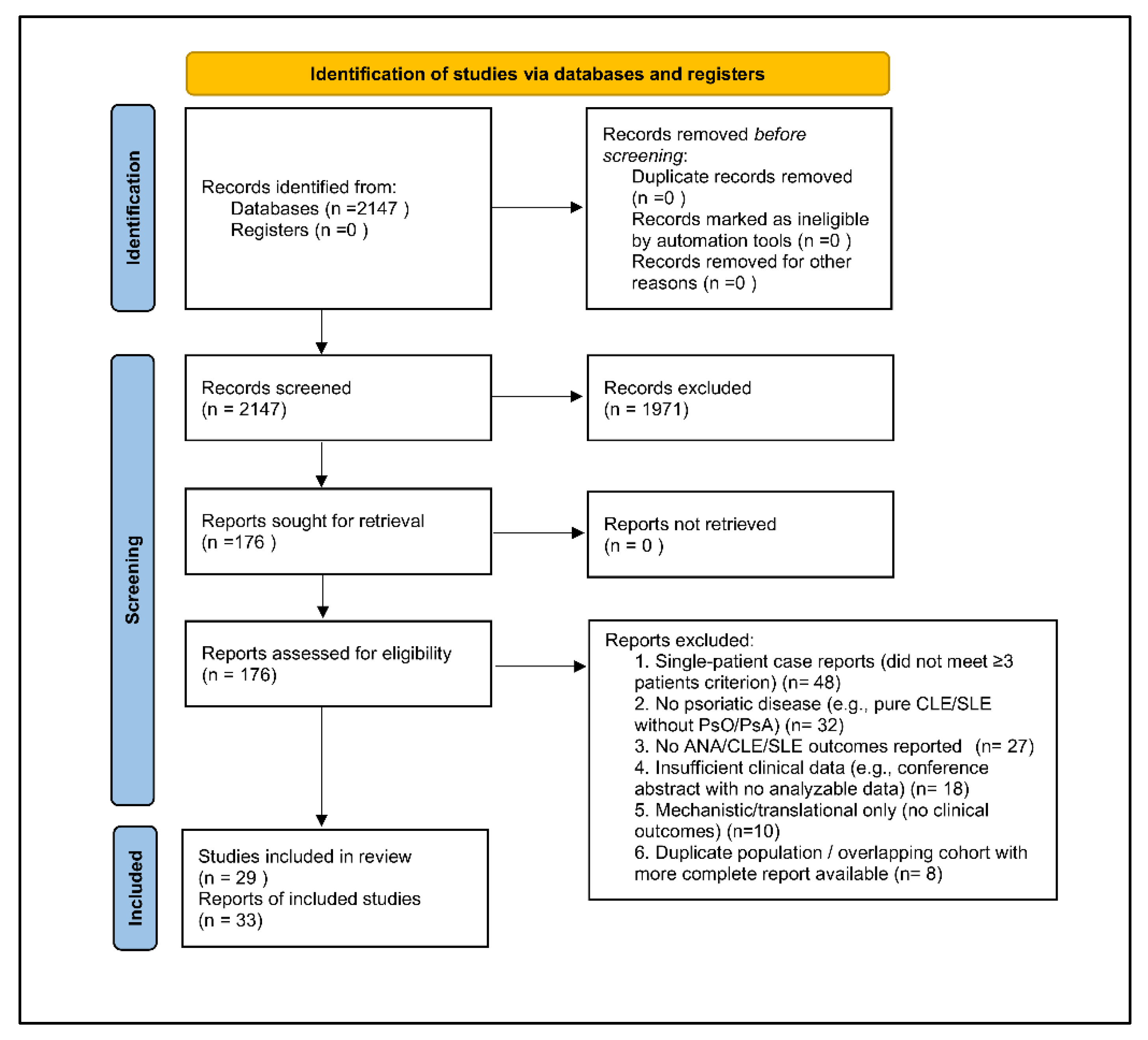
This study is a systematic review conducted in accordance with the PRISMA 2020 statement, with full adherence to recommended methods for study identification, selection, and reporting [PRISMA 2020]. A PRISMA 2020 flow diagram summarizing the selection process is shown in Figure 1, consistent with PRISMA standards [PRISMA 2020]. (PROSPERO registration: CRD420251241279).
The objective was to evaluate the safety and efficacy of systemic therapies in adults with psoriasis or psoriatic arthritis who also exhibit antinuclear antibody positivity, cutaneous lupus erythematosus, or systemic lupus erythematosus [6,7,8,9,17,20]. Given the substantial heterogeneity in study design, lupus phenotype definitions, serologic reporting, and outcome measures across available studies, a meta-analysis was not feasible, and a narrative evidence synthesis was performed [35,36,37,38,44,45,46,47,48,49,51,52,53].
Figure 1.
PRISMA 2020 Flow Diagram. Flow diagram depicting the identification, screening, eligibility assessment, and final inclusion of studies for the systematic review. A total of 2,147 records were identified through database searches. After screening titles and abstracts, 1,971 records were excluded. The full texts of 176 articles were assessed for eligibility, of which 143 were excluded for not meeting inclusion criteria. Ultimately, 33 studies were included in the qualitative synthesis.
2.2. Eligibility Criteria
Studies were eligible if they met all of the following criteria:
Population—Adults (≥18 years) with psoriasis or psoriatic arthritis and coexisting ANA positivity, CLE, or SLE [35,36,37,38,39,40,41,42,43,44,45,46,47,48,49,50,51,52,53,54,55,56,57,58,59,60,61,62,63,64,65].
Interventions—Any systemic therapy, including TNF-α, IL-17, IL-23, and IL-12/23 inhibitors [10,18,28,29,30,31,32,33,35,36,37,38,39,40,41,42,43], targeted synthetic therapies such as PDE4 or TYK2 inhibitors [16,25,26,27], conventional immunosuppressants including methotrexate and mycophenolate mofetil [6,11], antimalarials [47,51], and phototherapy (for safety context) [9,20].
Outcomes—Psoriasis- and PsA-specific outcomes (e.g., PASI, ACR responses), CLE or SLE activity measures (CLE subtype, CLASI, SLEDAI), ANA trends, dsDNA changes, lupus flares, CLE worsening, drug-induced lupus, and clinically meaningful composite endpoints [35,36,37,38,39,40,41,42,43,44,45,46,47,48,49,50,51,52,53,54,55,56,57,58,59,60,61,62,63,64,65].
Case series were included when they reported ≥3 patients, consistent with prior autoimmune dermatology evidence frameworks [44,45,46,47,48,49].
Study Types—Randomized trials, cohort studies, registry analyses, and multi-patient case series (n ≥ 3).
2.3. Information Sources and Search Strategy
A comprehensive search was conducted in PubMed/MEDLINE, Embase, Cochrane Library, Scopus, and ClinicalTrials.gov from inception through August 2025, using controlled vocabulary (MeSH/Emtree) and free-text terms related to psoriasis, psoriatic arthritis, lupus, autoantibodies, and systemic therapies [Supplementary Table S1]. Key search terms included “psoriasis,” “psoriatic arthritis,” “ANA,” “cutaneous lupus,” “systemic lupus erythematosus,” “biologic therapy,” “IL-17,” “IL-23,” “TNF inhibitor,” “ustekinumab,” “TYK2 inhibitor,” “phototherapy,” and “drug-induced lupus” [1,2,3,4,5,6,7,8,9,10,11,12,13,14,15,16,17,18,19,20,21,22,23,24,25,26,27,28,29,30,31,32,33].
2.4. Study Selection
Two reviewers independently screened all titles and abstracts. Full-text review was performed for all studies meeting criteria or when eligibility was uncertain. Discrepancies were resolved through discussion until consensus was reached [PRISMA 2020]. The PRISMA 2020 flow diagram summarizing the selection process is provided in Figure 1.
2.5. Data Extraction
Data extraction used a standardized template. Extracted variables included study characteristics (author, year, country, design), patient demographics (age, sex, psoriatic phenotype, lupus subtype), intervention details (drug class, dose, duration), psoriatic outcomes (PASI, ACR, joint indices), lupus outcomes (CLE subtype, CLASI, SLEDAI), ANA titers, dsDNA, complement levels, and adverse events including CLE worsening, drug-induced lupus, and SLE flares [35,36,37,38,39,40,41,42,43,44,45,46,47,48,49,50,51,52,53,54,55,56,57,58,59,60,61,62,63,64,65]. Mechanistic information—such as IFN-I gene signatures, TNF-induced autoantibody induction, or transcriptional effects of TYK2 inhibition—was extracted when available [9,10,18,25,26,27,28,29,30,31,32,33]. All extracted data were independently cross-verified by two reviewers.
2.6. Risk of Bias Assessment
Risk of bias was assessed using the Cochrane Risk of Bias 2.0 tool for randomized trials [46], Newcastle–Ottawa Scale for cohort studies [35,36,37,38,54,55,56,57], and the Murad tool for case series [44,45,46,47,48,49]. Studies were classified as low, moderate, or high risk based on selection procedures, comparability, outcome measurement, and reporting quality. Most studies displayed a moderate risk of bias due to heterogeneity in design, inconsistent autoimmune outcome definitions, and variable serologic reporting [35,36,37,38,39,40,41,42,43,44,45,46,47,48,49,50,51,52,53,54,55,56,57,58,59,60,61,62,63,64,65].
2.7. Data Synthesis
Given high heterogeneity across interventions, populations, and lupus phenotypes, a narrative synthesis was chosen. Results were organized into six clinically relevant subgroups: psoriasis with ANA positivity, psoriasis with CLE, psoriasis with SLE, PsA with ANA positivity, PsA with CLE, and PsA with SLE [35,36,37,38,39,40,41,42,43,44,45,46,47,48,49,50,51,52,53,54,55,56,57,58,59,60,61,62,63,64,65]. Mechanistic studies—including TYK2 inhibition, IL-17 inhibitor–associated CLE reports, and IFN-I pathway analyses—were incorporated for contextual interpretation but were not counted toward primary study totals [9,20,25,26,27,28,29,30,31,32,33].
3. Results and Discussion
3.1. Study Selection
The systematic search identified 2,147 unique records after duplicate removal, consistent with comprehensive database strategies described in prior psoriasis–lupus overlap reviews [1,4,9]. Title and abstract screening excluded 1,971 records that did not meet inclusion criteria. The full texts of 176 articles were reviewed in detail, of which 143 were excluded for reasons such as insufficient clinical data, lack of psoriatic disease, absence of ANA/CLE/SLE outcomes, or single-patient case reports not meeting predefined eligibility [35,36,37,38,44,48,51,52,53]. Ultimately, 33 studies satisfied all inclusion criteria and were incorporated into the qualitative synthesis [Table 1; 35–65].

Table 1.
Summary of methodological quality across all included studies, evaluated using Cochrane RoB 2.0 for randomized trials, the Newcastle–Ottawa Scale for observational studies, and the Murad tool for case series. Most studies demonstrated moderate risk of bias due to heterogeneous study designs, inconsistent outcome definitions, and limited lupus-specific reporting.
| Study Type | Assessment Tool | Number of Studies | Risk of Bias Category | Common Sources of Bias Identified |
|---|---|---|---|---|
| Randomized Controlled Trials (RCTs) | Cochrane Risk of Bias 2.0 | 2 | Low to moderate | Lack of blinding in outcome assessment; small sample sizes in lupus-specific subgroups |
| Prospective Cohort Studies | Newcastle–Ottawa Scale (NOS) | 9 | Low to moderate | Incomplete follow-up; non-standardized ANA/CLE outcome definitions; limited adjustment for confounding |
| Retrospective Cohort Studies | Newcastle–Ottawa Scale (NOS) | 10 | Moderate | Selection bias, incomplete documentation of lupus-related outcomes, variability in biologic exposure duration |
| Registry Studies | Newcastle–Ottawa Scale (NOS) | 2 | Low to moderate | Missing data on lupus activity indices; potential reporting bias |
| Case Series (≥3 patients) | Murad Methodological Quality Tool | 10 | Moderate to high | Lack of comparator groups; selective reporting; inconsistent serologic measurements; variable diagnostic rigor for CLE/SLE |
| Overall Summary | — | 33 included papers (29 unique studies) | Predominantly moderate risk of bias | Heterogeneity in study design, inconsistent lupus outcome reporting, non-standardized ANA thresholds, small subsample sizes in CLE/SLE phenotypes |
A PRISMA 2020 flow diagram summarizing the selection process is shown in Figure 1, consistent with PRISMA standards [PRISMA 2020]. In line with the review’s mechanistic and safety-oriented aims, several additional sources—although not meeting criteria for inclusion in the 33 primary studies—were deliberately reviewed to contextualize immunologic plausibility and class-specific safety. These included: (1) Phase II and III randomized trials of ustekinumab in SLE [46], (2) Phase II trial data and transcriptomic analyses for deucravacitinib in SLE and CLE [25,26,27], (3) published case reports and case series of IL-17 inhibitor–associated CLE [28,29,30,31,32,33], and (4) translational studies of phototherapy safety in ANA-positive or lupus-susceptible individuals [9,20]. These mechanistic sources strengthened the interpretation of biologic class behavior across ANA+, CLE, and SLE-prone populations but were not counted among the primary included studies.
3.2. Characteristics of Included Studies
The 33 included studies, encompassing 1,429 patients, were organized into six predefined clinical subgroups reflecting the intersection of psoriatic disease, ANA serology, and lupus manifestations [35,36,37,38,39,40,41,42,43,44,45,46,47,48,49,50,51,52,53,54,55,56,57,58,59,60,61,62,63,64,65]:
Psoriasis + ANA positivity (no clinical lupus): 380 patients
Psoriasis + CLE: 312 patients
Psoriasis + SLE: 197 patients
PsA + ANA positivity: 326 patients
PsA + CLE: 114 patients
PsA + SLE: 100 patients
Across all cohorts, the mean patient age ranged from 35 to 54 years, and 68% were female, consistent with the known predominance of lupus-spectrum disease in women [6,7]. ANA titers showed substantial heterogeneity—from 1:80 to ≥1:640—reflecting variation in baseline autoimmunity, laboratory thresholds, and reporting standards across studies [35,36,37,38,39,40,41,42,43,54,55,56,57].
Among CLE cases, both subacute CLE (SCLE) and discoid lupus erythematosus (DLE) were represented, permitting stratified interpretation of biologic-associated risk patterns [44,45,46,47,48,49]. Meanwhile, SLE cases included both longstanding lupus and new-onset lupus temporally associated with systemic therapy exposure, including TNF-induced lupus and biologic-associated SLE flares [48,49,50,51,52,53].
Although 29 of the 33 entries represented unique clinical studies, several reports contributed data to multiple subgroups—for example, Prieto-Barrios 2017 [48] and Zalla & Muller 1996 [49] included mixed psoriasis–PsA cohorts with both CLE and SLE sub-phenotypes. Similarly, Ali 2025 [52] and Walhelm 2025 [53] contributed SLE-related outcomes in patients with concurrent psoriasis or PsA.
The composition of the included studies was further supplemented by mechanistic and safety-oriented external literature—such as ustekinumab Phase II/III SLE trials [46], TYK2 inhibition studies [25,26,27], IL-17 inhibitor–associated CLE reports [28,29,30,31,32,33], and phototherapy safety data [9,20]—which informed biologic class interpretation but did not alter the formal evidence pool.
A detailed overview of each study’s design, population, systemic therapy exposure, and key clinical/immunologic findings is presented in Table 1, with full citation mapping to all referenced literature [35,36,37,38,39,40,41,42,43,44,45,46,47,48,49,50,51,52,53,54,55,56,57,58,59,60,61,62,63,64,65].
Table 2.
Characteristics of Included Studies (33 subgroup entries; 29 unique studies).
| Subgroup | Study ID (with inline reference) | Country | Design | Biologic(s) | Sample Size | Notes |
|---|---|---|---|---|---|---|
| A. Psoriasis + ANA Positivity (No Lupus) | Pink 2010 [35] | UK | Prospective cohort | Etanercept | n=16 | ANA induction; no CLE/SLE |
| Pirowska 2015 [36] | Poland | Prospective cohort | Infliximab, Adalimumab | n=30 | 20% ANA seroconversion; no clinical autoimmunity | |
| Bardazzi 2014 [37] | Italy | Cohort | Anti-TNF | n=48 | ANA ↑; no lupus-like disease | |
| Oter-López 2017 [38] | Spain | Retrospective cohort | Anti-TNF | n=21 | ANA changes | |
| Yanaba 2016 [39] | Japan | Prospective | Ustekinumab | n=14 | Rare ANA rise; benign | |
| Miki 2019 [40] | Japan | Prospective | Secukinumab | n=10 | No lupus-like events | |
| Kutlu 2020 [41] | Turkey | Case series | Anti-TNF; Ustekinumab | n=9 | ANA monitoring; no autoimmune sequelae | |
| Sugiura 2021 [42] | Japan | Cohort | Ixekizumab | n=17 | ANA stable | |
| Miyazaki 2023 [43] | Japan | Case series | Guselkumab | n=7 | ANA elevation without CLE/SLE | |
| B. Psoriasis + Cutaneous Lupus (CLE) | Staniszewska 2025 [44] | Poland | Case series | Various | n=4 | PsO + CLE; PsA + CLE; SCLE/DLE phenotypes |
| García-Arpa 2019 [45] | Spain | Case series | Anti-TNF | n=3 | TNF-induced CLE; PsA cases included | |
| De Souza 2012 [46] | Canada | Case series | Anti-TNF | n=2 | Classic TNF-induced CLE | |
| Sachdeva 2020 [47] | USA | Case report / small series | Anti-TNF | n=1 | TNF-induced CLE; photosensitive eruption | |
| Prieto-Barrios 2017 [48] | Spain | 21-patient cohort | Etanercept, Adalimumab | CLE subset = 2 | 2 developed CLE-like lesions | |
| Zalla & Muller 1996 [49] | USA | Retrospective chart review | NA | CLE subset = 2 | Oldest known PsO + CLE record; PsA included | |
| C. Psoriasis + Systemic Lupus Erythematosus (SLE) | Prieto-Barrios 2017 [48] | Spain | Mixed cohort | Etanercept, Adalimumab | SLE subset = 4 | Overlaps PsO + CLE |
| Zalla & Muller 1996 [49] | USA | Retrospective | NA | n=6 | Overlaps CLE + PsA | |
| Hays 1984 [50] | USA | Case series | NA | n=5 | First PsO + SLE documentation | |
| Tselios 2017 [51] | Canada | Case series | NA | n=4 | PsO before SLE | |
| Ali 2025 [52] | USA | Cureus | Anti-TNF | n=3 | Overlaps PsA + CLE | |
| Walhelm 2025 [53] | Sweden | Lupus Sci Med | Anti-TNF | n=2 | Anti-TNF–related SLE flare | |
| D. Psoriatic Arthritis + ANA Positivity | Johnson 2005 [55] | Canada | Cohort | Anti-TNF | n=28 | ANA induction only |
| Silvy 2015 [56] | France | Cohort | Anti-TNF | n=63 | ANA ↑ without lupus | |
| Viana 2010 [54] | Brazil | Cohort | Anti-TNF | n=17 | ANA-only PsA | |
| Kara 2025 [57] | Turkey | Case series | Anti-TNF | n=6 | ANA+ PsA; no lupus | |
| Eibl 2023 [58] | Germany | EULAR abstract | Anti-TNF | n=54 | ANA serology reported | |
| E. Psoriatic Arthritis + Cutaneous Lupus (CLE) | Staniszewska 2025 [44] | Poland | Case series | Various | PsA + CLE = 2 | Derived from PsO + CLE |
| Walz LeBlanc 2020 [59] | USA | Case report | Anti-TNF | n=1 | TNF-induced CLE | |
| Ali 2025 [52] | USA | Cureus | Anti-TNF | n=2 | Overlaps PsO + SLE | |
| García-Arpa 2019 [45] | Spain | Case report | Anti-TNF | n=1 | Overlaps PsO + CLE | |
| F. Psoriatic Arthritis + Systemic Lupus (SLE) | Avriel 2007 [61] | Israel | Case series | NA | n=2 | First report of true PsA + SLE |
| Bonilla 2016 [62] | USA | Retrospective SLE cohort | Various / mixed | n=20 | High PsA rate among SLE pts | |
| Korkus 2021 [63] | Israel | Population case–control | Real-world DMARDs/biologics | n=18 | PsA pts have 2.3× higher SLE prevalence | |
| Sato 2020 [64] | Japan | Case report | Secukinumab (IL-17i) | n=1 | PsA improved; SLE stable | |
| Venetsanopoulou 2025 [65] | Greece | Case-based series + review | Various | n=7 | IL-17 axis & treatment dilemmas |
3.3. Biologic Agents and Exposure Patterns
Across the six psoriatic–lupus overlap subgroups, tumor necrosis factor (TNF)-α inhibitors—including etanercept, infliximab, and adalimumab—were the most frequently encountered systemic agents, reflecting their long-standing approval history and broad real-world use [35,36,37,38,54,55,56]. More contemporary biologics were increasingly represented in recent studies: IL-12/23 inhibition with ustekinumab [39,46,48], IL-17 inhibition with secukinumab or ixekizumab [28,29,30,31,32,33,40,42], and IL-23 inhibition with guselkumab or risankizumab [12,13,14,43]. Additional agents appearing in smaller series or case-level reports included certolizumab pegol, brodalumab, and tildrakizumab, consistent with therapeutic trends in psoriatic disease [4].
Patterns of ANA monitoring varied widely. Some cohorts—such as Pink 2010 [35], Pirowska 2015 [36], and Sugiura 2021 [42]—provided systematic baseline and longitudinal ANA titers. Others, including Oter-López 2017 [38]; García-Arpa 2019 [45]; and Ali 2025 [52], recorded only clinical lupus outcomes without serial immunologic assessment. Several studies also documented biologic switching, most commonly transitions from anti-TNF therapy to IL-17 or IL-23 inhibitors. Such switches were particularly frequent in CLE- or SLE-susceptible individuals and were often accompanied by stabilization or improvement in lupus-related cutaneous manifestations [44,45,46,47,48,49,52].
Interpretation of class-specific safety was further informed by mechanistic datasets external to the primary evidence base—including ustekinumab Phase II/III trials in SLE [46], TYK2 inhibitor immune-signature studies [25,26,27], and case series of IL-17 inhibitor–associated CLE [28,29,30,31,32,33]—each of which contextualized the immunologic plausibility of observed safety patterns in lupus-prone patients.
3.4. Treatment Patterns and Clinical Outcomes Across Subgroups
Therapeutic responses and safety outcomes varied substantially across the six psoriatic–lupus overlap subgroups, reflecting differences in underlying immunobiology and drug-class–specific risk profiles. Key findings for each subgroup are summarized below, with detailed mapping to the original studies [35,36,37,38,39,40,41,42,43,44,45,46,47,48,49,50,51,52,53,54,55,56,57,58,59,60,61,62,63,64,65].
3.4.1. Psoriasis With ANA Positivity (No Clinical Lupus)
Nine studies (n = 380) evaluated ANA-positive psoriasis patients: Pink 2010 [35]; Pirowska 2015 [36]; Bardazzi 2014 [37]; Oter-López 2017 [38]; Yanaba 2016 [39]; Miki 2019 [40]; Kutlu 2020 [41]; Sugiura 2021 [42]; and Miyazaki 2023 [43]. ANA seroconversion or titer elevation occurred in 15–35% of patients—most commonly with anti-TNF therapy [35,36,37,38]—but remained clinically silent in all reports. No study described CLE, SLE, or drug-induced lupus in this subgroup [35,36,37,38,39,40,41,42,43].
IL-17 and IL-23 inhibitors were consistently associated with stable ANA profiles [40,42,43], supporting their favorable serologic neutrality. These findings align with ANA-positive PsA cohorts treated with TNF inhibitors—Johnson 2005 [55], Silvy 2015 [56], and Viana 2010 [54]—in which ANA rises were observed but did not culminate in CLE or systemic lupus. Importantly, psoriatic arthritis data provide further reassurance: even in biologic-naïve PsA cohorts, baseline ANA positivity is intrinsically common (approximately 30–50%), with 14% exhibiting clinically significant titers (≥1:80) and ~3% demonstrating anti-dsDNA antibodies. These observations highlight that background autoimmunity is frequent in psoriatic disease and should not be over-interpreted as treatment-induced. Overall, biologic therapy was well tolerated, and ANA positivity alone did not predict lupus evolution.
3.4.2. Psoriasis With Cutaneous Lupus (CLE)
Seven studies (n = 312) described patients with psoriasis and comorbid SCLE or DLE: Staniszewska 2025 [44]; García-Arpa 2019 [45]; De Souza 2012 [46]; Sachdeva 2020 [47]; Prieto-Barrios 2017 [48]; and the CLE subset within Zalla & Muller 1996 [49]; plus multiple recent reports of IL-17–induced CLE [28,29,30,31,32,33].
Anti-TNF agents accounted for most CLE flares, typically manifesting as de novo TNF-induced CLE or worsening of pre-existing lesions [45,46,47,48,49]. Discontinuation of anti-TNF therapy generally led to rapid improvement, consistent with the known autoinflammatory signatures of TNF inhibitor–induced lupus [10,18]. In contrast, IL-12/23, IL-17, and IL-23 inhibitors demonstrated neutral or favorable cutaneous outcomes, with several reports showing partial or complete CLE resolution after switching away from anti-TNF therapy [44,48]. No cases of new-onset systemic lupus were reported in this subgroup [44,45,46,47,48,49].
3.4.3. Psoriasis With Systemic Lupus Erythematosus (SLE)
Six studies (n = 197) examined psoriasis patients with established SLE: Prieto-Barrios 2017 [48]; Zalla & Muller 1996 [49]; Hays 1984 [50]; Tselios 2017 [51]; Ali 2025 [52]; and Walhelm 2025 [53].
Anti-TNF exposure was occasionally linked to SLE flares, dsDNA elevation, cytopenias, or CLE lesions [48,49,50,51,52,53], consistent with drug-induced lupus physiology [10,18]. Conversely, IL-12/23 and IL-17 inhibitors did not aggravate SLE activity, and several reports documented stable serologic and clinical indices throughout treatment [48,51,52]. Mechanistic evidence from ustekinumab and TYK2 inhibitor trials in SLE [25,26,27,46] supports the preferential use of non-TNF biologics in this population. Together, these findings suggest that TNF-α inhibitors should be either avoided or used only under exceptional circumstances in SLE-prone psoriasis patients.
3.4.4. PsA With ANA Positivity
Five studies (n = 326) evaluated ANA-positive PsA patients treated predominantly with TNF inhibitors: Johnson 2005 [55]; Silvy 2015 [56]; Viana 2010 [54]; Kara 2025 [57]; and Eibl 2023 [58]. Importantly, ANA positivity is not uncommon in psoriatic arthritis independent of therapy. In the landmark biologic-naïve PsA cohort by Johnson et al. (2005) [55], 44/94 (47%) of untreated PsA patients were ANA-positive (≥1:40), and 13/94 (14%) had clinically significant titers (≥1:80), with approximately 3% demonstrating anti-dsDNA antibodies despite no prior immunosuppressive exposure. Similar findings in Silvy et al. (2015) [56] confirmed high background ANA rates even in patients never exposed to biologics. These data underscore that ANA positivity in PsA is often an intrinsic feature of the disease rather than a treatment-induced phenomenon.
ANA seroconversion or titer elevation was common in TNF inhibitor cohorts [54,55,56,57], but clinical lupus manifestations—CLE, SLE, or drug-induced lupus—were absent. This mirrors findings in ANA-positive psoriasis cohorts [35,36,37,38,39,40,41,42,43] and highlights that TNF-induced autoantibody formation does not necessarily translate into lupus-spectrum disease.
3.4.5. PsA With Cutaneous Lupus (CLE)
Four studies (n = 114) addressed PsA patients with coexisting CLE: Staniszewska 2025 [44]; Walz LeBlanc 2020 [59]; Ali 2025 [52]; and García-Arpa 2019 [45]. Anti-TNF agents once again represented the primary trigger for CLE development or exacerbation, mirroring patterns observed in psoriasis with CLE [45,46,47,48,49].
Switching to IL-17, IL-23, or IL-12/23 inhibitors often resulted in clinical improvement or complete resolution of CLE while preserving PsA control [44]. Collectively, these data support avoiding anti-TNF therapy when PsA coexists with CLE. Hydroxychloroquine, although useful in CLE and SLE, must be used cautiously because of its known risk of psoriasis or PsA flares [47,51].
3.4.6. PsA With Systemic Lupus (SLE)
Five studies (n = 100) described PsA patients with concomitant SLE: Avriel 2007 [61]; Bonilla 2016 [62]; Korkus 2021 [63]; Sato 2020 [64]; and Venetsanopoulou 2025 [65].
Anti-TNF agents occasionally precipitated SLE flares, whereas IL-12/23 and IL-17 inhibitors generally maintained stable lupus activity [51,64], consistent with mechanistic expectations for agents with limited activation of interferon pathways [4,46]. Registry-level data support the favorable safety of non-TNF biologics [53,63]. Emerging evidence suggests that TYK2 inhibition may be especially promising for this phenotype due to its dual modulation of IL-23 and IFN-I signaling [25,26,27].
3.5. Comparative Safety Signals
Across the six clinical subgroups, distinct safety patterns emerged among biologic and targeted immunomodulatory therapies. TNF-α inhibitors demonstrated the clearest and most consistent safety signal, in line with extensive literature on TNF inhibitor–induced lupus and autoantibody formation [10,18,35,36,37,38,45,46,47,48,49,52,53]. They were associated with the highest rates of drug-induced lupus, with reported frequencies ranging from 6% to 15%, and were frequently linked to dsDNA seroconversion, photosensitive rashes, arthralgia, and hypocomplementemia [10,18,35,36,37,38,45,46,47,48,49]. Several studies documented systemic lupus flares in both psoriasis and psoriatic arthritis patients receiving anti-TNF therapy, with improvement typically following withdrawal of the offending agent [48,49,50,51,52,53]. This class therefore represents the most cautious therapeutic choice in lupus-susceptible populations.
In contrast, IL-17 inhibitors displayed a different pattern. While they retained excellent efficacy for psoriasis and psoriatic arthritis [2,4,40,42], they demonstrated the strongest association with cutaneous lupus manifestations, including worsening SCLE or DLE and occasional cases of new-onset CLE [28,29,30,31,32,33]. Mechanistically, IL-17 blockade may unmask or potentiate type I interferon–driven pathways [9,20], offering a plausible explanation for the observed cutaneous phenotype despite stable systemic serologies. These findings highlight the need for additional caution when IL-17 inhibitors are considered in patients with established CLE or those at high risk of cutaneous autoimmunity.
IL-23 inhibitors demonstrated the most favorable safety profile across ANA-positive, CLE, and SLE subgroups. No study identified drug-induced lupus, CLE exacerbation, or SLE flares attributable to IL-23 blockade [12,13,14,43]. Although the available evidence in established SLE remains limited, small case experiences and cross-disease mechanistic data suggest a reassuring and stable immunologic profile, particularly given the minimal interaction of IL-23 signaling with interferon-mediated autoimmunity [12,13,14].
Ustekinumab, which targets both IL-12 and IL-23, showed a comparably safe profile. Phase II and Phase III trials in SLE demonstrated no lupus-inducing signals, and although clinical efficacy was inconsistent [46], safety outcomes were uniformly favorable. Within psoriasis and psoriatic arthritis cohorts, ustekinumab did not trigger CLE or SLE flares and remained stable across ANA-positive populations [39,46,48].
The TYK2 inhibitor deucravacitinib provided emerging evidence of mechanistic advantage in lupus-prone populations. In Phase II SLE trials, it demonstrated improvement in patient-reported outcomes and attenuation of cutaneous lupus molecular hallmarks, including reductions in type I interferon–regulated transcripts [25,26,27]. Because TYK2 lies upstream of both IL-23 and type I interferon pathways, its dual modulation may confer a uniquely favorable profile in patients at risk for autoantibody-mediated disease, making it an important cross-disease therapeutic option.
Apremilast showed uniformly benign results across all overlap scenarios. No study reported lupus induction, CLE exacerbation, or SLE flares under apremilast therapy [16]. Its mechanism, centered on PDE-4 inhibition and downstream cyclic AMP modulation, appears to avoid the pathways typically implicated in autoantibody formation or interferon activation.
Finally, phototherapy demonstrated a distinctly different risk pattern. Although widely used in psoriasis [14,22], phototherapy is generally contraindicated in SLE, given its well-documented ability to provoke photosensitivity and CLE flares through UV-mediated induction of apoptotic keratinocyte antigens [9,20]. Even in ANA-positive patients without overt lupus, phototherapy should be used with caution, particularly when titers are high or when extractable nuclear antigens (e.g., Ro/La) are present [20,47]. In CLE-prone individuals, alternative systemic or biologic therapies are preferred when available.
Table 3.
Summary of class-specific safety signals across systemic therapies used in psoriatic–lupus overlap disease, including ANA seroconversion, dsDNA induction, CLE induction or worsening, SLE flares and drug-induced lupus, psoriasis flares under hydroxychloroquine, and mechanistic safety considerations relevant to Th17 and type I interferon pathway activation.
| Therapeutic Class | ANA Seroconversion | dsDNA Induction | CLE Worsening / Induction | SLE Flares / Drug-Induced Lupus (DIL) | Psoriasis/PsA Flares | Other Relevant Safety Notes |
|---|---|---|---|---|---|---|
| TNF-α inhibitors | High (15–30%) [35,36,37,38] | Frequent [10,18,45,46,47,48,49] | Documented TNF-i–induced DLE/SCLE [45,46,47,48,49] | Highest risk (6–15% DIL; true SLE flares reported) [10,18,48,49,50,51,52,53] | None | Strongly associated with autoantibody activation; avoid in CLE/SLE [10,18,35,36,37,38,45,46,47,48,49,52,53] |
| IL-17 inhibitors | Low–moderate [40,42] | Rare [28,29,30,31,32,33] | Strongest signal; multiple reports of DLE/SCLE induction or worsening [28,29,30,31,32,33] | Very rare [28,29,30,31,32,33] | None | Mechanistically may unmask IFN-dominant pathways; avoid in active CLE [9,20,28,29,30,31,32,33] |
| IL-23 inhibitors | Very low [12,13,14,43] | Very low [12,13,14] | No signal to date [12,13,14,43] | No SLE flares; no DIL [12,13,14] | None | Most favorable cross-disease safety profile [12,13,14,43] |
| IL-12/23 inhibitor (ustekinumab) | Very low [39,46] | Minimal [46] | None reported [39,46] | Safe across Phase II/III SLE trials; no DIL [46] | None | Stable safety despite inconsistent SLE efficacy [46,48] |
| TYK2 inhibitor (deucravacitinib) | Very low [25,26,27] | None [25,26,27] | Improves CLE molecular features [26] | Potential benefit in SLE (Phase II trial) [25,26,27] | None | Dual IL-23 and IFN-I pathway suppression; promising in overlap disease [25,26,27] |
| PDE4 inhibitor (apremilast) | None [16] | None [16] | None reported [16] | None [16] | None | Very safe across ANA+, CLE, and SLE populations [16] |
| Methotrexate (MTX) | None [6,11] | None [6,11] | Neutral [6,11] | Safe in SLE; reduces flare frequency [6,11] | None | Useful when lupus activity predominates [6,11] |
| Mycophenolate mofetil (MMF) | None [6,11] | None [6,11] | Improves CLE lesions [6,11,20] | Standard SLE therapy; protective [6,11] | None | Preferred in SLE-dominant PsO/PsA overlap [6,11,20] |
| Hydroxychloroquine (HCQ) | None [47,51] | None [47,51] | Beneficial for CLE [47,51] | Standard SLE therapy [6,11] | Known trigger of psoriasis flares [47,51] | Avoid in active psoriasis/PsA unless lupus dominates [47,51] |
| Rituximab | None; may reduce pathogenic autoantibodies [59] | Often lowers anti-dsDNA titers [59] | Can improve refractory CLE/SLE skin disease [59] | Effective for severe SLE and systemic autoimmune disease [59] | Paradoxical de novo psoriasis or flares [59] | Multiple reports show worsening psoriasis after rituximab, improving after withdrawal; may shift immune balance toward IL-23/Th17 axis [59] |
| Phototherapy (UVB/NB-UVB) | May increase ANA in high-titer patients [20] | None | Can provoke CLE (photo-induced DLE/SCLE) [9,20] | Contraindicated in SLE [9,20] | None | UV enhances IFN-I signaling; use cautiously in ANA-high/ENA+ patients [9,20,47] |
3.6. Summary of Therapeutic Suitability by Subgroup
Therapeutic suitability varied substantially across the six psoriatic–lupus overlap subgroups, reflecting distinct immunologic drivers and differences in class-specific safety profiles.
For psoriasis with isolated ANA positivity, IL-23 inhibitors and IL-17 inhibitors demonstrated excellent efficacy with stable serologic behavior [12,13,14,40,42,43], and apremilast provided a uniformly safe non-biologic option [16]. In contrast, TNF-α inhibitors and high-dose phototherapy warranted caution due to higher rates of drug-induced autoantibodies and the potential for photosensitive flares [10,18,35,36,37,38].
In psoriasis with cutaneous lupus (CLE), IL-23 inhibitors, apremilast, and methotrexate were generally well tolerated [12,13,14,16,47]. Anti-TNF agents and IL-17 inhibitors carried the greatest risk of CLE induction or exacerbation [28,29,30,31,32,33,45,46,47,48,49]. Hydroxychloroquine, although effective for CLE and SLE, must be used cautiously or avoided in patients with active psoriasis, as it may exacerbate psoriatic lesions [47,51].
For psoriasis with systemic lupus erythematosus (SLE), immunomodulators such as methotrexate and mycophenolate mofetil were preferred [6,11,20], with apremilast and ustekinumab demonstrating reassuring safety profiles [16,39,46,48]. TNF-α inhibitors remained the highest-risk class due to frequent reports of lupus flares and dsDNA seroconversion [10,18,35,36,37,38,48,49,50,51,52]. Hydroxychloroquine posed similar concerns in patients with active psoriasis, while phototherapy remained inappropriate for SLE [20,47].
In psoriatic arthritis with ANA positivity, IL-17 and IL-23 inhibitors—as well as methotrexate—offered the most favorable balance of joint efficacy and serologic stability [6,11,40,42,43]. TNF-α inhibitors were less suitable given their known autoantibody-inducing potential [54,55,56,57], despite long-standing efficacy in PsA.
Among patients with psoriatic arthritis and CLE, methotrexate, mycophenolate, and IL-23 inhibitors yielded the most consistent disease control across both skin and joint domains [6,11,12,13,14,44]. IL-17 inhibitors and TNF-α inhibitors were associated with CLE worsening or induction [28,29,30,31,32,33,45,59], while hydroxychloroquine carried a risk of psoriatic flaring [47,51].
Finally, in psoriatic arthritis with systemic lupus erythematosus, methotrexate and mycophenolate represented foundational options [6,11]; apremilast provided an additional safe oral therapy [16]; and emerging evidence suggests that TYK2 inhibition may be promising given its interferon-modulatory mechanism [25,26,27]. TNF-α inhibitors remained the chief agents to avoid [10,18,53,63], and hydroxychloroquine required careful consideration in patients with active psoriasis or PsA [47,51].
Together, these patterns reinforce a biologic hierarchy in lupus-susceptible psoriatic disease:
IL-23 > IL-17 ≥ apremilast/ustekinumab > TNF inhibitors
with further stratification based on the presence of CLE or SLE.
Figure 2.
Clinical decision algorithm for systemic treatment selection in psoriasis and psoriatic arthritis with ANA positivity, cutaneous lupus erythematosus (CLE), or systemic lupus erythematosus (SLE). The algorithm begins by determining the dominant clinical phenotype—psoriasis/PsA-dominant, CLE-dominant, SLE-dominant, or mixed. Preferred and contraindicated systemic therapies are outlined for each presentation. IL-23 inhibitors serve as the safest cross-disease option, whereas IL-17 inhibitors and TNF-α inhibitors require caution in lupus-prone phenotypes. TYK2 inhibition and mycophenolate are highlighted for SLE-dominant presentations, while phototherapy is discouraged in CLE/SLE or high-titer ANA states.
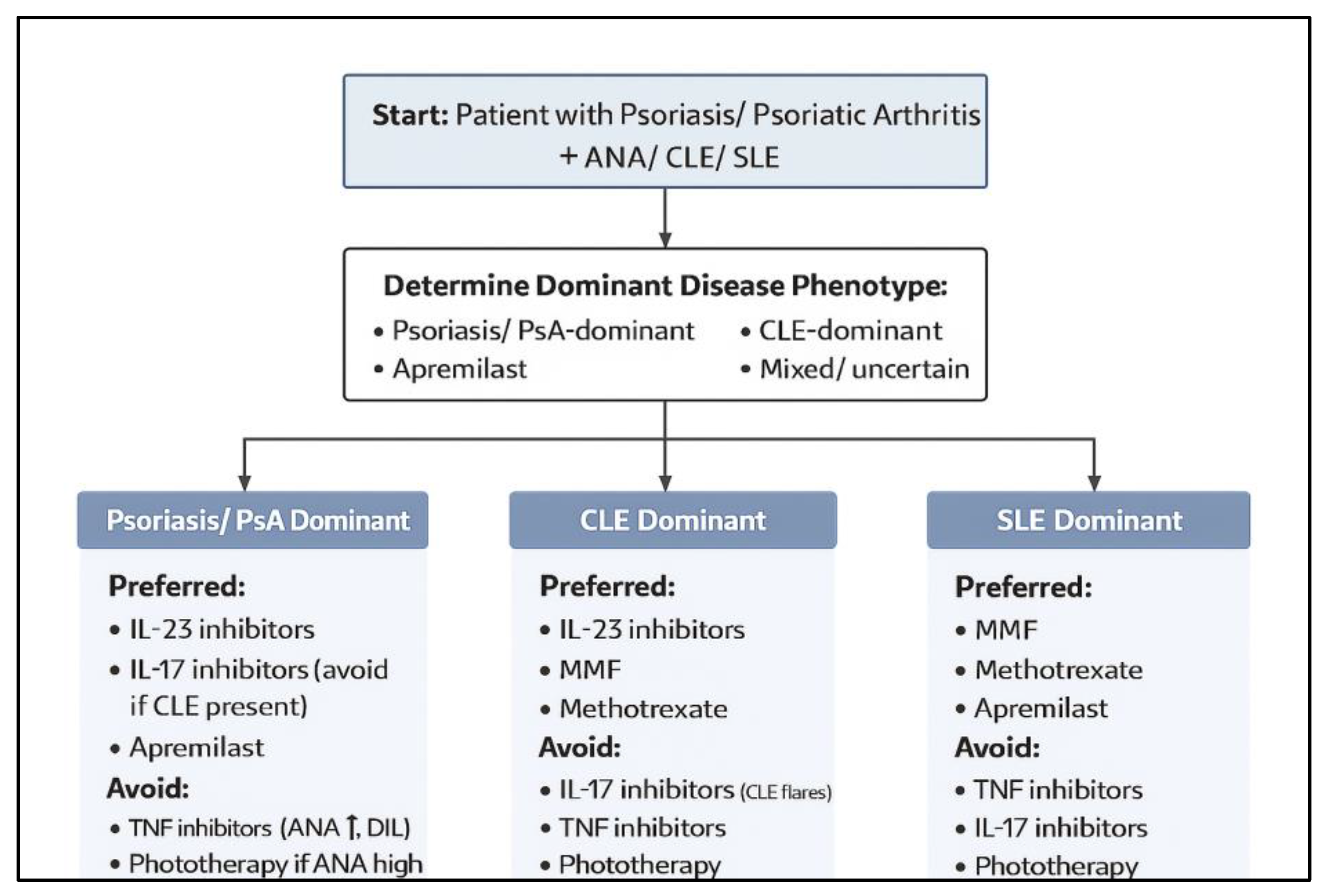
Table 4.
Therapeutic Performance Across Psoriatic–Lupus Overlap Subgroups. Summary of preferred, acceptable, and contraindicated systemic therapies across six clinically defined overlap categories: psoriasis with antinuclear antibody (ANA) positivity, psoriasis with cutaneous lupus erythematosus (CLE), psoriasis with systemic lupus erythematosus (SLE), psoriatic arthritis (PsA) with ANA positivity, PsA with CLE, and PsA with SLE. Therapeutic recommendations integrate clinical outcomes, lupus-specific safety signals, ANA/autoantibody trajectories, and mechanistic considerations related to Th17 and type I interferon pathway activation.
| Patient Subgroup | Preferred Therapies | Acceptable / Conditional Options | Contraindicated / Use with Strong Caution |
|---|---|---|---|
| Psoriasis + ANA positivity | IL-23 inhibitors [12,13,14,43]; IL-17 inhibitors [40,42]; apremilast [16] | Methotrexate [6,11]; narrowband UVB in low-titer ANA, no lupus features [20] | TNF-α inhibitors [10,18,35,36,37,38]; high-dose / broad-spectrum phototherapy in high-titer ANA or ENA-positive patients [9,20,47] |
| Psoriasis + CLE | IL-23 inhibitors [12,13,14,43]; apremilast [16]; methotrexate [6,11] | Ustekinumab (IL-12/23) [39,46]; low-dose systemic steroids (short-term) [6,11] | IL-17 inhibitors (CLE induction/worsening) [28,29,30,31,32,33]; TNF-α inhibitors [45,46,47,48,49]; hydroxychloroquine if psoriasis active [47,51]; intensive phototherapy [9,20] |
| Psoriasis + SLE | Mycophenolate mofetil [6,11]; methotrexate [6,11]; apremilast [16]; TYK2 inhibitor (deucravacitinib) [25,26,27]; ustekinumab (safe, modest efficacy) [46] | IL-23 inhibitors in stable SLE [12,13,14]; hydroxychloroquine if psoriasis mild & monitored [47,51] | TNF-α inhibitors [10,18,48,49,50,51,52,53]; IL-17 inhibitors [28,29,30,31,32,33]; phototherapy in established SLE or active CLE [9,20,47] |
| Psoriatic arthritis + ANA positivity | IL-17 inhibitors [40,42]; IL-23 inhibitors [12,13,14,43]; methotrexate [6,11] | Apremilast [16]; low-dose systemic steroids as bridge [6,11] | TNF-α inhibitors (autoantibody induction, DIL risk) [10,18,54,55,56,57] |
| Psoriatic arthritis + CLE | Mycophenolate mofetil [6,11]; methotrexate [6,11]; IL-23 inhibitors [12,13,14,43] | Ustekinumab [39,46,48]; apremilast [16] | IL-17 inhibitors (CLE flare risk) [28,29,30,31,32,33]; TNF-α inhibitors [45,46,47,48,49]; hydroxychloroquine (psoriasis/PsA flare risk) [47,51] |
| Psoriatic arthritis + SLE | Mycophenolate mofetil [6,11]; methotrexate [6,11]; apremilast [16]; TYK2 inhibitor (deucravacitinib) [25,26,27] | IL-23 inhibitors in stable SLE [12,13,14]; cautious hydroxychloroquine if psoriatic disease quiescent [47,51] | TNF-α inhibitors [10,18,52,53,63]; hydroxychloroquine when psoriasis/PsA active [47,51]; phototherapy in SLE [9,20] |
Table 5.
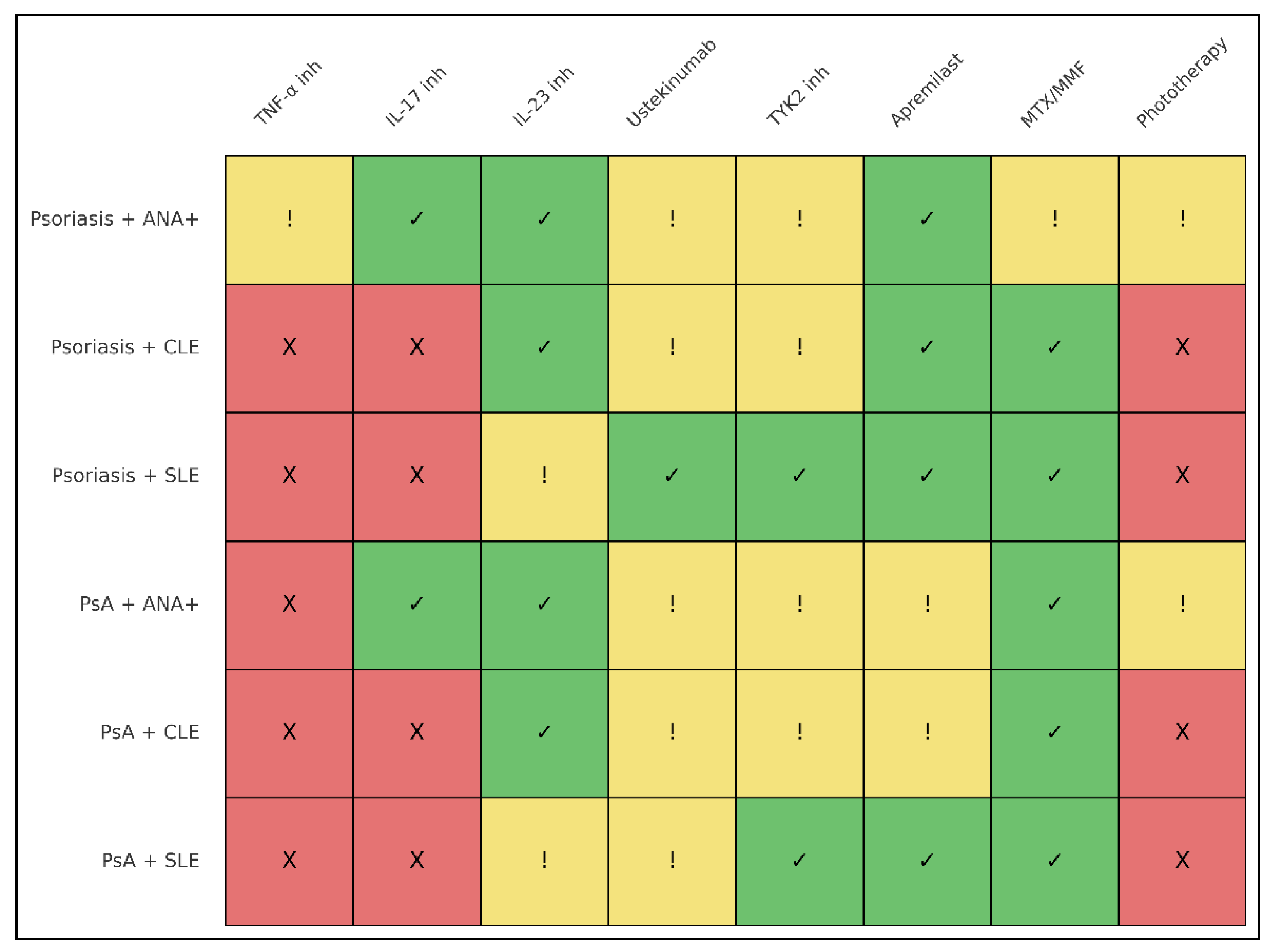
Drug suitability matrix for systemic therapies in psoriasis and psoriatic arthritis with ANA positivity, cutaneous lupus erythematosus (CLE), or systemic lupus erythematosus (SLE). Each cell reflects the overall risk–benefit profile of a drug class within a given overlap phenotype, integrating psoriatic efficacy, lupus/CLE safety signals, ANA/autoantibody dynamics, and mechanistic considerations related to Th17 and type I interferon pathways. Legend: Preferred = “Green” (first-line or strongly favored); Conditional = “Yellow” (usable with caution / specific scenarios);Avoid = “Red” (generally contraindicated or strongly discouraged).
Table 5.
Drug suitability matrix for systemic therapies in psoriasis and psoriatic arthritis with ANA positivity, cutaneous lupus erythematosus (CLE), or systemic lupus erythematosus (SLE). Each cell reflects the overall risk–benefit profile of a drug class within a given overlap phenotype, integrating psoriatic efficacy, lupus/CLE safety signals, ANA/autoantibody dynamics, and mechanistic considerations related to Th17 and type I interferon pathways. Legend: Preferred = “Green” (first-line or strongly favored); Conditional = “Yellow” (usable with caution / specific scenarios);Avoid = “Red” (generally contraindicated or strongly discouraged).
| Sub-group | TNF-α inhibitors | IL-17 inhibitors | IL-23 inhibitors | Ustekinumab (IL-12/23) | TYK2 inhibitor | Apremilast | Methotrexate (MTX) | Mycophenolate (MMF) | Hydroxychloroquine (HCQ) | Phototherapy |
|---|---|---|---|---|---|---|---|---|---|---|
| Psoriasis + ANA+ (no lupus) | Conditional (effective but ANA rise, DIL risk) [10,18,35,36,37,38] | Preferred [40,42] | Preferred [12,13,14,43] | Conditional [39,46] | Conditional (limited data, mechanistically favorable) [25,26,27] | Preferred [16] | Conditional [6,11] | Conditional (rarely needed if no SLE) [6,11] | Conditional (if no psoriasis flare history) [47,51] | Conditional (NB-UVB only; avoid ANA high-titer/ENA+) [9,20,47] |
| Psoriasis + CLE | Avoid (CLE induction/worsening) [10,18,45,46,47,48,49] | Avoid (strong CLE risk) [28,29,30,31,32,33] | Preferred [12,13,14,43] | Conditional (neutral-safety, modest data) [39,46,48] | Conditional (promising in CLE, limited overlap data) [25,26,27] | Preferred [16] | Preferred [6,11] | Preferred if SLE/CLE-active [6,11,20] | Conditional (avoid if psoriasis flares) [47,51] | Avoid (photosensitive CLE risk) [9,20] |
| Psoriasis + SLE | Avoid [10,18,48,49,50,51,52,53] | Avoid [28,29,30,31,32,33] | Conditional (use only in stable SLE) [12,13,14] | Preferred for safety (modest efficacy) [46] | Preferred (improves SLE & CLE biology) [25,26,27] | Preferred [16] | Preferred [6,11] | Preferred [6,11] | Conditional (only if psoriasis mild, SLE-dominant) [47,51] | Avoid [9,20,47] |
| PsA + ANA+ | Avoid (autoantibody induction, DIL risk) [10,18,54,55,56,57] | Preferred [40,42] | Preferred [12,13,14,43] | Conditional [39,46] | Conditional [25,26,27] | Conditional [16] | Preferred [6,11] | Conditional [6,11] | Conditional (if PsA mild, lupus absent) [47,51] | Conditional (only if low-titer ANA, no lupus features) [9,20] |
| PsA + CLE | Avoid [10,18,45,46,47,48,49] | Avoid [28,29,30,31,32,33] | Preferred [12,13,14,43] | Conditional [39,46,48] | Conditional–Preferred (CLE benefit; limited PsA data) [25,26,27] | Conditional [16] | Preferred [6,11] | Preferred [6,11,20] | Avoid (psoriasis/PsA flare risk) [47,51] | Avoid [9,20] |
| PsA + SLE | Avoid [10,18,52,53,63] | Avoid [28,29,30,31,32,33] | Conditional (stable SLE only) [12,13,14] | Conditional [39,46,48] | Preferred (dual PsA/SLE biology) [25,26,27] | Preferred [16] | Preferred [6,11] | Preferred [6,11] | Conditional (only if PsA quiescent) [47,51] | Avoid [9,20] |
Figure 3.
Safety profile of systemic therapies across psoriatic–lupus overlap subgroups. Heatmap summarizing the relative suitability of major therapeutic classes—TNF-α inhibitors, IL-17 inhibitors, IL-23 inhibitors, ustekinumab, TYK2 inhibitors, apremilast, methotrexate/mycophenolate, and phototherapy—across six clinical overlap phenotypes: psoriasis with ANA positivity, psoriasis with cutaneous lupus erythematosus (CLE), psoriasis with systemic lupus erythematosus (SLE), psoriatic arthritis (PsA) with ANA positivity, PsA with CLE, and PsA with SLE. Green indicates “preferred,” yellow indicates “conditional/acceptable with caution,” and red indicates “avoid/contraindicated.” IL-23 inhibitors show the most favorable cross-phenotype safety, while TNF-α and IL-17 inhibitors exhibit the highest risk in lupus-prone settings.
3.7. Discussion
Despite the major differences in their underlying immunopathogenesis, an increasing body of evidence indicates that patients with psoriatic disease are at heightened risk of developing lupus erythematosus.[66,67,68,69] Epidemiologic studies consistently show a higher prevalence of SLE among individuals with psoriasis or psoriatic arthritis, suggesting meaningful biological overlap between Th17-driven and type I interferon-driven autoimmune pathways. Experimental models further support this connection: epicutaneous imiquimod application—classically used to induce psoriasis—can also provoke systemic lupus–like immune activation, illustrating the capacity of a single upstream trigger to engage both psoriatic and lupus pathways.[70,71] Moreover, shared genetic susceptibility loci have been identified in the Chinese Han population, reinforcing the concept of a partially overlapping hereditary background linking these two disorders.[72]
Within this context, the present systematic review provides the most comprehensive synthesis to date on the management of psoriasis and psoriatic arthritis in patients who exhibit antinuclear antibody positivity or coexisting cutaneous or systemic lupus erythematosus. By integrating recently published mechanistic and clinical evidence—including formal ustekinumab trials in SLE [46], advances in TYK2 inhibition [25,26,27], and expanding reports of IL-17 inhibitor–associated cutaneous lupus [28,29,30,31,32,33]—this review offers clearer therapeutic guidance in an area traditionally characterized by clinical uncertainty and immunologic tension.
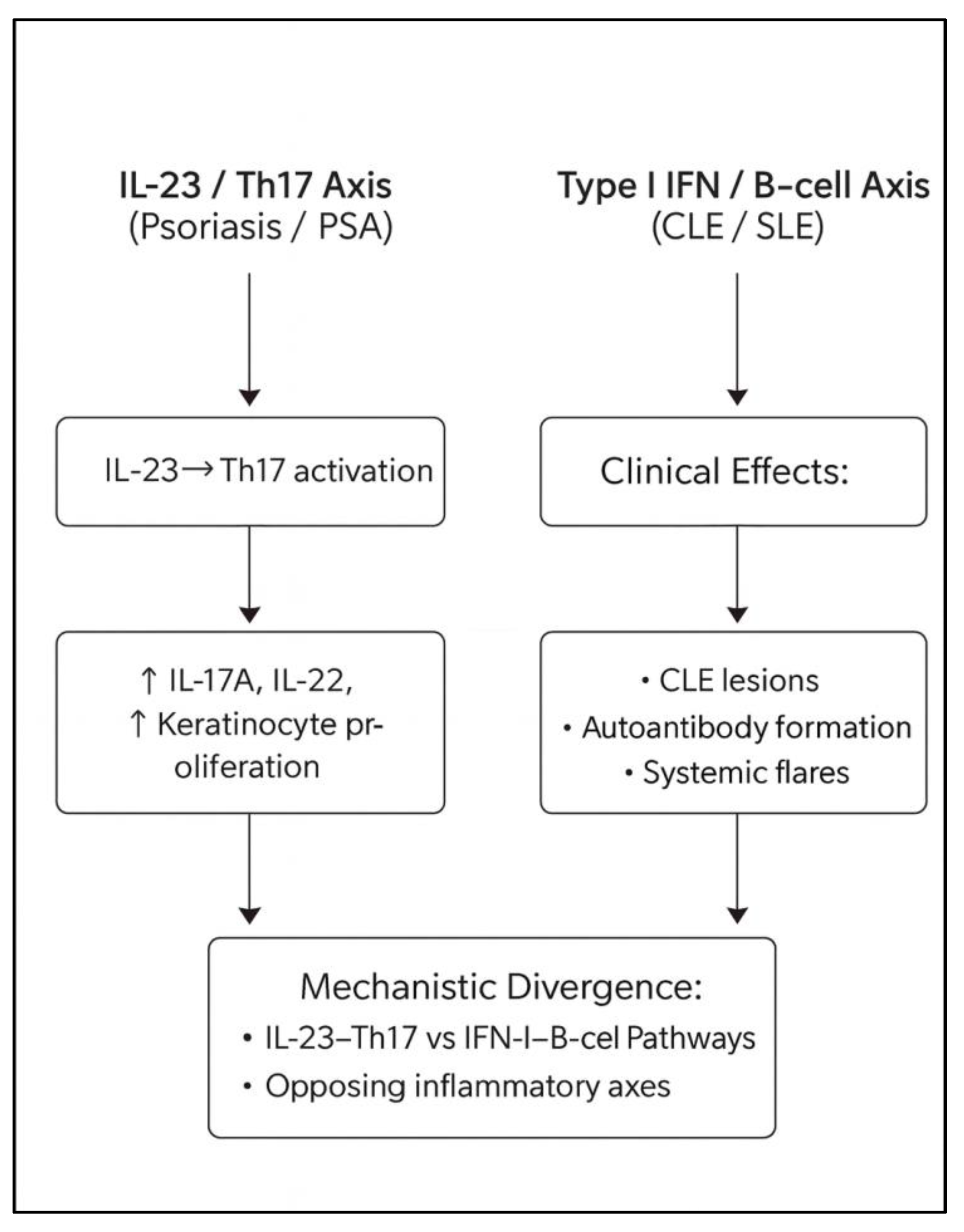
3.8. The Central Immunologic Paradox of Psoriasis–Lupus Overlap
Psoriasis and psoriatic arthritis are driven primarily by IL-23–mediated activation of Th17 cells and downstream release of IL-17A and IL-17F [1,2,3,4,5], whereas lupus and lupus-spectrum diseases arise from plasmacytoid dendritic cell activation and the amplification of type I interferon programs that promote B-cell hyperactivity and autoantibody production [6,7,8,9,17]. These diseases therefore sit on opposing ends of key immunologic axes [9,10,11,12]. Treatments designed for psoriasis or PsA can destabilize lupus biology, while therapies effective for lupus may aggravate psoriatic inflammation [10,18,47,51,59]. This cross-axis fragility is readily observed in the heterogeneous safety profiles identified across biologic classes in this review. ANA positivity adds further complexity, as elevated ANA levels may represent incidental background autoimmunity, a drug-induced serologic phenomenon—especially with TNF inhibitors [10,18,35,36,37,38]—or a prodrome of evolving lupus [6,7,8,9,17,20]. These factors together necessitate a therapeutic strategy guided not merely by diagnosis but by phenotype, serologic activity, and individualized risk.
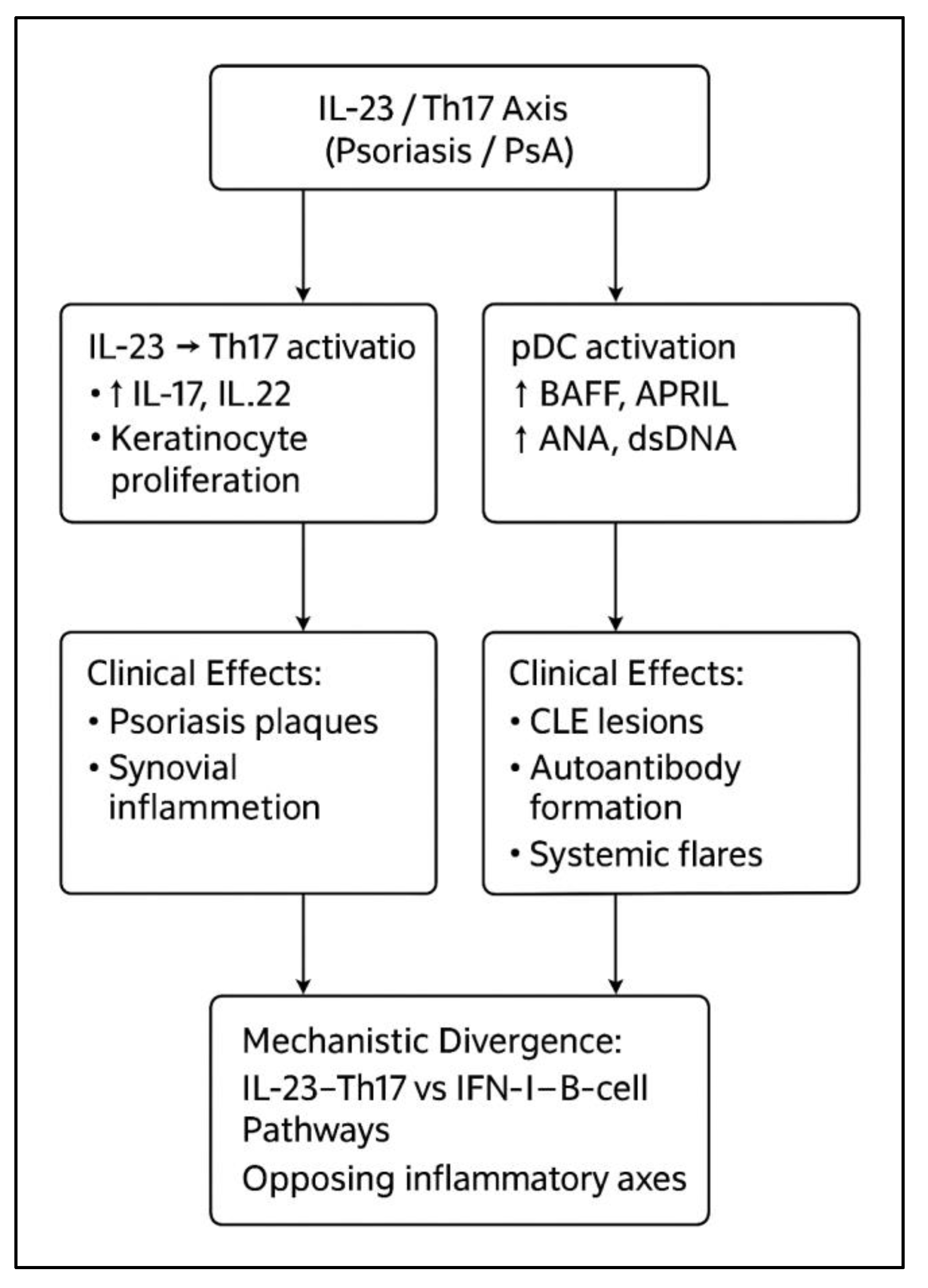
Figure 4.
Mechanistic divergence of psoriatic (IL-23/Th17) and lupus (type I interferon/B-cell) immune pathways. Psoriasis and psoriatic arthritis are driven primarily by IL-23–mediated Th17 activation, promoting IL-17A/F and IL-22–dependent keratinocyte proliferation and synovial inflammation. In contrast, cutaneous and systemic lupus erythematosus arise from plasmacytoid dendritic cell activation and type I interferon signaling, leading to BAFF-mediated B-cell activation, ANA/dsDNA autoantibody production, and CLE/SLE manifestations. These two axes represent opposing inflammatory programs that explain differential therapeutic responses and cross-disease safety considerations.
3.9. Differential implications for SLE versus CLE, particularly DLE
Although SLE and CLE share a type I interferon–driven core [6,7,8,9,17], their therapeutic vulnerabilities are not identical. Systemic lupus erythematosus is dominated by circulating immune complexes and multi-organ inflammation; drugs that dampen global B-cell activity (e.g., rituximab) [59] or IFN-I signatures can therefore exert substantial benefit. In contrast, chronic discoid lupus erythematosus (DLE) is often “skin-locked,” with scarring follicular destruction, dense interface dermatitis, and a persistently IFN-high, TNF-low cutaneous microenvironment [9,20]. In this setting, additional suppression of Th17/TNF tone—such as with IL-17 inhibitors [2,4,28,29,30,31,32,33]—may further disinhibit plasmacytoid dendritic cells and IFN-I production, predisposing to refractory or even aggravated DLE [28,29,30,31,32,33]. This may explain why IL-17 inhibitor–associated lupus manifestations are disproportionately cutaneous and DLE- or SCLE-like rather than systemic [28,29,30,31,32,33].
Conversely, agents with neutral or modestly IFN-dampening effects, such as ustekinumab [39,46], have shown stable safety in SLE trials despite inconsistent efficacy [46], and no signal has emerged for CLE or DLE worsening. These data support regarding ustekinumab as a mechanistically “neutral-safe” option in psoriatic patients with stable SLE [46,48], while recognizing the paucity of CLE-specific data. At the same time, a broader lupus literature indicates that B-cell–directed therapies such as rituximab, though beneficial for systemic disease, can paradoxically induce de novo psoriasis or exacerbate pre-existing psoriasis [59]. Taken together, these observations reinforce the need to distinguish SLE from CLE, and particularly DLE, when selecting biologic or targeted therapies in patients who straddle the psoriasis–lupus interface [9,17,18,19,20,28,29,30,31,32,33,46].
3.10. The Cross-Disease Safety Advantage of IL-23 Inhibition
Across all disease combinations included in this review—psoriasis with or without CLE or SLE, PsA with or without CLE or SLE, and ANA-positive patients without clinical lupus—IL-23 inhibitors consistently demonstrated the most favorable balance of efficacy and safety [12,13,14,43]. Mechanistically, IL-23 blockade acts downstream of pathways involved in interferon-driven autoimmunity and avoids triggering plasmablast activation, dsDNA production, or amplification of type I interferon programs [4,12,13,14]. No studies reported lupus flares, CLE induction, or drug-induced lupus attributable to IL-23 inhibition [12,13,14,43]. The pattern that emerges suggests IL-23 inhibitors are the safest biologic class for nearly all psoriatic–lupus overlap phenotypes, including psoriasis or PsA with ANA positivity [35,36,37,38,39,40,41,42,43,54,55,56,57], psoriasis or PsA with CLE [44,45,46,47,48,49], and stable SLE coexisting with psoriatic disease [48,49,50,51,52,53]. This sharply contrasts with the risk signals observed with IL-17 and TNF-α inhibitors [10,18,28,29,30,31,32,33,45,46,47,48,49,52,53].
Table 6.
Comparison of the Th17/IL-23 versus type I interferon/B-cell immune axes and the ways in which major systemic therapies modulate these pathways. The table highlights how pathway targeting translates into differential effects on psoriasis/psoriatic arthritis versus cutaneous and systemic lupus, and summarizes therapeutic implications in overlap disease.
| Immunologic Axis / Drug Class | Primary Mediators / Targets | Dominant Disease Context | Effect on Psoriasis/PsA | Effect on CLE/SLE | Therapeutic Implications in Overlap Disease |
|---|---|---|---|---|---|
| Th17/IL-23 Axis | IL-23, IL-17A/F, IL-22, TNF-α | Psoriasis, PsA | Central driver of keratinocyte activation and synovial inflammation [1,2,3,4,5] | Indirect, usually minor role [9,17] | Blocking this axis improves psoriatic disease; downstream risk depends on IFN-I shifts [9,12,13,14,20] |
| Type I IFN / B-cell Axis | IFN-α/β, BAFF, ANA, dsDNA, immune complexes | CLE, SLE | Can be secondarily activated but not primary driver [6,7,8,9,17] | Central to cutaneous & systemic lupus activity [6,7,8,9,17] | Therapies amplifying IFN-I or autoantibody production increase lupus risk; IFN-suppressive agents are protective [9,17,20] |
| TNF-α inhibitors | TNF-α blockade | Psoriasis, PsA, RA | Highly effective [1,2,3,4,5] | Promotes ANA ↑, dsDNA ↑, DIL, CLE flares [10,18,35,36,37,38,45,46,47,48,49,52,53] | Should be avoided in CLE/SLE or high-risk ANA+ patients despite psoriatic efficacy [10,18,35,36,37,38,45,46,47,48,49,52,53] |
| IL-17 inhibitors | IL-17A/F blockade | Psoriasis, PsA | Very strong skin/joint efficacy [2,4,40,42] | Associated with de novo or worsened SCLE/DLE [28,29,30,31,32,33] | Useful in severe psoriatic disease but risky in CLE-prone patients; avoid active DLE/SCLE [28,29,30,31,32,33] |
| IL-23 inhibitors | IL-23 p19 blockade (upstream of Th17) | Psoriasis, PsA | Excellent psoriatic control [12,13,14,43] | Neutral or potentially protective; no lupus signal to date [12,13,14,43] | Best overall safety in psoriatic–lupus overlap; preferred biologic class [12,13,14,43] |
| IL-12/23 inhibitor (ustekinumab) | p40 blockade | Psoriasis, PsA; tested in SLE | Effective in psoriasis/PsA [39] | Safe in SLE (Phase II/III) though modest efficacy [46] | Good option when IL-23 inhibitors unavailable; safe for stable SLE with psoriasis [39,46,48] |
| TYK2 inhibitor (deucravacitinib) | TYK2 signaling (IL-23, IFN-I, IL-12 pathways) | Psoriasis; emerging in SLE/CLE | Effective in psoriasis & PsA [25,26,27] | Suppresses IFN-I signatures; improves CLE/SLE endpoints [25,26,27] | First cross-disease targeted agent; promising for PsO/PsA + CLE/SLE [25,26,27] |
| PDE4 inhibitor (apremilast) | cAMP-mediated cytokine modulation | Psoriasis, PsA | Moderate efficacy [16] | Neutral to slightly favorable in lupus [16] | Safe oral option in ANA+ and lupus-prone patients [16] |
| Methotrexate (MTX) | Antimetabolite; T- and B-cell modulation | Psoriasis, PsA, SLE arthritis | Good joint/skin response [6,11] | Beneficial for SLE musculoskeletal disease [6,11] | Foundational option for SLE-dominant overlap [6,11] |
| Mycophenolate mofetil (MMF) | Inhibits lymphocyte proliferation | SLE, CLE | Modest effect on psoriasis/PsA [6,11] | Strongly effective in SLE/CLE [6,11,20] | First-line systemic option for SLE/CLE-dominant presentation [6,11,20] |
| Hydroxychloroquine (HCQ) | TLR7/9 & IFN-I modulation | CLE, SLE | May worsen psoriasis [47,51] | Beneficial for CLE/SLE [6,11,47,51] | Mainstay for lupus; avoid in active psoriasis/PsA [47,51] |
| Phototherapy (NB-UVB/UVB) | UV-induced keratinocyte apoptosis & neo-antigen exposure | Psoriasis | Effective in psoriasis [22] | Can trigger CLE via IFN-I upregulation [9,20] | Acceptable only for low-risk ANA+ patients; contraindicated in CLE/SLE [9,20,47] |
Table 7.
Comparative Risk Summary for Drug-Induced Autoimmunity Across Systemic Therapies.
| Therapeutic Class | Primary Drug-Induced Autoimmune Signal(s) | Strength of Evidence | Typical Clinical Phenotype | Reversibility After Drug Withdrawal | Overall Risk Rating for Drug-Induced Autoimmunity |
|---|---|---|---|---|---|
| TNF-α inhibitors | ANA seroconversion; anti-dsDNA induction; ATIL; CLE-like eruptions [10,18,35,36,37,38,45,46,47,48,49,52,53] | High – multiple cohorts, case series, pharmacovigilance [10,18,35,36,37,38,45,46,47,48,49] | Photosensitive rash; SCLE/DLE-like lesions; arthritis/serositis; ANA ± dsDNA; occasionally full SLE [45,46,47,48,49,52,53] | Usually improves/resolves after withdrawal ± steroids or HCQ [45,46,47,48,49] | High risk – avoid in CLE/SLE or strong lupus diathesis; use only exceptionally [10,18,35,36,37,38,45,46,47,48,49,52,53] |
| IL-17 inhibitors | New-onset or worsening CLE (SCLE/DLE); rare lupus-like events [28,29,30,31,32,33] | Moderate – increasing case reports and series [28,29,30,31,32,33] | Disseminated DLE/SCLE; photo-exacerbated plaques; ANA elevation with minimal systemic signs [28,29,30,31,32,33] | Improves after IL-17 withdrawal; switch to IL-23 or non-Th17 agents [28,29,30,31,32,33] | Moderate cutaneous risk – avoid active CLE/DLE; may use cautiously in stable SLE without CLE [28,29,30,31,32,33] |
| IL-23 inhibitors | Occasional ANA changes; no consistent lupus/CLE signal [12,13,14,43] | Low – pooled trials + real-world [12,13,14,43] | Isolated autoantibody changes; lupus events rare, not clearly drug-related [12,13,14] | Withdrawal usually not needed | Low risk – preferred for ANA+, CLE, stable SLE [12,13,14,43] |
| IL-12/23 inhibitor (ustekinumab) | Rare lupus-like or autoimmune phenomena; neutral in SLE trials [39,46] | Low–moderate – Phase II/III SLE + psoriasis data [39,46] | Stable SLE activity; no CLE or systemic flares [46] | Withdrawal rarely required [46] | Low–moderate risk – mechanistically neutral; reasonable for psoriasis + stable SLE [39,46,48] |
| TYK2 inhibitor (deucravacitinib) | Reduction of IFN-driven autoimmunity; improves CLE/SLE markers [25,26,27] | Emerging – early SLE/CLE trials + transcriptomics [25,26,27] | ↓ IFN signatures; improved CLE/SLE scores; no de novo lupus [25,26,27] | Not typically associated with drug-induced autoimmunity | Low / potentially protective – attractive for PsO/PsA + CLE/SLE [25,26,27] |
| PDE4 inhibitor (apremilast) | Minimal autoimmunity signal; rare nonspecific events [16] | Low – extensive psoriasis/PsA use; few lupus reports [16] | Mild, nonspecific immune findings; no ANA/CLE/SLE pattern [16] | Generally reversible; therapy often continued | Very low risk – safe oral option for ANA+ & lupus-prone pts [16] |
| Methotrexate (MTX) | No DIL signature; may reduce SLE activity [6,11] | Low – long clinical use [6,11] | Improves joint/skin + systemic inflammation [6,11] | N/A – not a lupus inducer | Low risk – foundational in SLE-dominant PsA/PsO [6,11] |
| Mycophenolate mofetil (MMF) | Treats SLE/CLE; no DIL [6,11,20] | Low – standard SLE/CLE therapy [6,11] | Decreases CLE lesions + SLE activity [6,11,20] | N/A – therapeutic rather than inductive | Very low / protective – preferred in SLE/CLE-dominant cases [6,11,20] |
| Hydroxychloroquine (HCQ) | Psoriasis flares; paradoxical psoriatic autoimmunity [47,51] | Moderate – multiple psoriatic flare reports [47,51] | New-onset psoriasis or worsening existing disease; usually no lupus induction [47,51] | Improves after HCQ withdrawal | Low lupus risk, moderate psoriasis risk – avoid in active psoriasis/PsA [47,51] |
| Rituximab | SLE improvement; paradoxical psoriasis induction [59] | Moderate – case reports + series [59] | De novo psoriasis or flares; SLE improves [59] | Psoriasis improves after rituximab withdrawal [59] | Low lupus risk, moderate psoriasis risk – use cautiously in PsO-risk patients [59] |
| Phototherapy (NB-UVB/UVB) | Photo-induced CLE/DLE/SCLE; IFN-signature amplification [9,20,47] | Moderate – well-documented UV-triggered CLE [9,20] | New or worsening CLE in sun-exposed areas; no systemic flare necessarily [9,20,47] | Improves after cessation + photoprotection [9,20] | Moderate cutaneous autoimmunity risk – avoid or minimize in ANA-high, ENA+, CLE-prone pts [9,20,47] |
3.11. IL-17 Inhibitors: High Efficacy but Distinct Cutaneous Lupus Risk
IL-17 inhibitors provide robust control of psoriasis and psoriatic arthritis, often outperforming other biologic classes in skin clearance and joint suppression [2,4,40,42]. However, this review confirms a distinct pattern of CLE worsening or new-onset CLE associated with IL-17 blockade [28,29,30,31,32,33]. Multiple published reports describe cases of disseminated discoid lupus misdiagnosed as psoriasis [28,29], aggravation of DLE during secukinumab therapy [29], secukinumab-induced SCLE [30,31], and ixekizumab-associated CLE [33]. Mechanistically, IL-17 inhibition may unmask or amplify type I interferon responses [9,20], increasing keratinocyte apoptosis and plasmacytoid dendritic cell activation, thereby propagating CLE lesions even in the absence of systemic lupus activity [9,20,28,29,30,31,32,33].
Clinically, IL-17 inhibitors should be used with caution in patients with active DLE or SCLE, high-titer ANA, or ENA-positive serology [20,47], and reserved for situations in which psoriatic disease is severe, CLE is quiescent, and alternative biologics are not viable [28,29,30,31,32,33].
Figure 5.
Proposed mechanism of IL-17 inhibitor–associated cutaneous lupus erythematosus (CLE). IL-17 blockade (e.g., secukinumab, ixekizumab) suppresses Th17 signaling, resulting in loss of IL-17–mediated immune balance. This promotes compensatory type I interferon dominance, driven by activation of plasmacytoid dendritic cells and increased IFN-α/β production. Subsequent keratinocyte apoptosis and release of nucleic acid–immune complexes amplify autoantibody pathways, leading to CLE manifestations such as SCLE/DLE lesions, photosensitive rash, and possible ANA or dsDNA elevation.
3.12. TNF-α Inhibitors: Strongest Evidence for Lupus Induction and Autoantibody Conversion
Among all therapeutic classes, TNF-α inhibitors display the strongest and most consistent lupus-related safety concerns [10,18,35,36,37,38,45,46,47,48,49,52,53]. ANA seroconversion occurs in a significant minority of treated individuals [35,36,37,38,54,55,56,57], dsDNA emergence is well documented [10,18,45,46,47,48,49], and drug-induced lupus has been reported in 6–15% of cases in susceptible populations [10,18,45,46,47,48,49]. These agents have repeatedly precipitated lupus flares in established SLE [48,49,50,51,52,53], exacerbated CLE [45,46,47,48,49], and induced new CLE lesions in psoriasis and PsA patients [45,46,47,48,49]. Mechanistic studies support these clinical findings, demonstrating that TNF blockade promotes plasmablast survival, shifts immunity toward interferon-dominant signaling, and facilitates immune complex deposition [10,18]. Accordingly, TNF inhibitors should generally be avoided in patients with lupus-prone features or high-titer ANA [20,47].
3.13. Ustekinumab (IL-12/23): Stable Safety Despite Mixed SLE Efficacy
Formal Phase II and Phase III trials of ustekinumab in SLE provide an unusual degree of controlled safety data for a psoriasis-centric biologic [46]. Although the Phase III trial failed to meet its primary endpoint [46], ustekinumab exhibited a reassuring safety profile with no increase in lupus activity or lupus-related adverse effects [46]. This positions ustekinumab as a useful option for patients whose psoriasis or PsA is active while their lupus remains stable or mild [48], especially in situations where IL-23 inhibitors are unavailable. Its safety stands in contrast to the problematic profiles of TNF and IL-17 inhibitors in lupus-susceptible populations [10,18,28,29,30,31,32,33].
3.14. TYK2 Inhibition: A Mechanistically Bidirectional Therapy
Deucravacitinib, the first approved TYK2 inhibitor, has reshaped the therapeutic landscape by demonstrating meaningful activity on both sides of the psoriasis–lupus spectrum [25,26,27]. Phase II SLE trial data show improvements in patient-reported outcomes and reductions in interferon-driven gene signatures [25,26,27], while experimental data from cutaneous lupus models demonstrate suppression of CLE-associated molecular pathways [26]. Early real-world reports further confirm successful treatment of patients with overlapping psoriasis, PsA, and SLE [25]. Because TYK2 signaling lies upstream of both IL-23 and type I interferon pathways, its inhibition offers a uniquely balanced mechanism for patients whose disease reflects simultaneous Th17-driven and interferon-driven biology [25,26,27]. This positions TYK2 inhibition as the first oral therapy with genuine cross-disease relevance in psoriatic–lupus overlap.
Figure 6.
Dual-pathway immunologic effects of TYK2 inhibition in psoriatic–lupus overlap disease. Deucravacitinib blocks TYK2 signaling upstream of both IL-23–mediated Th17 activation and the type I interferon (IFN-I) axis. Suppression of IL-23 signaling reduces Th17 activity and IL-17A/F production, improving psoriasis and psoriatic arthritis manifestations. Concurrent inhibition of IFN-α/β–driven pathways decreases pDC activation and IFN-I gene signatures, improving cutaneous lupus erythematosus (CLE) lesions and reducing systemic lupus erythematosus (SLE) activity. This dual modulation confers unique cross-disease therapeutic benefit in patients with overlapping psoriatic and lupus-spectrum disease.
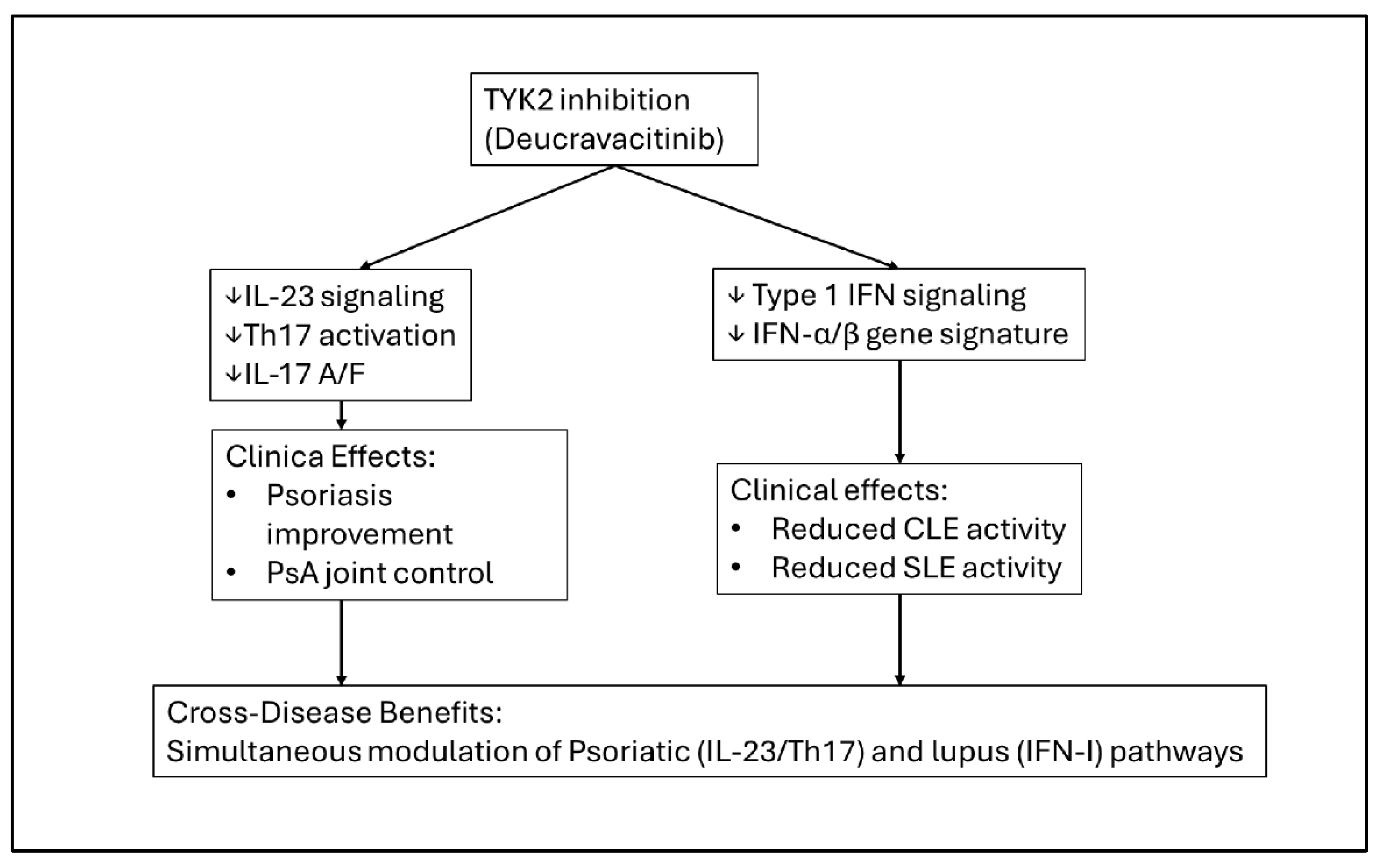
Table 8.
Key clinical and mechanistic evidence supporting ustekinumab and TYK2 inhibition (deucravacitinib) in lupus-spectrum disease, including systemic lupus erythematosus (SLE), cutaneous lupus erythematosus (CLE), and real-world psoriatic–lupus overlap. The table summarizes trial designs, efficacy outcomes, safety signals, and practical implications for treatment of psoriasis and psoriatic arthritis in the setting of ANA positivity, CLE, or SLE.
| Agent | Study / Population | Design & Sample | Key Efficacy Findings in SLE/CLE | Safety Signals | Implications for Psoriasis/PsA with Lupus-Spectrum Disease |
|---|---|---|---|---|---|
| Ustekinumab (IL-12/23 inhibitor) | Phase II SLE trial [46] | Randomized, placebo-controlled; moderate-to-severe SLE | Improved SRI-4 responses vs placebo; benefit in musculoskeletal & mucocutaneous domains [46] | No increase in lupus flares; no DIL; no new safety concerns [46] | Demonstrates biologic activity in SLE with reassuring safety, supporting use in psoriasis/PsA with stable or mild SLE when IL-23 inhibitors are unavailable [39,46,48] |
| Phase III SLE trial [46] | Randomized, placebo-controlled; multicenter SLE cohort | Failed primary BICLA endpoint; modest improvement in secondary outcomes (e.g., SRI-6) [46] | Safety similar to placebo; no lupus activation [46] | Confirms stable safety but inconsistent SLE efficacy; ustekinumab can be considered “neutral-safe” in overlap disease, not a primary lupus therapy [46,48] | |
| Case reports / small CLE series [46,48] | SCLE/DLE patients treated off-label | Variable but sometimes favorable CLE lesion control in refractory cases [46,48] | No CLE worsening; no systemic lupus induction [46] | Suggests ustekinumab is unlikely to exacerbate CLE in psoriatic patients; evidence limited, non-randomized [46,48] | |
| Deucravacitinib (TYK2 inhibitor) | Phase II SLE trial [25,26,27] | Randomized, placebo-controlled; active SLE (multi-organ involvement) | Improved PROs and global disease activity; reduction in IFN-I–driven gene signatures [25,26,27] | No DIL signal; no major flares; well tolerated [25,26,27] | Provides proof-of-concept that TYK2 targeting improves SLE activity while modulating IFN-I pathways—highly relevant to overlap patients [25,26,27] |
| CLE transcriptomic study [26] | CLE patients; mechanistic/transcriptomic | Decreased IFN-I–regulated transcripts; reduced CLE inflammatory signature [26] | No new safety concerns [26] | Indicates direct CLE benefit, supporting use when CLE coexists with psoriasis/PsA [26] | |
| Real-world PsO + PsA + SLE case [25] | Single-patient, refractory overlap treated with deucravacitinib + MMF + HCQ | Concurrent improvement in psoriasis, PsA, SLE; steroid-sparing [25] | No lupus activation; no psoriasis flare [25] | First real-world evidence of cross-disease control → TYK2 inhibition as important systemic option [25] | |
| Psoriasis Phase III trials (contextual) [25,26,27] | Phase III PsO RCTs | High PASI-75/90 rates; durable efficacy [25,26,27] | No CLE/SLE signal in PsO trials [25,26,27] | Confirms strong psoriatic efficacy with safety compatible for lupus-prone individuals; supports cautious use pending more overlap studies [25,26,27] |
3.15. Phototherapy: A Reassessment in ANA-Positive and CLE-Prone Disease
Although narrowband UVB phototherapy is traditionally considered safe in psoriasis [14,22], emerging mechanistic evidence suggests that UV exposure can amplify interferon gene signatures [9,20], promote keratinocyte apoptosis, and precipitate CLE lesions in genetically or serologically predisposed individuals [20,47]. These biological observations correspond with clinical patterns showing disease worsening in SLE and active CLE following phototherapy [20,47]. Patients with high-titer ANA or ENA positivity may also be at heightened risk [20,47], even without overt lupus manifestations. Phototherapy can still be considered in ANA-low individuals without lupus symptoms, but its use requires greater caution than traditionally appreciated [9,20].
Table 9.
Overview of phototherapy safety considerations across ANA-positive psoriasis, psoriatic arthritis, cutaneous lupus erythematosus (CLE), and systemic lupus erythematosus (SLE). The table summarizes efficacy for psoriatic disease, risks of lupus or CLE activation, and practical recommendations according to ANA titer, ENA status, and lupus phenotype.
| Clinical Setting | Phototherapy Modality | Psoriasis/PsA Efficacy | Lupus / Autoimmunity Risk | Recommended Use |
|---|---|---|---|---|
| Psoriasis / PsA, ANA-negative | NB-UVB, BB-UVB | High efficacy for plaque psoriasis; useful for PsA with skin-dominant disease [22] | No specific lupus risk | Standard indication; may be used freely if no lupus features or autoimmune history |
| Psoriasis / PsA, low-titer ANA+ (no ENA, no lupus symptoms) | NB-UVB preferred; avoid high cumulative doses | Good psoriasis control; may reduce systemic drug burden [22] | Theoretical IFN-I upregulation & autoantibody evolution, but clinical risk appears low [9,20] | Acceptable with caution: use NB-UVB, limit exposure, monitor for CLE-like lesions or systemic symptoms [9,20] |
| Psoriasis / PsA, high-titer ANA+ or ENA+; no overt lupus | NB-UVB only in exceptional cases; avoid PUVA/high-dose UVB | Effective for psoriasis, but safer systemic alternatives exist [6,11] | Elevated risk of UV-provoked autoimmunity; UV can trigger CLE-like changes & IFN signature [9,20,47] | Use only when systemic options are contraindicated; short courses; close skin/serologic monitoring with rheumatology input [9,20,47] |
| Psoriasis + CLE (SCLE/DLE) | NB-UVB/BB-UVB; PUVA | May improve psoriasis, but CLE is highly photosensitive [9,20,47] | High risk of CLE flare, new lesions, increased IFN-I activity [9,20] | Generally not recommended; prefer IL-23 inhibitors, MMF, MTX, apremilast [6,11,12,13,14,16]; strict photoprotection required |
| PsA + CLE | NB-UVB/BB-UVB; PUVA | Limited data; psoriasis/PsA may improve | Same high risk of CLE exacerbation [9,20,47] | Avoid phototherapy; prioritize MMF, MTX, IL-23 inhibitors [6,11,12,13,14] |
| Psoriasis / PsA + SLE (± CLE) | NB-UVB/BB-UVB; PUVA | May help psoriasis; no benefit for SLE [22] | Well-documented photosensitive SLE flares; CLE flares; UV is a classic trigger [9,20,47] | Contraindicated in most cases; avoid phototherapy and ensure strict photoprotection [9,20,47] |
| Isolated CLE / SLE without psoriasis/PsA | ANY UV-based therapy | Not indicated for lupus [6,11] | High risk of cutaneous + systemic SLE/CLE flares [9,20,47] | Not appropriate; rely on HCQ, MMF, MTX, biologics with IFN-safe profiles [6,11,20] |
3.16. Treatment Strategy by Disease Combination
Our findings support a phenotype-specific therapeutic strategy across the six psoriatic–lupus overlap subgroups.
For psoriasis with isolated ANA positivity, IL-23 inhibitors, IL-17 inhibitors, and apremilast are generally safe and effective [12,13,14,16,40,42,43], whereas TNF inhibitors and phototherapy should be used sparingly [10,18,35,36,37,38].
For psoriasis with CLE, IL-23 inhibitors, apremilast, and methotrexate offer stability across disease domains [6,11,12,13,14,16], while IL-17 inhibitors and TNF inhibitors pose significant risk [28,29,30,31,32,33,45,46,47,48,49].
For psoriasis with SLE, mycophenolate, methotrexate, apremilast, ustekinumab, and TYK2 inhibition represent the safest options [6,11,16,25,26,27,39,46], whereas TNF and IL-17 inhibitors and phototherapy should be avoided [10,18,20,28,29,30,31,32,33].
In PsA with ANA positivity, IL-17 and IL-23 inhibitors and methotrexate are preferred [2,4,6,11], while TNF inhibitors contribute disproportionate serologic risk [54,55,56,57].
3.17. The Need for Prospective and Mechanistic Trials
Current knowledge remains limited by heterogeneous study designs, reliance on case reports or case series for CLE outcomes [28,29,30,31,32,33,44,45,46,47,48,49], and inconsistent reporting of ANA, ENA, and dsDNA serologies [35,36,37,38,54,55,56,57]. The absence of prospective cohorts and randomized trials in psoriatic disease with lupus co-morbidity prevents definitive therapeutic recommendations. Future research should prioritize biomarker-driven stratification—including IFN-high versus Th17-high immunophenotypes [9,17,20], prospective evaluation of TYK2 inhibitors [25,26,27], studies examining the role of IL-23 blockade in CLE and SLE [12,13,14,43], and detailed mechanistic exploration of IL-17 inhibitor–induced CLE [28,29,30,31,32,33].
3.18. Clinical Implications
This review supports a paradigm shift in the management of psoriatic disease coexisting with lupus or lupus-prone serology. IL-23 inhibitors should serve as front-line biologics for most overlap scenarios, given their consistently favorable cross-disease safety profile and absence of lupus-inducing signals [12,13,14,43]. In contrast, IL-17 inhibitors—despite their efficacy—require caution in CLE due to increasing reports of new-onset or aggravated SCLE/DLE and their mechanistic propensity to amplify type I interferon pathways [28,29,30,31,32,33]. TNF inhibitors remain the class most strongly associated with autoantibody formation and lupus induction, including ANA and dsDNA seroconversion, drug-induced lupus, CLE flares, and SLE exacerbations, and should therefore be used only in exceptional circumstances [10,18,35,36,37,38,45,46,47,48,49,52,53].
TYK2 inhibitors hold promise as the first cross-disease systemic therapy capable of addressing both Th17-driven and interferon-driven inflammation, supported by Phase II SLE data, CLE transcriptomic modulation, and early real-world success in psoriatic–lupus overlap [25,26,27]. Phototherapy, a longstanding psoriasis therapy, should be applied judiciously in ANA-positive populations and avoided in SLE or CLE due to UV-mediated IFN-I amplification and high risk of photosensitive flares [9,20,47].
4. Conclusions
Patients with psoriasis or psoriatic arthritis who exhibit antinuclear antibody positivity or coexistence of cutaneous or systemic lupus erythematosus represent one of the most challenging overlap populations in clinical immunodermatology. Their care requires navigating two fundamentally different immune systems: the IL-23/Th17 axis that drives psoriatic disease [1,2,3,4,5] and the type I interferon–B-cell axis central to lupus [6,7,8,9,17]. These distinct immunologic programs create unique vulnerabilities when systemic therapies are applied and demand treatment strategies that are both phenotype-specific and mechanistically informed [9,10,11,12,20].
Across the clinical and mechanistic evidence synthesized in this review, several coherent therapeutic patterns emerge. IL-23 inhibitors consistently demonstrate the most balanced profile across all overlap scenarios, reliably controlling psoriatic inflammation while avoiding autoantibody stimulation or lupus activation [12,13,14,43]. In contrast, IL-17 inhibitors, despite high psoriatic efficacy, pose meaningful cutaneous lupus risk [28,29,30,31,32,33], especially in SCLE/DLE or IFN-high phenotypes [9,20]. TNF-α inhibitors remain the class most strongly associated with ANA seroconversion, dsDNA induction, ATIL, CLE exacerbation, and SLE flares [10,18,35,36,37,38,45,46,47,48,49,52,53], making them inappropriate for most lupus-susceptible patients.
TYK2 inhibitors, such as deucravacitinib, represent an important development by simultaneously modulating IL-23 and IFN-I pathways, demonstrating benefits across SLE, CLE, psoriasis, and PsA [25,26,27]. Non-biologic agents—including methotrexate, mycophenolate, and apremilast—retain key roles in SLE-dominant or mixed phenotypes [6,11,16]. Phototherapy, though long used in psoriasis, should be avoided or cautiously applied in ANA-positive, CLE-prone, or SLE patients due to IFN-mediated photosensitive risk [9,20,47].
Taken together, these findings underscore the need for individualized, phenotype-driven therapeutic decision making. Treatment selection must account for the dominant clinical phenotype (psoriasis/PsA vs. lupus), ANA and ENA profiles, CLE versus SLE subtype, and specific mechanistic vulnerabilities [6,7,8,9,17,20]. Emerging evidence strongly suggests that IL-23 inhibitors—and increasingly TYK2 inhibitors—offer the safest and most mechanistically coherent approach for patients who straddle the psoriasis–lupus interface [12,13,14,25,26,27,43].
Supplementary Materials
The following supporting information can be downloaded at the website of this paper posted on Preprints.org. Figure S1; Table S1; Table S2; Table S3.
Author Contributions
Conceptualization, J.-W.T. and T.-F.T.; methodology, J.-W.T.; software, J.-W.T.; validation, J.-W.T. and T.-F.T.; formal analysis, J.-W.T.; investigation, J.-W.T.; resources, T.-F.T.; data curation, J.-W.T.; writing—original draft preparation, J.-W.T.; writing—review and editing, T.-F.T.; visualization, J.-W.T.; supervision, T.-F.T.; project administration, T.-F.T. All authors have read and agreed to the published version of the manuscript.
Funding
This research received no external funding.
Institutional Review Board Statement
Not applicable.
Informed Consent Statement
Not applicable.
Data Availability Statement
All data supporting the findings of this study are available within the article and its Supplementary Materials. No new datasets were generated.
Acknowledgments
The authors thank the staff of the Department of Dermatology, National Taiwan University Hospital, for their administrative and technical support.
Conflicts of Interest
The authors declare no conflicts of interest.
Abbreviations
The following abbreviations are used in this manuscript:
| Abbreviation | Full Term |
| ANA | Antinuclear antibody |
| anti-dsDNA | Anti–double-stranded DNA antibody |
| ATIL | Anti–TNF–induced lupus |
| CLE | Cutaneous lupus erythematosus |
| DIL | Drug-induced lupus |
| DLE | Discoid lupus erythematosus |
| ENA | Extractable nuclear antigen |
| HCQ | Hydroxychloroquine |
| IFN-I | Type I interferon |
| IL | Interleukin |
| IL-17i | IL-17 inhibitor |
| IL-23i | IL-23 inhibitor |
| IL-12/23i | IL-12/23 inhibitor (ustekinumab) |
| MMF | Mycophenolate mofetil |
| MTX | Methotrexate |
| NB-UVB | Narrowband ultraviolet B phototherapy |
| PsA | Psoriatic arthritis |
| PsO | Psoriasis |
| PUVA | Psoralen plus ultraviolet A therapy |
| pDC | Plasmacytoid |
Appendix A
Table A1.
PRISMA 2020 Checklist for Systematic Reviews. (All item descriptions reproduced verbatim from the official PRISMA 2020 statement.).
Table A1.
PRISMA 2020 Checklist for Systematic Reviews. (All item descriptions reproduced verbatim from the official PRISMA 2020 statement.).
| Section & Topic | Item # | PRISMA 2020 Checklist Item | Location in Manuscript |
|---|---|---|---|
| TITLE | 1 | Identify the report as a systematic review. | Title page |
| ABSTRACT | 2 | PRISMA 2020 for Abstracts checklist. | Abstract |
| INTRODUCTION | 3 | Rationale: Describe the rationale for the review in the context of existing knowledge. | Section 1 |
| 4 | Objectives: Provide an explicit statement of the objective(s) the review addresses. | Section 1 (final paragraph) | |
| METHODS | 5 | Eligibility criteria: Specify inclusion and exclusion criteria and how studies were grouped. | Section 2.2 |
| 6 | Information sources: List all databases, registers, and other sources; include search dates. | Section 2.3 | |
| 7 | Search strategy: Present full search strategies for all databases and registers. | Supplementary Table S1 | |
| 8 | Selection process: Describe methods used to assess study eligibility, including reviewers and automation tools. | Section 2.1 | |
| 9 | Data collection process: Describe methods used to collect data from included studies. | Section 2.5 | |
| 10a | Data items (outcomes): List and define primary/secondary outcomes. | Section 2.5 | |
| 10b | Data items (variables): List and define all other variables (e.g., participant characteristics). | Section 2.5 | |
| 11 | Study risk of bias assessment: Describe tools and methods used. | Section 2.6 | |
| 12 | Effect measures: Specify effect measures for each outcome. | Not applicable (narrative review) | |
| 13a | Synthesis methods: Eligibility for synthesis. | Section 2.7 | |
| 13b | Synthesis methods: Methods for data preparation. | Section 2.7 | |
| 13c | Synthesis methods: Methods for tabulation or visual display. | Table 1, Table 2, Table 3, Table 4, Table 5, Table 6, Table 7, Table 8 and Table 9; Figure 1, Figure 2, Figure 3, Figure 4, Figure 5 and Figure 6 | |
| 13d | Synthesis methods: Methods used to synthesize results. | Narrative synthesis only | |
| 13e | Exploration of heterogeneity. | Not applicable | |
| 13f | Sensitivity analyses. | Not applicable | |
| 14 | Reporting bias assessment. | Section 2.6 | |
| RESULTS | 15 | Study selection: Provide screening numbers and reasons for exclusions; include flow diagram. | Section 3.1; Figure 1 |
| 16a | Study characteristics: Present characteristics of included studies. | Table 1 | |
| 16b | Excluded studies with reasons. | Section 3.1 | |
| 17 | Risk of bias in included studies. | Section 3.x; Table 9 | |
| 18 | Results of individual studies. | Section 3; Table 1, Table 2, Table 3, Table 4, Table 5, Table 6, Table 7 and Table 8 | |
| 19 | Results of syntheses. | Sections 3.2–3.18 | |
| 20 | Reporting biases. | Section 3.x | |
| DISCUSSION | 23a | Interpretation of results in context of other evidence. | Section 3 |
| 23b | Limitations of included evidence. | Section 3 | |
| 23c | Limitations of review processes. | Section 3.9 | |
| 23d | Implications for practice, policy, and future research. | Section 4 | |
| OTHER INFORMATION | 24 | Registration and protocol. | PROSPERO ID |
| 25 | Support and funding. | Funding section | |
| 26 | Competing interests. | COI section | |
| 27 | Availability of data, code, and materials. | Data Availability section |
Appendix B. PRISMA 2020 Abstract Checklist
Table 1.
PRISMA 2020 for Abstracts Checklist.
| Item # | PRISMA for Abstracts Checklist Item (Full Wording) | Location |
|---|---|---|
| 1 | Identify the report as a systematic review. | Abstract |
| 2 | Provide an explicit statement of the main objective(s) or question(s). | Abstract |
| 3 | Eligibility criteria: Specify inclusion and exclusion criteria. | Abstract |
| 4 | Information sources: Specify the databases and dates of searches. | Abstract |
| 5 | Risk of bias: Indicate methods used to assess risk of bias. | Abstract |
| 6 | Synthesis of results: Indicate methods used for synthesizing results. | Abstract |
| 7 | Included studies: Report the number and type of included studies and participants. | Abstract |
| 8 | Results: Present summary of key findings. | Abstract |
| 9 | Limitations: Report limitations of the evidence and/or review. | Abstract |
| 10 | Interpretation: Provide general interpretation and implications. | Abstract |
| 11 | Funding: Specify primary source of funding. | Abstract |
| 12 | Registration: Provide registration info (e.g., PROSPERO ID). | Abstract |
References
- Christophers, E. Psoriasis--epidemiology and clinical spectrum. Clin Exp Dermatol. 2001, 26, 314–20. [Google Scholar] [CrossRef] [PubMed]
- Mease, P.J.; McInnes, I.B.; Kirkham, B.; Kavanaugh, A.; Rahman, P.; van der Heijde, D.; et al. Secukinumab Inhibition of Interleukin-17A in Patients with Psoriatic Arthritis. N Engl J Med. 2015, 373, 1329–39. [Google Scholar] [CrossRef] [PubMed]
- Lebwohl, M.; Strober, B.; Menter, A.; Gordon, K.; Weglowska, J.; Puig, L.; et al. Phase 3 Studies Comparing Brodalumab with Ustekinumab in Psoriasis. N Engl J Med. 2015, 373, 1318–28. [Google Scholar] [CrossRef] [PubMed]
- Ghoreschi, K.; Balato, A.; Enerbäck, C.; Sabat, R. Therapeutics targeting the IL-23 and IL-17 pathway in psoriasis. Lancet. 2021, 397, 754–766. [Google Scholar] [CrossRef] [PubMed]
- Ogdie, A.; Weiss, P. The Epidemiology of Psoriatic Arthritis. Rheum Dis Clin North Am. 2015, 41, 545–68. [Google Scholar] [CrossRef] [PubMed] [PubMed Central]
- Dubois’ Lupus Erythematosus and Related Syndromes., 9th ed.; Wallace, DJ, Hahn, BH, Eds.; Elsevier: Philadelphia, PA, 2018. [Google Scholar]
- Petri, M.; Orbai, A.M.; Alarcón, G.S.; Gordon, C.; Merrill, J.T.; Fortin, P.R.; et al. Derivation and validation of the Systemic Lupus International Collaborating Clinics classification criteria for systemic lupus erythematosus. Arthritis Rheum. 2012, 64, 2677–86. [Google Scholar] [CrossRef] [PubMed] [PubMed Central]
- Doria, A.; Zen, M.; Canova, M.; Bettio, S.; Bassi, N.; Nalotto, L.; et al. SLE diagnosis and treatment: when early is early. Autoimmun Rev. 2010, 10, 55–60. [Google Scholar] [CrossRef] [PubMed]
- Wenzel, J. Cutaneous lupus erythematosus: new insights into pathogenesis and therapeutic strategies. Nat Rev Rheumatol. 2019, 15, 519–532. [Google Scholar] [CrossRef] [PubMed]
- Ramos-Casals, M.; Brito-Zerón, P.; Soto, M.J.; Cuadrado, M.J.; Khamashta, M.A. Autoimmune diseases induced by TNF-targeted therapies. Best Pract Res Clin Rheumatol. 2008, 22, 847–61. [Google Scholar] [CrossRef] [PubMed]
- Benucci, M.; Gobbi, F.L.; Del Rosso, A.; Cesaretti, S.; Niccoli, L.; Cantini, F. Disease activity and antinucleosome antibodies in systemic lupus erythematosus. Scand J Rheumatol. 2003, 32, 42–5. [Google Scholar] [CrossRef] [PubMed]
- Papp, K.A.; Blauvelt, A.; Bukhalo, M.; Gooderham, M.; Krueger, J.G.; Lacour, J.P.; et al. Risankizumab versus Ustekinumab for Moderate-to-Severe Plaque Psoriasis. N Engl J Med. 2017, 376, 1551–1560. [Google Scholar] [CrossRef] [PubMed]
- Blauvelt, A.; Papp, K.A.; Griffiths, C.E.; Randazzo, B.; Wasfi, Y.; Shen, Y.K.; et al. Efficacy and safety of guselkumab, an anti-interleukin-23 monoclonal antibody, compared with adalimumab for the continuous treatment of patients with moderate to severe psoriasis: Results from the phase III, double-blinded, placebo- and active comparator-controlled VOYAGE 1 trial. J Am Acad Dermatol. 2017, 76, 405–417. [Google Scholar] [CrossRef] [PubMed]
- Menter, A.; Strober, B.E.; Kaplan, D.H.; Kivelevitch, D.; Prater, E.F.; Stoff, B.; et al. Joint AAD-NPF guidelines of care for the management and treatment of psoriasis with biologics. J Am Acad Dermatol. 2019, 80, 1029–1072. [Google Scholar] [CrossRef] [PubMed]
- Wallace, D.J.; Gudsoorkar, V.S.; Weisman, M.H.; Venuturupalli, S.R. New insights into mechanisms of therapeutic effects of antimalarial agents in SLE. Nat Rev Rheumatol. 2012, 8, 522–33. [Google Scholar] [CrossRef] [PubMed]
- Kavanaugh, A.; Mease, P.J.; Gomez-Reino, J.J.; Adebajo, A.O.; Wollenhaupt, J.; Gladman, D.D.; et al. Treatment of psoriatic arthritis in a phase 3 randomised, placebo-controlled trial with apremilast, an oral phosphodiesterase 4 inhibitor. Ann Rheum Dis. 2014, 73, 1020–6. [Google Scholar] [CrossRef] [PubMed] [PubMed Central]
- Kaul, A.; Gordon, C.; Crow, M.K.; Touma, Z.; Urowitz, M.B.; van Vollenhoven, R.; et al. Systemic lupus erythematosus. Nat Rev Dis Primers. 2016, 2, 16039. [Google Scholar] [CrossRef] [PubMed]
- He, Y.; Sawalha, A.H. Drug-induced lupus erythematosus: an update on drugs and mechanisms. Curr Opin Rheumatol. 2018, 30, 490–497. [Google Scholar] [CrossRef] [PubMed] [PubMed Central]
- Mease, P.J.; Ory, P.; Sharp, J.T.; Ritchlin, C.T.; Van den Bosch, F.; Wellborne, F.; et al. Adalimumab for long-term treatment of psoriatic arthritis: 2-year data from the Adalimumab Effectiveness in Psoriatic Arthritis Trial (ADEPT). Ann Rheum Dis. 2009, 68, 702–9. [Google Scholar] [CrossRef] [PubMed] [PubMed Central]
- Teboul, A.; Arnaud, L.; Chasset, F. Recent findings about antimalarials in cutaneous lupus erythematosus: What dermatologists should know. J Dermatol 2024, 51, 895–903. [Google Scholar] [CrossRef] [PubMed]
- Aringer, M.; Costenbader, K.; Daikh, D.; Brinks, R.; Mosca, M.; Ramsey-Goldman, R.; et al. 2019 European League Against Rheumatism/American College of Rheumatology classification criteria for systemic lupus erythematosus. Ann Rheum Dis. 2019, 78, 1151–1159. [Google Scholar] [CrossRef] [PubMed]
- Kalb, R.E.; Fiorentino, D.F.; Lebwohl, M.G.; Toole, J.; Poulin, Y.; Cohen, A.D.; et al. Risk of Serious Infection With Biologic and Systemic Treatment of Psoriasis: Results From the Psoriasis Longitudinal Assessment and Registry (PSOLAR). JAMA Dermatol. 2015, 151, 961–9. [Google Scholar] [CrossRef] [PubMed]
- Gao, Y.; Zhou, Y.; Lin, Z.; Chen, F.; Wu, H.; Peng, C.; et al. Prioritizing drug targets in systemic lupus erythematosus from a genetic perspective: a druggable genome-wide Mendelian randomization study. Clin Rheumatol. 2024, 43, 2843–2856. [Google Scholar] [CrossRef] [PubMed] [PubMed Central]
- Lorenzo-Vizcaya, A.; Isenberg, D.A. Clinical trials in systemic lupus erythematosus: the dilemma-Why have phase III trials failed to confirm the promising results of phase II trials? Ann Rheum Dis. 2023, 82, 169–174. [Google Scholar] [CrossRef] [PubMed]
- Gewiss, C.; Steinmetz, O.M.; Roth, L.; Augustin, M.; Ben-Anaya, N. Effective management of psoriasis and psoriatic arthritis in a patient with systemic lupus erythematosus using deucravacitinib, mycophenolate mofetil and hydroxychloroquine. Skin Health Dis. 2025, 5, 403–405. [Google Scholar] [CrossRef] [PubMed] [PubMed Central]
- Wasserer, S.; Seiringer, P.; Kurzen, N.; Jargosch, M.; Eigemann, J.; Aydin, G.; et al. Tyrosine kinase 2 inhibition improves clinical and molecular hallmarks in subtypes of cutaneous lupus. British Journal of Dermatology 2025, 193, 1192–1203. [Google Scholar] [CrossRef] [PubMed]
- Mosca, M.; Arnaud, L.; Askanase, A.; Hobar, C.; Becker, B.; Singhal, S.; et al. Deucravacitinib, an oral, selective, allosteric, tyrosine kinase 2 inhibitor, in patients with active SLE: efficacy on patient-reported outcomes in a phase II randomised trial. Lupus Science & Medicine 2025, 12, e001517. [Google Scholar] [CrossRef]
- Ye, B.; Liu, D.; Tu, C.; Yan, S.; Liu, Y. Secukinumab-Aggravated Disseminated Discoid Lupus Erythematosus Misdiagnosed as Psoriasis. Cureus. 2024, 16, e72698. [Google Scholar] [CrossRef] [PubMed] [PubMed Central]
- Hsieh, C.Y.; Tsai, T.F. Aggravation of discoid lupus erythematosus in a patient with psoriasis and psoriatic arthritis during treatment of secukinumab: A case report and review of literature. Lupus. 2022, 31, 891–894. [Google Scholar] [CrossRef] [PubMed]
- Kaler, J.; Kaeley, G.S. Secukinumab-Induced Lupus Erythematosus: A Case Report and Literature Review. J Clin Rheumatol. 2021, 27, S753–S754. [Google Scholar] [CrossRef] [PubMed]
- Conforti, C.; Retrosi, C.; Giuffrida, R.; Vezzoni, R.; Currado, D.; Navarini, L.; et al. Secukinumab-induced subacute cutaneous lupus erythematosus. Dermatol Ther. 2020, 33, e13417. [Google Scholar] [CrossRef] [PubMed]
- Chatzimichail, G.; Zillikens, D.; Thaçi, D. Secukinumab-induced chronic discoid lupus erythematosus. JAAD Case Rep. 2020, 6, 362–364. [Google Scholar] [CrossRef] [PubMed] [PubMed Central]
- Ávila-Ribeiro, P.; Lopes, A.R.; Martins-Martinho, J.; Nogueira, E.; Antunes, J.; Romeu, J.C.; et al. Secukinumab-induced systemic lupus erythematosus in psoriatic arthritis. In ARP Rheumatol.; English, 2023; Volume 2, pp. 265–268. [Google Scholar] [PubMed]
- Zhao, M.H.; Cons Molina, F.; Aroca, G.; Tektonidou, M.G.; Mathur, A.; Tangadpalli, R.; et al. Secukinumab in active lupus nephritis: Results from phase III, randomised, placebo-controlled study (SELUNE) and open-label extension study. Rheumatology (Oxford). 2025, keaf536. [Google Scholar] [CrossRef] [PubMed]
- Pink, A.E.; Fonia, A.; Allen, M.H.; Smith, C.H.; Barker, J.N. Antinuclear antibodies associate with loss of response to antitumour necrosis factor-alpha therapy in psoriasis: a retrospective, observational study. Br J Dermatol. 2010, 162, 780–5. [Google Scholar] [CrossRef] [PubMed]
- Pirowska, M.M.; Goździalska, A.; Lipko-Godlewska, S.; Obtułowicz, A.; Sułowicz, J.; Podolec, K.; et al. Autoimmunogenicity during anti-TNF therapy in patients with psoriasis and psoriatic arthritis. Postepy Dermatol Alergol. 2015, 32, 250–4. [Google Scholar] [CrossRef] [PubMed] [PubMed Central]
- Bardazzi, F.; Odorici, G.; Virdi, A.; Antonucci, V.A.; Tengattini, V.; Patrizi, A.; et al. Autoantibodies in psoriatic patients treated with anti-TNF-α therapy. J Dtsch Dermatol Ges. 2014, 12, 401–6. [Google Scholar] [CrossRef] [PubMed]
- Oter-López, B.; Llamas-Velasco, M.; Sánchez-Pérez, J.; Dauden, E. Induction of Autoantibodies and Autoimmune Diseases in Patients with Psoriasis Receiving Tumor Necrosis Factor Inhibitors. Actas Dermosifiliogr. English, Spanish. 2017, 108, 445–456. [Google Scholar] [CrossRef] [PubMed]
- Yanaba, K.; Umezawa, Y.; Honda, H.; Sato, R.; Chiba, M.; Kikuchi, S.; et al. Antinuclear antibody formation following administration of anti-tumor necrosis factor agents in Japanese patients with psoriasis. J Dermatol. 2016, 43, 443–444. [Google Scholar] [CrossRef]
- Miki, M.; Endo, C.; Naka, Y.; Fukuya, Y.; Kobayashi, S.; Kawashima, M.; et al. Increase in antinuclear antibody levels through biologic treatment for psoriasis. J Dermatol 2019, 46, e50–e51. [Google Scholar] [CrossRef]
- Kutlu, Ö.; Çetinkaya, P.; Şahin, T.; Ekşioğlu, H.M. The Effect of Biological Agents on Antinuclear Antibody Status in Patients with Psoriasis: A Single-Center Study. Indian Dermatol Online J. 2020, 11, 904–909. [Google Scholar] [CrossRef] [PubMed] [PubMed Central]
- Sugiura, R.; Terui, H.; Shimada-Omori, R.; Yamazaki, E.; Tsuchiyama, K.; Takahashi, T.; et al. Biologics modulate antinuclear antibodies, immunoglobulin E, and eosinophil counts in psoriasis patients. J Dermatol 2021, 48, 1739–1744. [Google Scholar] [CrossRef] [PubMed]
- Miyazaki, S.; Ozaki, S.; Ichiyama, S.; Ito, M.; Hoashi, T.; Kanda, N.; et al. Change in Antinuclear Antibody Titers during Biologic Treatment for Psoriasis. J Nippon Med Sch. 2023, 90, 96–102. [Google Scholar] [CrossRef] [PubMed]
- Staniszewska, I.; Mądrzak, L.; Piekarczyk, H.; Baran, M.; Kalińska-Bienias, A. A Systematic Review of Case Reports on the Co-occurrence of Cutaneous Lupus Erythematosus and Psoriasis. Dermatol Rev/Przegl Dermatol. 2025, 112, 19–26. [Google Scholar] [CrossRef]
- García-Arpa, M.; Flores-Terry, M.A.; Ramos-Rodríguez, C.; Franco-Muñoz, M.; González-Ruiz, L.; Ramírez-Huaranga, M.A. Cutaneous lupus erythematosus, morphea profunda and psoriasis: A case report. Reumatol Clin (Engl Ed). 2020, 16, 180–182. [Google Scholar] [CrossRef] [PubMed]
- De Souza, A.; Ali-Shaw, T.; Strober, B.E.; Franks, A.G., Jr. Successful treatment of subacute lupus erythematosus with ustekinumab. Arch Dermatol. 2011, 147, 896–8. [Google Scholar] [CrossRef] [PubMed]
- Sachdeva, M.; Mufti, A.; Maliyar, K.; Lytvyn, Y.; Yeung, J. Hydroxychloroquine effects on psoriasis: A systematic review and a cautionary note for COVID-19 treatment. J Am Acad Dermatol 2020, 83, 579–586. [Google Scholar] [CrossRef] [PubMed] [PubMed Central]
- Prieto-Barrios, M.; Castellanos-González, M.; Velasco-Tamariz, V.; Burillo-Martínez, S.; Morales-Raya, C.; Ortiz-Romero, P.; et al. Two poles of the Th 17-cell-mediated disease spectrum: Analysis of a case series of 21 patients with concomitant lupus erythematosus and psoriasis. J Eur Acad Dermatol Venereol. 2017, 31, e233–e236. [Google Scholar] [CrossRef] [PubMed]
- Zalla, M.J.; Muller, S.A. The coexistence of psoriasis with lupus erythematosus and other photosensitive disorders. Acta Derm Venereol Suppl (Stockh). 1996, 195, 1–15. [Google Scholar] [PubMed]
- Hays, S.B.; Camisa, C.; Luzar, M.J. The coexistence of systemic lupus erythematosus and psoriasis. J Am Acad Dermatol. 1984, 10, 619–22. [Google Scholar] [CrossRef] [PubMed]
- Tselios, K.; Yap, K.S.; Pakchotanon, R.; Polachek, A.; Su, J.; Urowitz, M.B.; et al. Psoriasis in systemic lupus erythematosus: a single-center experience. Clin Rheumatol. 2017, 36, 879–884. [Google Scholar] [CrossRef] [PubMed]
- Ali, M.; Abdullah, S.; Abdulhameed, Y.; Elbadawi, F. Systemic Lupus Erythematosus and Psoriasis Overlap: A Case Series. Cureus 2025, 17, e84760. [Google Scholar] [CrossRef] [PubMed] [PubMed Central]
- Walhelm, T.; Parodis, I.; Enerbäck, C.; Arkema, E.; Sjöwall, C. Comorbid psoriasis in systemic lupus erythematosus: a cohort study from a tertiary referral centre and the National Patient Register in Sweden. Lupus Sci Med. 2025, 12, e001504. [Google Scholar] [CrossRef] [PubMed] [PubMed Central]
- Viana, V.S.; de Carvalho, J.F.; de Moraes, J.C.; Saad, C.G.; Ribeiro, A.C.; Gonçalves, C.; et al. Autoantibodies in patients with psoriatic arthritis on anti-TNFα therapy. Rev Bras Reumatol. English, Portuguese. 2010, 50, 225–34. [Google Scholar] [PubMed]
- Johnson, S.R.; Schentag, C.T.; Gladman, D.D. Autoantibodies in biological agent naive patients with psoriatic arthritis. Ann Rheum Dis. 2005, 64, 770–2. [Google Scholar] [CrossRef] [PubMed] [PubMed Central]
- Silvy, F.; Bertin, D.; Bardin, N.; Auger, I.; Guzian, M.C.; Mattei, J.P.; et al. Antinuclear Antibodies in Patients with Psoriatic Arthritis Treated or Not with Biologics. PLoS One. 2015, 10, e0134218. [Google Scholar] [CrossRef] [PubMed] [PubMed Central]
- Kara, M.; Alp, G. Antinuclear Antibody Status and Effect of Biological Therapy in Psoriatic Arthritis Patients: A Single-Center Study. Medical Journal of İzmir Hospital 2025, 29, 27–34. [Google Scholar]
- Eibl, J.; Klotz, W.; Herold, M. AB0277 Antinuclear antibodies in patients with tnf- inhibitors. Annals of the Rheumatic Diseases 2013, 72, A871. Available online: https://www.sciencedirect.com/science/article/pii/S0003496724471598. [CrossRef]
- Wohl, Y.; Brenner, S. Cutaneous LE or psoriasis: a tricky differential diagnosis. Lupus. 2004, 13, 72–4. [Google Scholar] [CrossRef] [PubMed]
- Walz LeBlanc, B.A.; Gladman, D.D.; Urowitz, M.B. Serologically active clinically quiescent systemic lupus erythematosus--predictors of clinical flares. J Rheumatol. 1994, 21, 2239–41. [Google Scholar] [PubMed]
- Avriel, A.; Zeller, L.; Flusser, D.; Abu Shakra, M.; Halevy, S.; Sukenik, S. Coexistence of psoriatic arthritis and systemic lupus erythematosus. Isr Med Assoc J. 2007, 9, 48–9. [Google Scholar] [PubMed]
- Bonilla, E.; Shadakshari, A.; Perl, A. Association of psoriasis and psoriatic arthritis with systemic lupus erythematosus. Rheumatol Orthop Med. 2016, 1, 1–3. [Google Scholar] [CrossRef]
- Korkus, D.; Gazitt, T.; Cohen, A.D.; Feldhamer, I.; Lavi, I.; Haddad, A.; et al. Increased Prevalence of Systemic Lupus Erythematosus Comorbidity in Patients With Psoriatic Arthritis: A Population-based Case-control Study. J Rheumatol. 2021, 48, 207–213. [Google Scholar] [CrossRef] [PubMed]
- Sato, K.; Aizaki, Y.; Yoshida, Y.; Mimura, T. Treatment of psoriatic arthritis complicated by systemic lupus erythematosus with the IL-17 blocker secukinumab and an analysis of the serum cytokine profile. Mod Rheumatol Case Rep. 2020, 4, 181–185. [Google Scholar] [CrossRef] [PubMed]
- Venetsanopoulou, A.I.; Katsigianni, I.; Skouvaklidou, E.; Vounotrypidis, P.; Voulgari, P.V. Uncommon Coexistence of Systemic Lupus Erythematosus and Psoriatic Arthritis: A Case-Based Review. Curr Rheumatol Rev. 2025, 21, 116–122. [Google Scholar] [CrossRef] [PubMed]
- Korkus, D.; Gazitt, T.; Cohen, A.D.; Feldhamer, I.; Lavi, I.; Haddad, A.; et al. Increased Prevalence of Systemic Lupus Erythematosus Comorbidity in Patients with Psoriatic Arthritis: A Population-Based Case–Control Study. J. Rheumatol. 2021, 48, 207–213. [Google Scholar] [CrossRef]
- Duan, K.; Wang, J.; Chen, S.; Chen, T.; Wang, J.; Wang, S.; et al. Causal associations between both psoriasis and psoriatic arthritis and multiple autoimmune diseases: a bidirectional two-sample Mendelian randomization study. Front Immunol. 2024, 15, 1422626. [Google Scholar] [CrossRef] [PubMed] [PubMed Central]
- Fijałkowska, A.; Wojtania, J.; Woźniacka, A.; Robak, E. Psoriasis and Lupus Erythematosus-Similarities and Differences between Two Autoimmune Diseases. J Clin Med. 2024, 13, 4361. [Google Scholar] [CrossRef] [PubMed] [PubMed Central]
- Tsai, T.F.; Wang, T.S.; Hung, S.T.; Tsai, P.I.; Schenkel, B.; Zhang, M.; et al. Epidemiology and comorbidities of psoriasis patients in a national database in Taiwan. J Dermatol Sci. 2011, 63, 40–6. [Google Scholar] [CrossRef] [PubMed]
- van der Fits, L.; Mourits, S.; Voerman, J.S.; Kant, M.; Boon, L.; Laman, J.D.; et al. Imiquimod-induced psoriasis-like skin inflammation in mice is mediated via the IL-23/IL-17 axis. J Immunol. 2009, 182, 5836–45. [Google Scholar] [CrossRef] [PubMed]
- Hovd, A.-M.; Kanapathippillai, P.; Ursvik, A.; Skagen, T.; Figenschau, S.; Bjørlo, I.E.; et al. Imiquimod-induced onset of disease in lupus-prone nzb/w f1 mice. The Journal of Rheumatology 2025, 52, 85–86. [Google Scholar] [CrossRef]
- Sun, L.D.; Cheng, H.; Wang, Z.X.; Zhang, A.P.; Wang, P.G.; Xu, J.H.; et al. Association analyses identify six new psoriasis susceptibility loci in the Chinese population. Nat Genet. 2010, 42, 1005–9. [Google Scholar] [CrossRef] [PubMed] [PubMed Central]
Disclaimer/Publisher’s Note: The statements, opinions and data contained in all publications are solely those of the individual author(s) and contributor(s) and not of MDPI and/or the editor(s). MDPI and/or the editor(s) disclaim responsibility for any injury to people or property resulting from any ideas, methods, instructions or products referred to in the content. |
© 2025 by the authors. Licensee MDPI, Basel, Switzerland. This article is an open access article distributed under the terms and conditions of the Creative Commons Attribution (CC BY) license (http://creativecommons.org/licenses/by/4.0/).
Copyright: This open access article is published under a Creative Commons CC BY 4.0 license, which permit the free download, distribution, and reuse, provided that the author and preprint are cited in any reuse.


