Preprint
Article

This version is not peer-reviewed.

Isolation and Functional Characterization of Margalitia shackletonii and Bacillus subtilis from Biofertilizer with Strong Antagonistic Activity Against Ganoderma boninense

Submitted:

04 December 2025

Posted:

05 December 2025

You are already at the latest version

Abstract

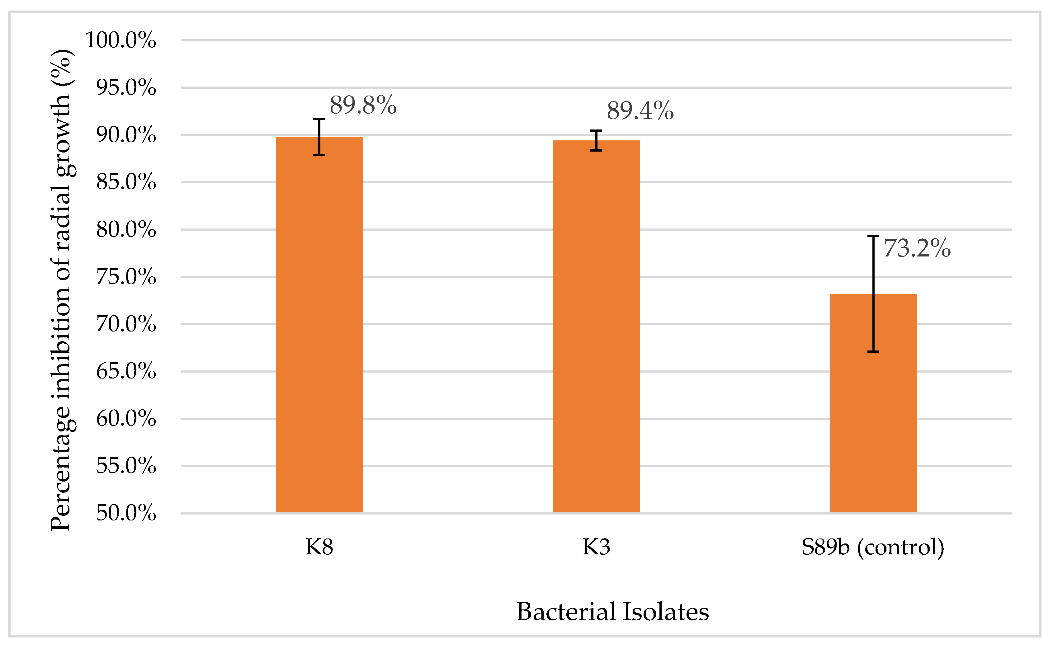
Basal stem rot (BSR), caused by Ganoderma boninense, is a major threat to oil palm plantations, leading to severe yield losses and significant economic impact. Early detection of BSR remains challenging because of the delayed onset of symptoms, while monoculture practices further exacerbate disease prevalence. G. boninense, a white rot fungus, invades root and vascular tissues of oil palm, impairing water and nutrient transport, ultimately causing plant decay and death. This study aimed to isolate, characterize, and identify bacterial isolates with both anti-Ganoderma and plant growth-promoting (PGP) abilities from a commercial biofertilizer, CRPO, specifically formulated for oil palm cultivations. Two isolates, K3 (Margalitia shackletonii) and K8 (Bacillus subtilis) displayed strong antagonistic activity against G. boninense, with percentage inhibition of radial growth (PIRG) values exceeding 89%. For PGP traits, K3 demonstrated broad potential, including phosphate and potassium solubilization, nitrogen fixation, 1-aminocyclopropane-1-carboxylate (ACC) deaminase activity, siderophore and high indole-3-acetic acid production. In contrast, K8 displayed positive results only for nitrogen fixation and ACC deaminase production. The dual functions of these isolates in suppressing G. boninense while enhancing plant growth confirms their presence as biocontrol agents. These advance sustainable BSR management strategies by reducing reliance on chemical inputs and strengthening oil palm resilience.

Keywords: 
;  ;  ;  ;  ;  ;  ;  

1. Introduction

Basal stem rot (BSR), caused by Ganoderma boninense, is a fungal disease of major economic significance in palm oil-producing countries, such as Malaysia and Indonesia. The progression of infection is difficult to detect because the symptoms of infection are only observable a few years after infection, depending on the age of the oil palm and the aggression level of G. boninense [1,2]. Symptoms of BSR disease include canopy mottling, unopened spears, and the presence of fruiting bodies on the lower part of the stem [3]. The prevalence of basal stem rot caused by G. boninense is most likely a result of monoculture practices in oil palm plantations. The plausible infection routes of G. boninense include latent inoculum from primary infection, root contact, and spores. Root contact may be the primary route of infection, and monoculture practices in oil palm plantations increase the risk of BSR infection via secondary inoculum through infected residual pieces [4,5].
G. boninense is also known as white rot fungus that infects living oil palms by secreting ligninolytic and cellulolytic enzymes capable of degrading plant cell walls, leading to the breakdown of basal stem tissues and ultimately causing the collapse of the palm. As the fungus invades and damages the internal vascular tissues of the palm, the transportation of water and nutrients to the upper parts of the tree is disrupted, eventually leading to the death of the palm [6,7]. G. boninense infection is thought to occur in two phases: an initial biotrophic phase and a later necrotrophic phase. During the biotrophic phase, hyphae grow to surround the surface of the root and secrete pathogen-associated molecular patterns (PAMPs) to trigger the plant immune response. After countering the oil palm’s defenses, G. boninense secretes enzymes that lyse the cell wall and cause cell death, eventually leading to the degradation of tissues at the base of the plant [8]. In general, losses caused by G. boninense can be direct or indirect; direct loss refers to the death of the oil palm, whereas indirect loss can be a decrease in fresh fruit bunch, fruit weight, and quality [3,9]. Apart from that, G. boninense is a soil-borne fungus with multiple persistent survival structures including resistant mycelium, basidiospores, chlamydospores, and pseudosclerotia that complicate the disease eradication [10]. Several management strategies such as soil mounding, surgical removal of infected tissues, and the use of chemical fungicides have been employed to control the disease. However, the effectiveness of these approaches is limited, often providing only temporary suppression, and their application is further constrained by cost considerations and environmental concerns [7,11]. Consequently, recent research has increasingly shifted towards environmentally sustainable strategies, particularly biological control approaches for managing BSR disease.
Today’s agricultural practices are driven by the need for more environmental-friendly and sustainable approaches, including the use of biofertilizers. Biofertilizers are formulations that incorporate living microorganisms, generally isolated from plant roots or rhizosphere soil, that establish symbiotic relationships with the host plant. These beneficial microbes enhance plant growth and productivity by facilitating nutrient acquisition and biodegradation, such as nitrogen fixation, phosphate and potassium solubilization, production of growth-promoting phytohormones, sequestration of vital nutrients and suppression of soil-borne pathogens, thereby reducing the reliance on chemical fertilizers and pesticides [12,13].
Generally, the composition of microbial inoculants in biofertilizers determines their overall effectiveness. In many cases, individual microbial strains within biofertilizer formulations exhibit multiple plant-beneficial functions. Beyond nitrogen fixation and phosphate solubilization, these microorganisms may also produce phytohormones, mobilize micronutrients such as zinc and iron, or secrete antimicrobial compounds that inhibit phytopathogens [14]. The presence of such multifunctional traits highlights the importance of microbial composition in determining biofertilizer efficacy and suggests that synergistic interactions among these traits can significantly enhance plant growth and stress tolerance [15]. This type of multifunctional microbe underscores the potential of biofertilizers as environmentally sustainable tools for improving crop productivity while concurrently providing biocontrol advantages.
Microorganisms in biofertilizers enhance plants growth by increasing the availability of macro- and micronutrients. Several members of genera Azotobacter and Rhizobium contribute to soil fertility through biological nitrogen fixation, converting atmospheric nitrogen (N₂), which plants cannot directly utilize, into ammonia that readily taken up by plants. In addition, certain plant-associated bacteria produce the enzyme ACC deaminase, an enzyme that mitigates stress-induced ethylene production. ACC deaminase producers, such as Burkholderia cepacia and Bacillus aryabhattai, degrade ACC, the immediate precursor of ethylene—thereby reducing ethylene levels that would otherwise inhibit root elongation and overall plant growth. Together, these microbes support plant nutrition and resilience under stress. Together, nitrogen-fixing microbes and ACC deaminase producers play complementary roles: one enhances nutrient availability, while the other mitigates stress responses, collectively promoting healthier plants. Potassium solubilization, phosphate solubilization, indole-3-acetic acid (IAA), ACC deaminase, and siderophore production are instances of nutrient acquisition processes [16].
Common groups of microorganisms employed in biofertilizers are plant growth-promoting bacteria (PGPB), free-living bacteria, and fungi [15]. Biofertilizers are formulated based on their ability to confer benefits to plants, survivability for field application, and antagonistic activity. It is also crucial to ensure microorganisms in the biofertilizer do not antagonize the host or other microorganisms present in the consortium [17]. Biofertilizers may possess multiple functions; fundamentally, they should supply nutrients to the plants and inhibit plant pathogens. Streptomyces spp. and Bacillus spp. have been reported to produce antibiotics and thrive in the rhizosphere. These traits provide significant advantages for biological agents used in biofertilizers. Most microorganisms that can combat plant pathogens engage in one or more of the following mechanisms: parasitism, competition for nutrients or space, antibiosis, inducing resistance, and promoting host growth [18].
In addition to plant growth-enhancing properties, microorganisms with protective properties have also been explored for inclusion in biofertilizers. PGPB can also protect against phytopathogenic infections. PGPB suppress pathogens through direct antagonism by producing antimicrobial compounds, such as antibiotics, lipopeptides, and hydrolytic enzymes that degrade fungal cell walls. Some PGPB also release volatile organic compounds (VOCs) that inhibit pathogen growth, offering an eco-friendly alternative to chemical pesticides [18,19].
Many recent studies have also focused on the VOCs released by soil bacteria. VOCs were first identified as signaling compounds among bacteria, or between the host and bacteria. Nevertheless, several studies have reported that aerobically and anaerobically generated VOCs influence the microbial community, inhibit fungal spore germination, strengthen plant host resistance, and promote growth [20]. An example of a VOC, nonan-2-one, can promote growth of rice plant whereas VOCs such as benzaldehyde, ketone, tetradecane, and 3-methylbutyric acid released by Bacillus spp. can antagonize a wide range of phytopathogen that causes diseases in various plants and post-harvest crops [21,22].
PGPB also acts through competition for nutrients and space, rapidly colonizing roots, and limiting pathogen access to essential resources such as iron. Many strains produce siderophores that chelate iron, depriving pathogens of crucial nutrients [23]. Additionally, biofilm formation by PGPB physically blocks the establishment of pathogens [18]. Beyond pathogen suppression, PGPB enhance plant growth and trigger induced systemic resistance (ISR). They produce phytohormones such as IAA for root development and exhibit ACC deaminase activity to reduce stress-related ethylene levels. Some strains activate plant defense pathways, priming crops for stronger resistance to pathogens [24].
This study aimed to isolate and characterize bacterial strains from CRPO biofertilizer, with a focus on strains exhibiting antagonistic activity against Ganoderma boninense and possessing PGP traits. Two isolates demonstrating the strongest anti-Ganoderma effects and PGP capabilities were selected for further analysis. These isolates were subsequently identified through 16S rRNA gene sequencing to evaluate their potential contribution to sustainable oil palm cultivation.

2. Materials and Methods

2.1. Isolation of Bacteria

Pellet form of CRPO biofertilizer manufactured by INO Nature Sdn. Bhd. was serially diluted using 0.85% (w/v) sodium chloride solution. Subsequently, 100 µL of each dilution (10× to 1,000,000×) was spread-plated onto eight different types of agar, namely, oatmeal agar (HiMedia, India), malt extract agar (20 g/L malt extract, 20 g/L glucose, 6 g/L peptone, and 15 g/L agar), soil extract agar (HiMedia, India), oil palm rhizosphere soil extract agar (500 mL of 50 mM NaOH-treated soil extract collected at an oil palm estate nearby, and 15 g agar was added per litre) for isolation of soil microorganisms, whereas streptomycin agar (10 g/L malt extract, 4 g/L yeast extract, 4 g/L glucose, 2 g/L calcium carbonate, 12 g/L agar supplemented with cycloheximide), yeast glycerol agar (5 g/L yeast extract, 50 mL/L glycerol and 15 g/L agar), actinomycetes isolation agar (BD Difco, USA), and starch casein agar (HiMedia, India) for isolation of actinomycetes. Isolated bacterial colonies were then maintained on Luria-Bertani (LB) agar.

2.2. Bacterial Antagonistic Activities Against G. boninense In-Vitro:

In this study, bacterial antagonistic activity against phytopathogenic fungi was evaluated using both dual culture and VOC assays on agar media, which allowed the underlying inhibitory mechanisms to be distinguished. Dual culture assay was performed independently in triplicate while bi-plate VOC assay was performed independently in quadruplicates, and the mean values of the results were calculated. For all antagonistic assays, the pathogenic fungus used was Ganoderma boninense PER71 provided Malaysian Palm Oil Board (MPOB), Malaysia.

2.2.1. Dual Culture Assay

A 5 mm plug from a 5-day-old G. boninense culture on potato dextrose agar (PDA) was transferred to the center of an LB plate. Bacterial cultures were adjusted to an OD600 of 0.2, using 0.05% NaCl solution and were then streaked in a straight line 3 cm away from the edge of the plug on both right and left sides. The percentage of inhibition was calculated after seven days of incubation at 28 °C using the formula described [10]. Burkholderia anthina strain S89b was used as the positive control for this assay. This bacterium was previously isolated from the soil of an oil palm plantation located at Teluk Intan, Perak, Malaysia. Our experimental results demonstrated that it exhibited strong antagonistic properties against G. boninense. The bacterium was identified through 16S rRNA sequencing and its genus and species were subsequently confirmed by whole genome sequencing (Loh et al., unpublished). Percentage inhibition of radial growth (PIRG) was determined as below:
PIRG   ( % )   =   [ A B A ] × 100 %
A: Distance in between the G. boninense plug and the edge of radial mycelial growth without bacterial inoculant (cm)
B: Distance in between the G. boninense plug and the edge of radial mycelial growth with bacterial inoculant (cm)

2.2.2. Volatile Organic Compound (VOC) Assay

To assess the suppression or inhibitory effect of bacterial VOC against G. boninense, the bi-plate method was used, with a slight modification [25]. A 5 mm plug from a 5-day-old G. boninense culture on PDA was introduced into one compartment of the bi-plate, and the other compartment was seeded with 50 µL of standardized overnight bacterial culture, which was adjusted to an OD600 of 0.2, using 0.05% NaCl solution. The percentage inhibition of mycelial growth (PIMG) was calculated using the formula described below after seven days of incubation at 28 °C [26]. Burkholderia anthina strain S89b was used as the positive control for this assay.
PIMG   ( % ) = [ X 0.5 Y 0.5 ( X 0.5 ) ] × 100 %
X: Colony diameter of fungus without bacterial inoculant (cm)
Y: Colony diameter of fungus after exposure to bacterial VOC (cm)

2.3. Plant Growth-Promoting Assay

Overnight cultures of each of the bacterial isolates were diluted accordingly and standardized to an OD600 of 0.2 using a spectrophotometer before being inoculated into the respective media for subsequent analyses. All these assays were performed independently in triplicates.

2.3.1. Phosphate Solubilization Assay

Phosphate solubilization assays were conducted with slight modifications [27]. Pikovskaya’s agar (0.5 g/L ammonium sulfate, 5 g/l calcium phosphate, 10 g/L glucose, 0.1 g/L magnesium sulfate heptahydrate, 0.2 g/L potassium chloride, 0.5 g/L yeast extract, 15 g/L agar, manganese sulfate heptahydrate and iron (II) sulfate heptahydrate in trace amount) was adjusted to pH 7.0 before autoclaving. Then, 10 μL of the OD600 adjusted culture of bacterial isolates was inoculated onto agar using the drop-plate method. The inoculated agar was incubated for seven days at 28 °C. The presence of clear zone around the bacterial colony was observed and recorded after seven days of incubation [28].

2.3.2. Potassium Solubilization Assay

Potassium solubilization assays were carried out on Aleksandrov agar using the drop-plate method with minor modifications [29]. The medium was prepared by dissolving 9.605 g/L of Aleksandrov broth powder (HiMedia, India) and 15 g/L of agar (BD Difco, USA) in distilled water, followed by sterilization at 121 °C for 15 min under 15 psi. After cooling, 10 μL of OD600 adjusted bacterial suspension as mentioned in Section 2.3 was spotted onto the Aleksandrov agar surface and incubated at 28 °C for seven days. The formation of clear halo zones surrounding the bacterial colonies was regarded as evidence of potassium solubilization.

2.3.3. Nitrogen Fixation Assay

This assay was performed to assess the ability of the bacterial isolates to fix atmospheric nitrogen. A volume of 10 μL each of bacterial culture was adjusted to OD600 of 0.2 and was subsequently streaked onto Jensen nitrogen-free agar (HiMedia, India), and growth of bacterial colonies was observed and recorded after seven days [30]. After incubation, colony growth on Jensen nitrogen-free agar suggests a positive outcome for nitrogen-fixing ability.

2.3.4. Siderophore Detection Assay

Overlaid Chrome Azurol S (O-CAS) was performed according to the method described [23]. About 5 μL each of OD-adjusted bacterial culture was spotted onto LB agar. After the iron-containing blue CAS agar was overlayed, the area surrounding the bacterial colony turned yellow or purple in the presence of siderophores. The color change was recorded within one hour after the CAS agar overlay was applied. Siderophores are required to sequester iron. A yellow or purple halo surrounding the bacterial colonies would be observed if the bacterial isolates were positive for siderophore production. A shift from blue to purple indicates the production of catechol-type siderophores, whereas a blue-to-orange transition signifies hydroxamate-type siderophores [23].

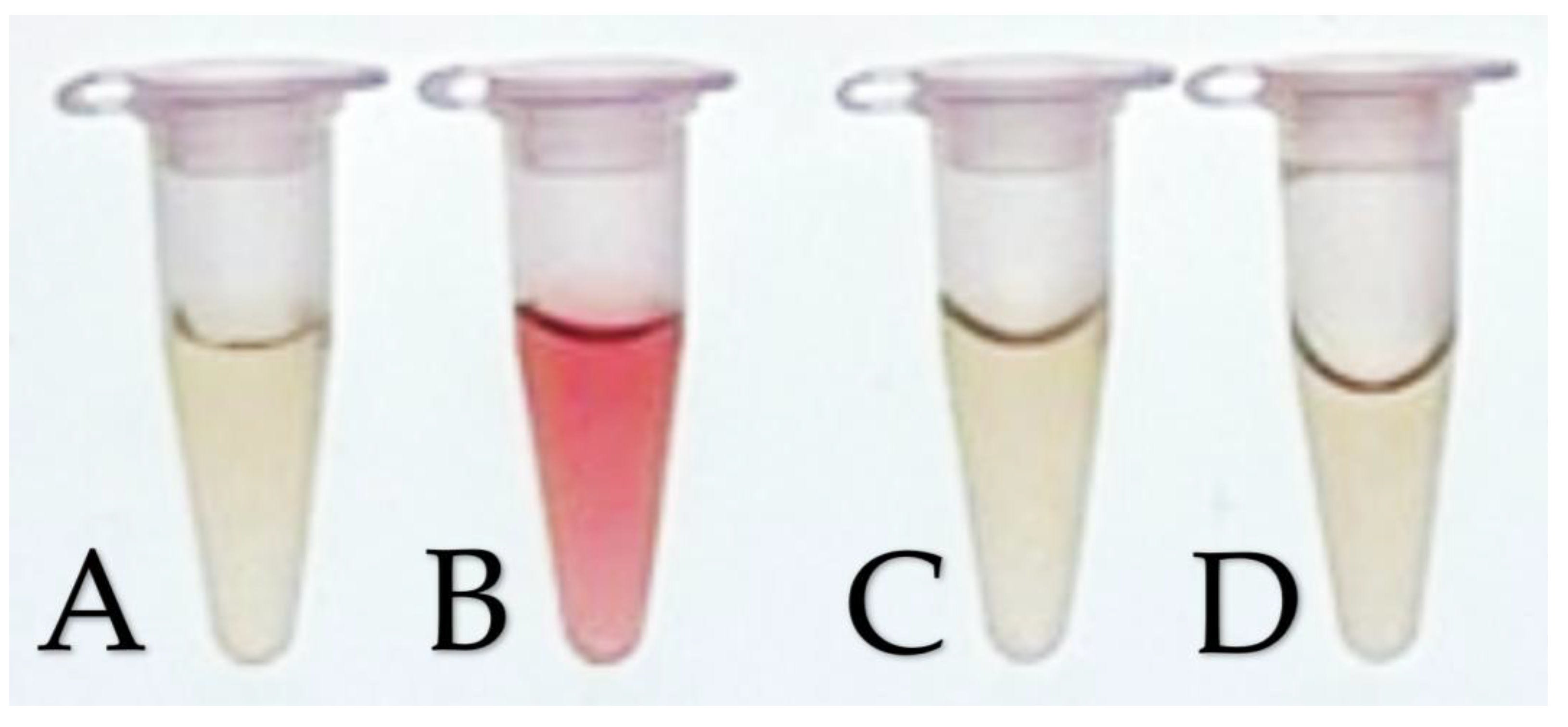
2.3.5. Quantitative Analysis of IAA Production

Bacterial isolates were screened for IAA production based on the following protocols with slight modifications [20,31]. Approximately 100 μl each of OD600 adjusted overnight bacterial culture was added to 3 mL of Lennox broth (LB) supplemented with L-tryptophan (1 mg/mL). The culture was incubated at 28 °C for 48 h with agitation at 150 rpm. The culture was centrifuged at 4 °C for 15 min at 7,500 rpm. The supernatant was collected in a fresh tube and Salkowski’s reagent was added to the supernatant at a 2:1 ratio. The mixture was incubated at room temperature in the dark for 30 min before readings were recorded at OD530.

2.3.6. Quantitative Analysis of ACC Deaminase Production

Bacterial isolates were also screened for their ability to produce ACC deaminase. The following protocol was used with minor modifications [32]. Approximately 100 μL each of OD600 adjusted overnight culture was added to 3 mL of Dworkin-Foster (DF) medium supplemented with 3 mM ACC. The culture was incubated at 28 °C for 48 h, with agitation at 200 rpm. The supernatant was collected and diluted 10x after centrifugation at 25 °C for 5 min at 8,000 rpm. A volume of 2 mL ninhydrin reagent was added to each milliliter of supernatant, followed by heating at 99 °C for 30 min before measuring at OD570.

2.3.7. Identification of Bacterial Isolates by 16S rDNA Sequencing

Bacterial genomic DNA, which served as a template for 16S rRNA PCR amplification, was extracted using a GF-1 Bacterial DNA Extraction Kit (Vivantis, Malaysia). The universal primers used for 16S rRNA gene amplification were 27F (5’-AGAGTTTGATCCTGGTCAG-3’) and 1492R (5’-GTTACCTTGTTACGACTT-3’). A standard 3-step PCR was used: initial denaturation at 95 °C for 3 min, followed by 30 cycles of denaturation at 95 °C for 30 s, annealing at 52 °C for 30 s, and extension at 72 °C for 90 s, and a final extension step at 72 °C for 5 min The PCR products were purified using USB® ExoSAP-IT® PCR Product Cleanup (Affymetrix, US). The purified PCR products were outsourced for DNA sequencing (Apical Scientific, Malaysia). BLASTn was used to search for nucleotide sequences homologous to the 16S rRNA-encoding gene.

3. Results

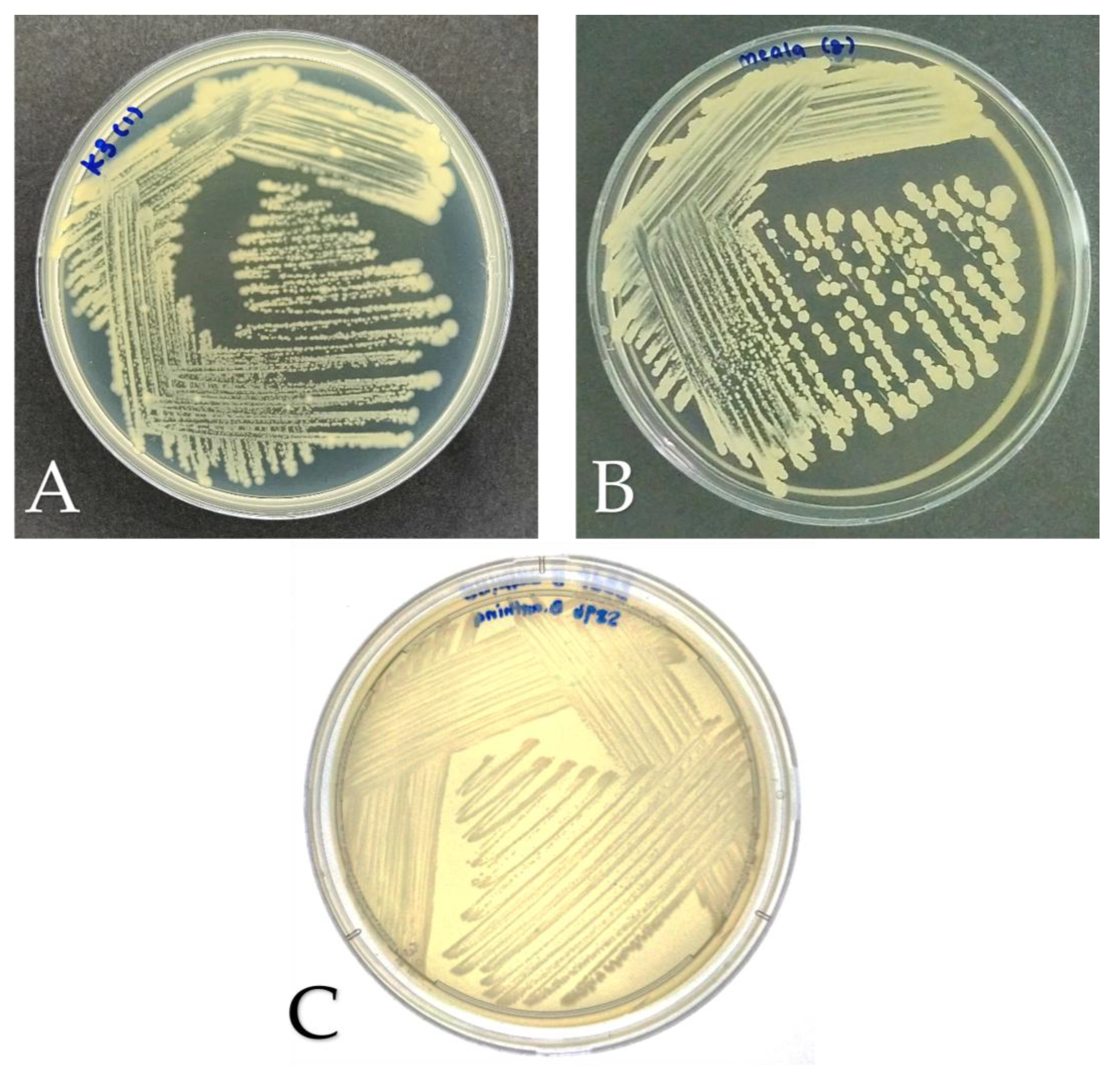
3.1. Bacterial Isolation and Morphological Study

A total of 84 morphologically distinct bacterial colonies were isolated from the CRPO biofertilizer, using eight types of media (Section 2.1). Colony morphology was observed in terms of shape, surface texture, color, elevation, and margin, along with the Gram staining results. About 90% of bacterial colonies were found to be Gram-positive; one bacterial colony among the 84 was observed to be a Gram-positive coccus, and the remaining 83 were either bacilli or coccobacilli.
In this study, Burkholderia anthina S89b was used as the positive control for antagonistic assays against G. boninense, whereas soil bacterium P. aeruginosa isolated from soil and maintained in our laboratory collection served as the control for plant growth–promoting assays [33,34]. Both strains were sourced from our laboratory culture collection. Strain S89b was chosen due to its consistently strong anti-Ganoderma activity, with PIRG and PIMG values exceeding 72% in both previous and current studies. In contrast, P. aeruginosa is a well-established model strain exhibiting multiple plant growth–promoting traits, including nitrogen fixation, phosphate and potassium solubilization, siderophore and ACC deaminase productions [35,36].
Based on the dual culture assays conducted on all 84 bacterial isolates, 23 bacteria isolates displayed PIRG more than 75.0%. Two bacteria isolates namely K3 and K8 were selected for further study due to their strong anti-Ganoderma activity. Figure 1 shows the colony appearance of isolates K3, K8 and the positive control S89b on Luria–Bertani (LB) agar after incubation at 28 °C for 48 h. Distinct variations in pigmentation, margins, and surface texture were observed. Their colony morphology is summarized in Table 1.
Table 1 shows that isolates K3 and K8 were Gram-positive bacilli, whereas the control strain S89b was Gram-negative bacilli. K3 formed dry, rough, white colonies with raised elevations and entire margins. K8 also produced dry, rough, white colonies but differed in having a flat elevation and undulated margins. The control strain S89b exhibited wet, smooth, yellow colonies with raised elevations and smooth entire margins. The phenotypic diversity among the isolates, with K3 and K8 belonging to the genera Margalitia and Bacillus, respectively, and S89b representing the distinct genus Burkholderia.

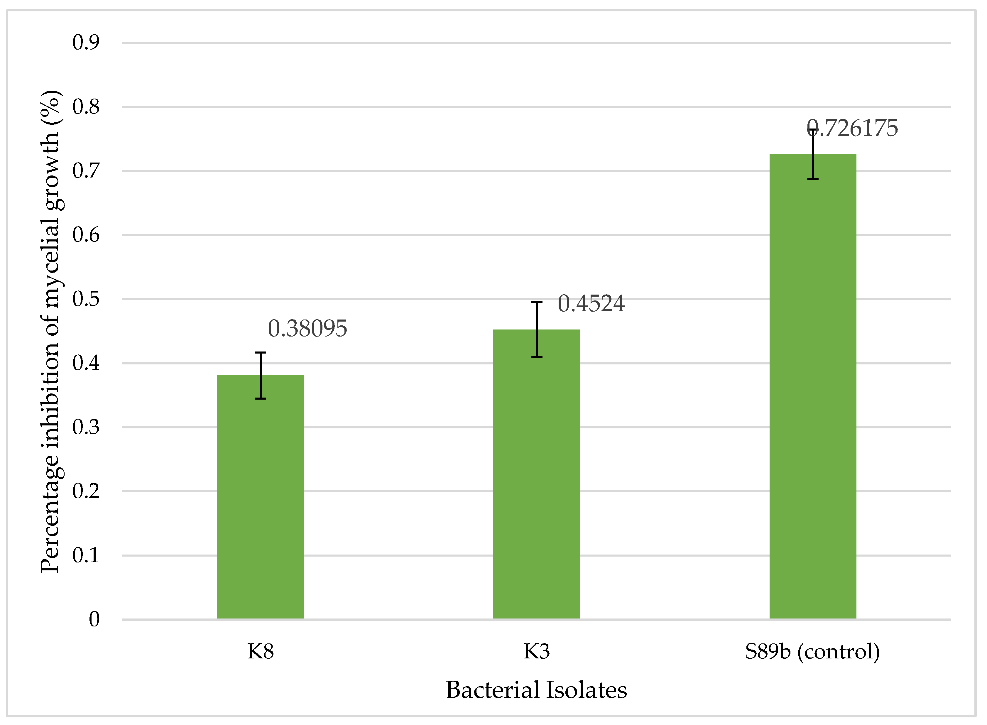
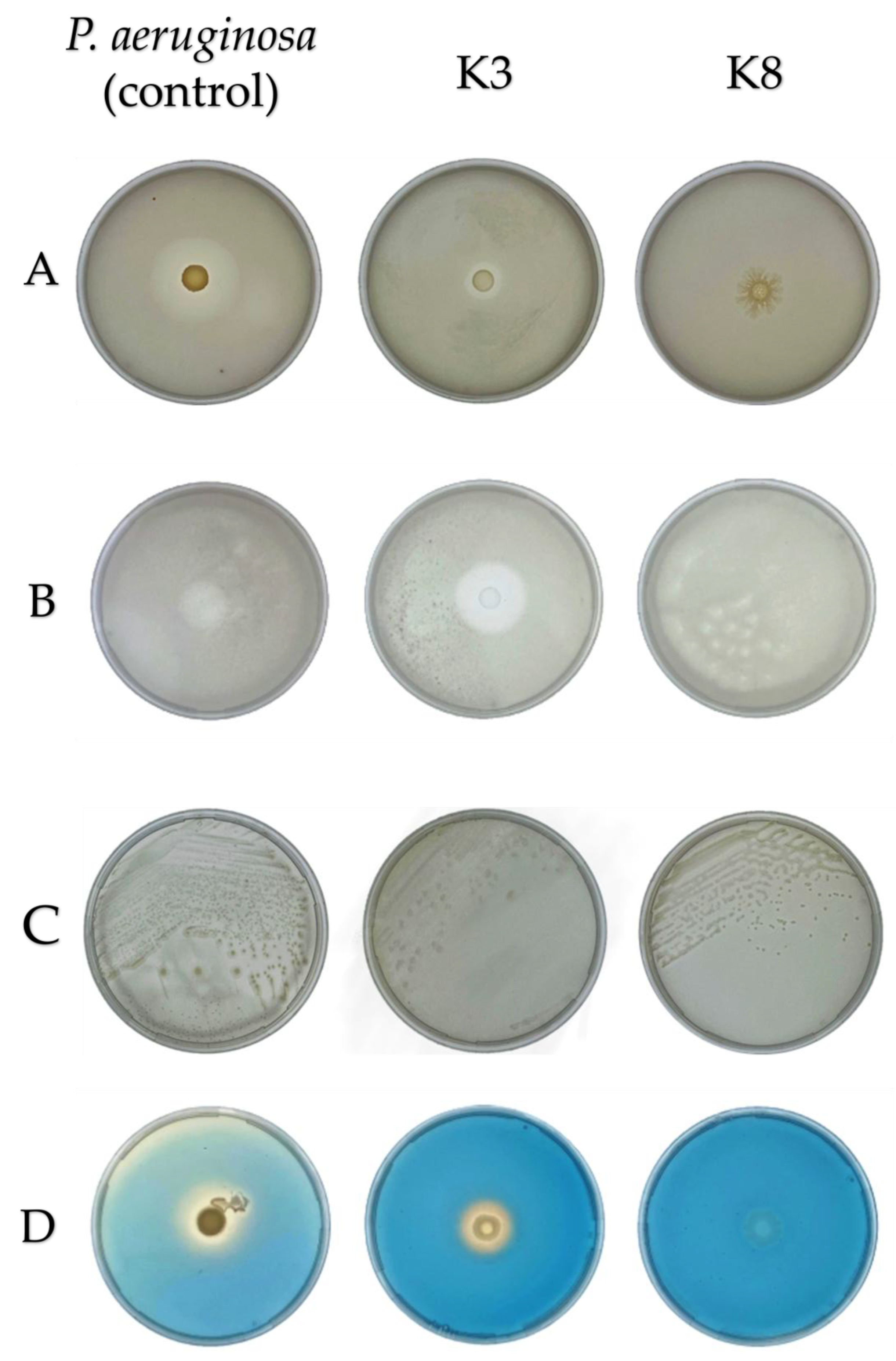
3.2. Bacterial Antagonistic Activities Against G. boninense

In this study, dual culture and VOC assays were used to evaluate bacterial antagonism against the phytopathogenic G. boninense. The dual culture assay measures the inhibition arising from direct contact interactions between the bacterium and the fungus G. boninense, including competition for nutrients and space, as well as the yield of diffusible antimicrobial metabolites, such as antibiotics, lipopeptides, and hydrolytic enzymes. In contrast, the VOC assay performed by incubating the bacterium and fungus in a center-compartmentalized bi-plate, specifically assesses antifungal effects mediated by airborne volatile metabolites, excluding any contact-dependent or diffusible compound effects.

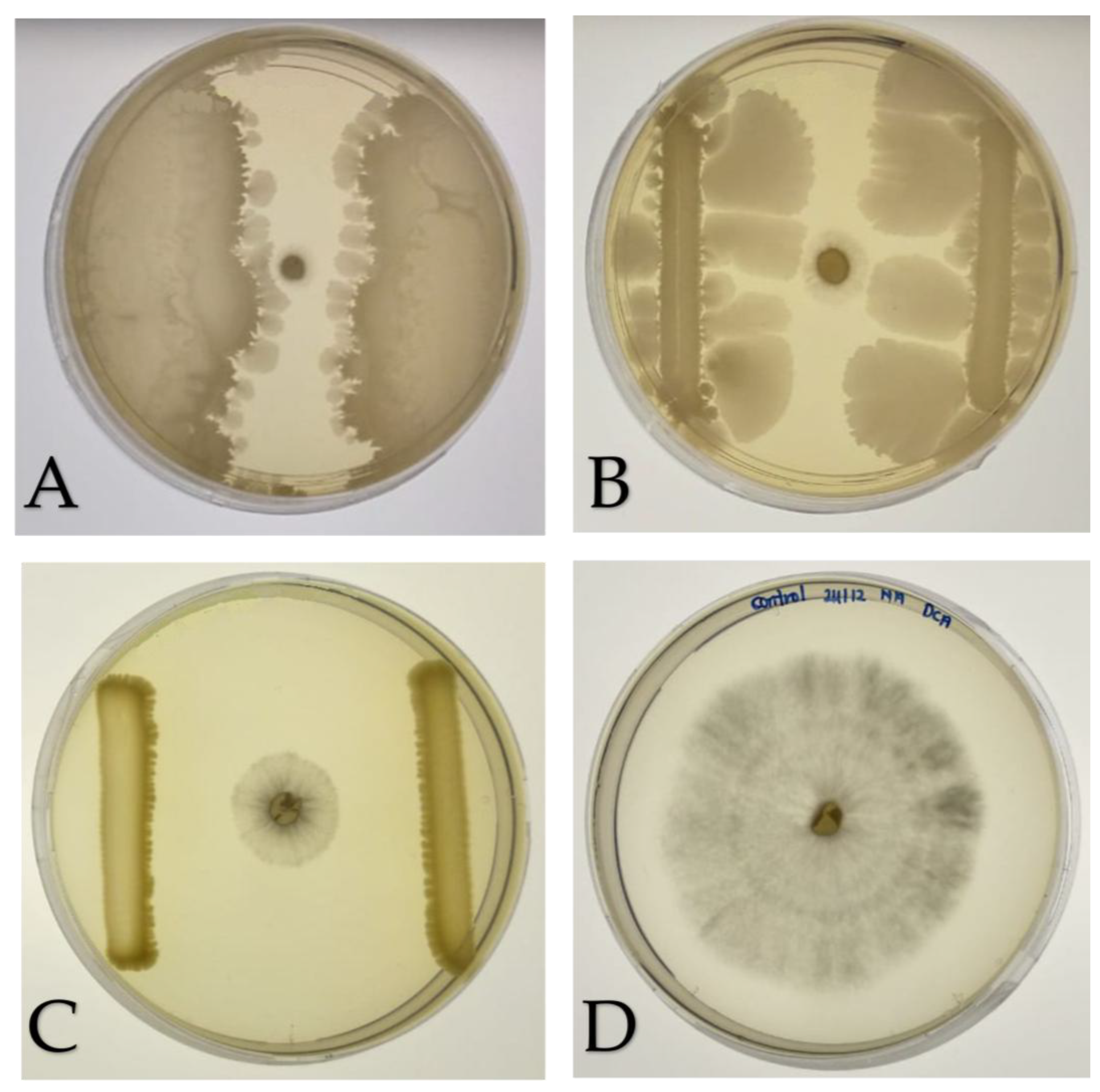
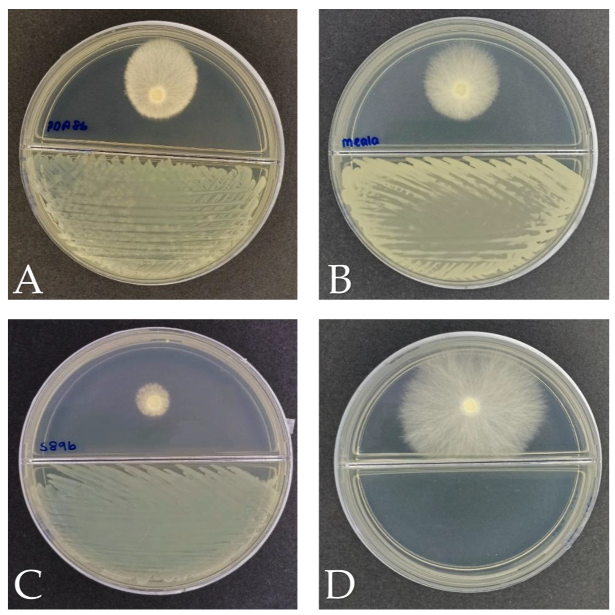
3.2.1. Dual Culture Assay

In the dual culture antagonism assay against G. boninense, 43 out of 84 bacterial colonies tested were able to suppress the growth of fungus. This assay was performed to determine the combined effects of volatile organic compounds and diffusible compounds hindering the growth of G. boninense. Among all isolates, the highest antagonistic activity was exhibited by K3 and K8, with PIRG values of 89.4% and 89.8%, respectively (Figure 2). In contrast, positive control S89b showed a slightly lower inhibition level of 73.2%. These findings demonstrate that K3 and K8 outperform the positive control in suppressing the radial growth of G. boninense, confirming their potential as highly effective antagonistic bacteria. Therefore, these two isolates were chosen for subsequent studies in VOC assay and plant growth-promoting activities. Figure 3 illustrates the quantitative results of the dual culture assay as a histogram.

3.2.2. Volatile Organic Compound (VOC) Assay

For the VOC assay, bi-plate assay was used to study the effect of bacterial VOC on the growth of G. boninense. The positive control, B. anthina, inhibited G. boninense growth, with PIMG value of 72.6%, indicating its robust volatile-based antagonism. Bacterial isolates K3 and K8 produce airborne metabolites capable of inhibiting the mycelial growth of G. boninense, although to different extents. In comparison, K3 and K8 demonstrated lower PIMG values of 45.2% and 38.1%, respectively. These results reveal that while K3 and K8 secrete moderate inhibitory VOCs, their activity is considerably moderate, and lower than that of the positive control under the conditions tested (Figure 4). Figure 5 presents a histogram summarizing the quantitative results of the VOC assay.

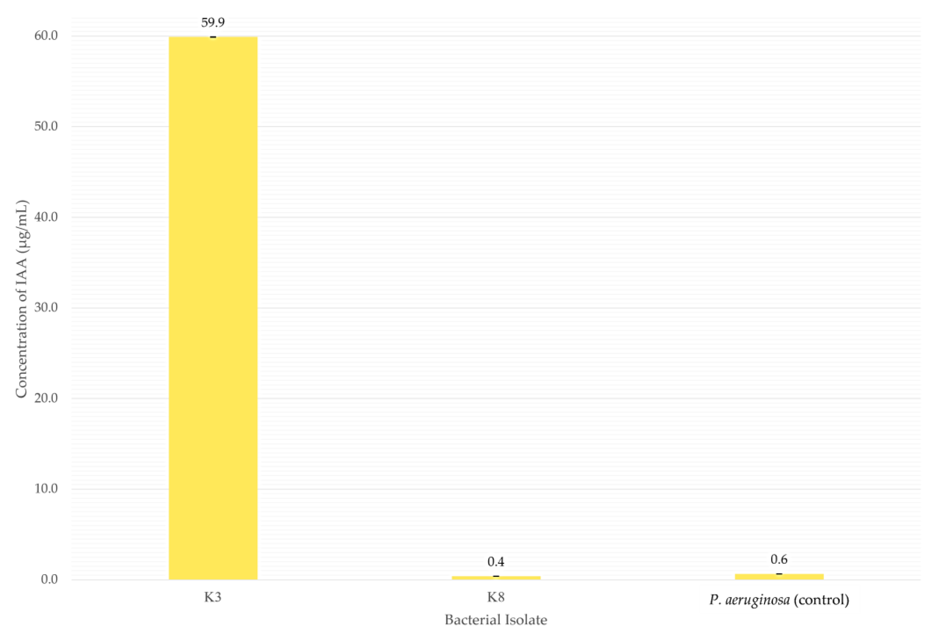
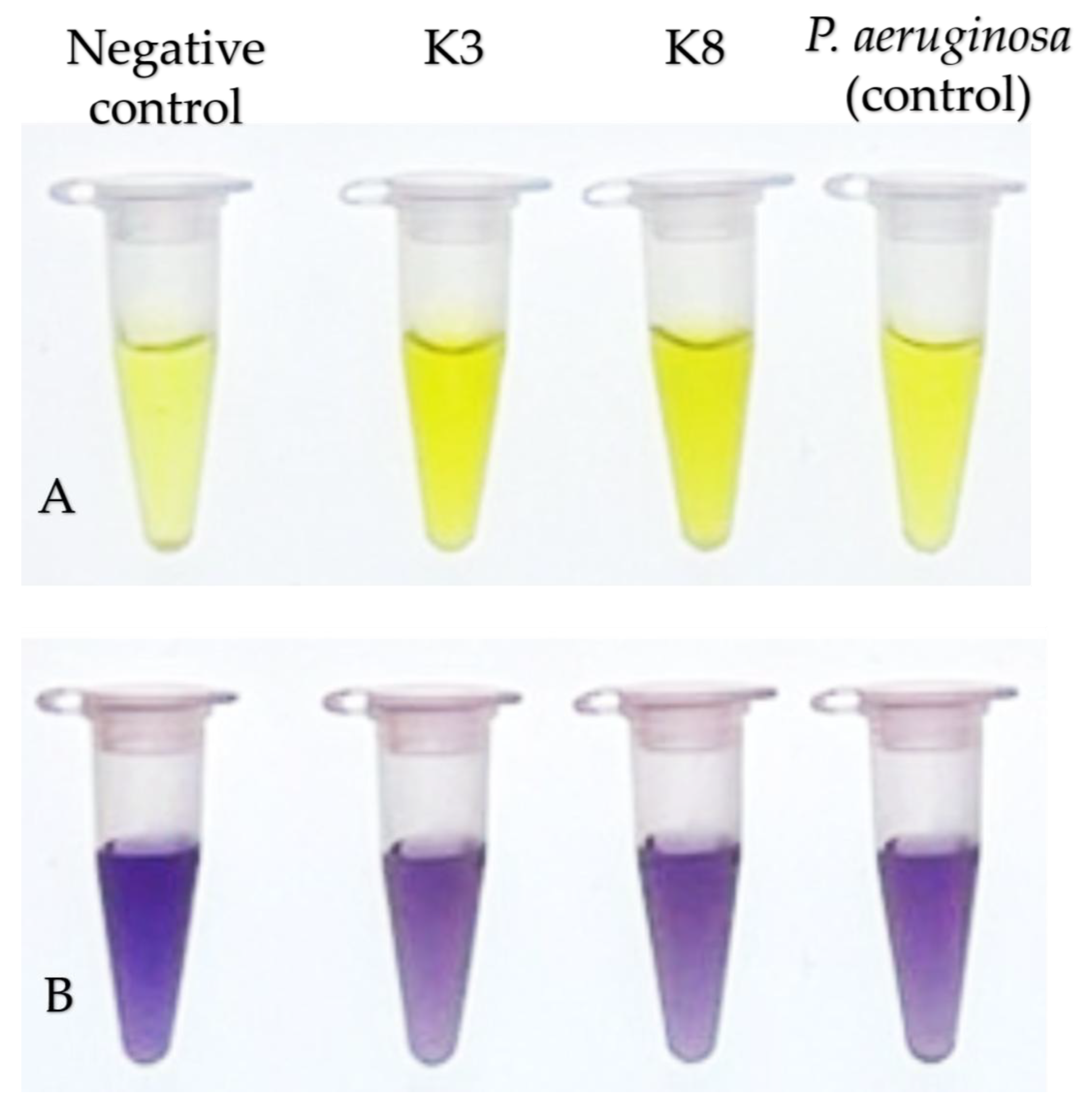
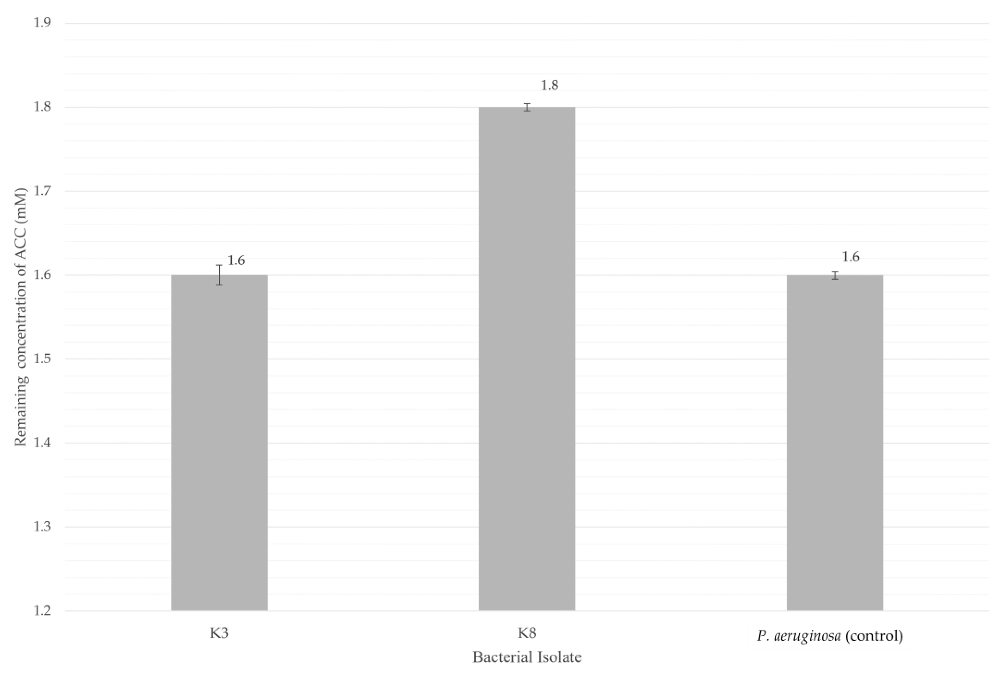
3.3. PGP Traits of Bacterial Isolates

In the PGP assays, both isolates K3 (M. shackletonii) and K8 (B. subtilis) tested positive for nitrogen fixation and ACC deaminase production. However, isolate K8 was negative for siderophore production, phosphate and potassium solubilization, and IAA production, indicating its ability to fix atmospheric nitrogen and synthesize ACC deaminase. On the other hand, isolate K3 was able to solubilize phosphate and potassium, produce siderophores, fix nitrogen, produce ACC deaminase and high amounts of IAA at approximately 60 µg/mL. In the plant growth-promoting assays, both bacterial isolates displayed positive results in the nitrogen fixation and ACC deaminase assays (Figure 6).
For quantitative analysis of IAA production, isolate K3 (M. shackletonii) produced the highest IAA concentration (59.9 µg/mL), whereas isolate K8 (B. subtilis) and the control P. aeruginosa yielded only 0.4 µg/mL and 0.6 µg/mL, respectively (Figure 7). In this assay, IAA production levels of 1.0–10.0 µg/mL are classified as weakly positive, 10.1–20.0 µg/mL as moderately positive, and values above 20.0 µg/mL as strongly positive. Figure 8 shows a histogram illustrating the quantitative results of IAA production.
For the ACC deaminase assay, isolate K3 showed the lowest remaining ACC concentration in the supernatant after 48 h of incubation (1.6 mM), followed by isolate K8 at 1.8 mM (Figure 9). In this assay, isolates with remaining ACC concentration between 2.0 mM to 2.7 mM is weakly positive, readings between 1.2 mM to 1.9 mM is moderately positive and lower than 1.2 mM as strongly positive for ACC deaminase production. DF broth was supplemented with 3.0 mM ACC as the sole nitrogen source; thus, a decrease in the ACC concentration indicated bacterial utilization of ACC. Therefore, a lower residual ACC indicates higher ACC deaminase activity. Figure 10 shows a histogram illustrating the quantitative results of ACC deaminase production.
Overall, the two bacterial isolates K3 and K8 recovered from the CRPO biofertilizer exhibited distinct PGP profiles when assessed across six functional assays (Table 2). Isolate K3 demonstrated broad PGP potential, showing positive activity for phosphate and potassium solubilization, nitrogen fixation, siderophore production, ACC deaminase activity, and strong IAA production. In contrast, isolate K8 displayed a narrower functional profile, being positive only for nitrogen fixation and ACC deaminase, while showing negative results for phosphate and potassium solubilization, siderophore production, and IAA synthesis. In contrast, the control strain P. aeruginosa exhibited positive responses for all the five traits tested except IAA production.

3.4. 16S rRNA Gene Sequencing and Taxonomic Identification

The identities of isolates K3 and K8 were determined through 16S rRNA gene sequencing followed by BLASTn analysis against the NCBI GenBank database (Table 3). Isolate K3 produced a 1473 bp sequence that matched M. shackletonii (accession number OQ552639.1) with 99.5% identity and 100% query coverage (E-value: 0.0). Meanwhile, isolate K8 yielded a 1399 bp sequence showing 99.7% identity and 100% query coverage to B. subtilis (accession number OP798061.1) with E-value of 0.0. These high identity values, together with full-length sequence coverage and statistically significant E-values, together with the supporting morphological characteristics (Table 1), confirm the species-level taxonomic assignment of both isolates.

4. Discussion

4.1. Isolation of Bacteria from CRPO Biofertilizer

CRPO is a semi-organic biofertilizer developed and produced by INO-Nature Sdn Bhd, has been reported to reduce the incidence of BSR in various local oil palm plantations, a devastating disease caused by Ganoderma spp. [35]. The formulation of CRPO combines beneficial microorganisms and organic nutrients, which are thought to enhance plant growth and simultaneously suppress pathogenic fungi, offering an environmentally friendly approach to disease management in oil palm cultivations. Understanding the microbial composition of CRPO is essential, as the beneficial effects of biofertilizers are largely attributed to the diverse bacteria they contain.
In this study, multiple culture media were used to recover bacteria from the CRPO biofertilizer, as soil-associated microbes differ widely in their nutritional requirements and physiological traits. Reliance on a single medium would bias the recovery toward fast-growing or readily culturable species while overlooking slow-growing, nutrient-specialized, oligotrophic, or stress-tolerant bacteria. By employing eight media with varying carbon sources, nutrient levels, and selective properties (Section 2.1), a broader and more representative fraction of the bacterial community of the biofertilizer could be isolated.
For example, the application of commercial soil extract agar that is nutrient-limited, and the manually prepared oil palm rhizosphere soil extract agar, simulate the native soil environment of oil palm plantations and therefore favor oligotrophic bacteria that may not grow on richer media, such as malt extract agar or yeast glycerol agar. Conversely, starch casein agar, streptomycin agar, and malt extract agar selectively encourage the growth of actinomycetes and heterotrophic bacteria.
Overall, the use of diverse media in the current study enhanced the likelihood of isolating morphologically and functionally distinct bacterial strains, including those with antagonistic capabilities against G. boninense or potential plant growth–promoting, as well as rare species that would not appear on standard media alone. In this study, approximately 90% of the recovered bacterial isolates were Gram-positive. This predominance is expected, as endospore-forming bacteria, particularly members of the Firmicutes such as genera Bacillus, Margalitia, and Lysinibacillus are well-adapted to survive desiccation, abiotic stress and nutrient limitation [36,37,38]. Because most commercial biofertilizers, including the CRPO formulation, are produced and stored in dried pellet forms, non-sporulating bacteria are far less likely to remain viable throughout processing, drying and storage [17]. Consequently, the isolation of primarily Gram-positive, endospore-forming taxa reflects both their intrinsic resilience and the selective pressures imposed by biofertilizer manufacturing.
4.2. 16S rRNA Gene Sequencing and Taxonomic Identification
To date, no curative or highly effective treatment is available for BSR disease in oil palms once being infected by G. boninense, the most aggressive species among the members of genus Ganoderma. Therefore, biofertilizers have recently emerged as environmentally friendly alternatives to conventional chemical pesticides. In this study, bacteria were isolated and characterized from the CRPO biofertilizer, with emphasis on identifying those capable of inhibiting G. boninense and subsequently, further determine their potential PGP traits. Among the isolates recovered, two strains K3 and K8 exhibiting the strongest anti-Ganoderma activity from dual culture assay were selected for detailed evaluation. Given the pressing demand for biocontrol agents to mitigate BSR disease besides improving the yield of palm oil productivity, identifying these high-performing isolates is critical for understanding their functional roles within the formulation.
Sequencing the 16S rRNA genes provided reliable species-level identification on bacteria, enabling a clearer understanding of their antagonistic and PGP capabilities and their potential contributions to the overall efficacy of CRPO. As summarized in Table 3, isolate K3 was identified as M. shackletonii with high sequence identity of 99.5%. It is a spore-forming, Gram-positive bacterium known for its ability to inhabit extreme or nutrient-limited environments [40,41]. Its identification aligns with the observed physiological traits in this study, including phosphate and potassium solubilization, nitrogen fixation, ACC deaminase activity, siderophore and substantial IAA production—traits also noted in our previous work on another foliar fertilizer [42].
M. shackletonii, formerly classified as Bacillus shackletonii, was originally described in 2004 from a volcanic soil sample collected on Candlemas Island in the sub-Antarctic region [40], but subsequent phylogenomic analyses led to major taxonomic revisions within the Bacillus genus. As part of this effort, Gupta et al. published a valid proposal in 2020 to reclassify several species into new genera, resulting in the reassignment of Bacillus shackletonii to M. shackletonii [41]. This reclassification reflects a broader refinement of Bacillus systematics to better align species with their evolutionary relationships. In this study, the ability of endospore-forming M. shackletonii to withstand diverse environmental conditions, and to remain viable in dried pellet form after biofertilizer production, likely contribute to its strong performance in both anti-Ganoderma and plant growth-promoting assays. These traits collectively support their suitability as a robust and reliable inoculant for biofertilizer formulations.
Isolate K8 was identified as B. subtilis, a species widely recognized for its agricultural relevance and broad-spectrum plant growth–promoting abilities [39,43]. The high sequence identity (99.7%) obtained in this study supports its accurate classification. The identification of B. subtilis, is consistent with its frequent recovery from soil and its well-documented role in promoting plant health and suppressing phytopathogens. Members of the Bacillus genus are known for their capacity to produce extracellular enzymes, phytohormones, and antimicrobial compounds, as well as to induce systemic resistance in plants. For example, Bacillus amyloliquefaciens is widely used in agriculture because it improves plant nutrient acquisition by solubilizing nitrogen, phosphate, potassium, and iron. This bacterium also promotes plant growth directly through the production of VOCs and phytohormones [39].
Overall, the 16S rRNA–based identification confirms that the bacterial isolates recovered from CRPO biofertilizer include taxa with established functional relevance in agriculture. The presence of K3 and K8 provides further support for the biofertilizer’s potential to deliver plant growth-promoting and biocontrol benefits to combat detrimental BSR disease in palm oil production industry.

4.3. Bacterial Antagonistic Activities Against G. boninense

4.3.1. Dual Culture Assay

The antagonistic activity of the bacterial isolates against the phytopathogenic fungus was evaluated using both dual culture and VOC assays to differentiate contact-dependent and airborne or gaseous compounds inhibitory mechanisms. Together, these assays distinguish between contact- or diffusible-based inhibition and volatile-mediated suppression, providing complementary insights into the mechanisms underlying bacterial antifungal activity and supports a more accurate assessment of their biocontrol potential.
The strong antagonistic activity demonstrated by the bacterial isolates K3 and K8 against G. boninense with PIRG values of 89.8% and 89.4%, respectively. These inhibition levels not only demonstrate strong antifungal activities but also exceed the performance of the positive control S89b, which achieved a PIRG of 73.2% (Figure 2). The high PIRG values suggest that both isolates can produce effective antifungal metabolites and/or engaging in competitive interactions that inhibit the radial expansion of G. boninense. Dual culture assay, typically reflects direct antagonism through multiple inhibitory mechanisms, including competition for nutrients and physical space (Figure 2, panels A and B), secretion of volatile and diffusible antimicrobial compounds (Figure 2, panel C) such as antibiotics, lipopeptides (e.g., iturin, fengycin), and siderophores, and the production of hydrolytic enzymes (e.g., chitinases and glucanases) capable of degrading fungal cell walls [39,44].
Physical interference with fungal hyphal expansion may also contribute to the observed suppression, a characteristic trait of the genus Bacillus. Similar findings were reported by Teixeira et al. (2021), where Bacillus velezensis strain CMRP 4490 inhibited the growth of Sclerotinia sclerotiorum and Botrytis cinerea by up to 60% and enhanced soybean germination rates [45]. Notably, B. subtilis as a dominant bacterium in water hyacinth biofertilizer, not only suppresses the growth of phytopathogen but it also enhances plant growth by fixing nitrogen, solubilizing phosphate and potassium, and secreting plant growth hormone IAA [46].
The superior performance of K3 and K8 relative to S89b implies that these isolates may produce a broader or more potent spectrum of inhibitory compounds than the known positive control strain. The strong fungal suppression by isolates K3 and K8, suggests enhanced antagonism through both physical interference by competing for nutrients and physical space, as well as the production of more potent inhibitory metabolites, in comparison to S89b. This antagonistic effect is further supported by the extensive and rapid expansion of the vertically streaked colonies of K3 and K8 observed in the dual culture assay. Such pronounced disruption of phytopathogen growth highlights their potential as effective biocontrol agents against persistently found soilborne pathogen G. boninense, crucial for early intervention in BSR of oil palm.

4.3.2. Volatile Organic Compound (VOC) Assay

The role of bacterial VOC in fungal inhibition was assessed through the VOC assay. Both isolates, K3 and K8 demonstrated inhibitory activity, confirming that volatile metabolites contribute to the suppression of G. boninense. Their PIMG values of 45.2% and 38.1%, respectively, placing both within the category of moderate inhibitors of G. boninense. Notably, K3 exhibited stronger inhibitory potential compared to K8, positioning it as the more effective VOC producer, although both isolates remain classified as moderate inhibitors.
Relatively to isolates K3 and K8, the positive control S89b showed a notably higher PIMG of 72.6%, suggesting that its VOC production is a major contributor to its strong antifungal activity and aligns closely with its strong direct antagonism observed in the dual culture assay (PIRG of 73.2%). Because VOC assay excludes physical contact and diffusible metabolites, the observed inhibition can be attributed solely to low molecular weight gaseous compounds. Such volatiles may include acetoin, 2,3-butanediol, dimethyl disulfide, hydrogen cyanide, and various alcohols and ketones, which are known to interfere with fungal growth and physiology [21]. For instance, VOCs produced by B. subtilis CF-3 have been reported to inhibit the growth of Colletotrichum gloeosporioides, the pathogenic fungus responsible for anthracnose in litchi plants, which manifests as dark, sunken lesions on leaves, stems, and fruits [47]. Beyond suppressing phytopathogens, another study highlights Bacillus pseudomycoides, which generates diverse VOCs such as ethyl ether, benzene, and 1-dodecanol, thereby enhancing drought tolerance and promoting growth in wheat [48].
Overall, the moderate VOC-mediated inhibition observed for K3 and K8 indicates that their antagonistic effects are driven mainly by diffusible metabolites or direct interaction mechanisms, as reflected by their stronger performance in the dual culture assay. Even so, their detectable VOC activity demonstrates that both isolates employ multiple modes of action, a trait advantageous in rhizosphere environments where physical contact with pathogens is inconsistent or spatially variable. Beyond direct inhibition, their ability to colonize the rhizosphere soil efficiently may also promote competitive exclusion, limiting G. boninense establishment by reducing available nutrients and space. Thus, incorporating these isolates into CRPO biofertilizer formulations designed for oil palm may help foster a protective microbial community that contributes to BSR management. Collectively, these findings emphasize the value of selecting bacterial strains with diverse and complementary functional traits to enhance the effectiveness and resilience of biofertilizer-based disease suppression strategies.

4.4. Functional Characterization of K3 (M. shackletonii) and K8 (B. subtilis) PGP Traits

To evaluate the growth-promoting potential of the bacterial isolates, six functional assays were conducted: phosphate solubilization, potassium solubilization, nitrogen fixation, siderophore production, ACC deaminase activity, and IAA production. All six assays yielded positive results for isolate K3 , indicating a broad spectrum of PGP traits and a comprehensive suite of functions commonly associated with enhanced plant growth, performance, and resilience. The ability of M. shackletonii to solubilize both phosphate and potassium suggests a strong capacity to mobilize essential nutrients in the rhizosphere, thereby increasing their bioavailability to oil palm roots. Positive nitrogen fixation further enhances its contribution to nutrient enrichment, while siderophore production may facilitate improved iron acquisition under limiting conditions and indirectly inhibit competing soil pathogens.
Notably, M. shackletonii expressed strong IAA production, a key hormone involved in root initiation and elongation. This trait is consistent with improved seedling vigor and greater root surface area, which may in turn enhance nutrient uptake efficiency. The presence of high ACC deaminase, an enzyme that lowers plant ethylene levels under stress, further supports a role for K3 in stress alleviation and improved root growth under adverse soil conditions.
Previous studies have reported that M. shackletonii possesses PGP abilities such as siderophore production, IAA synthesis, ACC deaminase activity, and phosphate solubilization [40], which are consistent with our findings for K3. Interestingly, Bacillus pinisoli, isolated from decaying pine trees, has been identified as a closely related species to M. shackletonii (96.7% similarity); however, its antagonistic properties and plant-beneficial traits remain largely unexplored [49]. Thus, while phylogenetic relatedness suggests a close evolutionary link, the functional characterization results highlight M. shackletonii as a more promising bacterium for biocontrol and plant growth promotion in agricultural systems.
Isolate K8 although more limited in its functional profile, showed positive results for nitrogen fixation and ACC deaminase activity. These traits may contribute to moderate growth promotion and stress mitigation, even in the absence of phosphate or potassium solubilization and IAA production. Consequently, B. subtilis may serve a complementary role within the bacterial consortia of CRPO, enhancing nitrogen availability and alleviating ethylene-mediated stress responses.
The functional broadness displayed by M. shackletonii, combined with its taxonomic identification as, suggests that this species could be one of the key contributors to biofertilizer’s reported field performance. Meanwhile, the more specialized traits of K8 reflect known mechanisms through which Bacillus spp. support plant health, including stress mitigation via ACC deaminase. The inclusion of soil bacterium P. aeruginosa as a control, validates the assay conditions were appropriate and that the observed bacterial activities were biologically meaningful.
Overall, these findings highlight that CRPO biofertilizer contains bacterial taxa with diverse and complementary PGP activities, together with the collection of bacteria isolates recovered from the formulation, likely act synergistically to enhance nutrient availability, promote root development, and improve plant stress tolerance. Such integrated functional interactions may contribute to its ability to support healthier oil palm growth and indirectly reduce susceptibility to BSR in field applications.
Oil palm requires exceptionally high nutrient inputs compared to most tropical crops, and its increasingly cultivation on low-fertility soils makes fertilizer management indispensable. Because substantial amounts of nutrients are removed in harvested bunches and stored in roots, trunks, and fronds, fertilizer regimes must balance these demands with nutrients recycled from crop residues such as empty fruit bunches and palm oil mill effluent [50]. In this context, biofertilizers provide a sustainable alternative by enhancing nutrient availability, improving soil fertility, and reducing reliance on synthetic inputs of chemical fertilizers and pesticides, thereby supporting both productivity and long-term sustainability of oil palm plantations. Importantly, their application also mitigates environmental impacts by minimizing chemical runoff, lowering greenhouse gas emissions, and promoting healthier soil ecosystems.

5. Conclusions

This study provides scientific evidence supporting the incorporation of two plant-beneficial bacteria, K3 (M. shackletonii) and K8 (B. subtilis) as biocontrol agents against G. boninense while simultaneously enhancing plant growth. Their demonstrated ability to inhibit fungal proliferation, compete for resources, and strengthen plant resilience highlights their potential as promising candidates for biofertilizer development. To translate these findings into practical applications, field trials are essential to validate their effectiveness under plantation conditions, and further research should focus on optimizing formulation and application protocols, particularly within the hot, humid, and high-rainfall environments characteristic of tropical regions. Molecular-level analyses of antifungal compounds and understanding of the underlying mechanisms of action will provide deeper insights into their biocontrol potential. Furthermore, integrating such biofertilizers with BSR management strategies and evaluating their compatibility with other beneficial microbes could enhance their efficacy and broaden their utility. Collectively, these findings contribute to advancing sustainable agricultural practices that reduce dependence on chemical inputs while improving oil palm productivity.

Author Contributions

Conceptualization, P.C.L. & H.L.W.; experiments carried out and investigation, H.R. & W.K.T.; 16S rRNA analysis, H.R. & W.K.T; data validation, H.R., P.C.L. W.K.T & H.L.W.; original draft preparation, H.R.; article review and editing, P.C. L., H.L.W., & H.R; supervision, P.C.L., H.L.W. & C.S.T.; project administration and funding acquisition, P.C.L. & H.L.W. All authors have read and agreed to the published version of the manuscript.

Funding

This research was fully funded by INO Nature Sdn. Bhd. under the Universiti Tunku Abdul Rahman, UTAR-INO Nature Sdn. Bhd. collaborative project vote no. 8043/001.

Acknowledgments

We are most grateful to Mr. G.S. Saw of INO Nature Sdn. Bhd. for providing the CRPO biofertilizer used in this study, and for supporting H.R. We extend our sincere appreciation to the Malaysian Palm Oil Board (MPOB) for providing the G. boninense PER71 strain, and to Prof. Chai-Ling Ho of UPM for technical support related to the G. boninense work.

Conflicts of Interest

The authors declare no conflicts of interest. The project funder maintained a neutral role and did not influence the research execution, data interpretation, or manuscript preparation.

References

  1. Sariah, M.; Hussin, M.Z.; Miller, R.N.G.; Holderness, M. Pathogenicity of Ganoderma boninense Tested by Inoculation of Oil Palm Seedlings. Plant Pathol. 1994, 43, 507–510. [Google Scholar] [CrossRef]
  2. Jazuli, N.A.; Kamu, A.; Chong, K.P.; Gabda, D.; Hassan, A.; Abu Seman, I.; Ho, C.M. A Review of Factors Affecting Ganoderma Basal Stem Rot Disease Progress in Oil Palm. Plants (Basel) 2022, 11, 2462. [Google Scholar] [CrossRef] [PubMed]
  3. Rees, R.W.; Flood, J.; Hasan, Y.; Potter, U.; Cooper, R.M. Basal Stem Rot of Oil Palm (Elaeis guineensis); Mode of Root Infection and Lower Stem Invasion by Ganoderma boninense. Plant Pathol. 2009, 58, 982–989. [Google Scholar] [CrossRef]
  4. Rees, R.W.; Flood, J.; Hasan, Y.; Cooper, R.M. Effects of Inoculum Potential, Shading and Soil Temperature on Root Infection of Oil Palm Seedlings by the Basal Stem Rot Pathogen Ganoderma boninense. Plant Pathol. 2007, 56, 862–870. [Google Scholar] [CrossRef]
  5. Alexander, A.; Sipaut, C.S.; Dayou, J.; Chong, K.P. Oil palm root colonisation by Ganoderma boninense: An Insight Study Using Scanning Electron Microscopy. J. Oil Palm Res. 2017, 29, 262–266. [Google Scholar] [CrossRef]
  6. Markom, M.A.; Shakaff, A.Y.M.; Adom, A.H.; Ahmad, M.N.; Hidayat, Wahyu; Abdullah, A.H.; Ahmad; Fikri, N. Intelligent Electronic Nose System for Basal Stem Rot Disease Detection. Comput. Electron. Agric. 2009, 66, 140–146. [Google Scholar] [CrossRef]
  7. Siddiqui, Y.; Surendran, A.; Paterson, R.R.M.; Ali, A.; Ahmad, K. Current Strategies and Perspectives in Detection and Control of Basal Stem Rot of Oil Palm. Saudi J. Biol. Sci. 2021, 28, 2840–2849. [Google Scholar] [CrossRef]
  8. Ying, W.K.; Khim, P.C. Ganoderma boninense: General Characteristics of Pathogenicity and Methods of Control. Front. Plant Sci. 2023, 14, 1156869. [Google Scholar] [CrossRef]
  9. Musa, H. Antagonistic Potential of Trichoderma Species against Ganoderma Disease of Oil Palm. Niger. J. Agric. Food Environ. 2017, 13, 60–67. [Google Scholar]
  10. Susanto, A.; Sudharto, P.S.; Purba, R.Y. Enhancing Biological Control of Basal Stem Rot Disease (Ganoderma boninense) in Oil Palm Plantations. Mycopathologia 2005, 159, 153–157. [Google Scholar] [CrossRef]
  11. Nur, R.R.; Maizatul, S.M.; Idris, A.S.; Madihah, A.Z.; Nasyaruddin, M. The Potential of Endophytic Bacteria as a Biological Control Agent for Ganoderma disease in Oil Palm. Sains Malaysiana 2016, 45, 401–409. [Google Scholar]
  12. Sneha, S.; Anitha, B.; Anjum Sahair, R.; Raghu, N; Gopenath, T.S.; Chandrashekrappa, G.K.; Basalingappa, K.M. Biofertilizer for Crop Production and Soil Fertility. Acad. J. Agric. Res. 2018, 6, 299–306. [Google Scholar] [CrossRef]
  13. Kumar, R.; Sahu, Y.K.; Kumawat, N. Role of Biofertilizers in Agriculture. Pop. Kheti 2017, 5(4), 63–66. [Google Scholar]
  14. Schütz, L.; Gattinger, A.; Meier, M.; Muller, A.; Boller, T.; Mader, P.; Mathimaran, N. Improving Crop Yield and Nutrient Use Efficiency via Biofertilization—A Global Meta-analysis. Front. Plant Sci. 2018, 8, 2204. [Google Scholar] [CrossRef] [PubMed]
  15. Pirttilä, A.M.; Tabas, H.M.P.; Baruah, N.; Koskimäki, J.J. Biofertilizers and Biocontrol Agents for Agriculture: How to Identify and Develop New Potent Microbial Strains and Traits. Microorganisms 2021, 9, 817. [Google Scholar] [CrossRef]
  16. Daniel, A.I.; Fadaka, A.O.; Gokul, A.; Bakare, O.O.; Aina, O.; Fisher, S.; Burt, A.F.; Mavumengwana, V.; Keyster, M.; Klein, A. Biofertilizer: The Future of Food Security and Food Safety. Microorganisms 2022, 10, 1220. [Google Scholar] [CrossRef]
  17. Kumar, S.; Diksha; Sindhu, S.S.; Kumar, R. Biofertilizers: An Ecofriendly Technology for Nutrient Recycling and Environmental Sustainability. Curr. Res. Microb. Sci. 2022, 3, 100094. [Google Scholar] [CrossRef]
  18. El-Saadony, M.T.; Saad, A.M.; Soliman, S.M.; Salem, H.M.; Ahmed, A.I.; Mahmood, M.; El-Tahan, A.M.; Ebrahim, A.A.M.; Abd El-Mageed, T.A.; Negm, S.H.; Selim, S.; Babalghith, A.O.; Elrys, A.S.; El-Tarabily, K.A.; Abu.Qamar, S.F. Plant Growth-Promoting Microorganisms as Biocontrol Agents of Plant Diseases: Mechanisms, Challenges, and Future Perspectives. Front. Plant Sci. 2022, 13, 923880. [Google Scholar] [CrossRef]
  19. Ngalimat, M.S.; Mohd Hata, E.; Zulperi, D.; Ismail, S.I.; Ismail, M.R.; Mohd Zainudin, N.A.I.; Saidi, N.B.; Yusof, M.T. Plant Growth-Promoting Bacteria as an Emerging Tool to Manage Bacterial Rice Pathogens. Microorganisms 2021, 9, 682. [Google Scholar] [CrossRef] [PubMed]
  20. Lee, C.; Jeon, M.S.; Kim, J.Y.; Lee, S.H. Effects of an Auxin-Producing Symbiotic Bacterium on Cell Growth of the Microalga Haematococcus pluvialis: Elevation of Cell Density and Prolongation of Exponential Stage. Algal Res. 2019, 41, 101547. [Google Scholar] [CrossRef]
  21. Zhao, X.; Zhou, J.; Tian, R.; Liu, Y. Microbial Volatile Organic Compounds: Antifungal Mechanisms, Applications, and Challenges. Front. Microbiol. 2022, 13, 922450. [Google Scholar] [CrossRef]
  22. Almeida, O.A.C.; de Araujo, N.O.; Mulato, A.T.N.; Persinoti, G.F.; Sforc, M.L.; Calderan-Rodrigues, M.J.; Oliveira, J.Vd.C. Bacterial Volatile Organic Compounds (VOCs) Promote Growth and Induce Metabolic Changes in Rice. Front. Plant Sci. 2023, 13, 1056082. [Google Scholar] [CrossRef] [PubMed]
  23. Pérez-Miranda, S.; Cabirol, N.; George-Téllez, R.; Zamudio-Rivera, L.S.; Fernández, F. O-CAS, a Fast and Universal Method for Siderophore Detection. J. Microbiol. Methods 2007, 70, 127–131. [Google Scholar] [CrossRef]
  24. Zhu, L.; Huang, J.; Lu, X.; Zhou, C. Development of Plant Systemic Resistance by Beneficial Rhizobacteria: Recognition, Initiation, Elicitation, and Regulation. Front. Plant Sci. 2022, 13, 952397. [Google Scholar] [CrossRef]
  25. Toral, L.; Rodríguez, M.; Martínez-Checa, F.; Montaño, A.; Cortés-Delgado, A.; Smolinska, A.; Llamas, I.; Sampedro. Identification of Volatile Organic Compounds in Extremophilic Bacteria and Their Effective Use in Biocontrol of Postharvest Fungal Phytopathogens. Front. Microbiol. 2021, 12, 773092. [Google Scholar] [CrossRef]
  26. Li, Q.; Wu, L.; Hao, J.; Luo, L.; Cao, Y.; Li, J. Biofumigation on Post-Harvest Diseases of Fruits Using a New Volatile-Producing Fungus of Ceratocystis fimbriata. PLoS ONE 2015, 10, e0132009. [Google Scholar] [CrossRef]
  27. Bhattacharyya, C.; Banerjee, S.; Acharya, U.; Mitra, A.; Mallick, I.; Haldar, A.; Haldar, S.; Ghosh, A.; Ghosh, A. Evaluation of Plant Growth Promotion Properties and Induction of Antioxidative Defence Mechanism by Tea Rhizobacteria of Darjeeling, India. Sci. Rep. 2020, 10, 72439. [Google Scholar] [CrossRef]
  28. Sudewi, S.; Ala, A.; Patandjengi, B.; Farid, M. Isolation of Phosphate Solubilizing Bacteria from The Rhizosphere of Local Aromatic Rice in Bada Valley Central Sulawesi, Indonesia. IOP Conf. Ser. Earth Environ. Sci. 2020, 575, 012017. [Google Scholar] [CrossRef]
  29. Adwani, M.S.; Pillai, T.S.; Isokar, S.; Rokade, G. Evaluation and Characterization of Potassium Solubilizing Bacteria. Int. J. Adv. Biochem. Res. 2024, 8(5), 245–248. https://www.biochemjournal.com/special-issue/2024.v8.i5S.d.1174. [CrossRef]
  30. Shomi, F.Y.; Uddin, M.B.; Zerin, T. Isolation and Characterization of Nitrogen-Fixing Bacteria from Soil Sample in Dhaka, Bangladesh. Stamford J. Microbiol. 2021, 11, 11–13. [Google Scholar] [CrossRef]
  31. Gordon, S.A.; Weber, R.P. Colorimetric Estimation of Indoleacetic Acid. Plant Physiol. 1951, 26, 192–195. [Google Scholar] [CrossRef]
  32. Li, Z.; Chang, S.; Lin, L.; Li, Y.; An, Q. A Colorimetric Assay of 1-Aminocyclopropane-1-Carboxylate (ACC) Based on Ninhydrin Reaction for Rapid Screening of Bacteria Containing ACC Deaminase. Lett. Appl. Microbiol. 2011, 53, 178–185. [Google Scholar] [CrossRef] [PubMed]
  33. Meyer, J.M.; Geoffroy, V.A.; Baida, N.; Gardan, L.; Izard, D.; Lemanceau, P.; Achouak, W.; Palleroni, N.J. Siderophore Typing, a Powerful Tool for the Identification of Fluorescent and Nonfluorescent Pseudomonads. Applied and Environmental Microbiology 2002, 68, 2745–2753. [Google Scholar] [CrossRef] [PubMed]
  34. Micromaster Laboratories Private Limited Pikovskaya’s Broth (Medium) (DM1841). 2018.
  35. Sasirekha, B.; Shivakumar, S.; Sullia, S.B. Statistical Optimization for Improved Indole-3-Acetic Acid (IAA) Production by Pseudomonas aeruginosa and Demonstration of Enhanced Plant Growth Promotion. Journal of Soil Science and Plant Nutrition 2012, 12, 863–873. [Google Scholar] [CrossRef]
  36. Roychowdury, R.; Tasneem, F.Q.; Pritam, M.; Madhumita, R. Isolation and Characterization of a Pseudomonas aeruginosa Strain PGP for Plant Growth Promotion. Proceedings of the National Academy of Sciences, India – Section B: Biological Sciences 2017, 89, 535–538. [Google Scholar] [CrossRef]
  37. Saw, G.S. Personal communication, INO Nature Sdn. Bhd.: Raub, Pahang, Malaysia, 2020.
  38. Pramanik, P.; Goswami, A.J.; Ghosh, S.; Kalita, C. An Indigenous Strain of Potassium-solubilizing Bacteria Bacillus pseudomycoides Enhanced Potassium Uptake in Tea Plants by Increasing Potassium Availability in the Mica Waste-Treated Soil of North-east India. J. Appl. Microbiol. 2018, 126, 215–222. [Google Scholar] [CrossRef]
  39. Luo, L.; Zhao, C.; Wang, E.; Raza, A.; Yin, C. Bacillus amyloliquefaciens as an Excellent Agent for Biofertilizer and Biocontrol in Agriculture: An Overview for Its Mechanisms. Microbiol. Res. 2022, 259, 127016. [Google Scholar] [CrossRef]
  40. Logan, N.A.; Lebbe, L.; Verhelst, A.; Goris, J.; Forsyth, G.; Rodríguez-Díaz, M.; Heyndrickx, M.; De Vos, P. Bacillus Shackletonii Sp. Nov., from Volcanic Soil on Candlemas Island, South Sandwich Archipelago. International Journal of Systematic and Evolutionary Microbiology 2004, 54, 373–376. [Google Scholar] [CrossRef]
  41. Gupta, R.S.; Patel, S.; Saini, N.; Chen, S. Robust Demarcation of 17 Distinct Bacillus Species Clades, Proposed as Novel Bacillaceae Genera, by Phylogenomics and Comparative Genomic Analyses: Description of Robertmurraya kyonggiensis Sp. Nov. And Proposal for an Emended Genus Bacillus Limiting It Only to the Members of the Subtilis and Cereus Clades of Species. International Journal of Systematic and Evolutionary Microbiology 2020, 70, 5753–5798. [Google Scholar] [CrossRef]
  42. Tor, X.Y.; Toh, W.K.; Loh, P.C.; Wong, H.L. Isolation of Bacteria with Plant Growth-Promoting Activities from a Foliar Biofertilizer. Malays. J. Microbiol. 2022, 18, 211365. [Google Scholar] [CrossRef]
  43. Ghazy, N.; El-Nahrawy, S. Siderophore Production by Bacillus subtilis MF497446 and Pseudomonas koreensis MG209738 and Their Efficacy in Controlling Cephalosporium maydis in Maize Plant. Arch. Microbiol. 2020, 203, 1195–1209. [Google Scholar] [CrossRef]
  44. Anggita, S.A.; Munif, A.; Nawangsih, A.A.; Tryono, R. The Endophytic Bacteria of Oil Palm and Areca Nut are Beneficial as Antagonist of Ganoderma boninense and Potential as Plant Growth Promoter. IOP Conf. Ser. Earth Environ. Sci. 2020, 457, 012055. [Google Scholar] [CrossRef]
  45. Teixeira, G.M.; Mosela, M.; Nicoletto, M.L.A.; Ribeiro, R.A.; Hungria, M.; Youssef, K.; Higashi, A.K.; Mian, S.; Ferreira, A.S.; Gonçalves, L.S.A.; Pereira, U.P.; de Oliveira, A.G. Genomic Insights into the Antifungal Activity and Plant Growth-promoting Ability in Bacillus velezensis CMRP 4490. Front. Microbiol. 2021, 11, 618415. [Google Scholar] [CrossRef] [PubMed]
  46. Sondang, Y.; Anty, K.; Siregar, R. Isolation and Identification of Effective Microorganisms from Water Hyacinth Biofertilizer. IOP Conf. Ser. Earth Environ. Sci. 2021, 709, 012064. [Google Scholar] [CrossRef]
  47. Wang, K.; Qin, Z.; Wu, S.; Zhao, P.; Zhen, C.; Gao, H. Antifungal Mechanism of Volatile Organic Compounds Produced by Bacillus subtilis CF-3 on Colletotrichum gloeosporioides Assessed Using Omics Technology. J. Agric. Food Chem. 2021, 69, 5267–5278. [Google Scholar] [CrossRef]
  48. Paul, G.K.; Mahmud, S.; Dutta, A.K.; et al. Volatile Compounds of Bacillus pseudomycoides Induce Growth and Drought Tolerance in Wheat (Triticum aestivum L.). Sci. Rep. 2022, 12, 22354. [Google Scholar] [CrossRef] [PubMed]
  49. Huang, Y.; Cai, H.; Qin, S.; et al. Bacillus pinisoli sp. nov., Isolated from Soil of a Decayed Pine Tree. Curr. Microbiol. 2023, 80, 55. [Google Scholar] [CrossRef]
  50. Yara Malaysia. Iron Deficiency—Oil Palm. Yara Malaysia; 2018.
Figure 1. Colony morphology of bacterial isolates on Luria Bertani (LB) agar after incubation at 28 °C for 48 h. (A) K3 (M. shackletonii), (B) K8 (B. subtilis), and (C) B. anthina S89b (control).
Preprints 188231 g001
Figure 2. Dual culture assay of bacterial isolates against Ganoderma boninense. Growth of bacterial isolates and alongside G. boninense on nutrient agar (NA) plates after incubation at 28 °C for seven days. A 5-day-old G. boninense plug was placed at the center of the LB plate. Each of the bacterial isolate was streaked 3 cm away from the fungal plug. (A) K3 (M. shackletonii), (B) K8 (B. subtilis), (C) S89b (control), and (D) G. boninense alone. Inhibition of fungal growth by the bacterial isolates was evaluated in triplicate and quantified as the percentage inhibition of radial growth (PIRG).
Preprints 188231 g002
Figure 3. Percentage inhibition of radial growth (PIRG) of bacterial isolates against G. boninense obtained from the dual culture assay. Bars indicate the mean average of triplicate results of PIRG, and the error bars represent the standard deviations. K8 (B. subtilis) and K3 (M. shackletonii), exhibited strong antifungal activity, achieving PIRG values of 89.8% and 89.4%, respectively, which were higher than the positive control S89b (73.2%). In this study, PIRG values > 75.0% were classified as strong inhibitors of G. boninense in vitro.
Preprints 188231 g003
Figure 4. Antagonistic activity VOCs produced by bacterial isolates against Ganoderma boninense on LB agar. Bacterial isolates were grown in the lower compartment and a G. boninense plug was placed in the upper compartment of center-partitioned bi-plates, followed by an incubation at 28 °C for seven days. (A) K3 (M. shackletonii), (B) K8 (B. subtilis), and (C) S89b (positive control), and (D) G. boninense alone. Fungal growth was inhibited by VOCs produced by the bacterial isolates, assessed in triplicate and quantified as the percentage inhibition of mycelial growth (PIMG), reflecting the extent of VOC-mediated antifungal activity.
Preprints 188231 g004
Figure 5. Percentage inhibition of mycelial growth (PIMG) of bacterial isolates against G. boninense obtained from the bi-plate VOC assay. Bars indicate the mean of triplicate PIMG values, and error bars represent standard deviations. Isolates K8 (B. subtilis) and K3 (M. shackletonii) exhibited moderate antifungal activity, achieving PIMG values of 38.1% and 45.2%, respectively, whereas the positive control S89b exhibited 72.6% PIMG value. In this study, bacterial isolates with PIMG values greater than 60.0% were classified as strong inhibitors of G. boninense in vitro by their ability to produce VOCs.
Preprints 188231 g005
Figure 6. Plate assays for plant growth-promoting activities of bacterial isolates after incubation at 28 °C for seven days. The soil-derived P. aeruginosa (positive control), along with isolates K3 (M. shackletonii) and K8 (B. subtilis), arranged from left to right, were evaluated on (A) Pikovskaya’s agar, (B) Aleksandrov agar (C) Jensen nitrogen-free agar, and (D) Chrome Azurol S (CAS) agar, respectively. Clear zones surrounding colonies in panels (A) and (B) indicate phosphate and potassium solubilization, respectively. Colonies growth on panel (C), illustrates the ability to fix atmospheric nitrogen. In panel (D), a yellow halo zone indicates siderophore production. K3 demonstrated broad PGP potential, showing positive activity for all four traits assessed, namely phosphate and potassium solubilization, nitrogen fixation, siderophore production. In contrast, K8 showed growth, only on Jensen’s nitrogen-free agar, thereby confirming its ability to fix atmospheric nitrogen but tested negative for the other traits tested.
Preprints 188231 g006
Figure 7. Quantitative assay for IAA production by bacterial isolates. Colorimetric assays of (A) Negative control (0 µg/mL IAA), (B) K3 (M. shackletonii), (C) K8 (B. subtilis), and (D) P. aeruginosa (non-IAA producer) for the IAA production assay. IAA production was inferred from the intensity of the color shift from yellow to red measured at OD530 after incubation with Salkowski reagent. Stronger color development indicating higher IAA levels. Isolate K3 exhibited strong IAA production, whereas isolate K8 and P. aeruginosa are classified as non-IAA producers.
Preprints 188231 g007
Figure 8. Concentration of IAA produced by bacterial isolates in the medium after incubation at 28 °C for 48 h. Bars indicate the mean IAA concentrations from triplicate assays, and error bars represent standard deviations. K8 (B. subtilis) and P. aeruginosa used in this study produced < 1.0 µg/mL IAA, therefore they are classified as non-IAA producers.
Preprints 188231 g008
Figure 9. Colorimetric assay of ACC deaminase production by bacterial isolates. Panels A and B show development of color before and after addition of ninhydrin reagent and heat treatment, respectively, for the negative control (3 mM ACC), K3 (M. shackletonii), K8 (B. subtilis) and P. aeruginosa (positive control). ACC deaminase activity was inferred from the intensity of purple color measured at OD570 after heating in the presence of ninhydrin. Stronger purple color indicates higher residual ACC concentration and therefore lower ACC deaminase activity. Both bacterial isolates produced ACC deaminase within the moderate category, with K3 as the highest ACC deaminase producer, among the three bacteria being tested.
Preprints 188231 g009
Figure 10. Quantitative assay of ACC deaminase activity for various bacteria after incubation at 28 °C for 48 h. Bars indicate the average of triplicates of the remaining 1-amino-cyclopropane-1-carboxylate (ACC), and error bars represent standard deviations. The remaining ACC concentration in the medium decreased to approximately 1.6 mM for both isolate K3 (M. shackletonii) and positive control P. aeruginosa. Residual ACC levels in the medium were higher in isolate K8 (B. subtilis), about 1.8 mM, indicating comparatively lower ACC deaminase activity. In this study, bacterial isolates K3 and K8 displayed moderate production of ACC deaminase.
Preprints 188231 g010
Table 1. Summarized results of the morphological study of bacteria grown on LB agar.
Bacteria Colony Morphology
Gram staining Shape Surface Texture Color Elevation Margin
K3 Positive Bacillus Dry Rough White Raised Entire
K8 Positive Bacillus Dry Rough White Flat Undulate
S89b (control) Negative Bacillus Wet Smooth Yellow Raised Entire
Table 2. Summarized results of PGP activities of bacteria.
Bacteria Plant Growth-Promoting Properties
Phosphate solubilization Potassium solubilization Nitrogen fixation Siderophore production IAA production ACC deaminase production
K3 Positive Positive Positive Positive +++ Positive
K8 Negative Negative Positive Negative - Positive
P. aeruginosa (control) Positive Positive Positive Positive - Positive
*‘-’ indicates negative, ‘+++’ strong positive*.
Table 3. Identity of bacterial isolates based on 16S rRNA sequencing results.
Isolate BLASTn identity Query length Query coverage (%) E-value Identity (%) Accession number
K3 Margalitia shackletonii 1473 100.0% 0.0 99.5 OQ552639.1
K8 Bacillus subtilis 1399 100.0% 0.0 99.7 OP798061.1
Disclaimer/Publisher’s Note: The statements, opinions and data contained in all publications are solely those of the individual author(s) and contributor(s) and not of MDPI and/or the editor(s). MDPI and/or the editor(s) disclaim responsibility for any injury to people or property resulting from any ideas, methods, instructions or products referred to in the content.
Copyright: This open access article is published under a Creative Commons CC BY 4.0 license, which permit the free download, distribution, and reuse, provided that the author and preprint are cited in any reuse.
Prerpints.org logo

Preprints.org is a free preprint server supported by MDPI in Basel, Switzerland.

Subscribe

Disclaimer

Terms of Use

Privacy Policy

Privacy Settings

© 2026 MDPI (Basel, Switzerland) unless otherwise stated