Preprint
Article

This version is not peer-reviewed.

Impact of Left Atrial Diameter on Long-Term Outcome After Catheter Ablation of Atrial Fibrillation

Submitted:

01 December 2025

Posted:

04 December 2025

You are already at the latest version

Abstract

Background/Objectives: Atrial fibrillation (AF) is closely associated with adverse remodeling of the left atrium (LA). This study evaluated the impact of LA diameter on long-term outcomes following radiofrequency ablation (RFA) of the pulmonary veins and assessed LA and left ventricular (LV) remodeling over a seven-year follow-up period. Methods: A total of 117 patients with symptomatic, drug-refractory AF underwent RFA. Structural remodeling was evaluated using echocardiography. Long-term outcomes were categorized using the Pulmonary Vein Isolation Outcome Degree (PVIOD), a four-level classification reflecting procedural and clinical success. Results: After seven years, 32.5% of patients who achieved successful sinus rhythm maintenance after a single RFA (PVIOD 1) demonstrated significant reverse remodeling of LA and LV. LA diameter decreased from 39.3±0.6 mm to 36.5±0.6 mm (p=0.0007); LV end-diastolic diameter (LVEDD) from 53.1±0.6 mm to 50.9±0.7 mm (p=0.008); LV end-systolic diameter (LVESD) from 34.7±0.8 mm to 32.0±0.1 mm (p=0.005); and LV ejection fraction (LVEF) increased from 56.8±0.8% to 62.1±1.1% (p=0.000008). Among patients with long-term success after multiple procedures (PVIOD 2; 29.1%), LA diameter decreased significantly from 41.9±0.7 mm to 40.2±0.6 mm (p=0.04), without significant ventricular changes. Patients achieving clinical success (PVIOD 3; 14.5%) showed no significant structural changes. Those with procedural and clinical failure (PVIOD 4; 23.9%) exhibited progressive negative remodeling: LA diameter increased from 44.7±0.7 mm to 47.4±0.7 mm (p=0.006); LVEDD from 52.8±0.9 mm to 57.1±0.6 mm (p=0.0006); LVESD from 36.5±1.1 mm to 40.7±1.2 mm (p=0.006); and LVEF decreased from 50.7±1.7% to 43.8±1.8% (p=0.004). Conclusions: Early and successful single RFA performed in patients with normal LA diameter is associated with complete reverse remodeling and prevention of AF recurrence. As LA size increases, the likelihood of achieving durable procedural success decreases, emphasizing the importance of timely intervention before significant left atrial enlargement develops.

Keywords: 
;  ;  ;  

1. Introduction

Atrial fibrillation (AF) is associated with negative remodeling of the left atrium (LA) and, in later stages of the disease, with negative remodeling of the left ventricle (LV) [1]. The LA and LV exist in a dynamic hemodynamic interplay, with the LA contributing significantly to LV filling and overall cardiac output. Importantly, elements of both LA and LV remodeling are potentially reversible during earlier stages of atrial and ventricular cardiomyopathy [2].
Whether pulmonary vein isolation (PVI) can effectively cure AF and prevent its progression remains a key question. Restoration of sinus rhythm, whether achieved through antiarrhythmic pharmacotherapy, electrical cardioversion, or catheter ablation, may ultimately improve LA size and function. LA remodeling and AF have a reciprocal relationship [1]. Consequently, early interventions aimed at preventing LA dilation may reduce arrhythmia progression. Prior studies by Tops and Beukema, have suggested that earlier catheter-based treatment may yield superior outcomes by minimizing or preventing atrial cardiomyopathy [2,3]. PVI has the potential to reverse LA remodeling, improve atrial function, and ultimately enhance the patient’s prognosis [4].
LA dilation and dysfunction are established predictors of stroke, AF recurrence following PVI, heart failure, and, consequently, overall mortality [4]. Moreover, LA enlargement is closely associated with LV function across the spectrum of left ventricular ejection fraction (LVEF) categories [5].
The objective of this study was to evaluate the impact of LA diameter on clinical outcomes following radiofrequency ablation (RFA) of the pulmonary veins, with a follow-up duration of seven years. The patients were also stratified into four groups based on the Pulmonary Vein Isolation Outcome Degree (PVIOD 1–4), a novel metric developed to quantify the efficacy of AF catheter ablation [6].

2. Materials and Methods

2.1. Study Design and Setting

This post-hoc analysis represents an extension of the previously published findings by Jurcevic et al. [6] and evaluates long-term outcomes in patients who underwent catheter-based treatment for AF. The study was conducted at the Institute for Cardiovascular Diseases Dedinje in Belgrade, Serbia. The original cohort consisted of consecutive patients who underwent RFA for symptomatic, drug-refractory paroxysmal or persistent AF between January 2012 and December 2013. The present research focuses on a seven-year follow-up period to assess structural and functional cardiac changes as well as clinical outcomes. The study protocol was approved by the Ethics Committee of the Institute for Cardiovascular Diseases Dedinje (protocol code 1935 and date of approval 24 April 2018). All clinical procedures were performed in accordance with the Declaration of Helsinki. Written informed consent was obtained from each participant before their inclusion in the study.

2.2. Study Population

A total of 124 patients met the initial eligibility criteria and underwent PVI. Seven individuals were subsequently lost to follow-up and were excluded, leaving 117 patients in the final dataset. All participants presented with symptomatic AF refractory to antiarrhythmic drug therapy and were considered suitable candidates for catheter ablation. Recurrences of atrial fibrillation, atrial flutter, or atrial tachycardia were defined as rhythm disturbances lasting at least 30 seconds, confirmed by electrocardiographic documentation. Throughout the study period, recurrent arrhythmias and patient-reported symptoms were carefully monitored.

2.2.1. Inclusion and Exclusion Criteria

All patients above 18 years of age were eligible for inclusion if they had symptomatic AF classified as paroxysmal, persistent, or longstanding persistent AF in accordance with the 2012 HRS/EHRA/ECAS Consensus Statement, were refractory to at least one Class I or III antiarrhythmic medication, and were referred for catheter ablation. All patients underwent comprehensive baseline clinical and echocardiographic evaluation before enrollment.
Exclusion criteria were permanent AF, pregnancy, acute reversible causes of AF, myocardial infarction within the preceding three months, moderate-to-severe valvular stenosis or regurgitation, significant coronary artery disease requiring revascularization, and inability to provide informed consent. These criteria are consistent with the methodology used in the original cohort described by Jurcevic et al. [6].

2.3. Procedures

2.3.1. Catheter Ablation Procedures

All index ablations were performed using a ThermoCool Navi-Star irrigated-tip catheter guided by the CARTO electroanatomical mapping system (Biosense Webster, CA, USA). The initial PVI procedure consisted of point-by-point antral ablation of the pulmonary veins (PV), and no additional substrate modification (ASM) was performed during the first intervention. In cases of arrhythmia recurrence associated with pulmonary vein reconnection, repeat RFAs were carried out to re-isolate the affected veins. When recurrent AF occurred without evidence of recovered pulmonary vein conduction, substrate modification was undertaken based on electrophysiological assessment. These additional interventions could involve creation of roof lines, mitral isthmus lines, anterior or inferior linear lesions, circumferential lines around the coronary sinus or superior vena cava, or ablation of complex fractionated atrial electrograms when clinically indicated.

2.3.2. Follow-Up Protocol

Patients underwent structured rhythm monitoring at 1, 3, and 6 months following the index ablation procedure, with subsequent annual evaluations extending through January 2021. Follow-up visits included clinical assessment, surface electrocardiography (ECG), and 7-day Holter monitoring of ECG. Arrhythmia recurrence was evaluated after the 3-month blanking period to ensure accurate classification of long-term rhythm outcomes. Antiarrhythmic drug use was documented throughout the follow-up period.

2.3.3. Echocardiographic Assessment

Transthoracic two-dimensional echocardiography was performed at baseline and after seven years of follow-up. LA diameter was measured in end-systole in the anteroposterior dimension from the parasternal long-axis (PLAX) view. Left ventricular end-diastolic diameter (LVEDD) and end-systolic diameter (LVESD) were assessed at the base of the heart from the septal endocardial border to the posterior wall endocardial border, also obtained from the PLAX view. LVEF was calculated using the biplane Simpson disk summation method. Changes in LA diameter and LVEF over the seven-year period were evaluated by comparing baseline and follow-up measurements, and complete recovery of LA size or LVEF was defined as normalization of respective parameters at long-term evaluation.

2.3.4. Pulmonary Vein Isolation Outcome Degree (PVIOD) Classification

Long-term clinical and procedural outcomes were categorized using Pulmonary Vein Isolation Outcome Degree (PVIOD), a four-level semi quantitative classification originally developed to assess the efficacy of AF catheter ablation [6]. Patients classified as PVIOD 1 demonstrated complete procedural success after a single PVI procedure and maintained freedom from recurrent atrial arrhythmias beyond the blanking period without the need for Class I or III antiarrhythmic drugs. Individuals categorized as PVIOD 2 achieved durable procedural success only after undergoing two or more ablation procedures, including repeat PVI or additional substrate modification. The PVIOD 3 category encompassed patients who achieved long-term clinical success regardless of the number of ablation procedures performed. Clinical success was defined as a significant reduction in the number and duration of AF episodes or the % time a patient was in AF, achieved with or without previously ineffective Class I or III antiarrhythmic pharmacotherapy. Patients placed in the PVIOD 4 category demonstrated persistent clinical and procedural failure despite PVI with or without ASM and regardless of the number of interventions performed.

2.4. Statistical Analysis

All statistical analyses were performed using STATISTICA 12 (StatSoft Inc., Tulsa, OK, USA). Continuous variables were expressed as mean (x̄) ± standard deviation (SD), and categorical variables were summarized as frequencies and percentages. Comparisons between PVIOD groups were performed using one-way analysis of variance (ANOVA), followed by Fisher’s least significant difference (LSD) test for post-hoc pairwise comparisons when appropriate. A two-tailed p value < 0.05 was considered indicative of statistical significance.

3. Results

Baseline demographic and clinical characteristics of the 117 patients included in the present analysis (93 males and 24 females; mean age 56.2 ± 8.5 years, range 38–73 years) have been published previously [6]. At the time of the initial procedure, the mean LA diameter was 41.9 ± 4.7 mm (range 31–53 mm), and the mean LVEF was 54.8 ± 6.9% (range 30–65%). Paroxysmal AF was present in 68.5% of our patients, persistent AF in 20.5%, and longstanding persistent AF in 13.7%. Changes of the baseline parameters (LA diameter, LVEDD, LVESD and LVEF) after PVI and 7-year follow-up period of study patients are shown in Table 1.
After seven years of follow-up, patients in the PVIOD 1 group, representing 32.5% of the cohort (n=38), demonstrated significant structural and functional improvement. LA diameter, LVEDD, and LVESD all decreased significantly, while LVEF increased markedly (Figure 1). LA diameter decreased from 39.3 ± 0.6 mm to 36.5 ± 0.6 mm (p = 0.0007), LVEDD from 53.1 ± 0.6 mm to 50.9 ± 0.7 mm (p = 0.008), and LVESD from 34.7 ± 0.8 mm to 32.0 ± 0.1 mm (p = 0.005). LVEF improved from 56.8 ± 0.8% to 62.1 ± 1.1% (p = 0.000008). These changes indicate meaningful reverse remodeling associated with long-term procedural success after a single ablation.
Patients in the PVIOD 2 group, accounting for 29.1% of the study population (n=34), also exhibited favorable structural changes, although on a more limited scale. After an average of 1.8 ablations per patient, LA diameter decreased significantly from 41.9 ± 0.7 mm to 40.2 ± 0.6 mm (p = 0.04), whereas LVEDD, LVESD and LVEF did not show significant changes (Figure 2). When combined, patients in the PVIOD 1 and PVIOD 2 groups contributed to a 61.6% cumulative procedural success rate at seven years.
In contrast, 14.5% patients classified as PVIOD 3 (n=17) achieved long-term clinical success without significant changes in cardiac chamber dimensions or LVEF over the seven-year period (Figure 3). These individuals had a baseline LA diameter of 43.2 ± 1.0 mm and an initial LVEF of 52.6 ± 1.49%. When added to the procedural success groups, the cumulative rate of procedural and clinical success reached 76.1%.
Patients in the PVIOD 4 group (23.9%; n=28), representing those with both procedural and clinical failure, demonstrated significant adverse structural remodeling during long-term follow-up. LA diameter increased from 44.7 ± 0.7 mm to 47.4 ± 0.7 mm (p = 0.006), LVEDD from 52.8 ± 0.9 mm to 57.1 ± 0.6 mm (p = 0.0006), and LVESD from 36.5 ± 1.1 mm to 40.7 ± 1.2 mm (p = 0.006), while LVEF decreased significantly from 50.7 ± 1.7% to 43.8 ± 1.8% (p = 0.004) (Figure 4). These findings reflect progressive LA and LV dilation and deterioration of systolic function in the absence of effective rhythm control.

4. Discussion

This study demonstrates that LA size is the single most important determinant of long-term success after PVI. LA diameter emerged as the strongest prognostic parameter for durable rhythm control, particularly when ablation is performed early in the course of AF. Patients with a normal baseline LA diameter who achieved successful sinus rhythm maintenance after a single ablation (PVIOD 1) exhibited complete reverse remodeling of both atrial and ventricular chambers at seven-year follow-up. Their mean LA diameter decreased significantly from 39.3 mm to 36.5 mm, and LVEF improved from 56.8% to 62.1%, reflecting complete recovery of LA and LV structure and function. These patients represent “super-responders,” in whom early intervention halts disease progression and promotes profound positive remodeling. The improvements in LA diameter appear to play an active role in facilitating beneficial LV remodeling.
Our findings also confirm that larger LA size is associated with reduced procedural efficacy. Patients in the PVIOD 2 group required multiple ablations to maintain sinus rhythm, and although they experienced significant improvement in LA diameter over seven years (from 41.9 mm to 40.2 mm), the degree of reverse remodeling was incomplete and did not translate into measurable improvements in LV parameters. This suggests that once LA enlargement is established, the potential for reverse remodeling becomes limited, even with multiple effective procedures.
Patients classified as PVIOD 3 achieved long-term clinical success without significant structural changes in LA or LV dimensions. Their baseline LA diameter (43.2 mm) and LVEF (52.6%) reflect more advanced atrial cardiomyopathy. Nevertheless, clinical success, achieved through PVI with or without Class I or III antiarrhythmic drugs, appeared sufficient to stabilize disease progression and prevent further adverse remodeling. These findings highlight the importance not only of procedural success but also of clinical success in mitigating the long-term consequences of AF.
In contrast, patients in the PVIOD 4 category experienced progressive reverse remodeling. At seven years, LA diameter increased significantly (44.7 mm to 47.4 mm) and LVEF declined markedly (50.7% to 43.8%). These trajectories emphasize that AF is inherently progressive when insufficiently treated and that delayed or ineffective intervention allows the remodeling cascade to continue unchecked, eventually leading to LV dysfunction and heart failure.
Previous studies have reported both favorable and unfavorable remodeling patterns following AF ablation [1,2,3]. However, the present study is the first to delineate outcomes across four distinct patient categories defined by procedural and clinical success. Our results align with findings by Tops et al., who demonstrated significant LA diameter reduction three months after catheter ablation only in patients maintaining sinus rhythm, whereas those with recurrence experienced LA enlargement [2]. Similarly, Beukema et al. reported marked LA diameter reduction six months after PVI in patients who remained arrhythmia-free, but worsening LA dilation in those with AF recurrence [3]. Other investigators have examined additional predictors, including LA emptying fraction [7], LA volume and LVEF [8], LA strain and LA volume index [9,10], highlighting the multidimensional nature of atrial remodeling.
Our findings reinforce the critical importance of timing. Ablation performed in the earlier stages of AF, when LA diameter is still normal, enables more robust reverse remodeling, whereas ablation performed after LA enlargement has occurred offers less potential for structural improvement. Current guidelines emphasize catheter ablation as a first-line option in patients with paroxysmal AF to prevent symptoms, recurrence, and disease progression [1]. Our data suggest that LA size should also be considered in determining the ideal timing for ablation, potentially serving as an additional criterion for selecting patients who would derive the greatest long-term benefit.
The mechanisms underlying AF progression involve a complex interplay of ionic, electrical, functional, and structural remodeling [11]. Functional remodeling includes “stunning” of the atrium or ventricle, with loss of LA contractility leading to impaired LV filling. Structural remodeling manifests as progressive dilation of both chambers [9,12]. Although there are no universally accepted thresholds for defining clinically meaningful LA reverse remodeling, many studies have used a ≥15% change in LA volume as a surrogate marker when measured by echocardiography or cardiac magnetic resonance (CMR) [10,1][15]. Similarly, an increase of ≥15% in LVEF is often used to characterize beneficial LV remodeling [1][18]. CMR studies by Kim and colleagues suggest that sustained AF may promote adverse LV remodeling through increased extracellular volume fraction, even after ablation [19].
Because the myocardium possesses substantial regenerative potential, timely intervention is essential. The presence of cycling myocytes throughout life supports the notion that earlier therapeutic action facilitates positive remodeling, whereas delayed treatment perpetuates negative remodeling [20,21]. As AF progresses, LA dysfunction contributes independently to LV dysfunction and heart failure, highlighting the bidirectional interdependence between these chambers [22].
The benefits of ablation extend beyond rhythm control. The CASTLE-AF trial demonstrated significant mortality reduction in patients with heart failure with reduced ejection fraction (HFrEF) undergoing PVI [23]. Additional evidence from the CABANA trial suggests that catheter ablation may be effective in patients with heart failure (observed in 79% of the trial population) with preserved ejection fraction (HFpEF) [24]. Likewise, the EAST-AFNET 4 trial showed reduced cardiovascular mortality and stroke among patients with HFmrEF and HFpEF treated early with rhythm-control strategies, including ablation [25]. These data collectively emphasize the broader cardiovascular implications of timely AF management.
Treatment decisions should be individualized, with the aim of selecting the right therapy for the right patient at the right time [26]. Future strategies must prioritize early detection of AF, aggressive management of modifiable risk factors, and streamlined selection of rhythm-control modalities, including antiarrhythmic drugs, cardioversion, and PVI, guided by established recommendations [1]. Although one small study suggested that delaying ablation for 12 months did not significantly affect arrhythmia-free survival in selected patients [27], the overall weight of evidence supports earlier intervention.
Preventing atrial cardiomyopathy remains a key objective of future AF strategies. Early and effective PVI in patients with normal LA diameter may prevent AF progression and associated complications such as cerebrovascular insult (CVI), which often represents the first manifestation of previously undiagnosed AF in asymptomatic individuals.

4.1. Study Limitations

This study has several limitations, including its relatively small sample size, single-center design, and predominance of male patients (79.5%). Similar male predominance has been reported in prior ablation cohorts [28,29], likely reflecting selection patterns in AF referral and treatment during the study period. Furthermore, although contemporary guidelines favor LA volume over LA diameter as a predictor of ablation outcomes [1], our protocol, which we initiated in 2012, reflects the standard practice at that time, when LA diameter was widely used, readily available, and well-validated. Updated guidelines have since refined techniques for accurate LA measurement in PLAX and emphasize biplane disk summation for LA volume assessment [30]. Whether LA function or LA volume better reflects the response to ablation remains an unresolved question.
Finally, the present analysis included only RFA, as alternative energy modalities were not available at our institution in 2012. A clearer definition of LA positive remodeling is needed, and future prospective trials should investigate its role as a biomarker of treatment response and long-term prognosis [10,31-34]. The influence of Class I and III antiarrhythmic drugs in the PVIOD 3 group could not be fully assessed due to the small sample size.

5. Conclusions

Single successful RFA of the pulmonary veins performed early in the course of AF, when the LA diameter is still normal, effectively prevents arrhythmia recurrence and leads to complete and significant reverse remodeling of both the LA and LV over long-term follow-up. Patients with normal baseline LA size represent “super responders” to PVI, exhibiting the most profound structural and functional recovery. As LA dimension increases, the likelihood of achieving durable procedural success decreases. Although multiple ablation procedures can slow AF progression, their efficacy is limited when performed in patients with pre-existing LA enlargement or when substantial time elapses between procedures. Long-term clinical success, which may include the adjunctive use of Class I or III antiarrhythmic drugs, remains an important component of AF management, particularly in preventing further adverse remodeling.
These findings reinforce that AF is a progressive disorder when not treated in a timely and effective manner. Future studies are needed to investigate whether LA reverse remodeling exerts an independent protective effect against the development of LV dysfunction and heart failure, and to more precisely define the role of atrial structural recovery as a therapeutic target in AF management.

Author Contributions

Conceptualization, R.J. and L.A.; methodology, V.J.; software, J.G.M.; validation, M.T., P.O. and N.T.; formal analysis, D.K.; investigation, R.J.; resources D.V.; data curation, V.R.; writing – original draft preparation, R.J.; writing - review and editing, M.P.; visualization, M.S, A.G and M.B.; supervision M.B.; project administration, R.J. and D.K.; funding acquisition, M.B. All authors have read and agreed to the published version of the manuscript.

Funding

This research received no external funding.

Institutional Review Board Statement

The study was conducted in accordance with the Declaration of Helsinki, and approved by the Institutional Review Board of the Institute for Cardiovascular Diseases Dedinje (protocol code 1935 and date of approval 24 April 2018).

Informed Consent Statement

Written informed consent was obtained from all subjects involved in the study.

Data Availability Statement

Available upon reasonable request form the corresponding author due to privacy reasons.

Acknowledgements

The results of this paper were previously presented as an abstract at ESC Congress 2024 titled Left atrial and left ventricular positive and negative remodeling after catheter ablation of atrial fibrillation.

Conflicts of Interest

The authors declare no conflicts of interest.

References

  1. Van Gelder CI, Rienstra M, Bunting VK, Casado-Arroyo R, Caso V, De Potter JRT, et al. 2024 ESC Guidelines for the management of atrial fibrillation developed in collaboration with the European Association for Cardio-Thoracic Surgery (EACTS). Eur Heart J 2024;00:1-101.
  2. Tops FL, Bax JJ, Zeppenfeld K, Jongbloed RMM, E van der Wall E, Schali JM. Effect of radiofrequency catheter ablation for atrial fibrillation on left atrial cavity size. Am J Cardiol 2006;97(8):1220-2.
  3. Beukema WP, Elvan A, Sie HT, Misier ARR, Wellens HJJ. Successful radiofrequency ablation in patients with previous atrial fibrillation results in a significant decrease in left atrial size. Circulation 2005;112:2089-95.
  4. Inciardi MR, Bonelli A, Biering-Sorensen T, Cameli M, Pagnesi M, Mario Lombardi C, et al. Left atrial disease and left atrial reverse remodeling across diferent stages of heart failure development and progression: a new target for prevention and treatment. Eur J Heart Fail 2022;24(6):959-75. [CrossRef]
  5. Meris A, Amigoni M, Uno H, Thune JJ, Verma A, Kober L, et al. Left atrial remodeling in patients with myocardial infarction complicated by heart failure, left ventricular dysfuction, or both: the VALIANT Echo study. Eur Heart J 2009;30:56-65.
  6. Jurcevic R, Angelkov L, Tasic N, Tomovic M, Kojic D, Otasevic P, Bojic M. Pulmonary Vein Isolation Outcome Degree Is a New Score for Efficacy of Atrial Fibrillation Catheter Ablation. J Clin Med 2021;10, 5827.
  7. Oka T, Tanaka K, Ninomiya Y, Hirao Y, Tanaka N, Okada M, et al. Impact of baseline left atrial fucion on long-term outcome after catheter ablation for paroxysmal atrial fibrillation. J Cardiol 2020;75(4):352-9.
  8. Kistler MP, Chieng D, Sugumar H, Ling L, Segan L, Azzopardi S, et al. Effect of Catheter Ablation Using Pulmonary Veiv Isolation With vs Without Posterior Left Atrial Wall Isolation on Atrial Arrhythmia Recurrence in Patients With Persistent Atrial Fibrillation. The CAPLA Randomized Clinical Trial. JAMA 2023;329(2):127-325.
  9. Crea F. Spotlight on hot topics: subclinical atrial fibrillation, risk stratification on channelopathies, and cardioprotection. Eur Heart J 2024;45:2579-83.
  10. Tops LF, Delgado V, Bertini M, Marsan NA, Den Uijl DW, Trines SAIP, et al. Left atrial strain predicts reverse remodeling after catheter ablation for atrial fibrillation. J Am Coll Cardiol 2011;57:324-31.
  11. Pathak R, Lau HD, Mahajan R, Sanders P. Structural and Functional Remodeling of the Left Atrium: Clinical and Therapeutic Implications for Atrial Fibrillation. J Atr Fibillation 2013;6(4):986-98.
  12. Casaclang-Verzosa G, Gersh JB, Tsang SMT. Structural and Functional Remodeling of the Left Atrium. J Am Coll Cardiol 2008;51(1):1-11.
  13. Westenberg JJM, van der Geest RJ, Lamb HJ, Versteegh MIM, Braun J, Doornbos J, et al. MRI to evaluate left atrial and ventricular reverse remodeling after restrictive mitral annuloplasty in dilated cardiomyopathy. Circulation 2005;112(9 Suppl):I437-42. I: Suppl).
  14. Gelsomino S, Lucà F, Rao CM, Parise O, Pison L, Wellens F, et al. Improvement of left atrial function and left atrial reverse remodeling after surgical treatment of atrial fibrillation. Ann Cardiothorac Surg 2014;3:70-4.
  15. Mathias A, Moss AJ, McNitt S, Zareba W, Goldenberg I, Solomon SD, et al. Clinical implications of complete left-sided reverse remodeling with cardiac resynchronization therapy: a MADIT-CRT substudy. J Am Coll Cardiol 2016;68:1268-76.
  16. Sunaga A, Hikoso H, Tanaka N, Masuda M, Witanable T, Inoue K, et al. Predictors of left ventricular reverse remodeling after catheter ablation for atrial fibrillation: sub analysis of randomized controlled EARNEST-PVI trial. Eur Heart J 2023;44(Suppl2). [CrossRef]
  17. Okada M, Tanaka N, Oka T, Tanaka K, Ninomiya Y, Hirao T, et al. Clinical significance of left ventricular reverse remodeling after catheter ablation of atrial fibrillation in patients with left ventricular systolic dysfunction. J Cardiol 2021;77(5):500-8. [CrossRef]
  18. Dujardin SK, Enriquez-Sarano M, Rossi A, Bailey RK, Seward BJ. Echocardiographic assessment of left ventricular remodeling: Are left ventricular diameters suitable tools? J Am Coll Cardiol 1997;30(6):1534-41.
  19. Kim S, Park S, Cho H, Cho Y, Oh Y, Kim Y, et al. Left ventricular remodeling after catheter ablation of atrial fibrillation: changes of myocardial extracellular volume fraction by cardiac MRI. Investig Magn Reson Imaging 2022;26(3):151-60. [CrossRef]
  20. Nadal-Ginard B, Kajstura B, Leri A, Anversa P. Myocyte death, growth, and regeneration in cardiac hypertrophy and failure. Circ Res 2003;7;92(2):139-50. [CrossRef]
  21. Bozkurt B, Coats AJS, Tsutsui H, Abdelhamid CM, Adamopoulos S, Albert N, et al. Universal definition and classification of heart failure: a report of the Heart Failure Society of America, Heart Failure Association of the European Society of Cardiology, Japanese Heart Failure Society and Writing Committee of the Universal Definition of Heart Failure: Endorsed by the Canadian Heart Failure Society, Heart Failure Association of India, Cardiac Society of Australia and New Zeland, and Chinese Heart Failure Association. Eur J Heart Fail 2021;23:352-80.
  22. Kloosterman M, Rienstra M, Mulder BA, Van Gelder IC, Maass AH. Atrial reverse remodelling is associated with outcome of cardiac resynchronization therapy. Europace 2016;18:1211-9.
  23. Marrouche NF, Brachmann J, Andresen D, Siebels J, Boersma L, Jordaens L, et al.; CASTLE-AF Investigators. Catheter ablation for atrial fibrillation with heart failure. N Engl J Med 2018;378:417-27.
  24. Packer DL, Mark DB, Robb RA, Monahan KH, Bahnson TD, Poole JE, et al.; CABANA Investigators. Effect of catheter ablation vs antiarrhythmic drug therapy on mortality, stroke, bleeding, and cardiac arrest among patients with atrial fibrillation: the CABANA randomized clinical trial. JAMA 2019;321:1261-74.
  25. Kirchof P, Camm J, Goette MD, Brandes MD, Eckardt L, Elvan A, et al.; EAST-AFNET 4 Investigators. Early Rhythm-Control Therapy in Patients with Atrial Fibrillation. N Engl J Med 2020;383:1305-16. [CrossRef]
  26. Jurcevic R, Angelkov L, Jakovljevic V, Grujic Milanovic J, Tomovic M, Kojic D, Ristic V, Vukajlovic D, Grbovic A, Babic M, Susic M, Otasevic P, Tasic N, Bojic M. Left atrial and left ventricular positive and negative remodeling after catheter ablation of atrial fibrillation. European Heart Journal 2024;45(Suppl.1). [CrossRef]
  27. Kalman JM, Al-Kaisey AM, Parameswaran R, Hawson J, Anderson RD, Lim M, et al. Impact of early vs. delayed atrial fibrillation catheter ablation on atrial arrhythmia recurrences. Eur Haert J 2023;44:2447-54. [CrossRef]
  28. Teunissen C, Kassenberg W, van der Heijden JF, Hassink RJ, van Driel VJ, Zuithoff NP, et al. Five-year efficacy of pulmonary venous antrum isolation as a primary ablation strategy for atrial fibrillation: A single-centre cohort study. Europace 2016;18:1335-42.
  29. Ouyang F, Tilz R, Chun J, Schmidt B, Wissner E, Zerm T, et al. Long-Term Results of Catheter Ablation in Paroxysmal Atrial Fibrillation: Lessons from a 5-year follow-up. Circulation 2010;122:2368-77.
  30. Lang MR, Badano PL, Mor-Avi V, Afilalo J, Armstrong A, Ernande L, et al. Recommendationes for cardiac chamber quantification by echocardiography in adults: an update from the American Society of Echocardiography and the European Association of Cardiovascular Imaging. J Am Soc Echocardiogr 2015;28(1):1-39. [CrossRef]
  31. Pannone L, Mouram S, Giovani Della Rocca D, Sorgente A, Monaco C, Del Monte A, et al. Hybrid atrial fibrillation ablation: long-term outcomes from a single-centre 10-year experience. Europace 2023;25:1-9. [CrossRef]
  32. De Raffele M, Teis A, Cediel G, Weerts J, Conte C, Juncà G, et al. Left atrial remodeling and function in various left ventricular hypertrophic phenotypes. Eur Heart J Cardiovasc Imaging 2025;26(5):853-62. [CrossRef]
  33. Yamaguchi T. Atrial structural remodeling and atrial fibrillation substrate: A histopathological perspective. Journal of Cardiology 2025;85(2):47-55.
  34. Xu T, Hu H., Zhu R, Hu W, Li X, Shen D, et al. Ultrasound assessment of the association between left atrial remodeling and fibrosis in patients with valvular atrial fibrillation: a clinical investigation. BMC Cardiovasc Disord 2025;25,149.
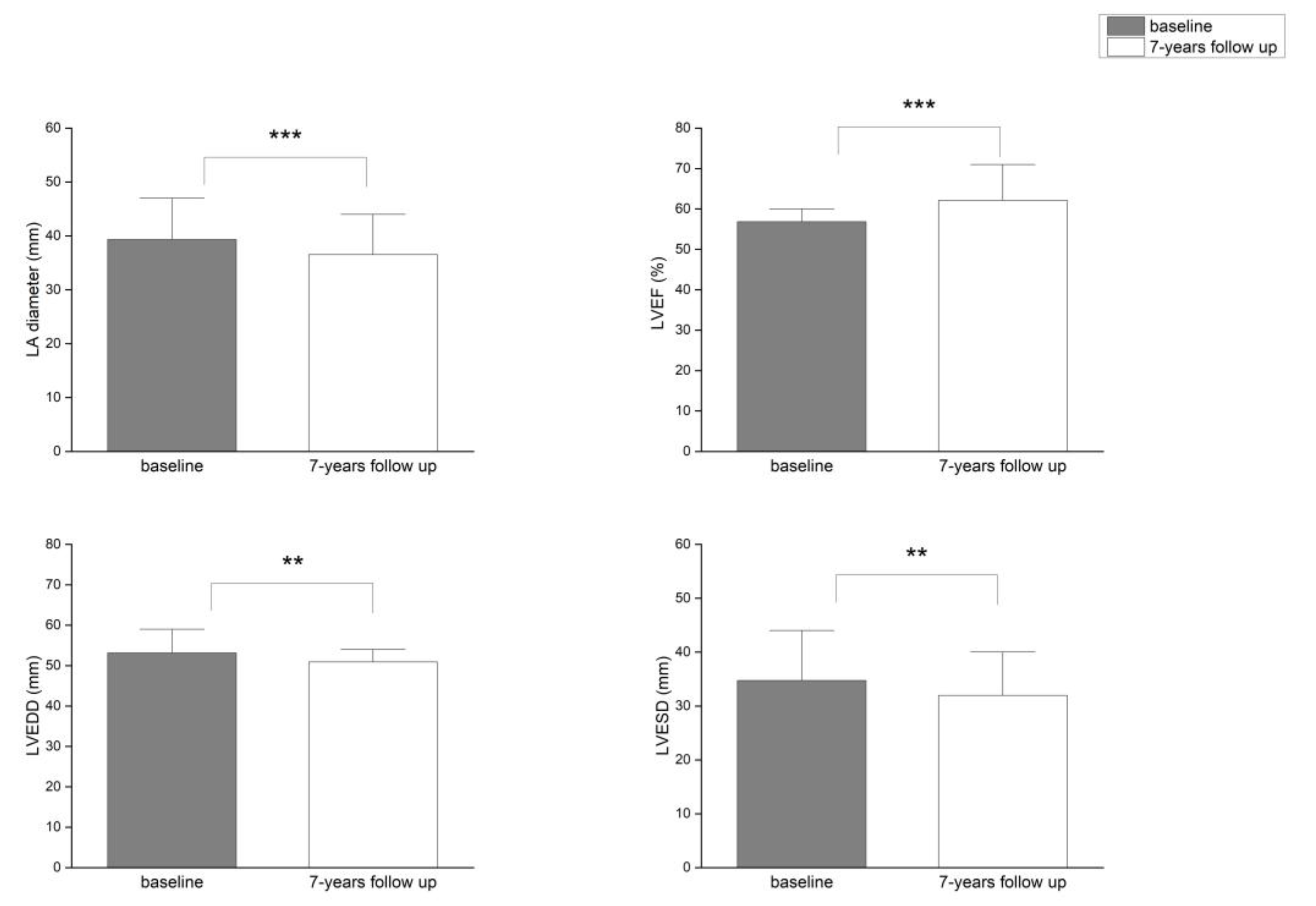
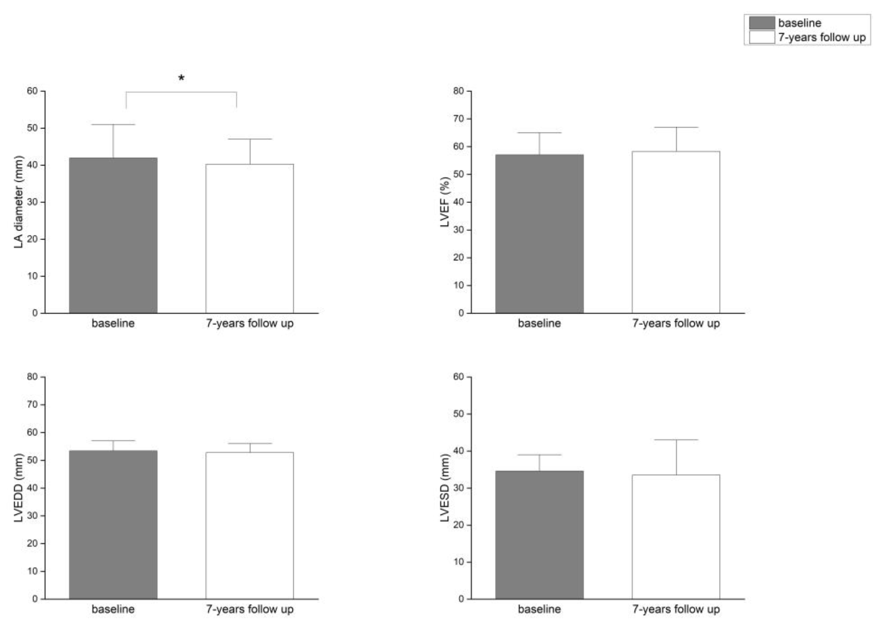
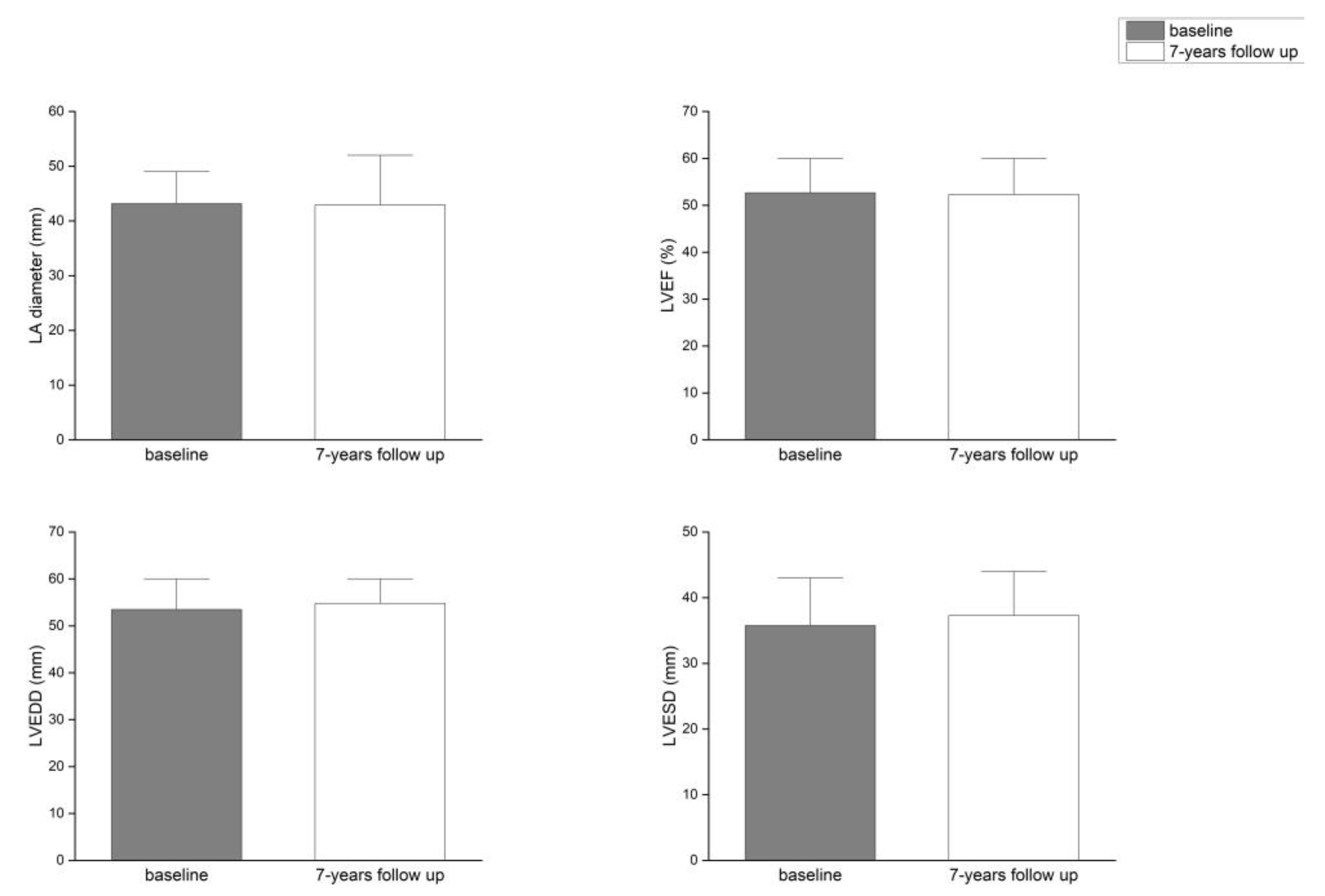
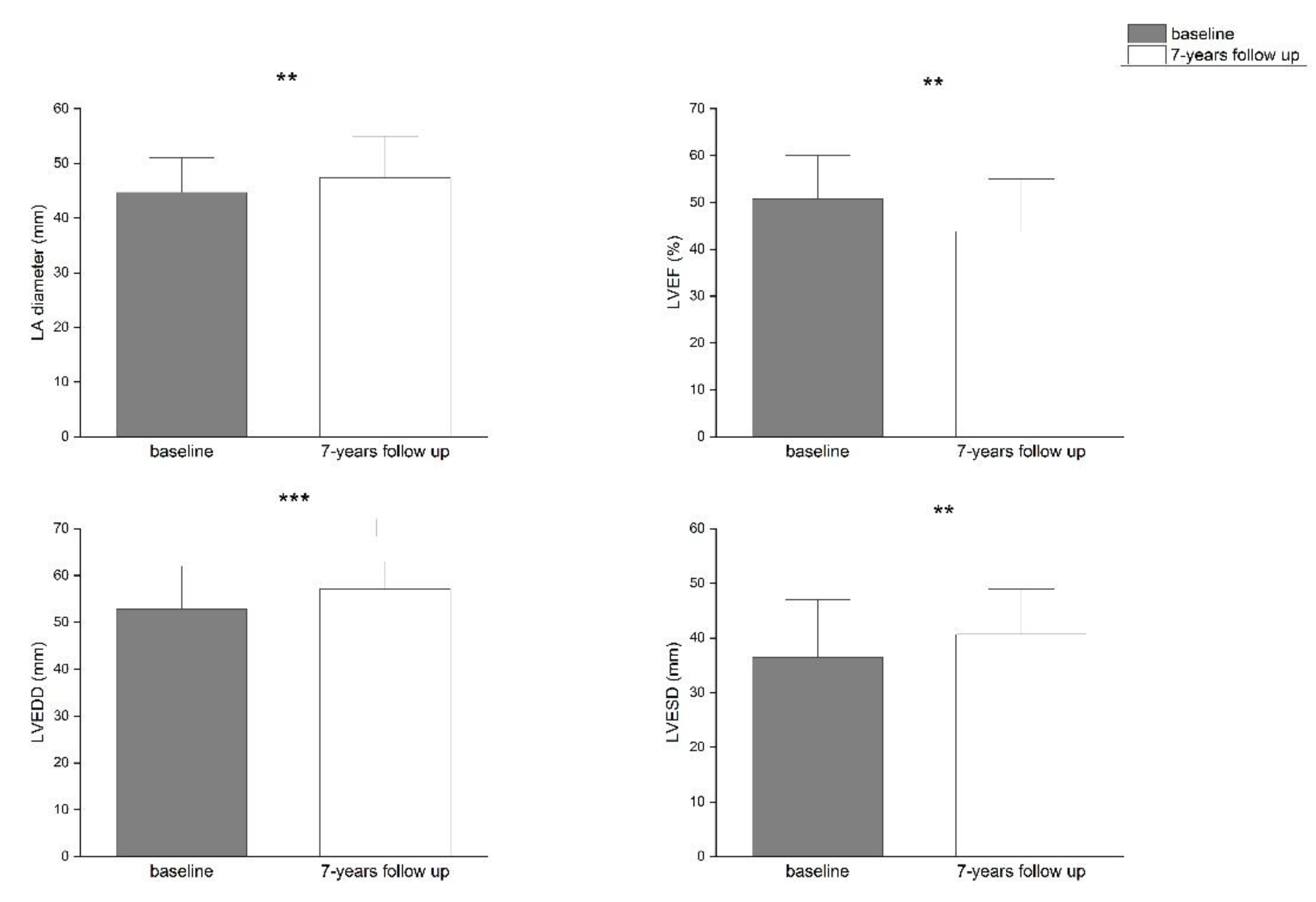
Figure 1. Significant positive changes of LA diameter, LV dimensions and LVEF in 32.5% of patients with successful single RFA of AF - PVIOD 1.
Preprints 187679 g001
Figure 2. Significant positive change of LA diameter in 29.1% of patients with success after multiple procedures - PVIOD 2.
Preprints 187679 g002
Figure 3. Significant changes of LA diameter, LV dimensions and LVEF did not occur in 14.5% of patients with clinical success - PVIOD 3.
Preprints 187679 g003
Figure 4. Significant negative changes of LA diameter, LV dimensions and LVEF in 23.9% pts with procedural and clinical failure - PVIOD 4.
Preprints 187679 g004
Table 1. Changes regarding left atrium diameter, left ventricular dimensions and left ventricular ejection fraction after PVI and 7-year follow-up period.
Parameter PVIOD 1 (n=38) PVIOD 2 (n=34) PVIOD 3 (n=17) PVIOD 4 (n=28)
baseline after 7 years baseline after 7 years baseline after 7 years baseline after 7 years
LAD (mm) 39.32±0.62 36.53±0.59 41.94±0.75 40.26±0.56 43.18±0.92 42.94±0.97 44.68±0.74 47.39±0.75
p-Value 0.000749 0.044408 0.444593 0.005874
LVEF (%) 56.84±0.76 62.11±1.08 57.06±0.97 58.29±1.02 52.63±1.49 52.29±1.76 50.71±1.68 43.82±1.81
p-Value 8.32686E-05 0.192093 0.43968 0.003668
LVEDD (mm) 53.10±0.64 50.89±0.66 53.44±0.46 52.76±0.58 53.47±0.81 54.76±0.73 52.85±0.89 57.07±0.63
p-Value 0.007842 0.201137 0.200228 0.000598
LVESD (mm) 34.74±0.81 31.97±0.09 34.62±0.68 33.56±0.67 35.76±1.21 37.29±1.24 36.46±1.13 40.68±1.21
p-Value 0.005266 0.13287 0.178529 0.005609
LAD: left atrial diameter; LVEF: left ventricular ejection fraction; LVEDD: left ventricular end-diastolic diameter; LVESD: left ventricular end-systolic diameter; PVIOD: pulmonary vein isolation outcome degree.
Disclaimer/Publisher’s Note: The statements, opinions and data contained in all publications are solely those of the individual author(s) and contributor(s) and not of MDPI and/or the editor(s). MDPI and/or the editor(s) disclaim responsibility for any injury to people or property resulting from any ideas, methods, instructions or products referred to in the content.
Copyright: This open access article is published under a Creative Commons CC BY 4.0 license, which permit the free download, distribution, and reuse, provided that the author and preprint are cited in any reuse.
Prerpints.org logo

Preprints.org is a free preprint server supported by MDPI in Basel, Switzerland.

Subscribe

Disclaimer

Terms of Use

Privacy Policy

Privacy Settings

© 2025 MDPI (Basel, Switzerland) unless otherwise stated