
Submitted:
28 November 2025
Posted:
02 December 2025
You are already at the latest version
Abstract
Extracellular polymeric substances (EPS) produced by Rhodococcus actinomycetes play crucial roles in their ecological success, metabolic versatility, and biotechnological value. This review summarizes existing studies of Rhodococcus EPS, emphasizing the biochemical composition, functional attributes, practical significance of EPS, and their importance in biomedicine, bioremediation and other applications (food industry, biomineralization) in regard to the EPS chemical composition and biological roles. Rhodococcus species synthesize complex EPS composed primarily of polysaccharides, proteins and lipids that support cell adhesion, aggregation, and biofilm formation. EPS produced by different species of Rhodococcus exhibit diverse structures, leading to variations in their biological activities. Notably, the EPS exhibit marked emulsifying and flocculating properties, contributing to their recognized role in bioremediation. Furthermore, EPS possess antiviral, antibiofilm, anti-inflammatory, anti-proliferating activities and high viscosity which are perspective in terms of biomedical and food applications. Despite extensive industrial and environmental interest, the molecular regulation, biosynthetic pathways, and structural diversity of Rhodococcus EPS remain insufficiently characterized. Advancing our understanding of these biopolymers could expand new applications in biomedicine, bioremediation, and biotechnology.
Keywords:
extracellular polymeric substances
; Rhodococcus actinomycetes
; biomedicine
; bioremediation
; viscosity properties
; biological activities
; emulsifying activity
; pollutant sorption
; flocculating activity
1. Introduction
Extracellular polymeric substances (EPS) represent high-molecular-weight biosynthetic polymers that are found outside the cell wall, either attached to it or secreted into the extracellular environment. Polysaccharides are considered to constitute a large proportion of EPS. Therefore, even in some sources, ‘exopolysaccharides’ is a synonym for extracellular polymeric substances [1,2,3]. But in fact, EPS in addition may consist of extracellular lipids, proteins, nucleic acids, and humic compounds, and differ between species [4,5]. In that way, in most research, bacterial EPS are being purified for further analyses and applications [6,7,8].
Great majority of bacteria live in forms of cell communities, such as biofilms. EPS, as a main component of biofilm matrix, exhibit ecological and physiological functions. They provide an instrument to survive in different conditions by binding xenobiotics, allowing the adhesion to substrates, and entrapment of nutrients [4,9]. Furthermore, bacterial polysaccharides are multifunctional, valuable compounds. Due to their biodegradability, non-toxicity, water-absorbing capacity, chelating and emulsifying properties, and biological activities, including anti-radiation, anti-fatigue, antioxidant, hypolipidemic, anti-inflammatory, antitumor, and immunomodulatory effects [8,10] EPS are applied in medicine, pharmaceuticals, cosmetics, the food industry, remediation, agriculture, and biotechnology [11,12].
EPS are positioned as prospective, universal, renewable sources. Bacterial polysaccharides compared to those from plants and animals and synthetic ones, differ in their numerous valuable qualities. In addition, the production of microbial EPS is more cost-effective and scalable [13]. They can be produced on a large scale using modern fermentation technology [13], which ensures stable product quality, easy production control, and eliminates concerns over raw material shortages [2].
Actinomycetes of the genus Rhodococcus (domain Bacteria, kingdom Bacillati, phylum Actinomycetota, class Actinomycetes, order Mycobacteriales, family Nocardiaceae, https://lpsn.dsmz.de/genus/rhodococcus, last accessed 16 September 2025) are well-known stress-tolerant biodegraders of a wide range of emergent ecopollutants, mainly hydrocarbons and their derivatives. Actinomycetes are known as abundant producers of metabolites, including fatty acids, proteins, amino acids, siderophores, carotenoids, biosurfactants, and polysaccharides [2,14,15]. Rhodococcus spp. have unique biosynthetic activities that contribute to their wide persistence, provide them a competitive advantage over other microorganisms, and could be utilized in biotechnologies. The beneficial ability of rhodococci to obtain high value-added products from low-cost substrates, like wastes of pulp, chemical, and pharma industries, livestock wastewaters and sludge, [16] and contaminants, offer the possibility to efficiently recover valuable resources and providing possible waste disposal solutions [17,18]. Rhodococcus spp. are considered to be perspective producers of novel bioactive molecules and so be potential for application in the framework of sustainable use, that highlighting the relevance of expanding the knowledge of their biosynthetic capacities and biotechnology products.
There is no comprehensive investigation about the Rhodococcus EPS’ diversity, their properties and capabilities, which brings further difficulties in completely utilizing their full potential. Variations in structures, coupled with potential applications and strain specificity, are supposed to be key factors in developing new products, optimizing extraction methods, and elucidating molecular mechanisms of action. Moreover, optimization of the procedures used for bacteria cultivation and further extracting EPS guarantees the preservation of its structure and functionality for various applications.
The review is focused on EPS produced by Rhodococcus actinomycetes. The data on chemical composition, biological functions, properties, and applications of rhodococcal EPS are summarized and discussed below.
2. Chemical Composition
There are fractions of microbial EPS. Slime, capsular, loosely bound, and tightly bound EPS are distinguished on the basis of the nature of their association with the cells or the extraction method used (Figure 1). It is considered there are two forms of EPS: bound EPS (sheaths, capsular polymers, condensed gels, loosely bound polymers, and attached organic materials) and soluble EPS (soluble macromolecules, colloids, and slimes) [7,19]. In terms of activated sludge used in wastewater treatment, it is important to divide tightly bound EPS (TB-EPS) located in the inner layer of the sludge floc and loosely bound EPS (LB-EPS) existing in the outer layer of the sludge floc. The contents, compositions, and properties of TB-EPS and LB-EPS were found to be different, as well as their influences on the flocculation, sedimentation and dewatering capabilities [20,21].
The chemical nature of EPS is diverse and varies in terms of carbohydrates, proteins, nucleic acids, lipids, and humic substances (Figure 1). The dominant components of numerous EPS are carbohydrates and proteins (75-90%) [12]. The EPS also may contain carbohydrate and protein derivatives such as lipopolysaccharides, glycoproteins, and lipoproteins [22]. EPS composition and properties are determined by environmental or cultivation conditions, and could differ greatly according to species-producers. Thus, chemical composition is believed to afford some insights into the relationship between its chemical structure and function.
Rhodococcus actinomycetes synthesized different types of EPS, such as acidic glucomanannes [23], heteroglycans [24], fatty acid containing polysaccharides [25,26,27,28], or complex of polymers [8,29]. The structure and completeness of EPS reveal their three-dimensional structure, which becomes more stable and complex with increasing molecular mass, because of multiple covalent bonds. EPS of Rhodococcus spp. have various molecular weights ranging from several hundred thousand to several million Daltons (Table 1).
2.1. Carbohydrates (Exopolysaccharides)
The key structure-forming component of EPS is carbohydrates, named ‘exopolysaccharides’. They are high-molecular compounds with a variety of carbohydrate compositions, consisting of monosaccharide residues forming linear or branched chains via diverse glycosidic bonds, what contributes a wide structural variety. It is noted that both the type of links between monomers and branching chains determine the physical properties of polysaccharides [49]. Variation in the composition of heteroglycans and their molecular weight may affect their ability to interact with proteins [50]. In biofilm matrix, carbohydrates usually contribute to the stability and integrity, and function as a protective barrier and pools for carbon and energy sources. However, most bacteria, unlike fungi, do not remetabolize their own polysaccharides [51], what was confirmed for Rhodococcus erythropolis [32].
Rhodococcus spp. form heteropolysaccharides, mainly containing glucose, galactose, mannose, rhamnose, and sometimes uronic acids. For example, Urai et al. actively investigate acidic, fatty acid containing polymers [25,27,28,44]. According to their results, exopolysaccharides from strains of Rhodococcus rhodochrous contain D-galactose, D-glucose, L-fucose, D-glucuronic acid and sometimes mannose [26,27,44], and from R. erythropolis PR4 also contain D-mannose and pyruvic acid [28], or N-acetylglucosamine and fucose [25]. Polysaccharide PLS-1 from R. erythropolis DSM 43215 had glucose and mannose in the molar ratio of 1 : 1 [32]. Exopolysaccharide from R. erythropolis HX-2 (HPS) had 4.79% of fucose in its composition, in addition to glucose, galactose, mannose and glucuronic acid, with a mass ratio of 27.29%, 24.83%, 26.66% and 15.84% respectively [8]. Mannose was shown to be the main residue in exopolysaccharides of R. rhodochrous R-202, Rhodococcus opacus 89 UMCS, Rhodococcus qingshengii QDR4-2 [29,40,43]. It was exhibited that monosaccharide compositions with higher proportions of mannose are associated with better scavenging activity of the hydroxyl radical, and consequently provide potential antioxidant capacity [43]. Exopolysaccharide PS-33 from Rhodococcus sp. and the other one from Rhodococcus strain 33 had another main monosaccharide and comprised of rhamnose, galactose, glucose, and glucuronic acid in the molar ratio of 2:1:1:1 [33,52]. Polysaccharide PS-33 is also acetylated and contained 6-deoxy sugars in the form of rhamnose, both in the backbone and in the side chain of the repeating unit. The presence of methyl groups and of O-acetyl are likely play an important role in the emulsifying activity [52]. The PS-33 was shown to consist of D-galactose, D-glucose, D-mannose, D-glucuronic, and pyruvic acids in ratio 1:1:1:1:1 [34]. It should be noticed that EPS from one strain – Rhodococcus sp. 33 – differs in researches [34,52], what is evidence for the fact that a conformation of EPS chains is easily affected by the various factors and conditions of the surrounding medium [53]. For Rhodococcus sp. p52, it was shown that the composition and proportion of extracellular polysaccharides greatly varied with the carbon source (dibenzofuran or sodium acetate) [54]. In case of using dibenzofuran as a carbon source, glucose (74.25%) was prevalent in monosaccharide composition. But, when using sodium acetate, exopolysaccharides consisted of guluronic acid (25.34%), mannuronic acid (24.09%), and galacturonic acid (22.65%) in approximately equal proportions [54]. Capsular polysaccharides of Rhodococcus equi serotypes were actively discovered by Richards’ research group. It was shown that they are mainly formed by glucose, mannose, glucuronic acid, rhamnose, and pyruvic acid [55,56,57,58]. EPS of 4 serotype contain unique 5-amino-3,5-dideoxynonulosonic (rhodaminic) acid [57].
In general, carbohydrates of EPS are varied. Exopolysaccharides of R. rhodochrous R-202 consist of reducing sugars, uronic acids, and amino sugars at the concentrations of 232.41 μg/mg, 45.29 μg/mg, and 15.07 μg/mg, respectively [40]. Exopolysaccharide of R. opacus 89 UMCS had these components in similar proportions: reducing sugars, uronic acids, and amino sugars at the concentrations of 184.79 µg/mg, 117.6 µg/mg, and 9.23 µg/mg, respectively [41]. 33 EPS was positioned as an acidic polysaccharide containing 66% of neutral sugars, 18.4% of uronic acids, and 15.6% of pyruvic acid [34]. On the contrary, QEPS from R. qingshengii QDR4 did not have any uronic acids, according to FTIR spectroscopy and monosaccharide composition analysis [43]. For Rhodococcus sp. p52 grown on sodium acetate, it was revealed that viscous uronic acids accounted for the vast majority of the polysaccharides (72.08%) [54]. In our study, Rhodococcus strains of various species produced low amounts of total EPS carbohydrates, ranging from 0.6 ± 0.2 mg/L (Rhodococcus aetherivorans IEGM 1250) to 58.2 ± 24.0 mg/L (Rhodococcus ruber IEGM 231), with median EPS carbohydrate production equaled 8.9 mg/L [5].
Most bacterial EPS exhibit anionic characteristics due to the presence of uronic acids containing carboxyl groups [4]. In general, exopolymers are mainly acidic molecules because of the mixed contributions of polysaccharide- or protein-associated COOH−, NH3+, and phosphate groups. Exopolymers are treated as weak and surface-grafted polyelectrolytes, since long chain macromolecules possessing ionizable groups [53]. Dobrowolski et al. actually noted that high amounts of functional groups on the Rhodococcus extracellular polymers’ surface make an interpretation of spectra more difficult [35]. Infrared spectrophotometry analysis revealed that the exopolymers contained carboxyl, hydroxyl, acetyl, and carboxylate groups, preferred for the flocculation and sorption processes [35,41]. In QEPS and HPS, a large amount of hydroxyl groups, and the hydrogen bonding, causing the cross-linking of molecular chains, are detected [8,43]. The typical polysaccharide functional groups, namely, hydroxyl (–OH), amino (–NH2), and acylamino (–CONH2) groups, were all observed in EPS of R. erythropolis ACCC 10543 [16]. Amine and amide groups are also presented in Rhodococcus EPS [16]. According to the FTIR and XPS spectra analyses, exopolymer R-202 contains hydroxyl, carboxylic, amide, and amine groups, whereby fractions contain high amounts of oxygen and nitrogen which may indicate a higher content of polar functional groups [40]. For EPS from R. rhodochrous 202 DSM and R. opacus 89 UMCS, it was shown that 52.1% and 62.7% of carbon is present in –CHx groups, respectively, and it was indicated the presence of acetoamido or amino groups, in addition to main hydroxyl groups [35]. Obviously, the FTIR analysis of HPS showed strong peaks characterised absorptions of C–O–C and C–O–H bonds of pyranose ring, and also peaks of methyl groups [8].
Aside from the components themselves, linkages between units also vary greatly. Pen et al. observed that the EPS of rhodococcal cells at the stationary phase are regarded as amorphous random coils, probably due to the 1,2-α- or 1,6-α-linkages between their exopolysaccharides [53], and the EPS of the late exponential phase, nevertheless, may possess 1,3-β- or 1,4-β-linkages between the exopolysaccharides, resulting in rigid helices that are less resilient to normal compression [59]. There are six key glycosidic linkages in QEPS: ← 2)-Manp-(1→, Manp-(1→, ← 2,6)-Manp-(1→, Glcp-(1→, ← 3)-Manp-(1→, and → 3) Glcp-(1→ [43]. What is suggested that ← 3)-Manp-(1→ composed the main chain of QEPS and that ← 2,6)-Manp-(1→ indicated the existence of 1→ 2,6 branching linkage. In PS-33, glucuronic acid, rhamnose and glucose are linked at position 3, galactose at positions 3 and 4, and that one rhamnose is a nonreducing terminus linked by (1→ 4) [52]. Extracellular polysaccharides of Rhodococcus sp. RHA1 is composed of tetrasaccharide repeating units linked in a such way: d-GlcpA-(1→, → 3,4)-l-Fucp(1→, → 4)-d-Glcp-(1→, and → 3)-d-Galp-(1→ ) [46].
2.2. Proteins
EPS always consist of few types of biopolymers, including carbohydrates, proteins, nucleic acids, and sometimes lipids. Because of special EPS components, matrix could show adsorption abilities, biodegradability and hydrophilicity/hydrophobicity. Depending upon chemical composition of exopolymers, they might exhibit various capabilities and so find applications in various spheres. The main constituents of Rhodococcus EPS are considered to be polysaccharides (15-95%) and proteins (0-85%). In less amounts (1-10%), there are lipids, nucleic acids, and humic substances (Table 1).
The proteins of exopolymers in the biofilm matrix contribute to its formation and stabilization, and provide metabolism and mediate intercellular communication [7]. They are also supposed to support biofilm cohesion, adhesion, communication, and environmental response [7]. These proteins are usually extracellular enzymes, S-layer proteins, electron transfer (ferredoxin, rubredoxin) proteins, transmembrane transporters (porin), lectins, stress response proteins [4,49]. In relation to practical properties, both proteins and polysaccharides play main roles in flocculation and biosorption. Their concentration and characteristics decide the fate of surface properties, as well as biodegradability of the EPS [12].
The amounts of proteins are varied in different species. For EPS from rhodococci, it is known that the polysaccharides’ content is at least 5 times higher than the proteins’ content. For example, the total carbohydrate and protein contents in EPS of R. erythropolis HX-2 were found to be 79.24% and 5.204%, respectively [8]. According to Czemierska et al., water-soluble fractions of exopolymers with flocculant activity contained proteins in 6 times less than carbohydrates. So, EPS of R. opacus 89 UMCS have 64.6% polysaccharides and 9.44% proteins [41], and EPS of R. rhodochrous R-202 have 62.86% and 10.36% of these components, respectively [40]. Extracellular polymer from R. erythropolis ACCC 10543 is described as a glycoprotein composed of 91.2% of polysaccharides, 7.6% of proteins, and 1.2% of DNA [16,37]. In our work, concentrations of proteins in Rhodococcus EPS were insignificant, with maximum detected for R. rhodochrous IEGM 107 (1.131 ± 0.091 mg/L). Other strains were shown to produce less than 0.5 mg/L proteins [5]. A purified polysaccharide PLS-1 had a trace of glucosamine and 3.3% of proteins with predominant glutamic acid and glycine, followed by alanine, serine, and leucine in amino acid composition [32]. After purification, the lack of proteins and nucleic acids is detected in all ATCC 53968 EPS, QEPS, and SM-1 EPS [27,43,44].
Protein content may be high or even predominant. In aerobic quinoline-degrading biofilms with a predominance of Rhodococcus polysaccharides and proteins were determined as two primary components accounting for 78−87% of total EPS [60]. Besides, saccharides (57.0 ± 10%) and proteins (43.0 ± 6.9%) were the most dominant composition of TB-EPS and LB-EPS, respectively [60]. For bioflocculant from Rhodococcus sp. R3, it was revealed that the total protein and sugar content were 84.6% and 15.2% [42]. A significant proportion of proteins are also found in EPS synthesized by Rhodococcus sp. p52 in the presence of dibenzofuran [54]. These compounds account for 20–33% of total EPS and dominate polysaccharides by 1.5–3.0 times in a fraction of loosely cell-bound EPS. It was defined, that in such toxic conditions the proportion of antitoxin proteins in EPS was five times greater than that in using sodium acetate as carbon source. And the percentages of cold shock proteins and DUF4193 domain-containing proteins, which are associated with the stress response, also greatly increased [54]. The content of protein in the biofilm of R. ruber C208 was similar to that of polysaccharides, with a maximal content (150-200 µg/cm2) after 10 days, but meanwhile, after 20 days the protein content of the EPS was up to 2.5 fold lower [47]. Extracellular organic matter of polychlorinated biphenyl (PCB) degrader Rhodococcus sp. SJ contains proteins and polysaccharides at concentrations of 24.33 mg/L and 1165 mg/L, respectively [48]. A protein-enriched fraction was optimized in composition of Rhodococcus sp. SJ EPS, since namely the proteins were shown to provide resuscitation-promoting activity [48]. Proteins of the optimized EPS constituted 223.58 mg/L and had a molecular weight more than 10 kDa. In its spectra analysis tryptophan-like proteins, tyrosine-like proteins, and tyrosine-like amino acids peaks were observed. The presence of these proteins is likely related to microbial stress responses and stability maintenance, and polysaccharides may serve as supplementary nutrient sources that support microbial activity and contribute to degradation [48].
2.3. Lipids
Exopolymers of Rhodococcus spp. are supposed to possess the hydrophobic properties due to polysaccharides-linked methyl and acetyl groups, lipids, and lipid derivatives in their composition [16]. Among exopolymers from Rhodococcus, a specific group of fatty acid containing polysaccharides has been described [25,28]. According to Urai et al., such saturated fatty acids as stearic and palmitic acids prevail in the lipids of Rhodococccus EPS [27,28,44]. It was demonstrated that fatty acids attach via ester bonds to the sugar backbone of exopolymers. So, FR2 EPS from R. erythropolis PR4 contain 2.9% of stearic and 4.4% of palmitic acid [28]. And polymers from R. rhodochrous ATCC 53968 and R. rhodochrous ATCC 12674 (SM-1) have these fatty acids in proportions of 1.3% and 4.1%, 1.2% and 2.3%, respectively [27,44]. According to unpublished data mucoid strains of R. rhodochrous S-2 produce an extracellular polysaccharide of several million daltons in size, which contains stearic acid, palmitic acid, and oleic acid [26]. In HX-2 EPS from R. erythropolis HX-2, composition 8.45% of total lipids were detected [8]. In our recent research, lipid-rich exopolymers of Rhodococcus IEGM strains were described [5]. It was shown that the presence of total lipids in EPS is similar to, or prevail over, the carbohydrate content. In particular, production of these compounds varies from 15.6 ± 1.6 mg/L (Rhodococcus sp. IEGM 1401) to 71.7 ± 7.2 mg/L (R. erythropolis IEGM 1415), with a median value of 32.0 mg/L [5].
2.4. Humic Substances
Humic substances can comprise significant parts of dissolved extracellular organic matter [61] and can be divided into three components: fulvic acids, humic acids, and humin, which facilitate aggregation and cohesion [12]. According to the multiple researches in Rhodococcus EPS composition, humic substances are rare or have not been defined. There is an example of detecting humic substances in exopolymers’ spectra. In extracellular organic matter of Rhodococcus strain SJ humic-like substances were identified [48]. However, it was shown that the optimization process substantially enhanced the protein content, but reduced the relative abundance of humic-like substances. Tong et al. showed that deposition of microbial EPS on silica surfaces could be significantly influenced by humic acids under solution conditions typical for subsurface environment, what is could be applied in bioremediation [62].
3. Biological Functions
EPS as major component of natural microbial systems play substantial functional role in the physiology and environmental interactions of the Rhodococcus cells. The physiological role of EPS depends on the natural environment in which the microorganisms live. Since Rhodococcus actinomycetes live in a wide variety of habitats and are able to withstand extreme conditions [63], most of the functions ascribed to their EPS are protective in nature. These biopolymers permit resistance under extreme environmental conditions, despite the fact that their synthesis and release require a lot of energy from the cells [53]. The basic role of polymers synthesized into surrounding media is to create and maintain favourable conditions for microorganisms. In this way, exopolymers form a hydrated barrier between the cells and the environment, reliably protecting the cells from adverse factors, firmly holding colonies to solid surface, participating in the substrate assimilation, contributing to the aggregation of microorganisms and the formation of biofilms [64].
EPS are also known as ‘adhesive polymers’ [49]. They are supposed to be important determinants of the biofilm formation on solid surfaces [65]. The authors believe that EPS are responsible for both adhesion and cohesion, and they play a key role in determining the structural unity of the biofilms and their corresponding physicochemical properties [66]. Bacterial biofilms are clearly more resistant to physical and chemical factors than are planktonic cells due to the extracellular polymeric matrix. Rhodococci are known to be capable of producing biofilms that have been shown to be more resistant to hydrocarbons, metal nanoparticles, and other stresses. For example, the revealed adaptation mechanisms of R. rhodochrous IEGM 1363 include increased surface roughness and intensive formation of an exopolymer matrix rich in lipids, which plays the main role in protecting cells from reactive oxidative stress [67]. Our study confirmed protective functions in dense R. ruber IEGM 231 biofilms formed in the presence of toxic n-hexane and diesel fuel [5].
A special function of exopolymers is considered to be the inactivation of chemical compounds, in addition to the effect on biofilm specificity [68]. Some authors have reported increased production of EPS in the presence of toxic organic compounds. It was shown for Rhodococcus pyridinivorans XB during growth on di-(2-ethylhexyl)phthalate [69]. Rhodococcus jostii RHA1, in the presence of toluene and perfluorocarboxylic acid aqueous solution, produced exopolymers at concentration above 2 mg/L [70]. R. erythropolis PR4, which degrades various alkanes, synthesizes a large amount of EPS, which are likely provided the hydrocarbon tolerance of this strain [25]. Moreover, EPS not only trap the toxic substances in the biofilms and act as a buffer zone between the toxic substances and bacteria, but also increase the adhesion of hydrophobic organic matter, thus improving the biodegradation efficiency [71]. An oxidation of anthracene or phenanthrene, for example, is associated with an attachment of rhodococcal cells to the substrate and the formation of biofilms on the surface of solid hydrocarbons [15].
The component structure of biofilms was also shown to be changeable in different conditions, thus to be a kind of adaptation. The increased content of polysaccharides was observed in the periphery of the cell conglomerates of the R. rhodochrous IEGM 1363 biofilms at the maximum CuO nanoparticles concentration, while lipid content remained stable when biofilms were exposed to increasing nanometal concentrations. Similarly, increased polysaccharide content was detected in R. ruber C208 biofilms grown on a polyethylene surface under nutrient-limited conditions [47], but meanwhile an increased protein content of this strain’s biofilms was revealed when growing on polystyrene [72].
Smooth colonies were supposed to be more resistant relative to rough ones due to produced EPS. Researches showed that the presence of exopolymers altered the level of cell exposure to solvents. Iwabuchi et al. showed that some R. rhodochrous strains designated smooth were found to produce biofilms with mucoidal hydrophilic EPS, which protected the cells [73]. In contrast, rough strains of R. rhodochrous with a more hydrophobic surface were shown to be more sensitive. So, EPS confer oil tolerance in members of Rhodococcus [73]. In the presence of organic solvents, part of the initially rough R. erythropolis DCL14 population started producing EPS [74,75]. The DCL14 cultures were found to form cell aggregates and biofilms in the presence of both water-immiscible and water-miscible solvents [76,77]. Aizawa et al. also considered that the mucoidal morphology positively influenced on growth and survival of cells in presence of xenobiotics. As a result, the addition of Rhodococcus sp. 33 EPS to rough strains improved their tolerance to benzene [34].
Biofilms, as well, having certain architecture, provide an optimal environment for an exchange of genetic material between cells. But, meanwhile exopolysaccharides can prevent binding of exogenous DNA by influencing horizontal gene transfer. Huang et al. confirmed that rich extracellular polymers prevent the entry of foreign DNA into cells, on the example of R. ruber YYL [78]. The YYL cells were shown to have mushroom-like substances surrounding them. So the electrotransformation efficiency was significantly improved for mutant cells with knocked out gmhD gene, responsible for the biosynthesis and assembly of capsular polysaccharides [78].
In fact, EPS could also play absolutely other physiological roles. For example, EPS of R. equi are determined as potential virulence factors, since they contribute to antigenic specificity and are recognized by the immune system [56,57]. Most R. equi strains were shown to form biofilms, which are favorable for its survival in the environment as well as in the host [79]. Tripathi et al. highlighted a possible relationship between conjugation and biofilm formation, showing a mechanism by which the virulence plasmid can move among R. equi in the soil [80].
4. Applications (Bioactivities)
Rhodococcus cells’ unique biochemical properties and adaptability to harsh conditions make their metabolites valuable for bioremediation, industrial, and potential medical applications. EPS are natural polymers that can be sustainably produced from renewable resources, they possess a competitive advantage over other polymers [8]. In certain environments, bacteria can stably produce EPS with high structural reproducibility; this is advantageous in terms of low fermentation costs, easy extraction, and short production cycles. Therefore, there is a huge commercial potential for EPS in industrial applications. Properties of Rhodococcus EPS in terms of their application areas are submitted further (Figure 2).
4.1 Biomedicine
Exopolysaccharides produced by Rhodococcus species are gaining attention for their promising biomedical applications due to their biocompatibility, non-toxicity, biodegrability and also unique structural, physicochemical, and biological properties. Recent research highlights their potential in anticancer therapy, antioxidant activity, bioemulsification, and as biocompatible materials for pharmaceutical use.
Rhodococcus EPS could have potential antiviral activity. For example, Rhodococcus spp. are able to attach to human Norovirus particles using their EPS. It was shown that exopolysaccharides derived from the five Rhodococcus strains bound to both GII.4 Sydney 2012 and GII.6 norovirus viral-like particles, but not to the rotavirus double-layer particles in negative control [30]. According to recent studies, the exopolysaccharides of some Rhodococcus strains [8,25] consist glucose, galactose, fucose, mannose and glucuronic acid, which are sugar moieties also found in Histo-blood group antigens on host cells and as such participate in molecular recognition. So, Santiso-Bellón et al. concluded that direct binding of norovirus to Rhodococcus cells appears via their exopolysaccharides, which is led to subsequent inability of these sequestered viruses to infect their target cells [30]. These investigations provide a basis for developing innovative antiviral strategies to prevent and treat NoV infections through EPS [30].
Li et al. described QEPS extracted from R. qingshengii QDR4-2 with potential antioxidant activity [43]. Exopolysaccharide demonstrated the scavenging activities against 2,2-diphenylpicrylhydrazyl (DPPH), 2,2′-azino-bis(3-ethylbenzothiazoline-6-sulfonic acid, and hydroxyl and superoxide radicals, that reached 30-49%. Moreover, QEPS could be used in pharmaceutical industries due to its good biocompatibility. Another exopolysaccharide from R. pyridinivorans ZZ47 demonstrated 54% and 22-27% DPPH and hydroxyl free radical removal respectively [31].
Exopolysaccharides could play a role in angiogenesis – a complex mechanism of the formation of new blood vessels, which is used for cancer treatment [81]. Güvensen et al. showed that exopolysaccharides from R. pyridinivorans ZZ47 have strong anti-angiogenic activity only at 2 mg/mL, so authors concluded that the exopolysaccharides have dose-dependent anti-angiogenic properties [6]. But, while exopolysaccharides of R. pyridinivorans ZZ47 showed low cytotoxic activities on human colorectal adenocarcinoma (HT-29) and human breast adenocarcinoma (MCF-7) cells [31]. Moreover, R. pyridinivorans EPS showed antibiofilm activity between 11-55% against strains of Salmonella typhimurium, Aeromonas hydrophila, Escherichia coli, Pseudomonas aeruginosa, Shigella dysenteriae, Staphylococcus aureus and Bacillus subtilis. Due to the studied biological activities and lack of genotoxicity and cytotoxicity of the polymers, they may be suitable for use in pharmaceutical, diagnostic, therapeutic industries [6].
Exopolymers synthesized by rhodococcal cells have the potential to act as agelling agents, thickeners, emulsifiers, stabilizers, water binders [27,43]. Hu et al. described a unique exopolysaccharide from R. erythropolis HX-2, which exhibited a stereoscopic network structure [43]. Due to its small pore size (< 1 μm), dense distribution and aggregation of polysaccharides, the HPS may retain more water molecules, which makes them good excipients. It was shown that the water solubility index and water holding capacity of HPS were 92.15 ± 3.05% and 189.45 ± 5.65%, respectively. So, HPS may have excellent hydrophilicity and the potential to hold a mass of water through hydrogen bonding. The solution viscosity of HPS equaled 18 mPa·s in 1 mg/mL and 30 rpm, which was lower than the viscosity of xanthan gum (61 mPa·s). Another exopolysaccharide from R. rhodochrous ATCC 53968 features high viscosity, with a value amount 1.39 m3/kg [27]. Medium and high viscosity indicates a thickening capacity of Rhodococcus exopolysaccharides, which was more beneficial for the application of excipients to the health industry. There is another exopolysaccharide with excellent moisture-retention and absorption capacities was described by Urai et al. [44]. SM-1 EPS from R. rhodochrous ATCC 12674 was capable of retaining the initially added water and also absorbing moisture in the desiccator under dry and high-temperature conditions. The authors claimed that SM-1 EPS absorption capability was much higher than that of the other moisture absorbents tested, such as silica gel and hyaluronic acid.
Bacterial exopolysaccharides are supposed to have a function of inhibiting cell proliferation [2]. HPS demonstrated high inhibition effect on cancer cells without affecting the normal ones [8]. Hu et al. showed that adding HPS in concentration 800 μg/mL decreased cell viability of A549 cells, SMMC-7721 and Hela cells by 21.86%, 31.24%, and 37.65%, respectively, but HPS had no cytotoxicity effect on L929 cells independently of concentration [8]. Although, it was noticed, that HPS can rapidly inhibit the proliferation of cancer cells on the first day and achieve a relatively high inhibition effect. On the one hand, Rhodococcus exopolysaccharides have been shown to exhibit anti proliferative activity [8], while on the other hand, some exopolymers were non-cytotoxic to cancer cells [6]. It is likely that extracellular polymers could provide specific activities according to the strain producer and cultivation conditions. Therefore, Rhodococcus exopolysaccharides could be used for various therapeutic applications depending on the intended goals.
Some EPS could provide such biological activities as anti-inflammatory effects [2]. According to unpublished data, the extracellular heteropolysaccharide of R. erythropolis DSM 43215 was found to exhibit anti-inflammatory activity [32].
In addition, potential biomedical applications of EPS are associated with vaccine development. Rhichards et al. have undertaken a detailed chemical study of the capsular polysaccharide antigens of R. equi cells, suggesting that the development of protective vaccines against these pathogens are of considerable interest as a means of controlling disease [56].
4.2 Bioremediation
Rhodococcus actinomycetes are known biodegraders, capable of decomposing xenobiotics to inorganic products or low molecular organic fragments. Their EPS could be actively used in bioremediation, since they enhance adhesion and biodegradation, have emulsification activity, and, moreover, protect cells against pollutant adverse effects. EPS, as cell-secreted polymers, influence the biofilm formation and the surface characteristics of bacteria, in turn positively affecting adhesion between the bacteria and contaminants what is led to improving degradation efficiency. Notably, the negatively charged functional groups presented in EPS have electrostatic interactions with various anions and pollutants in the surroundings and help in their biosorption [82].
EPS encapsulating bacterial cells help them to adhere to and dissolve hydrophobic organic matter, thereby promoting the utilization of the hydrophobic substances [83]. For example, EPS secreted by Rhodococcus sp. p52 affected both the biofilm formation and the dibenzofuran degradation [54]. It was shown that the absolute value of the zeta potential of p52 cells decreased with the degradation and the production of EPS, and this was beneficial for increasing the adhesion of strain p52 to dibenzofuran. Both tight and loosely bound EPS produced by strain p52 increased the solubility of the pollutant in water, and thus promoted the degradation. Authors assumed that the presence of dibenzofuran increased the cellular stress of strain p52, which in turn led to the secretion of more extracellular polysaccharides [54].
Shi et al. discovered that Rhodococcus strain SJ, isolated from PCB-contaminated soil, enters the viable but non culturable state under stress, and the possibility of its secreted exopolymers to facilitate resuscitation from this state and enhance degradation [48]. PCB degradation efficiencies in EPS-amended cultures reached 90.0%, 84.5%, and 80.7% at PCB concentrations of 1, 5, and 10 mg/L, respectively, compared to 80.5%, 65.9%, and 46.9% in the control, unamended cultures. It was indicated that the EPS protein fraction significantly enhanced the PCB degradation ability of strain SJ, with a stronger effect observed at higher PCB concentrations. Furthermore, it was concluded that utilizing EPS in stress recovery strategies for sustainable bioremediation represents a more practical and cost-effective approach compared to the use of purified resuscitation-promoting factors, a class of quorum sensing signaling molecules.
Semeniuk et al. described the exopolysaccharide from R. erythropolis Au-1 with potential as an effective emulsifying agent [45]. The emulsifying activity of its solutions with vaseline oil has been found to be equal 42-58%, depending on the exopolymer’s concentration. According to unpublished data, SM-1 EPS forms a water-in-oil emulsion and prevents evaporation of water under dry, high-temperature conditions, since SM-1 EPS is an efficient emulsifier of oil, polyaromatic hydrocarbons, and alkanes [44]. There is another example of EPS emulsification activity. S-2 EPS certainly enhanced the abilities of native marine bacteria to degrade some components of aromatic oil fractions [26]. It was shown that, when S-2 EPS was added to the medium, oil was immediately emulsified, whereas it either adhered to the inner surface of the culture tubes or formed oil clumps in the liquid when S-2 EPS was not added. The authors concluded that EPS produced by R. rhodochrous S-2 could be useful for the biodegradation of spilled oil in marine environments, and especially for the bioremediation of polyaromatic hydrocarbons, that remain in the environment even after a traditional bioremediation treatment [26]. In another research, it was noted that S-2 EPS protects rough Rhodococcus strains from the toxic oil [73].
The EPS synthesis was proposed to be a crucial mechanism for the assimilation of alkane substrates at low temperature by Rhodococcus sp. strain Q15, and it was described to have an emulsifying effect on alkane cell assimilation and oxidation [84]. With close contact between the strain Q15 cells and hydrocarbon surfaces achieved, extracellular compounds could then solubilize the alkane substrates, facilitating cellular uptake. The EPS may also play a role in floc formation in strain Q15, enabling the cells to remain in close physical contact with hydrocarbons. Such phenomena was described by Takeda et al. for biopolymer produced by R. erythropolis S-1 during growth on pentadecane [85].
The biodegradation process starts with attachment bacteria to the substrate. To facilitate adhesion to hydrophobic substrates, hydrocarbon-degrading bacteria may increase cell surface hydrophobicity by modifying cell surface components [86]. EPS are supposed to be one of the major causes of Rhodococcus cell surface hydrophobicity [33,52]. Urai et al. measured R. rhodochrous cell hydrophobicity, obtaining overestimated values for cells surrounded by loosely bound material that could be easily removed by physical impact, such as by stirring [44]. In addition, microbial cells may produce EPS in the form of capsules or mucoid secretions that may interact with hydrophobic substrates, such as hydrocarbons [87]. Polysaccharides carrying hydrophobic groups are involved in the labelling of hydrophobic interfaces by microorganisms [52]. It is known that polysaccharides rich in deoxy sugars engage in the adhesion of bacteria to hydrophobic interfaces [88]. Neu et al., in another research, described emulsion-stabilizing exopolysaccharide from the adhesive, hydrophobic Rhodococcus strain No. 33, which consists of different deoxy sugars [52]. Neu and Poralla extracted the amphiphilic polysaccharide from Rhodococcus sp. and confirmed that polymers contribute cell surface hydrophobicity and play role in adhesion [33]. But, meanwhile, S-2 EPS has been shown to lower the cell surface hydrophobicity of rough strains, indicating that S-2 EPS functions as a hydrophilin and that way establishes tolerance to oils and n-hexadecane [89].
EPS of Actinomycetes are also known as sorption agents for heavy metals. Exopolymers can act as adsorbents because of their particular structure, physicochemical properties and chemical stability, which are the result of the presence of chemical reactive groups in polymer chains. In the case of the heavy metals, the hydrophilicity is an essential property [90]. Such characteristic of EPS is attributed to chemical composition and culture conditions. Dobrowolski et al. described EPS of R. rhodochrous and R. opacus, which exhibited high adsorption affinity towards Cd(II), Pb(II), Ni(II), Co(II) and Cr(VI) ions, with the highest adsorption capacities obtained for Pb(II) and Cd(II) ions [35]. A rapid rate of ion adsorption is found to be a significant advantage of the EPS, what allows to use them in flow systems used for wastewater treatment [35]. In another study, Dobrowolski et al. used immobilized EPS for adsorption of Pb(II) and Cd(II) from aqueous solutions [36]. EPS extracted from R. opacus were immobilized on synthetic microspheres BES.DM-GMA-TETA, obtained via copolymerization of bis [4(2-hydroxy-3-methacryloyloxypropoxy) phenyl]sulfide with glycidyl methacrylate and modified with triethylenetetramine. Such fixing method is supposed to improve the solid phase properties and sorption capacities of the crude EPS. In the result, the coverage of the EPS on the synthetic microspheres was only 6.25%, but it enabled increase the adsorption capacities for both Pb(II) and Cd(II), by 47% and 25%, respectively [36].
EPS have good flocculant activity [13]. Bioflocculants are usually effective in aggregating colloids and widely used not only in remediation fields, but in industrial fields such as tap-water preparation, downstream techniques, fermentation process, and food industries [91]. Rhodococcus strains have been described as effective flocculant producers. For example, various exocellular polymers are produced by R. erythropolis ACCC 10543 in different conditions. Peng et al. cultivated R. erythropolis on sludge and livestock wastewater and got bioflocculant RCF, which was effective over a wide pH range from 2 to 12, with flocculating rates higher than 98% [16]. Another bioflocculant, NOC-1, is produced by R. erythropolis ACCC 10543 in standard conditions, and, unlike RCF, has only 30% flocculating rates at pH 2.0 [16,37]. Kurane et al. described bioflocculants from R. erythropolis S-1 and R. erythropolis 260-2, and reported that Rhodococcus flocculants have very interesting, unique flocculation characteristics, but low productivity [38]. In addition, exopolymers R-202, synthesized by R. rhodochrous with the highest flocculating activity at the pH value 7.2 and 10 mM concentration of salt solutions were obtained by Czemierscka et al [40].
R. opacus 89 UMCS cells with the affinity for calcite and magnesite surfaces produce exopolymers able to bind kaolin particles resulting in flocculation [41]. It was noted, that soluble fraction of EPS had molecular weight 760 kDa. This parameter is important for flocculation, so this process with a high-molecular-weight bioflocculant involves more adsorption points, stronger bridging ability, and higher flocculating activity [92]. Also, this macromolecule can interact with positively charged functional groups of particles suspended in water, causing their aggregation, as well as binding metal cations, e.g. Ca2+, Fe2+, Mg2+. Above confirms that bioflocculants from Rhodococcus EPS are suitable for mechanical treatment of water, for example, water with soil or clay particles.
4.3 Other applications
Since the microbial EPS possess different sugar compositions and different rheological characteristics, they represent a very big potential reserve within which it is possible to find the ideal EPS to use in a specific biotechnological and bioengineering application [11,93].
Rhodococcus bacteria could produce high-molecular exopolymers with potential in food industry. The high molecular weight of QEPS reached 9.450 × 10⁵ Da, which may contribute to its excellent emulsifying capabilities [43]. The emulsification indexes of QEPS for all six edible oils at a concentration of 2 mg/mL were not < 60%, with a higher index for olive oil. Moreover, QEPS is suggested to have potential as a bioemulsifier and antioxidant with applications in health, food, and pharmaceutical industries. The HPS exhibited higher molecular weight, which is equal to 1.04 × 10⁶ Da, which is suggesting that HPS may have a higher viscosity in addition to high water solubility [8]. Therefore, these hydrophilic exopolysaccharides may retain more moisture, which is a perfect characteristic for excipients in the food industry. Exopolysaccharide PLS-1 from R. erythropolis DSM 43215 also had an unusually high molecular weight, amounting 1.14 × 10⁶ Da [32]. The total exopolymer and the fraction of the water-soluble exopolymer from R. opacus show a fibrillar structure with a sheet-like texture. This indicates the thin web structure of these preparations with higher capillary forces to hold water molecules [41]. Additionally, these polymers have flocculation activity, which can be applied in food and fermentation industries for removing pollutants.
In terms of bacterial crystallization processes, the production of specific bacterial outer structures and their chemical nature might be crucial factors, influencing the mineralogy and morphology of calcium carbonate crystals [94]. An important aspect in biomineralization processes is the role played by functional groups at the surface of organic matrix, produced by microbial cells, especially acidic carboxyl and phosphate groups, which may bind cations, such as Ca2+, and hence act as the crystal nucleation sites [95]. It was found that the EPS from R. opacus strains are capable of binding calcium ions and hence act as nucleation centers influencing calcium carbonate precipitation, but the exopolymers do not affect the crystal structure [29,95]. Bacterially produced carbonate biominerals are known to be used for improving the durability of buildings, remediation of water or soil environments, and sequestration of atmospheric CO2 [93].
5. Conclusion
This article provides a detailed review of the chemical composition, biological functions, and activities of EPS produced by Rhodococcus actinomecetes, discovered over recent decades. EPS from Rhodococcus represent a unique class of biologically derived polymers, distinguished by their structural diversity, surface activity, and provided properties. As this review illustrates, Rhodococcus EPS have already demonstrated substantial value in their capacities for hydrocarbon emulsification, flocculation, ion sorption, and biofilm formation, providing powerful tools for bioremediation. Importantly, growing evidence now supports their emerging relevance in biomedical sciences. As discussed above, several independent studies have explored the potential activities of Rhodococcus exopolysaccharides, including antioxidant, antiviral, anticancer, antibiofilm, anti-inflammatory, and viscosity properties. Inherent biocompatibility, non-toxicity, and tunable chemical composition make Rhodococcus EPS promising candidates for next-generation biomaterials and active agents for biomedicine, environmental, food, and other industries. Despite the extensive research into Rhodococcus EPS activities, there are still no patented products in any of the promising areas of EPS application. Therefore, fundamental and applied investigations remain significant in a broad field of study.
Purified exopolysaccharides provide activities, making them ideal candidates for drug delivery systems and anticancer treatment procedures [6,8]. Besides, unpurified exopolymers, remaining less studied, could extend valuable properties and application possibilities. According to structural and physiological features of EPS described in literature, it should be noted that, for example, lipid-rich exopolymers with little amounts of proteins and nucleic acids [5,27] could have promising potentials, especially as prebiotics. Also, bacteria have an effect on pathological formation of minerals such as gallstones and kidney stones [96], and since EPS from Rhodococcus were shown to influence on mineralization process [95], this line of biomedicine research could be quite perspective.
Looking forward, expanding the biomedical potential of Rhodococcus EPS will require deeper mechanistic investigations into structure–function relationships, systematic methods for scalable and controlled biosynthesis, and advanced chemical or genetic engineering to tailor molecular architecture. Integrating omics technologies, synthetic biology, and polymer-engineering strategies may enable rational design of EPS-based biopolymers with precise physicochemical and biological properties. By bridging environmental biotechnology and biomedical polymer science, Rhodococcus EPS hold promise not only for sustainable remediation, but also for innovative therapeutic and biomedical material development.
Author Contributions
Conceptualization, I.I. and A.K.; data curation, D.N. and A.K.; writing—original draft preparation, D.N. and A.K.; writing—review and editing, A.K. and I.I.; visualization, D.N.; supervision, A.K.; project administration, I.I.; funding acquisition, I.I. All authors have read and agreed to the published version of the manuscript.
Funding
This research was funded by the Ministry for Science and Higher Education of the Russian Federation, state tasks 122031400671-1, 124020500028-4 and FSNF-2025-0013.
Institutional Review Board Statement
Not applicable.
Informed Consent Statement
Not applicable.
Data Availability Statement
Data is contained within the article
Acknowledgments
This study was carried out using equipment from the Core Facilities Centers “Regional Specialised Collection of Alkanotrophic Microorganisms” and “Research of Materials and Matter” at the Perm Federal Research Center of the Ural Branch of the Russian Academy of Sciences.
Conflicts of Interest
The authors declare no conflicts of interest. The funders had no role in the design of the study; in the collection, analyses, or interpretation of data; in the writing of the manuscript; or in the decision to publish the results.
Abbreviations
The following abbreviations are used in this manuscript:
| EPS | Extracellular polymeric substances |
| TB | Tightly bound |
| LB | Loosely bound |
| DPPH | Diphenylpicrylhydrazyl |
| PCB | Polychlorinated biphenyls |
References
- Naseem, M.; Chaudhry, A. N.; Jilani, G.; Naz, F.; Alam, T.; Zhang, D.-M. Biosynthesis and Characterization of Extracellular Polymeric Substances from Divergent Microbial and Ecological Bioresources. Arab J Sci Eng, 2024, 49 (7), 9043–9052. [CrossRef]
- Wei, X.-L.; Mo, S.-Y.; Ali-Saeed, R.; Zeng, H. Isolation, Structure, and Biological Activities of Exopolysaccharides from Actinomycetes: A Review. Curr Microbiol, 2025, 82 (1), 45. [CrossRef]
- Shanshoury, A.; Allam, N.; Basiouny, E.; Azab, M. Optimization of Exopolysaccharide Production by Soil Actinobacterium Streptomyces plicatus under Submerged Culture Conditions. Egypt. J. Exp. Biol. (Bot.), 2022, No. 0, 1. [CrossRef]
- Flemming, H.-C.; Wingender, J. The Biofilm Matrix. Nat Rev Microbiol, 2010, 8 (9), 623–633. [CrossRef]
- Krivoruchko, A.; Nurieva, D.; Luppov, V.; Kuyukina, M.; Ivshina, I. The Lipid- and Polysaccharide-Rich Extracellular Polymeric Substances of Rhodococcus Support Biofilm Formation and Protection from Toxic Hydrocarbons. Polymers, 2025, 17 (14), 1912. [CrossRef]
- Taşkaya, A.; Güvensen, N. C.; Güler, C.; Şanci, E.; Karabay, Ü. Exopolysaccharide from Rhodococcus pyridinivorans ZZ47 Strain: Evaluation of Biological Activity and Toxicity. Journal of Agricultural Production, 2023. [CrossRef]
- Atmakuri, A.; Yadav, B.; Tiwari, B.; Drogui, P.; Tyagi, R. D.; Wong, J. W. C. Nature’s Architects: A Comprehensive Review of Extracellular Polymeric Substances and Their Diverse Applications. Waste Dispos. Sustain. Energy, 2024, 6 (4), 529–551. [CrossRef]
- Hu, X.; Li, D.; Qiao, Y.; Wang, X.; Zhang, Q.; Zhao, W.; Huang, L. Purification, Characterization and Anticancer Activities of Exopolysaccharide Produced by Rhodococcus erythropolis HX-2. International Journal of Biological Macromolecules, 2020, 145, 646–654. [CrossRef]
- Weathers, T. S.; Higgins, C. P.; Sharp, J. O. Enhanced Biofilm Production by a Toluene-Degrading Rhodococcus Observed after Exposure to Perfluoroalkyl Acids. Environ. Sci. Technol., 2015, 49 (9), 5458–5466. [CrossRef]
- Freitas, F.; Alves, V. D.; Reis, M. A. M. Advances in Bacterial Exopolysaccharides: From Production to Biotechnological Applications. Trends in Biotechnology, 2011, 29 (8), 388–398. [CrossRef]
- Finore, I.; Di Donato, P.; Mastascusa, V.; Nicolaus, B.; Poli, A. Fermentation Technologies for the Optimization of Marine Microbial Exopolysaccharide Production. Marine Drugs, 2014, 12 (5), 3005–3024. [CrossRef]
- More, T. T.; Yadav, J. S. S.; Yan, S.; Tyagi, R. D.; Surampalli, R. Y. Extracellular Polymeric Substances of Bacteria and Their Potential Environmental Applications. Journal of Environmental Management, 2014, 144, 1–25. [CrossRef]
- Netrusov, A. I.; Liyaskina, E. V.; Kurgaeva, I. V.; Liyaskina, A. U.; Yang, G.; Revin, V. V. Exopolysaccharides Producing Bacteria: A Review. Microorganisms, 2023, 11 (6), 1541. [CrossRef]
- Saito, S.; Kato, W.; Ikeda, H.; Katsuyama, Y.; Ohnishi, Y.; Imoto, M. Discovery of “Heat Shock Metabolites” Produced by Thermotolerant Actinomycetes in High-Temperature Culture. J Antibiot, 2020, 73 (4), 203–210. [CrossRef]
- Ivshina, I. B.; Kuyukina, M. S.; Krivoruchko, A. V. Extremotolerant Rhodococcus as an Important Resource for Environmental Biotechnology. In Actinomycetes in Marine and Extreme Environments; CRC Press, 2024; pp 209–246.
- Peng, L.; Yang, C.; Zeng, G.; Wang, L.; Dai, C.; Long, Z.; Liu, H.; Zhong, Y. Characterization and Application of Bioflocculant Prepared by Rhodococcus erythropolis Using Sludge and Livestock Wastewater as Cheap Culture Media. Applied microbiology and biotechnology, 2014, 98 (15), 6847–6858.
- Ivshina, I. B. Current Situation and Challenges of Specialized Microbial Resource Centres in Russia. Microbiology, 2012, 81 (5), 509–516. [CrossRef]
- Cappelletti, M.; Presentato, A.; Piacenza, E.; Firrincieli, A.; Turner, R. J.; Zannoni, D. Biotechnology of Rhodococcus for the Production of Valuable Compounds. Appl Microbiol Biotechnol, 2020, 104 (20), 8567–8594. [CrossRef]
- Huang, L.; Jin, Y.; Zhou, D.; Liu, L.; Huang, S.; Zhao, Y.; Chen, Y. A Review of the Role of Extracellular Polymeric Substances (EPS) in Wastewater Treatment Systems. International Journal of Environmental Research and Public Health, 2022, 19 (19). [CrossRef]
- Iorhemen, O. T.; Hamza, R. A.; Zaghloul, M. S.; Tay, J. H. Aerobic Granular Sludge Membrane Bioreactor (AGMBR): Extracellular Polymeric Substances (EPS) Analysis. Water research, 2019, 156, 305–314.
- Li, D.; Xi, H. Layered Extraction and Adsorption Performance of Extracellular Polymeric Substances from Activated Sludge in the Enhanced Biological Phosphorus Removal Process. Molecules, 2019, 24 (18). [CrossRef]
- Simões, M.; Simões, L. C.; Vieira, M. J. A Review of Current and Emergent Biofilm Control Strategies. LWT - Food Science and Technology, 2010, 43 (4), 573–583. [CrossRef]
- Styazhkin, K.; Artemkina, I.; Zakshevskaya, L. Exogenous Polymers of Microorganisms from Natural and Technogenic Ecosystems of Oil Production Areas. Oilfield Engineering, 2007, No. 1, 21–25. in Russian.
- Botvinko, I. Exopolysaccharides of Bacteria. Uspekhi mikrobiologii (Advances in Microbiology), 1985, 20, 79–122. in Russianin Russian .
- Urai, M.; Yoshizaki, H.; Anzai, H.; Ogihara, J.; Iwabuchi, N.; Harayama, S.; Sunairi, M.; Nakajima, M. Structural Analysis of Mucoidan, an Acidic Extracellular Polysaccharide Produced by a Pristane-Assimilating Marine Bacterium, Rhodococcus erythropolis PR4. Carbohydrate Research, 2007, 342 (7), 927–932. [CrossRef]
- Iwabuchi, N.; Sunairi, M.; Urai, M.; Itoh, C.; Anzai, H.; Nakajima, M.; Harayama, S. Extracellular Polysaccharides of Rhodococcus rhodochrous S-2 Stimulate the Degradation of Aromatic Components in Crude Oil by Indigenous Marine Bacteria. Applied and Environmental Microbiology, 2002, 68 (5), 2337–2343. [CrossRef]
- Urai, M.; Anzai, H.; Iwabuchi, N.; Sunairi, M.; Nakajima, M. A Novel Viscous Extracellular Polysaccharide Containing Fatty Acids from Rhodococcus rhodochrous ATCC 53968. Actinomycetologica, 2004, 18, 15–17. [CrossRef]
- Urai, M.; Yoshizaki, H.; Anzai, H.; Ogihara, J.; Iwabuchi, N.; Harayama, S.; Sunairi, M.; Nakajima, M. Structural Analysis of an Acidic, Fatty Acid Ester-Bonded Extracellular Polysaccharide Produced by a Pristane-Assimilating Marine Bacterium, Rhodococcus erythropolis PR4. Carbohydrate Research, 2007, 342 (7), 933–942. [CrossRef]
- Szcześ, A.; Czemierska, M.; Jarosz-Wilkołazka, A. Calcium Carbonate Formation on Mica Supported Extracellular Polymeric Substance Produced by Rhodococcus opacus. Journal of Solid State Chemistry, 2016, 242, 212–221.
- Santiso-Bellón, C.; Randazzo, W.; Carmona-Vicente, N.; Peña-Gil, N.; Cárcamo-Calvo, R.; Lopez-Navarro, S.; Navarro-Lleó, N.; Yebra, M. J.; Monedero, V.; Buesa, J.; et al. Rhodococcus spp. Interacts with Human Norovirus in Clinical Samples and Impairs Its Replication on Human Intestinal Enteroids. Gut Microbes, 2025, 17 (1), 2469716. [CrossRef]
- Güvensen, N. C.; Alper, M.; Taşkaya, A. The Evaluation of Biological Activities of Exopolysaccharide from Rhodococcus pyridinivorans in vitro. The European Journal of Research and Development, 2022, 2 (2), 491–504.
- Rapp, P.; Beck, C. H.; Wagner, F. Formation of Exopolysaccharides by Rhodococcus erythropolis and Partial Characterization of a Heteropolysaccharide of High Molecular Weight. European journal of applied microbiology and biotechnology, 1979, 7 (1), 67–78.
- Neu, T. R.; Poralla, K. An Amphiphilic Polysaccharide from an Adhesive Rhodococcus Strain. FEMS microbiology letters, 1988, 49 (3), 389–392.
- Aizawa, T.; Neilan, B. A.; Couperwhite, I.; Urai, M.; Anzai, H.; Iwabuchi, N.; Nakajima, M.; Sunairi, M. Relationship between Extracellular Polysaccharide and Benzene Tolerance of Rhodococcus sp. 33. Actinomycetologica, 2005, 19 (1), 1–6.
- Dobrowolski, R.; Szcześ, A.; Czemierska, M.; Jarosz-Wikołazka, A. Studies of Cadmium(II), Lead(II), Nickel(II), Cobalt(II) and Chromium(VI) Sorption on Extracellular Polymeric Substances Produced by Rhodococcus opacus and Rhodococcus rhodochrous. Bioresource Technology, 2017, 225, 113–120. [CrossRef]
- Dobrowolski, R.; Krzyszczak, A.; Dobrzyńska, J.; Podkościelna, B.; Zięba, E.; Czemierska, M.; Jarosz-Wilkołazka, A.; Stefaniak, E. A. Extracellular Polymeric Substances Immobilized on Microspheres for Removal of Heavy Metals from Aqueous Environment. Biochemical Engineering Journal, 2019, 143, 202–211. [CrossRef]
- Yang, H.; Xiao, J.; Wang, F.; Zhang, L. Adsorption–Flocculation of Rhodococcus erythropolis on Micro-Fine Hemalitic. J. Cent. South Univ, 2013, 44, 874–879.
- Kurane, R.; Toeda, K.; Takeda, K.; Suzuki, T. Culture Conditions for Production of Microbial Flocculant by Rhodococcus erythropolis. Agricultural and biological chemistry, 1986, 50 (9), 2309–2313.
- Kurane, R.; Hatamochi, K.; Kakuno, T.; Kiyohara, M.; Hirano, M.; Taniguchi, Y. Production of a Bioflocculant by Rhodococcus erythropolis S-1 Grown on Alcohols. Bioscience, Biotechnology, and Biochemistry, 1994, 58 (2), 428–429. [CrossRef]
- Czemierska, M.; Szcześ, A.; Hołysz, L.; Wiater, A.; Jarosz-Wilkołazka, A. Characterisation of Exopolymer R-202 Isolated from Rhodococcus rhodochrous and Its Flocculating Properties. European Polymer Journal, 2017, 88, 21–33. [CrossRef]
- Czemierska, M.; Szcześ, A.; Pawlik, A.; Wiater, A.; Jarosz-Wilkołazka, A. Production and Characterisation of Exopolymer from Rhodococcus opacus. Biochemical Engineering Journal, 2016, 112, 143–152. [CrossRef]
- Guo, J.; Yang, C.; Zeng, G. Treatment of Swine Wastewater Using Chemically Modified Zeolite and Bioflocculant from Activated Sludge. Bioresource Technology, 2013, 143, 289–297. [CrossRef]
- Li, F.; Hu, X.; Li, J.; Sun, X.; Luo, C.; Zhang, X.; Li, H.; Lu, J.; Li, Y.; Bao, M. Purification, Structural Characterization, Antioxidant and Emulsifying Capabilities of Exopolysaccharide Produced by Rhodococcus qingshengii QDR4-2. J Polym Environ, 2023, 31 (1), 64–80. [CrossRef]
- Urai, M.; Anzai, H.; Iwabuchi, N.; Sunairi, M.; Nakajima, M. A Novel Moisture-Absorbing Extracellular Polysaccharide from Rhodococcus rhodochrous SM-1. Actinomycetologica, 2002, 16 (2), 26–31. [CrossRef]
- Semeniuk, I.; Koretska, N.; Kochubei, V.; Lysyak, V.; Pokynbroda, T.; Karpenko, E.; Midyana, H. Biosynthesis and Characteristics of Metabolites of Rhodococcus erythropolis AU-1 STRAIN. Journal of microbiology, biotechnology and food sciences, 2022, 11 (4), e4714–e4714.
- Perry, M.; Maclean, L.; Patrauchan, M.; Vinogradov, E. The Structure of the Exocellular Polysaccharide Produced by Rhodococcus sp. RHA1. Carbohydrate research, 2007, 342 15, 2223–2229. [CrossRef]
- Sivan, A.; Szanto, M.; Pavlov, V. Biofilm Development of the Polyethylene-Degrading Bacterium Rhodococcus ruber. Applied Microbiology and Biotechnology, 2006, 72 (2), 346–352. [CrossRef]
- Shi, J.; Dong, J.; Sun, F.; Dong, F.; Shen, C.; Su, X. Extracellular Organic Matter-Mediated Self-Regulation of Indigenous Rhodococcus sp. Enhances PCB Biodegradation under Environmental Stress: Self-Recovery Strategy for Sustained Bioremediation. Environmental Research, 2025, 285, 122716. [CrossRef]
- Czaczyk, K.; Myszka, K. Biosynthesis of Extracellular Polymeric Substances.
- Jurášková, D.; Ribeiro, S. C.; Silva, C. C. Exopolysaccharides Produced by Lactic Acid Bacteria: From Biosynthesis to Health-Promoting Properties. Foods, 2022, 11 (2), 156.
- Wilkinson, J. The Extracellular Polysaccharides of Bacteria. Bacteriological Reviews, 1958, 22 (1), 46–73.
- Neu, T. R.; Dengler, T.; Jann, B.; Poralla, K. Structural Studies of an Emulsion-Stabilizing Exopolysaccharide Produced by an Adhesive, Hydrophobic Rhodococcus Strain. Journal of General Microbiology, 1992, 138 (12), 2531–2537. [CrossRef]
- Pen, Y.; Zhang, Z. J.; Morales-García, A. L.; Mears, M.; Tarmey, D. S.; Edyvean, R. G.; Banwart, S. A.; Geoghegan, M. Effect of Extracellular Polymeric Substances on the Mechanical Properties of Rhodococcus. Biochimica et Biophysica Acta (BBA) - Biomembranes, 2015, 1848 (2), 518–526. [CrossRef]
- Chen, Y.; Wei, Q.; Wang, X.; Wu, Y.; Fu, C.; Wang, X.; Xu, H.; Li, L. Characterizing the Contaminant-Adhesion of a Dibenzofuran Degrader Rhodococcus sp. Microorganisms, 2025, 13 (1), 93. [CrossRef]
- Leitch, R.; Richards, J. Structural Analysis of the Specific Capsular Polysaccharide of Rhodococcus equi Serotype 1. Biochemistry and cell biology = Biochimie et biologie cellulaire, 1990, 68 4, 778–789. [CrossRef]
- Masoud, H.; Richards, J. Structural Elucidation of the Specific Capsular Polysaccharide of Rhodococcus equi Serotype 7. Carbohydrate research, 1994, 252, 223–233. [CrossRef]
- Severn, W.; Richards, J. The Structure of the Specific Capsular Polysaccharide of Rhodococcus equi Serotype 4. Carbohydrate research, 1999, 320 3-4, 209–222. [CrossRef]
- Severn, W.; Richards, J. Structural Analysis of the Specific Capsular Polysaccharide of Rhodococcus equi Serotype 2. Carbohydrate research, 1990, 206 2, 311–332. [CrossRef]
- Sutherland, I. W. Microbial Exopolysaccharides - Structural Subtleties and Their Consequences. Pure and Applied Chemistry, 1997, 69 (9), 1911–1918. [CrossRef]
- Tian, H.; Li, Y.; Chen, H.; Zhang, J.; Hui, M.; Xu, X.; Su, Q.; Smets, B. F. Aerobic Biodegradation of Quinoline under Denitrifying Conditions in Membrane-Aerated Biofilm Reactor. Environmental Pollution, 2023, 326, 121507. [CrossRef]
- Moura, M. N.; Martín, M. J.; Burguillo, F. J. A Comparative Study of the Adsorption of Humic Acid, Fulvic Acid and Phenol onto Bacillus subtilis and Activated Sludge. Journal of Hazardous Materials, 2007, 149 (1), 42–48. [CrossRef]
- Tong, M.; Zhu, P.; Jiang, X.; Kim, H. Influence of Natural Organic Matter on the Deposition Kinetics of Extracellular Polymeric Substances (EPS) on Silica. Colloids and Surfaces B: Biointerfaces, 2011, 87 (1), 151–158. [CrossRef]
- Pátek, M.; Grulich, M.; Nešvera, J. Stress Response in Rhodococcus Strains. Biotechnology Advances, 2021, 53, 107698. [CrossRef]
- Hommel, R. K. Formation and Physiological Role of Biosurfactants Produced by Hydrocarbon-Utilizing Microorganisms: Biosurfactants in Hydrocarbon Utilization. Biodegradation, 1990, 1 (2), 107–119.
- Liu, Y.-Q.; Liu, Y.; Tay, J.-H. The Effects of Extracellular Polymeric Substances on the Formation and Stability of Biogranules. Applied microbiology and biotechnology, 2004, 65 (2), 143–148.
- Chen, X.; Stewart, P. Role of Electrostatic Interactions in Cohesion of Bacterial Biofilms. Applied microbiology and biotechnology, 2002, 59 (6), 718–720.
- Kuyukina, M. S.; Bayandina, E. A.; Kostrikina, N. A.; Sorokin, V. V.; Mulyukin, A. L.; Ivshina, I. B. Adaptations of Rhodococcus rhodochrous Biofilms to Oxidative Stress Induced by Copper(II) Oxide Nanoparticles. Langmuir, 2025, 41 (2), 1356–1367. [CrossRef]
- Mosharaf, M.; Tanvir, M.; Haque, M.; Haque, M.; Khan, M.; Molla, A.; Alam, M. Z.; Islam, M.; Talukder, M. Metal-Adapted Bacteria Isolated from Wastewaters Produce Biofilms by Expressing Proteinaceous Curli Fimbriae and Cellulose Nanofibers. Frontiers in microbiology, 2018, 9, 1334.
- Zhao, H.-M.; Hu, R.-W.; Chen, X.-X.; Chen, X.-B.; Lü, H.; Li, Y.-W.; Li, H.; Mo, C.-H.; Cai, Q.-Y.; Wong, M.-H. Biodegradation Pathway of Di-(2-Ethylhexyl) Phthalate by a Novel Rhodococcus pyridinivorans XB and Its Bioaugmentation for Remediation of DEHP Contaminated Soil. Science of the Total Environment, 2018, 640, 1121–1131.
- Weathers, T. S.; Higgins, C. P.; Sharp, J. O. Enhanced Biofilm Production by a Toluene-Degrading Rhodococcus Observed after Exposure to Perfluoroalkyl Acids. Environ. Sci. Technol., 2015, 49 (9), 5458–5466. [CrossRef]
- Gupta, J.; Rathour, R.; Singh, R.; Thakur, I. S. Production and Characterization of Extracellular Polymeric Substances (EPS) Generated by a Carbofuran Degrading Strain Cupriavidus sp. ISTL7. Bioresource Technology, 2019, 282, 417–424.
- Mor, R.; Sivan, A. Biofilm Formation and Partial Biodegradation of Polystyrene by the Actinomycete Rhodococcus ruber: Biodegradation of Polystyrene. Biodegradation, 2008, 19 (6), 851–858.
- Iwabuchi, N.; Sunairi, M.; Anzai, H.; Nakajima, M.; Harayama, S. Relationships between Colony Morphotypes and Oil Tolerance in Rhodococcus rhodochrous. Applied and Environmental Microbiology, 2000, 66 (11), 5073–5077.
- De Carvalho, C. C.; Da Fonseca, M. M. R. Influence of Reactor Configuration on the Production of Carvone from Carveol by Whole Cells of Rhodococcus erythropolis DCL14. Journal of Molecular Catalysis B: Enzymatic, 2002, 19, 377–387.
- De Carvalho, C. C.; Da Fonseca, M. M. R. Preventing Biofilm Formation: Promoting Cell Separation with Terpenes. FEMS Microbiology Ecology, 2007, 61 (3), 406–413.
- de Carvalho, C. C. C. R. Adaptation of Rhodococcus to Organic Solvents. In Biology of Rhodococcus; Alvarez, H. M., Ed.; Springer International Publishing: Cham, 2019; pp 103–135. [CrossRef]
- De Carvalho, C. C.; Da Cruz, A. A.; Pons, M.-N.; Pinheiro, H. M.; Cabral, J. M.; Da Fonseca, M. M. R.; Ferreira, B. S.; Fernandes, P. Mycobacterium sp., Rhodococcus erythropolis, and Pseudomonas putida Behavior in the Presence of Organic Solvents. Microscopy Research and Technique, 2004, 64 (3), 215–222.
- Huang, H.; Liu, Z.; Qiu, Y.; Wang, X.; Wang, H.; Xiao, H.; Lu, Z. Efficient Electrotransformation of Rhodococcus ruber YYL with Abundant Extracellular Polymeric Substances via a Cell Wall-Weakening Strategy. FEMS Microbiol Lett., 2021, 368 (9), fnab049. [CrossRef]
- Gressler, L. T.; Vargas, A. C. de; Costa, M. M. da; Sutili, F. J.; Schwab, M.; Pereira, D. I. B.; Sangioni, L. A.; Botton, S. de A. Biofilm Formation by Rhodococcus equi and Putative Association with Macrolide Resistance. Pesquisa Veterinária Brasileira, 2015, 35, 835–841. [CrossRef]
- Tripathi, V.; Harding, W.; Willingham-Lane, J.; Hondalus, M. Conjugal Transfer of a Virulence Plasmid in the Opportunistic Intracellular Actinomycete Rhodococcus equi. Journal of Bacteriology, 2012, 194 (24), 6790–6801.
- Oguntade, A. S.; Al-Amodi, F.; Alrumayh, A.; Alobaida, M.; Bwalya, M. Anti-Angiogenesis in Cancer Therapeutics: The Magic Bullet. Journal of the Egyptian National Cancer Institute, 2021, 33 (1), 1–11.
- d’Abzac, P.; Bordas, F.; Joussein, E.; van Hullebusch, E. D.; Lens, P. N. L.; Guibaud, G. Metal Binding Properties of Extracellular Polymeric Substances Extracted from Anaerobic Granular Sludges. Environmental Science and Pollution Research, 2013, 20 (7), 4509–4519.
- Sheng, G.-P.; Yu, H.-Q.; Li, X.-Y. Extracellular Polymeric Substances (EPS) of Microbial Aggregates in Biological Wastewater Treatment Systems: A Review. Biotechnology Advances, 2010, 28 (6), 882–894.
- Whyte, L.; Slagman, S.; Pietrantonio, F.; Bourbonniere, L.; Koval, S.; Lawrence, J.; Inniss, W.; Greer, C. Physiological Adaptations Involved in Alkane Assimilation at a Low Temperature by Rhodococcus sp. Strain Q15. Applied and Environmental Microbiology, 1999, 65 (7), 2961–2968.
- Takeda, M.; Kurane, R.; Nakamura, I. Localization of a Biopolymer Produced by Rhodococcus erythropolis Grown on n-Pentadecane. Agricultural and Biological Chemistry, 1991, 55 (10), 2665–2666.
- Watkinson, R. J.; Morgan, P. Physiology of Aliphatic Hydrocarbon-Degrading Microorganisms. Biodegradation, 1990, 1 (2), 79–92.
- Wolfaardt, G. M.; Lawrence, J. R.; Headley, J. V.; Robarts, R. D.; Caldwell, D. E. Microbial Exopolymers Provide a Mechanism for Bioaccumulation of Contaminants. Microbial Ecology, 1994, 27 (3), 279–291.
- Neu, T. R.; Marshall, K. C. Bacterial Polymers: Physicochemical Aspects of Their Interactions at Interfaces. Journal of Biomaterials Applications, 1990, 5 (2), 107–133.
- Sunairi, M.; Iwabuchi, N.; Yoshizawa, Y.; Murooka, H.; Morisaki, H.; Nakajima, M. Cell-Surface Hydrophobicity and Scum Formation of Rhodococcus rhodochrous Strains with Different Colonial Morphologies. Journal of Applied Microbiology, 1997, 82 (2), 204–210.
- Crini, G. Recent Developments in Polysaccharide-Based Materials Used as Adsorbents in Wastewater Treatment. Progress in Polymer Science, 2005, 30 (1), 38–70.
- Salehizadeh, H.; Shojaosadati, S. Extracellular Biopolymeric Flocculants: Recent Trends and Biotechnological Importance. Biotechnology Advances, 2001, 19 (5), 371–385.
- Zhang, Z.-Q.; Bo, L.; others. Production and Application of a Novel Bioflocculant by Multiple-Microorganism Consortia Using Brewery Wastewater as Carbon Source. Journal of Environmental Sciences, 2007, 19 (6), 667–673.
- Dhami, N. K.; Reddy, M. S.; Mukherjee, A. Biomineralization of Calcium Carbonates and Their Engineered Applications: A Review. Front. Microbiol., 2013, 4. [CrossRef]
- Braissant, O.; Cailleau, G.; Dupraz, C.; Verrecchia, E. P. Bacterially Induced Mineralization of Calcium Carbonate in Terrestrial Environments: The Role of Exopolysaccharides and Amino Acids. Journal of Sedimentary Research, 2003, 73 (3), 485–490.
- Szcześ, A.; Czemierska, M.; Jarosz-Wilkołazka, A.; Magierek, E.; Chibowski, E.; Hołysz, L. Extracellular Polymeric Substance of Rhodococcus opacus Bacteria Effects on Calcium Carbonate Formation. Physicochemical Problems of Mineral Processing, 2018, 54 (1), 142–150.
- Kajander, E. O.; Çiftçioglu, N. Nanobacteria: An Alternative Mechanism for Pathogenic Intra-and Extracellular Calcification and Stone Formation. Proceedings of the National Academy of Sciences, 1998, 95 (14), 8274–8279.
Figure 1.
Fractions of Rhodococcus EPS and their illustrative composition. LB-EPS – loosely bound EPS, and TB-EPS – tightly bound EPS.
Figure 1.
Fractions of Rhodococcus EPS and their illustrative composition. LB-EPS – loosely bound EPS, and TB-EPS – tightly bound EPS.
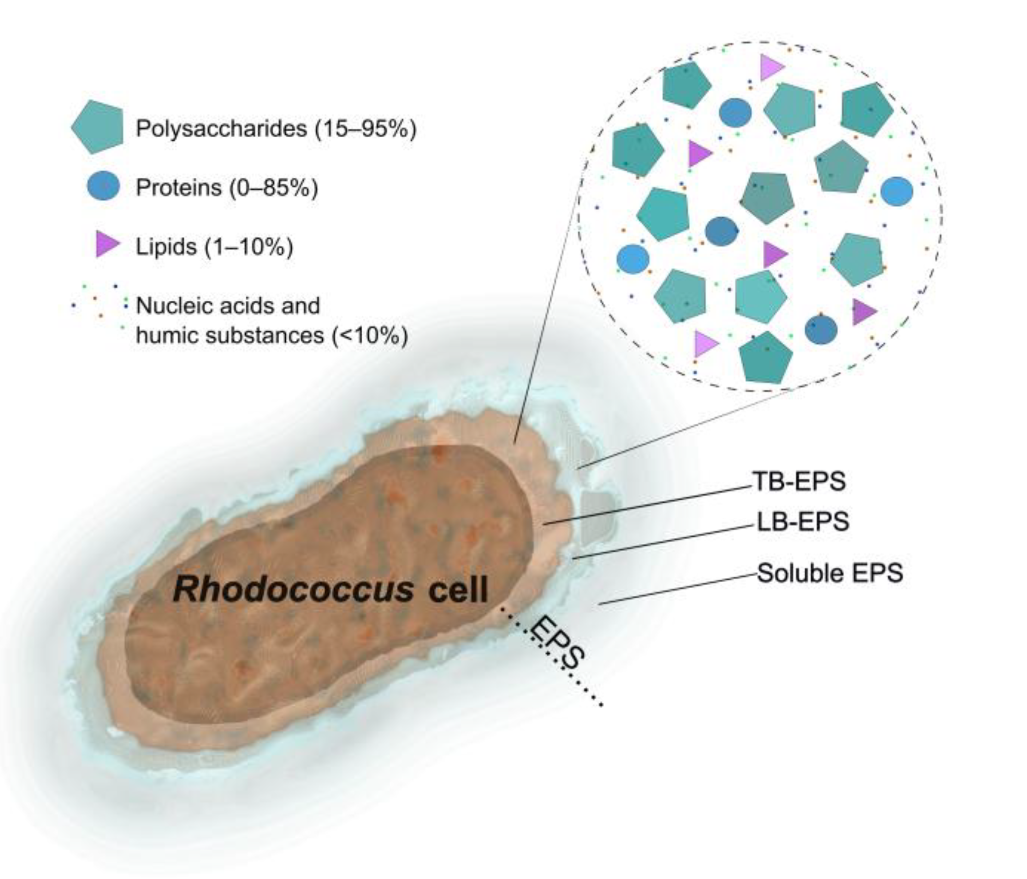
Figure 2.
The activities of Rhodococcus EPS and their causes, with examples of strains showing these activities. Mw – molecular weight.
Figure 2.
The activities of Rhodococcus EPS and their causes, with examples of strains showing these activities. Mw – molecular weight.
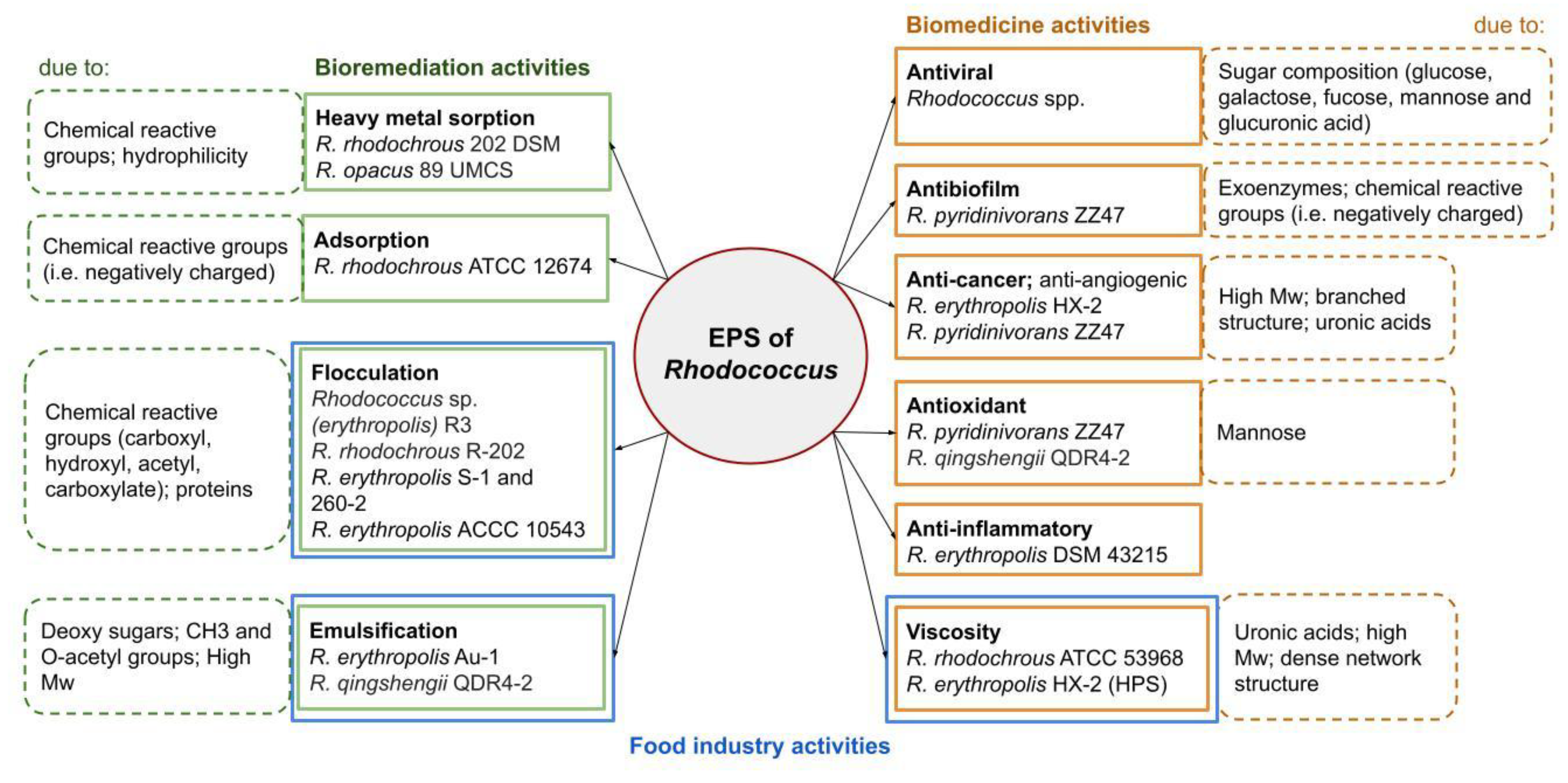

Table 1.
The diversity of EPS produced by Rhodococcus spp.
| Species of Rhodococcus | Compounds name | Molecular weight (Da) | Composition | Bioactivities | References | ||||||||||
| R. hoagii CECT555, R. erythropolis CECT3013, R. rhodochrous CECT5749, R. rhodnii CECT5750, R. coprophilus CECT5751 | No data | No data | No data | Antiviral; Binding Norovirus virus -like particles |
[30] |
||||||||||
| R. pyridinivorans ZZ47 | No data | No data | No data | As antibiofilm, anti-angiogenic, antioxidant agents | [6,31] | ||||||||||
| R. erythropolis HX-2 | HPS | 1.04 × 10⁶ | 79.24% carbohydrate, 5.2% protein and 8.45% lipid Glucose, galactose, fucose, mannose and glucuronic acid with a mass ratio of 27.29%, 24.83%, 4.79%, 26.66% and 15.84% |
As anticancer, viscosity agents |
[8] | ||||||||||
| R. rhodochrous ATCC 53968 | No data | No data | Galactose, glucose, fucose, and glucuronic acid at a molar ratio of 3: 2: 2: 2 1.3% stearic acid, 4.1% palmitic acid, 5.8% pyruvic acid |
As thickeners | [27] | ||||||||||
| R. erythropolis DSM 43215 | PLS-1 | 1.14 × 10⁶ | Glucose and mannose in the molar ratio of 1 : 1 3.3% of proteins |
As antiinflammatory agents | [32] | ||||||||||
| Rhodococcus strain 33 | No data | 1.05 × 10⁵ | Glucuronic acid, glucose, galactose and rhamnose in a molecular ratio of 1:1:1:2. | Adhesion; improving biodegradation | [33] | ||||||||||
| Rhodococcus strain 33 | 33 EPS; PS-33 | > 2 × 10⁶ | D-galactose, D-glucose, D-mannose, D-glucuronic, pyruvic acids in ratio 1:1:1:1:1 | Improving hydrocarbon tolerance | [34] | ||||||||||
| R. erythropolis PR4 | FR2 | No data | d-galactose, d-glucose, d-mannose, pyruvic acid and d-glucuronic acid in ratio 1:1:1:1:1; 2.9% stearic acid and 4.3% palmitic acid | Improving hydrocarbon tolerance | [28] | ||||||||||
| R. erythropolis PR4 | FACEPS | No data | Glucose, N-acetylglucosamine, glucuronic acid, and fucose at a molar ratio of 2:1:1:1. | Improving hydrocarbon tolerance | [25] | ||||||||||
| R. rhodochrous202 DSM and R. opacus 89 UMCS | No data | No data | 52.1% and 62.7% content of CHx groups | Heavy metals (Ni(II), Pb(II), Co(II), Cd(II) and Cr(VI)) sorption | [35] | ||||||||||
| R. opacus |
BES.DM-GMA-TETA-EPS Microspheres |
No data | No data | Pb(II), Cd(II) sorption | [36] | ||||||||||
| R. erythropolis ACCC 10543 | RSF; NOC-1 | No data | Proteoglycan (glycoprotein) composed of polysaccharides (91.2%), protein (7.6%), and DNA (1.2%). | Flocculation | [16] [37] |
||||||||||
| R. erythropolis S-1 and 260-2 | No data | No data | No data | Flocculation | [38,39] | ||||||||||
| R. rhodochrous R-202 | R-202 | 1.3 × 10⁶ | 62.86% of polysaccharide and 10.36% of protein. Mannose, glucose and galactose at the molar ratio of 12:6:1. |
Flocculation (Effective at pH around 7 and in the presence of salt solutions) |
[40] | ||||||||||
| R. opacus89 UMCS | No data | 7.6 × 10⁵ |
64.6% polysaccharide 9.44% protein Mannose, glucose, and galactose. |
Flocculation; Binding metal cations |
[41] | ||||||||||
| R. sp. (erythropolis) R3 | No data | 3.99 × 10⁵ | 84.6% protein and 15.2% sugar content | Flocculation | [42] | ||||||||||
| R. qingshengii QDR4-2 | QEPS | 9.450 × 10⁵ | Mannose and glucose in a molar ratio of 81.5:18.5 | As antioxidant agent; Emulsifying | [43] | ||||||||||
| R. rhodochrous ATCC 12674 | SM-1 EPS | No data | D-galactose, D-glucose, L-fucose, and D-glucuronic acid at a molar ratio of 6: 3: 2: 4 1.2% stearic acid, 2.3% palmitic acid, and 10.3% pyruvic acid |
Absorption | [44] | ||||||||||
| R. erythropolis Au-1 | No data | No data | No data | Emulsification | [45] | ||||||||||
| Rhodococcus sp. RHA1 (NRCC 6316) | No data | No data |
hexuronic acid and neutral glycose in the approximate ratio of 1:3. D-galactose, D-glucose, L-fucose, d-glucuronic acid in ratio 1:1:1:1 |
No data | [46] | ||||||||||
| R. ruber C208 | No data | No data | Polysaccharides and proteins in ratio 2.5:1 | Adhesion | [47] | ||||||||||
| Rhodococcus sp. SJ | EOM | No data | No data | Resuscitation of nonculturable cells; Improving polychlorbiphenyls degradation |
[48] | ||||||||||
Disclaimer/Publisher’s Note: The statements, opinions and data contained in all publications are solely those of the individual author(s) and contributor(s) and not of MDPI and/or the editor(s). MDPI and/or the editor(s) disclaim responsibility for any injury to people or property resulting from any ideas, methods, instructions or products referred to in the content. |
© 2025 by the authors. Licensee MDPI, Basel, Switzerland. This article is an open access article distributed under the terms and conditions of the Creative Commons Attribution (CC BY) license (http://creativecommons.org/licenses/by/4.0/).
Copyright: This open access article is published under a Creative Commons CC BY 4.0 license, which permit the free download, distribution, and reuse, provided that the author and preprint are cited in any reuse.


