Preprint
Article

This version is not peer-reviewed.

Nasal Colonization by Methicillin and Glycopeptide Resistant Staphylococcus aureus in Symptomatic and Asymptomatic Individuals in Maputo, Mozambique

Submitted:

29 November 2025

Posted:

05 December 2025

You are already at the latest version

Abstract

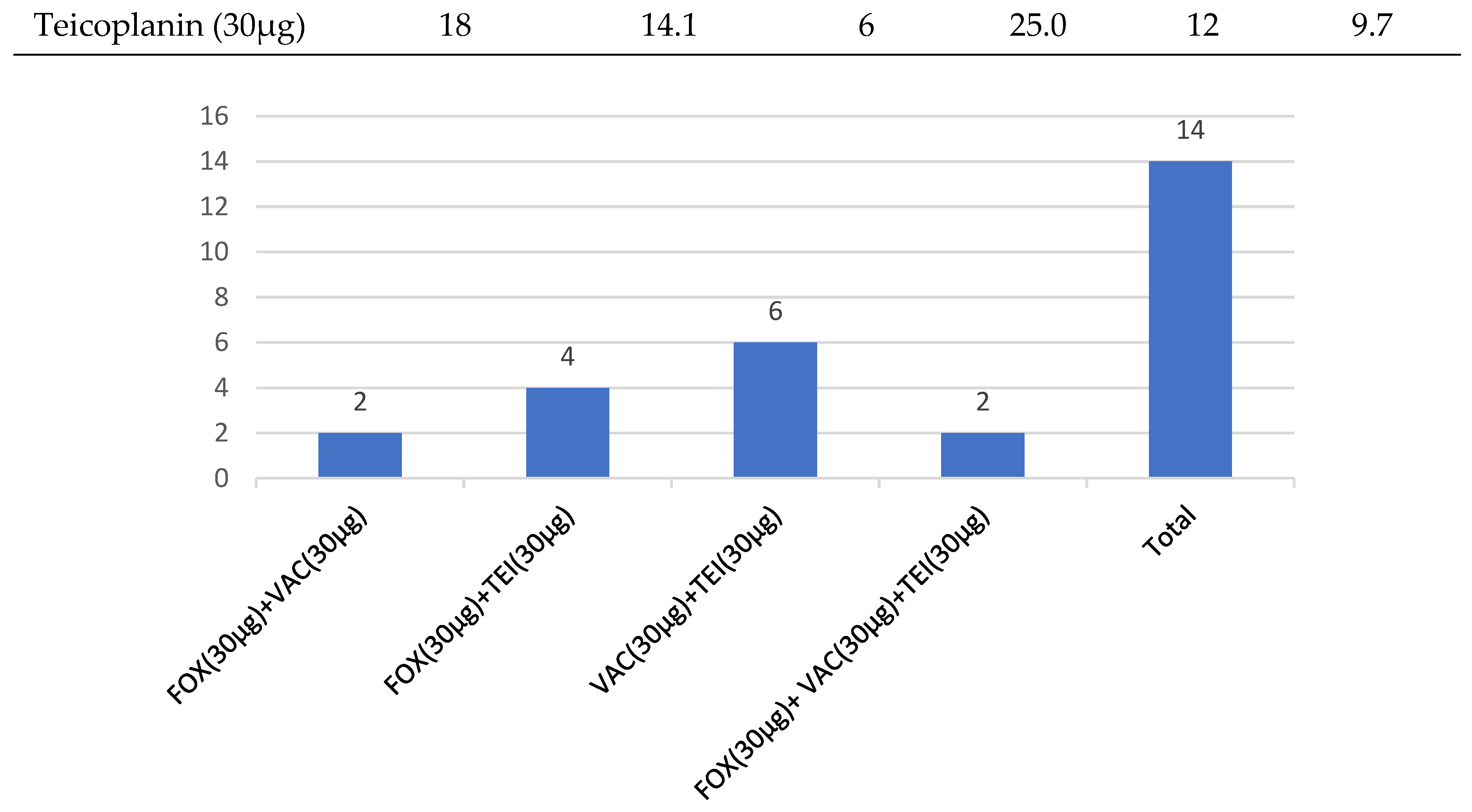
Background: Nasal Staphylococcus aureus is a major source of community and hospital associated staphylococcal infections, including Methicillin-resistant S. aureus (MRSA). Screening of MRSA nasal colonization is important in the prevention and control of infection and may provide useful information to guide antimicrobial therapy.Objective: The study aims to determine the prevalence of nasal colonization by S. aureus and investigated its antimicrobial resistance profile to methicillin and glycopeptides in hospital and non-hospital settings in Maputo, Mozambique. Methods: A cross-sectional study was conducted by collecting nasal specimens of symptomatic outpatients and asymptomatic students at Eduardo Mondlane University in Maputo. Using culture techniques on Mannitol salt and HiCrome™ Rapid MRSA agar, S. aureus was isolated based on the colonial characteristics and confirmed by Gram staining, catalase, and Microgen®Staph tests. The antibiotic susceptibility test was done using Kirby-Buer disk diffusion method on Salt Muellar Hinton agar for methicillin (cefoxitin), vancomycin and teicoplanin antibiotics. Results: A total of 50 (39.1%) S. aureus isolates were obtained from 128 collected nasal samples, in which 104 (31.3%) were from university students and 24 (7.8%) from outpatient’s form university clinic. The isolates showed a low overall 7.8% resistance to cefoxitin and, 14.1% and 11.7% presumptive resistance to teicoplanin and vancomycin, respectively. Conclusion: Methicillin and glycopeptide resistant S. aureus were highly prevalent in asymptomatic students. These observations call for strategies to prevent S. aureus spread to more vulnerable populations where the consequences can be severe.

Keywords: 
;  ;  ;  

Introduction

Staphylococcus aureus is a versatile commensal microorganism found in the human body and is associated with a wide variety of infections with high morbidity, mortality, and healthcare-associated costs [1,2]. S. aureus can efficiently adapt to multiple niches, establishing solid interactions with epithelial cells[2,3] and able to overcome these defenses and colonize this microenvironment[2,4,5]. The most frequent carriage site is the nose, with approximately 20 to 40% of the general population being permanently but asymptomatically colonized, and another 30% may carry the bacterium intermittently[2,6]. Interminent nasal colonization by S. aureus represents the major risk factor for infections such as cellulitis, endocarditis, impetigo, pneumonia, urinary tract infections, osteomyelitis, pneumonia, septicemia, and toxic shock syndrome (TSS) [2,4,7,8]. Its pathogenicity is multifaceted, encompassing antimicrobial resistance (AMR) and a high proficiency for biofilm formation[2,9,10].
S. aureus has become resistant to various antimicrobial agents, including the commonly used penicillin-related antibiotics such as methicillin, resulting in one of the top pathogens responsible for global deaths associated with AMR, with methicillin-resistant S. aureus (MRSA) causing more than 100,000 annual deaths[11]. For instance, most MRSA isolates have also shown resistance to vancomycin – a glycopeptide that was introduced and widely used as a preferred option for treating MRSA infections [12,13,14].
While globally, including in African context. the prevalence rates of MRSA and multidrug-resistant (MDR) S. aureus strains reach as high as 61.6% [15,16,17,18,19,20,21,22], data from sub-Saharan Africa, particularly in Mozambique, remain scarce. However, the few studies conducted in both hospital and community settings have documented S. aureus infection rates of up to 12.3% [22,23], with MDR and MRSA accounting for as much as 33.3% of these infections [23,24,25]. This has escalated into a global health challenge, as bacterial infections once easily treated have become increasingly difficult to manage [17,26].
The nasal colonization of both S. aureus and MRSA plays a key role in the epidemiology and pathogenesis of both MDR S. aureus and MRSA infections in various populations [13,17,27]. Screening for and eliminating nasal carriage of Staphylococcus aureus and MRSA has been associated with a reduction in infection rates[28,29,30]. In other hand, understanding the antimicrobial patterns of S. aureus is crucial for developing effective prevention and control measures – particularly in developing countries, where healthcare infrastructure and access to medical services remain limited [13,30,31].
This study aims to determine the prevalence of nasal carriage of methicillin and glycopeptide resistant S. aureus among outpatients attended at the university clinic, and students from the Department of Biological Sciences, Eduardo Mondlane University, Mozambique.

Materials and Methods

Study Type and Population

This was a cross-sectional study carried out from June to November 2022 (during Covid-19 pandemic), in a group of symptomatic outpatients attended at the Eduardo Mondlane University (UEM) clinic, and asymptomatic students from the Department of Biological Sciences, Eduardo Mondlane University (DBC-UEM).
Nasal specimens were collected aseptically from one hundred and twenty-four [128] participants, comprising outpatients and students. Willingness of the subjects to participate in the study was a strong criterion.
Nasal specimens were aseptically collected from one hundred and twenty-eight [128] participants, including both outpatients and students. A key criterion for inclusion in the study was the participants’ willingness to take part. Additionally, participants were asked if had undergone antibiotic treatment for at least two weeks prior to sample collection; and those who had, were excluded. The nasal specimens were collected with labelled sterile swabs sticks and taken within 2 hours of collection for inoculation on selective media in the Microbiology laboratory at the DBC-UEM.
Before the study, ethical approval was obtained from the Mozambican National Ethical Committee on June 06, 2022 (Approval No. 347/CNBS/22). The study was explained to the participants, and only those who provided written informed consent and informed assent in the case of minors under 18 years of age, were included. Confidentiality and privacy were maintained throughout the study.

Isolation and Characterization of S. aureus

Each of all the swab samples was inoculated onto sterilized Mannitol salt agar (MSA) and HiCrome™ Rapid MRSA agar (HiMedia, India) plates and incubated aerobically at 37°C for 24 to 48h. The culture media were prepared and sterilized according to the manufacturer’s instructions.
The isolates were characterized using standard established microbiological methods. Isolates with golden yellow-coloured colonies on MSA agar plates and give greenish yellow-coloured colonies on HiCrome™ Rapid MRSA agar were assumed to be S. aureus, and subsequently sub-cultured on MSA plates to obtain pure colonies, and confirmation by Gram staining, catalase, and agglutination (Microgen®Staph) tests. All isolates that were Gram positive cocci (grape-like clusters) and positive to catalase and agglutination tests, were considered as S. aureus and submitted to antimicrobial susceptibility testing.

Antimicrobial Susceptibility Test

Antimicrobial susceptibility testing was performed on Muller Hinton Agar (MHA) (HiMedia, India) using the disc diffusion method in accordance with the Clinical and Laboratory Standards Institute (CLSI) guidelines[32,33,34]. In summary, a brief suspension of multiple colonies from the pure culture of the S. aureus isolate was prepared with normal sterile saline solution and adjusted to a turbidity at 0.5 McFarland standards. Then, the cell suspension was swabbed, and the swab was spread on the surface of an MHA and left briefly to dry at room temperature. Thenceforth, the antibiotic disks were applied onto the agar; and allowed to stand for at least 30mins before incubated for 24 hours at 30 to 35°C, to allow the growth of methicillin-resistant strains [26,34,35]. Then, sterile cotton swabs were used to spread the cell suspension evenly on the surface of MHA, and left briefly the plates to dry at room temperature. Three (3) antibiotic discs (Oxoids U.K) including: cefoxitin (30µg) - for detection of MRSA, vancomycin (30µg) and teicoplanin (30µg) were placed on the surface of the inoculated MHA plates; and allowed to stand for at least 30mins before incubated for 24 hours at 30 to 35°C, for the growth of methicillin resistant strains [26,34,35). MRSA strain (ATCC 25923) provided by José Sumbana (Laboratory of Microbiology, Faculty of Medicine, UEM) was used as control. The diameter of inhibition zone was measured and antibiogram was determined according to the Clinical and Laboratory Standard Institute (CLSI) guidelines (Table 1), as described by Roy et al. [32], Deepak et al. [33] and Ennab et al. [34].

Statistical Analysis

Statistical data analysis was performed using SPSS 21 software (IBM Corp., Armonk, NY). Frequencies were obtained and percentages were calculated for study variables. Demographic characteristics were compared with the use of Chi-square test. All reported p-value of less than or equal to 0.05 were considered to be statistically significant (>0.05).

Results

One hundred and twenty-eight samples, were taken up for the study, 24 outpatients to represent the symptomatic resistant profile, while 104 were from students, representing the asymptomatic resistant profile. The modal age group for both groups was 1830 years with a total of 116 patients. The age ranges of the patients were from 1–62 years (Table 2).
Distribution of Staphylococcus aureus by participants characteristics
A total of fifty (39.1%) out of 128 nasal samples of overall subjects screened gave S. aureus isolates, and among the remainder number, 42.2% (n=54) gave Coagulase-Negative Staphylococcus (CoNS) isolates. The University students appeared to have high S. aureus colonization prevalence than the UEM clinic outpatients, but the difference is not statistically significant (P = 0.684). As shown in Table 2, the female volunteers were more colonized than their male counterpart, with a significant difference (P = 0.005). No statistically significant difference was found for the S. aureus colonization prevalence between the age group, but it was observed that most of the participants (n=116) were in 18-30 years age group, with 42 (32.8%) of them being positive for S. aureus colonization.
Table 2. The prevalence of S. aureus and Coagulase-Negative Staphylococcus among the participants.
Category Sample
Number (N)
S. aureus CoN Staphylococcus P value*
n (%) n (%)
Overall 128 50 39.1 54 42.2 -
Type of participants
UEM clinic Outpatients 24 10 7.8 11 8.6 0.684
DBC-UEM Students 104 40 31.3 43 33.6
Sex
Female 81 23 18.0 41 32.0 0.005
Male 47 27 21.1 13 10.2
Age group (years)
≤17 2 2 1.6 0 0.0 0.525
18-30 116 42 32.8 51 39.8
31-45 4 3 2.3 1 0.8
46-60 3 2 1.6 0 0.0
≥61 3 1 0.8 2 1.6
CoN Coagulase-Negative Staphylococcus; *Chi-square test
Staphylococcus aureus antibiotic resistance levels for both symptomatic (Outpatientes) andasymptomatic (Students) participants
In general, the isolates showed low resistance to the three (3) tested antibiotics, as shown in Table 2. Overall, 7.8% of the isolate’s showed methicillin (cefoxitin) resistance, while 14.1% and 11.7% presumptively resistant to teicoplanin and vancomycin, respectively. The observed differences in the isolates’ resistance pattern among the participants’ groups are not statistically significant (P > 0.05).
In this study we analysed the combined (to two antibiotics tested) or multi-drug (to all three antibiotics tested) resistance pattern of S. aureus isolates (Figure 1). Fourteen (28%) of all (n=50) the S. aureus isolates were at least presumptively resistant for two antibiotics tested, with two of them (4%) were presumptively multi-drug resistant (for methicillin vancomycin and teicoplanin) and six (12%) showed combined presumptive resistance to both glycopeptide antibiotics (vancomycin and teicoplanin).
Table 3. The Antimicrobial resistance profile of S. aureus isolates from the nares of the participants (students and outpatients).
Antibiotics Overall Resistance (n=128) Outpatientes (n=24) Students (n=124)
n (%) n (%) n (%)
Cefoxitin (30µg) 10 7.8 1 4.2 9 7.3
Vancomycin (30µg) 15 11.7 4 16.7 11 8.9
Teicoplanin (30µg) 18 14.1 6 25.0 12 9.7
Figure 1. The combined or multi-drug presumptive resistance pattern of S. aureus isolates from the nares of the volunteers. Legend: FOX, cefoxitin; VAC, vancomycin; TEI, teicoplanin.
Preprints 187373 g001

Discussion

S. aureus colonization has long been recognized as an important risk factor for subsequent infection caused by the colonizing strain [16,27]. This pathogen is capable of causing a wide spectrum of clinical manifestations, ranging from minor skin and soft tissue infections to invasive and life-threatening diseases such as pneumonia, bacteremia, and sepsis. Its clinical importance is amplified by its remarkable ability to acquire resistance to nearly all available classes of antibiotics [26,32]. Of particular concern is the global dissemination of multidrug-resistant (MDR) strains, especially methicillin-resistant S. aureus (MRSA), which complicates treatment and limits therapeutic options. Nasal colonization of S. aureus has been implicated in both community-associated infections, such as skin and soft tissue infections, and hospital-associated infections, including bacteremia and surgical site infections [17,27,36].
The present study provides insights into the prevalence of nasal carriage of S. aureus and MRSA among both symptomatic and asymptomatic individuals, while also exploring their glycopeptide antibiotic susceptibility profiles. Our findings demonstrate an overall S. aureus nasal carriage rate of 39.1%, which aligns with previous studies conducted in diverse geographic regions. For instance, Uemura et al. (2004) in Japan, Kuehnert et al. [37] in the United States, and Onanuga & Temedie [26] in Nigeria reported prevalence rates of 36.0%, 32.4%, and 33.3%, respectively, among healthy adults. This concordance suggests that a baseline prevalence of approximately one-third of the population being colonized appears consistent across different populations and continents.
Nevertheless, variations in prevalence have been reported. Higher colonization rates exceeding 43% were documented in both hospital and non-hospital populations in Abia State, Nigeria [38], as well as among medical students [39] and hospital personnel [40] in Nepal. In contrast, substantially lower colonization rates have also been observed. For instance, Adesida et al. [41] reported a prevalence of only 14.0% among students in Lagos, Nigeria, while Abie et al. [42]found 23.2% prevalence among janitors in Addis Ababa, Ethiopia. The recent study by Taqveem et al. [43] in Pakistan similarly noted low prevalence among students. Such discrepancies highlight that nasal carriage is influenced by multiple epidemiological and environmental factors, including antibiotic usage, healthcare exposure, population demographics, and even sampling and laboratory techniques [17,26].
Antibiotic exposure appears to be particularly influential. Populations receiving antibiotic treatment at the time of sampling often exhibit lower S. aureus prevalence due to suppression of colonization by susceptible strains. Conversely, hospital settings typically report higher colonization rates because of increased opportunities for bacterial transmission in environments with high infection burdens [41]. In contrast, non-hospitalized populations not under antibiotic treatment may exhibit relatively higher prevalence, reflecting natural colonization patterns. This has been confirmed in studies of community populations where nasal colonization typically ranges between 20–30% [2,6]. These findings collectively demonstrate that S. aureus colonization is shaped by a complex interplay between microbial ecology, host factors, and healthcare exposure.
In the present study, the anterior nares were confirmed as a major reservoir for S. aureus, consistent with previous reports [27]. Importantly, we found no significant difference in colonization rates between symptomatic and asymptomatic individuals, suggesting that carriage is not necessarily associated with acute infection risk at the individual level. However, colonization serves as a latent risk, providing a reservoir for potential infection and onward transmission. Interestingly, colonization was significantly associated with sex (P = 0.005), with males exhibiting higher carriage rates. This observation echoes findings by [43), where male students demonstrated higher prevalence of both S. aureus (57.7%) and MRSA (21.4%) compared to females (42.3% and 17.9%). Potential explanations include behavioral differences (e.g., hygiene practices, sports participation, or crowding exposures), as well as biological differences such as hormonal or immune factors.
Age-related colonization patterns have also been described globally, with higher rates observed shortly after birth and during adolescence [16]. In African populations, additional risk factors have been identified, including HIV infection, frequent hand-washing with harsh detergents, rural residence, and hospitalization, particularly in surgical wards [44,45]. Conversely, higher educational status and male sex have been reported as protective in some settings [16], suggesting that risk factors may be context-specific and interact with socioeconomic, environmental, and cultural variables.
Antibiotic susceptibility testing in our study revealed low overall resistance rates among nasal S. aureus isolates. However, MRSA prevalence was 7.8%, accompanied by presumptive resistance to teicoplanin (14.1%) and vancomycin (11.7%). These findings are consistent with both local and international studies. For example, Ceccarelli et al.[46] reported 4% MRSA prevalence, van der Meeren et al. [23] found 15.1%, and Vubil et al. [24] documented 8% in Mozambique. Comparable results were also seen in Pakistan, with Taqveem et al. [43] reporting 5.5%. While MRSA prevalence appears relatively low in healthy carriers, its presence remains clinically significant because colonization can lead to difficult-to-treat infections, especially in healthcare environments.
Comparisons with international studies underscore regional variability. Chen & Huang [47] reported MRSA prevalences from <5% to >35% in Asian countries, Olonitola et al. [48] 14.85% in Nigeria, Rijal et al.[49] 56.1% in Nepal, and Onanuga & Temedie [26] 27.5% in Nigeria. The variability may reflect differences in infection control practices, antibiotic prescribing patterns, and genetic background of circulating strains. Notably, our MRSA estimation was based on phenotypic resistance to cefoxitin rather than molecular detection of the mecA gene, which encodes the altered penicillin-binding protein (PBP2a). This methodological limitation means that some “phenotypic MRSA” isolates may not carry mecA, as reported in previous studies [46,50].
The public health implications of these findings are profound. Nasal carriage of S. aureus, particularly MRSA, is a major driver of subsequent infections, with estimates suggesting 7% of hospitalized patients and 2% of community populations carry MRSA [17]. Furthermore, healthcare-associated MRSA strains often display extensive multidrug resistance, whereas community-acquired strains remain more susceptible to non-β-lactam antibiotics[51]. This distinction is critical for guiding empirical therapy and infection control interventions.
Our findings on glycopeptide resistance merit particular attention. Resistance to vancomycin and teicoplanin was presumptively identified, though it is well established that conventional disc diffusion may underestimate or misclassify resistance due to poor diffusion of large molecules in agar [32,33]. Most studies report very low resistance rates (≤11.1%) and primarily intermediate resistance phenotypes [34,52]. However, in African context, the intermediate resistance of S. aureus to vancomycin has been reported in high rates (≤14.5) [14,53]. Nonetheless, the global emergence of vancomycin-resistant S. aureus (VRSA) remains a serious concern, particularly as vancomycin has long been considered the last line of defense against MRSA infections. Teicoplanin is increasingly viewed as a valuable alternative [33]. Our findings therefore highlight the urgent need for enhanced surveillance and reliable confirmatory testing, especially in resource-limited settings such as Mozambique.
This study has several limitations. First, susceptibility to vancomycin and teicoplanin was assessed using disc diffusion, which is not recommended by CLSI guidelines [54]. Thus, our categorization of isolates as “presumptively resistant” requires cautious interpretation and should ideally be validated using MIC or E-test methods. However, studies by Deepak et al. [33] indicate that disc diffusion and E-test may yield broadly comparable results, suggesting that disc diffusion remains a pragmatic and cost-effective option where resources are constrained. Second, the relatively small and specific study population limits generalizability, underscoring the need for larger-scale, heterogeneous studies across diverse Mozambican populations.
In conclusion, our findings reinforce the significance of nasal carriage of S. aureus and MRSA as reservoirs for subsequent infection and transmission. With a prevalence of 39.1% for S. aureus and 7.8% for MRSA, coupled with evidence of presumptive glycopeptide resistance, this study highlights both the ongoing burden of colonization and the emerging threat of antimicrobial resistance in Mozambique. Targeted surveillance, judicious antibiotic stewardship, and confirmatory testing are essential steps in mitigating the impact of MRSA and other resistant strains. Future research should focus on larger populations, incorporate molecular methods for resistance detection, and evaluate the effectiveness of decolonization strategies in reducing infection rates.

Acknowledgements

The authors acknowledge the Mozambican National Investigation Fund (FNI) for the research funding and are grateful to the UEM university clinic and DBC for the cooperation during sample collection. We are also thankful to the students and patients from whom we collected our community samples.

Author Contributions

All authors made a significant contribution to the work reported, whether that is in the conception, study design, execution, acquisition of data, analysis and interpretation, or in all these areas; took part in drafting, revising or critically reviewing the article; gave final approval of the version to be published; have agreed on the journal to which the article has been submitted; and agree to be accountable for all aspects of the work.

Disclosure

The authors declare no potential conflicts of interest with respect to the research, authorship, publication of this article, or this work.

References

  1. Thampi N, Showler A, Burry L, Bai AD, Steinberg M, Ricciuto DR, et al. Multicenter study of health care cost of patients admitted to hospital with Staphylococcus aureus bacteremia: Impact of length of stay and intensity of care. Am J Infect Control. 2015 Jul;43(7):739–44. [CrossRef]
  2. Sakr A, Brégeon F, Mège JL, Rolain JM, Blin O. Staphylococcus aureus Nasal Colonization: An Update on Mechanisms, Epidemiology, Risk Factors, and Subsequent Infections. Front Microbiol. 2018 Oct 8;9. [CrossRef]
  3. Bartlett AH, Hulten KG. Staphylococcus aureus Pathogenesis. Pediatric Infectious Disease Journal. 2010 Sep;29(9):860–1.
  4. Tong SYC, Davis JS, Eichenberger E, Holland TL, Fowler VG. Staphylococcus aureus Infections: Epidemiology, Pathophysiology, Clinical Manifestations, and Management. Clin Microbiol Rev. 2015 Jul;28(3):603–61. [CrossRef]
  5. Kluytmans J, van Belkum A, Verbrugh H. Nasal carriage of Staphylococcus aureus: epidemiology, underlying mechanisms, and associated risks. Clin Microbiol Rev. 1997 Jul;10(3):505–20. [CrossRef]
  6. Wertheim HF, Melles DC, Vos MC, van Leeuwen W, van Belkum A, Verbrugh HA, et al. The role of nasal carriage in Staphylococcus aureus infections. Lancet Infect Dis. 2005 Dec;5(12):751–62. [CrossRef]
  7. Wertheim HF, Vos MC, Ott A, van Belkum A, Voss A, Kluytmans JA, et al. Risk and outcome of nosocomial Staphylococcus aureus bacteraemia in nasal carriers versus non-carriers. The Lancet. 2004 Aug;364(9435):703–5. [CrossRef]
  8. Nouwen J, Schouten J, Schneebergen P, Snijders S, Maaskant J, Koolen M, et al. Staphylococcus aureus Carriage Patterns and the Risk of Infections Associated with Continuous Peritoneal Dialysis. J Clin Microbiol. 2006 Jun;44(6):2233–6. [CrossRef]
  9. Krismer B, Peschel A. Does Staphylococcus Aureus Nasal Colonization Involve Biofilm Formation? Future Microbiol. 2011 May 17;6(5):489–93. [CrossRef]
  10. Zacher AT, Mirza K, Thieme L, Nietzsche S, Senft C, Schwarz F. Biofilm formation of Staphylococcus aureus on various implants used for surgical treatment of destructive spondylodiscitis. Sci Rep. 2024 Aug 21;14(1):19364. [CrossRef]
  11. Murray CJL, Ikuta KS, Sharara F, Swetschinski L, Robles Aguilar G, Gray A, et al. Global burden of bacterial antimicrobial resistance in 2019: a systematic analysis. The Lancet. 2022 Feb;399(10325):629–55. [CrossRef]
  12. Foster TJ. Antibiotic resistance in Staphylococcus aureus. Current status and future prospects. Vol. 41, FEMS Microbiology Reviews. Oxford University Press; 2017. p. 430–49. [CrossRef]
  13. Gnanamani A, Hariharan P, Paul-Satyaseela M. Staphylococcus aureus: Overview of Bacteriology, Clinical Diseases, Epidemiology, Antibiotic Resistance and Therapeutic Approach. In: Frontiers in Staphylococcus aureus. InTech; 2017. [CrossRef]
  14. Adeoye-Isijola M, Olajuyigbe O, Adebola K, Coopoosamy R, Afolayan A. Vancomycin intermediate resistant Staphylococcus aureus in the nasal cavity of asymptomatic individuals: A potential public health challenge. Afr Health Sci. 2020;20(3):1109–17. [CrossRef]
  15. Conceição T, de Lencastre H, Aires-de-Sousa M. Carriage of Staphylococcus aureus among Portuguese nursing students: A longitudinal cohort study over four years of education. PLoS One. 2017 Nov 30;12(11):e0188855. [CrossRef]
  16. Schaumburg F, Alabi AS, Peters G, Becker K. New epidemiology of Staphylococcus aureus infection in Africa. Vol. 20, Clinical Microbiology and Infection. Blackwell Publishing Ltd; 2014. p. 589–96. [CrossRef]
  17. Lee AS, Huttner BD, Catho G, Harbarth S. Methicillin-Resistant Staphylococcus aureus: An Update on Prevention and Control in Acute Care Settings. Vol. 35, Infectious Disease Clinics of North America. W.B. Saunders; 2021. p. 931–52. [CrossRef]
  18. Lawal OU, Ayobami O, Abouelfetouh A, Mourabit N, Kaba M, Egyir B, et al. A 6-Year Update on the Diversity of Methicillin-Resistant Staphylococcus aureus Clones in Africa: A Systematic Review. Vol. 13, Frontiers in Microbiology. Frontiers Media S.A.; 2022. [CrossRef]
  19. Ezeh CK, Eze CN, Dibua MEU, Emencheta SC. A meta-analysis on the prevalence of resistance of Staphylococcus aureus to different antibiotics in Nigeria. Antimicrob Resist Infect Control. 2023 Apr 25;12(1):40. [CrossRef]
  20. Diop M, Bassoum O, Ndong A, Wone F, Ghogomu Tamouh A, Ndoye M, et al. Prevalence of multidrug-resistant bacteria in healthcare and community settings in West Africa: systematic review and meta-analysis. BMC Infect Dis. 2025 Feb 28;25(1):292. [CrossRef]
  21. Hasanpour AH, Sepidarkish M, Mollalo A, Ardekani A, Almukhtar M, Mechaal A, et al. The global prevalence of methicillin-resistant Staphylococcus aureus colonization in residents of elderly care centers: a systematic review and meta-analysis. Antimicrob Resist Infect Control. 2023 Jan 29;12(1):4. [CrossRef]
  22. Abdulgader SM, Shittu AO, Nicol MP, Kaba M. Molecular epidemiology of Methicillin-resistant Staphylococcus aureus in Africa: a systematic review. Front Microbiol. 2015 Apr 30;6.
  23. van der Meeren BT, Millard PS, Scacchetti M, Hermans MH, Hilbink M, Concelho TB, et al. Emergence of methicillin resistance and P anton- V alentine leukocidin positivity in hospital- and community-acquired S taphylococcus aureus infections in B eira, M ozambique. Tropical Medicine & International Health. 2014 Feb 11;19(2):169–76.
  24. Vubil D, Garrine M, Ruffing U, Acácio S, Sigaúque B, Alonso PL, et al. Molecular Characterization of Community Acquired Staphylococcus aureus Bacteremia in Young Children in Southern Mozambique, 2001–2009. Front Microbiol. 2017 May 4;8. [CrossRef]
  25. Garrine M, Quintó L, Costa SS, Messa A, Massinga AJ, Vubil D, et al. Epidemiology and clinical presentation of community-acquired Staphylococcus aureus bacteraemia in children under 5 years of age admitted to the Manhiça District Hospital, Mozambique, 2001–2019. European Journal of Clinical Microbiology & Infectious Diseases. 2023 May 18;42(5):653–9. [CrossRef]
  26. Onanuga A, Tc T. Nasal carriage of multi-drug resistant Staphylococcus aureus in healthy inhabitants of Amassoma in Niger delta region of Nigeria. Vol. 11, African Health Sciences. 2011.
  27. von Eiff C, Becker K, Machka K, Stammer H, Peters G. Nasal Carriage as a Source of Staphylococcus aureus Bacteremia. New England Journal of Medicine. 2001 Jan 4;344(1):11–6. [CrossRef]
  28. Robotham J V., Graves N, Cookson BD, Barnett AG, Wilson JA, Edgeworth JD, et al. Screening, isolation, and decolonisation strategies in the control of meticillin resistant Staphylococcus aureus in intensive care units: Cost effectiveness evaluation. BMJ (Online). 2011 Oct 15;343(7827). [CrossRef]
  29. Lee AS, de Lencastre H, Garau J, Kluytmans J, Malhotra-Kumar S, Peschel A, et al. Methicillin-resistant Staphylococcus aureus. Nat Rev Dis Primers. 2018 May 31;4(1):18033.
  30. Abebe A, Birhanu A. Methicillin Resistant Staphylococcus aureus: Molecular Mechanisms Underlying Drug Resistance Development and Novel Strategies to Combat. Infect Drug Resist. 2023 Dec;Volume 16:7641–62. [CrossRef]
  31. Tarai B, Das P, Kumar D. Recurrent Challenges for Clinicians: Emergence of Methicillin-resistant Staphylococcus aureus, Vancomycin Resistance, and Current Treatment Options. J Lab Physicians. 2013 Jul 7;5(02):071–8. [CrossRef]
  32. Roy PC, Shaheduzzaman Md, Sultana N, Jahid IK. Comparative Antibiotic Sensitivity Pattern of Hospital and Community Acquired <i>Staphylococcus aureus</i> Isolates of Jessore, Bangladesh. J Biosci Med (Irvine). 2015;03(10):17–23.
  33. K D. In-Vitro Activity of Teicoplanin against Clinical Methicillin Resistant Staphylococcus aureus Isolates. Open Access Journal of Microbiology & Biotechnology. 2017 Mar;2(1).
  34. Ennab R, Al-Momani W, Al-Titi R, Elayan A. Antibiotic Profile of Pathogenic Bacteria Isolated from Postsurgical Site Infections in Public Hospitals in Northern Jordan. Infect Drug Resist. 2022;15:359–66. [CrossRef]
  35. Brown DFJ, Wootton M, Howe RA. Antimicrobial susceptibility testing breakpoints and methods from BSAC to EUCAST. Journal of Antimicrobial Chemotherapy. 2016 Jan;71(1):3–5. [CrossRef]
  36. Techasupaboon T, Vasikasin V, Varothai N, Raknaisil N, Nasomsong W. Staphylococcus aureus nasal carriage and bloodstream infection among conventional hemodialysis patients in Thailand: a prospective multicenter cohort study. BMC Res Notes. 2022 Dec 1;15(1). [CrossRef]
  37. Kuehnert MJ, Kruszon-Moran D, Hill HA, McQuillan G, McAllister SK, Fosheim G, et al. Prevalence of Staphylococcus aureus Nasal Colonization in the United States, 2001–2002. J Infect Dis. 2006 Jan 15;193(2):172–9.
  38. Chigbu C. O, Ezeronye OU. Antibiotic resistant Staphylococcus aureus in Abia State of Nigeria. Afr J Biotechnol. 2003 Oct 31;2(10):374–8. [CrossRef]
  39. Rongpharpi SR, Hazarika NK, Kalita H. The prevalence of nasal carriage of Staphylococcus aureus among healthcare workers at a tertiary care hospital in Assam with special reference to MRSA. Journal of Clinical and Diagnostic Research. 2013 Feb 1;7(2):257–60.
  40. Mukhiya RK, Shrestha A, Rai SK, Panta K, Singh R, Rai G, et al. Prevalence of Methicillin-Resistant Staphylococcus aureus in Hospitals of Kathmandu Valley. Nepal J Sci Technol. 2013 Mar 10;13(2):185–90. [CrossRef]
  41. Adesida SA, Abioye OA, Bamiro BS, Brai BIC, Smith SI, Amisu KO, et al. Associated risk factors and pulsed field gel electrophoresis of nasal isolates of Staphylococcus aureus from medical students in a tertiary hospital in Lagos, Nigeria. Brazilian Journal of Infectious Diseases. 2007 Feb;11(1). [CrossRef]
  42. Abie S, Tiruneh M, Abebe W. Methicillin-resistant Staphylococcus aureus nasal carriage among janitors working in hospital and non-hospital areas: a comparative cross-sectional study. Ann Clin Microbiol Antimicrob. 2020 Dec 1;19(1). [CrossRef]
  43. Taqveem A, Rasool MH, Aslam B, Mujahid F, Ibrar A, Ejaz H, et al. Methicillin-Resistant Staphylococcus aureus among Students: Nasal Carriage Rate, Contributing Factors, and Antimicrobial Susceptibility. Medicina (B Aires). 2024 Sep 27;60(10):1590. [CrossRef]
  44. Schaumburg F, Nurjadi D, Dike AE, Ojurongbe O, Kolawole DO, Kun JF, et al. Clonal expansion accounts for an excess of antimicrobial resistance in Staphylococcus Adesola O.
  45. Kinabo GD, van der Ven A, Msuya LJ, Shayo AM, Schimana W, Ndaro A, et al. Dynamics of nasopharyngeal bacterial colonisation in HIV -exposed young infants in T anzania. Tropical Medicine & International Health. 2013 Mar 16;18(3):286–95.
  46. Ceccarelli D, Mondlane J, Sale M, Salvia AM, Folgosa E, Cappuccinelli P, et al. Sporadic methicillin resistance in community acquired Staphylococcus aureus in Mozambique. Vol. 28, THE NEW MICROBIOLOGICA. 2005.
  47. Chen CJ, Huang YC. New epidemiology of Staphylococcus aureus infection in Asia. Clinical Microbiology and Infection. 2014 Jul;20(7):605–23. [CrossRef]
  48. Olonitola OS, Olayinka BO, Sani FD. Antibiotic susceptibility of <i>Staphylococcus aureus</i> isolates from a Nigerian Federal Medical Center. Cameroon Journal of Experimental Biology. 2008 Feb 27;3(2). [CrossRef]
  49. Rijal KR, Pahari N, Shrestha BK, Nepal AK, Paudel B, Mahato P, et al. Prevalence of methicillin resistant Staphylococcus aureus in school children of Pokhara. Nepal Med Coll J. 2008 Sep;10(3):192–5.
  50. Olayinka BO, Olayinka AT, Obajuluwa AF, Onaolapo JA, Olurinola PF. Absence of meca gene in methicillin-resistant staphylococcus aureus isolates. Afr J Infect Dis. 2010 Jun 2;3(2). [CrossRef]
  51. Fey PD, Saïd-Salim B, Rupp ME, Hinrichs SH, Boxrud DJ, Davis CC, et al. Comparative Molecular Analysis of Community- or Hospital-Acquired Methicillin-Resistant Staphylococcus aureus. Antimicrob Agents Chemother. 2003 Jan;47(1):196–203. [CrossRef]
  52. Shariati A, Dadashi M, Moghadam MT, van Belkum A, Yaslianifard S, Darban-Sarokhalil D. Global prevalence and distribution of vancomycin resistant, vancomycin intermediate and heterogeneously vancomycin intermediate Staphylococcus aureus clinical isolates: a systematic review and meta-analysis. Sci Rep. 2020 Jul 29;10(1):12689. [CrossRef]
  53. Belete MA, Gedefie A, Alemayehu E, Debash H, Mohammed O, Gebretsadik D, et al. The prevalence of vancomycin-resistant Staphylococcus aureus in Ethiopia: a systematic review and meta-analysis. Antimicrob Resist Infect Control. 2023 Aug 30;12(1):86. [CrossRef]
  54. Humphries RM, Abbott AN, Hindler JA. Understanding and Addressing CLSI Breakpoint Revisions: a Primer for Clinical Laboratories. J Clin Microbiol. 2019 Jun;57(6). [CrossRef]
Table 1. CLSI standards of antibiotics inhibition zone diameter measurement.
Antimicrobial agente Inhibition/Diameter zone (mm)
R (resistant) I (intermediate) S (sensitive)
Cefoxitin (30µg) ≤ 21 - ≥ 22
Vancomycin (30µg) - - ≥15
Teicoplanin (30µg) - - ≥14
Disclaimer/Publisher’s Note: The statements, opinions and data contained in all publications are solely those of the individual author(s) and contributor(s) and not of MDPI and/or the editor(s). MDPI and/or the editor(s) disclaim responsibility for any injury to people or property resulting from any ideas, methods, instructions or products referred to in the content.
Copyright: This open access article is published under a Creative Commons CC BY 4.0 license, which permit the free download, distribution, and reuse, provided that the author and preprint are cited in any reuse.
Prerpints.org logo

Preprints.org is a free preprint server supported by MDPI in Basel, Switzerland.

Subscribe

Disclaimer

Terms of Use

Privacy Policy

Privacy Settings

© 2026 MDPI (Basel, Switzerland) unless otherwise stated