
Submitted:
26 November 2025
Posted:
27 November 2025
You are already at the latest version
Abstract
Sex-based variations are significant among the major subtypes of leukemia. This review briefly discusses the current understanding and knowledge gaps related to sex differences in epidemiology; mortality and survival rates; risk factors, and epigenetic, metabolomic, and sex-specific patterns. Males have higher incidence rates and mortality rates of leuke-mia than females do, highlighting the significance of biological and epidemiologic factors. Sex-based differences were reported in only 0.5% of the clinical trials, an underreporting may be resulting from a persistent lack of awareness or prioritization of integrating sex as a significant variable in research findings. Interesting sex-based patterns arise that sub-stantially impact disease epidemiology. An intriguing medical enigma in leukemia is treatment response, where women have higher overall survival rates but more severe treatment-related toxicity. However, ALL in pediatric patients contradicts this enigma, suggesting that gender differences may be less pronounced during childhood when hor-monal levels are low. Sex-based risk factors are discussed at both the environmental and genetic levels where epigenetic variations, such as DNA methylation, affect gene expres-sion differently in males versus females. Hematological and biochemical patterns are shown from a sex-based perspective in this review. Furthermore, there are clear signs of sex-based differences in the recognition of various metabolites in males and females, which could support the foundation for future research. We encourage researchers not only to perform sex-stratified analyses in their ongoing studies but also to design sex-based clinical trials.
Keywords:
sex differences
; leukemia
; risk factors
; sex stratification
; sex-based
; epigenetics
; metabolites
; enigma
1. Introduction
Males have higher incidence and mortality rates of leukemia than females do, highlighting the importance of biological and epidemiologic factors [1,2,3,4,5,6]. However, the incidence of cases and leukemia-related deaths are expected to increase globally until 2030 [6,7]. The causes of the differences in disease burden between males versus females are still unknown, even though a variety of factors, including genetic polymorphisms, epigenetics, hormones, senescence, immunity, and angiogenesis, may contribute to the explanation of this observation [8,9].
There is a significant knowledge gap regarding how sex hormones affect the progression of non-sex-specific malignancies, the increased risk of treatment-related side effects in female patients, and the effects of both donor and recipient sex on the outcome of allogeneic stem cell transplantation [10]. Remarkably, the Trialtrove database revealed that only 472 out of 89,221 cancer clinical trials included curated sex-based comparisons, accounting for just 0.5% of all trials [11]. Underreporting might be a result of a persistent lack of awareness or prioritization of integrating sex as a significant variable in research findings [12]. In light of this gap, we briefly review the current understanding and identify the knowledge gaps related to sex differences in epidemiology; mortality and survival rates; risk factors, and epigenetic, metabolomic, and sex-specific patterns.
2. Incidence of Leukemia in Males and Females
Males are not only more likely than females to develop acute lymphoblastic leukemia (ALL) but also to relapse. Age-related increases in the male-to-female ratio, especially in the 10–20 age range, indicate a potential hormonal influence [13].
There are sex differences in the incidence and overall survival of acute myeloid leukemia (AML) patients. Males account for approximately 57% of cases, reflecting a male predominance in incidence. Moreover, females tend to have better survival rates. Caregivers and public policies need to address treatment and health care factors to lower mortality rates and minimize current disparities between sexes [14].
The male-to-female ratios of chronic lymphocytic leukemia (CLL) ranged from 1.3:1 to 1.7:1 [15], with a slightly greater prevalence observed in males than in females. In the United States between 2017 and 2021, men (6.3 new cases per 100,000 persons) were diagnosed with CLL at a higher rate than women (3.3 new cases per 100,000 persons), regardless of race. In addition, males had a higher CLL mortality rate (1.6 deaths per 100,000) than women did (0.7 deaths per 100,000) between 2018 and 2022 [16].
In 2025, approximately 9550 children (aged birth to 14 years) and 5140 adolescents (aged 15 to 19 years) are estimated to be diagnosed with cancer, with 1050 and 600 deaths, respectively. Cancer is estimated to be diagnosed in every 264 children and adolescents before the age of 20 in the United States [10] where leukemia is the leading cause of childhood cancer, accounting for 28% of cases. Approximately 65,111 leukemia cases have been reported in children aged 0- 14 years, with 37,833 cases being male and 27,278 being female, according to cancer statistics in 2018 [11]. The most common cancer in adolescents is central nervous system tumors (22%), followed by leukemia (13%), then lymphoma (19%), where Hodgkin lymphoma is more prevalent in adolescents than non-Hodgkin lymphoma, in contrast to a reverse prevalence in pediatric patients [10].
3. Mortality of Leukemia Patients
Cancer is the second leading cause of mortality among children aged 1- 14 years (after accidents) and the fourth leading cause of death among adolescents aged 15- 19 years in the United States [10]. Brain and other nervous system tumors are the major causes of cancer death among children and adolescents under the age of 20, closely followed by leukemia in the United States [10]. Overall, leukemia deaths in children aged 0- 14 years in 2018 were 1,792 in males and 12,149 in females worldwide [11].
4. Sex Specific Patterns in Leukemia
Globally, the incidence of leukemia is higher in male than in female patients (269,503 vs. 205,016, respectively) [13]. Leukemia mortality is higher in male patients than in female patients globally (177,818 vs. 133,776, respectively) [13]. In leukemia, the global lifetime risk of incidence (LRI) and mortality (LRM) are generally higher in males than in females [22]. The global LRI for leukemia is higher in males (0.65%) compared to females (0.54%). Similarly, males have a higher global LRM (0.44%) compared to females (0.37%) [22]. Interesting patterns arise that substantially impact disease epidemiology (Figure 1).
5. Survival Rate of Leukemia PATIENTS
The 5-year relative survival rate for chronic myeloid leukemia (CML) has almost tripled, increasing from 22% in the mid-1970s to 70% between 2014 and 2020, with tyrosine kinase inhibitor (TKI) drugs offering the majority of patients a near-normal life expectancy [23]. Despite the approval of three generations of TKI, 5% to 10% CML patients develop drug resistance and a risk of progression to acute disease; thus, resistant CML is an active research area [24]. Metabolic intervention is an emerging therapeutic prospect for both resistant CML [25] and AML [26], which has a 5-year relative survival rate of only 32% [17].
There are significant sex-based variations among the major leukemia subtypes. Regardless of the underlying molecular processes, males consistently have greater incidence rates and poorer survival rates in patients with AML, ALL, and CML. An intriguing medical enigma in leukemia is treatment response, where women have higher overall survival rates but more severe treatment-related toxicity.
The observed differences in response to TKI therapy in CML patients indicate that females achieve major molecular response rates (80% vs. 45%, p = 0.018), despite higher treatment intolerance rates [27]. In ALL, females have better survival outcomes (42% vs. 16% of trials), despite having higher toxicity levels than males do [13]. In CLL, the incidence of treatment-induced toxicity was greater in females than in males (85% vs. 78%), especially gastrointestinal toxicity (57% vs. 42%), yet females responded better to treatment overall (83% in women vs. 71% for men), despite increased toxicity from treatment. These results suggest that better treatment outcomes may result from pharmacokinetic variations between the sexes and the action of hormones such as estrogen [28].
However, this isn’t always the case, as ALL pediatric patients contradict this enigma. Female pediatric patients with ALL have been reported to have a higher incidence of treatment related toxicity, treatment delays, and deaths than males [29]. A study in ALL pediatric patients contradicts this enigma, as it revealed that female treatment toxicity and the incidence of treatment-related deaths are greater than those in males. An increase in treatment-related late effects was reported in ALL pediatric females. Females were substantially more likely than males to experience grade 3 or 4 toxicities (liver, gastrointestinal), hospital days, therapy delays, and supportive care interventions (transfusions, intravenous antibiotics) among ALL high-risk patients. The cumulative incidence of treatment-related deaths was 1.2% for males and 2.6% for females five months after treatment initiation. Male and female patients tend to have varying risk profiles, with females being more likely to develop acute and long-term treatment-related toxicities [30]. Another study in pediatric patients with T-cell ALL showed that females had shorter 5-year survival than males. Overall, female patients had more deaths than male patients. Notably, among those treated with the Capizzi methotrexate regimen, females experienced a significantly inferior 5-year disease-free survival compared to males. This difference may be explained by studies suggesting that methotrexate clearance is lower in females, resulting in higher drug levels and more toxicities [31].
Hormonal influences in childhood and adulthood may explain the enigmatic contradiction described above. Research has indicated potential protective benefits of estrogen for females [32], and sex differences may be less pronounced during childhood when estrogen and androgen levels are low [33]. Recognizing and tackling these sex differences in drug use is essential for creating more personalized and effective cancer treatments while minimizing adverse effects [34].
6. Sex-Based Risk Factors
The global lifetime risk of developing (LRI) and dying from (LRM) all hematologic malignancies was 1.67% and 0.98% respectively, in 2022. Cancer-specific analysis showed that the LRI was highest for Non-Hodgkin Lymphoma (0.73%), followed by leukemia (0.60%), while the LRM was highest for leukemia (0.41%). LRIs and LRMs were higher in males than in females with leukemia [22].
The causes behind the higher incidence of leukemia in males are not well understood. There has been a limited focus on how risk factors might affect males and females differently in the development of leukemia, and several epidemiological studies acknowledge sex as a risk factor but fail to detail how exposure differences might vary between the sexes. Males and females might have different responses to the same toxic, environmental, and hormonal exposures [35].
6.1. Environmental, Lifestyle, and Parental Risk Factors
The potential leukemia-related mortality risk factors, which are based on the variables provided by the GBD database [36], are smoking, high body mass index (BMI), and exposure to carcinogens due to occupational exposure to benzene and formaldehyde [37]. Risk factors differ in terms of sex. The GBD database revealed that smoking had a greater impact on mortality and disability-adjusted life year (DALY) in males than in females. Among the 88,784.2 leukemia deaths globally associated with the risk factors listed above, smoking accounted for 72.74%, with males contributing 56.46% and females contributing 16.28%. However, leukemia-related mortality was more affected by high BMI in [37]. Notably, among females, smoking and high BMI contribute similarly to mortality [37].
A higher risk of ALL in daughters is associated with maternal obesity during pregnancy [36]. Further research investigating this mother‒daughter association may help clarify the potential etiology of sex hormone/chromosome-related ALL [38]. There was an elevated leukemia risk among the offspring of men employed in occupations in electromagnetic fields or radiation exposures, especially in males < 6 years old, but no significant relationship was detected in females [39]. The etiology of pediatric acute leukemia may be related to the domestic or garden use of pesticides three months before pregnancy [40]. In chronic myeloid leukemia (CML), males have a greater risk of developing CML, potentially because they have more target cells at risk. The hematopoietic stem cell in which BCR/ABL initiates CML is referred to as the target cell [41].
6.2. Genetic Risk Factors
The molecular effects of toxic exposure can also differ between sexes [35]. Extensive research has shown varying sex-based responses to genetic stimuli. For sex-specific outcomes, the two missense SNPs of the HLA-DQA1 gene, rs12722042 and rs12722039, had the largest effect sizes. The DQA1-01 HLADQA1 SNPs are linked to an increased risk of ALL in males [42]. Certain single-nucleotide polymorphisms (SNPs) in the ARID5B gene are associated with ALL in both males and females, even though some SNPs have sex-specific associations. For example, the SNPs rs10740055, rs10994982, and rs6479779 are significant in females, whereas the SNPs rs10821938 and rs7923074 are significant in males [43]. Gene polymorphisms such as del{GSTT1} and NQO1*2hom. are associated with an increased risk of acute leukemia, with a greater impact apparent in males [44]. The persistent correlation between SLX4IP deletion and male sex, along with the extension of this sex bias to TAL1 locus deletion, suggests that differential illegitimate V(D)J-mediated recombination at specific loci may contribute to the consistently higher incidence rates of childhood ALL in boys than in girls [45].
PHF6 mutations are seven times more common in males with AML than in females, whereas PHF6 alterations exclusively occur in males with T-cell acute lymphoblastic leukemia (T-ALL) [46,47]. UTX mutations are found only in male T-ALL patients [48]. In a study of recently diagnosed AML patients, NPM1 mutations were found in both sexes and were associated with slightly different distributions of the mutation types. Type A mutations are more prevalent in females than in males [49]. In adult AML patients, women are more likely than men to have a normal karyotype and FLT3-ITD, DNMT3A, NPM1, and WT1 mutations, whereas complex karyotypes and ASXL1, SRSF2, U2AF1, RUNX1, or KIT mutations are less common [50]. The 2022 European LeukemiaNet [51] classification revealed sex differences in the percentages of patients assigned to genetic-risk groups, whereas more women were in the intermediate-risk category, and more men were in the adverse-risk group [50].
In CLL, male sex has been identified as a risk factor for both CLL development and poor survival outcomes [28,52,53]. High-risk genetic and laboratory markers, including unmutated immunoglobulin heavy chain variable (IGHV) genes, mutated or deleted tumor protein 53 (TP53) genes, 11q deletions, elevated beta-2 microglobulin concentrations, ZAP-70 positivity, and CD38 positivity, are less common in women with CLL than in men [28].
Research revealed differing responses, based on sex, to genetic and environmental factors. Grasping the distinct impacts of exposure and sensitivity to hazardous stimuli in males and females would enhance the effectiveness of personalized prevention [35] .
7. Epigenetic Patterns in Males and Females
Dysregulation of epigenetic patterns and mutations in epigenetic modifiers that disturb normal blood cell formation play a role in the onset of various types of leukemia [54,55]. Most alterations in methylation-regulated gene expression focus on those genes that play roles in cell signaling, growth, differentiation, and programmed cell death. Methylation has proven valuable in predicting relapse possibilities and outcomes from a clinical perspective. Epigenetic variations, such as DNA methylation and histone modification, affect gene expression differently in males and females, influencing not only physiology but also susceptibility to the disease [56].
In ALL, a significant incidence of methylation is prevalent in adolescent and young adult ALL patients, with greater methylation related to particular clinicopathologic characteristics, such as male gender and an elevated WBC count. Patients frequently exhibit abnormal methylation, particularly males, which may indicate a common pathogenic mechanism [57].
In AML, male and female DNA methylation patterns are very different, particularly in CpG-rich promoter regions. Almost 10% of these promoters exhibit differential methylation between sexes. Functional analysis revealed that genes with differentially methylated promoters are associated primarily with homeobox, cell development, and morphogenesis, with men having a greater number of enrichments than women do (1893 and 322 enrichments, respectively). Potential epigenetic prognostic markers were found to be sex specific, with male and female survival-significant gene sets differing by 75% [58].
Research in CLL revealed 1043 sex-related differentially methylated positions (DMPs), including 987 on the X chromosome and 56 on autosomes. These DMPs are associated with variations in gene expression between male and female CLL patients, indicating that differences in DNA methylation may contribute to sex-related differences in CLL risk. Based on published B-cell RNA-sequencing data, 18 genes covered by DMPs presented varying levels of expression in male and female CLL patients. Among these genes, TRIB1 is an autosomal gene that has been demonstrated to suppress apoptosis and thus promote tumor growth. An epigenome-wide association analysis revealed that changes in DNA methylation may contribute to sex-related differences in the risk of developing CLL [59].
DAPK1 gene methylation was more common in females with advanced CML than in males, indicating that the epigenetic regulation of the disease differs between the sexes. Although the exact function of DNA methylation in CML development is not fully understood, researchers could discover new treatment options for the later stages of the disease by examining the epigenetic changes associated with genes that regulate CML [60].
Personalized epigenetic approaches can help overcome challenges such as chemoresistance in cancer therapy by tailoring treatment on the basis of individual epigenetic signatures [61].
8. Sex-Based Hematological and Biochemical Profiles
Hematological and biochemical profiles are crucial for determining patient prognosis before and after chemotherapy treatment [62]. Hematological parameters include complete blood count and white blood differential count, whereas biochemical parameters include renal function tests, liver function tests, serum electrolytes, and serum proteins [62].
Hematology parameters, such as red blood cells, hemoglobin, and platelet levels, are relatively low in pediatric B-ALL patients; however, white blood cell levels are elevated. There was a substantial difference in the biochemical profile based on sex and age between B-ALL patients, mostly males under 5 years, whereas the hematological profile did not significantly change [62].
Hematological data have shown that male adult ALL patients have more red blood cells, hemoglobin, and hematocrit, with significant mean differences compared with females [63]. Nevertheless, both sexes of ALL patients presented mean levels of red blood cells, hemoglobin, and hematocrit that were below the normal range for both sexes [64]. Notably, alterations in red blood cells are linked to the development of anemia [65]. Furthermore, biochemical data revealed that creatinine levels were higher in male ALL patients than in female patients, with a mean difference of 6686 mg/dL [66]. Patients of both sexes had creatinine levels less than normal compared with standard values [63]. Patients with ALL and variations in creatinine levels have already been reported; these patients mainly exhibit kidney injury caused by hematological disease [67,68].
Compared with male patients, female patients with CML have a significantly better survival rate. Compared with male patients, female patients had a longer median survival (58 vs 49 months, respectively), which was significantly due to better survival in the low- and intermediate-risk groups. Furthermore, CML female patients had lower hemoglobin levels and higher platelet counts than did male patients [69].
In chronic lymphocytic leukemia, male patients had a substantially higher average WBC count by 2.92 × 10³/μL than female patients did [70]. Importantly, white blood cell count, age, and sex are potential factors for patient risk assessment [71,72].
Biochemical parameters, such as the mean lactate dehydrogenase (LDH) levels in females, were found to be greater than those in males. A significant difference was identified only in individuals with chronic myeloid leukemia. Lactate dehydrogenase levels were significantly greater (p<0.001) in all types of leukemia with age. These findings suggest that increased cellular LDH activity indicates a shift toward anaerobic metabolism and glycolysis in malignant cells, which is associated with a high turnover rate. A further practical metric for assessing the clinical and prognostic aspects of leukemia may be the measurement of LDH levels in patients [73].
9. Sex-Specific Metabolites in Leukemia
"Omics" is a scientific discipline that explores the influence of genes, proteins, and metabolic pathways on disease susceptibility to enhance diagnosis, prognosis, and the development of new drugs. Advances in technology and informatics have enabled large-scale analyses of genes, proteins, and metabolites, resulting in fields such as genomics, proteomics, and metabolomics, which have improved our understanding of certain disorders [74]. In addition to the fields mentioned above, new omics approaches known as "sex omics" have been proposed to investigate sex-specific aspects of the biomedical sciences [74]. One of the most recent significant additions to -omics techniques is metabolomics, described as the qualitative and quantitative analysis of the metabolome, which is the whole set of metabolites found in cells, biological fluids, and tissues [75]. Owing to continuous advancements in the field, metabolomics seeks to offer personalized and optimal treatment plans in addition to a more rapid and precise diagnosis [76]. Taking sex differences into account could contribute to the discovery of biomarker metabolites for diagnosis, prognosis, and treatment, as well as clinical innovations aimed at improving men's and women's health [74].
Using newborn dried blood spots, untargeted metabolomics was performed to analyze neonatal exposure as a potential risk factor for AML. Metabolite features linked to AML were identified via sex-stratified analysis since variations in AML incidence rates indicate sex disparities. It appears that neonatal metabolomic profiles of pediatric AML risk are sex-specific because there was no overlap between the 16 predictors of AML in females and the 15 predictors in males. In males, the metabolite profile of AML predictors appears to be heterogeneous, while in females, ceramides, a class of metabolites associated with the proliferation of cancer cells, were putatively annotated as four predictors of AML. In females, breastfeeding duration was strongly associated with two metabolite predictors of AML, suggesting a potential biological link between childhood leukemia and this putative protective risk factor [77].
A comprehensive profiling of pituitary hormones and circulating sex steroids in CLL patients, both male and female, revealed a sex-specific association of these signaling molecules with treatment-free survival. The circulating hormone profiles of healthy donors and CLL patients differed significantly. Males had a lower median treatment-free survival (TFS) than females did (80 vs. 135 months, respectively). Shorter TFS in male CLL patients was associated with higher luteinizing hormone levels. An increased TFS was linked to female CLL patients with elevated levels of testosterone, dihydrotestosterone, and biologically active estrogen metabolites. CLL female patients who expressed high levels of the steroid-inactivating UGT2B17 enzyme showed reduced TFS. These results suggest that distinct biological mechanisms are linked to the progression of leukemia in males and females and support the idea that CLL is a hormone-responsive disease [78].
It is crucial to prioritize sex considerations during the stages of research planning and implementation, ensuring that studies include both sexes as subjects and ultimately perform omics studies in both mixed-sex groups and male/female-separated cohorts [79]. However, there are clear signs of sex-based differences in the recognition of various metabolites in males and females, which could support shaping the foundation for future research [80].
10. Molecular Mechanisms Underlying Sex Differences in Leukemia
Sex differences in leukemia are influenced by genetic, epigenetic, and immune responses (Table 1). Genetic factors such as loss of a sex chromosome (LOS) are common in hematological cancers. Among 868 patients with hematologic diseases, 5.1% exhibited LOS [81]. The highest frequency was observed in AML patients at 9.5%, compared to a range of 0-6% for ALL, CLL, and CML [81]. Epigenetic factors reflect the role of gender-specific H3K27 methylation imbalance in T-cell leukemogenesis. UTX mutant leukemias are more sensitive to H3K27me3 inhibitor therapy, providing new opportunities for epigenetically targeted therapy in T-ALL [82]. Males and females differ in the abundance of innate immunity cellular components [83]. Responses to cancer neoantigens are affected by normal sex variations in adaptive immunity [84,85,86,87]. Sex steroids (such as estrogens, testosterone, and progesterone), which bind to hormone receptors on the surface of immune cells and modulate the function of immune system molecules, are primarily responsible for the differences in disease prevalence and expression between males and females [88,89].
11. Conclusions
Despite the obvious sex-based patterns in leukemia, there are still significant knowledge gaps. Additional research is needed to clarify the mechanisms underlying sex-based disparities in disease biology. The inclusion of sex-specific factors could improve the prediction of outcomes and treatment approaches in leukemia patients. We encourage researchers not only to perform sex-stratified analyses in their ongoing studies but also to design sex-based clinical trials. Considering sex differences will pave the way for personalized medicine.
Institutional Review Board Statement
Not applicable.
Data Availability Statement
No new data were created.
Conflicts of Interest
The authors declare no conflicts of interest.
References
- Yang, X.; Chen, H.; Man, J.; Zhang, T.; Yin, X.; He, Q.; Lu, M. Secular Trends in the Incidence and Survival of All Leukemia Types in the United States from 1975 to 2017. J Cancer 2021, 12. [CrossRef]
- Rifat, R.H.; Poran, Md.S.; Islam, S.; Sumaya, A.T.; Alam, Md.M.; Rahman, M.R. Incidence, Mortality, and Epidemiology of Leukemia in South Asia: An Ecological Study. Open J Epidemiol 2023, 13. [CrossRef]
- Miranda-Filho, A.; Piñeros, M.; Ferlay, J.; Soerjomataram, I.; Monnereau, A.; Bray, F. Epidemiological Patterns of Leukaemia in 184 Countries: A Population-Based Study. Lancet Haematol 2018, 5. [CrossRef]
- Bertuccio, P.; Bosetti, C.; Malvezzi, M.; Levi, F.; Chatenoud, L.; Negri, E.; Vecchia, C. La Trends in Mortality from Leukemia in Europe: An Update to 2009 and a Projection to 2012. Int J Cancer 2013, 132. [CrossRef]
- Li, B.; Tang, H.; Cheng, Z.; Zhang, Y.; Xiang, H. The Current Situation and Future Trend of Leukemia Mortality by Sex and Area in China. Front Public Health 2020, 8. [CrossRef]
- Sharma, R.; Jani, C. Mapping Incidence and Mortality of Leukemia and Its Subtypes in 21 World Regions in Last Three Decades and Projections to 2030. Ann Hematol 2022, 101. [CrossRef]
- Du, M.; Chen, W.; Liu, K.; Wang, L.; Hu, Y.; Mao, Y.; Sun, X.; Luo, Y.; Shi, J.; Shao, K.; et al. The Global Burden of Leukemia and Its Attributable Factors in 204 Countries and Territories: Findings from the Global Burden of Disease 2019 Study and Projections to 2030. J Oncol 2022, 2022. [CrossRef]
- Stabellini, N.; Tomlinson, B.; Cullen, J.; Shanahan, J.; Waite, K.; Montero, A.J.; Barnholtz-Sloan, J.S.; Hamerschlak, N. Sex Differences in Adults with Acute Myeloid Leukemia and the Impact of Sex on Overall Survival. Cancer Med 2023, 12. [CrossRef]
- Rubin, J.B. The Spectrum of Sex Differences in Cancer. Trends Cancer 2022, 8.
- Özdemir, B.C.; Richters, A.; Oertelt-Prigione, S.; Adjei, A.A.; Borchmann, S.; Haanen, J.B.A.G.; Letsch, A.; Quaas, A.; Verhoeven, R.H.A.; Wagner, A.D. 1592P Awareness and Interest of Oncology Professionals in Sex and Gender Differences in Cancer Risk and Outcome: Analysis of an ESMO Gender Medicine Task Force Survey. Annals of Oncology 2024, 35, S959. [CrossRef]
- Kammula, A. V; Schäffer, A.A.; Rajagopal, P.S.; Kurzrock, R.; Ruppin, E. Outcome Differences by Sex in Oncology Clinical Trials. Nat Commun 2024, 15, 2608.
- van Eldik, M.J.A.; Ali, M.; Rietkerken, S.; Peters, S.A.E.; den Ruijter, H.M.; Ruigrok, Y.M. Underreporting of Sex-Specific Findings in Risk Factors for Unruptured Intracranial Aneurysms. Cerebrovascular Diseases 2025.
- Jaime-Pérez, J.C.; Hernández-De Los Santos, J.A.; Fernández, L.T.; Padilla-Medina, J.R.; Gómez-Almaguer, D. Sexual Dimorphism in Children and Adolescents with Acute Lymphoblastic Leukemia: Influence on Incidence and Survival. J Pediatr Hematol Oncol 2020, 42, e293–e298. [CrossRef]
- Stabellini, N.; Tomlinson, B.; Cullen, J.; Shanahan, J.; Waite, K.; Montero, A.J.; Barnholtz-Sloan, J.S.; Hamerschlak, N. Sex Differences in Adults with Acute Myeloid Leukemia and the Impact of Sex on Overall Survival. Cancer Med 2023, 12, 6711–6721. [CrossRef]
- Chennamadhavuni, A.; Lyengar, V.; Mukkamalla, S.K.R. Leukemia - StatPearls - NCBI Bookshelf. Ncbi 2023.
- NIH Cancer Stat Facts. Surveillance, Epidemiology, and End Results Program 2022.
- Siegel, R.L.; Kratzer, T.B.; Giaquinto, A.N.; Sung, H.; Jemal, A. Cancer Statistics, 2025. Ca 2025, 75, 10.
- Namayandeh, S.M.; Khazaei, Z.; Najafi, M.L.; Goodarzi, E.; Moslem, A. GLOBAL Leukemia in Children 0-14 Statistics 2018, Incidence and Mortality and Human Development Index (HDI): GLOBOCAN Sources and Methods. Asian Pacific Journal of Cancer Prevention 2020, 21. [CrossRef]
- Bray, F.; Laversanne, M.; Sung, H.; Ferlay, J.; Siegel, R.L.; Soerjomataram, I.; Jemal, A. Global Cancer Statistics 2022: GLOBOCAN Estimates of Incidence and Mortality Worldwide for 36 Cancers in 185 Countries. CA Cancer J Clin 2024, 74, 229–263.
- Sharma, R.; Jani, C. Mapping Incidence and Mortality of Leukemia and Its Subtypes in 21 World Regions in Last Three Decades and Projections to 2030. Ann Hematol 2022, 101, 1523–1534.
- Amini, M.; Sharma, R.; Jani, C. Gender Differences in Leukemia Outcomes Based on Health Care Expenditures Using Estimates from the GLOBOCAN 2020. Archives of public health 2023, 81, 151.
- Sun, K.; Wu, H.; Zhu, Q.; Gu, K.; Wei, H.; Wang, S.; Li, L.; Wu, C.; Chen, R.; Pang, Y. Global Landscape and Trends in Lifetime Risks of Haematologic Malignancies in 185 Countries: Population-Based Estimates from GLOBOCAN 2022. EClinicalMedicine 2025, 83.
- Osman, A.E.G.; Deininger, M.W. Chronic Myeloid Leukemia: Modern Therapies, Current Challenges and Future Directions. Blood Rev 2021, 49, 100825.
- Sasaki, K.; Strom, S.S.; O’Brien, S.; Jabbour, E.; Ravandi, F.; Konopleva, M.; Borthakur, G.; Pemmaraju, N.; Daver, N.; Jain, P. Relative Survival in Patients with Chronic-Phase Chronic Myeloid Leukaemia in the Tyrosine-Kinase Inhibitor Era: Analysis of Patient Data from Six Prospective Clinical Trials. Lancet Haematol 2015, 2, e186–e193.
- Li, Y.; Zeng, P.; Xiao, J.; Huang, P.; Liu, P. Modulation of Energy Metabolism to Overcome Drug Resistance in Chronic Myeloid Leukemia Cells through Induction of Autophagy. Cell Death Discov 2022, 8, 212.
- Chen, Y.; Chen, J.; Zou, Z.; Xu, L.; Li, J. Crosstalk between Autophagy and Metabolism: Implications for Cell Survival in Acute Myeloid Leukemia. Cell Death Discov 2024, 10, 46.
- Beaudet, M.-E.; Szuber, N.; Moussa, H.; Harnois, M.; Assouline, S.E.; Busque, L. Sex-Related Differences in CML Outcomes in a Real-World Prospective Registry (GQR LMC-NMP). Blood 2024, 144, 533.
- Catovsky, D.; Wade, R.; Else, M. The Clinical Significance of Patients’ Sex in Chronic Lymphocytic Leukemia. Haematologica 2014, 99. [CrossRef]
- Meeske, K.A.; Ji, L.; Freyer, D.R.; Gaynon, P.; Ruccione, K.; Butturini, A.; Avramis, V.I.; Siegel, S.; Matloub, Y.; Seibel, N.L.; et al. Comparative Toxicity by Sex Among Children Treated for Acute Lymphoblastic Leukemia: A Report From the Children’s Oncology Group. Pediatr Blood Cancer 2015, 62. [CrossRef]
- Meeske, K.A.; Ji, L.; Freyer, D.R.; Gaynon, P.; Ruccione, K.; Butturini, A.; Avramis, V.I.; Siegel, S.; Matloub, Y.; Seibel, N.L.; et al. Comparative Toxicity by Sex Among Children Treated for Acute Lymphoblastic Leukemia: A Report From the Children’s Oncology Group. Pediatr Blood Cancer 2015, 62, 2140 – 2149. [CrossRef]
- Jastaniah, W.; Elimam, N.; Abdalla, K.; AlAzmi, A.A.; Aseeri, M.; Felimban, S. High-Dose Methotrexate vs. Capizzi Methotrexate for the Treatment of Childhood T-Cell Acute Lymphoblastic Leukemia. Leuk Res Rep 2018, 10. [CrossRef]
- Dorak, M.T.; Karpuzoglu, E. Gender Differences in Cancer Susceptibility: An Inadequately Addressed Issue. Front Genet 2012, 3, 268.
- Ober, C.; Loisel, D.A.; Gilad, Y. Sex-Specific Genetic Architecture of Human Disease. Nat Rev Genet 2008, 9, 911–922.
- Delahousse, J.; Wagner, A.D.; Borchmann, S.; Adjei, A.A.; Haanen, J.; Burgers, F.; Letsch, A.; Quaas, A.; Oertelt-Prigione, S.; Oezdemir, B.C. Sex Differences in the Pharmacokinetics of Anticancer Drugs: A Systematic Review. ESMO Open 2024, 9, 104002.
- Paltiel, O.; Ratnasingam, S.; Lee, H. Are We Ignoring Sex Differences in Haematological Malignancies? A Call for Improved Reporting. Br J Haematol 2025, 206, 1315–1329.
- of Washington, U. Institute of Health Metrics and Evaluation. Global Health Data Exchange (GHDx). Http://Ghdx.Healthdata.Org/Gbd-Results-Tool?Params=Gbd-Api-2019-Permalink/D780Dffbe8a381B25E1416884959E88B 2019.
- Du, M.; Chen, W.; Liu, K.; Wang, L.; Hu, Y.; Mao, Y.; Sun, X.; Luo, Y.; Shi, J.; Shao, K.; et al. The Global Burden of Leukemia and Its Attributable Factors in 204 Countries and Territories: Findings from the Global Burden of Disease 2019 Study and Projections to 2030. J Oncol 2022, 2022. [CrossRef]
- Liu, J.; Kharazmi, E.; Liang, Q.; Chen, Y.; Sundquist, J.; Sundquist, K.; Fallah, M. Maternal Weight during Pregnancy and Risk of Childhood Acute Lymphoblastic Leukemia in Offspring: ACUTE LYMPHOBLASTIC LEUKEMIA. Leukemia 2025, 39, 590–598. [CrossRef]
- Pearce, M.S.; Hammal, D.M.; Dorak, M.T.; McNally, R.J.Q.; Parker, L. Paternal Occupational Exposure to Electro-Magnetic Fields as a Risk Factor for Cancer in Children and Young Adults: A Case-Control Study from the North of England. Pediatr Blood Cancer 2007, 49, 280–286. [CrossRef]
- Hernández-Morales, A.L.; Zonana-Nacach, A.; Zaragoza-Sandoval, V.M. Associated Risk Factors in Acute Leukemia in Children. A Cases and Controls Study; [Factores Asociados a Leucemia Aguda En Niños. Estudio de Casos y Controles.]. Rev Med Inst Mex Seguro Soc 2009, 47, 497 – 503.
- Radivoyevitch, T.; Jankovic, G.M.; Tiu, R. V; Saunthararajah, Y.; Jackson, R.C.; Hlatky, L.R.; Gale, R.P.; Sachs, R.K. Sex Differences in the Incidence of Chronic Myeloid Leukemia. Radiat Environ Biophys 2014, 53, 55–63. [CrossRef]
- Singh, S.K.; Lupo, P.J.; Scheurer, M.E.; Saxena, A.; Kennedy, A.E.; Ibrahimou, B.; Barbieri, M.A.; Mills, K.I.; McCauley, J.L.; Okcu, M.F.; et al. A Childhood Acute Lymphoblastic Leukemia Genome-Wide Association Study Identifies Novel Sex-Specific Risk Variants. Medicine (United States) 2016, 95. [CrossRef]
- Al-absi, B.; Noor, S.M.; Saif-Ali, R.; Salem, S.D.; Ahmed, R.H.; Razif, M.F.M.; Muniandy, S. Association of ARID5B Gene Variants with Acute Lymphoblastic Leukemia in Yemeni Children. Tumor Biology 2017, 39. [CrossRef]
- Bolufer, P.; Collado, M.; Barragán, E.; Cervera, J.; Calasanz, M.-J.; Colomer, D.; Roman-Gómez, J.; Sanz, M.A. The Potential Effect of Gender in Combination with Common Genetic Polymorphisms of Drug-Metabolizing Enzymes on the Risk of Developing Acute Leukemia. Haematologica 2007, 92, 308 – 314. [CrossRef]
- Meissner, B.; Bartram, T.; Eckert, C.; Trka, J.; Panzer-Grümayer, R.; Hermanova, I.; Ellinghaus, E.; Franke, A.; Möricke, A.; Schrauder, A.; et al. Frequent and Sex-Biased Deletion of SLX4IP by Illegitimate V(D)J-Mediated Recombination in Childhood Acute Lymphoblastic Leukemia. Hum Mol Genet 2014, 23, 590–601. [CrossRef]
- Van Vlierberghe, P.; Palomero, T.; Khiabanian, H.; Van Der Meulen, J.; Castillo, M.; Van Roy, N.; De Moerloose, B.; Philippé, J.; González-García, S.; Toribio, M.L.; et al. PHF6 Mutations in T-Cell Acute Lymphoblastic Leukemia. Nat Genet 2010, 42, 338–342. [CrossRef]
- Van Vlierberghe, P.; Patel, J.; Abdel-Wahab, O.; Lobry, C.; Hedvat, C. V; Balbin, M.; Nicolas, C.; Payer, A.R.; Fernandez, H.F.; Tallman, M.S.; et al. PHF6 Mutations in Adult Acute Myeloid Leukemia. Leukemia 2011, 25, 130–134. [CrossRef]
- Van Der Meulen, J.; Sanghvi, V.; Mavrakis, K.; Durinck, K.; Fang, F.; Matthijssens, F.; Rondou, P.; Rosen, M.; Pieters, T.; Vandenberghe, P.; et al. The H3K27me3 Demethylase UTX Is a Gender-Specific Tumor Suppressor in T-Cell Acute Lymphoblastic Leukemia. Blood 2015, 125, 13–21. [CrossRef]
- Lafta, S.A.; Abdulhassan, I.A. Prevalence of Classification of Nucleophosmin 1 Gene Mutations in Iraqi Cohort of Acute Myeloid Leukemia. Journal of Applied Hematology 2025, 16, 74–80. [CrossRef]
- Ozga, M.; Nicolet, D.; Mrózek, K.; Yilmaz, A.S.; Kohlschmidt, J.; Larkin, K.T.; Blachly, J.S.; Oakes, C.C.; Buss, J.; Walker, C.J.; et al. Sex-Associated Differences in Frequencies and Prognostic Impact of Recurrent Genetic Alterations in Adult Acute Myeloid Leukemia (Alliance, AMLCG). Leukemia 2024, 38, 45–57. [CrossRef]
- Döhner, H.; Wei, A.H.; Appelbaum, F.R.; Craddock, C.; DiNardo, C.D.; Dombret, H.; Ebert, B.L.; Fenaux, P.; Godley, L.A.; Hasserjian, R.P.; et al. Diagnosis and Management of AML in Adults: 2022 Recommendations from an International Expert Panel on Behalf of the ELN. Blood 2022, 140. [CrossRef]
- Radkiewicz, C.; Bruchfeld, J.B.; Weibull, C.E.; Jeppesen, M.L.; Frederiksen, H.; Lambe, M.; Jakobsen, L.; El-Galaly, T.C.; Smedby, K.E.; Wästerlid, T. Sex Differences in Lymphoma Incidence and Mortality by Subtype: A Population-Based Study. Am J Hematol 2023, 98. [CrossRef]
- Molica, S. Sex Differences in Incidence and Outcome of Chronic Lymphocytic Leukemia Patients. Leuk Lymphoma 2006, 47.
- Hu, D.; Shilatifard, A. Epigenetics of Hematopoiesis and Hematological Malignancies. Genes Dev 2016, 30, 2021–2041.
- Cullen, S.M.; Mayle, A.; Rossi, L.; Goodell, M.A. Hematopoietic Stem Cell Development: An Epigenetic Journey. Curr Top Dev Biol 2014, 107, 39–75.
- Rubin, J.B.; Abou-Antoun, T.; Ippolito, J.E.; Llaci, L.; Marquez, C.T.; Wong, J.P.; Yang, L. Epigenetic Developmental Mechanisms Underlying Sex Differences in Cancer. J Clin Invest 2024, 134.
- Dinardo, C.D.; Gharibyan, V.; Yang, H.; Wei, Y.; Pierce, S.; Kantarjian, H.M.; Garcia-Manero, G.; Rytting, M. Impact of Aberrant DNA Methylation Patterns Including CYP1B1 Methylation in Adolescents and Young Adults with Acute Lymphocytic Leukemia. Am J Hematol 2013, 88, 784–789. [CrossRef]
- Cecotka, A.; Krol, L.; O’Brien, G.; Badie, C.; Polanska, J. May Gender Have an Impact on Methylation Profile and Survival Prognosis in Acute Myeloid Leukemia? In Proceedings of the Lecture Notes in Networks and Systems; 2022; Vol. 325 LNNS, pp. 126–135.
- Lin, S.; Liu, Y.; Goldin, L.R.; Lyu, C.; Kong, X.; Zhang, Y.; Caporaso, N.E.; Xiang, S.; Gao, Y. Sex-Related DNA Methylation Differences in B Cell Chronic Lymphocytic Leukemia. Biol Sex Differ 2019, 10. [CrossRef]
- Elsayed, F.M.; Nafea, D.A.; El-Attar, L.M.; Saied, M.H. Epigenetic Silencing of the DAPK1 Gene in Egyptian Patients with Chronic Myeloid Leukemia. Meta Gene 2020, 26, 100779.
- Tollefsbol, T. Personalized Epigenetics; Elsevier, 2024; ISBN 0443238030.
- Khalid, A.; Ahmed, M.; Hasnain, S. Biochemical and Hematologic Profiles in B-Cell Acute Lymphoblastic Leukemia Children. J Pediatr Hematol Oncol 2023, 45, E867–E872. [CrossRef]
- Izu, A.; Yanagida, H.; Sugimoto, K.; Fujita, S.; Okada, M.; Takemura, T. Focal Segmental Glomerulosclerosis and Partial Deletion of Chromosome 6p: A Case Report. Clin Nephrol 2011, 76. [CrossRef]
- Sá, A.C.M.G.N. de; Bacal, N.S.; Gomes, C.S.; Silva, T.M.R. da; Gonçalves, R.P.F.; Malta, D.C. Blood Count Reference Intervals for the Brazilian Adult Population: National Health Survey. Revista Brasileira de Epidemiologia 2023, 26, e230004.
- Tefferi, A.; Hanson, C.A.; Inwards, D.J. How to Interpret and Pursue an Abnormal Complete Blood Cell Count in Adults. Mayo Clin Proc 2005, 80.
- Duarte, D. da S.; Teixeira, E.B.; de Oliveira, M.B.; Carneiro, T.X.; Leão, L.B.C.; Mello Júnior, F.A.R.; Carneiro, D.M.; Nunes, P.F.; Cohen-Paes, A.; Alcantara, D.D.F.Á.; et al. Hematological and Biochemical Characteristics Associated with Cytogenetic Findern Alterations in Adult Patients with Acute Lymphoblastic Leukemia (ALL) from the Northern Region of Brazil. Biomedicines 2024, 12. [CrossRef]
- Munker, R.; Hill, U.; Jehn, U.; Kolb, H.J.; Schalhorn, A. Renal Complications in Acute Leukemias. Haematologica 1998, 83.
- Zhou, Y.; Tang, Z.; Liu, Z.H.; Li, L.S. Acute Lymphoblastic Leukemia Complicated by Acute Renal Failure: A Case Report and Review of the Literature. Clin Nephrol 2010, 73. [CrossRef]
- Berger, U.; Maywald, O.; Pfirmann, M.; Lahaye, T.; Hochhaus, A.; Reiter, A.; Hasford, J.; Heimpel, H.; Hossfeld, D.K.; Kolb, H.-J.; et al. Gender Aspects in Chronic Myeloid Leukemia: Long-Term Results from Randomized Studies. Leukemia 2005, 19, 984–989. [CrossRef]
- Addisia, G.D.; Tegegne, A.S.; Belay, D.B.; Muluneh, M.W.; Kassaw, M.A. Risk Factors of White Blood Cell Progression Among Patients With Chronic Lymphocytic Leukemia at Felege Hiwot Referral Hospital, Bahir Dar, Ethiopia. Cancer Inform 2022, 21, 11769351211069902.
- Advani, A. Acute Lymphoblastic Leukemia (ALL). Best Pract Res Clin Haematol 2017, 30, 173–174.
- Niaz, H.; Malik, H.S.; Mahmood, R.; Mehmood, A.; Zaidi, S.A.; Nisar, U. CLINICO-HAEMATOLOGIC PARAMETERS AND ASSESSMENT OF POST-INDUCTION STATUS IN ACUTE LYMPHOBLASTIC LEUKAEMIA. Journal of Ayub Medical College 2022, 34. [CrossRef]
- Pujari, K.N.; Jadkar, S.P.; Belwalkar, G.J. Lactate Dehydrogenase Levels in Leukemias. 2012, 3, B454–B459.
- Gemmati, D.; Varani, K.; Bramanti, B.; Piva, R.; Bonaccorsi, G.; Trentini, A.; Manfrinato, M.C.; Tisato, V.; Carè, A.; Bellini, T. “Bridging the Gap” Everything That Could Have Been Avoided If We Had Applied Gender Medicine, Pharmacogenetics and Personalized Medicine in the Gender-Omics and Sex-Omics Era. Int J Mol Sci 2019, 21, 296.
- Wishart, D.S.; Jewison, T.; Guo, A.C.; Wilson, M.; Knox, C.; Liu, Y.; Djoumbou, Y.; Mandal, R.; Aziat, F.; Dong, E. HMDB 3.0—the Human Metabolome Database in 2013. Nucleic Acids Res 2012, 41, D801–D807.
- Szczepski, K.; Al-Younis, I.; Dhahri, M.; Lachowicz, J.I.; Al-Talla, Z.A.; Almahasheer, H.; Alasmael, N.; Rahman, M.; Emwas, A.-H.; Jaremko, Ł.; et al. Metabolic Biomarkers in Cancer. In Metabolomics: A Path Towards Personalized Medicine; 2023; pp. 173–198.
- Petrick, L.; Imani, P.; Perttula, K.; Yano, Y.; Whitehead, T.; Metayer, C.; Schiffman, C.; Dolios, G.; Dudoit, S.; Rappaport, S. Untargeted Metabolomics of Newborn Dried Blood Spots Reveals Sex-Specific Associations with Pediatric Acute Myeloid Leukemia. 2021, 106. [CrossRef]
- Allain, E.P.; Venzl, K.; Caron, P.; Turcotte, V.; Simonyan, D.; Gruber, M.; Le, T.; Lévesque, E.; Guillemette, C.; Vanura, K. Sex-Dependent Association of Circulating Sex Steroids and Pituitary Hormones with Treatment-Free Survival in Chronic Lymphocytic Leukemia Patients. 2018, 97, 1649–1661. [CrossRef]
- Bond, K.M.; McCarthy, M.M.; Rubin, J.B.; Swanson, K.R. Molecular Omics Resources Should Require Sex Annotation: A Call for Action. Nat Methods 2021, 18, 585–588.
- Costanzo, M.; Caterino, M.; Sotgiu, G.; Ruoppolo, M.; Franconi, F.; Campesi, I. Sex Differences in the Human Metabolome. Biol Sex Differ 2022, 13, 30.
- Huh, J.; Moon, H.; Chung, W.S. Incidence and Clinical Significance of Sex Chromosome Losses in Bone Marrow of Patients with Hematologic Diseases. Korean J Lab Med 2007, 27. [CrossRef]
- Meulen, J. Van Der; Sanghvi, V.; Mavrakis, K.; Durinck, K.; Fang, F.; Matthijssens, F.; Rondou, P.; Rosen, M.; Pieters, T.; Vandenberghe, P.; et al. The H3K27me3 Demethylase UTX Is a Gender-Specific Tumor Suppressor in T-Cell Acute Lymphoblastic Leukemia. Blood 2015, 125. [CrossRef]
- Scotland, R.S.; Stables, M.J.; Madalli, S.; Watson, P.; Gilroy, D.W. Sex Differences in Resident Immune Cell Phenotype Underlie More Efficient Acute Inflammatory Responses in Female Mice. Blood 2011, 118. [CrossRef]
- Lee, B.W.; Yap, H.K.; Chew, F.T.; Quah, T.C.; Prabhakaran, K.; Chan, G.S.H.; Wong, S.C.; Seah, C.C. Age- and Sex-Related Changes in Lymphocyte Subpopulations of Healthy Asian Subjects: From Birth to Adulthood. Communications in Clinical Cytometry 1996, 26. [CrossRef]
- Lisse, I.M.; Aaby, P.; Whittle, H.; Jensen, H.; Engelmann, M.; Christensen, L.B. T-Lymphocyte Subsets in West African Children: Impact of Age, Sex, and Season. Journal of Pediatrics 1997, 130. [CrossRef]
- Abdullah, M.; Chai, P.S.; Chong, M.Y.; Tohit, E.R.M.; Ramasamy, R.; Pei, C.P.; Vidyadaran, S. Gender Effect on in Vitro Lymphocyte Subset Levels of Healthy Individuals. Cell Immunol 2012, 272. [CrossRef]
- Uppal, S.S.; Verma, S.; Dhot, P.S. Normal Values of CD4 and CD8 Lymphocyte Subsets in Healthy Indian Adults and the Effects of Sex, Age, Ethnicity, and Smoking. Cytometry B Clin Cytom 2003, 52. [CrossRef]
- Klein, S.L.; Flanagan, K.L. Sex Differences in Immune Responses. Nat Rev Immunol 2016, 16, 626–638.
- Bupp, M.R.G.; Potluri, T.; Fink, A.L.; Klein, S.L. The Confluence of Sex Hormones and Aging on Immunity. Front Immunol 2018, 9.
- Dierlamm, J.; Michaux, L.; Criel, A.; Wlodarska, I.; Zeller, W.; Louwagie, A.; Michaux, J. -L; Mecucci, C.; Berghe, H. Van Den Isodicentric (X)(Q13) in Haematological Malignancies: Presentation of Five New Cases, Application of Fluorescence in Situ Hybridization (FISH) and Review of the Literature. Br J Haematol 1995, 91. [CrossRef]
- Adeyinka, A.; Smoley, S.; Fink, S.; Sanchez, J.; Dyke, D.L. Van; Dewald, G. Isochromosome (X)(P10) in Hematologic Disorders: FISH Study of 14 New Cases Show Three Types of Centromere Signal Patterns. Cancer Genet Cytogenet 2007, 179. [CrossRef]
- Danielsson, M.; Halvardson, J.; Davies, H.; Moghadam, B.T.; Mattisson, J.; Rychlicka-Buniowska, E.; Jaszczyński, J.; Heintz, J.; Lannfelt, L.; Giedraitis, V.; et al. Longitudinal Changes in the Frequency of Mosaic Chromosome Y Loss in Peripheral Blood Cells of Aging Men Varies Profoundly between Individuals. European Journal of Human Genetics 2020, 28. [CrossRef]
- Smith, A.; Watson, N.; Sharma, P. Frequency of Trisomy 15 and Loss of the Y Chromosome in Adult Leukemia. Cancer Genet Cytogenet 1999, 114. [CrossRef]
- Zhang, Q.; Zhao, L.; Yang, Y.; Li, S.; Liu, Y.; Chen, C. Mosaic Loss of Chromosome Y Promotes Leukemogenesis and Clonal Hematopoiesis. JCI Insight 2022, 7. [CrossRef]
- Snell, D.M.; Turner, J.M.A. Sex Chromosome Effects on Male–Female Differences in Mammals. Current Biology 2018, 28.
- Kovats, S. Estrogen Receptors Regulate Innate Immune Cells and Signaling Pathways. Cell Immunol 2015, 294. [CrossRef]
- Brooks, R.C.; Garratt, M.G. Life History Evolution, Reproduction, and the Origins of Sex-Dependent Aging and Longevity. Ann N Y Acad Sci 2017, 1389. [CrossRef]
- Fallahi, N.; Rafiee, M.; Azaryan, E.; Wilkinson, D.; Bagheri, V. Inhibitory Effects of Progesterone on the Human Acute Lymphoblastic Leukemia Cell Line. Gene Rep 2024, 36, 101991.
- Taneja, V. Sex Hormones Determine Immune Response. Front Immunol 2018, 9. [CrossRef]
Figure 1.
Sex specific patterns in leukemia. *LRI: global lifetime risk of incidence, **LRM: global lifetime risk of mortality.
Figure 1.
Sex specific patterns in leukemia. *LRI: global lifetime risk of incidence, **LRM: global lifetime risk of mortality.
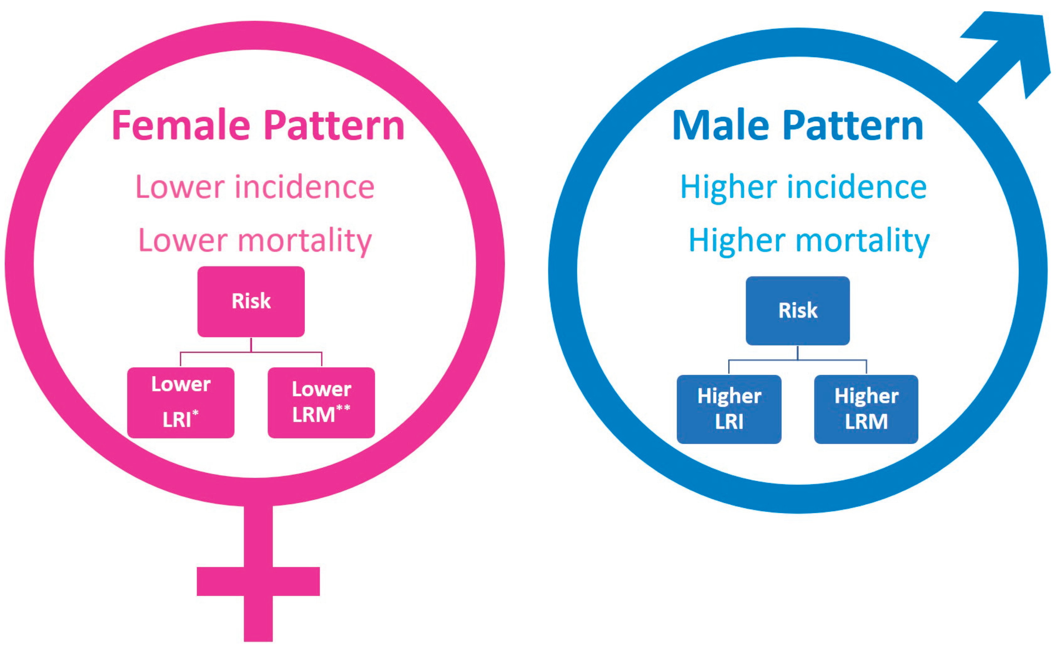

Table 1.
Molecular mechanisms behind sex differences in leukemia.
| Mechanism | Female characteristics | Male characteristics |
|---|---|---|
| Genetic Factors Sex chromosomes (XX vs. XY) |
Idic(X)(q13) is the most frequently observed structural anomaly of the X chromosome in hematologic malignancies [90]. This chromosomal abnormality has been reported exclusively in older female patients with myelodysplastic syndromes (MDS), chronic myeloproliferative disorders, and AML [90,91]. |
The most common chromosomal abnormality in adult male blood cells is mosaic loss of chromosome Y (mLOY) [92]. Leukocyte mLOY is a risk factor for hematological malignancies, such as myelodysplastic syndrome, ALL, and AML[93]. mLOY functionally drives leukemogenesis and clonal hematopoiesis [94]. |
| Epigenetic factors | The epigenetic modifier lysine demethylases KDM6A (UTX), which escapes X-chromosome inactivation, is expressed at higher levels in females [95]. In T cell acute lymphoblastic leukemia, KDM6A has been identified as a tumor suppressor [82]. |
Notably, UTX mutations were observed exclusively in male T-ALL patients [82]. UTX is the first identified X-linked tumor suppressor gene that may partially account for the skewed gender distribution (3:1) toward males in T-ALL on a genetic level [82]. |
| Innate immunity | Females have higher numbers of macrophages and neutrophils, resulting in greater phagocytic activity [83]. | Males have higher numbers of natural killer cells than females [83]. |
| Adaptive immunity | Females have higher levels of circulating CD4 T cells and B cells [84,85,86,87]. |
Males have higher levels of CD8 T cells [84,85,86,87]. |
| Sex hormones | Females have higher levels of progesterone and estrogen [96]. Estrogen receptors are detected in immune cell types such as lymphocytes, monocytes, and macrophages [96], which have protective properties and immunostimulant effects [97]. Progesterone could be effective in growth-inhibiting NALM6 cells by decreasing cell viability and lowering ROS levels [98]. |
Males have higher testosterone concentrations [96]. Testosterone has immunosuppressive effects [99] |
Disclaimer/Publisher’s Note: The statements, opinions and data contained in all publications are solely those of the individual author(s) and contributor(s) and not of MDPI and/or the editor(s). MDPI and/or the editor(s) disclaim responsibility for any injury to people or property resulting from any ideas, methods, instructions or products referred to in the content. |
© 2025 by the authors. Licensee MDPI, Basel, Switzerland. This article is an open access article distributed under the terms and conditions of the Creative Commons Attribution (CC BY) license (http://creativecommons.org/licenses/by/4.0/).
Copyright: This open access article is published under a Creative Commons CC BY 4.0 license, which permit the free download, distribution, and reuse, provided that the author and preprint are cited in any reuse.


