Preprint
Review

This version is not peer-reviewed.

iPSC-Derived Polyvalent Vaccines as Ontogenetically Informed Immunogens for Overcoming Immune Refractoriness in Microsatellite-Stable Colorectal Cancer: A New Frontier in Cancer Immunotherapy

Submitted:

24 November 2025

Posted:

25 November 2025

You are already at the latest version

Abstract

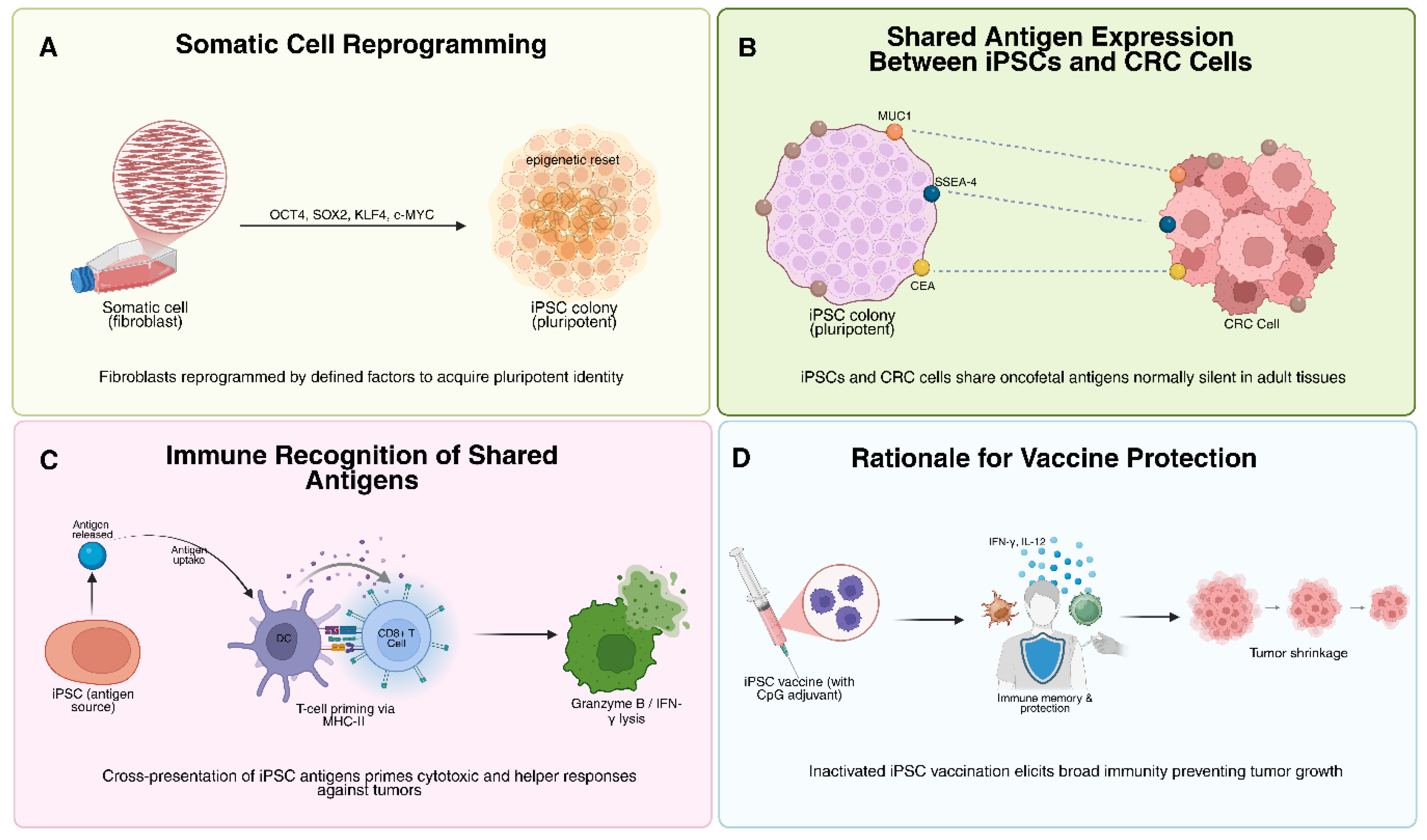
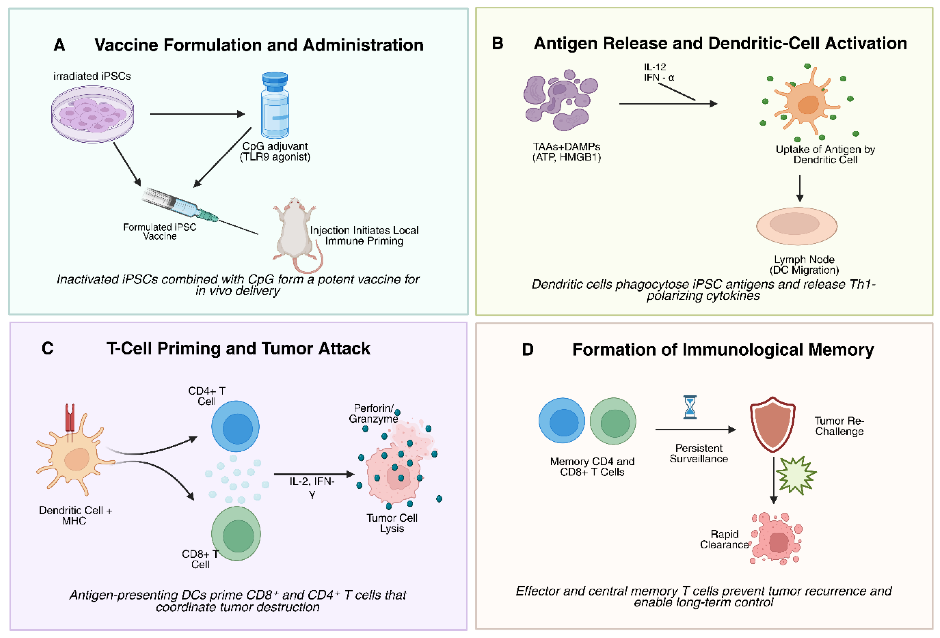
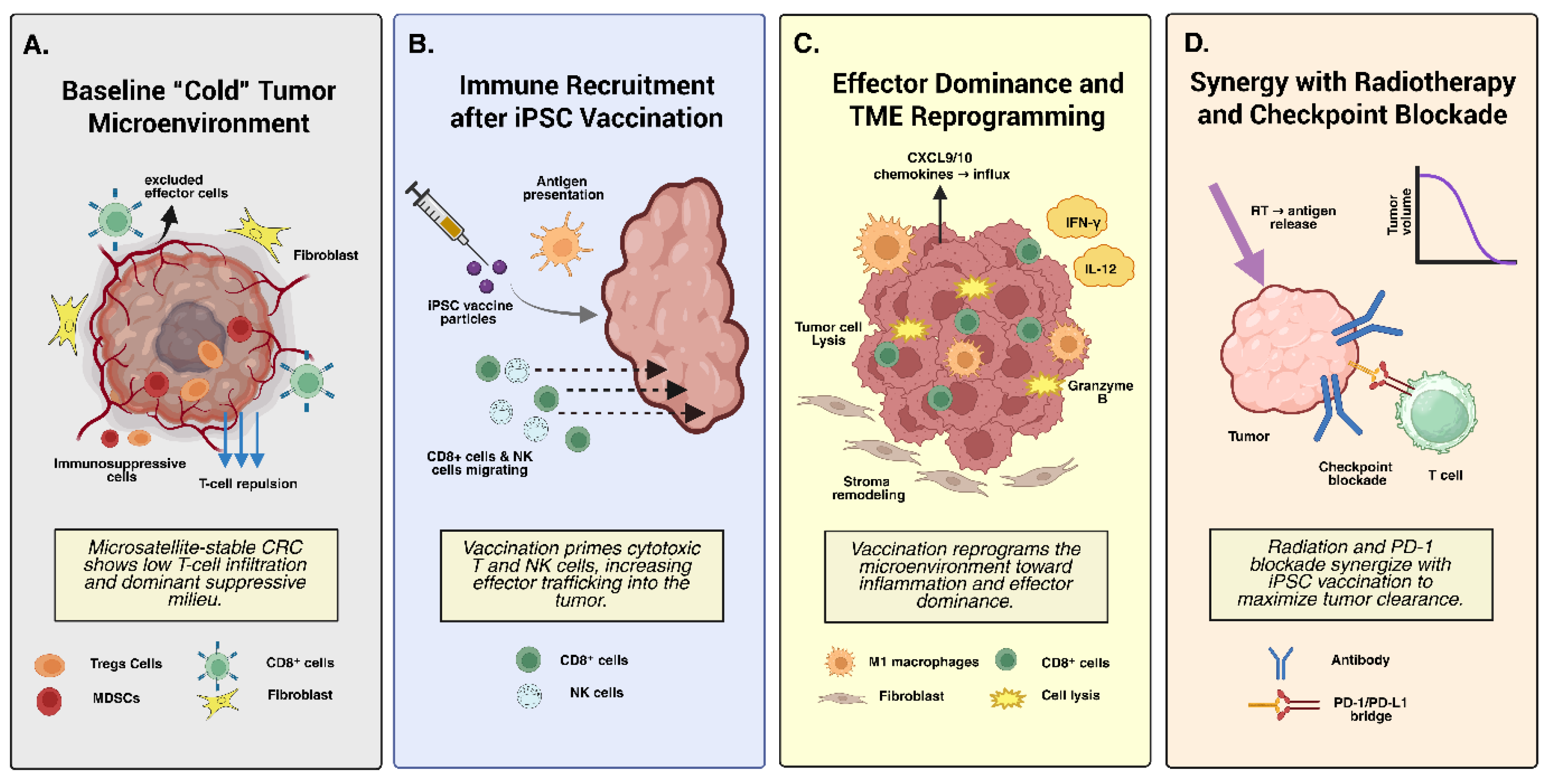
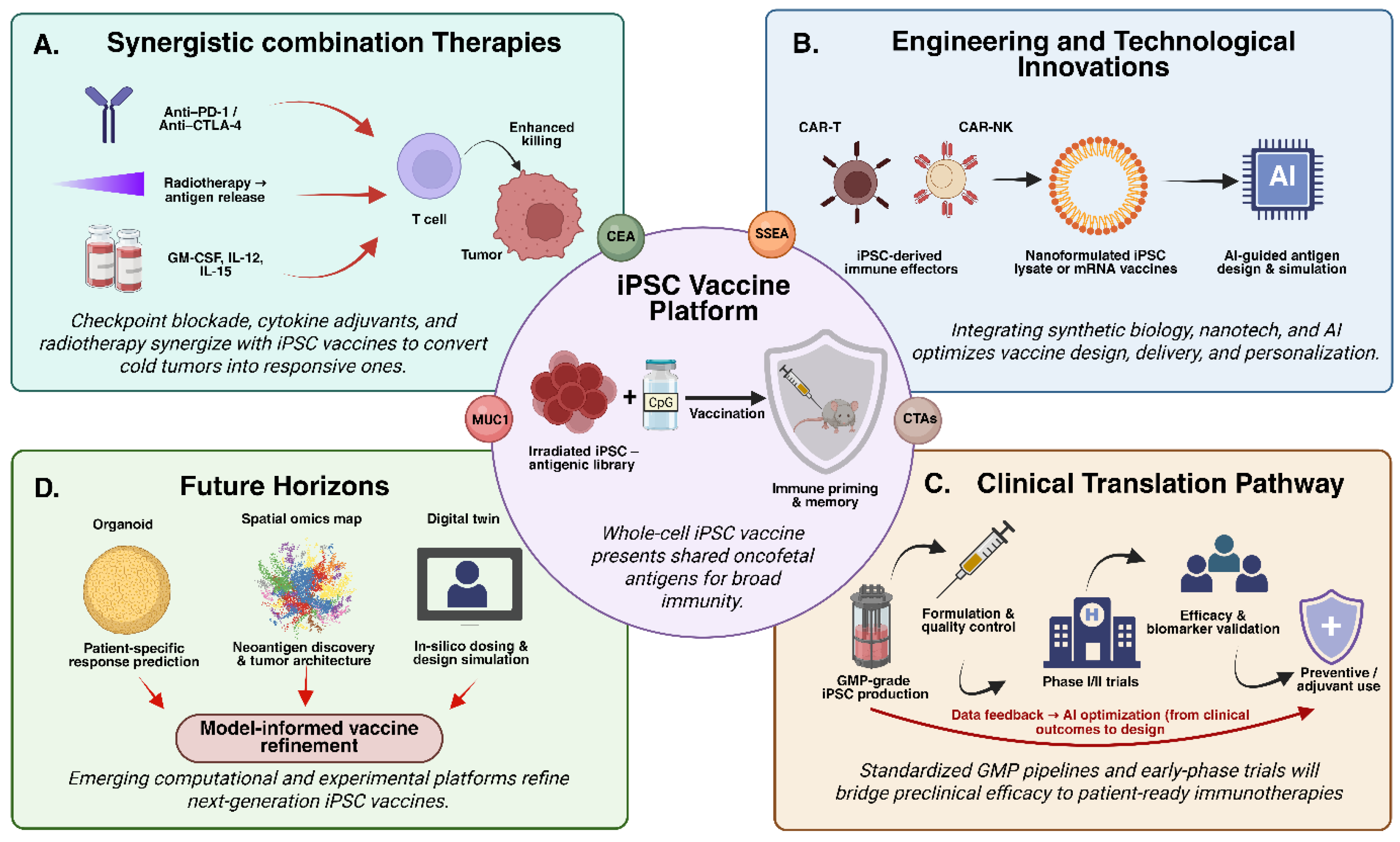
Colorectal carcinoma (CRC) exerts a growing global disease burden, with microsatellite-stable/proficient mismatch repair (MSS/pMMR) tumors exhibiting intrinsic refractoriness to immune-checkpoint blockade (ICB) owing to low tumor mutational burden, limited neoantigenicity, and an immunosuppressive tumor microenvironment (TME) dominated by regulatory T cells (Tregs) and myeloid-derived suppressor cells (MDSCs). This review evaluates induced pluripotent stem cell (iPSC)–derived polyvalent vaccines as ontogenetically recapitulative immunogens capable of reinstating broad antitumor immunity. Reprogramming induces re-expression of oncofetal tumor-associated antigens, including cancer-testis antigens (NY-ESO-1, MAGE-A3), aberrant glycoforms of CEA and MUC1, and clinically actionable neoepitopes such as KRAS^G12D/V, thereby promoting epitope spreading and immunogenic cell death. Irradiated autologous or syngeneic iPSCs, delivered with Toll-like receptor 9 agonists, facilitate robust MHC I/II cross-presentation, driving CD8⁺ cytotoxic T-cell activation, Th1 polarization, perforin/granzyme-mediated cytolysis, and favorable effector-to-suppressor ratios. Preclinical models of melanoma, pancreatic ductal adenocarcinoma, and MSS CRC demonstrate prophylactic and therapeutic efficacy, with neoantigen-enhanced iPSCs synergizing with radiotherapy-induced DAMPs to achieve durable regressions and memory T-cell formation. Translational priorities include CRISPR-engineered hypoimmunogenic iPSC platforms, GMP-compatible non-integrating reprogramming, and combinatorial integration with STING agonists, ICB, CAR-NK cells, and LNP-mRNA constructs to enable biomarker-guided clinical deployment in minimal-residual-disease CRC.

Keywords: 
;  ;  ;  ;  

1. Introduction

Colorectal cancer (CRC) represents one of the most formidable challenges in oncology, ranking among the top three cancers in global incidence and mortality. According to the Global Cancer Observatory, CRC accounted for approximately 1.93 million new cases and 940,000 deaths in 2020, and projections estimate an alarming increase to over 3.2 million new cases by 2040 [1]. This rising burden is driven by aging populations, Westernized diets, sedentary lifestyles, and increasing obesity rates. Despite advances in screening and multimodal treatment, CRC continues to impose a major clinical and socioeconomic burden worldwide [2]. The five-year survival rate exceeds 90% in localized disease but declines sharply to approximately 14% for metastatic CRC, reflecting the limited efficacy of current systemic therapies [3,4]. Conventional modalities, including surgery, cytotoxic chemotherapy regimens such as FOLFOX (folinic acid, fluorouracil, oxaliplatin) and FOLFIRI (folinic acid, fluorouracil, irinotecan), and targeted agents against VEGF (e.g., bevacizumab) or EGFR (e.g., cetuximab, panitumumab), have collectively improved median survival for advanced CRC from roughly 12 to 24 months over the past two decades [5,6]. Nonetheless, therapeutic resistance, relapse, and metastatic dissemination remain common, underscoring the urgent need for transformative therapeutic paradigms [7].
Molecularly, CRC exhibits profound heterogeneity, encompassing multiple subtypes with distinct genetic and immunological landscapes. Approximately 15% of CRCs display microsatellite instability-high (MSI-H) or mismatch repair deficiency (dMMR), characterized by a high tumor mutational burden, abundant neoantigen load, and increased T-cell infiltration, features that render these tumors responsive to immune checkpoint inhibitors (ICIs) such as pembrolizumab and nivolumab [8,9]. In stark contrast, the majority of cases (~85%) are microsatellite-stable (MSS) or mismatch repair proficient (pMMR), typified by low immunogenicity, sparse immune infiltration, and an immunosuppressive “cold” tumor microenvironment (TME) [10]. These MSS/pMMR tumors exhibit resistance to single-agent immunotherapy, highlighting a major therapeutic void. Even within responsive subsets, durable responses to ICIs are limited to a minority of patients, while toxicity, immune escape, and antigenic drift constrain long-term efficacy [10,11]. Thus, there is a pressing need for immunotherapeutic strategies capable of converting immunologically inert tumors into “hot” tumors amenable to durable immune surveillance.
In this context, cancer vaccines have re-emerged as a promising frontier for harnessing and directing the immune system against tumor cells. Unlike passive immunotherapies that rely on exogenous antibodies or adoptive cell transfer, cancer vaccines aim to induce a self-sustaining, antigen-specific immune response capable of recognizing and eradicating malignant cells while establishing long-term memory to prevent recurrence [12]. Early vaccine platforms in CRC, such as peptide vaccines targeting carcinoembryonic antigen (CEA), mucin-1 (MUC1), or mutant KRAS epitopes, as well as dendritic cell (DC) and irradiated tumor cell-based vaccines, have shown immunogenic potential but achieved only modest clinical efficacy [13]. Major barriers include tumor heterogeneity, immune tolerance, poor antigen presentation, and suppression within the TME. Moreover, the narrow antigenic repertoire of peptide or nucleic acid vaccines limits their effectiveness against highly heterogeneous tumors like CRC [13].
A paradigm-shifting concept has emerged with the use of induced pluripotent stem cells (iPSCs) as a multivalent, tumor-mimetic vaccine platform. Discovered by Shinya Yamanaka in 2006, iPSCs are generated by reprogramming adult somatic cells through defined transcription factors (Oct4, Sox2, Klf4, c-Myc), reverting them to an embryonic-like pluripotent state [14]. Intriguingly, this reprogramming process mirrors certain molecular hallmarks of oncogenesis, leading to the re-expression of oncofetal genes and tumor-associated antigens (TAAs) that are typically silenced in differentiated adult tissues [15]. Transcriptomic and proteomic analyses reveal that iPSCs share extensive similarities with embryonic stem cells (ESCs) and cancer stem cells, particularly in the expression of antigens such as SSEA-3/4, TRA-1-60, and various cancer-testis antigens (CTAs) [16,17]. This overlap forms the biological rationale for employing iPSCs as a broad-spectrum antigen source, essentially providing an “antigenic mirror” of tumor cells without relying on patient-specific tumor sequencing or biopsy-derived material.
Preclinical evidence has substantiated the immunogenic potential of iPSC-based vaccines. In murine models of melanoma, breast, lung, and pancreatic cancer, inactivated or irradiated iPSCs have been shown to elicit potent CD8⁺ T-cell responses, promote dendritic cell activation, and inhibit tumor growth and metastasis [18,19]. These effects are further enhanced when combined with immunostimulatory adjuvants such as CpG oligodeoxynucleotides, TLR agonists, or cytokine modulators (GM-CSF, IL-12). Importantly, iPSC vaccination can induce durable immune memory, conferring protection upon rechallenge with tumor cells [20,21]. Translational studies have also demonstrated the capacity of iPSC-derived vaccines to modulate the tumor immune milieu, increasing the infiltration of cytotoxic T lymphocytes and reducing regulatory T-cell (Treg) and myeloid-derived suppressor cell (MDSC) populations [21].
From a clinical and ethical standpoint, iPSCs offer significant advantages over embryonic stem cell–based platforms. They circumvent ethical concerns associated with embryo use and can be derived autologously, minimizing alloimmune rejection [22]. However, iPSCs also present unique safety considerations, including the potential risk of teratoma formation or autoimmune reactivity against shared self-antigens. Therefore, ensuring complete inactivation or differentiation of iPSC material prior to administration is critical for clinical translation [23]. Advances in irradiation protocols, mitomycin C treatment, and gene editing have largely mitigated these risks in preclinical studies [24]. Moreover, scalable “off-the-shelf” iPSC lines, engineered to express immune adjuvants or checkpoint modulators, are under exploration as universal vaccine platforms with standardized antigenic profiles.
In the context of CRC, iPSC-based vaccines hold particular promise for overcoming the limitations of the immunologically “cold” TME. By presenting a broad array of TAAs,including oncofetal antigens, neoantigens, and shared tumor epitopes,iPSCs may prime both innate and adaptive immunity, thereby enhancing tumor antigen visibility and T-cell infiltration [25]. When combined with immune checkpoint blockade (anti-PD-1, anti-CTLA-4), cytokine therapy, radiotherapy, or nanotechnology-based delivery systems, iPSC vaccines could transform the immunologic landscape of pMMR/MSS CRC [26]. Recent innovations in synthetic immunology and cellular engineering also envision using iPSCs not merely as vaccines but as versatile cellular chassis for generating CAR-T, CAR-NK, or CAR-macrophage therapies, offering integrated platforms for tumor targeting, antigen presentation, and immune modulation.
The present review provides a comprehensive synthesis of current advances in iPSC-based vaccine research, with a focus on their translational potential in colorectal cancer. It examines the immunogenic underpinnings of iPSCs, mechanisms of T-cell priming and memory formation, and strategies to enhance vaccine efficacy through adjuvants, nanodelivery, and combinatorial therapies. We also critically assess the safety, regulatory, and manufacturing challenges associated with iPSC-derived platforms and discuss their integration within the broader CRC immunotherapy landscape. By uniting stem cell biology, cancer immunology, and translational biotechnology, iPSC-based vaccines represent a compelling and multifaceted avenue toward next-generation immunoprevention and immunotherapy for CRC,offering the prospect of durable, systemic, and personalized anti-tumor immunity beyond the constraints of current therapeutic modalities.

2. Foundations of iPSC-Based Cancer Vaccines

2.1. Oncofetal Similarities and Historical Rationale

The idea that cancer cells and embryonic cells share common molecular features is not new. As early as the early 20th century, researchers observed that immunization with embryonic or fetal tissues could induce rejection of tumors in animal models. This observation led to the “oncofetal antigen” hypothesis , the idea that many proteins expressed during early embryonic development become aberrantly re-expressed in malignant tissues [27]. These proteins, often silenced after birth, act as tumor-associated antigens (TAAs) when re-emerging in cancer cells. Classic examples include carcinoembryonic antigen (CEA), alpha-fetoprotein (AFP), MUC1, and SSEA family members [28]. Because these molecules are largely absent from healthy adult tissues, they present an opportunity for selective immune targeting of tumors. Early experimental vaccines based on embryonic stem cells (ESCs) or fetal tissue extracts demonstrated that immunization could trigger immune responses capable of recognizing tumor cells sharing these embryonic antigens [29,30]. However, these early strategies faced major obstacles: ethical controversies surrounding the use of embryos, risks of uncontrolled proliferation or teratoma formation, and strong allogeneic immune rejection due to mismatched major histocompatibility complex (MHC) molecules between donors and recipients.
The discovery of induced pluripotent stem cells (iPSCs) in 2006 by Shinya Yamanaka and colleagues revolutionized this landscape. By reprogramming adult somatic cells (such as fibroblasts or blood cells) using a defined set of transcription factors (OCT4, SOX2, KLF4, and c-MYC), researchers could generate cells nearly identical to ESCs in morphology, self-renewal capacity, and gene expression profiles , without using embryos [14]. iPSCs express high levels of telomerase, maintain open chromatin states, and reactivate many genes typically expressed during early embryogenesis and in tumors [31]. Genome-wide studies have confirmed extensive overlap between the transcriptomes of iPSCs, ESCs, and cancer cells, particularly in pathways related to proliferation, metabolism, and stemness [17,32,33]. Importantly, reprogramming appears to awaken a set of genes and antigens collectively termed the “iPSC-cancer signature.” These include cancer-testis antigens (CTAs) such as MAGE-A and NY-ESO-1, as well as developmental regulators and proto-oncogenes (MYC, KLF4, LIN28, NANOG) [33]. This molecular resemblance forms the scientific rationale for iPSC-based vaccines: if the immune system can be safely exposed to the antigenic landscape of iPSCs, it may develop adaptive responses that recognize and eliminate cancer cells expressing similar antigens.
Early assumptions suggested that autologous iPSCs , derived from a patient’s own cells, would be immunologically inert, making them ideal for regenerative medicine. However, landmark studies by Araki et al. (2013) and others revealed that undifferentiated iPSCs are in fact immunogenic even in genetically identical (syngeneic) hosts [34]. Teratomas formed from autologous iPSCs were rejected by T-cell–mediated immune mechanisms, similar to allogeneic ESC-derived teratomas [35]. Later, studies by Ouyang and Kooreman demonstrated that irradiated, autologous iPSCs could function as potent vaccines [36,37]. Mice vaccinated with iPSCs developed robust CD8⁺ cytotoxic T-cell and antibody responses, producing long-term immunological memory and cross-reactivity against cancer cells [36]. These findings confirmed that undifferentiated iPSCs naturally express a wide spectrum of antigens recognized as “non-self,” providing a broad immunological training ground for tumor recognition. This shared “oncofetal mirror” relationship between iPSCs and CRC cells forms the conceptual foundation of pluripotent-cell-based cancer vaccines (Figure 1).
2.2. iPSC Versus ESC Vaccines and the Role of Adjuvants
Both ESCs and iPSCs share key immunogenic features, but iPSCs offer distinct advantages for translational use. ESCs are inherently allogeneic and ethically constrained, while iPSCs can be derived autologously or from universal donor lines. This flexibility reduces immune rejection risks and allows scalable vaccine production [38]. Comparative studies indicate that when matched for antigen expression, iPSCs and ESCs trigger comparable levels of immune activation. Yet, ESC vaccines often elicit stronger alloimmune responses due to mismatched MHC molecules, which can confound interpretation of tumor-specific immunity [29]. Autologous iPSCs, by contrast, avoid these background immune reactions, offering a cleaner model to study antigen-specific tumor responses. Despite their intrinsic immunogenicity, unmodified or adjuvant-free iPSC vaccines typically induce only partial or transient anti-tumor protection [29]. The immune system requires “danger signals” , molecular cues that alert antigen-presenting cells (APCs) such as dendritic cells (DCs) and macrophages to initiate adaptive immunity [39]. Therefore, iPSC-based vaccines are generally combined with strong immunostimulatory adjuvants to boost efficacy [40].
Among these, Toll-like receptor (TLR) agonists , particularly CpG oligodeoxynucleotides (TLR9 ligands) , have been most widely used. CpG motifs mimic bacterial DNA and activate plasmacytoid DCs, leading to secretion of type I interferons and IL-12, which enhance Th1 responses and CD8⁺ T-cell priming [40]. When CpG is co-administered with irradiated iPSCs, there is a marked increase in dendritic cell activation, cytokine production, and infiltration of cytotoxic T cells into tumors [36]. This combination has been shown to suppress tumor growth and even achieve complete tumor regression in murine melanoma, lung, and pancreatic cancer models. Other adjuvants under investigation include poly(I:C) (a TLR3 agonist that simulates viral RNA), GM-CSF (to recruit DCs to the injection site), and STING agonists, which activate innate DNA-sensing pathways [40]. The choice of adjuvant can be tailored to the tumor’s immunogenic profile or microenvironmental context. For example, in CRC vaccine studies, GM-CSF-based adjuvants (such as in GVAX formulations) have shown the ability to recruit and activate DCs within the TME, while CpG adjuvants are particularly effective at initiating systemic T-cell immunity [41].
The combination of iPSCs and adjuvants represents a synergistic approach: iPSCs provide a comprehensive library of tumor-relevant antigens, while adjuvants provide the inflammatory cues needed to activate innate and adaptive immunity. This balance is critical , without adjuvants, immune tolerance may occur; with excessive stimulation, systemic inflammation or autoimmunity could arise [42]. Preclinical data consistently show that optimized adjuvant pairing transforms iPSC vaccines from modestly immunogenic formulations into potent inducers of durable, tumor-specific immunity [36].

2.3. Safety Considerations: Irradiation and Autoimmunity

The greatest safety concern in iPSC-based vaccination is tumorigenicity. Undifferentiated iPSCs retain pluripotent potential and can form teratomas , benign but uncontrolled growths containing cells from all three germ layers. To eliminate this risk, all preclinical studies employ inactivation strategies before administration. The most established approach is gamma irradiation or X-ray exposure at high doses (typically 50–60 Gy), which halts cell proliferation while preserving antigen integrity [43]. Irradiated iPSCs can still present their antigenic repertoire to the immune system but cannot divide or form tumors. In all reported animal models, no teratoma formation has occurred following vaccination with properly irradiated iPSCs. Interestingly, irradiation may provide additional benefits beyond safety [44]. The process of cellular damage triggers the release of danger-associated molecular patterns (DAMPs) such as ATP, HMGB1, and calreticulin, which further stimulate innate immune receptors on APCs [45]. This “immunogenic cell death” amplifies antigen presentation and promotes a pro-inflammatory environment conducive to effective T-cell activation [46].
Autoimmunity represents another theoretical concern, as many embryonic antigens are shared between iPSCs and certain adult tissues (for instance, testis or placental antigens). However, preclinical data thus far have shown minimal evidence of autoimmune toxicity (19,36,47). Vaccinated mice maintain stable body weight, normal organ histology, and absence of circulating autoantibodies [36,47]. The immune responses appear focused on tumor-associated antigens rather than indiscriminate self-reactivity. Nevertheless, careful immune monitoring , including serum autoantibody assays and organ pathology , remains essential as this technology moves closer to human application [47,48].
Additional safety innovations are being explored to ensure translational viability. These include genetic “suicide switches” such as inducible caspase-9 systems, which can selectively ablate any residual proliferative iPSCs upon administration of a small-molecule activator, and cell-sorting techniques to remove undifferentiated pluripotent cells before vaccine formulation [49,50]. iPSC-based cancer vaccines integrate a century-old concept of oncofetal antigen immunology with cutting-edge stem cell technology. Through inactivation, adjuvant optimization, and immune safety profiling, these vaccines offer a highly versatile, multivalent, and potentially transformative platform for cancer immunotherapy. By training the immune system against a broad repertoire of tumor-associated antigens, iPSC vaccines hold the promise of generating durable, cross-reactive immunity , a crucial step toward combating refractory and immunologically “cold” malignancies such as colorectal cancer.

3. Mechanisms of Anti-Tumor Immunity via iPSC Vaccines

3.1. Activation of Cellular and Humoral Immunity: T-Cell Priming, Effector Responses, and Tumor Infiltration

The antitumor efficacy of iPSC-based vaccines fundamentally depends on their ability to activate the adaptive immune system,most notably CD8⁺ cytotoxic T lymphocytes (CTLs),supported by CD4⁺ helper T cells and, to a lesser extent, B-cell–mediated humoral responses. The sequence of immune activation begins when irradiated iPSCs are administered along with an appropriate adjuvant (such as CpG oligodeoxynucleotides) [48]. The irradiation process induces immunogenic cell death, releasing a complex mixture of tumor-associated antigens (TAAs) and damage-associated molecular patterns (DAMPs) such as ATP, calreticulin, and HMGB1. These molecular signals recruit and activate antigen-presenting cells (APCs),particularly dendritic cells (DCs) and macrophages,within the injection site [51].
Activated DCs internalize iPSC-derived debris and process the TAAs via two major antigen-presentation pathways. The MHC class I pathway leads to cross-presentation of intracellular or exogenous iPSC antigens to CD8⁺ T cells, while the MHC class II pathway presents processed peptides to CD4⁺ T helper (Th) cells [52]. This dual presentation results in the clonal expansion of both effector and helper T-cell subsets [53]. Together, these events outline the mechanistic framework of iPSC vaccine–induced anti-tumor immunity (Figure 2). CD8⁺ CTLs recognize tumor cells expressing shared iPSC-derived antigens and eliminate them through the release of perforin and granzymes, initiating apoptosis in target cells. Meanwhile, CD4⁺ Th1 cells produce interleukin-2 (IL-2) and interferon-γ (IFN-γ), reinforcing CD8⁺ activation and promoting macrophage cytotoxic functions [54].
Kooreman and colleagues provided compelling mechanistic evidence for this process using a syngeneic melanoma model. Mice vaccinated with autologous, irradiated iPSCs combined with CpG displayed a significant increase in CD8⁺ T-cell frequency and IFN-γ production in tumor-draining lymph nodes [19]. When CD8⁺ cells were depleted, the antitumor efficacy of the vaccine was completely abrogated, establishing CD8⁺ T cells as the central mediators of vaccine-induced protection. Adoptive transfer experiments further validated this: transferring CD8⁺ T cells from vaccinated mice conferred tumor resistance to naïve recipients, confirming that the immune protection is antigen-specific and transferable [21,55].
iPSC vaccination also remodels the tumor microenvironment (TME) by altering immune cell composition. Vaccinated mice exhibit enhanced infiltration of CD8⁺ effector T cells and a concomitant reduction in immunosuppressive regulatory T cells (Tregs) and myeloid-derived suppressor cells (MDSCs) [21]. This shift increases the effector-to-suppressor (E:T) ratio, tipping the TME toward an immunostimulatory state. In pancreatic and colorectal cancer (CRC) models, this effect correlated with elevated expression of granzyme-B, IFN-γ, and TNF-α within the tumor, demonstrating a transition from an immune-excluded to an immune-permissive phenotype [56,57].
While cellular immunity dominates, humoral responses also contribute to the vaccine’s efficacy. B cells exposed to iPSC antigens differentiate into plasma cells that secrete IgG antibodies recognizing both iPSC and tumor surface antigens [58,59]. These antibodies may facilitate antibody-dependent cellular cytotoxicity (ADCC) by engaging Fc receptors on NK cells or macrophages, or promote opsonization and phagocytic clearance of tumor cells. In the melanoma model, vaccinated mice generated tumor-reactive IgG capable of binding tumor cells and enhancing immune cell–mediated killing. However, the humoral response mainly complements rather than drives the protective effect [60].
An additional hallmark of iPSC-based vaccination is epitope spreading [61]. As iPSC-primed T cells attack tumor cells expressing shared TAAs, the ensuing tumor cell death releases new tumor-specific antigens, broadening the immune repertoire. APCs capture and present these secondary antigens, allowing the immune system to mount responses against previously unrecognized tumor epitopes. This cascade amplifies the antitumor effect beyond the original antigenic scope of the vaccine, generating polyclonal and diverse T-cell responses,an essential feature for tackling tumor heterogeneity in CRC [62,63].

3.2. Development of Immunological Memory and Long-Term Tumor Protection

One of the most desirable features of cancer vaccines is their capacity to generate long-lasting immunological memory. iPSC-based vaccines excel in this regard by inducing both effector-memory (T_EM) and central-memory (T_CM) T-cell subsets [19,48]. Following antigen priming, a fraction of activated T cells persists in secondary lymphoid organs, where they differentiate into memory populations that provide continuous immune surveillance against tumor recurrence or metastasis [64].
In murine studies, vaccinated animals demonstrate remarkable durability of protection. Ouyang et al. reported that mice immunized with iPSCs + CpG in a pancreatic ductal adenocarcinoma model remained tumor-free for several months after initial challenge. Upon re-exposure to tumor cells, these animals rapidly mounted a recall response characterized by the expansion of CD44^hiCD62L^hi (central-memory) CD8⁺ T cells producing IFN-γ and IL-2 [36]. In colorectal cancer models, similar patterns were observed: iPSC vaccination led to the persistence of memory CD8⁺ T cells capable of recognizing tumor peptides in vitro long after the last immunization [21].
The protective effect of these memory populations has been confirmed through tumor rechallenge experiments. Mice cured of primary tumors by iPSC vaccination often reject secondary tumor inoculations months later, without requiring booster doses. Adoptive transfer of splenic memory T cells from such animals to naïve hosts reproduces the protective effect, establishing a direct causal link between memory formation and long-term antitumor immunity [19,36].
Mechanistically, the generation of durable memory depends on CD4⁺ T-cell help during priming. CD4⁺ Th1 cells secrete cytokines that promote the survival of activated CD8⁺ cells and program them into long-lived memory subsets [65]. These T_CM and T_EM cells maintain proliferative potential and can rapidly differentiate into cytotoxic effectors upon antigen re-exposure [66].
The establishment of memory is particularly significant for microsatellite-stable (MSS) or proficient mismatch-repair (pMMR) CRC, where relapse after resection is common and adjuvant therapy options remain limited. iPSC vaccines could serve as prophylactic agents for high-risk patients,those with residual microscopic disease or genetic predispositions,providing ongoing immunological surveillance capable of intercepting early metastatic lesions [67,68].
Interestingly, the quality of the memory response induced by iPSC vaccination differs from that elicited by conventional peptide or dendritic-cell vaccines. Because iPSCs present an exceptionally broad antigen repertoire, the resulting memory pool recognizes multiple antigens, reducing the risk of immune escape due to tumor antigen loss [64,69]. This breadth of recognition may explain why iPSC-vaccinated animals often exhibit protection across different tumor types that share stemness or oncofetal antigens. The major tumor-associated and neoantigenic targets that underpin colorectal cancer vaccine design,including KRAS mutations, oncofetal antigens, and cancer-testis antigens,are summarized in Table 1.

3.3. Integration of Neoantigens and Synergy with Radiotherapy

Although native iPSCs express a vast array of TAAs and oncofetal proteins, these shared antigens are sometimes insufficient to overcome immune tolerance in established, low-mutational-burden cancers such as CRC. To increase antigenic precision and potency, researchers have engineered iPSCs to express tumor-specific neoantigens,mutant peptides arising from nonsynonymous somatic mutations unique to cancer cells. These neoantigen-augmented iPSCs (NA-iPSCs) serve as customized vaccine platforms capable of stimulating highly specific T-cell responses [74].
A pioneering study used adeno-associated viral vectors to insert genes encoding multiple CRC-associated neoepitopes,such as mutant KRAS, TP53, and SMAD4 peptides,into mouse iPSCs. When vaccinated, these NA-iPSCs induced strong CD8⁺ T-cell responses specific to the introduced neoantigens [75,76]. However, the therapeutic efficacy was dramatically enhanced when vaccination was combined with localized radiotherapy (RT).
Radiotherapy acts as an immunological amplifier. Ionizing radiation induces tumor cell apoptosis and necrosis, releasing a diverse array of TAAs and neoantigens into the TME. This process increases MHC class I expression on tumor cells, activates the STING–type I interferon pathway, and promotes recruitment of DCs to the irradiated site. Essentially, RT converts the tumor into an in situ vaccine, enhancing antigen availability and immune infiltration [77].
When NA-iPSC vaccination was coupled with RT in murine models of microsatellite-stable CRC, approximately 60% of treated animals achieved complete tumor regression. These responses were accompanied by high frequencies of neoantigen-specific CD8⁺ T cells secreting IFN-γ and TNF-α, along with dense infiltration of cytotoxic lymphocytes and CD11c⁺ DCs within the tumors. Importantly, these effects extended beyond local control: vaccinated and irradiated mice exhibited reduced lung and liver metastases, demonstrating systemic immune activation [75].
Mechanistically, the synergy between NA-iPSC vaccination and RT arises from complementary processes. The vaccine primes a systemic pool of neoantigen-specific T cells, while RT enhances local antigen presentation and facilitates T-cell trafficking into tumors. RT-induced cytokines such as CXCL10 and CCL5 act as chemoattractants, guiding effector T cells into the tumor bed [78]. Moreover, RT downregulates immunosuppressive factors (e.g., TGF-β, VEGF) and transiently increases PD-L1 expression, providing a window for combination with immune checkpoint inhibitors. This integrated approach,combining NA-iPSCs, RT, and checkpoint blockade,represents a powerful strategy to convert immunologically “cold” MSS tumors into “hot,” inflamed lesions responsive to immunotherapy [78].

3.4. Innate Immune Activation and Remodeling of the Tumor Microenvironment

Although the adaptive immune response drives specificity, innate immunity provides the essential foundation for vaccine efficacy. iPSC vaccination inherently engages innate sensors through the release of DAMPs and the inclusion of TLR agonists as adjuvants. CpG oligodeoxynucleotides, the most common adjuvant, activate Toll-like receptor 9 (TLR9) on DCs and B cells, leading to secretion of IL-12, IL-6, and type I interferons. These cytokines create a pro-inflammatory environment that favors Th1 polarization and enhances cross-priming of CD8⁺ T cells (39,42,65).
Simultaneously, innate effector cells such as natural killer (NK) cells are activated. NK cells recognize stress-induced ligands like NKG2D on tumor cells and mediate direct cytotoxicity, bridging innate and adaptive immunity. Vaccination with iPSCs plus CpG has been shown to increase NK-cell infiltration into tumors, elevating IFN-γ production and promoting DC maturation via reciprocal cytokine crosstalk [75].
The iPSC vaccine also reshapes the immunosuppressive components of the TME. Preclinical CRC and melanoma models report a significant decline in FoxP3⁺ Tregs and CD11b⁺Gr1⁺ MDSCs following vaccination [26]. This reduction is associated with increased M1-polarized macrophages expressing iNOS and TNF-α, and decreased M2-associated cytokines such as IL-10 and Arginase-1. The resulting microenvironment becomes conducive to antigen presentation, T-cell infiltration, and effector function [79].
Beyond cellular remodeling, soluble factors play an important role. iPSC vaccination enhances the expression of chemokines (CXCL9, CXCL10) that attract CXCR3⁺ effector T cells to tumor sites, while reducing angiogenic factors like VEGF, thereby limiting tumor vascularization and metastasis. The net result is a coordinated reprogramming of the TME from an immunosuppressive “cold” niche to an inflamed, cytotoxic milieu capable of sustaining long-term tumor control [56,80].
Collectively, iPSC-based vaccines act as multivalent immunogenic platforms that engage both innate and adaptive immunity through a cascade of interconnected processes:
a)
Antigen release and presentation – Irradiated iPSCs release a broad array of TAAs and DAMPs that recruit and activate APCs, enabling MHC-I and MHC-II antigen presentation [45,52].
b)
Adaptive immune activation – DC-mediated priming leads to expansion of CD8⁺ CTLs and CD4⁺ Th1 cells, generating a cytotoxic and cytokine-rich immune response [53].
c)
Humoral response and epitope spreading – B-cell–derived antibodies complement cellular immunity, while tumor cell death releases additional antigens that broaden recognition [61].
d)
Memory formation – Effector and central memory T cells provide long-term surveillance, protecting against recurrence and metastasis [66].
e)
Innate amplification – Adjuvants like CpG and RT-induced inflammatory cues reinforce innate activation, bridging to adaptive responses [46,78].
f)
Microenvironment reprogramming – The balance of immune infiltrates shifts toward effector dominance (CD8⁺, NK, M1 macrophages) with reduced suppressive populations (Tregs, MDSCs) [79].
Through these mechanisms, iPSC vaccines can effectively train the immune system to recognize the shared antigenic landscape between pluripotent stem cells and malignant cells. When combined with modern immunomodulatory agents or radiotherapy, they have the potential to overcome immune exclusion, achieve systemic tumor control, and establish long-lasting antitumor immunity,an especially critical advance for refractory malignancies such as microsatellite-stable colorectal cancer.
4. iPSC Vaccines in the Context of Colorectal Cancer and Frontiers in CRC Vaccine Platforms

4.1. Cross Cancer Foundations and Translational Relevance to CRC

Much of the foundational understanding of induced pluripotent stem cell (iPSC)–based vaccines comes from preclinical models in other epithelial cancers, such as melanoma, breast carcinoma, lung adenocarcinoma, mesothelioma, and pancreatic ductal adenocarcinoma (PDAC) [36]. In these models, autologous iPSC vaccines formulated with potent adjuvants like CpG oligodeoxynucleotides elicited durable, antigen specific T cell responses capable of preventing tumor establishment or slowing growth of established disease [36]. For instance, in PDAC,a tumor with a profoundly immunosuppressive and fibrotic microenvironment analogous to colorectal cancer (CRC),syngeneic iPSC + CpG vaccines prevented tumor formation in over 70% of mice, highlighting their ability to convert immunerefractory malignancies into immunologically active states [81]. The translational relevance of these findings to CRC lies in the shared molecular and immunological hallmarks between PDAC and pMMR/MSS CRC: both display low tumor mutational burden (TMB), high stromal density, limited immune infiltration, and poor responsiveness to checkpoint inhibitors [82]. Furthermore, both malignancies express overlapping oncofetal and stemness associated antigens, such as carcinoembryonic antigen (CEA), MUC1, SSEA3/4, and TRA160, which are also abundant in iPSCs [19,82,83]. This antigenic convergence suggests that the same vaccine principles proven in PDAC and breast cancer may be extrapolated to CRC, particularly in subtypes characterized by immune exclusion and low neoantigen load. Moreover, success in mesothelioma and lung carcinoma models,diseases with low immunogenicity,underscores that iPSC vaccines need not rely on high TMB but can instead exploit shared developmental antigens [19,82]. These findings collectively establish iPSCs as a cross cancer immunogen platform capable of priming broad spectrum antitumor immunity, providing a strong biological rationale for their application to colorectal malignancies.

4.2. CRC-Specific iPSC Vaccine Studies and Antigen Discovery

Recent experimental evidence has extended the iPSC vaccine paradigm directly into colorectal cancer. The landmark murine studies by Jwo et al. (2025) established both prophylactic and therapeutic efficacy of autologous, irradiated iPSC vaccines in syngeneic CRC models [26]. In prophylactic settings, vaccination prior to tumor challenge prevented tumor establishment in a majority of mice, whereas in therapeutic settings, vaccination significantly delayed tumor progression and improved overall survival. Mechanistically, these outcomes were associated with heightened intratumoral infiltration of CD8⁺ cytotoxic T lymphocytes and increased secretion of IFN γ and granzyme B, indicative of a potent Th1type immune response [26]. Depletion experiments confirmed that both CD4⁺ and CD8⁺ T cells were indispensable for vaccine induced protection, validating a dual arm adaptive mechanism. Importantly, proteomic and transcriptomic profiling of iPSCs and CRC cell lines identified two nuclear proteins,heterogeneous nuclear ribonucleoprotein U (HNRNPU) and nucleolin (NCL),as shared immunogenic targets. These proteins were overexpressed in both CRC and iPSCs but not in normal adult tissues, fulfilling the ideal criteria for tumor associated antigens (TAAs). MHC class I binding predictions further indicated strong affinity of HNRNPU and NCL derived peptides for common murine H2 alleles [26]. Subsequent dendritic cell (DC) vaccination using these peptides (with CpG adjuvant) recapitulated the cytotoxic T-cell activation and tumor control observed with whole iPSC vaccines, confirming that iPSCs can serve not only as vaccines but also as antigen discovery platforms for identifying novel, shared CRC antigens [26]. Hybrid strategies further extend this utility: iPSCs engineered to overexpress HNRNPU or NCL, when irradiated and combined with CpG, produced enhanced CD8⁺ responses and induced partial tumor regressions in established CRC models. These data underscore a dual role for iPSCs,as both polyvalent antigen sources and customizable carriers for disease specific antigen expression,thereby bridging whole cell and targeted vaccine paradigms [26].

4.3. Integrating iPSC Vaccines into the CRC Immunotherapy Landscape

Colorectal cancer exemplifies the principle of cancer immunoediting, wherein immune pressure drives the selection of resistant tumor clones and progressive immune evasion. In most CRC cases,particularly the 85–90% that are microsatellite stable (MSS) and mismatch repair–proficient (pMMR),the tumor microenvironment (TME) remains immunologically “cold,” characterized by high densities of Tregs, MDSCs, and stromal fibroblasts that suppress effector T-cell infiltration [84,85]. Immune checkpoint inhibitors (ICIs), such as anti–PD1 (pembrolizumab, nivolumab) and anti–CTLA4 (ipilimumab), have demonstrated remarkable efficacy in microsatellite instability–high (MSIH) CRC but remain largely ineffective in pMMR/MSS subtypes. iPSC vaccines have the potential to reshape this immunological landscape by providing a multivalent antigenic stimulus that broadens T-cell repertoires and increases immune infiltration [86]. When combined with ICIs, iPSC vaccines could supply the missing priming signal required for checkpoint blockade to succeed in MSS tumors [87]. In concept, an iPSC vaccine could “ignite” the TME, transforming it from cold to hot by increasing chemokine gradients (CXCL9, CXCL10), recruiting effector T cells, and reducing suppressor populations [88,89]. Parallel immunotherapy modalities, such as CART, CARNK, and CAR macrophage (CARM) therapies, further illustrate the complementary potential of iPSC-based vaccination [90,91]. CART therapies targeting CEA, GUCY2C, and EpCAM have shown limited efficacy in CRC due to on target toxicity and the physical barriers of the TME [90]. An iPSC vaccine that primes systemic immunity could augment CART function by preconditioning the TME with activated T cells and inflammatory cytokines, improving CART infiltration and persistence [92]. Conversely, iPSC derived stem cells can be genetically engineered into CAR bearing effector cells themselves,offering a closed loop system where the same iPSC platform provides both antigenic priming (via vaccine) and effector cytotoxicity (via differentiated CAR immune cells) [93,94]. Dendritic cell vaccines, peptide vaccines, and viral vector–based vaccines have historically shown safety but modest efficacy in CRC, often due to limited antigen breadth or inadequate T-cell priming [95,96]. By presenting the full complement of TAAs,including shared developmental proteins and neoantigens,iPSC vaccines overcome these constraints [75]. As such, they may serve as a priming backbone in combination immunotherapy regimens that include checkpoint blockade, radiation, or adoptive cell transfer [90,92].

4.4. Expanding CRC Vaccine Strategies: KRAS, CT Antigens, and Neoantigens

Advances in CRC vaccine development provide synergistic frameworks for integrating iPSC technology. One major frontier is KRAS targeted vaccination. Approximately 40–50% of CRCs harbour activating KRAS mutations (e.g., G12D, G12V, G13D), which represent ideal neoantigenic targets due to their tumor exclusivity and shared recurrence across patients [97]. The ELI002 2P “off the shelf” vaccine exemplifies this approach. It delivers KRAS mutant peptides conjugated to amphiphile adjuvants that enhance lymph node delivery [97]. In the Phase I AMPLIFY201 trial, ELI002 induced KRAS specific CD4⁺ and CD8⁺ T-cell responses in over 80% of patients with resected CRC or PDAC, translating to prolonged relapse free survival among high immune responders [98]. Notably, antigen spreading was observed in two thirds of patients, mirroring the epitope broadening effects characteristic of iPSC vaccination [97]. Another promising avenue involves cancer–testis antigens (CTAs) such as NYESO1, MAGEA3, LAGE1, DKKL1, and FBXO39, which are normally restricted to germline tissues but re-expressed in CRC [99]. Bioinformatic epitope prediction and multi peptide design enable vaccines that cover intertumoral heterogeneity [100]. These epitope driven vaccines, often formulated with GMCSF or TLR agonists, induce robust IFN γ–secreting CTL responses in preclinical models [26] . Incorporating CTA epitopes into iPSC vaccines,either via transduction or pulsing,could create hybrid formulations that merge the breadth of iPSC antigens with the specificity of known CTA targets [75]. Personalized neoantigen vaccines based on next generation sequencing (NGS) of patient tumors represent another transformative area. By mapping the patient’s mutanome and selecting predicted HLA binding peptides, vaccines can be designed to elicit tailored Tcell responses [101]. However, for MSS CRC, which often exhibits low TMB, the number of actionable neoantigens is limited. Here, iPSC platforms could bridge the gap by serving as scaffolds for integrating known neoepitopes or delivering shared developmental antigens to complement the sparse mutational repertoire [75]. The emerging neoantigen augmented iPSC (NAiPSC) approach,where iPSCs are genetically modified to express tumor specific mutations,demonstrates this convergence, achieving significant tumor regression and immune activation when combined with local radiotherapy in MSS CRC models [75].

4.5. Future Integration: Delivery Innovations and Combination Therapies

The delivery and formulation of CRC vaccines are rapidly evolving with the advent of mRNA and nanotechnology-based systems. mRNA vaccines, validated during the SARS-CoV-2 pandemic, offer scalable, safe, and flexible platforms for encoding tumor antigens [102,103]. Lipid nanoparticle (LNP)–encapsulated mRNA vaccines can deliver multiple TAAs or neoantigens directly to dendritic cells [103,104]. In CRC, preclinical mRNA formulations encoding KRAS mutations, CTAs, or oncofetal antigens have induced potent CD8⁺ Tcell responses and suppressed tumor growth [102,105]. The modularity of mRNA design allows for combination with iPSC derived lysates or adjuvants, generating hybrid vaccine regimens that couple the precision of mRNA with the broad antigenic spectrum of iPSCs [103]. Nano vaccines further enhance antigen delivery and presentation. Poly (lactic-co-glycolic acid) (PLGA), iron oxide, or virus like particle (VLP) carriers can encapsulate iPSC lysates or TAA peptides alongside CpG or STING agonists, ensuring colocalized antigen–adjuvant delivery and sustained release within lymphoid organs. Such systems improve antigen uptake by DCs and facilitate cross presentation to T cells. Moreover, targeted nanoparticles can be engineered to release contents in response to pH or enzymatic cues within tumors, offering precise spatiotemporal control of immune activation [106]. From a systems immunology perspective, the most promising horizon for CRC lies in combination therapies. iPSC vaccines could serve as a priming platform to be followed by checkpoint inhibitors (anti–PD1, anti–CTLA4), adoptive transfer (CART, CARNK), or immunogenic radiotherapy. Radiotherapy enhances antigen release and MHC upregulation, synergizing with iPSC induced Tcell priming [102,105]. Similarly, coadministration of cytokine adjuvants (e.g., IL12, GMCSF) or oncolytic viruses could further potentiate the response by amplifying antigen exposure and immune cell recruitment [107]. In future clinical designs, sequential regimens might involve iPSC vaccination to establish antigen specific immunity, checkpoint inhibition to prevent exhaustion, and CAR or NK cell infusions to deliver direct tumor cytotoxicity. Together, these integrative strategies could finally breach the immunological barriers that have long defined MSS CRC.

4.6. Perspective

The intersection of iPSC-based vaccines and colorectal cancer immunotherapy represents a transformative frontier. By leveraging the shared antigenic landscape between pluripotent stem cells and tumors, integrating neoantigen or CTA engineering, and adopting next generation delivery systems, iPSC vaccines offer an unprecedentedly versatile and immunologically rich platform. Their unique capacity to simultaneously provide antigenic diversity, innate activation, and adaptive priming situates them at the convergence of whole cell and precision immunotherapy. In the coming decade, systematic evaluation of iPSC vaccines,alone or in combination with checkpoint blockade, radiotherapy, or cellular therapies,could redefine immune management in CRC, especially for the vast MSS population that remains unresponsive to current treatments.

5. Tumor Microenvironment (TME): Challenges and Strategies, and Preclinical to Early Clinical Evidence of iPSC-Based Vaccines in Colorectal Cancer

5.1. Immunosuppressive Tumor Microenvironment in Colorectal Cancer

One of the principal barriers to effective immunotherapy in colorectal cancer (CRC) is the profoundly immunosuppressive tumor microenvironment (TME) [108]. The majority of CRCs,especially the microsatellite-stable (MSS) and proficient mismatch repair (pMMR) subtypes,exhibit a “cold” immune phenotype characterized by poor infiltration of cytotoxic T lymphocytes (CTLs) and antigen-presenting dendritic cells (DCs) [109]. Instead, these tumors are heavily populated with regulatory and suppressive immune subsets that create a hostile milieu for effector immunity [110]. Key immunosuppressive elements include regulatory T cells (Tregs), myeloid-derived suppressor cells (MDSCs), and tumor-associated macrophages (TAMs) of the M2 phenotype, all of which secrete inhibitory cytokines such as TGF-β, IL-10, and VEGF [111]. These soluble factors suppress effector T-cell activation, inhibit DC maturation, and promote angiogenesis and tumor proliferation [112]. Metabolic constraints within the TME,such as hypoxia, acidosis, and nutrient competition (glucose and amino acids),further blunt T-cell function and promote exhaustion through expression of inhibitory receptors (PD-1, TIM-3, LAG-3) [113].
This immunosuppressive network is reinforced by the structural properties of the CRC microenvironment. Dense desmoplastic stroma, abnormal vasculature, and fibrotic extracellular matrix hinder the trafficking of immune cells into tumor nests [114]. Tumor epithelial cells overexpress PD-L1 and indoleamine-2,3-dioxygenase (IDO), while stromal fibroblasts secrete CXCL12 and TGF-β to actively exclude T cells from tumor cores [115,116]. As a result, the dominant immune phenotypes in pMMR CRC are “immune-desert” or “immune-excluded,” with minimal T-cell infiltration and negligible cytotoxic activity [115]. By contrast, the minority of CRCs with high microsatellite instability (MSI-H/dMMR) exhibit abundant CD8⁺ T-cell infiltration and respond robustly to immune checkpoint inhibitors (ICIs) such as pembrolizumab and nivolumab [117]. The challenge, therefore, lies in reprogramming the pMMR/MSS microenvironment into an immune-permissive state capable of sustaining T-cell infiltration and activity,an objective that iPSC-based vaccination strategies are beginning to address.

5.2. iPSC Vaccine–Mediated Modulation of the Tumor Microenvironment

Induced pluripotent stem cell (iPSC) vaccines have demonstrated the capacity to partially remodel the suppressive CRC TME [48]. In preclinical models, iPSC vaccination,particularly when formulated with the TLR9 agonist CpG,induces a strong systemic Th1-skewed immune response that translates into increased infiltration of CD8⁺ CTLs and natural killer (NK) cells into the tumor [36]. These changes occur in parallel with a decline in immunosuppressive populations, including FoxP3⁺ Tregs and CD11b⁺Gr1⁺ MDSCs, thus improving the effector-to-suppressor ratio within the tumor [21,26]. In a 2025 murine CRC study using a CpG-adjuvanted iPSC vaccine, histological analyses revealed a marked increase in intratumoral CD8⁺ T cells expressing granzyme B and perforin, accompanied by a significant reduction in FoxP3⁺ cells [26,36]. Flow cytometry confirmed that the CD8⁺/Treg ratio increased by more than threefold compared to unvaccinated controls. Cytokine profiling of tumor tissue showed elevated IFN-γ, TNF-α, and IL-12p70, alongside reduced IL-10 and TGF-β, reflecting a shift toward an inflammatory, cytotoxic milieu [118,119].
When the same vaccine was combined with focal radiotherapy (RT), the effect was amplified. RT induced immunogenic cell death in tumor cells, upregulated MHC class I expression, and released endogenous damage-associated molecular patterns (DAMPs) such as calreticulin and HMGB1 [120]. These signals enhanced local antigen presentation and DC recruitment, allowing systemic iPSC-primed T cells to home to the tumor [121]. Immunohistochemistry revealed a dense infiltration of CD8⁺ and NK1.1⁺ cells within irradiated tumors, with increased expression of the activation marker NKG2D. The combined treatment also decreased intratumoral MDSCs and polarized TAMs toward an M1-like phenotype expressing iNOS and CD86. These data indicate that iPSC vaccination not only induces systemic immunity but can also locally “heat up” the CRC microenvironment by recruiting and activating cytotoxic effectors while dismantling suppressive networks [122]. Although the degree of modulation is modest compared to viral or checkpoint-based immunotherapies, it establishes a foundation for combinatorial approaches that can further amplify immune reprogramming in MSS CRC [123,124]. This reprogramming of the colorectal tumor microenvironment,from a suppressive, immune-excluded niche to an effector-dominant and inflamed state,is illustrated in Figure 3.
Table 2. Vaccine Platforms Evaluated in Colorectal Cancer and Current Development Status.
Vaccine Platform Example / Target Clinical Stage / Trial Key Outcomes / Challenges
Whole-cell tumor vaccines OncoVAX (irradiated autologous colon tumor + BCG) Phase III (stage II/III CRC) Reduced recurrence in stage II; modest benefit in stage III; logistic and cost barriers due to individualized manufacturing [125,126].
Vigil™ (autologous tumor + GM-CSF + furin inhibitor) Phase I / case reports Safe; occasional long-term remissions in metastatic CRC; remains investigational with limited scalability [127,128,129].
Dendritic-cell vaccines CEA-RNA/DC, p53-SLP/DC, tumor-lysate/DC Phase I–II Induced tumor-specific T cells in many patients; clinical responses modest; DC yield and standardization remain technical challenges [130].
Peptide vaccines KRAS G12D/V (ELI-002 2P) Phase I (AMPLIFY-201) 84 % of patients developed KRAS-specific T-cell responses; higher responders had prolonged RFS / OS; expanding to Phase II with PD-1 blockade [131].
MUC1 long-peptide (OCV-501) Phase II (adenoma prevention) Immunogenic and well-tolerated; trend toward reduced adenoma recurrence [132,133].
CEA, NY-ESO-1 peptides Phase I–II Safe; variable T-cell induction; limited efficacy alone → supports multi-antigen combinations [134].
Nucleic-acid vaccines DNA plasmid (pVAX1-CEA, pGS-21) Early-phase / preclinical Generated humoral + cellular responses; development shifting toward mRNA platforms.
mRNA (neoantigen / shared antigen) Early clinical (Moderna, BioNTech) Rapid, customizable; encouraging immunogenicity in GI tumors; CRC-focused trials ongoing.
Viral-vector vaccines MVA-CEA, MVA-MUC1 Phase I Strong innate activation; safe; combinable with adjuvants or checkpoint inhibitors [135].
Oncolytic viruses (T-VEC, adenoviral constructs) Preclinical / Phase I Induce tumor lysis + immune priming; under evaluation for CRC metastases [136].
iPSC-based vaccines Autologous iPSCs + CpG (syngeneic) Preclinical (murine) Multivalent antigen display; prophylactic and therapeutic efficacy; potent T-cell activation; teratoma-safe after irradiation; no human trials yet [37].

5.3. Combination Strategies to Overcome Immunosuppression

Given the complexity of the CRC TME, monotherapy with vaccines is unlikely to suffice for durable tumor control. Rational combinations that simultaneously activate the immune system and neutralize suppression are essential. Several strategies have shown preclinical efficacy in enhancing iPSC vaccine potency:
  • Innate immune agonists: CpG (TLR9), poly(I:C) (TLR3), and imiquimod (TLR7) are among the most effective adjuvants for triggering DC maturation and type I interferon production. CpG has been used in nearly all iPSC vaccine formulations, but new synthetic TLR and STING agonists may provide stronger and more sustained DC activation [137]. STING agonists, for instance, promote cGAS-mediated interferon signaling, facilitating cross-presentation of tumor antigens to CD8⁺ cells [138].
  • Checkpoint inhibitors: Checkpoint blockade is a logical partner for iPSC vaccines. Anti–PD-1, anti–PD-L1, or anti–CTLA-4 antibodies can release inhibitory constraints on iPSC-induced T cells, enhancing effector proliferation and cytotoxicity [139,140]. While MSS CRC rarely responds to checkpoint blockade alone, vaccines that expand tumor-reactive T cells can create sufficient immune infiltration to render these antibodies effective. Preclinical CRC models are currently testing iPSC + anti–PD-1 combinations, showing promising synergy in tumor control and survival extension [141].
  • Cytokine adjuvants: Cytokines such as IL-12, IL-15, and GM-CSF can enhance T-cell survival and effector differentiation. GM-CSF, widely used in whole-cell vaccines (e.g., GVAX), recruits and matures DCs at vaccination sites. Low-dose IL-2 selectively supports effector T-cell expansion when carefully titrated to avoid Treg proliferation. IL-15 strengthens NK and memory CD8⁺ compartments, offering a complementary axis to checkpoint or vaccine-induced activation [142,143].
  • Chemotherapy and radiotherapy: Certain cytotoxic agents have immunomodulatory roles. Oxaliplatin and cyclophosphamide induce immunogenic cell death, releasing tumor antigens and transiently depleting Tregs, respectively. Combining these with iPSC vaccination enhances antigen availability and reduces suppression [144]. RT not only provides local tumor control but also serves as an in-situ vaccine by increasing antigen release and vascular permeability, facilitating immune cell trafficking [120].
  • Anti-angiogenic and metabolic modulators: Agents like bevacizumab (anti–VEGF) normalize tumor vasculature, improving T-cell access to the tumor parenchyma. Targeting tumor metabolism,such as IDO inhibitors to prevent tryptophan depletion or lactate blockers to reverse acidosis,could further enhance T-cell persistence and function after vaccination [145,146].
Through such integrative approaches, iPSC vaccines can serve as a “priming hub” in multimodal regimens designed to convert cold tumors into inflamed, targetable lesions. A comparative summary of current colorectal cancer vaccine platforms,including whole-cell, dendritic-cell, peptide, nucleic-acid, and iPSC-based strategies,and their developmental status is provided in Table 2.

6. Preclinical and Early Clinical Data Supporting iPSC-Based Cancer Vaccines

6.1. Murine Model Evidence

Across preclinical models, the hallmark finding is that iPSC-based vaccines, especially in combination with adjuvants, confer strong prophylactic immunity and measurable therapeutic effects. In melanoma, lung, and PDAC models, weekly administrations of irradiated syngeneic iPSCs (1–2 × 10⁶ cells per dose) with CpG achieved tumor rejection in 60–75% of vaccinated mice. The Ouyang et al., 2021 PDAC study demonstrated that 6 of 8 vaccinated mice completely resisted tumor challenge, whereas all controls succumbed to progressive disease. In therapeutic settings with established tumors, single-agent iPSC vaccines slowed tumor growth but rarely eradicated large lesions [36]. However, combination strategies yielded striking improvements. In the Huang et al., 2024 study, a neoantigen-augmented iPSC (NA-iPSC) vaccine combined with localized RT achieved complete regression in 60% of mice bearing MSS CRC tumors. Treated tumors exhibited increased infiltration by IFN-γ⁺ CD8⁺ T cells and reduced lung and liver metastases [75]. Similarly, combining iPSC vaccines with subtherapeutic chemotherapy or agonistic anti-OX40 antibodies enhanced tumor clearance, suggesting that immune costimulation or tumor debulking can synergize with vaccination [147]. Immunological analyses consistently show robust activation of both arms of adaptive immunity. Splenocytes from vaccinated animals display high IFN-γ secretion upon tumor antigen stimulation, and serum antibodies recognize shared epitopes on tumor cells. In vivo cytotoxic assays confirm that CD8⁺ T cells from vaccinated mice kill target cells in an MHC-I–restricted manner. Long-term follow-up indicates the presence of memory CD44^hiCD62L^hi CD8⁺ cells capable of mediating rejection upon tumor rechallenge months later.

6.2. Pancreatic Cancer iPSC Vaccine Models as CRC Analogs

PDAC provides an excellent surrogate for MSS CRC due to its low TMB, desmoplastic stroma, and poor immune infiltration. In Ouyang et al. (Stem Cell Reports, 2021), PDAC-bearing mice vaccinated with autologous iPSCs + CpG showed robust systemic immunity, elevated granzyme B and perforin levels in splenocytes, and increased tumor-infiltrating CD8⁺ T cells with reduced Tregs [36]. Tumor-free survival was significantly prolonged following resection, modeling adjuvant immunotherapy for minimal residual disease,a clinically analogous scenario to post-colectomy CRC patients at high recurrence risk. Transcriptomic comparison revealed substantial overlap between PDAC and CRC “iPSC-cancer signature” gene sets, validating iPSC-derived antigens as relevant to both [36].

6.3. Early Human Data: Lessons from KRAS Peptide Vaccines

Although iPSC vaccines have not yet entered human trials, insights can be drawn from related peptide vaccine studies in CRC. The ELI-002 2P KRAS-mutant vaccine (Phase I, AMPLIFY-201, 2024–2025) enrolled 25 patients (20 PDAC, 5 CRC) with residual disease post-surgery. The vaccine, consisting of KRAS G12D/G12V peptides conjugated to amphiphile adjuvants, was well tolerated with no serious adverse events [148]. Immunogenicity was robust,84% of patients developed KRAS-specific T-cell responses, encompassing both CD4⁺ and CD8⁺ compartments. Notably, 67% exhibited epitope spreading to additional tumor antigens beyond KRAS, paralleling the poly-antigenic immunity elicited by iPSC vaccination in animal models [148].
Clinically, patients with high immune responses achieved markedly superior outcomes: median relapse-free survival was not reached compared to 3.0 months in low responders (p = 0.0002), and median overall survival was also not reached versus 15.9 months (p = 0.0099). Approximately 24% of patients cleared circulating tumor DNA (ctDNA), providing molecular evidence of disease control [148]. These findings demonstrate that robust, mutation-directed vaccination can generate clinically meaningful immune activity in CRC and related solid tumors, supporting the translational trajectory of iPSC-based vaccines. A summary of representative preclinical iPSC vaccine studies demonstrating prophylactic and therapeutic efficacy across tumor models is presented in Table 3.
Preclinical evidence establishes iPSC vaccines as potent inducers of both systemic and intratumoral immunity, capable of preventing tumor initiation and synergizing with radiotherapy and adjuvants to eradicate established disease. The translational insights from KRAS peptide vaccine trials further underscore that antigen-targeted vaccination can prolong survival in CRC patients, validating the underlying immunological framework of vaccine-induced tumor control [97,98]. The next steps involve first-in-human iPSC vaccine trials, most feasibly in high-risk, post-resection CRC or PDAC patients with minimal residual disease,settings where the immune system is least suppressed and antigen load is lowest. Critical questions include the optimal iPSC dose, frequency, adjuvant formulation, and integration with standard-of-care regimens such as oxaliplatin-based chemotherapy or PD-1 blockade. As the field moves toward clinical translation, the convergence of stem cell biology, immunoengineering, and precision oncology positions iPSC-based vaccines as a new frontier in solid tumor immunotherapy. Key experimental and clinical approaches to remodel the colorectal tumor microenvironment and enhance vaccine responsiveness,including innate agonists, cytokines, radiotherapy, and anti-angiogenic agents,are summarized in Table 4.

7. Safety, Translational Considerations, and Opportunities for Innovation

7.1. Safety and Translational Considerations

The clinical translation of induced pluripotent stem cell (iPSC)–based cancer vaccines demands careful balancing of immunogenic efficacy with biological safety and manufacturing feasibility. Two main safety issues dominate the translational landscape: autoimmunity and tumorigenicity.
Autoimmunity Risks: Because iPSCs express a broad spectrum of oncofetal and developmental antigens, there is theoretical concern that immune responses could cross-react with normal adult tissues expressing low levels of the same antigens [154]. However, extensive murine data indicate minimal evidence of autoimmunity. Mice vaccinated with irradiated autologous or syngeneic iPSCs did not develop pathological inflammation or autoimmune lesions in organs such as the liver, kidney, or skin [155]. This suggests that while iPSC vaccines elicit broad immune activation, the responses remain focused on tumor-associated antigens rather than self-tissues. Nevertheless, clinical translation requires stringent monitoring, including serum autoantibody assays, histopathological surveillance, and immune profiling to detect subclinical autoimmunity [156].
Tumorigenicity Risks: Residual undifferentiated iPSCs can form teratomas if not adequately inactivated. Therefore, gamma irradiation (typically 50–60 Gy) or X-ray exposure is mandatory to eliminate proliferative potential while preserving antigenicity. All preclinical studies have confirmed complete prevention of teratoma formation using this approach. For clinical-grade production, comprehensive quality control,including viability assays, sterility testing, and colony-forming unit checks,is required to ensure no live pluripotent cells persist. Genetic “suicide switches” such as inducible caspase-9 (iCasp9) or thymidine kinase systems are being developed as redundant safeguards [157].
Autologous vs. Off-the-Shelf Models: Translational application of iPSC vaccines involves a trade-off between autologous personalization and off-the-shelf scalability. Autologous vaccines,generated from the patient’s own somatic cells,ensure full HLA compatibility and include personalized tumor-associated and neoantigens [158]. However, they are labor-intensive, costly, and time-consuming, often requiring several weeks to generate clinical-grade material. This limits use in fast-progressing cancers. Conversely, allogeneic or “universal donor” iPSC vaccines can be manufactured at scale, cryopreserved, and deployed rapidly. To overcome immune rejection, researchers are engineering hypoimmunogenic iPSC lines via CRISPR-mediated deletion of MHC-I/II genes combined with overexpression of immune-inhibitory molecules such as CD47. Such “immune-cloaked” iPSCs may act as universal antigen sources while minimizing host rejection. A hybrid approach,rapidly deploying an off-the-shelf iPSC vaccine followed by a patient-specific booster containing personalized neoantigens,represents a practical translational path [159].
Manufacturing and Regulatory Oversight: Clinical manufacturing must follow Good Manufacturing Practice (GMP) standards. iPSCs should be derived using non-integrating vectors (Sendai virus, episomal plasmids, or synthetic mRNA) to prevent insertional mutagenesis. Each batch requires characterization for pluripotency (OCT4, SOX2, NANOG), sterility, genomic stability, and absence of replication-competent vectors. After irradiation, the vaccine can be formulated as whole-cell suspensions, lysates, or membrane fractions combined with adjuvants such as CpG or GM-CSF. Release criteria must include potency assays (e.g., induction of IFN-γ in dendritic cell co-culture) and stability validation during storage. Regulatory agencies will evaluate iPSC vaccines as complex biologics combining features of cellular therapy and prophylactic vaccines. Documentation of safety, reproducibility, and lot consistency will be critical [160]. Early-phase human trials are expected to enroll post-resection or minimal-residual-disease CRC patients, where the immune system is intact and tumor burden is low. Ethical oversight focuses mainly on donor consent, genetic data privacy, and equitable access. Unlike embryonic stem cell–derived products, iPSCs raise few ethical controversies, but transparency in donor use and data governance will remain essential [161].
In essence, the safe and reproducible translation of iPSC vaccines hinges on three pillars: biological containment (irradiation and genetic safeguards), immunological specificity (avoiding self-reactivity), and manufacturing standardization (GMP workflows with clear regulatory endpoints).

7.2. Opportunities for Innovation

The iPSC platform is uniquely positioned to drive innovation at the intersection of stem cell biology, synthetic immunology, and nanotechnology. Beyond acting as antigen sources, iPSCs can be engineered into integrated therapeutic systems encompassing cellular vaccines, immune effectors, and smart monitoring tools.
iPSC-Derived Immune Effectors: One of the most promising innovations is the differentiation of iPSCs into immune effector cells,iPSC-derived NK cells, macrophages, and T cells,that can be genetically modified with chimeric antigen receptors (CARs) [162]. Several iPSC-NK cell products (e.g., FT500, FT576) are already in early clinical trials for solid tumors, including gastrointestinal cancers [163]. These “off-the-shelf” cytotoxic cells combine the uniformity of a cell line with the potency of adoptive immunotherapy. The future integration of iPSC vaccines with iPSC-derived CAR-NK or CAR-T cells could enable a two-phase treatment: the vaccine primes tumor-specific immunity, and the engineered effector cells deliver direct cytotoxic clearance [164].
Synthetic Immunology and Controlled Activation: Synthetic biology tools allow precise regulation of immune responses. Incorporating inducible “safety switches” or logic-gated CAR designs can prevent off-target cytotoxicity,a crucial consideration for CRC antigens such as CEA that are expressed on normal mucosa [165]. Engineering iPSCs or their derivatives to secrete immune-modulating cytokines (IL-12, IL-15) or co-stimulatory ligands under conditional promoters could fine-tune the amplitude and duration of immune activation. Such programmable iPSC vaccines could achieve robust immunity while minimizing systemic inflammation [166].
Nanotechnology-Enhanced Vaccine Delivery: Nanotechnology provides new opportunities for optimizing antigen presentation. Encapsulation of iPSC lysates or membranes within biodegradable nanoparticles containing CpG or STING agonists can ensure synchronized delivery of antigen and adjuvant to dendritic cells in draining lymph nodes [167]. Lipid nanoparticles (LNPs), proven in mRNA vaccine platforms, could carry RNA transcripts derived from iPSC antigens, effectively combining the antigenic diversity of iPSCs with the tunability of mRNA technology. These systems improve stability, targeting efficiency, and scalability for clinical deployment [168].
Real-Time Immune Monitoring and AI-Driven Optimization: To accelerate translation, real-time assessment of immune dynamics is essential. Imaging techniques, such as PET using radiolabeled PD-L1 tracers or MRI with superparamagnetic iron oxide–labeled immune cells,can noninvasively track T-cell infiltration and vaccine response in vivo [169]. Integration of bioinformatics and AI pipelines can guide antigen selection, predict patient-specific HLA binding, and optimize dosing regimens based on immune signatures or circulating tumor DNA (ctDNA) kinetics. Machine learning–driven modeling could further help distinguish productive immune responses from tolerance or exhaustion [170,171].
System Integration for CRC: For colorectal cancer, where the immune landscape is often cold, iPSC technology could combine several of these innovations into a unified platform: irradiated iPSC vaccines as antigen sources; CAR-engineered iPSC-derived effector cells for cytolytic function; nano-vaccine formulations for efficient delivery; and real-time imaging for immune tracking. Collectively, these strategies can convert immunologically inert MSS CRC into responsive disease states [168,172,173]. The translational continuum of iPSC vaccine development, from GMP manufacturing and clinical testing to integration with computational, nanotechnological, and synthetic-biology innovations, is summarized in Figure 4. iPSC vaccines occupy a unique translational niche, bridging conventional vaccination and advanced cell therapy. Through safe manufacturing, rigorous regulation, and integration with next-generation technologies such as CAR engineering, nanodelivery, and AI-guided immune monitoring, they represent a flexible and evolvable platform poised to redefine tumor immunotherapy.

8. Conclusion and Future Directions

8.1. Conclusion

Induced pluripotent stem cell (iPSC)–based vaccines represent a transformative step in the evolution of cancer immunotherapy, offering a unified framework that merges developmental biology, immunology, and translational oncology. Their rationale is grounded in a simple yet powerful concept , iPSCs recapitulate the antigenic landscape of embryonic and malignant cells, enabling the immune system to recognize a wide array of tumor-associated antigens (TAAs) and neoantigens that traditional peptide or dendritic cell vaccines may overlook. Preclinical evidence across multiple cancer models, including colorectal cancer (CRC), has consistently demonstrated that irradiated iPSC vaccines can elicit robust CD8⁺ cytotoxic T-cell and Th1-polarized immune responses, generating durable immune memory and significantly delaying or preventing tumor growth. When combined with immune adjuvants such as CpG oligodeoxynucleotides or granulocyte-macrophage colony-stimulating factor (GM-CSF), iPSC vaccines amplify dendritic cell activation, enhance cytokine release (IFN-γ, IL-12), and improve antigen cross-presentation. These responses translate into improved CD8⁺/Treg ratios and heightened tumor infiltration by effector lymphocytes , outcomes critical for converting “cold,” immune-desert CRC tumors into immunologically “hot” lesions responsive to therapy.
In particular, studies integrating iPSC vaccines with radiotherapy, chemotherapy, or checkpoint blockade (anti–PD-1, anti–CTLA-4) have shown synergistic effects, underscoring the flexibility of this platform in multi-modal regimens. While direct clinical trials for iPSC vaccines in CRC are pending, analogous data from the KRAS peptide vaccine trial (ELI-002) have validated key translational principles: vaccines targeting shared tumor mutations can induce strong, durable T-cell immunity and correlate with improved relapse-free survival. These findings indirectly reinforce the feasibility of iPSC-based vaccines as broad-spectrum immunogens capable of producing clinically meaningful outcomes. At the conceptual level, iPSC vaccines blur the boundary between prophylactic and therapeutic immunotherapy. They can prime the immune system against shared oncofetal antigens (preventive potential) and simultaneously boost existing anti-tumor immunity in established disease. With their ability to incorporate both tumor-wide and patient-specific antigens, iPSC vaccines provide a versatile foundation for next-generation, multi-epitope immunotherapies against CRC.

8.2. Future Directions

Future progress in iPSC vaccine research will hinge on a translational continuum that connects molecular innovation to clinically measurable benefit. The immediate priority lies in conducting biomarker-driven, early-phase trials designed to evaluate safety, immunogenicity, and preliminary efficacy in patients with minimal residual disease (e.g., post-resection high-risk CRC). This setting minimizes tumor-induced immune suppression and maximizes vaccine responsiveness. Biomarkers such as tumor mutational burden (TMB), immune cytolytic (IC) score, CD8⁺ infiltration, and PD-L1 expression should guide patient stratification and correlate with immune response metrics. Parallel mechanistic studies must refine antigen selection and vaccine formulation , determining whether irradiated whole cells, lysates, or RNA-loaded nanoparticles achieve optimal cross-presentation. Comparative analyses between autologous versus allogeneic (hypoimmunogenic) iPSC lines will define the balance between personalization and scalability. Manufacturing pipelines should standardize reprogramming (using non-integrating vectors), irradiation protocols, and potency assays to meet global GMP requirements. The field’s trajectory also points toward integration with next-generation immunotherapeutics. iPSC-based vaccines can synergize with checkpoint inhibitors, to release T-cell inhibition in MSS CRC, cytokine therapy (IL-12, IL-15), to reinforce effector and memory cell function, nanotechnology-based delivery systems, to enhance lymphoid targeting and control antigen release kinetics, and iPSC-derived immune effectors (CAR-T or CAR-NK), to provide immediate cytotoxic reinforcement following immune priming.
Emerging computational approaches will play a pivotal role. Artificial intelligence and immunoinformatics pipelines can identify shared CRC antigens within iPSC transcriptomes, predict epitope-HLA affinities, and model immune dynamics, thereby optimizing vaccine design and dosing. Real-time immune monitoring using molecular imaging (PET, MRI) and liquid biopsy (ctDNA, T-cell receptor sequencing) will transform vaccine trials into adaptive feedback systems capable of individualized therapeutic adjustment. Long-term, iPSC vaccines could redefine cancer care through preventive immunization in genetically predisposed populations or as adjuvant immunotherapy following curative surgery. The ultimate goal is a scalable, universal platform , potentially an “off-the-shelf” iPSC-derived vaccine line covering common CRC antigens , integrated with patient-tailored boosters encoding neoantigens. In essence, iPSC-based vaccines represent the convergence of stem cell engineering, immunogenomics, and precision oncology. Their future depends on disciplined translational execution: moving from mechanistic validation to human efficacy through rational trial design, biomarker integration, and global regulatory collaboration. If realized, this platform could reshape the therapeutic landscape of colorectal cancer , transforming it from a late-stage, treatment-resistant disease into one that can be durably controlled, or even pre-empted, through immune memory.

Funding

The author(s) acknowledge the funds received by Uddalak Das from the Department of Biotechnology (Grant No: DBTHRDPMU/JRF/BET-24/I/2024-25/376), the Council of Scientific and Industrial Research (Grant No: 24J/01/00130), and the Indian Council of Medical Research (Grant No: 3/1/3/BRET-2024/HRD (L1)).

Authorship Contribution Statement

Abhishek Kumar: Writing - Review & Editing; Dyumn Dwivedi: Writing - Original Draft, Writing - Review & Editing, Visualization; Kesavaperumal Gopalakrishnan: Writing - Review & Editing; Mohamed Rahamathulla: Writing - Review & Editing; Mohammed Muqtader Ahmed: Writing - Review & Editing; Chandralekha Nair: Visualization, Writing - Original Draft; Dewang Singh: Visualization, Randhir Singh: Writing - Original Draft, Writing - Review & Editing; Uddalak Das: Conceptualization, Project Administration, Supervision, Funding Acquisition, Review & Editing. All authors critically revised the paper and approved the final version.

Declaration of Competing Interest

The author(s) report no conflict of interest.

Acknowledgments

The authors extend their appreciation to the Deanship of Research and Graduate Studies at King Khalid University, Saudi Arabia, for funding this work through the Large Program (Grant Number: RGP-2/233/46).

Ethical Statements

None.

Declaration of generative AI and AI-assisted technologies in the writing process

The writing of this review paper involved the use of generative AI and AI-assisted technologies only to enhance the clarity, coherence, and overall quality of the manuscript. The authors acknowledge the contributions of AI in the writing process while ensuring that the final content reflects the author's own insights and interpretations of the literature. All interpretations and conclusions drawn in this manuscript are the sole responsibility of the author.

References

  1. Xi, Y.; Xu, P. Global colorectal cancer burden in 2020 and projections to 2040. Transl Oncol. 2021, 14, 101174. [Google Scholar] [CrossRef]
  2. He, K.; Liu, Z.; Gong, G. Addressing the rising colorectal cancer burden in the older adult: examining modifiable risk and protective factors for comprehensive prevention strategies. Front Oncol. 2025, 15, 1487103. [Google Scholar] [CrossRef] [PubMed]
  3. Colorectal Cancer Survival Rates | Colorectal Cancer Prognosis [Internet]. [cited 2025 Nov 11]. Available online: https://www.cancer.org/cancer/types/colon-rectal-cancer/detection-diagnosis-staging/survival-rates.html.
  4. Marcellinaro, R.; Spoletini, D.; Grieco, M.; Avella, P.; Cappuccio, M.; Troiano, R.; et al. Colorectal Cancer: Current Updates and Future Perspectives. J Clin Med. 2023, 13, 40. [Google Scholar] [CrossRef] [PubMed]
  5. Kumar, A.; Gautam, V.; Sandhu, A.; Rawat, K.; Sharma, A.; Saha, L. Current and emerging therapeutic approaches for colorectal cancer: A comprehensive review. World J Gastrointest Surg. 2023, 15, 495–519. [Google Scholar] [CrossRef] [PubMed]
  6. Brenner, R.; Amar-Farkash, S.; Klein-Brill, A.; Rosenberg-Katz, K.; Aran, D. Comparative Analysis of First-Line FOLFOX Treatment With and Without Anti-VEGF Therapy in Metastatic Colorectal Carcinoma: A Real-World Data Study. Cancer Control. 2023, 30, 10732748231202470. [Google Scholar] [CrossRef]
  7. Gmeiner, W.H. Recent Advances in Therapeutic Strategies to Improve Colorectal Cancer Treatment. Cancers. 2024, 16, 1029. [Google Scholar] [CrossRef]
  8. Saoudi González, N.; Salvà, F.; Ros, J.; Baraibar, I.; Rodríguez-Castells, M.; García, A.; et al. Unravelling the Complexity of Colorectal Cancer: Heterogeneity, Clonal Evolution, and Clinical Implications. Cancers (Basel). 2023, 15, 4020. [Google Scholar] [CrossRef]
  9. Inamura, K. Colorectal Cancers: An Update on Their Molecular Pathology. Cancers. 2018, 10, 26. [Google Scholar] [CrossRef]
  10. Chen, E.; Zhou, W. Immunotherapy in microsatellite-stable colorectal cancer: Strategies to overcome resistance. Critical Reviews in Oncology/Hematology. 2025, 212, 104775. [Google Scholar] [CrossRef]
  11. Lizardo, D.Y.; Kuang, C.; Hao, S.; Yu, J.; Huang, Y.; Zhang, L. Immunotherapy efficacy on mismatch repair-deficient colorectal cancer: From bench to bedside. Biochim Biophys Acta Rev Cancer. 2020, 1874, 188447. [Google Scholar] [CrossRef]
  12. Bilusic, M.; Madan, R.A. Therapeutic Cancer Vaccines: The Latest Advancement in Targeted Therapy. Am J Ther. 2012, 19, e172–e181. [Google Scholar] [CrossRef]
  13. Gallio, C.; Esposito, L.; Passardi, A. Therapeutic Cancer Vaccines in Colorectal Cancer: Platforms, Mechanisms, and Combinations. Cancers. 2025, 17, 2582. [Google Scholar] [CrossRef] [PubMed]
  14. Takahashi, K.; Yamanaka, S. Induction of pluripotent stem cells from mouse embryonic and adult fibroblast cultures by defined factors. Cell. 2006, 126, 663–676. [Google Scholar] [CrossRef] [PubMed]
  15. Sarker, D.B.; Xue, Y.; Mahmud, F.; Jocelyn, J.A.; Sang, Q.X.A. Interconversion of Cancer Cells and Induced Pluripotent Stem Cells. Cells. 2024, 13, 125. [Google Scholar] [CrossRef]
  16. Kim, J.; Orkin, S.H. Embryonic stem cell-specific signatures in cancer: insights into genomic regulatory networks and implications for medicine. Genome Med. 2011, 3, 75. [Google Scholar] [CrossRef] [PubMed]
  17. Yeo, J.C.; Ng, H.H. Transcriptomic analysis of pluripotent stem cells: insights into health and disease. Genome Med. 2011, 3, 68. [Google Scholar] [CrossRef]
  18. Wang, R.; Zhu, T.; Hou, B.; Huang, X. An iPSC-derived exosome-pulsed dendritic cell vaccine boosts antitumor immunity in melanoma. Molecular Therapy. 2023, 31, 2376–2390. [Google Scholar] [CrossRef]
  19. Kooreman, N.G.; Kim, Y.; de Almeida, P.E.; Termglinchan, V.; Diecke, S.; Shao, N.Y.; et al. Autologous iPSC-Based Vaccines Elicit Anti-tumor Responses In Vivo. Cell Stem Cell. 2018, 22, 501–513.e7. [Google Scholar] [CrossRef]
  20. Yaddanapudi, K.; Li, C.; Eaton, J.W. Vaccination with induced pluripotent stem cells confers protection against cancer. Stem Cell Investig. 2018, 5, 23. [Google Scholar] [CrossRef]
  21. Bernardes de Jesus, B.; Neves, B.M.; Ferreira, M.; Nóbrega-Pereira, S. Strategies for Cancer Immunotherapy Using Induced Pluripotency Stem Cells-Based Vaccines. Cancers. 2020, 12, 3581. [Google Scholar] [CrossRef]
  22. Arifa, N. Induced Pluripotent Stem Cells (iPSCs): The Ethical Alternative? Stem Cell Research and Regenerative Medicine. 2024, 7, 251–252. [Google Scholar]
  23. Attwood, S.W.; Edel, M.J. iPS-Cell Technology and the Problem of Genetic Instability,Can It Ever Be Safe for Clinical Use? J Clin Med. 2019, 8, 288. [Google Scholar] [CrossRef] [PubMed]
  24. Budach, W.; Paulsen, F.; Welz, S.; Classen, J.; Scheithauer, H.; Marini, P.; et al. Mitomycin C in combination with radiotherapy as a potent inhibitor of tumour cell repopulation in a human squamous cell carcinoma. Br J Cancer. 2002, 86, 470–476. [Google Scholar] [CrossRef] [PubMed]
  25. Huang, K.C.Y.; Chen, W.T.L.; Chen, J.Y.; Lee, C.Y.; Wu, C.H.; Lai, C.Y.; et al. Neoantigen-augmented iPSC cancer vaccine combined with radiotherapy promotes antitumor immunity in poorly immunogenic cancers. npj Vaccines. 2024, 9, 95. [Google Scholar] [CrossRef] [PubMed]
  26. Jwo, S.H.; Ng, S.K.; Li, C.T.; Chen, S.P.; Chen, L.Y.; Liu, P.J.; et al. Dual prophylactic and therapeutic potential of iPSC-based vaccines and neoantigen discovery in colorectal cancer. Theranostics. 2025, 15, 5890–5908. [Google Scholar] [CrossRef]
  27. Sharma, A.; Blériot, C.; Currenti, J.; Ginhoux, F. Oncofetal reprogramming in tumour development and progression. Nat Rev Cancer. 2022, 22, 593–602. [Google Scholar] [CrossRef]
  28. Kankanala, V.L.; Zubair, M.; Mukkamalla, S.K.R. Carcinoembryonic Antigen. In: StatPearls [Internet]. Treasure Island (FL): StatPearls Publishing; 2025 [cited 2025 Nov 11]. Available online: http://www.ncbi.nlm.nih.gov/books/NBK578172/.
  29. Ouyang, X.; Telli, M.L.; Wu, J.C. Induced Pluripotent Stem Cell-Based Cancer Vaccines. Front Immunol [Internet]. 2019 July 8 [cited 2025 Nov 11];10. Available online: https://www.frontiersin.org/journals/immunology/articles/10.3389/fimmu.2019.01510/full.
  30. Wan, G.; Huang, J.D.; Xu, R.H.; Jin, M. Advances and Challenges in Pluripotent Stem Cell-Based Whole-Cell Vaccines for Cancer Treatment. Medicine Bulletin. 2025, 1, 60–76. [Google Scholar] [CrossRef]
  31. Cerneckis, J.; Cai, H.; Shi, Y. Induced pluripotent stem cells (iPSCs): molecular mechanisms of induction and applications. Sig Transduct Target Ther. 2024, 9, 112. [Google Scholar] [CrossRef]
  32. Brooks, I.R.; Garrone, C.M.; Kerins, C.; Kiar, C.S.; Syntaka, S.; Xu, J.Z.; et al. Functional genomics and the future of iPSCs in disease modeling. Stem Cell Reports. 2022, 17, 1033–1047. [Google Scholar] [CrossRef]
  33. Ben-David, U.; Benvenisty, N. The tumorigenicity of human embryonic and induced pluripotent stem cells. Nat Rev Cancer. 2011, 11, 268–277. [Google Scholar] [CrossRef]
  34. Araki, R.; Uda, M.; Hoki, Y.; Sunayama, M.; Nakamura, M.; Ando, S.; et al. Negligible immunogenicity of terminally differentiated cells derived from induced pluripotent or embryonic stem cells. Nature. 2013, 494, 100–104. [Google Scholar] [CrossRef] [PubMed]
  35. Kaneko, S.; Yamanaka, S. To Be Immunogenic, or Not to Be: That’s the iPSC Question. Cell Stem Cell. 2013, 12, 385–386. [Google Scholar] [CrossRef] [PubMed]
  36. Ouyang, X.; Liu, Y.; Zhou, Y.; Guo, J.; Wei, T.T.; Liu, C.; et al. Antitumor effects of iPSC-based cancer vaccine in pancreatic cancer. Stem Cell Reports. 2021, 16, 1468–1477. [Google Scholar] [CrossRef] [PubMed]
  37. Kooreman, N.G.; Kim, Y.; de Almeida, P.E.; Termglinchan, V.; Diecke, S.; Shao, N.Y.; et al. Autologous iPSC-Based Vaccines Elicit Anti-tumor Responses In Vivo. Cell Stem Cell. 2018, 22, 501–513.e7. [Google Scholar] [CrossRef]
  38. Yamanaka, S. Pluripotent Stem Cell-Based Cell Therapy,Promise and Challenges. Cell Stem Cell. 2020, 27, 523–531. [Google Scholar] [CrossRef]
  39. Gallucci, S.; Matzinger, P. Danger signals: SOS to the immune system. Curr Opin Immunol. 2001, 13, 114–119. [Google Scholar] [CrossRef]
  40. Zhao, T.; Cai, Y.; Jiang, Y.; He, X.; Wei, Y.; Yu, Y.; et al. Vaccine adjuvants: mechanisms and platforms. Sig Transduct Target Ther. 2023, 8, 283. [Google Scholar] [CrossRef]
  41. Jia, W.; Zhang, T.; Huang, H.; Feng, H.; Wang, S.; Guo, Z.; et al. Colorectal cancer vaccines: The current scenario and future prospects. Front Immunol. 2022, 13, 942235. [Google Scholar] [CrossRef]
  42. Joffe, A. The Promise and Predicament of Combining Adjuvants in Vaccines. Vaccine Insights. 2025, 4, 27–30. [Google Scholar]
  43. Zhong, C.; Liu, M.; Pan, X.; Zhu, H. Tumorigenicity risk of iPSCs in vivo: nip it in the bud. Prec Clin Med. 2022, 5, pbac004. [Google Scholar] [CrossRef]
  44. Qiao, Y.; Agboola, O.S.; Hu, X.; Wu, Y.; Lei, L. Tumorigenic and Immunogenic Properties of Induced Pluripotent Stem Cells: a Promising Cancer Vaccine. Stem Cell Rev Rep. 2020, 16, 1049–1061. [Google Scholar] [CrossRef]
  45. Ma, M.; Jiang, W.; Zhou, R. DAMPs and DAMP-sensing receptors in inflammation and diseases. Immunity. 2024, 57, 752–771. [Google Scholar] [CrossRef] [PubMed]
  46. Pandolfi, F.; Altamura, S.; Frosali, S.; Conti, P. Key Role of DAMP in Inflammation, Cancer, and Tissue Repair. Clinical Therapeutics. 2016, 38, 1017–1028. [Google Scholar] [CrossRef] [PubMed]
  47. Ouyang, X.; Telli, M.L.; Wu, J.C. Induced Pluripotent Stem Cell-Based Cancer Vaccines. Front Immunol [Internet]. 2019 July 8 [cited 2025 Nov 11];10. Available online: https://www.frontiersin.org/journals/immunology/articles/10.3389/fimmu.2019.01510/full.
  48. Ouyang, X.; Telli, M.L.; Wu, J.C. Induced Pluripotent Stem Cell-Based Cancer Vaccines. Front Immunol. 2019, 10, 1510. [Google Scholar] [CrossRef] [PubMed]
  49. Wu, C.; Hong, S.G.; Winkler, T.; Spencer, D.M.; Jares, A.; Ichwan, B.; et al. Development of an inducible caspase-9 safety switch for pluripotent stem cell–based therapies. Molecular Therapy Methods & Clinical Development [Internet]. 2014 Jan 1 [cited 2025 Nov 11];1. Available online: https://www.cell.com/molecular-therapy-family/methods/abstract/S2329-0501(16)30122-X.
  50. Precision installation of a highly efficient suicide gene safety switch in human induced pluripotent stem cells - Shi - 2020 - STEM CELLS Translational Medicine - Wiley Online Library [Internet]. [cited 2025 Nov 11]. Available online: https://stemcellsjournals.onlinelibrary.wiley.com/doi/10.1002/sctm.20-0007?utm_source=chatgpt.com.
  51. Jin, M.Z.; Wang, X.P. Immunogenic Cell Death-Based Cancer Vaccines. Front Immunol [Internet]. 2021 May 31 [cited 2025 Nov 11];12. Available online: https://www.frontiersin.org/journals/immunology/articles/10.3389/fimmu.2021.697964/full.
  52. Colbert, J.D.; Cruz, F.M.; Rock, K.L. Cross-presentation of exogenous antigens on MHC I molecules. Curr Opin Immunol. 2020, 64, 1–8. [Google Scholar] [CrossRef]
  53. Ostroumov, D.; Fekete-Drimusz, N.; Saborowski, M.; Kühnel, F.; Woller, N. CD4 and CD8 T lymphocyte interplay in controlling tumor growth. Cell Mol Life Sci. 2018, 75, 689–713. [Google Scholar] [CrossRef]
  54. Ekkens, M.J.; Shedlock, D.J.; Jung, E.; Troy, A.; Pearce, E.L.; Shen, H.; et al. Th1 and Th2 Cells Help CD8 T-Cell Responses. Infect Immun. 2007, 75, 2291–2296. [Google Scholar] [CrossRef]
  55. Yaddanapudi, K.; Li, C.; Eaton, J.W. Vaccination with induced pluripotent stem cells confers protection against cancer. Stem Cell Investigation [Internet]. 2018 July 23 [cited 2025 Nov 11];5(6). Available online: https://sci.amegroups.org/article/view/20487.
  56. Kishi, M.; Asgarova, A.; Desterke, C.; Chaker, D.; Artus, J.; Turhan, A.G.; et al. Evidence of Antitumor and Antimetastatic Potential of Induced Pluripotent Stem Cell-Based Vaccines in Cancer Immunotherapy. Front Med [Internet]. 2021 Dec 10 [cited 2025 Nov 11];8. Available online: https://www.frontiersin.org/journals/medicine/articles/10.3389/fmed.2021.729018/full. 7290. [Google Scholar]
  57. Florescu, D.N.; Boldeanu, M.V.; Șerban, R.E.; Florescu, L.M.; Serbanescu, M.S.; Ionescu, M.; et al. Correlation of the Pro-Inflammatory Cytokines IL-1β, IL-6, and TNF-α, Inflammatory Markers, and Tumor Markers with the Diagnosis and Prognosis of Colorectal Cancer. Life (Basel). 2023, 13, 2261. [Google Scholar] [CrossRef]
  58. Wang, L.; Pegram, M.D.; Wu, J.C. Induced pluripotent stem cells as a novel cancer vaccine. Expert Opin Biol Ther. 2019, 19, 1191–1197. [Google Scholar] [CrossRef]
  59. Yu, F.; Zhang, Z.; Chang, X.; Ye, X.; Cheng, H.; Li, Y.; et al. Immunization with Embryonic Stem Cells/Induced Pluripotent Stem Cells Induces Effective Immunity against Ovarian Tumor-Initiating Cells in Mice. Stem Cells International. 2023, 2023, 8188324. [Google Scholar] [CrossRef]
  60. Pinto, S.; Pahl, J.; Schottelius, A.; Carter, P.J.; Koch, J. Reimagining antibody-dependent cellular cytotoxicity in cancer: the potential of natural killer cell engagers. Trends in Immunology. 2022, 43, 932–946. [Google Scholar] [CrossRef] [PubMed]
  61. Kellermann, G.; Leulliot, N.; Cherfils-Vicini, J.; Blaud, M.; Brest, P. Activated B-Cells enhance epitope spreading to support successful cancer immunotherapy. Front Immunol [Internet]. 2024 Mar 19 [cited 2025 Nov 11];15. Available online: https://www.frontiersin.org/journals/immunology/articles/10.3389/fimmu.2024.1382236/full.
  62. Crews, D.W.; Dombroski, J.A.; King, M.R. Prophylactic Cancer Vaccines Engineered to Elicit Specific Adaptive Immune Response. Front Oncol [Internet]. 2021 Mar 29 [cited 2025 Nov 11];11. Available online: https://www.frontiersin.org/journals/oncology/articles/10.3389/fonc.2021.626463/full. 6264. [Google Scholar]
  63. Sellars, M.C.; Wu, C.J.; Fritsch, E.F. Cancer vaccines: Building a bridge over troubled waters. Cell. 2022, 185, 2770–2788. [Google Scholar] [CrossRef] [PubMed]
  64. Wang, L.; Pegram, M.D.; Wu, J.C. Induced pluripotent stem cells as a novel cancer vaccine. Expert opinion on biological therapy. 2019, 19, 1191. [Google Scholar] [CrossRef] [PubMed]
  65. Huang, H.; Hao, S.; Li, F.; Ye, Z.; Yang, J.; Xiang, J. CD4+ Th1 cells promote CD8+ Tc1 cell survival, memory response, tumor localization and therapy by targeted delivery of interleukin 2 via acquired pMHC I complexes. Immunology. 2007, 120, 148–159. [Google Scholar] [CrossRef]
  66. Frontiers | Defining Memory CD8 T Cell [Internet]. [cited 2025 Nov 11]. Available online: https://www.frontiersin.org/journals/immunology/articles/10.3389/fimmu.2018.02692/full?utm_source=chatgpt.com.
  67. Sahin, I.H.; Ciombor, K.K.; Diaz, L.A.; Yu, J.; Kim, R. Immunotherapy for Microsatellite Stable Colorectal Cancers: Challenges and Novel Therapeutic Avenues. Am Soc Clin Oncol Educ Book. 2022, 42, 242–53. [Google Scholar] [CrossRef]
  68. Qu, F.; Wu, S.; Yu, W. Progress of Immune Checkpoint Inhibitors Therapy for pMMR/MSS Metastatic Colorectal Cancer. Onco Targets Ther. 2024, 17, 1223–1253. [Google Scholar] [CrossRef]
  69. Diao, L.; Liu, M. Rethinking Antigen Source: Cancer Vaccines Based on Whole Tumor Cell/tissue Lysate or Whole Tumor Cell. Advanced Science. 2023, 10, 2300121. [Google Scholar] [CrossRef]
  70. Raza, A.; Merhi, M.; Inchakalody, V.P.; Krishnankutty, R.; Relecom, A.; Uddin, S.; et al. Unleashing the immune response to NY-ESO-1 cancer testis antigen as a potential target for cancer immunotherapy. Journal of Translational Medicine. 2020, 18, 140. [Google Scholar] [CrossRef]
  71. Wu, Z.; Yang, M.; Cao, Y. Tumor antigens and vaccines in colorectal cancer. Medicine in Drug Discovery. 2022, 16, 100144. [Google Scholar] [CrossRef]
  72. Wagner, S.; Mullins, C.S.; Linnebacher, M. Colorectal cancer vaccines: Tumor-associated antigens vs neoantigens. World J Gastroenterol. 2018, 24, 5418–5432. [Google Scholar] [CrossRef]
  73. Sun, P.; Wang, L.; Liu, Z.; Xu, Z. Design and immunogenic evaluation of multi-epitope vaccines for colorectal cancer: insights from molecular dynamics and In-Vitro studies. Front Oncol [Internet]. 2025 June 17 [cited 2025 Nov 11];15. Available online: https://www.frontiersin.org/journals/oncology/articles/10.3389/fonc.2025.1592072/full.
  74. Neoantigen-augmented iPSC cancer vaccine combined with radiotherapy promotes antitumor immunity in poorly immunogenic cancers | npj Vaccines [Internet]. [cited 2025 Nov 11]. Available online: https://www.nature.com/articles/s41541-024-00881-5. 4154.
  75. Huang, K.C.Y.; Chen, W.T.L.; Chen, J.Y.; Lee, C.Y.; Wu, C.H.; Lai, C.Y.; et al. Neoantigen-augmented iPSC cancer vaccine combined with radiotherapy promotes antitumor immunity in poorly immunogenic cancers. NPJ Vaccines. 2024, 9, 95. [Google Scholar] [CrossRef]
  76. Ho, S.Y.; Chang, C.M.; Liao, H.N.; Chou, W.H.; Guo, C.L.; Yen, Y.; et al. Current Trends in Neoantigen-Based Cancer Vaccines. Pharmaceuticals. 2023, 16, 392. [Google Scholar] [CrossRef]
  77. Guo, S.; Yao, Y.; Tang, Y.; Xin, Z.; Wu, D.; Ni, C.; et al. Radiation-induced tumor immune microenvironments and potential targets for combination therapy. Sig Transduct Target Ther. 2023, 8, 205. [Google Scholar] [CrossRef]
  78. Hartley, F.; Ebert, M.; Cook, A.M. Leveraging radiotherapy to improve immunotherapy outcomes: rationale, progress and research priorities. Clinical & Translational Immunology. 2025, 14, e70030. [Google Scholar]
  79. Haist, M.; Stege, H.; Grabbe, S.; Bros, M. The Functional Crosstalk between Myeloid-Derived Suppressor Cells and Regulatory T Cells within the Immunosuppressive Tumor Microenvironment. Cancers (Basel). 2021, 13, 210. [Google Scholar] [CrossRef] [PubMed]
  80. Gąbka-Buszek, A.; Kwiatkowska-Borowczyk, E.; Jankowski, J.; Kozłowska, A.K.; Mackiewicz, A. Novel Genetic Melanoma Vaccines Based on Induced Pluripotent Stem Cells or Melanosphere-Derived Stem-Like Cells Display High Efficacy in a murine Tumor Rejection Model. Vaccines (Basel). 2020, 8, 147. [Google Scholar] [CrossRef] [PubMed]
  81. He, R.; Weng, Z.; Liu, Y.; Li, B.; Wang, W.; Meng, W.; et al. Application of Induced Pluripotent Stem Cells in Malignant Solid Tumors. Stem Cell Rev and Rep. 2023, 19, 2557–2575. [Google Scholar] [CrossRef] [PubMed]
  82. Krog, R.T.; de Miranda, N.F.C.C.; Vahrmeijer, A.L.; Kooreman, N.G. The Potential of Induced Pluripotent Stem Cells to Advance the Treatment of Pancreatic Ductal Adenocarcinoma. Cancers. 2021, 13, 5789. [Google Scholar] [CrossRef]
  83. Wang, L.; Pegram, M.D.; Wu, J.C. Induced pluripotent stem cells as a novel cancer vaccine. Expert Opin Biol Ther. 2019, 19, 1191–1197. [Google Scholar] [CrossRef]
  84. Al Zein, M.; Boukhdoud, M.; Shammaa, H.; Mouslem, H.; El Ayoubi, L.M.; Iratni, R.; et al. Immunotherapy and immunoevasion of colorectal cancer. Drug Discovery Today. 2023, 28, 103669. [Google Scholar] [CrossRef]
  85. Sillo, T.O.; Beggs, A.D.; Morton, D.G.; Middleton, G. Mechanisms of immunogenicity in colorectal cancer. Br J Surg. 2019, 106, 1283–1297. [Google Scholar] [CrossRef] [PubMed]
  86. André, T.; Cohen, R.; Salem, M.E. Immune Checkpoint Blockade Therapy in Patients With Colorectal Cancer Harboring Microsatellite Instability/Mismatch Repair Deficiency in 2022. Am Soc Clin Oncol Educ Book. 2022, 42, 1–9. [Google Scholar] [CrossRef] [PubMed]
  87. Santos, P.M.; Adamik, J.; Howes, T.R.; Du, S.; Vujanovic, L.; Warren, S.; et al. Impact of checkpoint blockade on cancer vaccine–activated CD8+ T cell responses. J Exp Med. 2020, 217, e20191369. [Google Scholar] [CrossRef] [PubMed]
  88. From cold to hot tumors: feasibility of applying therapeutic insights to TNBC - PMC [Internet]. [cited 2025 Nov 12]. Available online: https://pmc.ncbi.nlm.nih.gov/articles/PMC12540225/?utm_source=chatgpt.com.
  89. Wu, B.; Zhang, B.; Li, B.; Wu, H.; Jiang, M. Cold and hot tumors: from molecular mechanisms to targeted therapy. Signal Transduct Target Ther. 2024, 9, 274. [Google Scholar] [CrossRef]
  90. Hushmandi, K.; Imani Fooladi, A.A.; Reiter, R.J.; Farahani, N.; Liang, L.; Aref, A.R.; et al. Next-generation immunotherapeutic approaches for blood cancers: Exploring the efficacy of CAR-T and cancer vaccines. Experimental Hematology & Oncology. 2025, 14, 75. [Google Scholar]
  91. Jing, J.; Chen, Y.; Chi, E.; Li, S.; He, Y.; Wang, B.; et al. New power in cancer immunotherapy: the rise of chimeric antigen receptor macrophage (CAR-M). Journal of Translational Medicine. 2025, 23, 1182. [Google Scholar] [CrossRef]
  92. Kaczmarek, M.; Poznańska, J.; Fechner, F.; Michalska, N.; Paszkowska, S.; Napierała, A.; et al. Cancer Vaccine Therapeutics: Limitations and Effectiveness,A Literature Review. Cells. 2023, 12, 2159. [Google Scholar] [CrossRef]
  93. Zong, J.; Li, Y.R. iPSC Technology Revolutionizes CAR-T Cell Therapy for Cancer Treatment. Bioengineering (Basel). 2025, 12, 60. [Google Scholar] [CrossRef]
  94. Alidadi, M.; Barzgar, H.; Zaman, M.; Paevskaya, O.A.; Metanat, Y.; Khodabandehloo, E.; et al. Combining the induced pluripotent stem cell (iPSC) technology with chimeric antigen receptor (CAR)-based immunotherapy: recent advances, challenges, and future prospects. Front Cell Dev Biol [Internet]. 2024 Nov 18 [cited 2025 Nov 12];12. Available online: https://www.frontiersin.org/journals/cell-and-developmental-biology/articles/10.3389/fcell.2024.1491282/full.
  95. Makker, S.; Galley, C.; Bennett, C.L. Cancer vaccines: from an immunology perspective. immunother advanc. 2024, 4, ltad030. [Google Scholar] [CrossRef]
  96. Wang, L.; Pegram, M.D.; Wu, J.C. Induced pluripotent stem cells as a novel cancer vaccine. Expert Opin Biol Ther. 2019, 19, 1191–1197. [Google Scholar] [CrossRef]
  97. Wainberg, Z.A.; Weekes, C.D.; Furqan, M.; Kasi, P.M.; Devoe, C.E.; Leal, A.D.; et al. Lymph node-targeted, mKRAS-specific amphiphile vaccine in pancreatic and colorectal cancer: phase 1 AMPLIFY-201 trial final results. Nat Med. 2025, 1–6. [Google Scholar] [CrossRef] [PubMed]
  98. O’Reilly, E.M.; Wainberg, Z.A.; Weekes, C.D.; Furqan, M.; Kasi, P.M.; Devoe, C.E.; et al. AMPLIFY-201, a first-in-human safety and efficacy trial of adjuvant ELI-002 2P immunotherapy for patients with high-relapse risk with KRAS G12D- or G12R-mutated pancreatic and colorectal cancer. J Clin Oncol. 2023, 41 (16_suppl), 2528–2528. [Google Scholar] [CrossRef]
  99. Ai, H.; Yang, H.; Li, L.; Ma, J.; Liu, K.; Li, Z. Cancer/testis antigens: promising immunotherapy targets for digestive tract cancers. Front Immunol [Internet]. 2023 June 16 [cited 2025 Nov 12];14. Available online: https://www.frontiersin.org/journals/immunology/articles/10.3389/fimmu.2023.1190883/full.
  100. Liu, Z.; Lv, J.; Dang, Q.; Liu, L.; Weng, S.; Wang, L.; et al. Engineering neoantigen vaccines to improve cancer personalized immunotherapy. International Journal of Biological Sciences. 2022, 18, 5607–5623. [Google Scholar] [CrossRef] [PubMed]
  101. Xie, N.; Shen, G.; Huang, C.; Zhu, H. Neoantigen-driven personalized tumor therapy: An update from discovery to clinical application. Chinese Medical Journal. 2025, 138, 2057. [Google Scholar] [CrossRef]
  102. Zhu, Y.; Li, X.; Chen, T.; Wang, J.; Zhou, Y.; Mu, X.; et al. Personalised neoantigen-based therapy in colorectal cancer. Clin Transl Med. 2023, 13, e1461. [Google Scholar] [CrossRef]
  103. Pardi, N.; Hogan, M.J.; Porter, F.W.; Weissman, D. mRNA vaccines, a new era in vaccinology. Nat Rev Drug Discov. 2018, 17, 261–279. [Google Scholar] [CrossRef]
  104. Fu, Q.; Zhao, X.; Hu, J.; Jiao, Y.; Yan, Y.; Pan, X.; et al. mRNA vaccines in the context of cancer treatment: from concept to application. Journal of Translational Medicine. 2025, 23, 12. [Google Scholar] [CrossRef]
  105. Muthukutty, P.; Woo, H.Y.; Yoo, S.Y. Therapeutic Colorectal Cancer Vaccines: Emerging Modalities and Translational Opportunities. Vaccines. 2025, 13, 689. [Google Scholar] [CrossRef]
  106. Sun, Z.; Zhao, H.; Ma, L.; Shi, Y.; Ji, M.; Sun, X.; et al. The quest for nanoparticle-powered vaccines in cancer immunotherapy. Journal of Nanobiotechnology. 2024, 22, 61. [Google Scholar] [CrossRef]
  107. Genetic Adjuvants Interleukin-12 and Granulocyte-Macrophage Colony Stimulating Factor Enhance the Immunogenicity of an Ebola Virus Deoxyribonucleic Acid Vaccine in Mice | The Journal of Infectious Diseases | Oxford Academic [Internet]. [cited 2025 Nov 15]. Available online: https://academic.oup.com/jid/article/218/suppl_5/S519/5056373?utm_source=chatgpt.com&login=false.
  108. Ma, S.X.; Li, L.; Cai, H.; Guo, T.K.; Zhang, L.S. Therapeutic challenge for immunotherapy targeting cold colorectal cancer: A narrative review. World J Clin Oncol. 2023, 14, 81–88. [Google Scholar] [CrossRef]
  109. Wang, D.; Zhang, H.; Xiang, T.; Wang, G. Clinical Application of Adaptive Immune Therapy in MSS Colorectal Cancer Patients. Front Immunol [Internet]. 2021 Oct 13 [cited 2025 Nov 15];12. Available online: https://www.frontiersin.org/journals/immunology/articles/10.3389/fimmu.2021.762341/full.
  110. Predictors of response to immunotherapy in colorectal cancer | The Oncologist | Oxford Academic [Internet]. [cited 2025 Nov 15]. Available online: https://academic.oup.com/oncolo/article/29/10/824/7699403?utm_source=chatgpt.com&login=false.
  111. Immunosuppressive cells in cancer: mechanisms and potential therapeutic targets | Journal of Hematology & Oncology | Full Text [Internet]. [cited 2025 Nov 15]. Available online: https://jhoonline.biomedcentral.com/articles/10.1186/s13045-022-01282-8?utm_source=chatgpt.com.
  112. Tumor-infiltrating myeloid cells; mechanisms, functional significance, and targeting in cancer therapy | Cellular Oncology [Internet]. [cited 2025 Nov 15]. Available online: https://link.springer.com/article/10.1007/s13402-025-01051-y?utm_source=chatgpt.com.
  113. Davern, M.; Donlon, N.E.; O’Connell, F.; Gaughan, C.; O’Donovan, C.; McGrath, J.; et al. Nutrient deprivation and hypoxia alter T cell immune checkpoint expression: potential impact for immunotherapy. J Cancer Res Clin Oncol. 2023, 149, 5377–5395. [Google Scholar] [CrossRef] [PubMed]
  114. Xiao, Z.; Todd, L.; Huang, L.; Noguera-Ortega, E.; Lu, Z.; Huang, L.; et al. Desmoplastic stroma restricts T cell extravasation and mediates immune exclusion and immunosuppression in solid tumors. Nat Commun. 2023, 14, 5110. [Google Scholar] [CrossRef]
  115. Zheng, S.; Wang, W.; Shen, L.; Yao, Y.; Xia, W.; Ni, C. Tumor battlefield within inflamed, excluded or desert immune phenotypes: the mechanisms and strategies. Exp Hematol Oncol. 2024, 13, 80. [Google Scholar] [CrossRef] [PubMed]
  116. Liu, X.; Yan, G.; Xu, B.; Yu, H.; An, Y.; Sun, M. Evaluating the role of IDO1 macrophages in immunotherapy using scRNA-seq and bulk-seq in colorectal cancer. Front Immunol [Internet]. 2022 Sept 29 [cited 2025 Nov 15];13. Available online: https://www.frontiersin.org/journals/immunology/articles/10.3389/fimmu.2022.1006501/full.
  117. Xie, Q.; Liu, X.; Liu, R.; Pan, J.; Liang, J. Cellular mechanisms of combining innate immunity activation with PD-1/PD-L1 blockade in treatment of colorectal cancer. Molecular Cancer. 2024, 23, 252. [Google Scholar] [CrossRef] [PubMed]
  118. Elkoshi, Z. On the Prognostic Power of Tumor-Infiltrating Lymphocytes – A Critical Commentary. Front Immunol [Internet]. 2022 May 12 [cited 2025 Nov 16];13. Available online: https://www.frontiersin.org/journals/immunology/articles/10.3389/fimmu.2022.892543/full.
  119. Solis-Castillo, L.A.; Garcia-Romo, G.S.; Diaz-Rodriguez, A.; Reyes-Hernandez, D.; Tellez-Rivera, E.; Rosales-Garcia, V.H.; et al. Tumor-infiltrating regulatory T cells, CD8/Treg ratio, and cancer stem cells are correlated with lymph node metastasis in patients with early breast cancer. Breast Cancer. 2020, 27, 837–849. [Google Scholar] [CrossRef]
  120. Zhu, S.; Wang, Y.; Tang, J.; Cao, M. Radiotherapy induced immunogenic cell death by remodeling tumor immune microenvironment. Front Immunol. 2022, 13, 1074477. [Google Scholar] [CrossRef]
  121. Yi, M.; Li, T.; Niu, M.; Mei, Q.; Zhao, B.; Chu, Q.; et al. Exploiting innate immunity for cancer immunotherapy. Molecular Cancer. 2023, 22, 187. [Google Scholar] [CrossRef]
  122. Cheng, J.N.; Luo, W.; Sun, C.; Jin, Z.; Zeng, X.; Alexander, P.B.; et al. Radiation-induced eosinophils improve cytotoxic T lymphocyte recruitment and response to immunotherapy. Sci Adv. 2021, 7, eabc7609. [Google Scholar] [CrossRef]
  123. Wu, B.; Zhang, B.; Li, B.; Wu, H.; Jiang, M. Cold and hot tumors: from molecular mechanisms to targeted therapy. Sig Transduct Target Ther. 2024, 9, 274. [Google Scholar] [CrossRef]
  124. Lei, W.; Zhou, K.; Lei, Y.; Li, Q.; Zhu, H. Cancer vaccines: platforms and current progress. Mol Biomed. 2025, 6, 3. [Google Scholar] [CrossRef]
  125. Hanna Jr, M.G. Immunotherapy with autologous tumor cell vaccines for treatment of occult disease in early stage colon cancer. Hum Vaccin Immunother. 2012, 8, 1156–1160. [Google Scholar] [CrossRef]
  126. Uyl-de Groot, C.A.; Vermorken, J.B.; Hanna, M.G.; Verboom, P.; Groot, M.T.; Bonsel, G.J.; et al. Immunotherapy with autologous tumor cell-BCG vaccine in patients with colon cancer: a prospective study of medical and economic benefits. Vaccine. 2005, 23, 2379–2387. [Google Scholar] [CrossRef] [PubMed]
  127. Nemunaitis, J.; Stanbery, L.; Walter, A.; Wallraven, G.; Nemunaitis, A.; Horvath, S.; et al. Gemogenovatucel-T (Vigil): bi-shRNA plasmid-based targeted immunotherapy. Future Oncol. 20, 2149–2164. [CrossRef] [PubMed]
  128. Dörrie, J.; Schaft, N.; Schuler, G.; Schuler-Thurner, B. Therapeutic Cancer Vaccination with Ex Vivo RNA-Transfected Dendritic Cells,An Update. Pharmaceutics. 2020, 12, 92. [Google Scholar] [CrossRef] [PubMed]
  129. Zhu, T.; Li, Y.; Wang, Y.; Li, D. The Application of Dendritic Cells Vaccines in Tumor Therapy and Their Combination with Biomimetic Nanoparticles. Vaccines. 2025, 13, 337. [Google Scholar] [CrossRef]
  130. Sheykhhasan, M.; Ahmadieh-Yazdi, A.; Heidari, R.; Chamanara, M.; Akbari, M.; Poondla, N.; et al. Revolutionizing cancer treatment: The power of dendritic cell-based vaccines in immunotherapy. Biomedicine & Pharmacotherapy. 2025, 184, 117858. [Google Scholar]
  131. Wainberg, Z.A.; Weekes, C.D.; Furqan, M.; Kasi, P.M.; Devoe, C.E.; Leal, A.D.; et al. Lymph node-targeted, mKRAS-specific amphiphile vaccine in pancreatic and colorectal cancer: phase 1 AMPLIFY-201 trial final results. Nat Med. 2025, 31, 3648–3653. [Google Scholar] [CrossRef]
  132. Stergiou, N.; Urschbach, M.; Gabba, A.; Schmitt, E.; Kunz, H.; Besenius, P. The Development of Vaccines from Synthetic Tumor-Associated Mucin Glycopeptides and their Glycosylation-Dependent Immune Response. The Chemical Record. 2021, 21, 3313–3331. [Google Scholar] [CrossRef]
  133. Soares, M.M.; Mehta, V.; Finn, O.J. Three Different Vaccines Based on the 140-Amino Acid MUC1 Peptide with Seven Tandemly Repeated Tumor-Specific Epitopes Elicit Distinct Immune Effector Mechanisms in Wild-Type Versus MUC1-Transgenic Mice with Different Potential for Tumor Rejection1. J Immunol. 2001, 166, 6555–6563. [Google Scholar] [CrossRef]
  134. Zhou, H.; Ma, Y.; Liu, F.; Li, B.; Qiao, D.; Ren, P.; et al. Current advances in cancer vaccines targeting NY-ESO-1 for solid cancer treatment. Front Immunol [Internet]. 2023 Sept 5 [cited 2025 Nov 16];14. Available online: https://www.frontiersin.org/journals/immunology/articles/10.3389/fimmu.2023.1255799/full.
  135. Mayer, L.; Weskamm, L.M.; Fathi, A.; Kono, M.; Heidepriem, J.; Krähling, V.; et al. MVA-based vaccine candidates encoding the native or prefusion-stabilized SARS-CoV-2 spike reveal differential immunogenicity in humans. NPJ Vaccines. 2024, 9, 20. [Google Scholar] [CrossRef]
  136. Bartlett, D.L.; Liu, Z.; Sathaiah, M.; Ravindranathan, R.; Guo, Z.; He, Y.; et al. Oncolytic viruses as therapeutic cancer vaccines. Mol Cancer. 2013, 12, 103. [Google Scholar] [CrossRef]
  137. Baljon, J.J.; Kwiatkowski, A.J.; Pagendarm, H.M.; Stone, P.T.; Kumar, A.; Bharti, V.; et al. A Cancer Nanovaccine for Co-Delivery of Peptide Neoantigens and Optimized Combinations of STING and TLR4 Agonists. ACS Nano. 2024, 18, 6845–6862. [Google Scholar] [CrossRef]
  138. Kaur, A.; Baldwin, J.; Brar, D.; Salunke, D.B.; Petrovsky, N. Toll-like receptor (TLR) agonists as a driving force behind next-generation vaccine adjuvants and cancer therapeutics. Current Opinion in Chemical Biology. 2022, 70, 102172. [Google Scholar] [CrossRef] [PubMed]
  139. Alturki, N.A. Review of the Immune Checkpoint Inhibitors in the Context of Cancer Treatment. J Clin Med. 2023, 12, 4301. [Google Scholar] [CrossRef] [PubMed]
  140. Sun, Q.; Hong, Z.; Zhang, C.; Wang, L.; Han, Z.; Ma, D. Immune checkpoint therapy for solid tumours: clinical dilemmas and future trends. Sig Transduct Target Ther. 2023, 8, 320. [Google Scholar] [CrossRef]
  141. Gandini, A.; Puglisi, S.; Pirrone, C.; Martelli, V.; Catalano, F.; Nardin, S.; et al. The role of immunotherapy in microsatellites stable metastatic colorectal cancer: state of the art and future perspectives. Front Oncol. 2023, 13, 1161048. [Google Scholar] [CrossRef]
  142. ResearchGate [Internet]. [cited 2025 Nov 16]. (PDF) Whole tumor cell vaccine adjuvants: Comparing IL-12 to IL-2 and IL-15. Available online: https://www.researchgate.net/publication/308725229_Whole_tumor_cell_vaccine_adjuvants_Comparing_IL-12_to_IL-2_and_IL-15.
  143. Rahman, T.; Das, A.; Abir, M.H.; Nafiz, I.H.; Mahmud, A.R.; Sarker, M.d.R.; et al. Cytokines and their role as immunotherapeutics and vaccine Adjuvants: The emerging concepts. Cytokine. 2023, 169, 156268. [Google Scholar] [CrossRef]
  144. Principe, D.R.; Kamath, S.D.; Korc, M.; Munshi, H.G. The immune modifying effects of chemotherapy and advances in chemo-immunotherapy. Pharmacology & Therapeutics. 2022, 236, 108111. [Google Scholar]
  145. Wang, L.; Liu, W.Q.; Broussy, S.; Han, B.; Fang, H. Recent advances of anti-angiogenic inhibitors targeting VEGF/VEGFR axis. Front Pharmacol. 2023, 14, 1307860. [Google Scholar] [CrossRef]
  146. Azimi, M.; Manavi, M.S.; Afshinpour, M.; Khorram, R.; Vafadar, R.; Rezaei-Tazangi, F.; et al. Emerging immunologic approaches as cancer anti-angiogenic therapies. Clin Transl Oncol. 2025, 27, 1406–1425. [Google Scholar] [CrossRef]
  147. Collins, J.M.; Redman, J.M.; Gulley, J.L. Combining vaccines and immune checkpoint inhibitors to prime, expand, and facilitate effective tumor immunotherapy. Expert Rev Vaccines. 2018, 17, 697–705. [Google Scholar] [CrossRef]
  148. Elicio Therapeutics. First in Human Phase 1 Trial of ELI-002 Immunotherapy as Treatment for Subjects With Kirsten Rat Sarcoma (KRAS) Mutated Pancreatic Ductal Adenocarcinoma and Other Solid Tumors [Internet]. clinicaltrials.gov; 2025 Aug [cited 2025 Nov 16]. Report No.: NCT04853017. Available online: https://clinicaltrials.gov/study/NCT04853017.
  149. Wang, A.Y.L.; Aviña, A.E.; Liu, Y.Y.; Kao, H.K. Pluripotent Stem Cells: Recent Advances and Emerging Trends. Biomedicines. 2025, 13, 765. [Google Scholar] [CrossRef]
  150. Cerneckis, J.; Cai, H.; Shi, Y. Induced pluripotent stem cells (iPSCs): molecular mechanisms of induction and applications. Sig Transduct Target Ther. 2024, 9, 112. [Google Scholar] [CrossRef] [PubMed]
  151. Kuzmich, N.N.; Sivak, K.V.; Chubarev, V.N.; Porozov, Y.B.; Savateeva-Lyubimova, T.N.; Peri, F. TLR4 Signaling Pathway Modulators as Potential Therapeutics in Inflammation and Sepsis. Vaccines (Basel). 2017, 5, 34. [Google Scholar] [CrossRef] [PubMed]
  152. Chinthareddy, A.; Oroszi, T. Exploration of Kras Mutations and Their Potential for Being a Target Molecule in Cancer Chemotherapy. Journal of Cancer Therapy. 2023, 14, 257–276. [Google Scholar] [CrossRef]
  153. Ferrucci, P.F.; Pala, L.; Conforti, F.; Cocorocchio, E. Talimogene Laherparepvec (T-VEC): An Intralesional Cancer Immunotherapy for Advanced Melanoma. Cancers (Basel). 2021, 13, 1383. [Google Scholar] [CrossRef]
  154. Ouyang, X.; Telli, M.L.; Wu, J.C. Induced Pluripotent Stem Cell-Based Cancer Vaccines. Front Immunol [Internet]. 2019 July 8 [cited 2025 Nov 17];10. Available online: https://www.frontiersin.org/journals/immunology/articles/10.3389/fimmu.2019.01510/full.
  155. Chehelgerdi, M.; Behdarvand Dehkordi, F.; Chehelgerdi, M.; Kabiri, H.; Salehian-Dehkordi, H.; Abdolvand, M.; et al. Exploring the promising potential of induced pluripotent stem cells in cancer research and therapy. Mol Cancer. 2023, 22, 189. [Google Scholar] [CrossRef]
  156. Scheiner, Z.S.; Talib, S.; Feigal, E.G. The Potential for Immunogenicity of Autologous Induced Pluripotent Stem Cell-derived Therapies *. Journal of Biological Chemistry. 2014, 289, 4571–4577. [Google Scholar] [CrossRef]
  157. Watanabe, T.; La Shu, S.; Rio-Espinola, A.D.; Ferreira, J.R.; Bando, K.; Lemmens, M.; et al. Evaluating teratoma formation risk of pluripotent stem cell-derived cell therapy products: a consensus recommendation from the Health and Environmental Sciences Institute’s International Cell Therapy Committee. Cytotherapy. 2025, 27, 1072–1084. [Google Scholar] [CrossRef]
  158. Madrid, M.; Sumen, C.; Aivio, S.; Saklayen, N. Autologous Induced Pluripotent Stem Cell-Based Cell Therapies: Promise, Progress, and Challenges. Curr Protoc. 2021, 1, e88. [Google Scholar] [CrossRef]
  159. Asadi-Sarabi, P.; Rismani, E.; Shabanpouremam, M.; Hendi, Z.; Nikoubin, B.; Rahimi, S.; et al. Hypoimmunogenic pluripotent stem cells: A game-changer in cell-based regenerative medicine. International Immunopharmacology. 2025, 162, 115134. [Google Scholar] [CrossRef]
  160. Madrid, M.; Lakshmipathy, U.; Zhang, X.; Bharti, K.; Wall, D.M.; Sato, Y.; et al. Considerations for the development of iPSC-derived cell therapies: a review of key challenges by the JSRM-ISCT iPSC Committee. Cytotherapy. 2024, 26, 1382–1399. [Google Scholar] [CrossRef]
  161. Covarrubias, C.E.; Rivera, T.A.; Soto, C.A.; Deeks, T.; Kalergis, A.M. Current GMP standards for the production of vaccines and antibodies: An overview. Front Public Health. 2022, 10, 1021905. [Google Scholar] [CrossRef]
  162. Cichocki, F.; van der Stegen, S.J.C.; Miller, J.S. Engineered and banked iPSCs for advanced NK- and T-cell immunotherapies. Blood. 2023, 141, 846–855. [Google Scholar] [CrossRef]
  163. Qiao, W.; Dong, P.; Chen, H.; Zhang, J. Advances in Induced Pluripotent Stem Cell-Derived Natural Killer Cell Therapy. Cells. 2024, 13, 1976. [Google Scholar] [CrossRef]
  164. Wang, C.; Liu, J.; Li, W. ‘Off the shelf’ immunotherapies: Generation and application of pluripotent stem cell-derived immune cells. Cell Prolif. 2023, 56, e13425. [Google Scholar] [CrossRef] [PubMed]
  165. Roybal, K.T.; Lim, W.A. Synthetic Immunology: Hacking Immune Cells to Expand Their Therapeutic Capabilities. Annu Rev Immunol. 2017, 35, 229–253. [Google Scholar] [CrossRef] [PubMed]
  166. Fang, M.; Allen, A.; Luo, C.; Finn, J.D. Unlocking the potential of iPSC-derived immune cells: engineering iNK and iT cells for cutting-edge immunotherapy. Front Immunol [Internet]. 2024 Aug 30 [cited 2025 Nov 17];15. Available online: https://www.frontiersin.org/journals/immunology/articles/10.3389/fimmu.2024.1457629/full.
  167. Márquez, P.G.; Wolman, F.J.; Glisoni, R.J. Nanotechnology platforms for antigen and immunostimulant delivery in vaccine formulations. Nano Trends. 2024, 8, 100058. [Google Scholar] [CrossRef]
  168. Eslami, M.; Fadaee Dowlat, B.; Yaghmayee, S.; Habibian, A.; Keshavarzi, S.; Oksenych, V.; et al. Next-Generation Vaccine Platforms: Integrating Synthetic Biology, Nanotechnology, and Systems Immunology for Improved Immunogenicity. Vaccines (Basel). 2025, 13, 588. [Google Scholar] [CrossRef]
  169. McCarthy, C.E.; White, J.M.; Viola, N.T.; Gibson, H.M. In vivo Imaging Technologies to Monitor the Immune System. Front Immunol. 2020, 11, 1067. [Google Scholar] [CrossRef]
  170. Olawade, D.B.; Teke, J.; Fapohunda, O.; Weerasinghe, K.; Usman, S.O.; Ige, A.O.; et al. Leveraging artificial intelligence in vaccine development: A narrative review. Journal of Microbiological Methods. 2024, 224, 106998. [Google Scholar] [CrossRef]
  171. Gururaj, A.E.; Scheuermann, R.H.; Lin, D. AI and immunology as a new research paradigm. Nat Immunol. 2024, 25, 1993–1996. [Google Scholar] [CrossRef]
  172. Zhang, Y.; Guan, H.; Feng, X.; Liu, M.; Shao, J.; Liu, M.; et al. Emerging strategies in colorectal cancer immunotherapy: enhancing efficacy and survival. Front Immunol. 2025, 16, 1616414. [Google Scholar] [CrossRef]
  173. Steup, C.; Kennel, K.B.; Neurath, M.F.; Fichtner-Feigl, S.; Greten, F.R. Current and emerging concepts for systemic treatment of metastatic colorectal cancer. 2025 Dec 1 [cited 2025 Nov 17]. Available online: https://gut.bmj.com/content/74/12/2070.
Figure 1. Conceptual basis of iPSC-based cancer vaccines: the oncofetal mirror hypothesis. A schematic overview lillustrating how induced pluripotent stem cells (iPSCs) recapitulate the antigenic and molecular features of colorectal carcinoma (CRC) cells, providing a rationale for their use as polyvalent cancer vaccines. (A) Somatic cell reprogramming: Adult fibroblasts are reprogrammed with the Yamanaka factors (OCT4, SOX2, KLF4, c-MYC) to generate pluripotent colonies exhibiting epigenetic resetting. (B) Shared antigen expression: iPSCs and CRC cells display overlapping oncofetal antigens such as SSEA-4, MUC1, and CEA, normally silent in adult tissues. (C) Immune recognition of shared antigens: Dendritic cells (DCs) internalize iPSC-derived antigens and present them via MHC molecules to activate CD8⁺ T cells, leading to cytotoxic responses through granzyme B and IFN-γ release. (D) Rationale for vaccine protection: Vaccination with inactivated iPSCs (with CpG adjuvant) elicits robust innate and adaptive immune responses, induces immune memory, and promotes tumor regression.
Preprints 186520 g001
Figure 2. Mechanistic frameworks of iPSC-based vaccines and induced anti-tumor immunity. (A) Irradiated iPSCs combined with CpG adjuvant form an immunogenic vaccine that initiates local immune priming. (B) Dendritic-cell uptake of iPSC antigens releases cytokines (IL-12, IFN-α) and drives Th1 polarization. (C) Activated antigen-presenting cells prime CD4⁺ and CD8⁺ T cells, which mediate tumor-cell lysis via perforin and granzyme. (D) Effector and central memory T cells persist to provide durable surveillance and rapid recall responses upon tumor rechallenge. Abbreviations: TAA, tumor-associated antigen; DAMP, damage-associated molecular pattern.
Preprints 186520 g002
Figure 3. Remodeling of the colorectal tumor microenvironment (TME) by iPSC vaccination. (A) Baseline “cold” microsatellite-stable (MSS) colorectal tumor characterized by fibroblast-rich stroma, myeloid-derived suppressor cells (MDSCs), and T-cell exclusion. (B) Following iPSC vaccination, antigen presentation and cytokine signaling (IL-12, IFN-γ) promote infiltration of CD8⁺ T cells and NK cells and stromal remodeling. (C) Effector dominance emerges through M1 macrophage polarization, increased CXCL9/10-mediated chemotaxis, and tumor-cell lysis via granzyme B. (D) Combination with radiotherapy and PD-1 blockade amplifies antigen release and T-cell–mediated tumor clearance. Abbreviations: TME, tumor microenvironment; MDSC, myeloid-derived suppressor cell; RT, radiotherapy.
Preprints 186520 g003
Figure 4. Translational integration and future directions of iPSC vaccines in colorectal cancer (CRC). The schematic outlines the roadmap for advancing iPSC-based vaccines from laboratory concept to clinical implementation. (A) Synergistic combination therapies are checkpoint inhibitors, cytokine adjuvants, and radiotherapy enhance iPSC-induced immune priming and tumor clearance. (B) Engineering and technological innovations involves integration of CAR-engineered iPSC-derived effectors, nanovaccine formulations, and AI-driven antigen design improves precision and personalization. (C) Clinical translation pathway are GMP-grade iPSC production, formulation, and Phase I/II trial design enable safe and standardized deployment. (D) Future horizons includes data-driven optimization using organoid and digital-twin platforms refines vaccine efficacy and patient-specific response prediction. Abbreviations: GMP, Good Manufacturing Practice; CAR, chimeric antigen receptor; AI, artificial intelligence; CRC, colorectal cancer.
Preprints 186520 g004
Table 1. Principal Antigenic Targets Investigated in Colorectal Cancer Vaccinology.
Antigen / Target Type / Class Prevalence in CRC Vaccine Strategy / Relevance
NY-ESO-1 Cancer-testis antigen (CTA) 10–20 % of CRCs; serum antibody ≈ 10 % Used in peptide and DC vaccines; highly immunogenic; often combined with CEA / MUC1 for broader coverage [70,71].
MAGE-A3 / MAGE-A4 CTA 5–15 % Tested in multi-epitope peptide constructs; limited single-antigen efficacy; promising for combination vaccines [71,72].
DKKL1 (Dickkopf-like 1) CTA Overexpressed; absent in normal colon Identified as novel CRC antigen via immunoinformatics; CTL epitopes validated in vitro; suitable for peptide/DC vaccines [73].
FBXO39 CTA Elevated in CRC tissues Incorporated in in-silico multi-epitope constructs; elicits human CTL activation [73].
OIP5 (Opa-interacting protein 5) CTA Frequently upregulated in CRC Immunogenic in vitro; included in composite CTA vaccine designs [73].
Mutant KRAS (G12D/V) Neoantigen ~50 % of metastatic CRC Validated by ELI-002 trial; induced durable CD4⁺ and CD8⁺ responses; correlated with improved RFS/OS [71,72].
Personalized neoantigens Neoantigen Rare in MSS; abundant in MSI-H Basis for individualized mRNA or peptide vaccines; early clinical testing ongoing [71].
CEA (Carcinoembryonic antigen) Oncofetal glycoprotein Highly expressed (>80 % CRC) Central biomarker and vaccine target; peptides less immunogenic alone; used with GM-CSF or viral vectors [71,72].
MUC1 (aberrant glycoform) Oncofetal glycoprotein Widely expressed Studied in peptide/DC vaccines; moderate efficacy; potential preventive use in adenoma recurrence [71,72].
Table 3. This table concisely summarizes all major murine and preclinical iPSC vaccine experiments discussed in the text.
Study (Year) Cancer Model iPSC Source / Adjuvant Key Experimental Findings
Kooreman et al. (2018, Cell Stem Cell) Murine melanoma, breast carcinoma, mesothelioma Autologous mouse iPSCs; CpG (TLR9 agonist) Prophylactic vaccination prevented tumor growth in ~60 % of mice; induced strong CD8⁺ cytotoxic T-cell and antibody responses; adoptive transfer of T cells from vaccinated mice conferred protection; demonstrated cross-reactivity between iPSC and tumor antigens [37].
Ouyang et al. (2021, Stem Cell Reports) Murine pancreatic ductal adenocarcinoma (PDAC) Autologous mouse iPSCs; CpG adjuvant 75 % of vaccinated mice rejected tumors completely; vaccine elicited robust effector/memory CD8⁺ T cells and humoral immunity; reduced intratumoral FoxP3⁺ Tregs and MDSCs; prolonged survival in adjuvant (post-surgical) setting [36].
Huang et al. (2024, Cancer Immunol Res) Microsatellite-stable (MSS) colorectal carcinoma and TNBC Autologous iPSCs engineered with eight CT26 neoantigens via AAV; combined with focal radiotherapy iPSC + RT yielded ~60 % complete regression vs <10 % with single modality; generated strong neoantigen-specific CD8⁺ T-cell responses, high IFN-γ / granzyme B expression, and reduced metastasis; validated synergy between iPSC vaccination and RT-induced antigen release [75].
Jwo et al. (2025, Nat Commun, preclinical) Murine colorectal carcinoma (CT26, MC38) Autologous iPSCs; CpG adjuvant Demonstrated both prophylactic and therapeutic efficacy; increased tumor-infiltrating CD8⁺ cells, elevated IFN-γ; identified shared antigens HNRNPU and NCL as dominant targets; peptide vaccines against these antigens reproduced cytotoxic and memory responses [26].
Multiple groups (2000s–2020s) Melanoma, lung, ovarian, colon models Allogeneic/ syngeneic ESCs or iPSCs ± CpG, poly(I:C), GM-CSF Proof-of-concept studies established pluripotent stem cells as broad antigen sources; antitumor efficacy observed only with potent adjuvants or in combination with checkpoint blockade, highlighting need for multi-modal design [149,150].
Table 4. Experimental and Clinical Strategies to Modulate the Colorectal Tumor Microenvironment (TME).
Strategy / Agent Primary TME Target Mechanism / Functional Effect Representative Example / Status
TLR agonists (CpG, poly(I:C)) Dendritic cells / macrophages Activate innate sensors → type I IFN + IL-12 release; promote cross-presentation and Th1 polarization. CpG used in iPSC vaccine protocols; multiple TLR agonists in clinical testing as adjuvants [138].
Checkpoint inhibitors (anti-PD-1, anti-CTLA-4) Exhausted T cells Block inhibitory signaling → restore effector function; synergize with vaccines by sustaining T-cell activity. Approved for MSI-H CRC; trials ongoing in MSS CRC with vaccine combination [86,140,147].
TLR2/4 agonists (MPLA, poly-ICLC) APC activation Trigger NF-κB and IRF pathways; enhance antigen presentation; safe adjuvants. MPLA used in HPV vaccine (Cervarix); adapted for experimental cancer vaccines [137,138,151].
Cytokines (GM-CSF, IL-2, IL-12, IL-15) DCs / T cells / NK cells Recruit and activate APCs and effector cells; strengthen cytotoxic responses. GM-CSF in GVAX and iPSC vaccines; IL-12/IL-15 potent but dose-limited clinically [142,143].
Radiotherapy (RT) Tumor cells and vasculature Induces immunogenic cell death, increases MHC I expression, releases DAMPs; normalizes vasculature. Synergy demonstrated in NA-iPSC + RT CRC model; clinical validation underway [78,120].
Chemotherapy (Oxaliplatin, Cyclophosphamide) Tumor + immune cells Oxaliplatin triggers immunogenic death; low-dose cyclophosphamide transiently depletes Tregs. Combined in CRC vaccine trials to enhance immune priming [144,152].
Anti-angiogenic therapy (Bevacizumab) Tumor vasculature / hypoxia Normalizes vessels, increases T-cell infiltration, reduces MDSCs. Widely used in metastatic CRC; under evaluation with vaccines + ICI [145,146].
Oncolytic viruses (T-VEC, AdV, VV) Tumor cells and innate pathways Cause direct tumor lysis and release neoantigens; viral PAMPs activate STING / TLR. T-VEC approved for melanoma; engineered adenoviral/vaccinia vectors in CRC Phase I [136,153]
Disclaimer/Publisher’s Note: The statements, opinions and data contained in all publications are solely those of the individual author(s) and contributor(s) and not of MDPI and/or the editor(s). MDPI and/or the editor(s) disclaim responsibility for any injury to people or property resulting from any ideas, methods, instructions or products referred to in the content.
Copyright: This open access article is published under a Creative Commons CC BY 4.0 license, which permit the free download, distribution, and reuse, provided that the author and preprint are cited in any reuse.
Prerpints.org logo

Preprints.org is a free preprint server supported by MDPI in Basel, Switzerland.

Subscribe

Disclaimer

Terms of Use

Privacy Policy

Privacy Settings

© 2025 MDPI (Basel, Switzerland) unless otherwise stated