

Submitted:
21 November 2025
Posted:
24 November 2025
You are already at the latest version
Abstract
Plant protease inhibitors (PPIs) play a significant role against microbes, insects, and, to a considerable extent, against human pathogens. PPIs inactivate hydrolase enzymes or depolarize the plasma membrane of the pathogens, thereby inhibiting their growth, replication, and invasion. Here, an active serine protease inhibitor was isolated and purified from the seeds of Cleome viscosa. The purified inhibitor was homogenous and exhibited a molecular weight of around 12 kDa as a monomer. The secondary structure analysis indicated that the inhibitor is predominantly composed of α-helical content. The kinetics experiments demonstrated a non-competitive mode of inhibition towards serine protease when casein has been used as the substrate. The inhibitor formed a stable complex with serine protease having likely 1:1 stoichiometry as inferred from ITC, and the dissociation constant was examined to be Kd = 1.9 ×10⁻⁶ M with Gibb’s free energy ΔG = -8.079 (Kcal/mol). Further, in vitro preliminary studies revealed its inhibitory effects against HSV-2 function, evidence it may have a role in the treatment of viral infections.
Keywords:
Cleome viscosa
; trypsin
; serine proteases
; protease inhibitor
; anti-HSV-2
; viral infections
1. Introduction
Protease inhibitors (PIs) are frequently being investigated for their biological potential and their increasing use in pharmaceutical and biotechnological industries [1,2]. In this context, they have been isolated and characterized from various sources, including bacteria, fungi, protozoa, animals, fish, crabs, and different parts of plants [3]. These small molecular polypeptides are widely present in plants and constitute 10% of plant proteins [4]. In addition, plant protease inhibitors (PPIs) play a potentially defensive role against microbes, insects, and, to a considerable extent, against human pathogens [5]. The most extensively characterized PPIs are Serpins and Cystatins [6]. Among the PPIs, known serine protease inhibitor families are the Bowman-Birk & Kunitz family, Barley protease inhibitor family, Potato I inhibitor, Potato II inhibitor, and squash inhibitor family [7]. PIs are present in plants and act by inactivating the hydrolase enzymes or depolarization of the plasma membrane of the pathogens, thereby inhibiting their growth and invasion [8]. In addition to its defensive role, its inhibitory property could be extrapolated to treat several diseases [9]. They also help to regulate function such as apoptosis, cellular signaling, homeostasis and pathophysiology [10].
Proteases are vital molecules involved in signaling pathways, and any dysregulation in their activity can lead to cardiovascular disease, inflammatory disease, cancer, and neurological disorders. Proteases are critical to the life cycle of many pathogenic viruses, including HIV, Hepatitis C [11], Herpes Simplex Virus (HSV) [12,14] and Human Rhinovirus (HRV). In general, viral proteases cleave large, non-functional polyproteins into smaller, mature proteins essential for viral replication, assembly, and infection. Serine protfvalueease inhibitor, aprotinin characterized against dengue NS2B-NS3 protease and west Nile virus protease [13] This makes viral proteases key targets for antiviral drug development. PIs in the form of drugs targeted against these proteases could help in the treatment of the above pathological conditions. Hence, the knowledge of the structure-function relationship becomes inevitable to understand the interaction between the inhibitor and target enzyme in the process of drug designing for several infections [15,16].
Proteases exhibit their substrate hydrolysis action through their catalytic triad residues. In serine proteases, histidine, serine, and aspartic acid are the three key residues within the catalytic pocket [16]. Enzyme inhibition can target the binding site by attacking the nucleophilic pocket, resulting in competitive inhibition. Alternatively, the enzyme can be inhibited by binding to a site other than the catalytic pocket, which constitutes noncompetitive inhibition, this form of inhibition prevents the substrate from being converted into the product. In this study, we have successfully isolated, purified, and characterized a serine protease inhibitor from Cleome viscosa Figure 1a), a medicinal plant belonging to Brassicaceae family. Conventionally, Cleome viscosa seeds Figure 1b). have been used to treat genetic boils in southern India. In addition, various parts of this plant are also used to treat liver diseases, chronic joint pain, and mental disorders.
The seeds also have been reported to be carminative, anthelmintic, rubefacient, and vesicant [17]. Despite its extensive use in traditional medicine, the mode of action, as well as the biophysical and biochemical properties of its biological macromolecules remain largely unexplored. Therefore, in the current study, a serine protease inhibitor was purified and characterized to better understand its biological significance. This study provides significant insights into the multi-faceted roles of this serine protease inhibitor in disease treatment and its potential therapeutic applications.
2. Results and Discussion
2.1. Isolation of the CVTI
Seeds of Cleome viscosa were collected from mature plants in Tamilnadu, India and processed to obtain a crude protein extract. The seeds were initially washed thoroughly to remove surface contaminants, air-dried, and finely powdered. The powdered material was subjected to successive defatting and de-pigmentation treatments using ice-cold acetone and hexane to eliminate lipophilic and pigment components, resulting in a pale, protein-rich seed powder. The crude lysate was prepared by homogenizing the pretreated seed powder in 50 mM Tris buffer (pH 7.8) containing 150 mM NaCl, followed by centrifugation to remove insoluble debris. The supernatant containing soluble proteins represented the crude extract. For partial purification, the crude extract was subjected to stepwise ammonium sulfate precipitation in the ranges of 0–40% and 40–80% saturation. These fractions were then dialyzed extensively against the same buffer to remove residual ammonium sulfate, yielding partially purified protein samples suitable for further biochemical characterization.
The C. viscosa crude (CVC) lysate, 0-40%, and 40%-80% ammonium sulfate fractions were assayed for trypsin protease inhibition by agar radial diffusion method where 1% of casein was used as substrate. Interestingly, 40%-80% ammonium sulfate fraction was shown to have significant protease inhibition activity, which is witnessed in [Figure 2a,b], indicating the presence and inhibition of trypsin function from Cleome viscosa samples.
2.2. CVTI Purification and Trypsin Inhibition
The active functional ammonium sulfate fraction (40%-80%) was subjected to the G-100 size exclusion chromatography column, which is equilibrated with SEC buffer, yielding two distinct peaks (Figure 3a), which were subsequently analyzed for the purity of the inhibitor by SDS PAGE (Figure 3b). Further, peak 2 from the SEC showed significant trypsin inhibition on 1% casein agar plate (Figure 3c). The inhibition function of the protease inhibitor samples (CVC, 40%-80% and SEC peak2) were recorded, measured in millimeter scale and tabulated (Supplementary Information Tables S1 and S2). In addition, CVTI also exhibited chymotrypsin inhibition (Supplementary Materials Figure S2). Together, electrophoresis and SEC results displayed that; the purified trypsin inhibitor was found to be a single polypeptide chain of close to12 kDa (Figure 3d). The inhibitor’s molecular weight correlates with the other plant protease inhibitors from ragi [28], maize [29], velvet bean [30], and corn [31].
2.3. CVTI Higher Helical Content Inhibitor
The secondary structure characterization of purified CVTI was performed using a far UV CD spectrum (190-240 nm). The negative peak at 210 nm and the broadness of the negative peak up to 220 nm indicate that CVTI predominantly composed of alpha-helical content (Figure 4). The maximum positive peak at 190 nm also suggests that the CVTI also contains beta-sheet content. Dichroweb software predicts the secondary structure content composition of CVTI have about 69.6% helices, beta-sheets and others. Hence, CVTI could be grouped into helical-type protease inhibitors.
Most of the serine protease inhibitors from plants are beta sheet in nature, for instance inhibitors from Vigna unguiculate [32] , Inga cylindrica [33], or it can be a combination in which high beta content and less alpha content. Few of the protease inhibitors have majorly helical content. From crystal structure analysis, 12 kDa, bifunctional amylase/trypsin inhibitor from ragi [34] and Hageman factor/amylase trypsin inhibitor from maize seeds were reported to have more than 70% of helices [29] and trypsin inhibitor from Veronica seeds which have the helix turn helix binding motif [35].
2.4. CVTI a Serine Protease Inhibitor
The CVTI was further characterized using mass spectrometry. The m/z values in Figure 5, represents protonated molecular ions; their corresponding peptides were analyzed using mascot server engine peptide mass fingerprinting. From the obtained peptide fragments, the fingerprinting result could locate four cysteines displayed in Table 1. While the top-ranked hit was identified based on p-score analysis, it should be noted that the confidence score for this assignment is modest due to insufficient sequence information available for comparison.
The cysteine richness of the inhibitor suggests the potential to form intramolecular disulfide bonds, which is characteristic of many protease inhibitors. The observed structural features probable disulfide-links which are typical hallmarks of serine protease inhibitors, though these findings warrant careful interpretation given the analytical constraints of confirming the exact sequences from Mass spectroscopic based measurement.
Peptide fragments showed sequence matches with protease inhibitors from various plant sources including Triticum urartu, Lathyrus sativus, Cucumis sativus, Solanum phureja, Veronica hederifolia, Oryza sativa, Brassica napus, and ragi seed by protein blast analysis with mass errors of 0.005 - 0.016% [36]. The sequence alignment of the prominent peptide fragments with related inhibitors is shown in SI. Figure 1 using clustal omega. While these alignments provide preliminary insights into potential evolutionary relationships and structural conservation among plant-derived protease inhibitors, the interpretations should be considered provisional pending additional validation studies.
2.5. CVTI Enzyme Kinetics Studies with Serine Protease
The enzyme kinetics of Cleome viscosa trypsin inhibitor (CVTI) toward the serine protease trypsin were systematically evaluated using UV-spectrophotometric assays to elucidate its inhibitory mechanism. The substrate–velocity profiles of trypsin-mediated caseinolytic were determined both in the absence and presence of CVTI (15 µM), as illustrated in Figure. 6a,b. In the uninhibited system, trypsin exhibited the typical Michaelis–Menten behavior, showing a progressive increase in reaction velocity with increasing substrate concentration, ultimately reaching a plateau corresponding to the maximal catalytic rate Vmax of 20.23 15 µM/min and Km about 2.1 µM. However, upon the introduction of CVTI, a noticeable decline in Vmax was observed to 16.7 µM/min and km about 1.9 µM. This kinetic behavior strongly indicates that CVTI exerts a noncompetitive mode of inhibition, wherein the inhibitor interacts the other site distinct from the active catalytic center. Such binding likely induces conformational alterations in the enzyme structure that reduce catalytic efficiency without interfering with substrate binding.
Further evidence supporting this inhibitory mechanism was derived from the Lineweaver–Burk (LB) double reciprocal plot [1/V] versus [1/S], shown in Figure 6b. The linear plots obtained for both the control and inhibited reactions intersected on the x-axis, reflecting a close Km values, while the distinct y-intercepts demonstrated a reduced Vmax. This pattern is characteristic of noncompetitive inhibition, confirming that CVTI does not compete with the substrate for the active site but rather modulates enzyme activity through secondary interactions.
Comparable noncompetitive inhibition patterns have been documented for other plant-derived protease inhibitors, including those isolated from Inga laurina [37], chickpea (Cicer arietinum) [26], and Dimorphandra mollis [38]. The similarity in inhibition profiles suggests that such inhibitors may share conserved structural or mechanistic motifs. However, these findings highlight the biochemical significance of CVTI as a potent serine protease inhibitor that employs a classical noncompetitive mechanism. Its high efficacy and stability underscore the evolutionary conservation of plant-derived protease inhibitors as part of natural defense systems and emphasize CVTI’s potential applicability in therapeutic or biotechnological contexts where protease regulation is desired.
2.6. CVTI Interaction with Serine Protease
Isothermal titration calorimetry (ITC) experiments were performed to characterize the interaction between the serine protease trypsin and the trypsin inhibitor CVTI. The binding thermograms were analyzed using the Nano Analyzer software to extract the thermodynamic parameters of the interaction (Figure 7) [40]. The analysis revealed a high association constant (Ka = 5.13 × 10⁵ M⁻¹) and a corresponding dissociation constant (Kd = 1.949 × 10⁻⁶ M), indicating that CVTI exhibits strong affinity toward trypsin and forms a stable inhibitor–enzyme complex [39].The binding process was characterized by a positive enthalpy change (ΔH = 76.83 kcal mol⁻¹) along with a positive entropy contribution (ΔS = 273.9 cal mol⁻¹ K⁻¹), suggesting that the interaction is predominantly entropy-driven. Such thermodynamic signatures typically indicate the involvement of non-covalent stabilizing forces, including hydrogen bonding and van der Waals interactions, which collectively facilitate proper molecular recognition between the inhibitor and trypsin. The stoichiometry of binding (n = 1.101) shows that one molecule of CVTI interacts with one molecule of trypsin, consistent with the 1:1 binding reported for several other plant-derived trypsin inhibitors, such as Archidendron ellipticum trypsin inhibitor (AeTI) [41] and mustard trypsin inhibitor (MTI) [42]. This similarity in binding stoichiometry supports the classification of CVTI as a typical canonical trypsin inhibitor. A comparison with previously reported trypsin–inhibitor complexes further reinforces the strength of CVTI binding,the calculated Gibbs free energy of binding for CVTI (ΔG = –8.079 kcal mol⁻¹) is very close to that of the white mustard trypsin inhibitor (ΔG = –11.6 kcal mol⁻¹), highlighting the energetically favorable nature of the CVTI–trypsin interaction.
2.7. Thermally Stable Inhibitor
The trypsin inhibitory activity of CVTI exhibited remarkable stability across a temperature range of 40°C to 90 °C at pH 7.8. Notably, UV spectroscopy revealed a gradual increase in activity, particularly between 50 and 70 °C (Figure 8). The retention of inhibitory activity even at 90 °C highlights its exceptional thermal stability, likely attributed to the preservation of the secondary structure that may possible due to disulfide linkages formed by cysteine residues. This thermal stability aligns with similar properties observed in trypsin inhibitors isolated from Sinapis alba (white mustard) [42] and chymotrypsin/subtilisin inhibitors from Brassica nigra (black mustard) [43].
2.8. Antiviral Role of CVTI Against HSV-2 Proteases
Several protease inhibitors (PIs) have been reported to exhibit antiviral, anticancer, anti-proliferative, anti-inflammatory, and anti-neurodegenerative properties [44,45,46]. The antiviral activity of CVTI was evaluated in vitro against Herpes Simplex Virus-2 (HSV-2) using HEp2 cells and a controlled viral invasion assay. The cytopathic effect (CPE) caused by HSV-2 on HEp2 cells was observed under an inverted phase-contrast microscope. Severe CPE [Figure 9a] was noted in HSV-2-infected cells, but it was effectively inhibited by acyclovir at 1.56 μg/ml [Figure 9b] and CVTI at 3.12 µg/ml [Figure 9.]. The absence of visible CPE confirmed the inhibitory action, with CVTI demonstrating antiviral efficacy comparable to that of acyclovir.
While many plant-derived serine protease inhibitors are known to have antibacterial, antifungal, and antiviral activities [47,48,49], there are limited reports on their ability to inhibit HSV. This may be due to differences in the catalytic triad of HSV proteases, which include Ser/His/His, compared to the Ser/His/Asp configuration in other trypsin-like serine proteases. HSV proteases play a crucial role in DNA packaging, facilitating successful viral replication and invasion. Inhibitors of HSV-1 and HSV-2 have been shown to modulate viral invasion effectively [50]. Given the traditional use of Cleome viscosa seeds to treat boils, the purified CVTI was investigated for anti-HSV-2 activity and demonstrated significant antiviral properties.
Protease inhibitors in seeds play a critical role in plant survival by protecting seeds from insects and microbes. These seed proteins also provide essential nutrition to animals and humans. Approximately 10% of the soluble proteins in seeds are trypsin inhibitors, which regulate endogenous plant proteases and inhibit gut proteases. In the mustard family, several low molecular weight trypsin inhibitors have been identified. For instance, Sinapis alba (white mustard) produces single polypeptide inhibitors with chymotrypsin-inhibitory activity, while Brassica juncea (Indian mustard) produces trypsin inhibitors as 2S seed storage proteins. The mustard trypsin inhibitor MT2 has shown anti-insect activity against lepidopteran pests. MT2 expression in Arabidopsis was effective against Plutella xylostella larvae [51], in Nicotiana tabacum (tobacco) against Spodoptera littoralis larvae [52], and in oilseeds against Mamestra brassicae larvae. Recombinant MT2 expressed in Pichia pastoris was also an active inhibitor of Spodoptera gut proteases [53].
Additionally, a 14 kDa dimeric molecule consisting of 4 kDa and 9 kDa chains linked by disulfide bonds has been reported in seeds of Sinapis arvensis (charlock mustard) and Brassica nigra (black mustard). These proteins, part of the serpin family, exhibit bifunctional properties and can inhibit subtilisin and trypsin.
However, no reports of serine protease inhibitors exist for Cleome viscosa (wild mustard). Here, we identify a trypsin, chymotrypsin inhibitor from Cleome viscosa (CVTI) with dual inhibitory properties, highlighting its potential as a promising candidate for antiviral therapeutics. Tough these preliminary findings display CVTI dual effect, including serine protease inhibition, and significantly demonstrates the preliminary inhibitory activity against the HSV-2 function. Further structural study will give insight mechanism of CVTI and mode of action towards the enzymes.
3. Materials and Methods
3.1. Materials
Cleome viscosa seeds were collected directly from fields for isolation. Bovine pancreatic β-trypsin (3x Crystallized-salt free), Casein, Hen egg-white lysozyme, polyvinyl pyrrolidine, Low range molecular weight marker, and dialysis bags were purchased from Sigma Aldrich for purification, characterization, and binding studies. Gel filtration resin Sephadex (G100) was purchased from GE Life Sciences. HSV-2 Vero cell [18], acyclovir, Minimum Essential medium eagle (MEM), Earle’s salts, L-glutamine, sodium bicarbonate and antibiotic solutions such as penicillin (100 µg/ml), streptomycin (100 µg/ml), kanamycin (50 µg/ml) and amphotericin B (25µg/ml) were purchased from Sigma Aldrich for anti-viral studies.
3.2. Extraction, Isolation of the Inhibitor
Dry seeds of Cleome viscosa were collected and thoroughly washed with distilled water to remove dust, then allowed to air dry. Approximately 15 grams of seeds were finely powdered and subjected to depigmentation and defatting using 3 volumes of ice-cold acetone followed by 2 volumes of ice-cold hexane. After air drying, the powder was soaked overnight in 50 mM Tris (pH 7.8+150mM NaCl) containing 1% polyvinyl pyrrolidine at 4°C to effectively remove phenolic compounds. The suspension was then centrifuged at 12,000 rpm for 30 minutes at 4°C to remove debris. The resulting supernatant was incubated at 70°C for 60 minutes to deactivate any endogenous protease activity. The clear supernatant, termed Cleome viscosa crude (CVC), was collected for further analysis.
3.3. Ammonium Sulfate Fractionation
The crude Cleome viscosa extract (CVC) was fractionated using ammonium sulfate precipitation. Pulverized solid ammonium sulfate was added gradually to achieve 40% saturation with constant stirring. After standing for 2 hours at 4°C, the mixture was centrifuged at 12,000 rpm for 30 minutes at 4°C. The resulting pellet was resuspended in 3 mL of extraction buffer. The supernatant obtained was further subjected to ammonium sulfate precipitation to achieve 40-80% saturation, following the same procedure. These fractions were dialyzed using 10 kDa cutoff dialysis bags against 50 mM Tris pH 7.8, 150 mM NaCl buffer at 4°C overnight, followed by an additional 4-hour dialysis with freshly prepared buffer [19].
3.4. Trypsin Inhibition Activity
The isolated protein extract crude was assessed for trypsin inhibition activity as described [20]. Briefly, one percent (1 % w/v) casein agar solution adjusted to pH 7.8 was autoclaved, plated, and the wells were made [21]. To 5 μg of bovine pancreatic β-trypsin, by varying concentrations (used various amount of volumes) of Cleome viscosa samples were mixed and incubated at 37 °C for 30 min. The preincubated samples with trypsin were loaded into punched wells along with proper positive and negative controls. Plates were incubated overnight at 37° C temperature to examine the protease inhibition activity from the digestion of zone in millimeter and tabulated.
3.5. Size Exclusion Chromatography
The ammonium sulfate fraction (40%-80%) sample that showed anti-trypsin activity were subjected to size exclusion chromatography (SEC) using a Sephadex G-100 column connected to the FPLC (ÄKTA purifier, GE) system. The column was pre-equilibrated with SEC buffer [50 mM Tris pH 7.8, 150 mM NaCl] by 2 column volumes, and fractions were collected at the flow rate of 0.5 ml/min. All the obtained peak fractions at 280nm were analyzed for their trypsin inhibition activity by the radial diffusion method, as mentioned above. Samples were further analyzed on 15% SDS PAGE to confirm the purity of the sample under denaturing conditions with a standard protein marker. The SDS gel was stained by freshly prepared Coomassie blue R-250 and completely destained to visualize the protein bands [22].
3.6. Circular Dichroism Analysis of Cleome Viscosa Trypsin Inhibitor (CVTI)
The far UV CD measurements of CVTI have been carried out in Jasco J 815 polarimeter with 0.1mg/ml of CVTI in 50 mM Tris pH 7.8+150mM NaCl at room temperature. The instrument has been calibrated with a standard solution of (+)-10-camphor sulfonic acid. Quartz cuvettes of 0.1 cm path length (Hellma, United States) were used to collect the data at the far-UV (190-240 nm) region with a scanning speed of 50 nm/min. Data were collected as triplets, and the average spectrum has been taken for processing after baseline-correction with the buffer spectrum. Mean Residue ellipticity was calculated and has been utilized for secondary structure determination. Dichroweb software was used to determine the secondary structure content analysis [23].
3.7. Protein Identification Using Peptide Mass Fingerprinting
The purified 12 kDa protein was excised from Coomassie blue R-250 stained SDS Polyacrylamide gel and trypsinized as mentioned [24]. The trypsinized protein was loaded onto a mass spectrometer (Bruker, MALDI-TOF/TOF). The obtained peptide peaks (Bio Tools version 2.2 software Bruker Daltonics) and their corresponding masses were analyzed by the MASCOT search tool. All were compared to the NCBI database (Matrix Science Inc., USA) [25].
3.8. UV Based Kinetics
Enzyme-enzyme inhibitor kinetics of CVTI against trypsin was performed using the UV spectroscopic method. Casein was used as the substrate for the kinetics experiment, fixed concentration of CVTI with 5 µM with trypsin at the fixed reaction time, the rate of proteolysis was measured and compared in the presence and absence of inhibitor as described by [26]. Trypsin (5 µM) with the substrate was taken separately and pre-incubated with CVTI (15 µM) for 20 min at 25 °C in a buffer containing 50 mM Tris pH 7.8+150mM NaCl. To measure the residual protease activity, substrate of varying concentrations up to 20 µM was taken. The hydrolysis rate was monitored by measuring the peptidyl substrate under UV absorbance at 280 nm[27]. Lineweaver-Burk linear regression plots [1/V] Vs [1/S] obtained with Graph pad Prism6.0 (San Diego, CA)., assays were carried out in triplicates.
3.9. Isothermal Titration Calorimetry Binding Study
The Nano ITC instrument (TA Instruments, Lindon, Utah, USA) [14] was used in analyzing the enthalpy and entropy changes resulting from the titration of CVTI with bovine trypsin. All the solutions used were degassed for about 60 mins with 270 rpm under a 176 Hg vacuum. 300 µl buffer (50 mM Tris pH 7.8+150mM NaCl) was injected in both the cells and the baseline correction was carried out. To confirm the absence of dilution factor, various concentrations of buffer to protein (Trypsin) and inhibitor (CVTI) based experiments were performed. A quantity of 2.02 μl of CVTI (200µM) was injected sequentially into a 170 μl titration cell initially containing bovine trypsin (20 µM). A time interval of 250 seconds was maintained for successive injections of samples. A rotating Hamilton micro-syringe (50 μl) ensures a homogeneous phase by the constant stirring of the solution at a speed of 200 rpm [16]. The heat of dilution from the blank titration of ‘buffer to buffer’ was measured, and these heats of dilution were subtracted from the raw data. Results were analyzed using Nano ITC (7.0) software.
The variation of Gibbs free energy of mixing was calculated using the well-known relationship:
ΔGb° = - RT ln Kb,
Change in entropy was calculated using:
ΔGb° = Δ Hb - T Δ Sb.
Data acquisition and analyses were performed using Nano Analyzer.
3.10. Herpes Simplex Virus-2 Inhibition
The HEp2 cells were cultured in 25 cm2 tissue culture flask containing Minimum Essential Medium Eagle (MEM) supplemented with 10 % FBS, Earle′s salts, L-glutamine, sodium bicarbonate and an antibiotic solution containing: Penicillin (100 µg/ml), Streptomycin (100 µg/ml), Kanamycin (50 µg/ml) and Amphotericin B (25 µg/ml). Cultured cells were kept at 37 °C in a humidified 5% CO2 incubator. The toxicity/viability of HEp2 cells was evaluated by direct observation of treated cells using an inverted phase-contrast microscope. Two days old confluent monolayer of HEp2 cells was trypsinized, and the cells were suspended in 10 % growth medium. About 100 µl of cell suspension (5 x 10⁴ cells/well) was seeded with HSV-2 cells in 96 well tissue culture plate and was incubated at 37 °C in a humidified 5 % CO2 incubator. After 24 hrs, the cells were observed for at least 90 % of confluency following which the spent medium was removed from all wells. The test compounds (CVTI, Acyclovir) were freshly prepared using 5 % MEM to a stock concentration of 1 mg/ml and was eight times serially diluted by two-fold dilution method (100 µg, 50 µg, 25 µg, 12.5 µg, 6.25 µg, 3.125 µg, 1.5625 µg, 0.78125 µg in 100 µl of 5 % MEM). 100 µl of each concentration was added to the respective wells and incubated at 37 °C in a humidified 5% CO2 incubator. The plate was observed under an inverted phase-contrast microscope at 24th and 48th hours; test wells were compared for cytopathic effect (CPE) with the drug (CVTI) treated, untreated (HSV-2), and uninfected cells [18]. The assay was carried out in triplicates.
3.11. Thermal Stability of CVTI:
Purified CVTI in 50mM Tris (7.8), 150mM NaCl was heated at various temperatures from 40°C to 90°C for 20 min and centrifuged about 8,000 rpm at 4°C. The clear supernatant of 40 µg of CVTI was incubated with 5µg of bovine trypsin for 30 min at room temperature. The trypsin inhibition activity was then assessed where 1%w/v of casein as substrate by residual caseinolysis as described earlier in section 2.8.
4. Conclusion
Nearly, a 12 kDa noncompetitive serine protease inhibitor from Cleome viscosa seeds was purified in three step process. The negative ΔG value from ITC results judges the tight binding by following 1:1 stoichiometry. Enzyme inhibition assays confirmed that CVTI may exhibits a stronger affinity toward trypsin than chymotrypsin, highlighting its potential selectivity within the serine protease family. High alpha helical content of CVTI makes it as an αhelical rich protein while many other plant serine protease inhibitors falls under beta case. The correlation of medicinal importance of the plant with relevance to HSV-2 viral protease is demonstrated through cell line based preliminary anti-HSV-2 assays. Comprehensive structural, mechanistic, and in-vivo studies are needed to fully elucidate its mode of action towards these enzymes and validate its therapeutic potential against HSV-2. Many of these kind of protease inhibitors are of low molecular weight with flexible structure. Crystallization process of such proteins is generally a difficult task and only possible upon complex formation with suitable protease, will stabilize the inhibitor structure. Crystallization of CVTI with trypsin is currently underway in our lab.
Supplementary Materials
The following supporting information can be downloaded at: The following supporting information can be downloaded at the website of this paper posted on Preprints.org.
Author Contributions
For research articles with several authors, a short paragraph specifying their individual contributions must be provided. The following statements should be used “Conceptualization, K.G. and M.R.; methodology, M.R.; software, M.R.; validation, K.G., M.R. and S.K.; formal analysis, N.E.; investigation, K.G.; resources, M.R.; data curation, S.V.; writing—original draft preparation, M.R.; writing—review and editing, .N.E.; visualization, S.K.; supervision, K.G.; project administration, X.X.; funding acquisition, Y.Y. All authors have read and agreed to the published version of the manuscript.” Please turn to the CRediT taxonomy for the term explanation. Authorship must be limited to those who have contributed substantially to the work reported.
Funding
Please add: “This research received no external funding”.
Data Availability Statement
We encourage all authors of articles published in MDPI journals to share their research data. In this section, please provide details regarding where data supporting reported results can be found, including links to publicly archived datasets analyzed or generated during the study. Where no new data were created, or where data is unavailable due to privacy or ethical restrictions, a statement is still required. Suggested Data Availability Statements are available in section “MDPI Research Data Policies” at https://www.mdpi.com/ethics.
Acknowledgments
The authors sincerely thank for Isothermal Titration Calorimetry facility at Pondicherry University, India, and University Grant Commission-India for financial support.
Conflicts of Interest
Declare conflicts of interest or state “The authors declare no conflicts of interest.”.
Abbreviations
The following abbreviations are used in this manuscript:
| PPI | Plant Protease Inhibitor |
| CVC | Cleome Viscosa crude |
| CVTI | Cleome viscosa trypsin inhibitor |
| HSV | Herpes simplex virus |
| CPE | Cytopathic effect |
References
- Kaspar, A.A., and Reichert, J.M. (2013). Future directions for peptide therapeutics development. Drug discovery today 18, 807-817.
- Polli, J.W., Jarrett, J.L., Studenberg, S.D., Humphreys, J.E., Dennis, S.W., Brouwer, K.R., and -Woolley, J.L. (1999). Role of P-glycoprotein on the CNS disposition of amprenavir (141W94), an HIV protease inhibitor. Pharmaceutical research 16, 1206-1212.
- Chaudhary, N.S., Shee, C., Islam, A., Ahmad, F., Yernool, D., Kumar, P., and Sharma, A.K. (2008). Purification and characterization of a trypsin inhibitor from Putranjiva roxburghii seeds. Phytochemistry 69, 2120-2126.
- Kowalska, J., Pszczoła, K., Wilimowska-Pelc, A., Lorenc-Kubis, I., Zuziak, E., Ługowski, M., Łęgowska, A., Kwiatkowska, A., Śleszyńska, M., and Lesner, A. (2007). Trypsin inhibitors from the garden four o’clock (Mirabilis jalapa) and spinach (Spinacia oleracea) seeds: isolation, characterization and chemical synthesis. Phytochemistry 68, 1487-1496.
- Ryan, C.A. (1990). Protease inhibitors in plants: genes for improving defenses against insects and pathogens. Annual review of phytopathology 28, 425-449.
- Brito, M.S.d., Melo, M.B., Alves, J.P.d.A., Fontenelle, R.O.d.S., Mata, M.F., and Andrade, L.B.d.S. (2016). Partial purification of trypsin/papain inhibitors from Hymenaea courbaril L. seeds and antibacterial effect of protein fractions. Hoehnea 43, 11-18.
- Jamal, F., Pandey, P.K., Singh, D., and Khan, M. (2013). Serine protease inhibitors in plants: nature’s arsenal crafted for insect predators. Phytochemistry reviews 12, 1-34.
- Vg, M.K., and Murugan, K. (2016). SOLANUM PROTEASE INHIBITORS AND THEIR THERAPEUTIC POTENTIALITIES: A REVIEW. International Journal of Pharmacy and Pharmaceutical Sciences 8.
- Zhu-Salzman, K., and Zeng, R. (2015). Insect response to plant defensive protease inhibitors. Annual review of entomology 60, 233-252.
- Clemente, M.; Corigliano, M.G.; Pariani, S.A.; Sánchez-López, E.F.; Sander, V.A.; Ramos-Duarte, V.A. Plant Serine Protease Inhibitors: Biotechnology Application in Agriculture and Molecular Farming. Int. J. Mol. Sci. 2019, 20, 1345. [CrossRef]
- Vilarinho, S., and Lifton, R.P. (2016). Pioneering a Global Cure for Chronic Hepatitis C Virus Infection. Cell 167, 12-15.
- Waxman, L., and Darke, P.L. (2000). The herpesvirus proteases as targets for antiviral chemotherapy. Antiviral Chemistry and Chemotherapy 11, 1-22.
- Mueller, N.H., Yon, C., Ganesh, V.K., and Padmanabhan, R. (2007). Characterization of the West Nile virus protease substrate specificity and inhibitors. The international journal of biochemistry & cell biology 39, 606-614.
- Sheaffer, A.K., Newcomb, W.W., Gao, M., Yu, D., Weller, S.K., Brown, J.C., and Tenney, D.J. (2001). Herpes simplex virus DNA cleavage and packaging proteins associate with the procapsid prior to its maturation. Journal of virology 75, 687-698.
- Gustafson, K.R., Sowder, R.C., Henderson, L.E., Parsons, I.C., Kashman, Y., Cardellina, J.H., McMahon, J.B., Buckheit Jr, R.W., Pannell, L.K., and Boyd, M.R. (1994). Circulins A and B. Novel human immunodeficiency virus (HIV)-inhibitory macrocyclic peptides from the tropical tree Chassalia parvifolia. Journal of the American Chemical Society 116, 9337-9338.
- Manohar, R., Kutumbarao, N.H.V., Krishna Nagampalli, R.S., Velmurugan, D., and Gunasekaran, K. (2018). Structural insights and binding of a natural ligand, succinic acid with serine and cysteine proteases. Biochem Biophys Res Commun 495, 679-685. 10.1016/j.bbrc.2017.11.033.
- Lavate, S. M., Kamble, G. S., & Deshpande, N. R. (2010). Detection of amino acids from an edible Cleome viscosa seeds.
- López-Muñoz, A. D., Rastrojo, A., Martín, R., & Alcamí, A. (2021). Herpes simplex virus 2 (HSV-2) evolves faster in cell culture than HSV-1 by generating greater genetic diversity. PLoS pathogens, 17(8), e1009541.
- Radhakrishnan, M., Palayam, M., Altemimi, A. B., Karthik, L., Krishnasamy, G., Cacciola, F., & Govindan, L. (2022). Leucine-Rich, Potent Anti-Bacterial Protein against Vibrio cholerae, Staphylococcus aureus from Solanum trilobatum Leaves. Molecules, 27(4), 1167. [CrossRef]
- Radhakrishnan, M.; Balu, K.E.; Karthik, L.; Nagampalli, R.S.K.; Nadendla, E.K.; Krishnasamy, G. Exploring the Antibiotic Potential of a Serine Protease from Solanum trilobatum Against Staphylococcus aureus Biofilms. Infect. Dis. Rep. 2025, 17, 50. [CrossRef]
- Cheeseman, G. (1963). Action of rennet and other proteolytic enzymes on casein in casein-agar gels. J. Dairy Res 30, 17-22.
- Laemmli, U.K. (1970). Cleavage of structural proteins during the assembly of the head of bacteriophage T4. nature 227, 680-685.
- Whitmore, L., and Wallace, B. (2004). DICHROWEB, an online server for protein secondary structure analyses from circular dichroism spectroscopic data. Nucleic acids research 32, W668-W673.
- Williams, K.R., and Stone, K.L. (1995). In gel digestion of SDS PAGE-separated proteins: observations from internal sequencing of 25 proteins. Techniques in Protein Chemistry 6, 143-152.
- Jensen, O.N., Podtelejnikov, A., and Mann, M. (1996). Delayed extraction improves specificity in database searches by matrix-assisted laser desorption/ionization peptide maps. Rapid Communications in Mass Spectrometry 10, 1371-1378.
- Karthik, L., Manohar, R., Elamparithi, K., & Gunasekaran, K. (2019). Purification, characterization and functional analysis of a serine protease inhibitor from the pulps of Cicer arietinum L.(Chick Pea). Indian Journal of Biochemistry and Biophysics (IJBB), 56(2), 117-124.
- Anson, M.L. (1938). The estimation of pepsin, trypsin, papain, and cathepsin with hemoglobin. The Journal of General Physiology 22, 79-89.
- Gourinath, S., Alam, N., Srinivasan, A., Betzel, C., and Singh, T. (2000). Structure of the bifunctional inhibitor of trypsin and α-amylase from ragi seeds at 2.2 Å resolution. Acta Crystallographica Section D: Biological Crystallography 56, 287-293.
- BLANCO-LABRA, A., CHAGOLLA-LOPEZ, A., MARÍNEZ-GALLARDO, N., and VALDES-RODRIGUEZ, S. (1995). Further characterization of the 12 kDa protease/alpha amylase inhibitor present in maize seeds. Journal of Food Biochemistry 19, 27-41.
- Chandrashekharaiah, K. S. (2013). Physico-chemical and antifungal properties of trypsin inhibitor from the seeds of Mucuna pruriens. Oriental journal of Chemistry, 29(3), 1061.
- Chen, Z.-Y., Brown, R.L., Lax, A.R., Cleveland, T.E., and Russin, J.S. (1999). Inhibition of plant-pathogenic fungi by a corn trypsin inhibitor overexpressed in Escherichia coli. Applied and Environmental Microbiology 65, 1320-1324.
- Álvares, A.d.C.M., Schwartz, E.F., Amaral, N.O., Trindade, N.R., Pedrino, G.R., Silva, L.P., and de Freitas, S.M. (2014). Bowman-Birk Protease Inhibitor from Vigna unguiculata Seeds Enhances the Action of Bradykinin-Related Peptides. Molecules 19, 17536-17558.
- Calderon, L.A., Almeida Filho, H.A., Teles, R.C., Medrano, F.J., Bloch Jr, C., Santoro, M.M., and Freitas, S.M. (2010). Purification and structural stability of a trypsin inhibitor from Amazon Inga cylindrica [Vell.] Mart. seeds. Brazilian Journal of Plant Physiology 22, 73-79.
- Gourinath, S., Alam, N., Srinivasan, A., Betzel, C., & Singh, T. P. (2000). Structure of the bifunctional inhibitor of trypsin and α-amylase from ragi seeds at 2.2 Å resolution. Biological Crystallography, 56(3), 287-293.
- Conners, R., Konarev, A. V., Forsyth, J., Lovegrove, A., Marsh, J., Joseph-Horne, T., ... & Brady, R. L. (2007). An unusual helix-turn-helix protease inhibitory motif in a novel trypsin inhibitor from seeds of Veronica (Veronica hederifolia L.). Journal of Biological Chemistry, 282(38), 27760-27768.
- Altschul, S.F., Madden, T.L., Schäffer, A.A., Zhang, J., Zhang, Z., Miller, W., and Lipman, D.J. (1997). Gapped BLAST and PSI-BLAST: a new generation of protein database search programs. Nucleic acids research 25, 3389-3402.
- Macedo, M.L.R., Garcia, V.A., Maria das Graças, M.F., and Richardson, M. (2007). Characterization of a Kunitz trypsin inhibitor with a single disulfide bridge from seeds of Inga laurina (SW.) Willd. Phytochemistry 68, 1104-1111.
- Macedo, M.L.g.R., de Matos, D.G.G., Machado, O.L., Marangoni, S., and Novello, J.C. (2000). Trypsin inhibitor from Dimorphandra mollis seeds: purification and properties. Phytochemistry 54, 553-558.
- Ladbury, J.E., and Chowdhry, B.Z. (1996). Sensing the heat: the application of isothermal titration calorimetry to thermodynamic studies of biomolecular interactions. Chemistry & biology 3, 791-801.
- Pereira, M., Silva-Alves, J., Martins-José, A., Lopes, J., and Santoro, M. (2005). Thermodynamic evaluation and modeling of proton and water exchange associated with benzamidine and berenil binding to ß-trypsin. Brazilian journal of medical and biological research 38, 1593-1601.
- Bhattacharyya, A., Mazumdar, S., Leighton, S.M., and Babu, C.R. (2006). A Kunitz proteinase inhibitor from Archidendron ellipticum seeds: purification, characterization, and kinetic properties. Phytochemistry 67, 232-241.
- Menegatti, E., Tedeschi, G., Ronchi, S., Bortolotti, F., Ascenzi, P., Thomas, R.M., Bolognesi, M., and Palmieri, S. (1992). Purification, inhibitory properties and amino acid sequence of a new serine proteinase inhibitor from white mustard (Sinapis alba L.) seed. FEBS letters 301, 10-14.
- Genov, N., Goshev, I., Nikolova, D., Georgieva, D. N., Filippi, B., & Svendsen, I. (1997). A novel thermostable inhibitor of trypsin and subtilisin from the seeds of Brassica nigra: amino acid sequence, inhibitory and spectroscopic properties and thermostability. Biochimica et Biophysica Acta (BBA)-Protein Structure and Molecular Enzymology, 1341(2), 157-164.
- Patick A, Potts K (1998) Protease inhibitors as antiviral agents. Clinical microbiology reviews 11:614-627.
- Peanasky RJ, Martzen MR, Homandberg GA, Cash JM, Babin DR, Litweiler B, MacInnis A (1987) Proteinase inhibitors from intestinal parasitic helminths: structure and indications of some possible functions Molecular paradigms for eradicating helminthic parasites Proceedings of an Upjohn-UCLA Symposium, Steamboat Springs, Colorado, USA, 24-31 January, 1987. Alan R. Liss, Inc., pp 349-366.
- .
- de Paula, C.A.A., Coulson-Thomas, V.J., Ferreira, J.G., Maza, P.K., Suzuki, E., Nakahata, A.M., Nader, H.B., Sampaio, M.U., and Oliva, M.L.V. (2012). Enterolobium contortisiliquum trypsin inhibitor (EcTI), a plant proteinase inhibitor, decreases in vitro cell adhesion and invasion by inhibition of Src protein-focal adhesion kinase (FAK) signaling pathways. Journal of Biological Chemistry 287, 170-182.
- Luo, Y., Xu, X., Liu, J., Li, J., Sun, Y., Liu, Z., Liu, J., Van Damme, E., Balzarini, J., and Bao, J. (2007). A novel mannose-binding tuber lectin from Typhonium divaricatum (L.) Decne (family Araceae) with antiviral activity against HSV-II and anti-proliferative effect on human cancer cell lines. Journal of biochemistry and molecular biology 40, 358-367.
- Fear, G., Komarnytsky, S., and Raskin, I. (2007). Protease inhibitors and their peptidomimetic derivatives as potential drugs. Pharmacology & therapeutics 113, 354-368.
- Hoog, S.S., Smith, W.W., Qiu, X., Janson, C.A., Hellmig, B., McQueney, M.S., O'Donnell, K., O'Shannessy, D., DiLella, A.G., and Debouck, C. (1997). Active site cavity of herpesvirus proteases revealed by the crystal structure of herpes simplex virus protease/inhibitor complex. Biochemistry 36, 14023-14029.
- De Leo, F., Bonadé-Bottino, M., Ceci, L. R., Gallerani, R., & Jouanin, L. (2001). Effects of a mustard trypsin inhibitor expressed in different plants on three lepidopteran pests. Insect Biochemistry and Molecular Biology, 31(6-7), 593-602.
- De Leo, F., Bonadé-Bottino, M. A., Ceci, L. R., Gallerani, R., & Jouanin, L. (1998). Opposite effects on Spodoptera littoralis larvae of high expression level of a trypsin proteinase inhibitor in transgenic plants. Plant Physiology, 118(3), 997-1004.
- dVolpicella, M., Schipper, A., Jongsma, M. A., Spoto, N., Gallerani, R., & Ceci, L. R. (2000). Characterization of recombinant mustard trypsin inhibitor 2 (MTI2) expressed in Pichia pastoris. FEBS letters, 468(2-3), 137-141.
Figure 1.
a). Cleome viscosa plant b). Seeds used for the study.
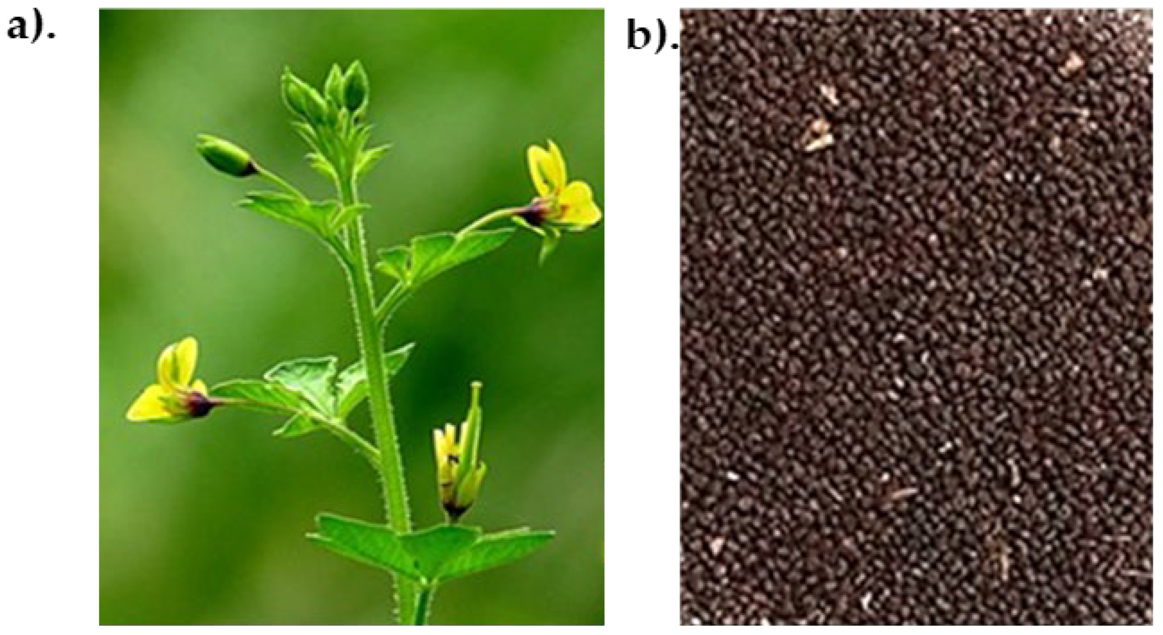
Figure 2.
(a). Trypsin inhibition activity of Cleome viscosa aqueous crude (CVC) extract at pH 7.8; Well No. 1. Trypsin 5 µg (Positive control), Well No. 2- 6. Trypsin 5 µg + 10, 20, 30, 40 and 50 µl of CVC, respectively, 7 - Buffer (Negative control).(b). Trypsin inhibition activity of 40%-80% fraction; Well No: 1 - Trypsin 5 µg, Well No. 2- 5: Trypsin 5 µg + 10, 20, 30, 40 µl of 0-80% respectively, 6 - Buffer (Negative Control).
Figure 2.
(a). Trypsin inhibition activity of Cleome viscosa aqueous crude (CVC) extract at pH 7.8; Well No. 1. Trypsin 5 µg (Positive control), Well No. 2- 6. Trypsin 5 µg + 10, 20, 30, 40 and 50 µl of CVC, respectively, 7 - Buffer (Negative control).(b). Trypsin inhibition activity of 40%-80% fraction; Well No: 1 - Trypsin 5 µg, Well No. 2- 5: Trypsin 5 µg + 10, 20, 30, 40 µl of 0-80% respectively, 6 - Buffer (Negative Control).
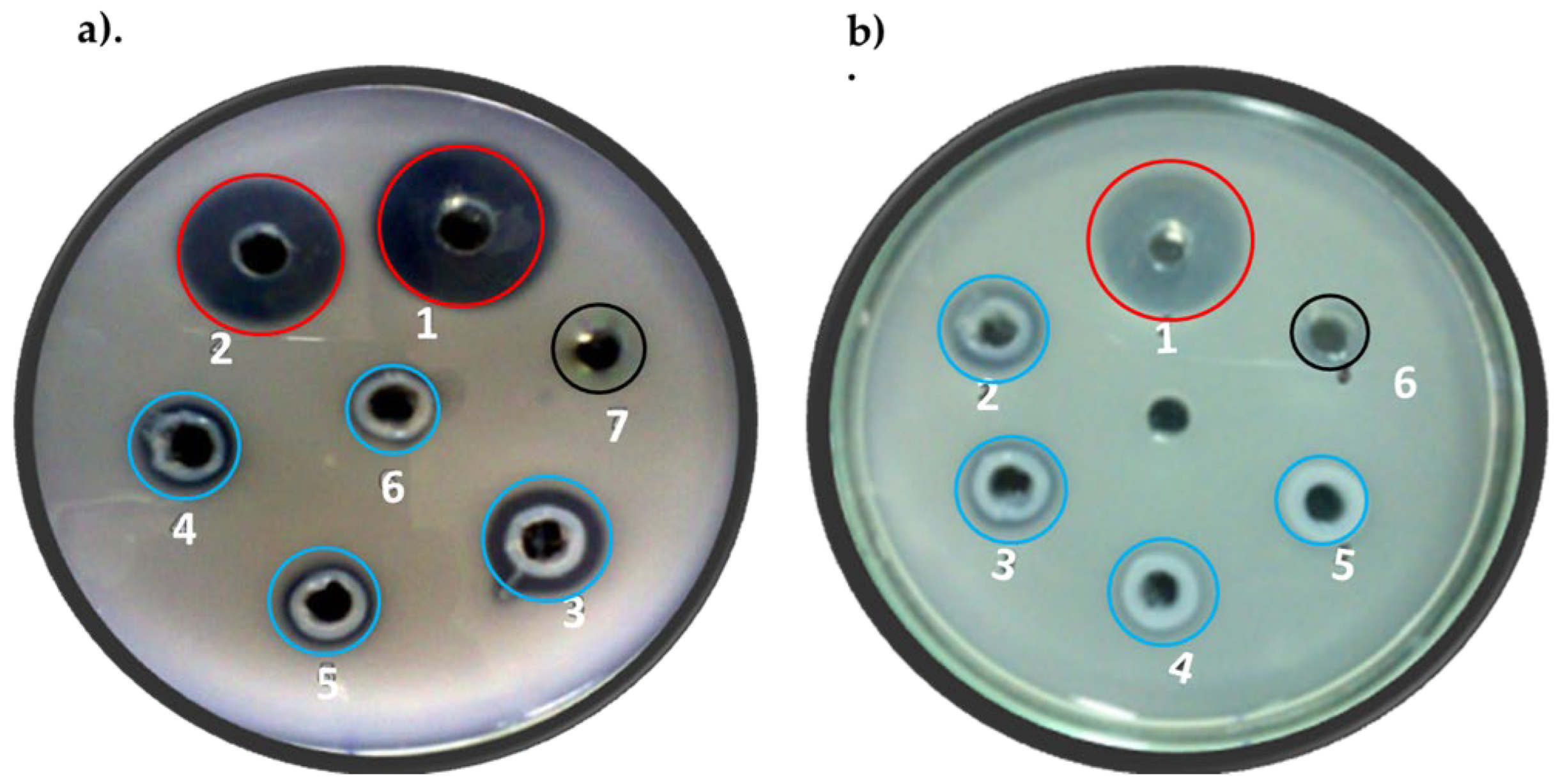
Figure 3.
Purification and trypsin inhibition of CVTI: (a). Size exclusion chromatography of 40-80% fraction on G-100 resin, (b) Trypsin inhibition by Peak 2 fraction of the size exclusion chromatography.[ Well No 1 - Trypsin 5 µg (Positive control), 2 - Trypsin 5µg + 10 µg of peak 2 fraction, 3 - Trypsin 5 µg + 20 µg of peak 2 fraction, 4 - Trypsin 5 µg + 30 µg of peak 2 fraction, 5 - Trypsin 5 µg + 40 µg of peak 2 fraction, 6 - Trypsin 5 µg + 50 µg of peak 2 fraction, 7 - Buffer (Negative control)].(c) SDS PAGE (15 %) with peak 1 and 2. Lysozyme (14 kDa) has been loaded as a molecular weight marker. (d). CVTI with standard protein marker on 12% SDS ploy acrylamide gel electrophoresis.
Figure 3.
Purification and trypsin inhibition of CVTI: (a). Size exclusion chromatography of 40-80% fraction on G-100 resin, (b) Trypsin inhibition by Peak 2 fraction of the size exclusion chromatography.[ Well No 1 - Trypsin 5 µg (Positive control), 2 - Trypsin 5µg + 10 µg of peak 2 fraction, 3 - Trypsin 5 µg + 20 µg of peak 2 fraction, 4 - Trypsin 5 µg + 30 µg of peak 2 fraction, 5 - Trypsin 5 µg + 40 µg of peak 2 fraction, 6 - Trypsin 5 µg + 50 µg of peak 2 fraction, 7 - Buffer (Negative control)].(c) SDS PAGE (15 %) with peak 1 and 2. Lysozyme (14 kDa) has been loaded as a molecular weight marker. (d). CVTI with standard protein marker on 12% SDS ploy acrylamide gel electrophoresis.
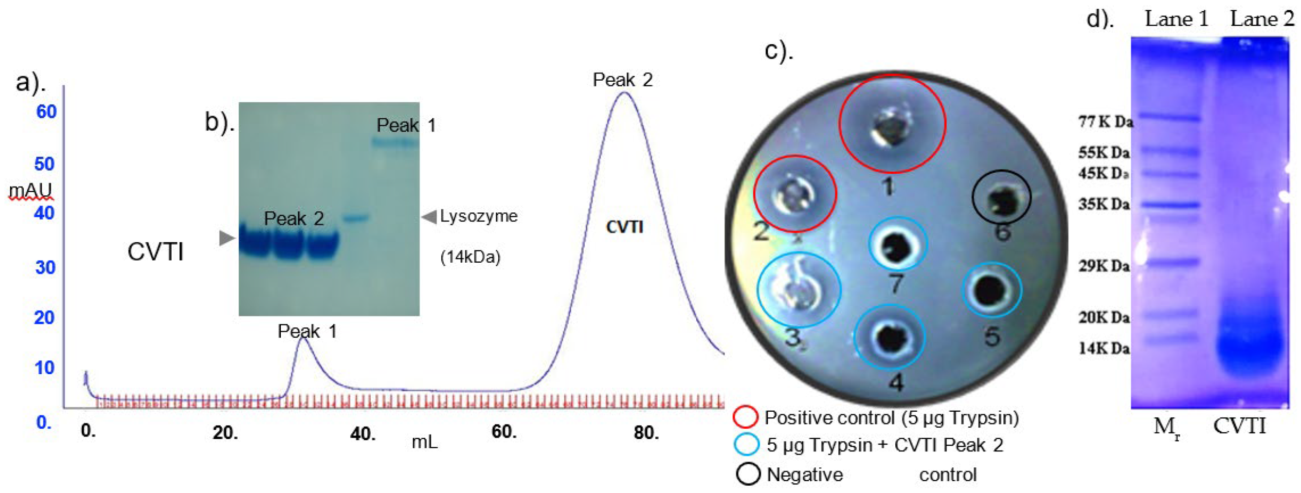
Figure 4.
Circular Dichroism spectra of CVTI in the far UV region(190nm-240nm), reveals majority of helical content (Alpha helix: 69.6%).
Figure 4.
Circular Dichroism spectra of CVTI in the far UV region(190nm-240nm), reveals majority of helical content (Alpha helix: 69.6%).
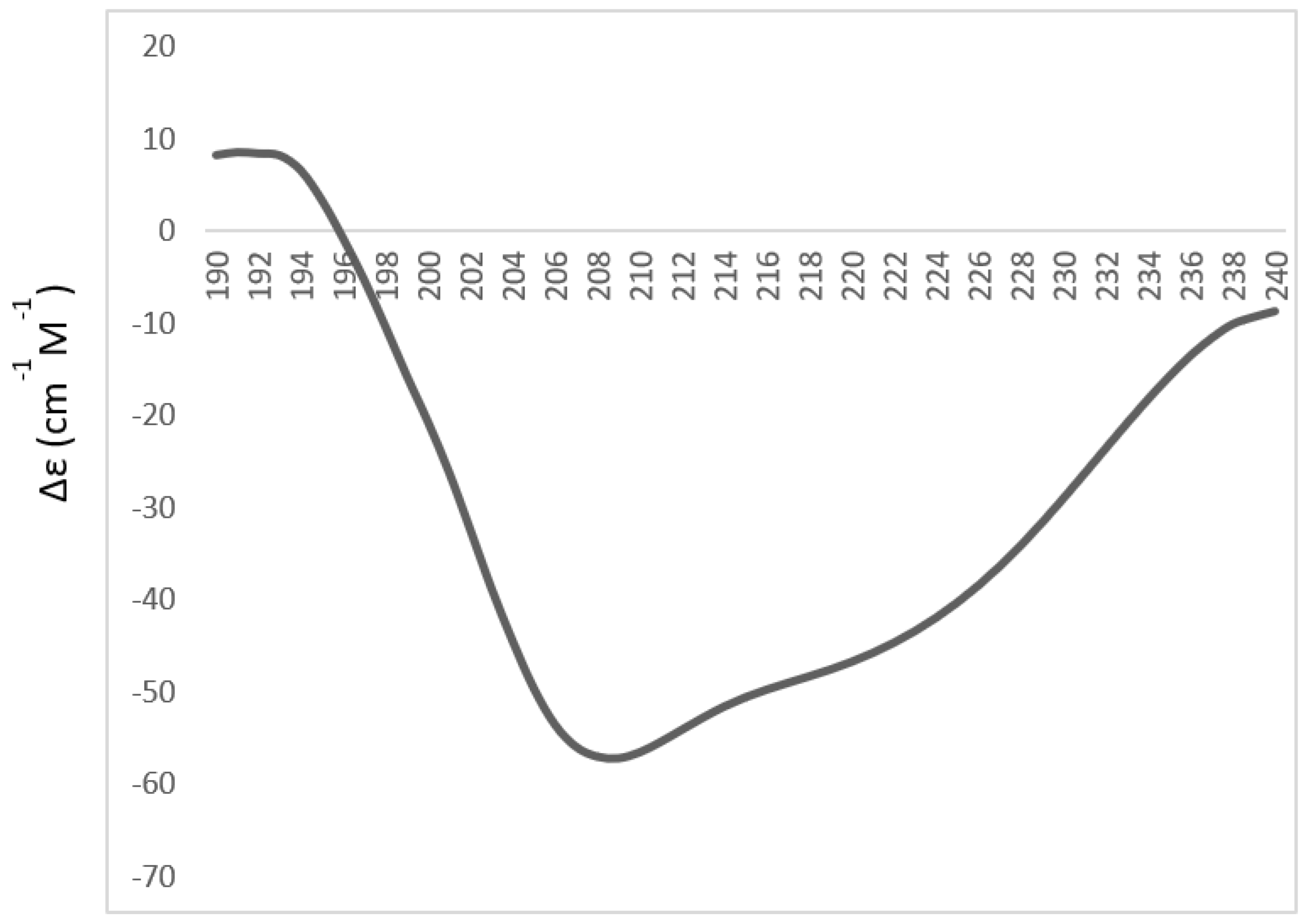
Figure 5.
Mass Spectrum Peptide finger printing pattern of CVTI fragments and their corresponding M/Z values.
Figure 5.
Mass Spectrum Peptide finger printing pattern of CVTI fragments and their corresponding M/Z values.
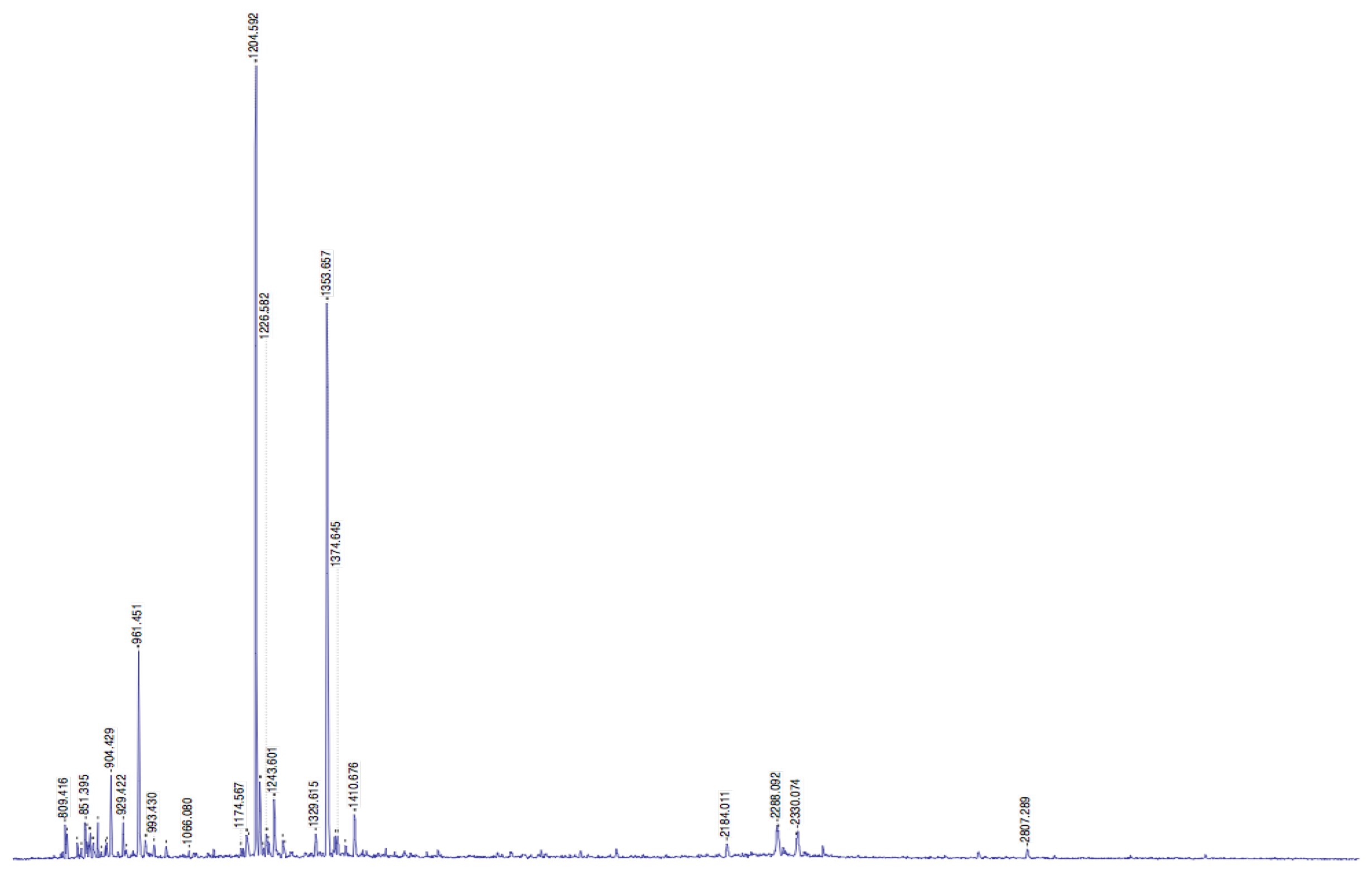
Figure 6.
a). Substrate Velocity curve (b) LB plot of CVTI vs Trypsin.
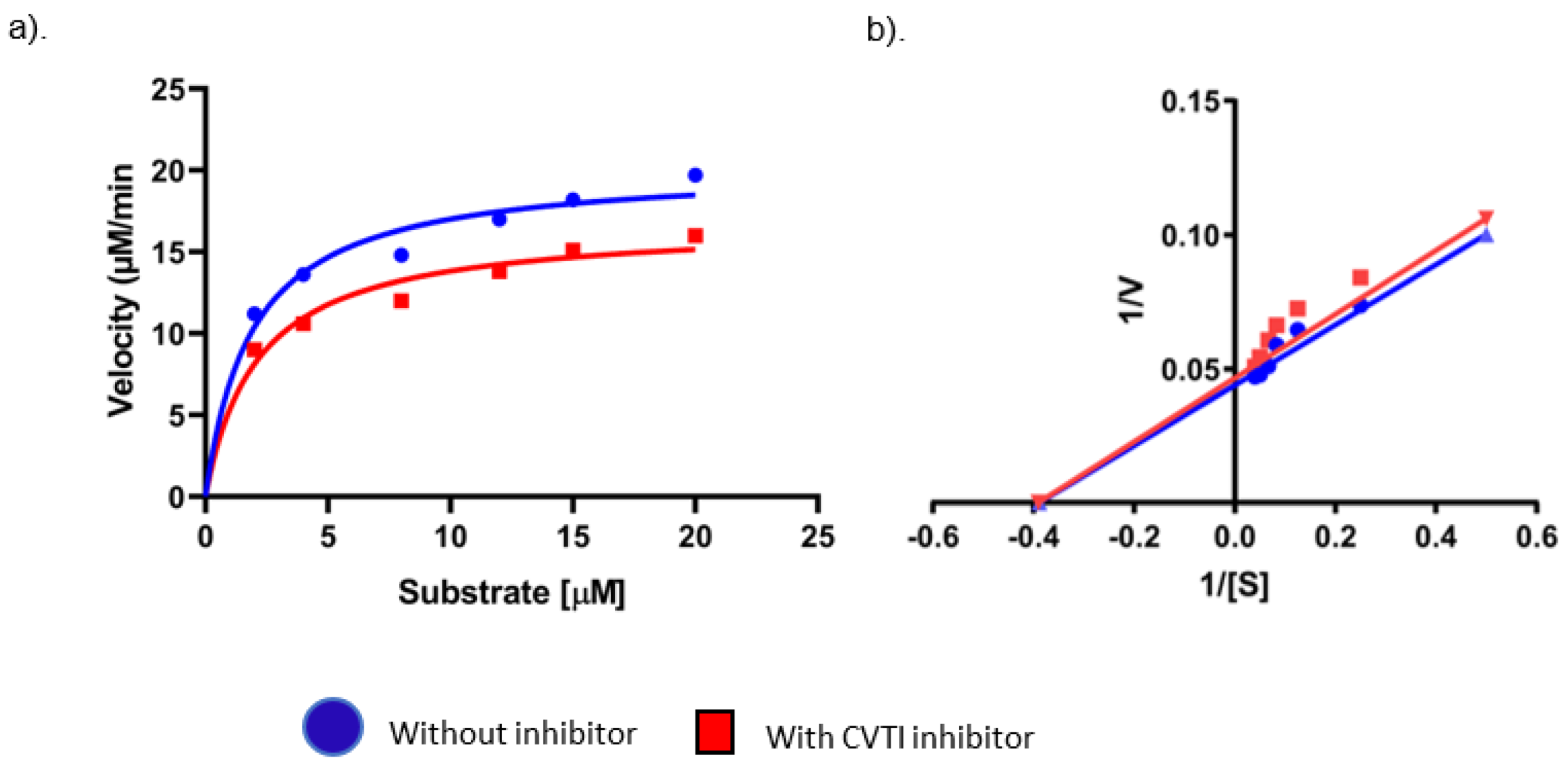
Figure 7.
Interaction by Isothermal titration of purified CVTI with trypsin.
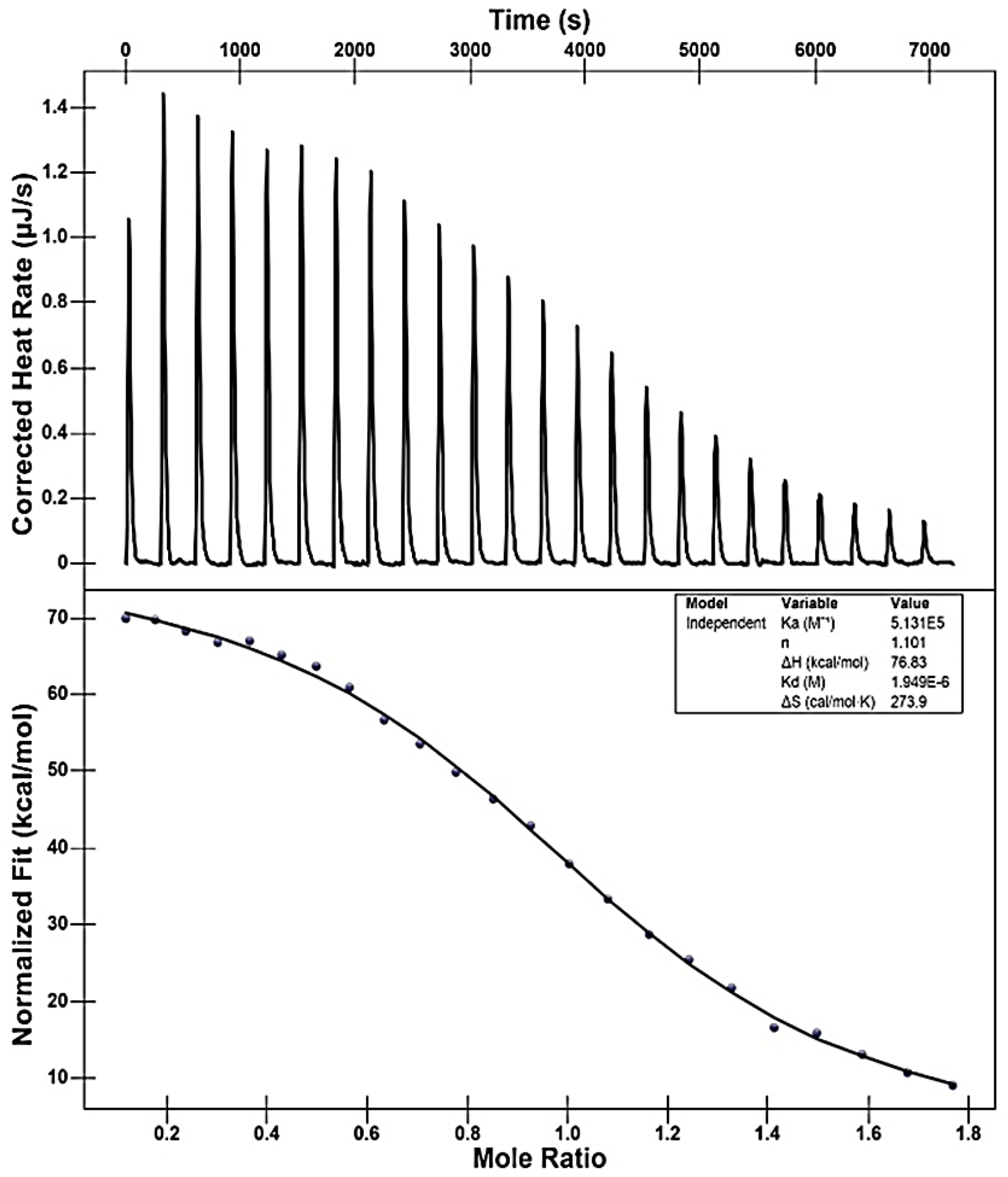
Figure 8.
Thermal stability of CVTI.
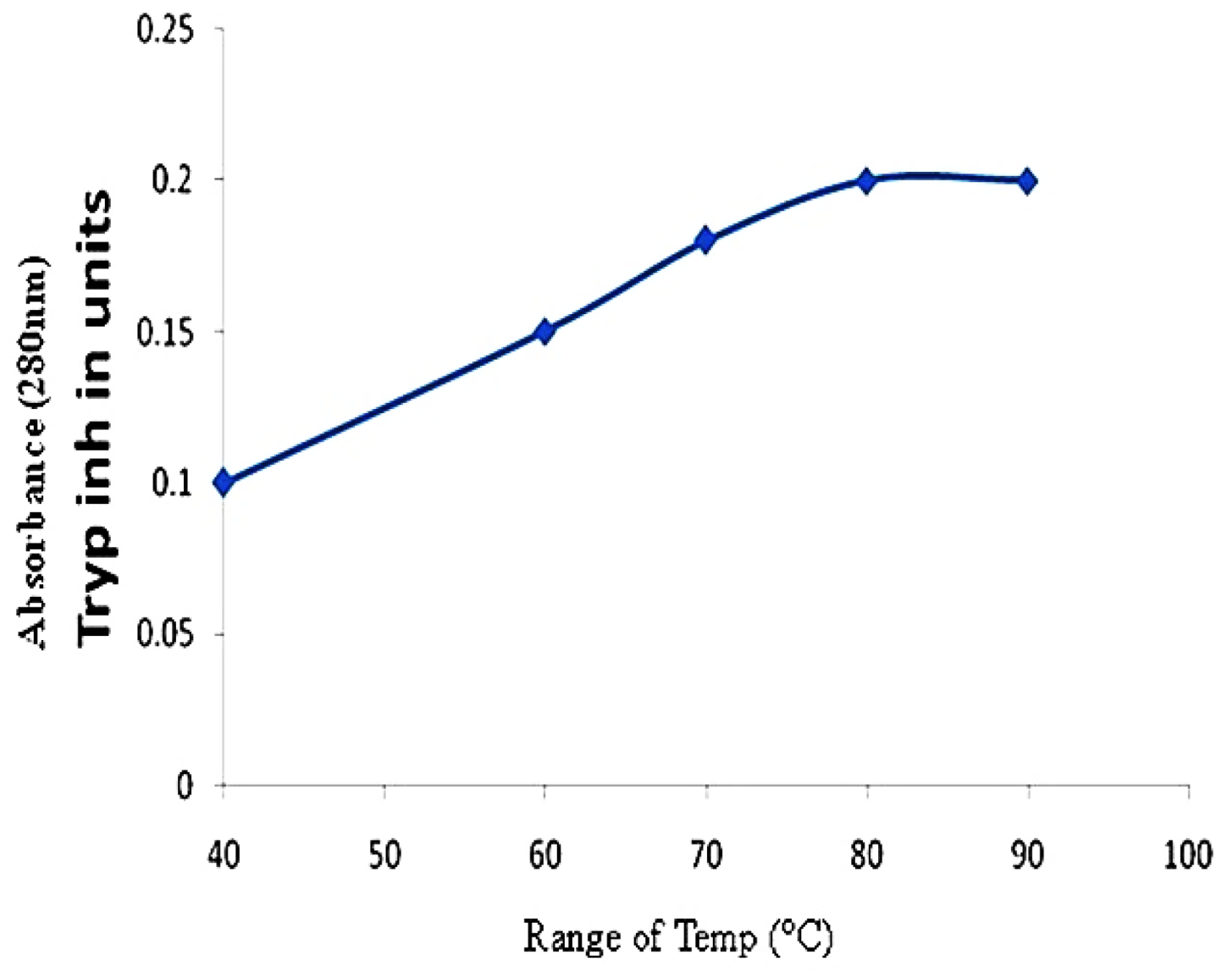
Figure 9.
Direct observation of viral infected HEp2 cells under (40 X) phase-contrast inverted microscope. (Arrow indicates the cytopathic effect by viral invasion). (a). HSV-2 infected cell; (b). Acyclovir 1.56 µg/ml; (c). CVTI 3 µg/ml.
Figure 9.
Direct observation of viral infected HEp2 cells under (40 X) phase-contrast inverted microscope. (Arrow indicates the cytopathic effect by viral invasion). (a). HSV-2 infected cell; (b). Acyclovir 1.56 µg/ml; (c). CVTI 3 µg/ml.
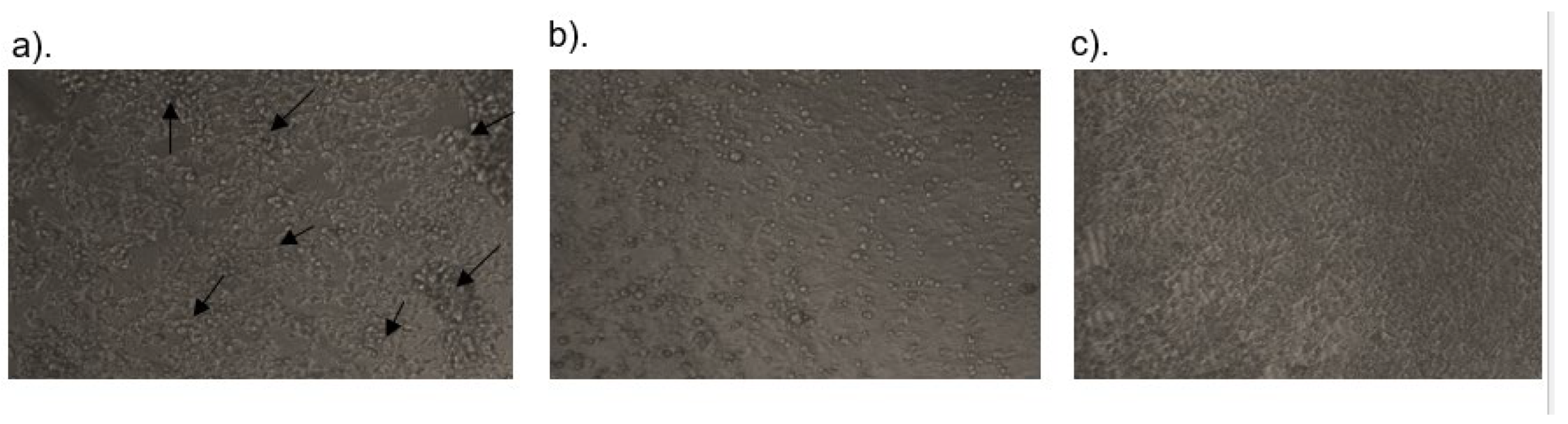

Table 1.
Maldi TOF/TOF Mass fingerprinting of CVTI sequence fragments.
| Peptide | Data submitted | MH+ Matched | Obtained Peptide sequences |
|---|---|---|---|
| 1 | 2807.387 | 2807.4566 | EEAKKIILKDKPDANIVVL |
| 2 | 834.3790 | 834.3905 | CVDIRET |
| 3 | 861.0650 | 859.4772 | CPRILMK |
| 4 | 2288.02 | 2289.421 | CPRNCDTNIAYSKCPRS |
| 5 | 120.569 | 120.461 | CLDNCEKEHD |
Disclaimer/Publisher’s Note: The statements, opinions and data contained in all publications are solely those of the individual author(s) and contributor(s) and not of MDPI and/or the editor(s). MDPI and/or the editor(s) disclaim responsibility for any injury to people or property resulting from any ideas, methods, instructions or products referred to in the content. |
© 2025 by the authors. Licensee MDPI, Basel, Switzerland. This article is an open access article distributed under the terms and conditions of the Creative Commons Attribution (CC BY) license (http://creativecommons.org/licenses/by/4.0/).
Copyright: This open access article is published under a Creative Commons CC BY 4.0 license, which permit the free download, distribution, and reuse, provided that the author and preprint are cited in any reuse.


