Submitted:
23 November 2025
Posted:
24 November 2025
You are already at the latest version
Abstract
Cystic fibrosis (CF) airway mucus is thick and sticky, which makes it hard for nanoparticles to move and deliver drugs effectively. In this study, degradable PLGA nanoparticles were coated with polyethylene glycol (PEG) of different molecular weights (2, 5, and 10 kDa) to test their movement and depth of penetration in CF-like mucus. The nanoparticles had a uniform size of 150 ± 10 nm and were measured using fluorescence recovery after photobleaching (FRAP) and confocal imaging. The PEG (5 kDa) coating showed the best results, with a diffusion rate of 1.45 ± 0.12 µm²/s, about 3.8 times higher than that of uncoated particles. In a 6-hour test, the particles moved over 70% deeper into the mucus and showed much higher uptake by airway cells (P < 0.01). These results show that medium-length PEG chains reduce mucus adhesion and help nanoparticles move more easily through dense airway mucus. This work provides a practical way to improve drug carriers for CF and other lung diseases with mucus blockage.
Keywords:
PEG coating
; PLGA nanoparticles
; mucus transport
; cystic fibrosis
; drug carrier
; diffusion
; airway model
1. Introduction
Cystic fibrosis (CF) airway mucus forms a dense and adhesive barrier that blocks inhaled therapeutics from reaching the epithelial surface. Excessive secretion of mucins, together with elevated levels of extracellular DNA and actin, significantly increases viscoelasticity and decreases pore size, which collectively hinder nanoparticle mobility [1]. These biophysical constraints restrict drug transport to the cell surface, impair absorption, and contribute to persistent infection and inflammatory cycles that characterize CF pathology [2]. Poly(lactic-co-glycolic acid) (PLGA) nanoparticles are widely explored as inhaled drug carriers because of their biodegradability, biocompatibility, and capacity for controlled drug release [3]. Nonetheless, unmodified PLGA nanoparticles readily adhere to mucin fibers through hydrophobic and electrostatic interactions, resulting in limited diffusion and shallow penetration into CF sputum [4]. Surface grafting with polyethylene glycol (PEG) offers an accessible strategy to reduce mucus adhesion. Hydrophilic PEG chains can form a steric hydration layer that minimizes contact with mucins and enhances nanoparticle movement within the mucus network [5]. Prior studies have shown that PEG-modified nanoparticles can improve transport and lung delivery efficiency, although reported outcomes vary with PEG chain length, surface density, and coating uniformity [6]. These inconsistencies arise partly from the use of artificial or healthy mucus models that do not fully reflect the concentrated, ion-rich microenvironment of CF sputum [7]. Moreover, differences in nanoparticle formulation, PEG attachment chemistry, and testing methodology complicate direct comparison across studies [8]. Advanced analytical methods—such as multiple particle tracking, fluorescence recovery after photobleaching (FRAP), and confocal imaging—are now employed to examine nanoparticle dynamics in mucus, enabling quantification of both ensemble diffusion and depth-dependent penetration [9]. Yet, most investigations assess only a single PEG configuration or rely on non-degradable systems, leaving limited understanding regarding how PEG molecular weight influences transport performance and subsequent cellular interaction in a CF-like environment [10. Recent work further underscores that optimizing the physicochemical architecture of nanocarriers is key to overcoming mucus barriers and improving drug delivery efficiency in pulmonary disease [11,12].
The study prepared PLGA nanoparticles coated with PEG chains of three molecular weights (2, 5, and 10 kDa), maintaining a uniform diameter of approximately 150 nm. Using FRAP, we quantified nanoparticle mobility, and confocal imaging was applied to evaluate penetration depth and resulting uptake in a CF-like mucus model. The work compares the diffusion behavior of different PEG chain lengths, establishes the connection between faster diffusion and deeper penetration with enhanced cell uptake, and identifies an optimal PEG chain length that balances mobility with mucus interaction. Collectively, these results elucidate how PEG architecture governs the transport of degradable nanoparticles through CF-like sputum and provide design guidelines for developing efficient inhaled drug delivery systems targeting mucus-obstructive lung disease.
2. Materials and Methods
2.1. Sample and Model Description
This study used a simulated cystic fibrosis (CF) mucus model that mimicked the main physical and chemical properties of patient sputum. The mixture contained mucin (5% w/v), DNA (2% w/v), and actin (0.5% w/v) in phosphate-buffered saline (PBS, pH 7.4), giving a final viscosity of 1.8 ± 0.2 Pa·s at 25 °C. To validate the model, three CF sputum samples were collected from patients at the Shanghai Pulmonary Hospital with ethical approval. All samples were gently homogenized, stored at 4 °C, and used within 24 hours. Before testing, each sample was pre-incubated at 37 °C for 1 hour to reach steady-state viscosity.
2.2. Experimental Design and Control Setup
Poly(lactic-co-glycolic acid) (PLGA) nanoparticles were made by a double-emulsion solvent evaporation method. The surfaces were modified with polyethylene glycol (PEG) chains of 2 kDa, 5 kDa, and 10 kDa using carbodiimide chemistry. Uncoated PLGA nanoparticles were used as controls. All particles had an average size of 150 ± 10 nm and a surface charge between –5 mV and –10 mV. Three independent batches were tested for each group. Nanoparticles were dispersed in the mucus samples at 0.1 mg/mL, and diffusion was tracked for 6 hours at 37 °C. The comparison focused on particle diffusion, penetration depth, and uptake by airway epithelial cells.
2.3. Measurement Methods and Quality Control
Fluorescence recovery after photobleaching (FRAP) was used to measure particle diffusion. Nanoparticles were labeled with fluorescein isothiocyanate (FITC), and fluorescence recovery was recorded with a laser confocal microscope (Leica SP8, Germany) over a 10 µm × 10 µm area. The diffusion coefficient (D) was obtained by fitting the recovery curve. Penetration depth was determined by z-stack confocal imaging to a depth of 200 µm. For cell uptake tests, 16HBE bronchial epithelial cells were grown on Transwell membranes and exposed to nanoparticles for 6 hours. All tests were repeated three times, and results with more than 5% deviation from the mean were removed. Each instrument was calibrated before use, and temperature was controlled at 37 °C during measurements.
2.4. Data Processing and Analytical Formulas
All data were processed with Origin 2023 and GraphPad Prism 10.0. The diffusion coefficient was calculated as:
where is the radius of the bleached area and is the half-time for fluorescence recovery. Penetration efficiency was defined as [13]:
where is the fluorescence intensity at a given depth , and is the surface intensity. One-way ANOVA followed by Tukey’s test was used to determine differences, and P < 0.05 was considered significant.
2.5. Experimental Conditions and Reproducibility
All nanoparticle samples were freshly prepared before each experiment to avoid aggregation. Particle size and uniformity were checked by dynamic light scattering (Malvern Zetasizer Nano ZS), and morphology was observed by transmission electron microscopy (TEM). Blank mucus controls were used to confirm fluorescence stability during FRAP tests. Three trained operators repeated all measurements under the same temperature (37 ± 0.2 °C) and humidity (50 ± 5%) conditions to ensure reproducibility.
3. Results and Discussion
3.1. Effect of PEG Chain Length on Nanoparticle Diffusion
FRAP analysis showed that PEG modification greatly improved the movement of PLGA nanoparticles (NPs) in CF-like mucus. The PEG (5 kDa) group reached a diffusion coefficient of 1.45 ± 0.12 µm²/s, about 3.8 times higher than unmodified PLGA. The 2 kDa PEG coating showed moderate improvement, while 10 kDa PEG gave no further gain, likely because longer chains increased friction. This indicates that a medium-length PEG coating forms a compact hydration layer that reduces adhesion without adding excess drag. The trend matches previous findings showing that proper PEG coverage increases transport in thick mucus layers [14].
Figure 1.
Diffusion and penetration of PEG-coated PLGA nanoparticles in CF-like mucus.
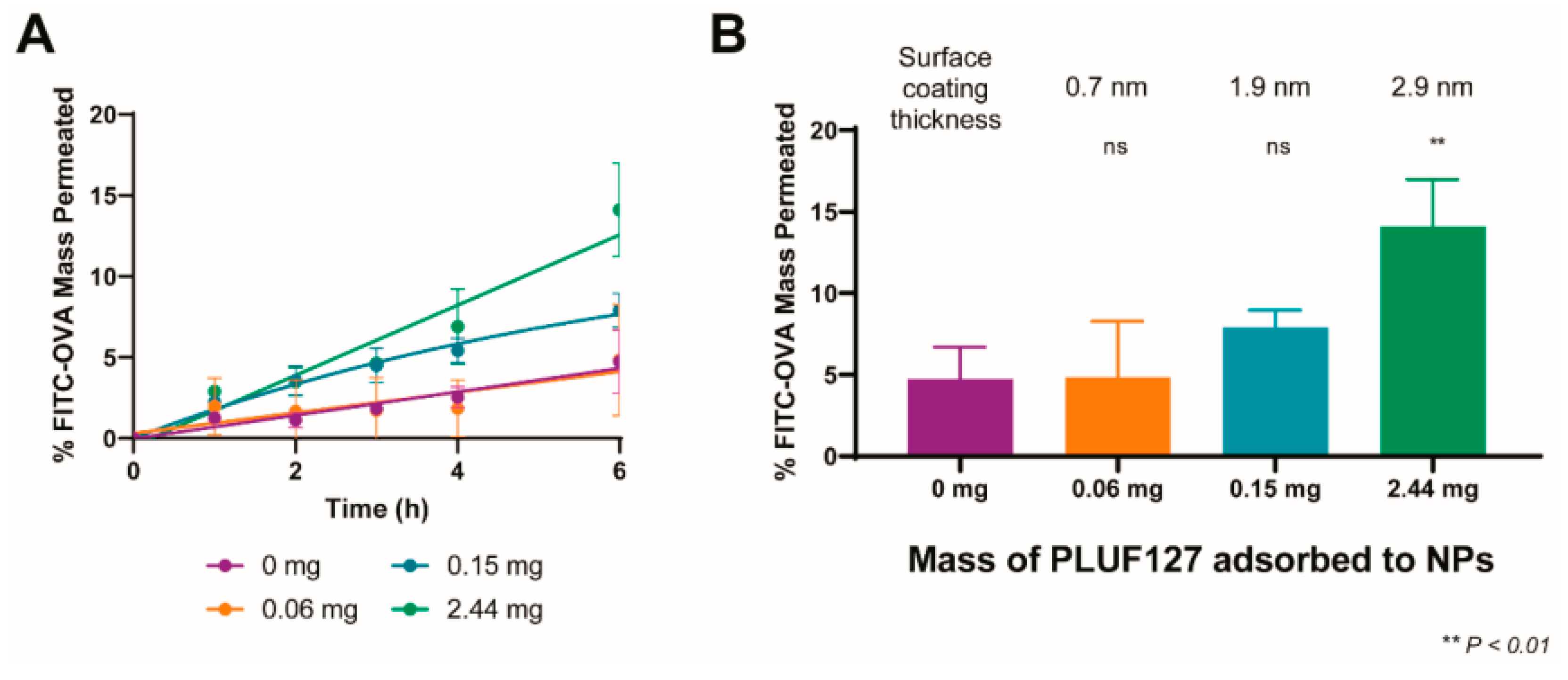
3.2. Mucus Penetration and Cellular Delivery
Confocal depth imaging showed that PEG modification increased particle penetration through the mucus layer by more than 70% within 6 hours. The PEG (5 kDa) NPs showed the deepest average penetration, followed by 2 kDa and 10 kDa coatings. The higher penetration also led to significantly stronger uptake by 16HBE bronchial epithelial cells (P < 0.01). These findings suggest that PEG helps nanoparticles overcome the initial adhesion barrier, improving both diffusion and access to target cells. This agrees with previous mucus transport studies using similar biosimilar mucus systems, where PEG surface coatings improved deep-layer mobility compared with uncoated PLGA particles [15].
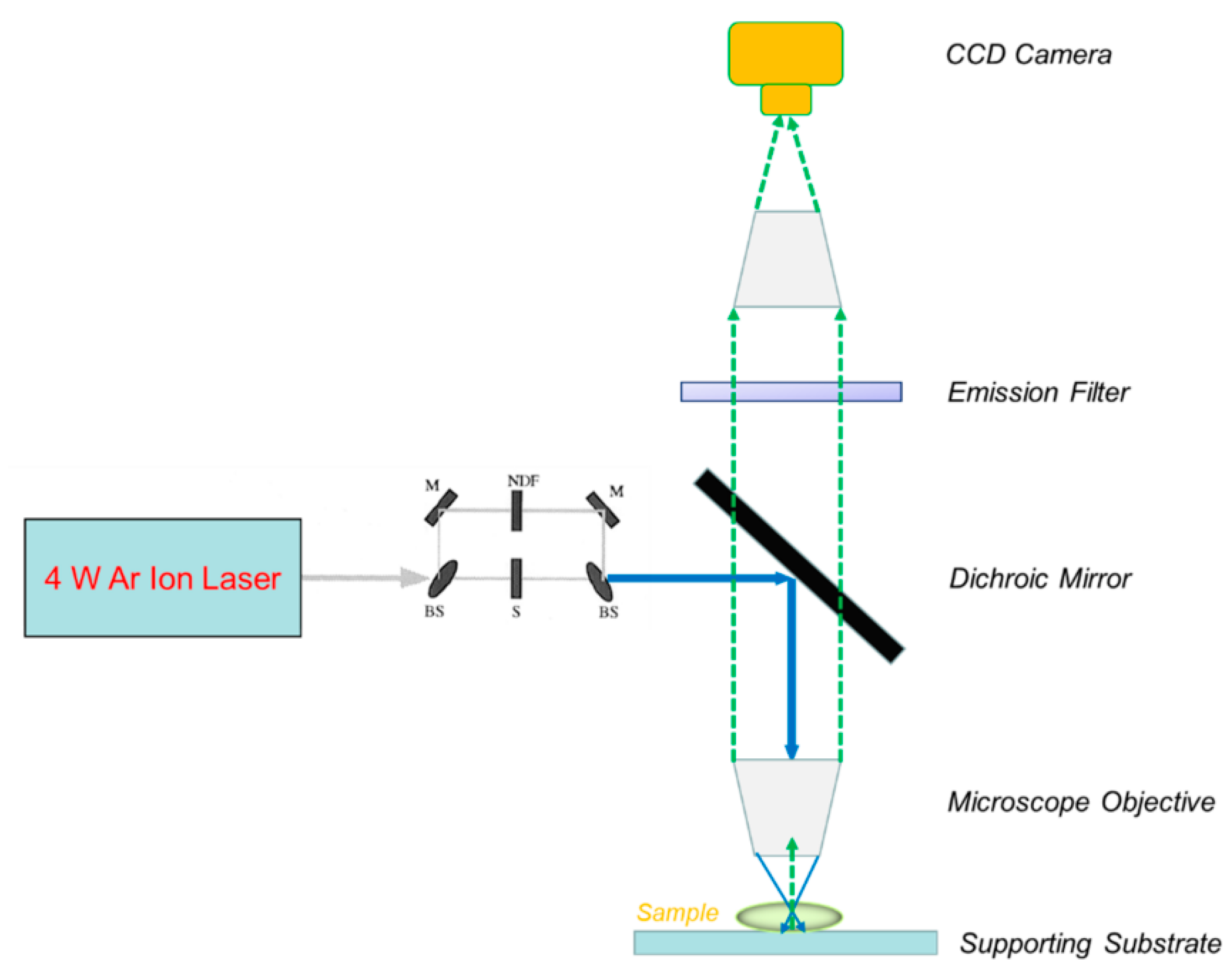
3.3. FRAP Validation and Consistency Across Methods
FRAP recovery curves were fitted using the standard spot-FRAP model, giving consistent diffusion results across PEG variants [16]. Bleaching radius and recovery half-time remained stable, confirming the reliability of the setup.
Figure 2.
Diagram of the FRAP system used to measure nanoparticle movement.
3.4. Comparison with Previous Studies and Practical Significance
Compared with studies using variable particle sizes or low PEG density, this work used uniform 150 nm PLGA nanoparticles with controlled surface charge, isolating the effect of PEG chain length. The results indicate that PEG (5 kDa) provides the best trade-off between mobility and coating stability [17]. High-density PEG coatings prevent adhesion to mucin fibers while maintaining moderate drag, improving both short-term diffusion and long-term penetration. Similar findings were reported in mucus model studies showing that dense PEG coatings allow efficient transport without aggregation [18]. These results provide design guidance for pulmonary nanoparticle systems, where optimized PEG coatings can enhance drug delivery efficiency in CF and other mucus-rich airway diseases.
4. Conclusion
This study explored how PEG coating changes the movement of degradable PLGA nanoparticles in cystic fibrosis–like mucus. The results showed that PEG coating, especially with 5 kDa chains, clearly increased nanoparticle diffusion, mucus penetration, and cell uptake compared with uncoated particles. The PEG (5 kDa) group reached a diffusion rate of 1.45 ± 0.12 µm²/s, about 3.8 times higher than the control, and moved more than 70% deeper into the mucus after six hours. These results show that a medium-length PEG layer forms a stable water shell that lowers adhesion to mucus while keeping good mobility. The findings give a simple and reliable way to improve nanoparticle movement in thick mucus and can support the design of better drug carriers for lung diseases such as cystic fibrosis and chronic bronchitis. The main limitation of this study is that tests were done in model mucus and limited clinical samples. Future work should include more patient data and in vivo studies to confirm long-term safety and clearance of PEG-coated particles.
References
- Abrami, M., Biasin, A., Tescione, F., Tierno, D., Dapas, B., Carbone, A., ... & Grassi, M. (2024). Mucus structure, viscoelastic properties, and composition in chronic respiratory diseases. International Journal of Molecular Sciences, 25(3), 1933.
- Zha, D. , Gamez, J., Ebrahimi, S. M., Wang, Y., Verma, N., Poe, A. J.,... & Saghizadeh, M. (2025). Oxidative stress-regulatory role of miR-10b-5p in the diabetic human cornea revealed through integrated multi-omics analysis. Diabetologia, 1-16.
- Costa, M. P. , Abdu, J. O. C., de Moura, M. F. C. S., Silva, A. C., Zacaron, T. M., de Paiva, M. R. B.,... & Tavares, G. D. (2025). Exploring the Potential of PLGA Nanoparticles for Enhancing Pulmonary Drug Delivery. Molecular Pharmaceutics.
- Omidian, H. , & Wilson, R. L. (2025). PLGA-Based Strategies for Intranasal and Pulmonary Applications. Pharmaceutics, 17(2), 207.
- Tafech, B. , Rokhforouz, M. R., Leung, J., Sung, M. M., Lin, P. J., Sin, D. D.,... & Hedtrich, S. (2024). Exploring Mechanisms of Lipid Nanoparticle-Mucus Interactions in Healthy and Cystic Fibrosis Conditions. Advanced Healthcare Materials, 13(18), 2304525.
- Chen, D., Liu, J., Wu, J., & Suk, J. S. (2021). Enhancing nanoparticle penetration through airway mucus to improve drug delivery efficacy in the lung. Expert opinion on drug delivery, 18(5), 595-606.
- Xu, J. (2025). Building a Structured Reasoning AI Model for Legal Judgment in Telehealth Systems.
- Ho, K. W. , Liu, Y. L., Liao, T. Y., Liu, E. S., & Cheng, T. L. (2024). Strategies for non-covalent attachment of antibodies to PEGylated nanoparticles for targeted drug delivery. International Journal of Nanomedicine, 10045-10064.
- Tjakra, M. , Chakrapeesirisuk, N., Jacobson, M., Sellin, M. E., Eriksson, J., Teleki, A., & Bergström, C. A. (2025). Optimized Artificial Colonic Mucus Enabling Physiologically Relevant Diffusion Studies of Drugs, Particles, and Delivery Systems. Molecular Pharmaceutics.
- Wang, Y. , Wen, Y., Wu, X., Wang, L., & Cai, H. (2025). Assessing the Role of Adaptive Digital Platforms in Personalized Nutrition and Chronic Disease Management.
- Ali, T. (2025). Nanomedicine Approaches to Overcome Barriers in Pulmonary Drug Delivery for Respiratory Diseases. Current Pharmaceutical Research, 30-44.
- Wen, Y. , Wu, X., Wang, L., Cai, H., & Wang, Y. (2025). Application of Nanocarrier-Based Targeted Drug Delivery in the Treatment of Liver Fibrosis and Vascular Diseases. Journal of Medicine and Life Sciences, 1(2), 63-69.
- Zha, D. , Mahmood, N., Kellar, R. S., Gluck, J. M., & King, M. W. (2025). Fabrication of PCL Blended Highly Aligned Nanofiber Yarn from Dual-Nozzle Electrospinning System and Evaluation of the Influence on Introducing Collagen and Tropoelastin. ACS Biomaterials Science & Engineering.
- Tafech, B. , Rokhforouz, M. R., Leung, J., Sung, M. M., Lin, P. J., Sin, D. D.,... & Hedtrich, S. (2024). Exploring Mechanisms of Lipid Nanoparticle-Mucus Interactions in Healthy and Cystic Fibrosis Conditions. Advanced Healthcare Materials, 13(18), 2304525.
- Semitela, A. , Marques, P. A., & Completo, A. (2024). Strategies to engineer articular cartilage with biomimetic zonal features: a review. Biomaterials Science, 12(23), 5961-6005.
- Xu, K., Lu, Y., Hou, S., Liu, K., Du, Y., Huang, M., ... & Sun, X. (2024). Detecting anomalous anatomic regions in spatial transcriptomics with STANDS. Nature Communications, 15(1), 8223.
- Tsukamoto, Y. , Kawamura, A., Yurtsever, A., Suzuki, H., Rojas-Chaverra, N. M., Sato, H.,... & Sakai, K. (2025). Condensation-dependent interactome of a chromatin remodeler underlies tumor suppressor activities. Nature Communications, 16(1), 9599.
- Dash, P. K. , Chen, C., Kaminski, R., Su, H., Mancuso, P., Sillman, B.,... & Khalili, K. (2023). CRISPR editing of CCR5 and HIV-1 facilitates viral elimination in antiretroviral drug-suppressed virus-infected humanized mice. Proceedings of the National Academy of Sciences, 120(19), e2217887120.
Disclaimer/Publisher’s Note: The statements, opinions and data contained in all publications are solely those of the individual author(s) and contributor(s) and not of MDPI and/or the editor(s). MDPI and/or the editor(s) disclaim responsibility for any injury to people or property resulting from any ideas, methods, instructions or products referred to in the content. |
© 2025 by the authors. Licensee MDPI, Basel, Switzerland. This article is an open access article distributed under the terms and conditions of the Creative Commons Attribution (CC BY) license (http://creativecommons.org/licenses/by/4.0/).
Copyright: This open access article is published under a Creative Commons CC BY 4.0 license, which permit the free download, distribution, and reuse, provided that the author and preprint are cited in any reuse.


