Preprint
Review

This version is not peer-reviewed.

Prenols and Prenoic Acids in Metabolism, Disease, and Aging

Submitted:

18 November 2025

Posted:

20 November 2025

You are already at the latest version

Abstract

Acyclic terpene derivatives are well known as components of plant essential oils and insect hormones, yet their active biosynthesis also occurs in mammals. The terpenic alcohols—or prenols—geranylgeraniol (GGOH) and farnesol (FOH), together with their prenoic acids and derivatives, were identified in mammalian cells over sixty years ago but remain largely overlooked. These metabolites display diverse biological functions: they induce autophagy, inhibit tumor growth and inflammation, suppress cholesterol synthesis, enhance insulin sensitivity and cognition, regulate sexual characteristics, and promote healthy aging. In mammals, prenols arise from an age-dependent, bidirectional pathway that interconverts polyprenyl diphosphates, prenols, and prenoic acids. They can be oxidized into aldehydes and carboxylic acids or reconverted into diphosphate forms for use in protein prenylation and in the biosynthesis of ubiquinone, cholesterol, and dolichol. While enzymes catalyzing polyprenyl diphosphate dephosphorylation and oxidation steps have been partly characterized, the kinases mediating their reverse phosphorylation remain unidentified. This review summarizes current advances in the understanding of prenol metabolism in mammals, emphasizing its role in metabolic regulation, disease prevention, and longevity. By integrating biochemical and physiological evidence, we highlight the emerging view that these small terpenes constitute a fundamental yet underexplored layer of metabolic control. Greater attention to this pathway may reveal novel strategies for maintaining metabolic health and mitigating age-related disorders.

Keywords: 
;  ;  ;  ;  ;  ;  ;  

1. Introduction

1.1. Cellular Mechanisms of Aging

Among multicellular organisms, the transition to reproductive maturity is typically preceded by a juvenile stage. This transition is tightly regulated by hormonal and genetic programs. However, the progression from maturity to senescence remains far less understood and is characterized by a gradual decline in physiological and reproductive functions, known as aging (Lopez-Otín et al., 2013). Unlike the predictable onset of adulthood, aging is influenced by a complex interplay of genetic, metabolic, and environmental factors.
In humans, aging is evident both externally and internally. Outwardly, changes such as skin wrinkling, graying hair, and reduced elasticity become visible. Internally, aging leads to losses in muscle mass, cognitive abilities, and immune responsiveness (Kirkwood, 2005). At the cellular level, aging is marked by genomic instability, telomere attrition, altered gene expression, mitochondrial functional decline, and cellular senescence (López-Otín et al., 2013). The cumulative effect of these processes increases vulnerability to chronic diseases and conditions, among them cardiovascular disorders, neurodegeneration, cancer, diabetes, osteoporosis, and sarcopenia (Franceschi et al., 2018; Kennedy et al., 2014).
Biochemically, aging involves multiple impaired cues. Defective mitochondrial oxidative phosphorylation leads to reduced ATP production and elevated levels of reactive oxygen species (ROS), promoting oxidative stress and damage to biomolecules (Bratic & Larsson, 2013). Concomitantly, antioxidant defenses such as the glutathione redox cycle and superoxide anion dismutation decline, exacerbating redox imbalance (Harman, 2001). Insulin resistance rises with age, disrupting glucose uptake and contributing to type 2 diabetes (Barzilai et al., 2012). Serum lipid profiles also worsen, with increased LDL, reduced HDL, and accumulation of toxic lipid species that foster cardiovascular pathology (Aschner et al., 2021).
In addition, a chronic pro-inflammatory state emerges, driven by increased cytokines such as IL-6 and TNF-α (Franceschi et al., 2018). Cellular processes like autophagy and proteostasis decline, reducing the clearance of damaged proteins and organelles (Rubinsztein et al., 2011). Levels of NAD+, crucial for mitochondrial metabolism and DNA repair, also fall with age (Imai & Guarente, 2014). Finally, aging also affects cofactors like S-adenosyl-L-methionine (SAM), acetyl-CoA, and α-ketoglutarate, influencing gene expression and accelerating cellular aging (Hayashi et al., 2022; Naeini et al., 2023; Schroeder et al., 2014).
Emerging research suggests that aging is not merely a passive deterioration but may be subject to modulation. Reprogramming experiments have shown that partial induction of pluripotency factors (e.g., OCT4, SOX2, KLF4, and c-MYC) can reverse epigenetic aging markers without erasing cellular identity (Ocampo et al., 2016). Furthermore, energy-sensing pathways such as AMPK and mTOR regulate longevity through autophagy and nutrient balance. While high mTOR activity inhibits autophagy, fasting and caloric restriction activate AMPK, suppress mTOR, and restore cellular cleanup mechanisms clearing macromolecules and organelle debris (Laplante & Sabatini, 2012). Impaired autophagy and hyperglycemia synergize to exacerbate tissue damage and aging via accumulation of advanced glycation end-products (Uribarri et al., 2005).
Pharmacological reversion of aging has been attempted. Among the most promising strategies in this respect is the use of rapamycin, an mTOR inhibitor that enhances autophagy, improves stem cell maintenance, and delays age-associated diseases. In rodents, rapamycin prolongs lifespan and reduces incidence of cancer, neurodegeneration, and immune dysfunction, even when administered late in life (Mannick et al., 2014; Wilkinson et al., 2012).
A central, yet underappreciated player in the aging process is the mevalonate (MVA) pathway—a metabolic route classically known primarily for cholesterol biosynthesis, but also responsible for producing bioactive isoprenoids like ubiquinone-10 (coenzyme Q 10), dolichols, and prenyl groups (Goldstein & Brown, 1990). These metabolites are critical for mitochondrial respiration, protein maturation and glycosylation, membrane integrity, and intracellular signaling (Gillespie et al., 2016). Dysregulation of this pathway contributes to mitochondrial dysfunction, defective proteostasis, and chronic inflammation—key features of aging (Buhaescu & Izzedine, 2007).

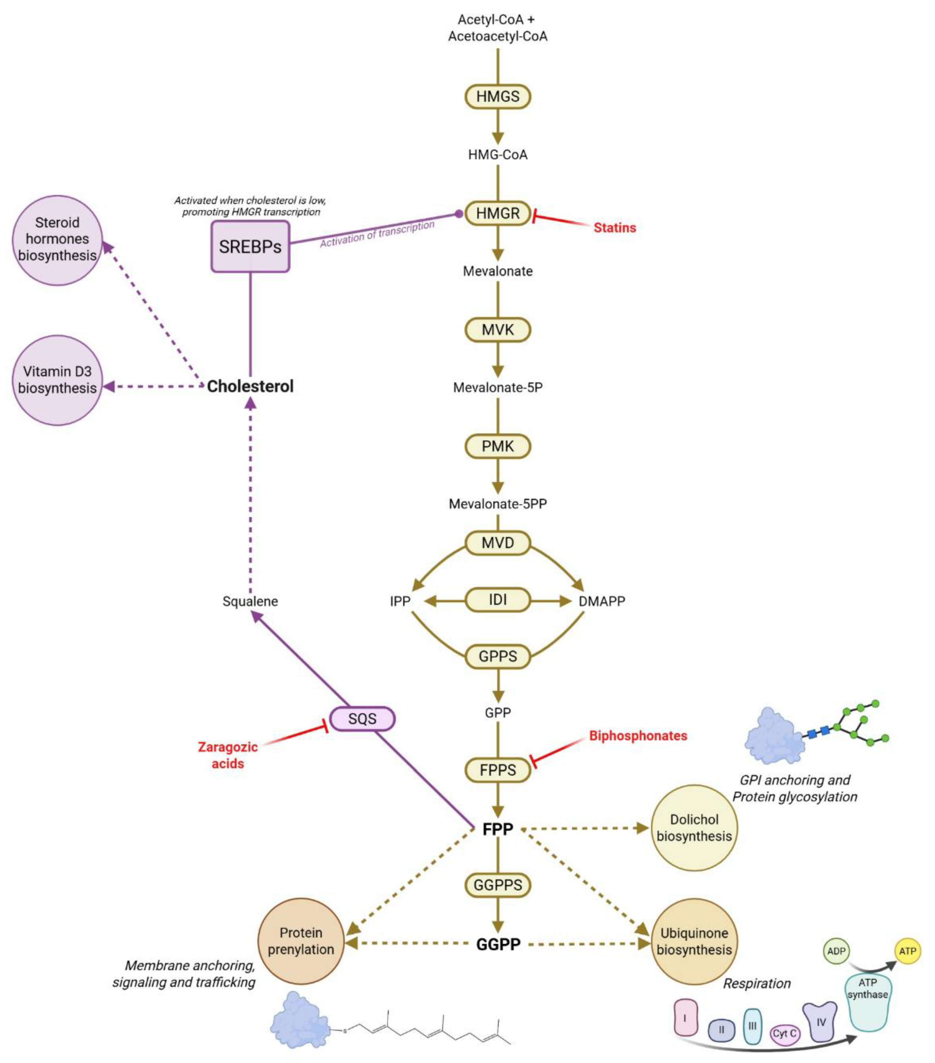
1.2. The Mevalonate Pathway at the Heart of Metabolic Control

Isoprenoids—also known as terpenes or terpenoids—constitute one of the most ancient and structurally diverse classes of lipids. Interested readers in isoprenoid biochemical pathways and evolutionary aspects are referred to that work (Crispim et al., 2024b). In the present review, however, we will focus on the fundamental principles of isoprenoid biology, particularly as they relate to human disease and aging.
All isoprenoids are conceptually derived from the five-carbon building block isoprene (C₅H₈). Their biosynthesis begins with the formation of two key precursors: isopentenyl diphosphate (IPP) and its isomer dimethylallyl diphosphate (DMAPP). Through sequential condensation of these units, a wide range of larger compounds is produced, which are classified according to chain length into groups such as monoterpenes (C₁₀), sesquiterpenes (C₁₅), diterpenes (C₂₀), and triterpenes (C₃₀) (Crispim et al., 2024b).
In animals, isoprenoid biosynthesis proceeds through the cytosolic MVA pathway (Figure 1), which begins with the condensation of acetyl-CoA molecules to form HMG-CoA. This is then reduced to mevalonic acid by HMG-CoA reductase (HMGR). Remarkably, this is the pathway’s rate-limiting enzyme and the molecular target of statin drugs. Mevalonic acid is then phosphorylated and decarboxylated to generate IPP and DMAPP; these two are interconvertible by the action of the enzyme IPP isomerase (IDI). At any rate, these C₅ units serve as building blocks for larger molecules. Through prenylsynthase enzymes, they form geranyl diphosphate (GPP, a monoterpene; by GPP synthase), farnesyl diphosphate (FPP, a sesquiterpene; by FPP synthase, which also is the target of nitrogen-containing bisphosphonates used in bone-related diseases), and geranylgeranyl diphosphate (GGPP, a diterpene; by GGPP synthase). FPP and GGPP are precursors to various essential compounds: dolichols (used in N-linked glycosylation), and ubiquinone (the sole lipid electron carrier in mitochondria with great antioxidant activity). These intermediates also participate in protein prenylation, modulating numerous cell processes (Crispim et al., 2024b).
Another important derivative of the mevalonate (MVA) pathway is cholesterol, together with all steroid hormones derived from it. The cholesterol biosynthetic pathway begins with the activity of squalene synthase (SQS), which involves two sequential chemical reactions. This enzyme catalyzes the dimerization of farnesyl diphosphate (FPP) to form the intermediate presqualene diphosphate (PSDP), followed by its reduction to squalene using NADPH as a cofactor. In one hand, presqualene diphosphate can be transformed to presqualene monophosphate by the enzyme pyrophosphatase/phosphatase domain-containing protein 2 (PPAPDC2). This molecule acts as a regulatory lipid signal in human immune cells. Specifically, presqualene monophosphate has been shown to inhibit phospholipase D and to reduce superoxide anion production, thereby modulating inflammatory responses (Fukunaga et al., 2006). On the other hand, squalene continues along the cholesterol biosynthetic pathway. Among the multistep monooxygenase reactions required for cholesterol formation, squalene epoxidase (SQLE) catalyzes the first oxygenation step in the committed branch of cholesterol synthesis, upstream of the Kandutsch–Russell (predominant in specific tissues such as brain and skin) and Bloch (mainly in liver) pathways. These branches use lanosterol-derived 7-dehydrocholesterol and desmosterol, respectively, as the immediate precursors for cholesterol synthesis, through reactions catalyzed by 7-dehydrocholesterol reductase (DHCR7) and 24-dehydrocholesterol reductase (DHCR24) (Figure 2).
In turn, cholesterol plays a key role in regulating the MVA pathway, as low cholesterol levels activate Sterol Regulatory Element-Binding Proteins (SREBPs), which upregulate the transcription of several lipid metabolism–related enzymes, including HMGR (Shimano & Sato, 2017).
In addition to regulating its own synthesis, isoprenoids contribute to cellular signaling and cancer biology. They are involved in the regulation of Hedgehog signaling, control of nuclear localization of transcription factors such as Yes-associated protein (YAP), and transcriptional co-activation through PDZ-binding motifs—pathways often implicated in oncogenesis and cell proliferation (Verdaguer et al., 2022). In humans, not all cellular isoprenoids are endogenously synthesized. Several isoprenoid-based cofactors and other compounds are classified as vitamins because they must be obtained through the diet. Key examples are the carotenoids, a group of C₄₀ isoprenoids found abundantly in orange and green vegetables. Once ingested, carotenoids are enzymatically cleaved into retinal, which can then be converted into retinol or retinoic acid. Retinoic acid functions as a signaling molecule by binding to nuclear receptors such as RAR and RXR, modulating gene expression related to cell differentiation, proliferation, and immune responses, particularly in epithelial and neural tissues (Crispim et al., 2024b). Another isoprenoid-derived nutrient is vitamin E, primarily in the form of α-tocopherol, which is synthesized from homogentisic acid and phytyl-derived isoprenoid chains. It serves as a major lipid-soluble antioxidant, protecting cell membranes and lipoproteins from oxidative damage. Vitamin K₁, found in leafy green vegetables, and vitamin K₂, produced by gut microbiota, also depend on isoprenoid side chains for their activity. These vitamins are synthesized from GGPP and are essential for the γ-carboxylation of specific proteins, a modification required for blood coagulation and bone metabolism (Nowicka & Kruk, 2010; Tetali et al., 2019).
The MVA pathway produces various isoprenoids that are crucial for cellular function in the brain across the life span. Studies show that this pathway is dynamically regulated during both development and aging. For example, in C. elegans and mammalian systems, the first committed enzyme HMGR synthase is controlled by posttranslational modification. Thus, age dependent SUMO attachment (and its removal by ULP protease), together with ubiquitin-mediated proteolysis of the enzyme, are involved in fine-tunning MVA flux as organisms age (Sapir et al., 2014). Such modulation likely evolved to adjust pathway activity with age, and its disruption is linked to age-related diseases (Sapir et al., 2014). Consistent with this, metabolic profiling of rodent brains indicates that MVA pathway outputs shift over time: during early postnatal development there is vigorous synthesis of cholesterol and dolichol (for membrane growth and glycosylation), which tapers off in adulthood. Certain end-products of the isoprenoid pathway accumulate during aging. For instance, aged mouse brains exhibit increased levels of farnesyl diphosphate (FPP) and geranylgeranyl diphosphate (GGPP), which may affect protein prenylation and contribute to neuronal aging and dysfunction (Hooff et al., 2012). In contrast, ubiquinone synthesis remains relatively high and stable in the nervous system (Andersson et al., 1995), whereas it declines with age in other tissues (Díaz-Casado et al., 2019).
Proper cholesterol synthesis is also essential for neurodevelopment and neurodegeneration (Arenas et al., 2017). Cholesterol, the end-product of this pathway, is indispensable for myelination and brain growth; indeed, blocking cholesterol synthesis in developing neurons causes severe brain malformation and perinatal lethality in animal models (Tonini et al., 2020) and mental retardation is a common trait among patients of cholesterol biosynthesis disorders such as SLOS, X-Linked Chondrodysplasia Punctata and Greenberg Syndrome (Porter, 2011). Accordingly, disturbances in this pathway during prenatal or early postnatal periods can have lasting physiological effects. It has been observed that the MVA pathway in the brain is modulated in a sex-specific manner from infancy through adulthood, partly under hormonal influence (Tonini et al., 2020). For instance, estrogen in female rodents suppresses HMGR activity around puberty, leading to lower cholesterol synthesis compared to males; however, females compensate via this feedback inhibition by increasing dietary cholesterol absorption, leading to a similar maintenance of overall cholesterol balance in both sexes (Tonini et al., 2020). With aging, both males and females experience hyperactivation of the pathway (contributing to hypercholesterolemia), but via different mechanisms: in aged males oxidative stress upregulates HMGR, whereas in females the age-related drop in estrogen leads to reduced AMPK activity and de-repression of cholesterol synthesis (Tonini et al., 2020).
Cholesterol homeostasis and ageing are bidirectionally interconnected. Emerging evidence suggests that inhibiting SQS (e.g. zaragozic acid A or YM-53601) or SQLE (e.g. terbinafine), may offer a novel strategy to treat cancer, such as hepatocellular carcinoma, as well as mitigate age-related cellular decline (Montero et al., 2008; Liu et al., 2018; Ha & Lee, 2020; Ziegler et al., 2024). By acting at a critical branch point in the MVA pathway, this intervention appears to reduce the buildup of cholesterol and its associated metabolic stress. In human fibroblasts, both genetic and pharmacological inhibition of SQS leads to lower expression of senescence markers (such as p21 and IL-8), reduced ROS production, and extended proliferative capacity (Ziegler et al., 2024). These effects are thought to reflect relief from cholesterol-induced mitochondrial dysfunction and DNA damage.
In stress-prone environments, zaragozic acid has also demonstrated superior cytoprotective effects compared to upstream inhibitors like statins. For instance, in stromal cells challenged by cholesterol-sensitive toxins, zaragozic acid better preserved membrane integrity and viability (Griffin et al., 2017). This supports the idea that stress resilience can be enhanced without broadly suppressing isoprenoid metabolism, through targeting committed cholesterol synthesis pathways downstream of HMGR.
Additionally, interventions in this pathway may influence cellular senescence dynamics more broadly. Statins, which act upstream in the MVA pathway, have shown senolytic activity, selectively clearing senescent cells in culture — an effect reversible by MVA supplementation (Belakova et al., 2023). Although zaragozic acid targets a more distal enzymatic step, its downstream positioning may still allow it to modulate similar senescence-related processes.
Longevity benefits have also been observed in model organisms. In C. elegans, statin treatment extended lifespan by around 25%, largely via DAF-16/FoxO signaling and enhanced stress resistance (Jahn et al., 2020). Likewise, simvastatin prolonged lifespan and improved cardiac function in Drosophila, likely through reduced protein prenylation (Spindler et al., 2012). In support of this, a landmark study using a mouse model of Hutchinson–Gilford Progeria Syndrome found that combined inhibition of HMGR with statins and FPPS with bisphosphonate drugs extended lifespan and improved multiple aging-related parameters (Varela et al., 2008). While direct lifespan studies with zaragozic acid in mammals are lacking, the metabolic shift it induces—favoring ubiquinone synthesis while preventing that of cholesterol—suggests a conserved mechanism that could support tissue maintenance over time.
In short, blocking the activity of enzymes such as FPPS, HMGR, and SQS may help slow down the aging process. These enzymes participate in cholesterol synthesis and other interconnected pathways that, when overactive, can damage cells. Their inhibition helps prevent cholesterol overload, maintains mitochondrial health, reduces protein prenylation, and lowers inflammation. In the following chapters, we will explore a pathway that generates special molecules known as prenols (dephosphorylated polyprenyl diphosphates) and prenoic acids. As will be discussed, the formation of these compounds naturally limits cholesterol production and the availability of intermediates required for protein prenylation. Furthermore, similar to how SQS inhibition promotes ubiquinone synthesis and may exert anti-aging effects, the biosynthesis of prenols and prenoic acids is also enhanced by SQS inhibition. Altogether, these observations suggest that the anti-aging benefits associated with FPPS, HMQGR, and SQS inhibition may, in fact, be related to the activation of the prenol and prenoic acid biosynthetic pathway.

2. Discovery and Characterization of Prenols and Prenoic Acids Biosynthesis

2.1. Occurrence of Prenols and Prenoic Acids Biosynthesis Across Animal and Microbial Life

Historically, prenols and prenoic acids (Figure 3) have been most studied in entomology. In the 1950s, insect juvenile hormone activity was first identified in the accessory glands of male Hyalophora cecropia moths, initially described as a farnesol (FOH) ester—the simplest natural isoprenoid hormone (Williams, 1952; Williams, 1956; Sláma, 2013). Later, additional juvenile hormones were found in various insects, including FOH esters with terminal epoxide and methyl or ethyl esters (Figure 3). These hormones regulate development, metamorphosis, and reproduction by maintaining larval immaturity and controlling oogenesis and accessory gland activity in adults.
Structurally related molecules, such as FOH and farnesoic acid, also act as quorum-sensing signals in Candida albicans, inhibiting the yeast-to-hypha transition via Ras1–cAMP–PKA signaling—key to morphogenesis, virulence, biofilm formation, and drug resistance (Riekhof & Nickerson, 2017). Juvenile hormone-like compounds were later identified in other invertebrates, including crustaceans (Sláma, 2013).
In a landmark study, Williams et al. (1959) reported juvenile hormone-like activity across diverse invertebrate phyla—including Hydrozoa, Polychaeta, Oligochaeta, Holothuroidea, Balanoglossida, and Decapoda. Ether extracts from these organisms induced characteristic juvenile hormone responses in Antheraea polyphemus pupae, suggesting that such signaling may be a widespread biochemical trait among invertebrates. Surprisingly, similar activity was also detected in mammalian tissues such as thymus, placenta, and adrenal glands, challenging the notion that juvenile hormones are exclusive to arthropods and hinting at terpenoid-based signaling in vertebrates.
The first evidence of terpene oxidation to carboxylic acids in mammals dates back to 1900, when Hildebrandt identified dicarboxylic derivatives (later termed “Hildebrandt acid”) in rabbit urine after citral administration (Hildebrandt, 1900; Kuhn et al., 1936). Later, Dituri et al. (1957) and Christophe & Popják (1961) demonstrated that mammalian liver can synthesize farnesoic acid de novo from prenyl diphosphates (DMAPP, GPP, FPP) through dephosphorylation to prenols followed by sequential oxidation via alcohol and aldehyde dehydrogenases. Radiolabeled MVA tracing confirmed this both in vitro and in vivo, with 16% of labeled MVA recovered as free prenols, 23% as prenoic acids, and only 4% in allyl diphosphates—the canonical sterol precursors.
Given the high energetic cost and abundant production of prenols and prenoic acids, these compounds are unlikely to be mere by-products. Christophe and Popják (1961) proposed that farnesoic acid might serve as a cholesterol precursor, but radiolabeling experiments failed to show its incorporation into sterols. They instead hypothesized that prenoic acid formation regulates cholesterol biosynthesis by diverting prenyl diphosphates from sterol production and possibly inhibiting key enzymes. Supporting this, farnesoic acid inhibited cholesterol synthesis from MVA in liver homogenates (Wright & Cleland, 1957) and modestly inhibited MVA kinase (Levy & Popják, 1960).
Later, Fliesler and Schroepfer (1986) found that retinal tissue converts 20–40% of radiolabeled MVA into saponifiable lipids, mainly farnesoic and geranylgeranoic acids (GGA), revealing active prenol metabolism in neural tissue and representing the first natural occurrence of GGA. Seven years later, Foster et al. (1993) observed [¹⁴C]-MVA incorporation into geraniol (GOH), geranylgeraniol (GGOH), GGA, and 2,3-dihydroGGA in adult Schistosoma mansoni parasites, with both GGOH and 2,3-dihydroGGA found in esterified forms. Subsequent studies showed that FOH can also be oxidized into carboxylic and dicarboxylic acids—mainly trans-3,7-dimethyl-2-octene-1,8-dioic acid and 3,7-dimethyloctane-1,8-dioic acid. In mice, these FPP-derived acids are markedly increased by zaragozic acid, an SQS inhibitor, which redirects isoprenoid flux; after three days, about 11 mg/day of these metabolites were excreted in urine (Vaidya et al., 1998).
Subsequent studies on prenols focused mainly on the mammalian catabolism of citral, GOH, FOH, and phytol—a hydrogenated GGOH derivative forming the side chain of chlorophyll, tocopherol, and vitamin K₁. GOH/citral and phytol are oxidized to carboxylic and dicarboxylic acids—geranoic acid (also known as geranic acid) as well as phytanic, and pristanic acids—which are excreted or degraded via β-oxidation (Chadha & Madyastha, 1984; Verhoeven et al., 1998). These metabolites show little biological activity under normal conditions but accumulate pathologically in Refsum syndrome, where defective peroxisomal α-oxidation due to PHYH mutations leads to phytanic and pristanic acid buildup, causing neuropathy, ataxia, retinopathy, and cardiac abnormalities (Wanders et al., 2001). At this point, it is important to distinguish endogenous from exogenous prenols. Exogenous compounds like phytol and citral, mainly from dietary sources, will not be further discussed as they show limited activity in non-pathological states.
It was not until the 21st century that the pioneering studies of Shidoji and colleagues on GGA and its reduced form, 2,3-dihydroGGA, reignited research into prenol metabolism in health and disease (Shidoji, 2023). First, Shidoji & Ogawa (2004) identified GGA in several medicinal herbs, indicating a plant-specific biosynthetic route. They also detected partially hydrogenated derivatives—2,3-dihydroGGA, 14,15-dihydroGGA—and phytanic acid in extracts of Curcuma longa (turmeric), Schisandra chinensis (schisandra), Glycyrrhiza uralensis (licorice), Emblica officinalis (Indian gooseberry), and Rheum palmatum (rhubarb).
Shidoji and Tabata (2019) demonstrated the de novo biosynthesis of GGA in mammals through [¹³C]-mevalonolactone labeling in HuH-7 cells, showing that ~80% of cellular GGA originates from MVA via FPP and GGPP intermediates. Inhibition of cholesterol synthesis with zaragozic acid increased GGA production, likely by redirecting isoprenoid flux. In rats, GGA was highest in liver and reproductive organs, while 2,3-dihydroGGA predominated in thymus and brain; the latter can also form from GGOH in thymocytes (Kodaira et al., 2002).
Although primarily synthesized endogenously, dietary intake contributes to GGA levels. In healthy volunteers (20–25 years), turmeric ingestion elevated plasma GGA within two hours, peaking at four and remaining steady thereafter, suggesting homeostatic regulation (Mitake et al., 2010). Tabata et al. (2021) reported an age-related biphasic decline in hepatic GGA in mice—beginning after 20 weeks and becoming nearly undetectable by 93 weeks. Supplementation with GGA, GGOH, or zaragozic acid restored hepatic levels, and a single GGA oral dose at 11 months (when GGA normally declines) markedly reduced liver tumor burden by 24 months. Regarding prenol biosynthesis, available data are still limited. So far, it has been reported that the calculated concentrations of endogenous FOH and GGOH are relatively high, specifically 0.197 and 2.063 µg/g in rat liver, and 0.186 and 0.483 µg/g in rat testis, respectively (Teshima & Kondo, 2008). Recently, our group detected GGOH in human plasma as well as in bovine plasma derivatives, although not yet quantified (Crispim et al., 2024). Despite this limitation, biological assays demonstrated that the concentrations of these compounds are sufficient to counteract the efficacy of isoprenoid biosynthesis inhibitors in malaria parasites.

2.2. Prenoic Acid Biosynthesis from Prenols: A Converging Pathway Linking Inflammation, Cancer, Hormonal Control, Lipid Homeostasis, and Cell Signaling

As previously mentioned, the endogenous biosynthesis of prenols and prenoic acids, as well as their derivatives, begins with the dephosphorylation of prenyl diphosphates (Figure 4). This initial step in the pathway remained unclear until 2010, when the mammalian gene encoding the phosphatase responsible for FPP and GGPP dephosphorylation—PDP1/PPAPDC2—was identified (Miriyala et al., 2010). Further research revealed that GGOH disrupts the interaction between UBIAD1—a key regulator of HMGR stability—and the reductase itself. By preventing UBIAD1 from binding to HMGR, GGOH promotes the degradation of the enzyme within the endoplasmic reticulum. Thus, overexpression of PDP1 or PPAPDC2 induces statin-like effects, including altered protein prenylation patterns, impaired cell growth, disruption of cytoskeletal organization, and interference with Rho-family GTPase signaling (Miriyala et al., 2010; Schumacher et al., 2015; Elsabrouty et al., 2021). Further, PDP1/PPAPDC2 also catalyze the transformation of presqualene diphosphate into presqualene monophosphate, a cholesterol intermediate that also acts as a regulatory lipid signal in human immune cells, inhibiting phospholipase D, reducing superoxide anion production, and thus, modulating inflammatory responses (Fukunaga et al., 2006).
The conversion of GGOH to geranylgeranyl aldehyde (GGal) is mediated by an oxygen-dependent oxidase rather than a dehydrogenase and does not require NAD⁺. However, the subsequent oxidation of GGal to GGA is NAD⁺-dependent. The enzyme monoamine oxidase B (MAO-B) was identified as the primary catalyst for the conversion of GGOH to GGal, based on inhibition studies using tranylcypromine, as well as assays with recombinant MAO-B protein. Interestingly, MAO-B expression decreases with age, which may potentially explain the age-dependent reduction in hepatic GGA levels (Tabata et al., 2021). However, cells deficient in MAO-B still retained ability to produce GGA. Further investigations demonstrated that CYP3A4, a member of the cytochrome P450 enzyme family - known to oxidize structurally similar molecules, such as retinol and GOH - compensates for the loss of MAO-B and helps maintain intracellular GGA levels. Inhibitors of cytochrome P450 enzymes, such as 1-aminobenzotriazole and bergamottin, also led to a decrease in GGA content in hepatocytes (Hagvall et al., 2008; Tabata & Shidoji, 2020). Another enzyme capable of producing GGal from GGOH is alcohol dehydrogenase 1A (ADH1A). However, knockdown of ADH1A did not reduce GGA levels in cells, suggesting a limited or redundant role in this specific metabolic context (Tabata & Shidoji, 2020). Again, all these enzymes involved in prenol oxidation also participate in other processes of clinical relevance. CYP3A4 plays a central role in phase I xenobiotic metabolism (Luo et al., 2004). Monoamine oxidase B (MAO-B) catalyzes the oxidative deamination of biogenic amines such as dopamine, phenylethylamine, and benzylamine, and is critically involved in neurotransmitter catabolism, oxidative stress regulation in neural tissues, and Alzheimer’s disease (Youdim & Bakhle, 2006). Alcohol dehydrogenase 1A (ADH1A) catalyzes the oxidation of primary and secondary alcohols to their corresponding aldehydes, including retinoids (Duester, 2000).
Importantly, the conversion of FOH/GGOH to its respective aldehydes is a reversible reaction (Endo et al., 2009; Endo et al., 2011). The reduction of prenals is believed to function as part of a recycling mechanism for prenylcysteines released during degradation of prenylated proteins. In more detail, the enzyme prenylcysteine oxidase catalyzes the breakdown of prenylcysteines into their respective free amino acid and prenal. Interestingly, prenylcysteine oxidase has been positively associated with lipid peroxidation, atherosclerosis, elevated plasma lipid levels, and systemic inflammation (Banfi et al., 2021). Once formed, prenals can be reduced to prenols and subsequently phosphorylated, enabling their reintegration into the isoprenoid biosynthetic pathway as diphosphate intermediates. Two aldo-keto reductases (AKRs)—AKR1B10 and AKR1C3—are the most efficient at converting the aldehyde forms of FOH and GGOH back into their respective alcohols. In concordance with this, overexpression of AKR1C15 slowed the usual conversion of prenols to prenoic acids. Conversely, inhibition of AKR1C3 using tolfenamic acid and R-flurbiprofen accelerated this metabolic flow, highlighting the regulatory role of AKRs in this reversible step. Importantly, both AKR1B10 and AKR1C3 are also involved in the metabolism of steroids, prostaglandins, and other lipid mediators, with major implications for redox homeostasis and tumor biology. AKR1B10 is notably overexpressed in multiple cancers, including hepatocellular carcinoma, lung cancer, and pancreatic cancer, where it supports cancer cell survival, lipogenesis, and resistance to chemotherapy (Fukumoto et al., 2005; Jin et al., 2006). AKR1C3 functions as a 17β-hydroxysteroid dehydrogenase and prostaglandin F synthase, catalyzing the conversion of androstenedione to testosterone and prostaglandin D₂ to 9α,11β-PGF₂, thereby influencing androgen, estrogen, and prostaglandin signaling (Penning et al., 2003; Byrns et al., 2008). Its expression is often elevated in hormone-related cancers such as prostate and breast cancer, where it contributes to tumor growth, endocrine therapy resistance, and inflammation.

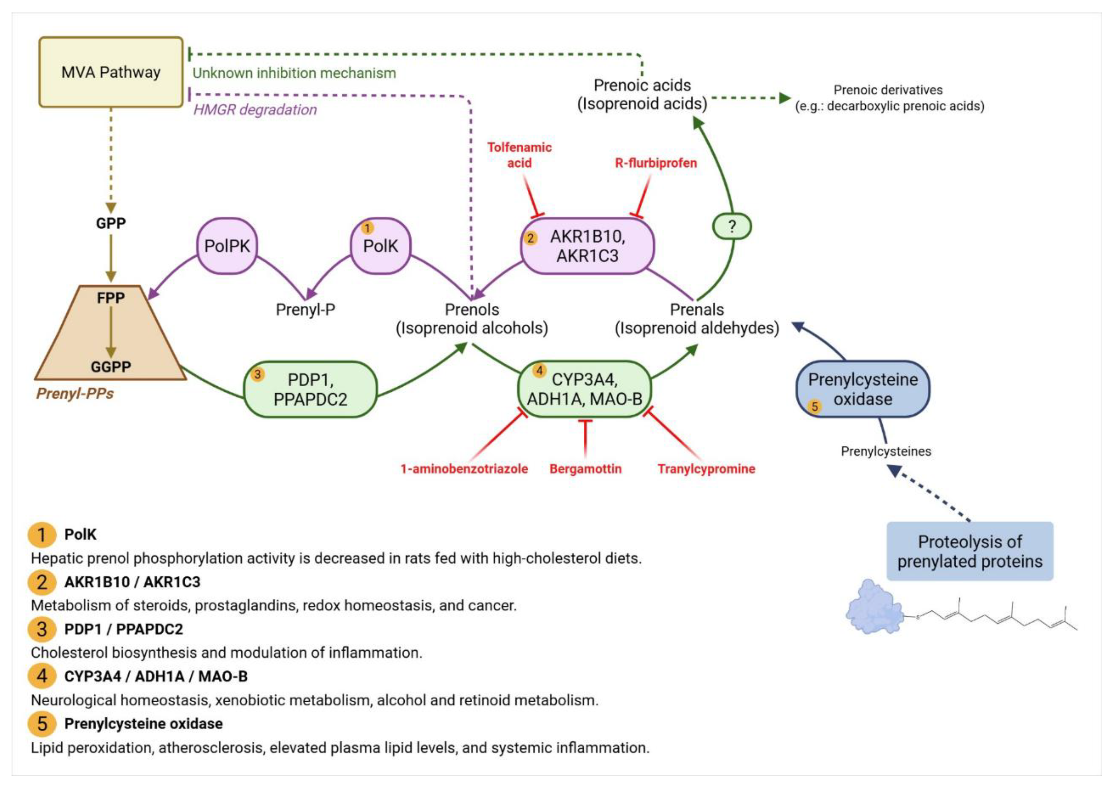
2.3. The Missing kinases of Prenols: A Key Regulator of Isoprenoid Homeostasis?

As previously mentioned, GGOH—and potentially FOH in the future—are emerging as paradoxical modulators of the MVA pathway, capable of both inhibiting this metabolic route and counteracting the effects of its pharmacological inhibition by statins. This dual functionality stems from a prenol salvage pathway involving the reversible phosphorylation and dephosphorylation of prenols and their corresponding diphosphates (Bentinger et al., 1998; Crick et al., 1995; Crick et al., 1997; Verdaguer et al., 2022). While the dephosphorylation branch of this cycle-mediated by the previously discussed PDP1/PPAPDC2 enzymes—is well established, the phosphorylation of free prenols back into their phosphorylated forms remains poorly understood in animals. This regulatory gap is particularly intriguing given that all other enzymatic steps in prenol metabolism discussed in the previous chapter appear to be constitutively active throughout the lifespan (e.g., PDP1/PPAPDC2 activity, and prenol oxidation mediated by MAO-B or CYP3A4). Therefore, it is plausible to hypothesize that prenol kinases act as critical modulators of isoprenoid homeostasis during development, aging, and disease. However, the genes encoding prenol kinases in animals have yet to be definitively identified. In contrast, a small number of these enzymes have been functionally characterized in plants—such as the genes VTE5 (phytol kinase), FOLK (FOH kinase), and VTE6 (phytyl phosphate kinase) (Valentin et al., 2006; Fitzpatrick et al., 2011; Vom Dorp et al., 2015). Recently, our group identified the first FOH/GGOH kinase, encoded by the gene Pf3D7_0710300 of the malaria parasite, Plasmodium falciparum (Crispim et al., 2024a). The fact that all known prenol kinases capable of phosphorylating prenols have been identified in photosynthetic organisms and in a parasite phylogenetically related to algae is not coincidental. Rather, it underscores the essential role of the prenol salvage pathway in these organisms, where it constitutes one of the most widespread and quantitatively significant biochemical processes of life. A striking example of this pathway’s relevance occurs during autumn leaf senescence, when chlorophyll, a phytol-containing pigment, is degraded. This process releases phytol, which is then rapidly phosphorylated by phytol/phytyl-P kinases. The resulting phosphorylated intermediates are subsequently funneled into the biosynthesis of vital isoprenoid-derived compounds, including tocopherols (vitamin E) and phylloquinones (vitamin K1) (Valentin et al., 2006; Fitzpatrick et al., 2011).
To date, evidence for prenol kinase activity in mammalian cells has remained largely indirect and based on biochemical inference. Radiolabeling experiments published in 90s demonstrated that tissue homogenates are capable of converting [3H] FOH and [3H] GGOH into their respective monophosphates—farnesyl monophosphate (FP) and geranylgeranyl monophosphate (GGP)—via a presumed enzyme termed prenol kinase (PolK) (Bentinger et al., 1998; Crick et al., 1995; Crick et al., 1997). The second enzyme of the prenol salvage pathway, a CTP-dependent prenyl-phosphate kinase (PolPK), was also observed to be active in mammal tissues. Subcellular fractionation studies further showed that PolK activity is associated with the luminal side of both rough and smooth microsomes, whereas PolPK localizes to the cytoplasmic face. Notably, the activity of these enzymes was found to be sensitive to dietary modulation: tissues from animals fed cholesterol-enriched diets displayed a reduction in liver prenol kinase activity (Westfall et al., 1997). These findings support the notion that the prenol salvage pathway is metabolically connected to cholesterol regulation, becoming downregulated once cellular cholesterol demands are met. In 2013, advanced mass spectrometry techniques combined with innovative chemical probes and stable isotope-labeled tracers enabled the direct observation of this pathway in human cancer cell lines (Onono et al., 2013). Researchers traced the metabolic fate of externally supplied FOH, GGOH, and synthetic prenol analogs bearing an aniline moiety, revealing their conversion into isoprenoid diphosphates and incorporation into protein isoprenylcysteines in MDA-MB-231 cells (breast ductal carcinoma cell line). Remarkably, when the concentration of exogenous isoprenols exceeded 10 μM, this salvage process was capable of supplying up to 50% of the total intracellular isoprenoid diphosphate pool required for protein prenylation.
The scientific interest for the prenol salvage pathway appears to be gradually emerging, as recent discoveries increasingly link it to various diseases. In oncology, the therapeutic interest in isoprenoid metabolism stems from its involvement in protein prenylation, a post-translational modification essential for the function of many oncogenic proteins. Mutated isoforms of Ras, Rho, and Rac, which rely on prenyl anchors for membrane localization, are dysregulated in nearly one-third of all human cancers (Alan & Lundquist, 2013). Thus, prenyltransferase and prenyl synthase inhibitors have been extensively studied and continue to be explored as potential strategies for cancer treatment. Similarly, numerous studies have investigated the potential anticancer effects of statins, but their clinical efficacy in this context has been limited. While some epidemiological data suggest modest cancer prevention and improved survival outcomes (Verdaguer et al., 2022), mechanistic and pharmacokinetic limitations remain significant. Chief among these is the fact that the concentrations of statins required to inhibit tumor cell growth are often too high to be safely achieved in patients (Verdaguer et al., 2022).
A further complicating factor is the ability of cancer cells to bypass statin-induced inhibition by incorporating exogenous prenols, particularly FOH and GGOH, into their metabolic networks. This has been demonstrated in various cell types, including OVCAR3 ovarian carcinoma cells, neuroblastoma lines, and NIH3T3 fibroblasts (Ownby & Hohl, 2002; Marcuzzi et al., 2012; de Wolf et al., 2017; Verdaguer et al., 2022). Of particular concern is the observation that dietary sources of GGOH, such as vegetable oils, can interfere with the pro-apoptotic effects of statins. For instance, de Wolf et al. showed that GGOH-rich food extracts can block pitavastatin-induced apoptosis in ovarian cancer cells, raising the possibility that nutritional intake of prenols may influence the outcome of clinical trials involving MVA pathway inhibitors. Interestingly, experiments in MDA-MB-231 breast cancer cells, mutations that activate the p53 protein lead to increased activity of the MVA pathway, which in turn promotes tumor invasiveness (Onono et al., 2013). When p53 is silenced or HMGR is pharmacologically inhibited, the synthesis of endogenous prenyl diphosphates and their incorporation into proteins is reduced but cells still resist by increasing their use of externally supplied FOH / GGOH for protein prenylation. This shift suggests that the utilization of exogenous isoprenols operates through a mechanism that is at least partially independent of the canonical MVA pathway.

3. Metabolic Effects of Prenols and Prenoic Acids

As observed so far, the metabolism of prenols and prenoic acids appears to be connected to aging, metabolic regulation, and human diseases. Stemmed from the pioneering proposal by De Loof et al. (2015) in the following section we provide a concise overview, highlighting some of the most notable and representative findings on the metabolic impact of prenols and prenoic acids in disease and aging.

3.1. Prenols

In this section, we will focus primarily on GOH, FOH and GGOH, as these compounds can be produced endogenously or obtained through dietary sources. Regarding specifically FOH and GGOH, we have previously mentioned that these compounds can undergo phosphorylation and subsequently participate in general isoprenoid metabolism. As a result, they have the potential to compensate for genetic or pharmacological disruptions in isoprenoid biosynthesis, particularly those induced by statins or bisphosphonates (e.g. osteonecrosis of the jaw, rhabdomyolysis) (Verdaguer et al., 2022). Since the primary aim of this review is to examine the biological properties of prenols and prenoic acids, we will not explore these salvage-related pathways in detail. Instead, we will highlight effects that appear to be independent of prenol salvage mechanisms. Readers interested in this topic are encouraged to consult our previously published review on the subject (Verdaguer et al., 2022). The following Table shows the main biologic activities of prenols discussed in this review (Table 1).

3.1.1. Geraniol

GOH is an acyclic monoterpenoid alcohol found in the essential oils of various aromatic plants such as lemongrass, rose, and citronella. Widely used in the fragrance and food industries, GOH has attracted increasing attention due to its broad spectrum of pharmacological activities, including antioxidant, anti-inflammatory, anticancer, and neuroprotective effects (Mączka et al., 2020).
Unlike FOH and GGOH, which are discussed later, we found no evidence in the literature indicating that this isoprenoid can be phosphorylated and incorporated into the isoprenoid metabolism. Nevertheless, this compound exhibits numerous interesting pharmacological properties. First, GOH acts as a reactive oxygen species (ROS) scavenger and enhances endogenous antioxidant defense systems, including upregulation of glutathione, superoxide dismutase, and catalase activities (El Azab et al., 2020). These properties are associated with reduced oxidative damage in models of cardiovascular and gastrointestinal diseases. GOH also suppresses inflammatory pathways by inhibiting NF-κB activation and reducing levels of cytokines such as TNF-α, IL-1β, and IL-6 (Wu et al., 2020).
GOH has demonstrated anticancer effects in in vitro and in vivo models of various types of cancer, including breast, lung, colon, prostate, and liver cancers. It induces apoptosis by modulating pro- and anti-apoptotic proteins, including upregulation of Bax and downregulation of Bcl-2, and activation of caspases (Kuzu et al., 2021). GOH also causes cell cycle arrest, inhibits the PI3K/Akt/mTOR pathway, and interferes with the MVA pathway, leading to decreased Ras prenylation and signaling (Mączka et al., 2020). In combination therapies, GOH enhances the efficacy of chemotherapeutic agents such as 5-fluorouracil and gemcitabine (Cho et al., 2016). GOH triggers cell death in human prostate cancer cells through disruption of mitochondrial function (Yu et al., 2024). Another study demonstrated that the combination of simvastatin and GOH, at sub-inhibitory concentrations, synergistically inhibits cholesterol biosynthesis and proliferation in Hep G2 hepatocarcinoma cells, suggesting that GOH may not be phosphorylated by tissues, but instead acts as a natural negative feedback regulator of the MVA pathway (Polo et al., 2011). In line with this, a more recent study showed that GOH reduces obesity by promoting the browning of white adipose tissue through interaction with HMGR in a high-fat diet–induced obesity model in rats (Chand et al., 2023).
GOH has shown neuroprotective effects in models of neurodegenerative disease. It mitigates oxidative stress-induced neuronal damage, likely via activation of the Nrf2/HO-1 axis and suppression of inflammatory mediators (Rajendran et al., 2024). In Parkinson’s disease models, GOH preserved dopaminergic neuron integrity and function (Rekha and Sivakamasundari, 2018).

3.1.2. Farnesol

As previously mentioned, FOH is the structural core of insect juvenile hormones, whose function is to maintain larval states and prevent premature metamorphosis. Analogously, De Loof et al. (2015) proposed that vertebrates may possess endogenous FOH-like sesquiterpenoids that act as “inbromes” (intramembrane signals), regulating cellular calcium homeostasis and contributing to the maintenance of youthful physiology. Disruptions in farnesylation have been implicated in accelerated aging disorders, such as Hutchinson–Gilford progeria, where the persistence of a farnesyl group on the aberrant protein progerin which alters nuclear architecture and accelerates senescence (De Loof et al., 2015). In the same work, De Loof et al. (2015) proposed that FOH functions as a physiological regulator of the juvenile state in mammals by maintaining low cytosolic Ca²⁺ levels and supporting high Ca²⁺-ATPase activity (e.g., SERCA). As puberty approaches, reductions in FOH signaling may allow Ca²⁺ to rise, triggering differentiation and aging. We have known for some time that nanomolar concentrations of FOH inhibit neuronal N-type Ca²⁺ channels by altering gating dynamics (Roullet et al. 1999). At higher concentrations, it can also block L-type and other high-voltage-activated channels. This makes FOH a functional “molecular valve”, limiting excessive calcium entry, which in turn regulates key processes such as neurotransmitter release, apoptosis, and excitotoxicity (De Loof & Schoofs, 2019). Although direct evidence for a FOH endocrine axis in mammals is still lacking, its detection in human blood and brain tissues and its diverse regulatory roles support its function as a bioactive molecule (Roullet et al., 1999; De Loof et al., 2015).
Besides the main role of FOH as a juvenile hormone in animals, it is a well-established fact that FOH exhibits significant anticancer activity across various tumor models. It induces cell cycle arrest and promotes apoptosis by modulating signaling pathways involved in cell survival and proliferation. For example, in prostate cancer cells (DU145), FOH upregulated p53, Bax, and cleaved caspases 3 and 9, while inhibiting PI3K/Akt and ERK pathways, as well as the anti-apoptotic Bcl-2 protein (Park et al., 2014). Activation of JNK and p38 MAPK further supports pro-apoptotic signaling. In vivo, FOH administration (50 mg/kg) significantly reduced tumor volumes in xenograft models, with increased apoptotic activity observed histologically (Park et al., 2014). At the molecular level, FOH induces intrinsic apoptosis via mitochondrial depolarization, cytochrome c release, caspase activation, and PARP cleavage (Joo et al., 2010). It also activates the PERK–eIF2α–ATF4/CHOP pathway, linking ER stress to apoptotic execution. These effects are associated with ionic dysregulation (altered Ca²⁺/K⁺ levels) and are more pronounced in cancer cells than in normal ones, suggesting a selective cytotoxic window (Öztürk et al., 2022). Finally, FOH also promotes HMGR degradation (Meigs et al., 1996).
FOH modulates autophagy, a process often linked to cell survival or death. In lung (A549) and colon (Caco-2) carcinoma cells, FOH induced autophagic vacuole formation along with apoptotic markers, whereas normal cells remained largely unaffected (Öztürk et al., 2022). Mechanistically, it suppresses the PI3K/Akt/mTOR pathway, relieving autophagy inhibition. In neuroinflammation models, this autophagy modulation has protective effects by promoting clearance of damaged organelles and reducing necrosis under chronic stress.
FOH is a known inhibitor of the NF-κB pathway, suppressing transcription of inflammatory genes such as TNF-α, IL-6, COX-2, and iNOS (Joo and Jetten, 2010). FOH also has anti-inflammatory and anti-arthritic activity (Ahmed et al., 2025). It modulates upstream regulators like Ras and IκB kinase. In vivo, FOH attenuates inflammation in asthma, arthritis, and neuroinflammatory conditions. In arthritis models, it reduced joint damage and inflammation while shifting immune responses toward regulatory T cells (Ahmed et al., 2025).

3.1.3. Geranylgeraniol

GGOH is present in small amounts in Bixa orellana (annatto) (Batista et al., 2022), vegetable oils, and other plant-derived sources and food (Jaward et al., 2022). It has attracted increasing scientific interest due to its diverse biological effects, particularly in the contexts of metabolic regulation, musculoskeletal health, and neuroprotection (Chung et al., 2021). In the endocrine system, a randomized clinical trial found that GGOH supplementation increased testosterone levels in men with low baseline hormone levels, with potential modulation of hormonal profiles even in eugonadal individuals (Gheith et al., 2023). Supporting these findings, in vitro experiments demonstrated that GGOH enhances the production of testosterone and its precursor progesterone via activation of adenylate cyclase and the cAMP/PKA pathway, independent of phosphodiesterase inhibition (Ho et al., 2018). Notably, as GGPP is not a precursor for steroid hormones, these hormonal effects appear to be intrinsic to GGOH and may involve hormone-like signaling properties.
In metabolic disease models, GGOH has shown beneficial effects on glucose homeostasis. In mice subjected to a high-fat diet, GGOH improved glucose tolerance and insulin sensitivity, preserved pancreatic islet architecture, reduced levels of pro-inflammatory cytokines such as IL-6 and MCP-1, and promoted the growth of beneficial butyrate-producing gut microbiota (Chung et al., 2021; Shen et al., 2023). These actions are consistent with GGOH’s known anti-inflammatory and mitochondrial-supportive roles (Tan & Chin, 2023). Additional studies suggest indirect benefits of GGOH in pain relief (Spindola et al., 2010).
Neuroprotective effects have also been reported. GGOH inhibits microglial activation, suppresses NF-κB signaling, and reduces the expression of pro-inflammatory mediators in vitro (Saputra et al., 2021). In neuron-like cells under MVA pathway inhibition, GGOH preserved mitochondrial integrity and inhibited NLRP3 inflammasome activation, suggesting potential therapeutic applications in neurodegenerative diseases and age-related cognitive decline (Jiwan et al., 2024). Similarly, in animal models of mevalonate kinase deficiency — a disease characterized by impaired isoprenoid biosynthesis and marked inflammation — supplementation with GGOH, and to a lesser extent with geraniol and FOH, has been shown to prevent inflammation (Marcuzzi et al., 2008). In rats with type 2 diabetes, GGOH also helps preserve muscle fiber size, possibly by increasing the expression of the MyoD protein and maintaining the pool of satellite cells (Jiwan et al., 2024). In terms of its mechanism of action, GGOH has been shown to induce Peroxisome proliferator-activated receptor γ (PPARγ) expression and enhance the biological effects of PPARγ agonists in adipocyte lineage cells, thereby contributing to the regulation of glucose metabolism (Matsubara et al., 2018).
From an oncological perspective, GGOH has demonstrated antitumor potential by downregulating HMGR, the rate-limiting enzyme of the MVA pathway. This downregulation limits the production of intermediates essential for the prenylation of oncogenic proteins. For example, in DU145 prostate cancer cells, GGOH reduced cell viability in a dose-dependent manner, induced G1 phase arrest, decreased cyclin D1 levels, and activated caspase-3-dependent apoptosis. These effects correlated with decreased HMGR protein expression, highlighting its chemopreventive potential (Fernandes et al., 2013). However, because GGOH can also act as a downstream product of the pathway—being potentially phosphorylated into GGPP—its antitumor effects must be interpreted with caution. These effects may only occur at high concentrations, where GGOH accumulates and suppresses the MVA pathway—a scenario that may be difficult to replicate in vivo.

3.2. Prenoic Acids

In this section, we will discuss farnesoic acid and geranoic acid but we will focus mostly on GGA and related natural or synthetic compounds, as it is the prenoic acid for which substantial data are currently available in the literature. For further details on this compound, we once again refer the reader to the comprehensive review by Shidoji (2023). The following Table shows the main biologic activities of prenols discussed in this review (Table 2).

3.2.1. Geranoic Acid 

Literature on the biological activities of geranoic acid is scarce. We found only limited evidence suggesting that this compound is a potent tyrosinase inhibitor, which directly affects melanin production. Masuda et al. (2008) first identified geranoic acid as the active tyrosinase-inhibiting component in lemongrass extracts. Follow-up studies investigated its anti-melanogenic properties in cellular models: Choi (2012), for example, tested geranoic acid derivatives in melanocyte cultures and demonstrated that geranoic acid effectively reduces melanin synthesis with minimal cytotoxicity, highlighting its potential as a skin-lightening agent.

3.2.2. Farnesoic Acid 

Farnesoic acid, as an endogenous metabolite, has attracted interest for its ability to modulate key metabolic regulators. One of its most relevant pharmacological properties is its interaction with PPARα, a nuclear receptor that regulates lipid metabolism and inflammation. As previously discussed, farnesoic acid, along with FOH, is believed to act as a PPARα agonist in cells (Rizzo, 2014). Activation of PPARα is associated with well-established therapeutic effects: it enhances fatty acid oxidation in the liver and muscles, reduces triglyceride levels, exerts anti-inflammatory effects in vascular tissues and skin, and promotes keratinocyte differentiation (O’Brien et al., 2000).
The role of farnesoic acid in human health may extend to certain rare metabolic and dermatological disorders. For example, in Sjögren–Larsson syndrome, patients are unable to convert FOH into farnesoic acid, leading to the accumulation of the corresponding prenol. This contributes to symptoms such as ichthyosis, neurological impairments, and a deeply disturbed isoprenoid metabolism (Rizzo, 2014). Specifically, a downregulated MVA pathway has been observed in these patients, resulting in decreased levels of cholesterol, dolichols, and protein prenylation. Additionally, this metabolic impairment disrupts the expression of genes regulated by PPARs, compromising epidermal homeostasis and proper keratinocyte maturation.

3.2.3. Geranylgeranoic Acid and Related Compounds

GGA is an acyclic diterpenoid with notable bioactivity across multiple physiological systems. It exhibits selective antitumor effects, modulates autophagy, and acts through both retinoid-mimetic and independent cellular signaling pathways. Studies have consistently demonstrated that GGA selectively induces apoptosis in hepatocellular carcinoma (HCC) cell lines, such as HuH-7 and PLC/PRF-5, while sparing non-tumoral hepatocytes (Shidoji & Tabata, 2019; Shidoji & Ogawa, 2004). These findings suggest that the prenol salvage pathway may function as a tumor-suppressive mechanism—potentially a “cancer stop” pathway—that remains inactive or downregulated in malignant cells.
The antineoplastic activity of GGA appears to be related to its capacity to induce endoplasmic reticulum stress and disturb proteostasis, culminating in dysfunctional autophagy (Iwao & Shidoji, 2015). Unlike canonical autophagy, which serves a cytoprotective role, the form elicited by GGA is incomplete and contributes to cell death through mechanisms likely linked to both ER perturbation and impaired lipid metabolism. Furthermore, GGA has been shown to trigger pyroptosis—a caspase-1 and 4 dependent inflammatory form of cell death—via Toll-like receptor 4 (TLR4) signaling. In HuH-7 cells, GGA induces caspase-1 activation and pro-inflammatory cytokine production, leading to gasdermin D cleavage and pore formation in the cell membrane, classic features of pyroptotic execution (Yabuta & Shidoji, 2020). Upon exposure to GGA, HuH-7 cells exhibited a rapid activation of genes involved in the metabolic transition toward oxidative phosphorylation. In particular, the expression of TIGAR, which downregulates glycolytic flux, and SCO2, essential for the assembly of cytochrome c oxidase complex IV, was markedly enhanced, consistent with a shift from glycolytic to respiratory energy production — which, in broader terms, represents a reversal of the Warburg effect (Iwao & Shidoji, 2015).
The hepatoprotective and anticarcinogenic effects of GGA have been especially explored in liver cancer models. Remarkably, studies in C3H/HeN mice revealed that a single oral administration of GGA during middle age significantly lowered both the frequency and size of spontaneous liver tumors by 24 months of age (Tabata et al., 2021). Its clinical translational potential is underscored by trials involving peretinoin (3,7,11,15-Tetramethyl-2,4,6,10,14-hexadecapentaenoic acid) —a synthetic GGA analog—which showed a reduction in HCC recurrence following curative resection (Muto et al., 1999).
Despite being structurally distinct from retinoids like all-trans-retinoic acid (ATRA), GGA exhibits ligand activity against retinoid nuclear receptors RAR and RXR (Araki et al., 1995). In HL-60 promyelocytic leukemia cells, GGA induces granulocytic differentiation in a manner akin to ATRA (Araki et al., 1995; Kodaira et al., 2007). Similarly, GGA also induces growth suppression and neural differentiation in SH-SY5Y human neuroblastoma cells (Sakane & Shidoji, 2011). Within the skeletal system, GGA stimulates osteoblastic activity and suppresses osteoclastogenesis. Experimental data show that GGA enhances alkaline phosphatase activity and osteopontin expression in bone-forming cells, while inhibiting osteoclast formation driven by RANKL (Wang et al., 2002). These effects translated in vivo to increased bone mineral density in osteopenic aged mice, supporting its potential application in bone-related disorders. Another study compared the effects of GGA and 2,3-dihydroGGA on HL-60 cells. GGA induced neutrophil differentiation, similar to retinoic acid (Kodaira et al., 2007). In contrast, 2,3-dihydroGGA did not induce neutrophils but promoted lipid droplet formation. Under low serum conditions (0.1% FBS), both compounds triggered apoptosis. Interestingly, when a caspase-3 inhibitor was added, apoptosis was blocked, and lipid droplet formation was observed again with 2,3-dihydroGGA.
Beyond its pharmacological relevance, GGA may also play physiological roles. High endogenous concentrations have been detected in the liver and testes of mice, and its reduced form, 2,3-dihydroGGA, in the thymus, implying involvement in hepatic and reproductive functions (Shidoji, 2023). Notably, dietary supplementation with GGA during mating and gestation enhanced fertility and reproductive success in murine models, suggesting roles in embryogenesis and hormonal regulation (Tabata et al., 2020).

4. Future Perspectives and Conclusions

The evidence reviewed here highlights the remarkable role of acyclic terpene derivatives, particularly prenols and prenoic acids, in mammalian physiology. Originally recognized for their importance in plants and insects, these compounds are now emerging as endogenously produced metabolites in mammals, with several biologic effects. Studies have demonstrated that the endogenous production of terpenic alcohols and their derivatives in mammals is age-dependent, suggesting the existence of a regulatory metabolic mechanism intimately linked to development, senescence, and cellular bioenergetics. Based on this, some researchers have proposed that mammals may harbor a terpenoid-based signaling system involved in the regulation of aging—paralleling mechanisms observed in arthropods and fungi. Prenol metabolism in mammals appears to operate through a bidirectional pathway, interconverting polyprenyl diphosphates and their corresponding prenols, which can subsequently be oxidized to their respective aldehydes and prenoic acids. In the phosphorylative direction, this pathway is thought to support cholesterol biosynthesis and, in experimental contexts, potentially contribute to statin resistance in the treatment of dyslipidemia or cancer. Conversely, in the dephosphorylative direction, it may downregulate the MVA pathway and promote a range of benefits, including the induction of autophagy, endocrine effects, tumor and inflammation suppression, enhanced insulin sensitivity, cholesterol-lowering activity, and improvements in cognitive function—collectively supporting a broad range anti-aging effect. Considering that the oxidative and carboxylation steps of the pathway appear to remain active throughout life, it is assumed that the missing kinases may be the most affected by aging.
Therefore, strategies aimed solely at supplementing prenols precursors could possess limited effects, as these compounds can be rapidly converted into their diphosphate forms. The exploration of this pathway for research and biomedical purposes will necessarily require the augmentation of intracellular pool of both prenoic acids and prenols, which will likely require the identification and inhibition of the missing prenol salvage pathway. Another validated strategy to stimulate the prenol pathway is the inhibition of squalene synthase, which, in our view, deserves further investigation for its potential anti-aging effects.
In light of these findings, the therapeutic potential of the prenol pathway, particularly in the context of aging, appears highly promising. It is our hope that this review stimulates renewed scientific interest in the long-overlooked prenol pathway, guiding future research toward its molecular dissection and clinical application. As we rediscover this ancient metabolic circuit, we may reveal a powerful axis of longevity regulation conserved across diverse kingdoms of life.

Author Contributions

All authors contributed to the conceptualization of the review, literature search and analysis, manuscript drafting, figure design, and critical revision for intellectual content.

Funding

IBV is fellow from the Fundação de Amparo à Pesquisa do Estado de São Paulo (FAPESP); IBV FAPESP process number: 2022/09526-4. M.C. was supported by a Beatriu de Pinós fellowship from AGAUR (Government of Catalonia). This work received support from the grants PID2022-142956OB-100, PID2022-140169OB-C22 and PID2023-148803OB-100 from Plan Nacional de I+D funded by the Agencia Estatal de Investigación (AEI) and the Fondo Europeo de Desarrollo Regional (FEDER) and from the CIBEREHD; as well as support from National Cancer Institute PO1 CA281819-01A1 from the NIH, and from the European Horizon’s research and innovation program HORIZON-HLTH-2022-STAYHLTH-02 under agreement No 101095679.

Acknowledgments

We thank Professor Yoshihiro Shidoji (University of Nagasaki, Nagayo, Nagasaki, Japan) for his valuable advice and insightful corrections on specific sections of the manuscript related to geranylgeranoic acid.

Conflicts of Interest

The author declare that he has no competing interests.

Abbreviations

  • Acetyl-CoA – Acetyl-coenzyme A
  • ADH1A – Alcohol dehydrogenase 1A
  • AGEs – Advanced glycation end-products
  • AKR1B10 – Aldo-keto reductase family 1 member B10
  • AKR1C3 – Aldo-keto reductase family 1 member C3
  • Akt – Protein kinase B
  • AMPK – AMP-activated protein kinase
  • ATF4 – Activating transcription factor 4
  • Bax – Bcl-2-associated X protein
  • Bcl-2 – B-cell lymphoma 2 (anti-apoptotic protein)
  • cAMP/PKA – Cyclic adenosine monophosphate / protein kinase A
  • CHOP – C/EBP homologous protein
  • c-MYC – Cellular myelocytomatosis oncogene
  • COX-2 – Cyclooxygenase-2
  • CYP3A4 – Cytochrome P450 3A4
  • DHCR24 – 24-Dehydrocholesterol reductase
  • DHCR7 – 7-Dehydrocholesterol reductase
  • DMAPP – Dimethylallyl diphosphate
  • ERK – Extracellular signal-regulated kinase
  • FP – Farnesyl monophosphate
  • FPP – Farnesyl diphosphate
  • FPPS – Farnesyl diphosphate synthase
  • FXR – Farnesoid X receptor
  • GGP – Geranylgeranyl monophosphate
  • GGPP – Geranylgeranyl diphosphate
  • GGPPS – Geranylgeranyl diphosphate synthase
  • GOH - Geraniol
  • GPP – Geranyl diphosphate
  • GPPS – Geranyl diphosphate synthase
  • HMG-CoA – 3-Hydroxy-3-methylglutaryl-coenzyme A
  • HMGR – 3-Hydroxy-3-methylglutaryl-CoA reductase
  • HMGS – 3-Hydroxy-3-methylglutaryl-CoA synthase
  • HO-1 – Heme oxygenase-1
  • IDI – Isopentenyl-diphosphate isomerase
  • IL-1β – Interleukin-1 beta
  • IL-6 – Interleukin-6
  • iNOS – Inducible nitric oxide synthase
  • IPP – Isopentenyl diphosphate
  • JNK – c-Jun N-terminal kinase
  • K⁺ – Potassium ion
  • KLF4 – Krüppel-like factor 4
  • MAO-B – Monoamine oxidase B
  • MAPK – Mitogen-activated protein kinase
  • mTOR – Mechanistic target of rapamycin
  • MVA – Mevalonate
  • MVK – Mevalonate kinase
  • NAD⁺ – Nicotinamide adenine dinucleotide (oxidized form)
  • NF-κB – Nuclear factor kappa-light-chain-enhancer of activated B cells
  • NLRP3 – NOD-, LRR-, and pyrin domain-containing protein 3
  • Nrf2 – Nuclear factor erythroid 2–related factor 2
  • OCT4 – Octamer-binding transcription factor 4
  • PARP – Poly(ADP-ribose) polymerase
  • PDP1 / PPAPDC2 – Pyrophosphatase/phosphatase domain-containing protein 2
  • PDZ – PSD-95/Dlg/ZO-1 (protein–protein interaction domain)
  • PERK – Protein kinase RNA-like endoplasmic reticulum kinase
  • PI3K – Phosphoinositide 3-kinase
  • PMK – Phosphomevalonate kinase
  • PolK – Polyprenol kinase
  • PolPK – Polyprenyl phosphate kinase
  • PPAR – Peroxisome proliferator-activated receptor
  • PPARγ - Peroxisome proliferator-activated receptor γ
  • RAR – Retinoic acid receptor
  • RXR – Retinoid X receptor
  • SAM – S-Adenosylmethionine
  • SERCA – Sarco/endoplasmic reticulum Ca²⁺-ATPase
  • SOX2 – SRY-box transcription factor 2
  • SQLE – Squalene epoxidase
  • SQS – Squalene synthase
  • SREBPs – Sterol regulatory element-binding proteins
  • SQS – Squalene synthase
  • TNF-α – Tumor necrosis factor alpha
  • UBIAD1 – UbiA prenyltransferase domain-containing protein 1
  • YAP – Yes-associated protein
  • α-Ketoglutarate – Alpha-ketoglutarate (a Krebs cycle intermediate)

References

  1. Ahmed, S. R.; Uttra, A. M.; Usman, M.; Qasim, S.; Jahan, S.; Roman, M.; Rashwan, E. K. Deciphering farnesol’s anti-arthritic and immunomodulatory potential by targeting multiple pathways: A combination of network pharmacology guided exploration and experimental verification. Journal of Pharmacy and Pharmacology 2025, 77(1), 127–141. [Google Scholar] [CrossRef]
  2. Alan, J. K.; Lundquist, E. A. Mutationally activated Rho GTPases in cancer. Small GTPases 2013, 4(3), 159–163. [Google Scholar] [CrossRef]
  3. Andersson, M.; Åberg, F.; Teclebrhan, H.; Edlund, C.; Appelkvist, E. L. Age-dependent modifications in the metabolism of mevalonate pathway lipids in rat brain. Mechanisms of Ageing and Development 1995, 85(1), 1–14. [Google Scholar] [CrossRef] [PubMed]
  4. Araki, H.; Shidoji, Y.; Yamada, Y.; Moriwaki, H.; Muto, Y. Retinoid agonist activities of synthetic geranylgeranoic acid derivatives. Biochemical and Biophysical Research Communications 1995, 209(1), 66–72. [Google Scholar] [CrossRef] [PubMed]
  5. Arenas, F.; Garcia-Ruiz, C.; Fernandez-Checa, J. C. Intracellular cholesterol trafficking and impact in neurodegeneration. Frontiers in molecular neuroscience 2017, 10, 382. [Google Scholar] [CrossRef] [PubMed]
  6. Aschner, M.; Nguyen, T.T.; Sinitskii, A.I.; Santamaría, A.; Bornhorst, J.; Ajsuvakova, O.P.; da Rocha, J.B.T.; Skalny, A.V.; Tinkov, A.A. Isolevuglandins (isoLGs) as toxic lipid peroxidation byproducts and their pathogenetic role in human diseases. Free Radical Biology and Medicine 2021, 162, 266–273. [Google Scholar] [CrossRef]
  7. Banfi, C.; Baetta, R.; Barbieri, S. S.; Brioschi, M.; Guarino, A.; Ghilardi, S.; Sandrini, L.; Eligini, S.; Polvani, G.; Bergman, O.; Eriksson, P.; Tremoli, E. Prenylcysteine oxidase 1, an emerging player in atherosclerosis. Communications biology 2021, 4(1), 1109. [Google Scholar] [CrossRef]
  8. Barzilai, N.; Huffman, D. M.; Muzumdar, R. H.; Bartke, A. The critical role of metabolic pathways in aging. Diabetes 2012, 61(6), 1315–1322. [Google Scholar] [CrossRef]
  9. Batista, M. A.; de Lima Teixeira dos Santos, A. V. T.; do Nascimento, A. L.; Moreira, L. F.; Souza, I. R. S.; da Silva, H. R.; Carvalho, J. C. T. Potential of the compounds from Bixa orellana purified annatto oil and its granules (Chronic®) against dyslipidemia and inflammatory diseases: in silico studies with geranylgeraniol and tocotrienols. Molecules 2022, 27(5), 1584. [Google Scholar] [CrossRef]
  10. Belakova, B.; Wedige, N. K.; Awad, E. M.; et al. Lipophilic statins eliminate senescent endothelial cells by inducing anoikis-related cell death. Cells 2023, 12(24), 2836. [Google Scholar] [CrossRef]
  11. Bentinger, M.; Grünler, J.; Peterson, E.; Swiezewska, E.; Dallner, G. Phosphorylation of farnesol in rat liver microsomes: Properties of farnesol kinase and farnesyl phosphate kinase. Arch. Biochem. Biophys. 1998, 353, 191–198. [Google Scholar] [CrossRef]
  12. Bratic, A.; Larsson, N. G. The role of mitochondria in aging. The Journal of Clinical Investigation 2013, 123(3), 951–957. [Google Scholar] [CrossRef]
  13. Buhaescu, I.; Izzedine, H. Mevalonate pathway: A review of clinical and therapeutical implications. Clinical Biochemistry 2007, 40(9-10), 575–584. [Google Scholar] [CrossRef]
  14. Byrns, M. C.; Mindnich, R.; Duan, L.; Penning, T. M. Overexpression of aldo-keto reductase 1C3 (AKR1C3) in prostate cancer: Implications for resistance to hormone therapy and opportunities for therapeutic intervention. Chemico-Biological Interactions 2008, 178(1-3), 103–110. [Google Scholar]
  15. Chadha, A.; Madyastha, K. M. Metabolism of geraniol and linalool in the rat and effects on liver and lung microsomal enzymes. Xenobiotica 1984, 14(5), 365–374. [Google Scholar] [CrossRef]
  16. Chand, S.; Tripathi, A.S.; Hasan, T.; Ganesh, K.; Cordero, M.A.W.; Yasir, M.; Zaki, M.E.A.; Tripathi, P.; Mohapatra, L.; Maurya, R.K. Geraniol reverses obesity by improving conversion of WAT to BAT in high fat diet induced obese rats by inhibiting HMGCoA reductase. Nutr Diabetes 2023, 13(1), 26. [Google Scholar] [CrossRef]
  17. Cho, M.; So, I.; Chun, J.N.; Jeon, J.H. The antitumor effects of geraniol: Modulation of cancer hallmark pathways (Review). Int J Oncol. 2016, 48(5), 1772–82. [Google Scholar] [CrossRef] [PubMed]
  18. Choi, S. Y. Inhibitory effects of geranic acid derivatives on melanin biosynthesis. Journal of Cosmetic Science 2012, 63(5), 351–358, microbialcellfactories.biomedcentral.com. [Google Scholar] [PubMed]
  19. Christophe, J.; Popják, G. Studies on the biosynthesis of cholesterol: XIV. The origin of prenoic acids from allyl diphosphates in liver enzyme systems. Journal of Lipid Research 1961, 2(3), 244–257. [Google Scholar] [CrossRef]
  20. Chung, E.; Kim, H. J.; Kwon, J. Y.; Cho, S. Y. Beneficial effect of dietary geranylgeraniol on glucose homeostasis and bone microstructure in obese mice. Nutrition Research 2021, 93, 27–37. [Google Scholar] [CrossRef] [PubMed]
  21. Crick, D.C.; Andres, D.A.; Waechter, C.J. Farnesol is utilized for protein isoprenylation and the biosynthesis of cholesterol in mammalian cells. Biochem. Biophys. Res. Commun. 1995, 211, 590–599. [Google Scholar] [CrossRef]
  22. Crick, D.C.; Andres, D.A.; Waechter, C.J. Novel salvage pathway utilizing farnesol and geranylgeraniol for protein isoprenylation. Biochem. Biophys. Res. Commun. 1997, 237, 483–487. [Google Scholar] [CrossRef]
  23. Crispim, M.; Bofill Verdaguer, I.; Mazzine Filho, M.; Santos, M. F.; Hernández, A. Isoprenoids, the versatile lipids: Their functions in intracellular membranes. In Advances in Biology; Nova Science Publishers, September 2024; pp. 1–66. [Google Scholar]
  24. Crispim, M.; Verdaguer, I. B.; Hernández, A.; Kronenberger, T.; Fenollar, À.; Yamaguchi, L. F.; Izquierdo, L. Beyond the MEP Pathway: A novel kinase required for prenol utilization by malaria parasites. PLoS Pathogens 2024, 20(1), e1011557. [Google Scholar] [CrossRef] [PubMed]
  25. De Loof, A.; Marchal, E.; Rivera-Perez, C.; Noriega, F. G.; Schoofs, L. Farnesol-like endogenous sesquiterpenoids in vertebrates: the probable but overlooked functional “inbrome” anti-aging counterpart of juvenile hormone of insects? Frontiers in endocrinology 2015, 5, 222. [Google Scholar] [CrossRef] [PubMed]
  26. De Loof, A.; Schoofs, L. Mode of Action of Farnesol, the “Noble Unknown” in Particular in Ca2+ Homeostasis, and Its Juvenile Hormone-Esters in Evolutionary Retrospect. Frontiers in Neuroscience 2019, 13, 141. [Google Scholar] [CrossRef]
  27. Díaz-Casado, M. E.; Quiles, J. L.; Barriocanal-Casado, E.; González-García, P.; Battino, M.; López, L. C.; Varela-López, A. The Paradox of Coenzyme Q10 in Aging. Nutrients 2019, 11(9), 2221. [Google Scholar] [CrossRef] [PubMed]
  28. Dituri, F.; Rabinowitz, J. L.; Hullin, R. P.; Gurin, S. G. Precursors of squalene and cholesterol. Journal of Biological Chemistry 1957, 229(2), 825–836. [Google Scholar] [CrossRef]
  29. Duester, G. Families of retinoid dehydrogenases regulating vitamin A function: Production of visual pigment and retinoic acid. European Journal of Biochemistry 2000, 267(14), 4315–4324. [Google Scholar] [CrossRef]
  30. El Azab, E.F.; Elguindy, N.M.; Yacout, G.A.; Elgamal, D.A. Hepatoprotective Impact of Geraniol Against CCl4-Induced Liver Fibrosis in Rats. Pakistan Journal of Biological Sciences 2020, 23(12), 1650–1658. [Google Scholar] [CrossRef]
  31. Elsabrouty, R.; Jo, Y.; Hwang, S.; Jun, D.-J.; DeBose-Boyd, R. A. Type 1 polyisoprenoid diphosphate phosphatase modulates geranylgeranyl-mediated control of HMG CoA reductase and UBIAD1. eLife 2021, 10, e64688. [Google Scholar] [CrossRef]
  32. Endo, S.; Matsunaga, T.; Kitade, Y.; Soda, M.; Zhao, Y.; Murata, K.; El-Kabbani, O. Roles of aldo-keto reductases in the reduction of farnesal and geranylgeranial in human and murine cells. Chemico-Biological Interactions 2009, 178(1-3), 115–121. [Google Scholar]
  33. Endo, S.; Matsunaga, T.; Ohta, C.; Soda, M.; Kanamori, A.; Kitade, Y.; et al. Roles of rat and human aldo-keto reductases in metabolism of farnesol and geranylgeraniol. Chemico-Biological Interactions 2011, 191(1-3), 261–268. [Google Scholar] [CrossRef]
  34. Fernandes, N. V.; Yeganehjoo, H.; Katuru, R.; DeBose-Boyd, R. A.; Morris, L. L.; Michon, R.; Yu, Z. L.; Mo, H. Geranylgeraniol suppresses the viability of human DU145 prostate carcinoma cells and the level of HMG CoA reductase; Experimental biology and medicine: Maywood, N.J., 2013; Volume 238, 11, pp. 1265–1274. [Google Scholar]
  35. Fitzpatrick, A.H.; Bhandari, J.; Crowell, D.N. Farnesol kinase is involved in farnesol metabolism, ABA signaling and flower development in Arabidopsis. Plant J. 2011, 66, 1078–1088. [Google Scholar] [CrossRef]
  36. Fliesler, S. J.; Schroepfer, G. J., Jr. Metabolism of mevalonic acid in cell-free homogenates of bovine retinas: Formation of novel isoprenoid acids. Journal of Biological Chemistry 1983, 258(24), 15062–15070. [Google Scholar] [CrossRef]
  37. Foster, J. M.; Pennock, J. F.; Marshall, I.; Rees, H. H. Biosynthesis of isoprenoid compounds in Schistosoma mansoni. Molecular and Biochemical Parasitology 1993, 61(2), 275–284. [Google Scholar] [CrossRef]
  38. Franceschi, C.; Garagnani, P.; Parini, P.; Giuliani, C.; Santoro, A. Inflammaging: a new immune-metabolic viewpoint for age-related diseases. Nature reviews. Endocrinology 2018, 14(10), 576–590. [Google Scholar] [PubMed]
  39. Fukumoto, S.; Yamauchi, N.; Moriguchi, H.; Hippo, Y.; Watanabe, A.; Shibahara, J.; Aburatani, H. Overexpression of the aldo-keto reductase family member AKR1B10 is highly correlated with smokers’ non-small cell lung carcinomas. Clinical Cancer Research 2005, 11(5), 1776–1785. 29. [Google Scholar] [CrossRef] [PubMed]
  40. Fukunaga, K.; Arita, M.; Takahashi, M.; Morris, A. J.; Pfeffer, M.; Levy, B. D. Identification and functional characterization of a presqualene diphosphate phosphatase. Journal of Biological Chemistry 2006, 281(14), 9490–9497. [Google Scholar] [CrossRef] [PubMed]
  41. Gillespie, Z. E.; Pickering, J.; Eskiw, C. H. Better living through chemistry: Caloric restriction (CR) and CR mimetics alter genome function to promote increased health and lifespan. Frontiers in Genetics 2016, 12, 662320. [Google Scholar] [CrossRef]
  42. Goldstein, J. L.; Brown, M. S. Regulation of the mevalonate pathway. Nature 1990, 343(6257), 425–430. [Google Scholar] [CrossRef]
  43. Griffin, S.; Preta, G.; Sheldon, I. M. Inhibiting mevalonate pathway enzymes increases stromal cell resilience to a cholesterol-dependent cytolysin. Scientific Reports 2017, 7, 17050. [Google Scholar] [CrossRef]
  44. Ha, N. T.; Lee, C. H. Roles of Farnesyl-Diphosphate Farnesyltransferase 1 in Tumour and Tumour Microenvironments. Cells 2020, 9(11), 2352. [Google Scholar] [CrossRef]
  45. Hagvall, L.; Baron, J. M.; Börje, A.; Weidolf, L.; Merk, H.; Karlberg, A. T. Cytochrome P450-mediated activation of the fragrance compound geraniol forms potent contact allergens. Toxicology and Applied Pharmacology 2008, 233(2), 308–313. [Google Scholar] [CrossRef] [PubMed]
  46. Harman, D. Aging: Overview. Annals of the New York Academy of Sciences 2001, 928(1), 1–21. 35. [Google Scholar] [CrossRef] [PubMed]
  47. Hayashi, Y.; Kashio, S.; Murotomi, K.; Hino, S.; Kang, W.; Miyado, K.; Nakao, M.; Miura, M.; Kobayashi, S.; Namihira, M. Biosynthesis of S-adenosyl-methionine enhances aging-related defects in Drosophila oogenesis. Science Reports 2022, 12(1), 5593. [Google Scholar] [CrossRef]
  48. Hildebrandt, H. Ueber Synthesen im Thierkörper (2. mittheilung). Archiv für experimentelle Pathologie und Pharmakologie 1900, 45, 110–129. [Google Scholar] [CrossRef]
  49. Hooff, G. P.; Wood, W. G.; Kim, J. H.; Igbavboa, U.; Ong, W. Y.; Muller, W. E.; Eckert, G. P. Brain isoprenoids farnesyl pyrophosphate and geranylgeranyl pyrophosphate are increased in aged mice. Molecular neurobiology 2012, 46(1), 179–185. [Google Scholar] [CrossRef]
  50. Imai, S. I.; Guarente, L. NAD+ and sirtuins in aging and disease. Trends in Cell Biology 2014, 24(8), 464–471. [Google Scholar] [CrossRef]
  51. Iwao, C.; Shidoji, Y. Upregulation of energy metabolism-related, p53-target TIGAR and SCO2 in HuH-7 cells with p53 mutation by geranylgeranoic acid treatment. Biomedical Research 2015, 36(6), 371–381. [Google Scholar] [CrossRef] [PubMed]
  52. Jahn, A.; Scherer, B.; Fritz, G.; Honnen, S. Statins Induce a DAF-16/Foxo-dependent Longevity Phenotype via JNK-1 through Mevalonate Depletion in C. elegans. Aging Disease 2020, 11(1), 60–72. [Google Scholar]
  53. Jawad, M. J.; Ibrahim, S.; Kumar, M.; Burgert, C.; Li, W. W.; Richardson, A. Identification of foods that affect the anti-cancer activity of pitavastatin in cells. Oncology Letters 2022, 23(3), 73. [Google Scholar] [CrossRef]
  54. Jin, Y.; Penning, T. M. Aldo-keto reductases and bioactivation/detoxication. Annual Review of Pharmacology and Toxicology 2006, 47, 263–292. [Google Scholar] [CrossRef] [PubMed]
  55. Jiwan, N. C.; Appell, C. R.; Sterling, R.; Shen, C. L.; Luk, H. Y. The Effect of Geranylgeraniol and Ginger on Satellite Cells Myogenic State in Type 2 Diabetic Rats. Current issues in molecular biology 2024, 46(11), 12299–12310. [Google Scholar] [CrossRef] [PubMed]
  56. Joo, J. H.; Jetten, A. M. Molecular mechanisms involved in farnesol-induced apoptosis. Cancer Letters 2010, 287(2), 123–135, (Discusses farnesol and farnesoic acid in apoptosis signaling)pmc.ncbi.nlm.nih.gov. [Google Scholar] [CrossRef] [PubMed]
  57. Kennedy, B. K.; Berger, S. L.; Brunet, A.; Campisi, J.; Cuervo, A. M.; Epel, E. S.; Sierra, F. Geroscience: Linking aging to chronic disease. Cell 2014, 159(4), 709–713. 48. [Google Scholar] [CrossRef]
  58. Kirkwood, T. B. L. Understanding the odd science of aging. Cell 2005, 120, 437–447. 50, Kuhn R., Kohler F., Kohler L. Uber methyl-oxidationen im tierkorper. Hoppe Seylers Z. Physiol. Chem. 1936;242:171–197. 51.. [Google Scholar]
  59. Kodaira, Y.; Kusumoto, T.; Takahashi, T.; Matsumura, Y.; Miyagi, Y.; Okamoto, K.; Sagami, H. Formation of lipid droplets induced by 2, 3-dihydrogeranylgeranoic acid distinct from geranylgeranoic acid. Acta Biochimica Polonica 2007, 54(4), 777–782. [Google Scholar] [CrossRef]
  60. Kodaira, Yuichi; et al. Formation of (R)-2, 3-dihydrogeranylgeranoic acid from geranylgeraniol in rat thymocytes. The journal of biochemistry 2002, 132.2, 327–334. [Google Scholar] [CrossRef]
  61. Kuhn, R.; Köhler, F.; Köhler, L. Über Methyl-Oxidationen im Tierkörper. Hoppe-Seyler’s Zeitschrift für physiologische Chemie 1936, 242, 171–197. [Google Scholar] [CrossRef]
  62. Kuzu, B.; Cüce, G.; Ayan, I.Ç.; Gültekin, B.; Canbaz, H.T.; Dursun, H.G.; Şahin, Z.; Keskin, I.; Kalkan, S.S. Evaluation of Apoptosis Pathway of Geraniol on Ishikawa Cells. Nutrition and Cancer 2021, 73(11-12), 2532–2537. [Google Scholar] [CrossRef]
  63. Laplante, M.; Sabatini, D. M. mTOR signaling in growth control and disease. Cell 2012, 149(2), 274–293. [Google Scholar] [CrossRef]
  64. Levy, H. R.; Popjak, G. Studies on the biosynthesis of cholesterol. 10. Mevalonic kinase and phosphomevalonic kinase from liver. Biochemical Journal 1960, 75.3, 417. [Google Scholar] [CrossRef]
  65. Liu, D.; Wong, C. C.; Fu, L.; Chen, H.; Zhao, L.; Li, C.; Yu, J. Squalene epoxidase drives NAFLD-induced hepatocellular carcinoma and is a pharmaceutical target. Science translational medicine 2018, 10(437), eaap9840. [Google Scholar] [CrossRef] [PubMed]
  66. López-Otín, C.; Blasco, M. A.; Partridge, L.; Serrano, M.; Kroemer, G. The hallmarks of aging. Cell 2013, 153(6), 1194–1217. [Google Scholar] [CrossRef]
  67. Luo, G.; Guenthner, T.; Gan, L. S.; Humphreys, W. G. CYP3A4 induction by xenobiotics: biochemistry, experimental methods and impact on drug discovery and development. Current drug metabolism 2004, 5(6), 483–505. [Google Scholar] [CrossRef]
  68. Mączka, W.; Wińska, K.; Grabarczyk, M. One Hundred Faces of Geraniol. Molecules (Basel, Switzerland) 2020, 25(14), 3303. [Google Scholar] [CrossRef]
  69. Mannick, J. B.; Del Giudice, G.; Lattanzi, M.; Valiante, N. M.; Praestgaard, J.; Huang, B.; Klickstein, L. B. mTOR inhibition improves immune function in the elderly. Science Translational Medicine 2014, 6(268), 268ra179. [Google Scholar] [CrossRef] [PubMed]
  70. Marcuzzi, A.; Piscianz, E.; Valencic, E.; Kleiner, G.; Vecchi Brumatti, L.; Monasta, L.; Crovella, S. Geranylgeraniol rescues neuron-like cells from mevalonate pathway blockade: link to inflammasome inhibition. International Journal of Molecular Sciences 2016, 17(3), 365. 57. [Google Scholar]
  71. Marcuzzi, A.; Pontillo, A.; Leo, L. D.; Tommasini, A.; Decorti, G.; Not, T.; Ventura, A. Natural isoprenoids are able to reduce inflammation in a mouse model of mevalonate kinase deficiency. Pediatric Research 2008, 64(2), 177–182. [Google Scholar] [CrossRef]
  72. Masuda, T.; Odaka, Y.; Ogawa, N.; Nakamoto, K.; Kuninaga, H. Identification of geranic acid, a tyrosinase inhibitor in lemongrass (Cymbopogon citratus). Journal of Agricultural and Food Chemistry 2008, 56(2), 597–601, pubmed.ncbi.nlm.nih.gov. [Google Scholar] [CrossRef]
  73. Matsubara, T.; Takakura, N.; Urata, M.; Muramatsu, Y.; Tsuboi, M.; Yasuda, K.; Addison, W. N.; Zhang, M.; Matsuo, K.; Nakatomi, C.; Shigeyama-Tada, Y.; Kaneuji, T.; Nakamichi, A.; Kokabu, S. Geranylgeraniol Induces PPARγ Expression and Enhances the Biological Effects of a PPARγ Agonist in Adipocyte Lineage Cells. In vivo (Athens, Greece) 2018, 32(6), 1339–1344. [Google Scholar] [CrossRef] [PubMed]
  74. Meigs, TE; Roseman, DS; Simoni, RD. Regulation of 3-hydroxy-3- methylglutaryl-coenzyme A reductase degradation by the nonsterol mevalonate metabolite farnesol in vivo. J Biol Chem. 1996, 271, 7916–7922. [Google Scholar] [CrossRef]
  75. Miriyala, S.; Subramanian, T.; Panchatcharam, M.; Ren, H.; McDermott, M. I.; Sunkara, M.; et al. Functional characterization of the atypical integral membrane lipid phosphatase PDP1/PPAPDC2 identifies a pathway for interconversion of isoprenols and isoprenoid phosphates in mammalian cells. Journal of Biological Chemistry 2010, 285(18), 13918–13929. [Google Scholar] [CrossRef]
  76. Mitake, M.; Ogawa, H.; Uebaba, K.; Shidoji, Y. Increase in plasma concentrations of geranylgeranoic acid after turmeric tablet intake by healthy volunteers. Journal of Clinical Biochemistry and Nutrition 2010, 46(3), 252–258. [Google Scholar] [CrossRef]
  77. Montero, J.; Morales, A.; Llacuna, L.; Lluis, J. M.; Terrones, O.; Basanez, G.; Fernández-Checa, J. C. Mitochondrial cholesterol contributes to chemotherapy resistance in hepatocellular carcinoma. Cancer research 2008, 68(13), 5246–5256. [Google Scholar] [CrossRef]
  78. Muto, Y.; Moriwaki, H.; Saito, A. Prevention of second primary tumors by an acyclic retinoid in patients with hepatocellular carcinoma. New England Journal of Medicine 1999, 340(13), 1046–1047. [Google Scholar] [CrossRef]
  79. Naeini, S.H.; Mavaddatiyan, L.; Kalkhoran, Z.R.; Taherkhani, S.; Talkhabi, M. Alpha-ketoglutarate as a potent regulator for lifespan and healthspan: Evidences and perspectives. Experimental Gerontology 2023, 175, 112154. [Google Scholar] [CrossRef] [PubMed]
  80. Nowicka, B.; Kruk, J. Occurrence, biosynthesis and function of isoprenoid quinones. Biochimica et Biophysica Acta (BBA)-Bioenergetics 2010, 1797(9), 1587–1605. [Google Scholar] [CrossRef]
  81. O’Brien, P. J.; Marcus, A. H.; Bazan, N. G. Farnesol stimulates differentiation in epidermal keratinocytes via PPARα activation. Journal of Biological Chemistry 2000, 275(15), 11484–11491. [Google Scholar]
  82. Ocampo, A.; Reddy, P.; Martinez-Redondo, P.; Platero-Luengo, A.; Hatanaka, F.; Hishida, T.; Belmonte, J. C. I. In vivo amelioration of age-associated hallmarks by partial reprogramming. Cell 2016, 167(7), 1719–1733.e12. 61. [Google Scholar] [CrossRef]
  83. Okamoto, K.; Sakimoto, Y.; Imai, K.; Senoo, H.; Shidoji, Y. Induction of an incomplete autophagic response by cancer-preventive geranylgeranoic acid (GGA) in a human hepatoma-derived cell line. The Biochemical journal 2011, 440(1), 63–71. [Google Scholar] [CrossRef]
  84. Onono, F.; Subramanian, T.; Sunkara, M.; Subramanian, K. L.; Spielmann, H. P.; Morris, A. J. Efficient use of exogenous isoprenols for protein isoprenylation by MDA-MB-231 cells is regulated independently of the mevalonate pathway. The Journal of biological chemistry 2013, 288(38), 27444–27455. [Google Scholar] [CrossRef]
  85. Ownby, S. E.; Hohl, R. J. Farnesol and geranylgeraniol: prevention and reversion of lovastatin-induced effects in NIH3T3 cells. Lipids 2002, 37(2), 185–192. [Google Scholar] [CrossRef]
  86. Öztürk, B.Y.; Feyzullazade, N.; Dağ, İ.; Şengel, T. The investigation of in vitro effects of farnesol at different cancer cell lines. Microscopy Research and Technique 2022, 85(8), 2760–2775. [Google Scholar] [CrossRef]
  87. Park, J. S.; Kwon, J. K.; Kim, H. R.; Kim, H. J.; Kim, B. S.; Jung, J. Y. Farnesol induces apoptosis of DU145 prostate cancer cells through the PI3K/Akt and MAPK pathways. International Journal of Molecular Medicine 2014, 33(5), 1169–1176. [Google Scholar] [CrossRef] [PubMed]
  88. Penning, T. M.; Burczynski, M. E.; Jez, J. M.; Hung, C. F.; Lin, H. K.; Ma, H.; Ratnam, K. Human 3α-hydroxysteroid dehydrogenase isoforms (AKR1C1–AKR1C4) of the aldo-keto reductase superfamily: functional plasticity and tissue distribution reveals roles in the inactivation and formation of male and female sex hormones. Biochemical Journal 2003, 351 Pt 1, 67–77. [Google Scholar] [CrossRef]
  89. Polo, M.P.; Crespo, R.; de Bravo, M.G. Geraniol and simvastatin show a synergistic effect on a human hepatocarcinoma cell line. Cell Biochemistry and Function 2011, 29(6), 452–8. [Google Scholar] [CrossRef] [PubMed]
  90. Popják, G.; Cornforth, R.; Clifford, K. Inhibition of cholesterol biosynthesis by farnesoic acid and its analogues. The Lancet 1960, 275(7137), 1270–1273. [Google Scholar] [CrossRef]
  91. Porter, F.D.; Herman, G.E. Malformation syndromes caused by disorders of cholesterol synthesis. Journal of Lipid Research 2011, 52(1), 6–34. [Google Scholar] [CrossRef] [PubMed]
  92. Rajendran, P.; Al-Saeedi, F.J.; Ammar, R.B.; Abdallah, B.M.; Ali, E.M.; Al Abdulsalam, N.K.; Tejavat, S.; Althumairy, D.; Veeraraghavan, V.P.; Alamer, S.A.; Bekhet, G.M.; Ahmed, E.A. Geraniol attenuates oxidative stress and neuroinflammation-mediated cognitive impairment in D galactose-induced mouse aging model. Aging (Albany NY) 2024, 16(6), 5000–5026. [Google Scholar]
  93. Rekha, K.R.; Sivakamasundari, R.I. Geraniol Protects Against the Protein and Oxidative Stress Induced by Rotenone in an In Vitro Model of Parkinson’s Disease. Neurochem Res. 2018, 43(10), 1947–1962, Epub 2018 Aug 23. [Google Scholar] [CrossRef]
  94. Riekhof, W. R.; Nickerson, K. Quorum sensing in Candida albicans: farnesol versus farnesoic acid. 2017. [Google Scholar] [CrossRef] [PubMed]
  95. Rizzo, W. B. Fatty aldehyde and fatty alcohol metabolism: Review and importance for epidermal structure and function. Biochimica et Biophysica Acta 2014, 1841(3), 377–392pmc, ncbi.nlm.nih.govpmc.ncbi.nlm.nih.gov. [Google Scholar] [CrossRef] [PubMed]
  96. Roullet, J.B.; Spaetgens, R.L.; Burlingame, T.; Feng, Z.P.; Zamponi, G.W. Modulation of neuronal voltage-gated calcium channels by farnesol. Journal of Biological Chemistry 1999, 274(36), 25439–46. [Google Scholar] [CrossRef] [PubMed]
  97. Rubinsztein, D. C.; Marino, G.; Kroemer, G.; Sapir, A.; Tsur, A.; Koorman, T.; Ching, K.; Mishra, P.; Bardenheier, A.; Sternberg, P. W. Autophagy and aging. Cell 2011, 146, 682–695. 72, Sapir, A., Tsur, A., Koorman, T., Ching, K., Mishra, P., Bardenheier, A., … Sternberg, P. W. (2014). Controlled sumoylation of the mevalonate pathway enzyme HMGS-1 regulates metabolism during aging. Proceedings of the National Academy of Sciences, 111(37), E3880–E3889.. [Google Scholar]
  98. Sakane, C.; Shidoji, Y. Reversible upregulation of tropomyosin-related kinase receptor B by geranylgeranoic acid in human neuroblastoma SH-SY5Y cells. Journal of neuro-oncology 2011, 104, 705–713. [Google Scholar] [CrossRef]
  99. Sapir, A.; Tsur, A.; Koorman, T.; Ching, K.; Mishra, P.; Bardenheier, A.; Sternberg, P. W. Controlled sumoylation of the MEVALONATE pathway enzyme HMGS-1 regulates metabolism during aging. Proceedings of the National Academy of Sciences 2014, 111(37), E3880–E3889. [Google Scholar] [CrossRef]
  100. Saputra, W. D.; Saputri, F. C.; Widodo, N. geranylgeraniol inhibits LPS-induced inflammation in microglial cells via NF-κB modulation. International Journal of Molecular Sciences 2021, 22(19), 10543. [Google Scholar] [CrossRef]
  101. Schroeder, S.; Zimmermann, A.; Carmona-Gutierrez, D.; Eisenberg, T.; Ruckenstuhl, C.; Andryushkova, A.; Pendl, T.; Harger, A.; Madeo, F. Metabolites in aging and autophagy. Microbial Cell 2014, 1(4), 110–114. [Google Scholar] [CrossRef]
  102. Schumacher, M. M.; Elsabrouty, R.; Seemann, J.; Jo, Y.; DeBose-Boyd, R. A. The prenyltransferase UBIAD1 is the target of geranylgeraniol in degradation of HMG CoA reductase. Elife 2015, 4, e05560. [Google Scholar] [CrossRef]
  103. Shen, C. L.; Chyu, M. C.; Pence, B. C.; Yeh, J. K. Effect of geranylgeraniol and green tea polyphenols on glucose homeostasis and bone in obese mice. International Journal of Molecular Sciences 2023, 24(2), 979. [Google Scholar]
  104. Shidoji, Y.; Ogawa, H. Natural occurrence of cancer-preventive geranylgeranoic acid in medicinal herbs. Journal of Lipid Research 2004, 45(6), 1092–1103. [Google Scholar] [CrossRef] [PubMed]
  105. Shidoji, Y.; Tabata, Y. Unequivocal evidence for endogenous geranylgeranoic acid biosynthesized from mevalonate in mammalian cells. Journal of Lipid Research 2019, 60(3), 579–593. [Google Scholar] [CrossRef]
  106. Shidoji, Yoshihiro. Geranylgeranoic acid, a bioactive and endogenous fatty acid in mammals: a review. Journal of Lipid Research 2023, 64.7, 100396. [Google Scholar] [CrossRef] [PubMed]
  107. Shimano, H.; Sato, R. SREBP-regulated lipid metabolism: convergent physiology—divergent pathophysiology. Nature Reviews Endocrinology 2017, 13(12), 710–730. [Google Scholar] [CrossRef]
  108. Sláma, K. Insect hormones: more than 50-years after the discovery of insect juvenile hormone analogues (JHA, juvenoids). Terrestrial Arthropod Reviews 2013, 6(4), 257–333. [Google Scholar] [CrossRef]
  109. Spindler, S.R.; Li, R.; Dhahbi, J.M.; Yamakawa, A.; Mote, P.; Bodmer, R.; Ocorr, K.; Williams, R.T.; Wang, Y.; Ablao, K.P. Statin treatment increases lifespan and improves cardiac health in Drosophila by decreasing specific protein prenylation. PLoS ONE 2012, 7(6), e39581. [Google Scholar] [CrossRef]
  110. Spindola, H. M.; Fernandes, F. R.; Dos Santos, M. H.; Braga, F. C.; Vieira Filho, S. A. Antinociceptive effect of geranylgeraniol isolated from Pterodon pubescens. Journal of Natural Medicines 2010, 64(4), 402–408. 82. [Google Scholar]
  111. Tabata, Y.; Shidoji, Y. Hepatic monoamine oxidase B is involved in endogenous geranylgeranoic acid synthesis in mammalian liver cells. Journal of Lipid Research 2020, 61(5), 778–789. [Google Scholar] [CrossRef]
  112. Tabata, Y.; Omori, M.; Shidoji, Y. Age-dependent decrease of hepatic geranylgeranoic acid content in C3H/HeN mice and its oral supplementation prevents spontaneous hepatoma. Metabolites 2021, 11(9), 634. [Google Scholar] [CrossRef]
  113. Tabata, Y.; Uematsu, S.; Shidoji, Y. Supplementation with geranylgeranoic acid during mating, pregnancy and lactation improves reproduction index in C3H/HeN mice. Journal of Pet Animal Nutrition 2020, 23, 1–8. [Google Scholar]
  114. Tan, B. L.; Chin, K. Y. Potential role of geranylgeraniol in managing statin-associated muscle symptoms: a COVID-19 related perspective. Frontiers in Physiology 2023, 14, 1246589. [Google Scholar] [CrossRef]
  115. Teshima, K.; Kondo, T. Analytical method for determination of allylic isoprenols in rat tissues by liquid chromatography/tandem mass spectrometry following chemical derivatization with 3-nitrophtalic anhydride. Journal of pharmaceutical and biomedical analysis 2008, 47(3), 560–566. [Google Scholar] [CrossRef]
  116. Tetali, S. D. Terpenes and isoprenoids: a wealth of compounds for global use. Planta 2019, 249(1), 1–8. [Google Scholar] [CrossRef] [PubMed]
  117. Tonini, C.; Segatto, M.; Pallottini, V. Impact of sex and age on the mevalonate pathway in the brain: A focus on effects induced by maternal exposure to exogenous compounds. Metabolites 2020, 10(8), 304. [Google Scholar] [CrossRef]
  118. Uribarri, J.; Cai, W.; Sandu, O.; Peppa, M.; Goldberg, T.; Vlassara, H. Diet-derived advanced glycation end products are major contributors to the body’s AGE pool and induce inflammation in healthy subjects. Annals of the New York Academy of Sciences 2005, 1043, 461–466. [Google Scholar] [CrossRef] [PubMed]
  119. Vaidya, S.; Bostedor, R.; Kurtz, M. M.; Bergstrom, J. D.; Bansal, V. S. Massive production of farnesol-derived dicarboxylic acids in mice treated with the squalene synthase inhibitor zaragozic acid A. Archives of biochemistry and biophysics 1998, 355(1), 84–92. [Google Scholar] [CrossRef] [PubMed]
  120. Valentin, H.E.; Lincoln, K.; Moshiri, F.; Jensen, P.K.; Qi, Q.; Venkatesh, T.V.; Last, R.L. The Arabidopsis vitamin E pathway gene5-1 mutant reveals a critical role for phytol kinase in seed tocopherol biosynthesis. Plant Cell. 2006, 18, 212–224. [Google Scholar] [CrossRef]
  121. Varela, I.; Pereira, S.; Ugalde, A. P.; et al. Combined treatment with statins and aminobisphosphonates extends longevity in a mouse model of human premature aging. Nature Medicine 2008, 14(7), 767–772. [Google Scholar] [CrossRef]
  122. Verdaguer, I. B.; Crispim, M.; Hernández, A.; Katzin, A. M. The Biomedical Importance of the Missing Pathway for Farnesol and Geranylgeraniol Salvage. Molecules (Basel, Switzerland) 2022, 27(24), 8691. [Google Scholar] [CrossRef]
  123. Verhoeven, N. M.; Wanders, R. J. A.; Poll-The, B. T.; Saudubray, J. M.; Jakobs, C. The metabolism of phytanic acid and pristanic acid in man: a review. Journal of inherited metabolic disease 1998, 21, 697–728. [Google Scholar] [CrossRef] [PubMed]
  124. Vom Dorp, K.; Hölzl, G.; Plohmann, C.; Eisenhut, M.; Abraham, M.; Weber, A.P.; Dörmann, P. Remobilization of phytol from chlorophyll degradation is essential for tocopherol synthesis and growth of Arabidopsis. Plant Cell. 2015, 27, 2846–2859. [Google Scholar] [CrossRef]
  125. Wanders, R. J.; Jakobs, C.; Skjeldal, O. H. Refsum disease. The metabolic and molecular bases of inherited disease 2001, 2, 3303–3321. [Google Scholar]
  126. Wang, X.; Wu, J.; Shidoji, Y.; Muto, Y.; Ohishi, N.; Yagi, K. Effects of geranylgeranoic acid in bone: induction of osteoblast differentiation and inhibition of osteoclast formation. Journal of Bone and Mineral Research 2002, 17(1), 91–100. [Google Scholar] [CrossRef]
  127. Westfall, D.; Aboushadi, N.; Shackelford, J.E.; Krisans, S.K. Metabolism of farnesol: Phosphorylation of farnesol by rat liver microsomal and peroxisomal fractions. Biochem. Biophys. Res. Commun. 1997, 230, 562–568. [Google Scholar] [CrossRef] [PubMed]
  128. Wilkinson, J. E.; Burmeister, L.; Brooks, S. V.; Chan, C. C.; Friedline, S.; Harrison, D. E.; Miller, R. A. Rapamycin slows aging in mice. Aging Cell 2012, 11(4), 675–682. [Google Scholar] [CrossRef]
  129. Williams, C. M. Physiology of insect diapause. IV. The brain and prothoracic glands as an endocrine system in the Cecropia silkworm. Biologial Bulletin 1952, 103, 120–138. 92. [Google Scholar] [CrossRef]
  130. Williams, C. M. The juvenile hormone of insects. Nature 1956, 178, 212–213. 93. [Google Scholar] [CrossRef]
  131. Williams, C. M.; Moorhead, L. V.; Pulis, J. F. Juvenile hormone in thymus, human placenta and other mammalian organs. Nature 1959, 183(4658), 405. [Google Scholar] [CrossRef]
  132. Wright, L. D.; Cleland, M. Factors influencing incorporation of mevalonic acid into cholesterol by rat liver homogenates. Proceedings of the Society for Experimental Biology and Medicine. Society for Experimental Biology and Medicine (New York, N.Y.) 1957, 96(1), 219–224. [Google Scholar] [CrossRef] [PubMed]
  133. Wu, Y.; Wang, Z.; Fu, X.; Lin, Z.; Yu, K. Geraniol-mediated osteoarthritis improvement by down-regulating PI3K/Akt/NF-κB and MAPK signals: In vivo and in vitro studies. International Immunopharmacology 2020, 86, 106713. [Google Scholar] [CrossRef]
  134. Yabuta, S.; Shidoji, Y. TLR4-mediated pyroptosis in human hepatoma-derived HuH-7 cells induced by a branched-chain polyunsaturated fatty acid, geranylgeranoic acid. Bioscience Reports 2020, 40(4), BSR20194118. [Google Scholar] [CrossRef] [PubMed]
  135. Youdim, M. B. H.; Bakhle, Y. S. Monoamine oxidase: Isoforms and inhibitors in Parkinson’s disease and depressive illness. British Journal of Pharmacology 2006, 147(S1), S287–S296. [Google Scholar] [CrossRef] [PubMed]
  136. Yu, H.; Ning, N.; He, F.; Xu, J.; Zhao, H.; Duan, S.; Zhao, Y. Targeted Delivery of Geraniol via Hyaluronic Acid-Conjugation Enhances Its Anti-Tumor Activity Against Prostate Cancer. International Journal Nanomedicine 2024, 19, 155–169. [Google Scholar] [CrossRef] [PubMed]
  137. Ziegler, D. V.; Czarnecka-Herok, J.; Vernier, M.; et al. Cholesterol biosynthetic pathway induces cellular senescence through ERRα. NPJ Aging 2024, 10(5), 21. [Google Scholar] [CrossRef]
Figure 1. Isoprenoid biosynthesis in animals. The figure illustrates the mevalonate (MVA) pathway, the synthesis of polyprenyl diphosphates, and the metabolic routes involved protein prenylation, as well as the biosynthesis of cholesterol, ubiquinone (coenzyme Q), and dolichol. Enzymes and enzymatic processes are represented in boxes, while metabolites are shown in regular style. Italic letters indicate biological processes, and circles indicate biosynthetic routes. Arrows with circular ends indicate regulatory steps within the pathway and red arrows denote enzymatic inhibition by drugs. Dashed arrows indicate multiple enzymatic steps, whereas solid arrows correspond to single enzymatic reactions. The figure also includes representations of glycosylated proteins, prenylated proteins, and the mitochondrial electron transport chain. Purple arrows highlight the biosynthetic pathway of cholesterol and its derivatives, whereas brown arrows indicate the mevalonate pathway, the biosynthesis of ubiquinone and dolichol, and protein prenylation. Enzymes abbreviations: 3-Hydroxy-3-methylglutaryl-CoA synthase (HMGS), 3-Hydroxy-3-methylglutaryl-CoA reductase (HMGR), Mevalonate kinase (MVK), Phosphomevalonate kinase (PMK), Mevalonate diphosphate decarboxylase (MVD), Isopentenyl-diphosphate isomerase (IDI), Geranyl diphosphate synthase (GPPS), Farnesyl diphosphate synthase (FPPS), Geranylgeranyl diphosphate synthase (GGPPS), Squalene synthase (SQS), Sterol regulatory element-binding proteins (SREBPs), 3-Hydroxy-3-methylglutaryl-Coenzyme A (HMG-CoA), isopentenyl diphosphate (IPP), dimethylallyl diphosphate (DMAPP), geranyl diphosphate (GPP), farnesyl diphosphate (FPP), and geranylgeranyl diphosphate (GGPP). Created with BioRender.com.
Preprints 185727 g001
Figure 2. Sterols biosynthesis. The figure illustrates the biosynthetic pathway of presqualene monophosphate and cholesterol, including both the Kandutsch–Russell arm (predominant in tissues such as the brain and skin; green) and the Bloch arm (mainly in the liver; pink). The figure also depicts the transformation of cholesterol into vitamin D₃ and steroid hormones. Enzymes and enzymatic processes are represented in boxes, whereas metabolites are shown in regular font. Dashed arrows indicate multiple enzymatic steps, whereas solid arrows represent single enzymatic reactions. Red arrows denote enzyme inhibition by drugs. The figure also includes images representing the locations of specific enzymatic steps (liver, brain, and immune cells). It is important to note that the cholesterol biosynthetic pathway begins with the activity of squalene synthase (SS), which catalyzes two sequential chemical reactions, both of which are represented in the figure. Abbreviations: farnesyl diphosphate (FPP), squalene synthase (SQS), pyrophosphatase/phosphatase domain-containing protein 2 (PPAPDC2), squalene epoxidase (SQLE), lanosterol synthase (LSS), 7-dehydrocholesterol reductase (DHCR7), and 24-dehydrocholesterol reductase (DHCR24).
Preprints 185727 g002
Figure 3. Chemical structures of terpenes. Structure I shows prenols: geraniol (n = 2), farnesol (n = 3), and geranylgeraniol (n = 4). Structure II shows prenoic acids: geranoic acid (n = 2), farnesoic acid (n = 3), and geranylgeranoic acid (n = 4). Structure III displays 2,3-dihydrogeranylgeranoic acid, and Structure IV shows peretinoin. Structures created with ChemDraw Professional (PerkinElmer).
Preprints 185727 g003
Figure 4. Isoprenoid metabolism in animals. The figure illustrates the mevalonate (MVA) pathway and the synthesis of polyprenyl diphosphates (both shown in brown arrows), together with the metabolic routes involved in prenoic acid biosynthesis (green), prenol/isoprenoid aldehyde salvage (purple), and prenylated protein degradation (blue). Enzymes and enzymatic processes are represented in boxes, whereas metabolites are shown in regular font. Line-ending arrows indicate regulatory steps within the pathway, and red arrows denote enzymatic inhibition by drugs or metabolites. The symbol “?” represents steps catalyzed by unknown enzymes. Yellow numbers and their corresponding legends (below) indicate biological processes in which specific enzymes participate, although they are not directly related to prenol or prenoic acid metabolism. Dashed arrows indicate multiple enzymatic steps, whereas solid arrows represent single enzymatic reactions. Abbreviations: Farnesyl diphosphate synthase (FPPS), Geranylgeranyl diphosphate synthase (GGPPS), Pyrophosphatase/phosphatase domain-containing protein 2 (PDP1 / PPAPDC2), Polyprenol kinase (PolK), Polyprenyl phosphate kinase (PolPK), Aldo-keto reductase family 1 member B10 (AKR1B10), Aldo-keto reductase family 1 member C3 (AKR1C3), Cytochrome P450 3A4 (CYP3A4), Alcohol dehydrogenase 1A (ADH1A), Monoamine oxidase B (MAO-B), geranyl diphosphate (GPP), farnesyl diphosphate (FPP), and geranylgeranyl diphosphate (GGPP). Created with BioRender.com.
Preprints 185727 g004
Table 1. Biological properties of prenols. Main biological activities of prenols discussed in this review, along with their respective references.
Compound Biological activity References
Geraniol Antioxidant activity El Azab et al., 2020; Mączka et al., 2020
Anti-inflammatory effect Wu et al., 2020
Anticancer activity Mączka et al., 2020; Kuzu et al., 2021; Cho et al., 2016; Yu et al., 2024
Synergistic inhibition of cholesterol biosynthesis/MVA pathway regulation Polo et al., 2011
Anti-obesity effect (browning of white adipose tissue) Chand et al., 2023
Neuroprotective effect Rajendran et al., 2024; Rekha & Sivakamasundari, 2018
Farnesol Anticancer activity (apoptosis, mitochondrial depolarization, ER stress, selective cytotoxicity) Park et al., 2014; Joo et al., 2010; Öztürk et al., 2022
Induction of autophagy Öztürk et al., 2022
Anti-inflammatory and anti-arthritic effects (NF-κB pathway inhibition) Joo & Jetten, 2010; Ahmed et al., 2025
HMGR degradation / regulation of mevalonate pathway Meigs et al., 1996
Geranylgeraniol Hormonal modulation (increased steroid hormones levels) Ho et al., 2018; Gheith et al., 2023
GGOH protects muscle fibers in type 2 diabetic rats by supporting muscle regeneration, improving glucose metabolism and insulin sensitivity / gut microbiota modulation Ho et al., 2018; Gheith et al., 2023; Jiwan et al., 2024
Anti-inflammatory and mitochondrial-supportive effects Chung et al., 2021; Shen et al., 2023; Tan & Chin, 2023
Neuroprotective effects Marcuzzi et al., 2016; Saputra et al., 2021
Analgesic Spindola et al., 2010
HMGR degradation / regulation of mevalonate pathway Fernandes et al., 2013
Anticancer
Table 2. Biological properties of prenoic acids and its derivatives. Main biological activities of prenoic acids and its derivatives discussed in this review, along with their respective references.
Compound Biological activity References
Geranoic acid Tyrosinase inhibitor Masuda et al. 2008; Choi et al., 2012
Farnesoic acid PPARα activation (lipid metabolism and anti-inflammatory regulation) Rizzo, 2014; O’Brien et al., 2000
Enhanced fatty acid oxidation and reduced triglycerides Rizzo, 2014; O’Brien et al., 2000
Promotion of keratinocyte differentiation / skin homeostasis O’Brien et al., 2000; Rizzo, 2014
Regulation of the MVA pathway Rizzo, 2014
Geranylgeranoic acid and its derivatives Selective antitumor and pro-apoptotic effects in liver cancer cells; pyroptosis Shidoji & Tabata, 2019; Shidoji & Ogawa, 2004; Yabuta & Shidoji, 2020
Induction of ER stress and proteostasis disruption (cytotoxic autophagy) Okamoto et al., 2011; Iwao & Shidoji, 2015
Reversal of glycolytic to oxphos effect Iwao & Shidoji, 2015
Hepatoprotective and anticarcinogenic effects in vivo Tabata et al., 2021
Induction of cellular differentiation Kodaira et al., 2007; Sakane & Shidoji, 2011
Stimulation of osteoblastic activity / inhibition of osteoclastogenesis Wang et al., 2002
Enhanced fertility and embryonic development after dietary supplementation Tabata et al., 2020
Physiological roles in hepatic, reproductive, and thymic functions — Shidoji, 2023 Shidoji, 2023
Disclaimer/Publisher’s Note: The statements, opinions and data contained in all publications are solely those of the individual author(s) and contributor(s) and not of MDPI and/or the editor(s). MDPI and/or the editor(s) disclaim responsibility for any injury to people or property resulting from any ideas, methods, instructions or products referred to in the content.
Copyright: This open access article is published under a Creative Commons CC BY 4.0 license, which permit the free download, distribution, and reuse, provided that the author and preprint are cited in any reuse.
Prerpints.org logo

Preprints.org is a free preprint server supported by MDPI in Basel, Switzerland.

Subscribe

Disclaimer

Terms of Use

Privacy Policy

Privacy Settings

© 2025 MDPI (Basel, Switzerland) unless otherwise stated