Submitted:
18 November 2025
Posted:
19 November 2025
You are already at the latest version
Abstract
Development of new therapeutic approaches and strategies for common neuropsychiatric disorders, including Major Depressive Disorder, anxiety disorders, and Post-Traumatic Stress Disorder, represent a significant global health challenge. Recent research indicates that emotional dysregulation and persistent inflammation are closely linked and serve as key pathophysiological features of these conditions. Emotional dysregulation is mechanistically coupled to the activity of the locus coeruleus and norepinephrine (LC-NE; noradrenergic) system. Arising from chronic stress, persistently elevated activity of the noradrenergic system leads to hypervigilance, anxious states, and depressed mood. Concurrently, these disorders are marked by systemic inflammation as indicated by elevated pro-inflammatory cytokines, and central neuroinflammation indicated by microglial activation in brain regions and networks involved in mood regulation and emotional control. In turn, chronic inflammation increases sympathetic tone and LC-NE activity resulting in a vortex of psychoneuroimmunological dysfunction that worsens mental health. Transcutaneous auricular vagus nerve stimulation (taVNS) in a non-invasive neuromodulation method uniquely positioned to address both noradrenergic dysfunction and chronic inflammation in neuropsychiatric applications. Evidence spanning the past couple decades demonstrates taVNS works via two complementary mechanisms. An ascending pathway engages vagal afferents projecting to the LC-NE system in the brain stem, which has been shown to modulate cortical arousal, cognitive function, mood, and stress responses. Through descending circuits taVNS can modulate the cholinergic anti-inflammatory pathway to suppresses the production of pro-inflammatory cytokines like TNF-α and IL-6 mitigating poor health outcomes caused by inflammation. By enhancing both central brain function and peripheral immune responses, taVNS has shown significant potential for recalibrating perturbed affective-cognitive processing. The present article describes and discusses recent evidence suggesting that taVNS offers a promising network-based paradigm for restoring psychoneuroimmunological homeostasis in common neuropsychiatric conditions.
Keywords:
mental health
; depression
; anxiety
; PTSD
; inflammation
; neuromodulation
; vagus nerve
; cognition
; emotion
; autonomic nervous system
Introduction
Common neuropsychiatric disorders including Major Depressive Disorder (MDD), anxiety disorders, and Post-Traumatic Stress Disorder (PTSD) affect hundreds of millions of individuals worldwide, with prevalence rates of approximately 4 – 6% each. Collectively, these conditions are leading contributors to global disability and impose a substantial economic burden, with annual costs reaching hundreds of billions of dollars due to healthcare expenditures and lost productivity [1,2,3,4]. Although conventional treatments such as established pharmacotherapies and psychotherapies are available, efficacy is often limited to partial relief and high rates of treatment resistance. This leaves many individuals suffering from persistent, debilitating neuropsychiatric symptoms. This enduring clinical challenge underscores a critical need for alternative, network-based therapeutic strategies capable of addressing the core, interconnected pathophysiological mechanisms underlying these conditions. Emerging research has identified two critical, deeply entangled, network dysfunctions that are exacerbated in these mental health conditions thereby presenting interesting therapeutic targets. Profound emotional dysregulation and chronic, low-grade neuroinflammation are common clinical features of depression, anxiety, and PTSD [5,6,7,8,9,10,11,12].
The locus coeruleus (LC) acts as the brain's principal source of norepinephrine (NE) or noradrenaline (NA) and functions as a fundamental regulator of arousal, attention, learning, and the stress response [13,14,15,16,17]. The LC-NE system is highly responsive to threat, and under conditions of chronic stress, can become maladaptive leading to hyperactivity and hypersensitivity of LC neurons [14,16,17]. This hyperactive state is closely linked to the persistent anxious states, hyperarousal, and exaggerated startle responses seen across anxiety disorders and PTSD [14,16,17]. Given strong pharmacological evidence suggesting that modulating NE neurotransmission is a necessary component for effective antidepressant treatment [18], therapeutic focus is increasingly shifting toward complex system-level dysregulation of the LC-NE axis. This requires an examination of new therapeutic strategies beyond serotonin-centric, pharmacological approaches.
A vast body of evidence also now implicates chronic, low-grade inflammation as a common pathophysiological pathway across mental health disorders [7,10,12,19]. Patients with MDD, anxiety, and PTSD consistently exhibit elevated peripheral and central levels of key pro-inflammatory cytokines, including interleukin-1 beta (IL-1β), interleukin-6 (IL-6), and tumor necrosis factor-alpha (TNF-α) [12,19,20]. This systemic inflammation extends into the central nervous system (CNS) as neuroinflammation, characterized by the activation of resident immune cells, microglia [21,22,23]. This microglial activation can be visualized in vivo using Positron Emission Tomography (PET) imaging targeting the translocator protein 18 kDa (TSPO). Studies have demonstrated significantly elevated TSPO levels in mood control brain regions of depressed individuals [10,24]. This inflammatory milieu can disrupt synaptic plasticity, impair neurogenesis, and contribute to network dysfunction, suggesting that interventions capable of restoring neuroimmune balance offer a critical, novel therapeutic avenue [7,10,11,12,22]. This evolving psychoneuroimmunological understanding establishes a framework for the investigation of neuromodulation methods, such as transcutaneous auricular vagus nerve stimulation (taVNS), which offers a unique potential to address these dual pathologies simultaneously [25,26].
Vagus nerve stimulation (VNS) can involve surgically implanted electrodes as used to treat epilepsy and depression. Several methods of noninavsive VNS (nVNS) have also been shown to produce effects on brain activity and immunological function. Of particular interest here is a taVNS which is a noninvasive method that targets the auricular branch of the vagus nerve (ABVN), which engages the brain through two primary, complementary mechanisms [25,27,28,29,30,31,32,33,34,35]. The first is an ascending pathway involves activation of vagal afferents projecting to the nucleus of the solitary tract (NTS), which directly modulates the activity of the LC-NE system, influencing central brain networks associated with arousal, attention, and cognitive control [25,27,28,29,30,31,32]. A second pathway involves the descending engagement of the cholinergic anti-inflammatory pathway (CAP), which is an autonomic reflex that inhibits peripheral inflammation by causing the efferent release of acetylcholine (ACh) [33,34,35,36]. ACh suppresses the production of pro-inflammatory cytokines like TNF-α and IL-6 via binding to alpha-7 nicotinic acetylcholine receptors (α7nAChR) on immune cells [19,35,36,37]. By simultaneously targeting the dysregulated LC-NE stress response and the pervasive inflammatory pathways, taVNS offers a novel paradigm for restoring both neural and immune homeostasis in neuropsychiatric disorders [19,25,26,37]. This article offers a critical review of recent taVNS research, addressing key unresolved questions and outlining future directions for its application in treating common neuropsychiatric disorders.
Psychophysiological Arousal and Noradrenergic Activity in Anxiety and Depression
Emotional dysregulation and neuroinflammation are two convergent pathophysiological pathways that contribute significantly to the symptomatology of depression, anxiety, and PTSD. These are not separate phenomena but rather interconnected processes that create a vicious cycle of dysfunction. The LC-NE system is a fundamental component of the brain's arousal and stress-response networks. Nearly 250 years ago, scientists first identified the LC, a small, bilateral nucleus located in the dorsal pontine tegmental area of the brainstem [17]. Despite its small size, the LC is a powerhouse controlling neurophysiological arousal. It contains a cluster of NE neurons which have widespread projections throughout the entire central nervous system (CNS), from the spinal cord to the cerebral cortex [17].
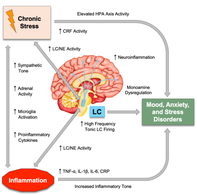
As the main source of NE in the CNS, the LC is crucial for regulating arousal, attention, stress responses, sleep/wake cycles, learning, and memory, particularly for emotionally salient and threat-related events [13,14,16,17]. The LC-NE system is functionally modular with discrete regions and neuronal populations enabling both specific behavioral modulation and general arousal regulation while robust, aversive stimuli can trigger unified responses [13,14,16,38]. In response to chronic stress, this system can become dysregulated, leading to tonic hyperactivity and hypersensitivity of LC neurons (Figure 1).
Studies have shown that corticotropin-releasing factor (CRF) containing afferents originating in the central nucleus of the amygdala provide direct synaptic inputs to dendrites of NA neurons in the LC [39,40,41]. This synaptic circuitry serves to consolidate the emotional and cognitive components of the stress response, tightly linking the perception of a threat with the physiological state of arousal [42]. Direct electrical, chemical, or optogenetic activation of the LC produces significant increases in anxiety and fear-like behavior in both rodents and monkeys [43,44,45]. In fact, panic symptoms can be induced with optogenetic or electrical LC activation, as well as with pharmacological agents that increase LC-NE activity [16,17,43,45]. These maladaptive states are closely linked to the hyperarousal, exaggerated startle responses, and persistent anxious states characteristic of anxiety disorders and PTSD. Importantly this network highlights a critical feedback loop where the amygdala's threat processing and CRF-mediated chronic stress response can increase tonic LC activity, which in turn promotes anxiety (Figure 1) [40,46,47].
The involvement of the LC-NE system in depression is not a simple matter of too much or too little NE. Instead, evidence highlights a complicated dysregulation of the entire system [18]. The function of LC-NE signaling often follows an inverted U-shape function, where both low and high rates of tonic LC neuron firing are associated with cognitive and emotional dysfunction (Figure 1B). Optimal performance occurs only when moderate, phasic firing rates [13,48]. Importantly, this indicates that both the timing and pattern of NE release, not just the quantity, are crucial for healthy brain function. Dysregulation of NA signaling can directly contribute to cognitive symptoms of depression, such as poor concentration and memory deficits [17,49,50].
Additional robust evidence supporting the role of LC-NE activity in depression comes from pharmacology. For decades, chronic treatment with virtually all classes of antidepressants including older monoamine oxidase inhibitors (MAOIs), adrenergic receptor (AR) agonists and antagonists, and tricyclics to newer, more selective compounds has been shown to exert some effect on the NE system. For example, chronic treatment with antidepressants like imipramine and reboxetine primarily acts by increasing synaptic NE concentrations through reuptake inhibition implying NE neurotransmission is a necessary component for effective antidepressant treatment [18,51]. The actions of LC/NE signaling through both α-AR and β-AR subtypes has been shown to underlie symptoms in depression, anxiety, and PTSD (Figure 1B) [52,53,54,55,56,57,58]. Human studies have shown β-AR agonists and α2-AR antagonists can trigger frequent and intense panic attacks in individuals with panic disorder [59,60,61]. In contrast, clonidine, an α2-AR agonist, has been shown to alleviate anxiety symptoms and panic in certain contexts (Figure 1B) [62,63].
Positron emission tomography (PET) imaging studies have shown elevated platelet and brain expression levels of the α2-AR in depressed patients and depressed suicide victims compared to healthy controls [64,65,66]. Treatment with tricyclic antidepressants decreases the elevated α2-AR expression levels observed by PET imaging in depression [64,67]. The α2A-AR has been shown to play a protective role in rodent models of depression and anxiety [68]. Other evidence from animal models suggests the α2C-AR may also be a promising therapeutic target for depression [69]. Patients with depression especially those exhibiting psychomotor agitation have shown decreased β-AR function as indicated by significantly lower isoproterenol-stimulated cyclic AMP levels compared to controls [70,71,72]. In anxiety disorders, patients diagnosed with panic disorder or agoraphobia with panic attacks demonstrate lower number of lymphocyte β-AR binding sites coupled with a higher affinity of binding compared to healthy subjects [73]. Furthermore, β-AR are demonstrably involved in stress-related behavioral changes, as studies show that the β-AR antagonist L-propranolol prevents anxiety-like behavior induced by repeated social defeat in mice and attenuates stress-induced changes in defensive withdrawal in rats [55,74]. Together, these findings underscore that LC-NE dysfunction in neuropsychiatric dysfunction reflects a complex and critical imbalance in NA signaling dynamics.
Inflammatory Signatures of Anxiety, Stress, and Mood Disorders
Across major psychiatric disorders, a distinct inflammatory signature has emerged, characterized by elevated biomarkers and specific patterns of brain changes. A growing body of evidence now implicates chronic, low-grade inflammation as a common pathophysiological pathway in mental health disorders. The influence of inflammation on mental health originates in the periphery, where the immune system first detects a threat. The immune system is activated by different molecular signals that indicate either an external threat (not self) or internal damage (self). Pathogen-Associated Molecular Patterns (PAMPs) are conserved molecules derived from microorganisms that signal a pathogen intrusion. A classic example is lipopolysaccharide (LPS), a component of the outer membrane of gram-negative bacteria, which is a potent inducer of inflammation [75,76,77,78]. Damage-Associated Molecular Patterns (DAMPs) are endogenous molecules released from cells during tissue injury, trauma, or metabolic stress. A key example is adenosine triphosphate released from damaged cells [75,76,77,78]. Whether triggered by PAMPs or DAMPs, these molecular patterns activate toll-like receptors (TLRs) and critical transcription factors, such as nuclear factor kappa B (NF-κB). Once activated, NF-κB promotes the transcription of genes responsible for producing proinflammatory cytokines, including TNF-α, IL-1β, and IL-6 [75,76,77,78]. The initial release of these cytokines triggers the production of even more, creating a self-amplifying feedback loop known as the cytokine cascade or cytokine storm.
Inflammation is signaled to the brain through several mechanisms including a humoral and neural pathway. The humoral pathway involves the circulation of cytokines and other inflammatory mediators in the bloodstream. While large molecules like TNF-α and IL-1β do not readily diffuse across the blood-brain barrier (BBB). They can leak across damage or compromised BBB, they can be actively transported across the BBB, they can affect circumventricular organs, and they can signal through endothelial messengers like prostaglandins and nitric oxide [79]. The neural pathway involves direct transmission of inflammatory signals to the brain via nerve afferents, with the vagus nerve serving as the primary conduit. The vagus conveys afferent signals from a wide range of visceral, somatic, and immunological signals to the brain. These signals in turn activate microglia and astrocytes triggering neuroinflammation providing a direct link between systemic inflammation and the symptoms of mood disorders. The brain can actively regulate peripheral inflammation when it detects inflammation via vagal afferents. Through this cholinergic anti-inflammatory pathway (CAP) discussed further below, the brain sends signals back down the vagus nerve to suppress cytokine production in organs like the spleen, demonstrating a bidirectional neuroimmune reflex loop (Figure 2B) [34,36].
Patients with depression, anxiety, and PTSD consistently show elevated peripheral and central levels of pro-inflammatory cytokines, including IL-1β, IL-6, TNF-α in both blood and cerebrospinal fluid (Figure 1A) [6,7,8,12,19,80,81,82]. Elevated C-reactive protein (CRP) is frequently elevated in individuals with depression and anxiety as a function of symptom severity [6,83,84]. At the cellular level in the brain, neuroinflammation involves the activation of microglia. The translocator protein 18 kDa (TSPO) has been established as a key biomarker of microglial activation that can be measured in the living human brain using PET imaging. PET studies have demonstrated significantly elevated TSPO levels in critical mood-regulating brain regions, such as the prefrontal cortex and anterior cingulate cortex, of individuals experiencing a major depressive episode [10,24,85]. Inflammation influences other critical physiological systems that affect mood and behavior. For example, proinflammatory cytokines can directly stimulate the hypothalamic-pituitary-adrenal (HPA) axis, contributing to the hypercortisolemia commonly observed in depression [86,87,88]. Other recent evidence underscores the fundamental role of the gut-brain axis, mediated by the vagus nerve, in neuropsychiatric health. Perturbations in the diversity and functionality of the gut microbiota have been shown to trigger inflammatory responses that influence mood and behavior [89,90]. Reflecting these actions, diets high in sugar including soft drink consumption have been shown to elicit depression and anxiety symptoms by increasing inflammation driven by gut microbiota alterations [91,92,93,94].
Together, the dysregulated LC-NE system and inflammatory pathways described above represent prime targets for therapeutic intervention. Addressing these dual pathologies has been a major challenge for conventional pharmacotherapy, which often targets one system at the expense of the other. As discussed below, this presents a major opportunity for using taVNS to engage both dysfunctional pathways simultaneously in the treatment of depression, anxiety, and PTSD (Figure 2B and Table 1).
Figure 2.
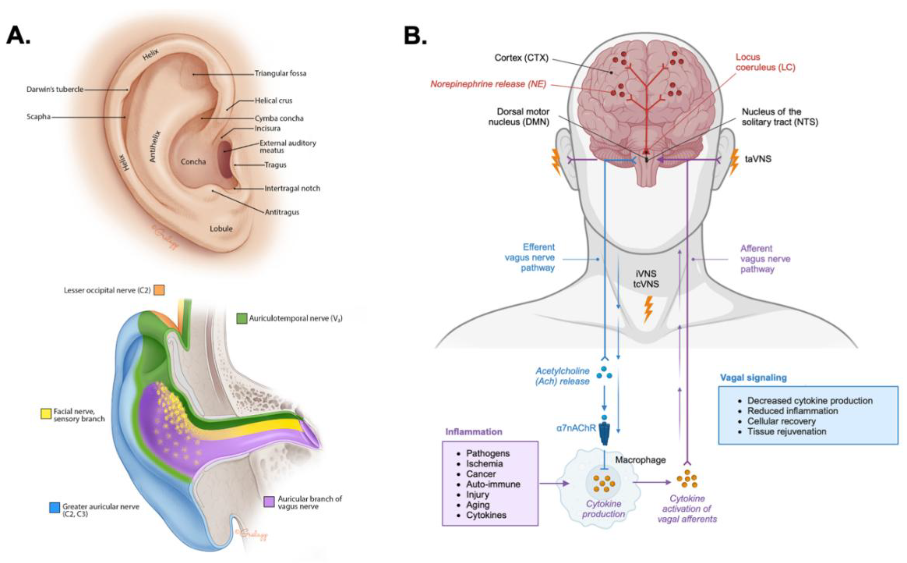
Neuroanatomy of transcutaneous auricular vagus nerve stimulation. A. The anatomical illustration depicts the anatomy of the human external ear showing prominent structures (top). The illustration (bottom) depicts the sensory innervation of the external ear by the lesser occipital nerve (C2; orange), the great auricular nerve (C2, C3; blue), the facial nerve (yellow), the auriculotemporal nerve (ATN) or third branch of the trigeminal nerve (V3; green), and the auricular branch of the vagus nerve (ABVN; purple). The images in panel A were reproduced from reference [95] with permission from the illustrator Chris Gralapp. B. A schematic circuit representation of transcutaneous auricular vagus nerve stimulation (taVNS), transcutaneous cervical vagus nerve stimulation (tcVNS), and invasive vagus nerve stimulation (iVNS) targeting vagal afferents projecting to the nucleus tractus solitarius (NTS) locus coeruleus (LC), and dorsal motor nucleus (DMN). Norepinephrine (NE) release from the LC is shown projecting to large scale brain networks. Efferent vagal signaling engages the cholinergic anti-inflammatory pathway, where acetylcholine (ACh) release activates α7 nicotinic acetylcholine receptors (α7nAChR) on immune cells reducing pro-inflammatory cytokine production to promoting cellular recovery and tissue restoration.
Figure 2.
Neuroanatomy of transcutaneous auricular vagus nerve stimulation. A. The anatomical illustration depicts the anatomy of the human external ear showing prominent structures (top). The illustration (bottom) depicts the sensory innervation of the external ear by the lesser occipital nerve (C2; orange), the great auricular nerve (C2, C3; blue), the facial nerve (yellow), the auriculotemporal nerve (ATN) or third branch of the trigeminal nerve (V3; green), and the auricular branch of the vagus nerve (ABVN; purple). The images in panel A were reproduced from reference [95] with permission from the illustrator Chris Gralapp. B. A schematic circuit representation of transcutaneous auricular vagus nerve stimulation (taVNS), transcutaneous cervical vagus nerve stimulation (tcVNS), and invasive vagus nerve stimulation (iVNS) targeting vagal afferents projecting to the nucleus tractus solitarius (NTS) locus coeruleus (LC), and dorsal motor nucleus (DMN). Norepinephrine (NE) release from the LC is shown projecting to large scale brain networks. Efferent vagal signaling engages the cholinergic anti-inflammatory pathway, where acetylcholine (ACh) release activates α7 nicotinic acetylcholine receptors (α7nAChR) on immune cells reducing pro-inflammatory cytokine production to promoting cellular recovery and tissue restoration.
Anatomical Basis for taVNS
Understanding the specific anatomy of the external ear is important for the application and efficacy of taVNS. The success of taVNS is entirely dependent on the precise targeting of the cutaneous distribution of the ABVN. The location and density of these nerve fibers determine where stimulation should be applied to ensure that the electrical signal is transmitted to the central nervous system to produce its therapeutic effects. The sensory nerve supply of the human auricle is characterized by a heterogeneous distribution of cranial and cervical nerves (Figure 2A). Cadaveric studies have meticulously mapped these distributions, revealing that the ABVN predominantly innervates specific regions of the external ear. The cymba conchae is innervated by the ABVN in all cases (Peuker & Filler, 2002; Butt et al., 2019). The innervation of the cavity of the conchae is more variable where in 45% of specimens, the ABVN solely innervates this region and in the remaining 55%, it receives dual innervation from both the ABVN and the great auricular nerve (GAN) (Peuker & Filler, 2002). Other areas, such as the tragus and antihelix, show significant overlap with other nerves, including the GAN and the auriculotemporal nerve (ATN) branch of the trigeminal nerve (Peuker & Filler, 2002). The colocation of vagal and trigeminal nerves highlights their shared ascending pathways to the brainstem through the spinal trigeminal sensory nuclear complex, as well as common descending pathways along the brain-heart axis as observed through the trigeminocardiac reflex and mammalian diving reflex [96,97,98,99,100,101,102]. The external acoustic meatus (EAM) is also innervated by the ABVN and ATN, as well as the facial nerve [103,104,105,106]. Methods and device for transcutaneous electrical nerve stimulation (TENS) of the external ear are generally referred to collectively as taVNS although different degrees of cranial and cervical nerve afferents may be influenced. Beyond nerve targeting considerations, the structural features of the external ear require different human factors and biomedical engineering approaches to conduct taVNS safely, reliably, and comfortably for users [25] (Figure 2A and Figure 3).
The therapeutic effects of taVNS are mediated through a well-characterized central afferent pathway. Electrical stimulation of the ABVN on the external ear transmits signals along vagal afferents to the nucleus of the solitary tract (NTS) in the brainstem (Figure 2B). The NTS serves as a key central vagal relay center, integrating sensory information and projecting to several higher-order brain structures. Among its most critical projections are those to the LC. The NTS and LC are considered key targets for the therapeutic effects of taVNS [27]. This direct anatomical link between the periphery and key brainstem nuclei provides a direct conduit to the LC and interconnected brainstem nuclei [29,30,31], whose dysregulation in response to chronic stress and inflammation contributes to the pathophysiology of these disorders [13,16,17,18,38].
Ascending Modulation of Noradrenergic Signaling by taVNS
The primary CNS mechanism of taVNS involves the ascending modulation of the LC-NE system (Table 1). As mentioned above the process begins with the transcutaneous electrical modulation of ABVN afferents, which transmit signals to the NTS in the brainstem. The NTS, in turn, projects directly to and modulates the firing rate and pattern of neurons within the LC. Several functional neuroimaging studies have shown that taVNS directly modulates BOLD fMRI signals in the NTS and LC [30,107,108,109,110]. Norepinephrine receptor engagement by VNS has been demonstrated in vivo using PET imaging of 11C-yohimbine, a radiolabeled α2-AR antagonist. These investigations showed that acute VNS leads to a marked reduction in receptor binding, a finding consistent with the widespread release of NE in cortical, limbic, and thalamic brain regions [111]. Interestingly, it has been shown the local activation of α2-ARs is required for VNS-induced motor cortical plasticity [112] while β-AR blockade enhances it [113]. Human psychophysiological investigations have also demonstrated that taVNS activates NA signaling as observed through modulation of pupil diameter a known biomarker of LC-NE activity and cortical arousal [29,31,32]. Several other EEG and functional neuroimaging studies have shown large scale modulation of cortical networks in response to taVNS [107,108,114,115,116,117,118,119,120,121,122]. This modulation of the LC-NE system by taVNS has significant functional consequences, including enhanced arousal, heightened attention, and improved cognitive control—functions that are often impaired in neuropsychiatric disorders [25,114,118,123,124,125,126,127,128].
Modulation of the Descending Cholinergic Anti-Inflammatory Pathway by taVNS
The second major mechanism of taVNS is its ability to engage the CAP, a critical neuroimmune reflex circuit that regulates systemic inflammation (Table 1). Stimulation of afferent vagus nerve fibers triggers a descending, efferent vagal response that can inhibit peripheral inflammation (Figure 2B) [19,34,35,36,37]. This pathway is mediated by the release of acetylcholine (ACh) targeting the α7nAChR located on the surface of immune cells like macrophages. In turn, activation of α7nAChR suppresses intracellular inflammatory signaling cascades by inhibiting the activation of NF-κB, as well as decreasing production and release of pro-inflammatory cytokines like TNF-α [19,34,35,36,37]. As discussed below several lined of evidence corroborate these mechanisms and demonstrate that taVNS can suppress inflammation by modulating NF-κB and cytokine signaling.
Additional studies across various animal models and human diseases of inflammation demonstrate that taVNS effectively modulates key inflammatory biomarkers, including CRP and numerous cytokines. The data show taVNS produces a consistent pattern of reducing pro-inflammatory and increasing anti-inflammatory markers. In human trials, taVNS significantly reduced post-surgical inflammation following lung lobectomy, decreasing serum concentrations of CRP and IL-6 while elevating IL-10 on the first postoperative day [129]. For patients with COVID-19, taVNS led to a significant reduction in both CRP and IL-6 [130]. In sepsis patients, taVNS resulted in significant reductions of serum pro-inflammatory cytokines like TNF-α and IL-1β, accompanied by increases in anti-inflammatory IL-4 and IL-10 [131]. For neurological conditions, taVNS has been shown to mitigate the inflammatory response after subarachnoid hemorrhage SAH, significantly reducing TNF-α and IL-6 in both plasma and cerebrospinal fluid [132]. In large vessel occlusion stroke patients, taVNS significantly reduced normalized aggregate pro-inflammatory cytokines and IL-6 levels [133]. In mouse models of colitis, taVNS decreased colonic pro-inflammatory markers such as IL-1β and TNF-α, while upregulating anti-inflammatory TGF-β and IL-10 [134]. In rat models of depression and anxiety, taVNS has been shown to decrease central and peripheral pro-inflammatory cytokines , including IL-1β and TNF-α, by inhibiting the activation of inflammatory pathways like NF-κB in the hypothalamus, hippocampus, and prefrontal cortex [135,136,137]. Presently, there is a lack of human studies that investigate whether taVNS can similarly reduce inflammation in depression and anxiety. Encouraging preliminary results have shown however that transcutaneous cervical nerve stimulation (tcVNS) can reduce IL-6 and interferon-γ in subjects with PTSD compared to healthy controls [138]. Future clinical trials examining the influence of taVNS on mental health should include the quantification of inflammatory biomarkers to expand knowledge in the field while addressing putative therapeutic targets.

Table 1.
Dual Mechanisms of Action of Transcutaneous Auricular Vagus Nerve Stimulation.
| Mechanistic Pathway | Neurobiological and Physiological Effects |
|---|---|
| Ascending Neuromodulation | Transcutaneous auricular vagus nerve stimulation (taVNS) activates afferent vagal fibers from the external ear projecting to the nucleus of the solitary tract (NTS). The NTS then modulates activity in the locus coeruleus (LC), leading to widespread release of norepinephrine (NE). This helps to stabilize mood and arousal, as well as enhance attention, cognitive control, and facilitates neuroplasticity related to learning and memory. |
| Descending Anti-Inflammatory Modulation | Afferent vagal activation triggers the efferent cholinergic anti-inflammatory pathway (CAP). Activation of the CAP involves the release of acetylcholine (ACh), which binds to α7 nicotinic ACh receptors (α7nAChR) on peripheral immune cells like macrophages to inhibit the activation of NF-κB and production of pro-inflammatory cytokines such as TNF-α and IL-6. |
The two distinct but complementary mechanisms, central neuromodulation and peripheral anti-inflammatory signaling, described above position taVNS as an exceptional method for addressing the core pathologies of emotional dysregulation and inflammation in neuropsychiatry. The sections below discuss evidence from studies translating these mechanistic actions into measurable changes in autonomic tone, affective processing, and clinical symptom severity.
Figure 3.
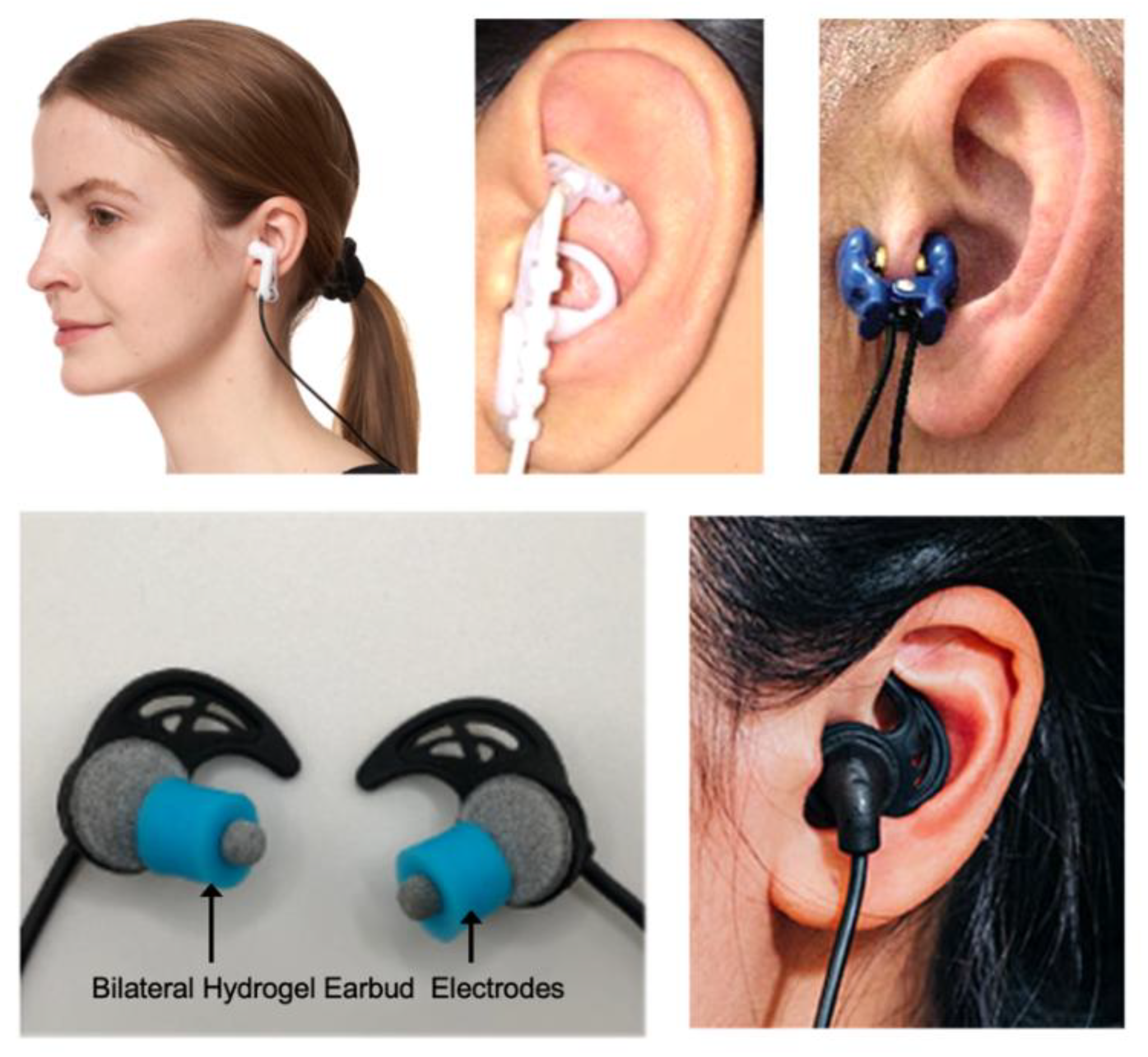
Methods of transcutaneous auricular vagus nerve stimulation. Several common methods of conducting transcutaneous auricular vagus nerve stimulation (taVNS) or transcutaneous electrical nerve stimulation of the external ear are shown. Electrode ear clips (top left and top right) can be affixed the tragus while customized electrode assemblies can deliver stimuli to the concha (top middle). Bilateral hydrogel earbud electrodes (BRAIN Buds, IST, LLC) can also be used to target afferent vagal and trigeminal fibers lining the external acoustic meatus as shown in the bottom panel of images.
Figure 3.
Methods of transcutaneous auricular vagus nerve stimulation. Several common methods of conducting transcutaneous auricular vagus nerve stimulation (taVNS) or transcutaneous electrical nerve stimulation of the external ear are shown. Electrode ear clips (top left and top right) can be affixed the tragus while customized electrode assemblies can deliver stimuli to the concha (top middle). Bilateral hydrogel earbud electrodes (BRAIN Buds, IST, LLC) can also be used to target afferent vagal and trigeminal fibers lining the external acoustic meatus as shown in the bottom panel of images.
Effects of taVNS on Autonomic Balance and Heart Rate Variability
An interesting therapeutic indicator of taVNS is its ability to rebalance autonomic homeostasis by suppressing sympathetic dominance of vagal cardiac tone. This modulation is frequently measured by improvements in heart rate variability (HRV), which is associated with better cardiac autonomic control and stress recovery. Studies have shown taVNS can produce significant increases in HRV metrics such as the root mean square of successive differences (RMSSD), the percentage of adjacent intervals differing by more than 50 msec (pRR50), and the high-frequency (HF) component, which collectively indicate a shift towards parasympathetic predominance under resting conditions [139,140]. It has also been shown to directly dampen sympathetic nervous system activation as measured during sympathetic muscle nerve activity recordings [140]. This vagal-mediated increase in HRV extends to experimental challenges and stressful conditions. For example, taVNS has been shown to increase HRV correlating with significant suppression of experimentally induced pain [141].
Other studies have shown taVNS can mitigate physiological responses to acute mental and psychosocial stress [142,143], and improves autonomic functioning in extreme environmental conditions, particularly when the stimulation is applied repeatedly [144]. The downstream consequences of this autonomic regulation are significant for the HPA axis and cognitive function. Notably, taVNS has been shown to inhibit the release of salivary cortisol during mental stress, suggesting therapeutic potential for chronic inflammatory conditions associated with glucocorticoid dysfunction [142]. Cognitively, taVNS can improve cognitive flexibility by reducing perseverative thinking following a psychosocial stressor [143], while repeated stimulation can enhance overall cognitive performance [144]. The efficacy of taVNS appears more dependent on the individual's baseline physiological state than on the specific stimulation parameters used, provided the correct anatomical target is stimulated. This is supported by evidence that taVNS does not produce parameter-specific effects on HRV [139,145]. In contrast, individual variability is a powerful predictor of outcomes. For instance, a higher baseline sympathovagal balance (LF/HF ratio) predicts a greater decrease in this ratio [139], the degree of parasympathetic activation during stimulation predicts the subsequent magnitude of pain suppression [141], and taVNS is particularly effective at reducing perseverative thinking in individuals who exhibit greater autonomic inflexibility during a stressor [143]. The findings with respect to HRV are clinically relevant given low vagal tone is strongly associated with impaired recovery from stress, poor mental health, and is associated with a pro-inflammatory state further linking autonomic dysregulation to the symptoms observed in stress, mood, and anxiety disorders [26,146].
Modulation of Stress, Cognition, and Emotion by taVNS
Current evidence indicates that taVNS beneficially impacts cognition, with notable effects on executive functions [119,147,148,149]. More specifically, taVNS can enhance cognitive flexibility, particularly in conditions of high cognitive demand [144,147,150] and improve emotional inhibitory control by enabling more efficient suppression of prepotent emotional responses [151,152]. In addition to executive control, taVNS has demonstrated a causal link with the formation of emotional memories by increasing recollection-based memory specifically for emotionally relevant information [153]. Another study has shown taVNS can significantly improve visual memory [154]. This effect may extend to social memory and threat processing functions of the vagus [155,156]. Consistent with these functions, treatment with taVNS has been shown to mitigate negative emotional bias by improving the detection of positive facial expressions and reducing negative emotions [157]. Psychological markers of resilience are improved by taVNS as indicated by significant reductions in cognitive rigidity and perseverative thinking following a psychosocial stressor [143]. Similarly, taVNS also prevents increases in perceived stress following cognitively or physically demanding tasks [146,158]. These behavioral and cognitive outcomes highlight a targeted effect of taVNS on the brain's arousal, emotional regulation, and attention systems.
The cognitive and emotional benefits of taVNS are also rooted in specific, measurable changes in brain activity and network dynamics. At a fundamental level, taVNS enhances cortical arousal and alertness by attenuating high-frequency alpha oscillations and reducing the duration of EEG microstate C, a neural signature associated with alertness [114], while also increasing power in the delta frequency band [159]. This general enhancement in cortical arousal provides the foundation for more targeted, network-level changes that support executive function. At the network level, taVNS promotes more efficient brain communication following a stress task as it enhances global network efficiency evidenced by a reduction in prefrontal alpha and theta band activity [158]. It also strengthens functional connectivity to support emotional control. This is achieved by reducing alpha band power spectral density and increasing phase locking within prefrontal networks, as well as enhancing neurofunctional coupling between the inferior and orbital frontal cortex [151,158]. This increased network efficiency translates into more parsimonious use of neural resources for cognitive control, as demonstrated by a significant reduction of the frontal N2 potential in NoGo tasks, which suggests fewer cognitive resources are needed to successfully inhibit a response [148,151]. Functional neuroimaging corroborates these findings, showing that taVNS modulates neural activity in key emotion regulation regions, including the bilateral precuneus and temporal gyrus, which is thought to facilitate the integration of emotional responses and memories [160].
The capacity of taVNS to reliably modulate cognitive, emotional, and neurophysiological systems gives it significant therapeutic potential for psychiatric disorders. More specifically, the ability of taVNS to modulate perseverative cognition [143], negative emotional bias [157], and inhibitory control [151] positions it as a promising therapeutic tool. Furthermore, the causal role of taVNS in modulating emotional memory formation suggests potential applications for disorders with altered memory functions, such as PTSD [153]. More broadly, its ability to improve emotional regulation by modulating prefrontal and temporal brain regions points to its utility in treating a range of conditions characterized by emotional dysregulation [108,115,117,122,151,160,161,162]. However, to translate this potential into clinical practice, future research must address several critical areas. There is a clear need for longitudinal studies with larger, more diverse samples to confirm the observed health benefits [28,119,144]. Concurrently, researchers must work to identify optimal stimulation parameters to maximize efficacy [25,29,119] and validate robust biomarkers that can illuminate its mechanisms of action and help predict therapeutic outcomes in individuals with neuropsychiatric diseases [25,144,163,164,165].
Clinical and Translational Efficacy of taVNS in Depression, Anxiety, and PTSD
Clinical trial data indicate taVNS is an effective approach for reducing symptoms of major depressive disorder (MDD). Studies have shown taVNS treatment produces significant decreases in depression scores in depressed subjects as measured with the Hamilton Depression Rating Scale (HAM-D), Beck Depression Inventory (BDI), and other clinically valid scales [126,128,166,167,168,169,170,171]. When directly compared to pharmacotherapy, taVNS demonstrated therapeutic effects comparable to citalopram; however, patients treated with taVNS experienced significantly higher remission rates at weeks four and six than those treated with citalopram [167]. Li et al (2022) also showed that taVNS treatment decreased plasma serotonin, NA, and γ-aminobutyric acid (GABA) while increasing dopamine producing effects like citalopram on these neurotransmitters [167]. Enhancing fear extinction is a critical goal in PTSD treatment. It has been shown that taVNS paired with extinction training strengthens the consolidation of fear extinction memory [121,172]. A recent pilot study demonstrated that taVNS treatment of subjects diagnosed with PTSD can significantly improve sleep quality (depth) and autonomic function which are typically disrupted by the disorder [173]. Other evidence from the study of VNS in the treatment of PTSD suggest it provides a viable treatment option worthy of further investigation [138,174,175,176,177]. Translational investigations of the use of taVNS for the treatment of anxiety have also shown encouraging results. Several recent feasibility and exploratory clinical trials have reported that taVNS treatment can significantly reduce anxiety across different health and experimental conditions as measured by the Generalized Anxiety Disorder-7 (GAD-7), Beck Anxiety Inventory (BAI), and other scales [170,178,179,180,181]. For example, a feasibility study demonstrated that at-home taVNS improved anxiety scores in youth with comorbid autism spectrum disorder and anxiety [182]. While these initial results on depression, PTSD, and anxiety are encouraging, there remains a need to conduct larger randomized controlled trials with a focus on gaining regulatory approvals for neuropsychiatric indications.
Beyond symptom reduction, taVNS can also induce specific behavioral and cognitive improvements. In patients with MDD, it has been shown to enhance motivation, invigorate effort, and increase the subjective wanting of rewards [118,146,183], as well as improve emotional processing by increasing accuracy in detecting positive facial expressions [157]. For individuals with high trait anxiety, taVNS has been found to reduce negative thought intrusions and perseverative thinking [143,184] These targeted benefits are complemented by broader improvements in quality of life, including better sleep [185,186,187,188] and a reduction in pain perception, a common comorbidity in psychiatric conditions [141,179]. Neuroimaging findings further illustrate that taVNS modulates activity across large-scale brain networks implicated in psychiatric disorders [120,161,189,190,191]. These effects include the normalization of functional connectivity within the limbic system, particularly the amygdala, in patients with depression [191], and the regulation of the cortico-striatal-thalamic-cortical circuit, evidenced by the downregulation of hyperactivity in the ventral striatum [192]. Furthermore, taVNS has been shown to alter connectivity between core cognitive control and emotional processing networks, including the default mode network (DMN), frontoparietal network (FPN), and cingulo-opercular network (CON) [108,115,117,122,161,162]. These findings collectively support the therapeutic potential of taVNS, demonstrating its ability to appropriately modulate neurophysiological outcomes and improve clinical symptoms across a spectrum of common neuropsychiatric disorders.
Conclusions
Transcutaneous auricular vagus nerve stimulation is a promising, non-invasive therapeutic modality that directly addresses the core, interconnected pathologies of emotional dysregulation and neuroinflammation in neuropsychiatric disorders. Through its dual mechanisms of action, taVNS modulates the central locus coeruleus-norepinephrine system to enhance cognitive control and emotional processing, while simultaneously engaging the descending cholinergic anti-inflammatory pathway to suppress peripheral inflammation. This unique combination of effects allows taVNS to restore autonomic and immune homeostasis, leading to measurable improvements in clinical symptoms. The convergence of evidence suggests that taVNS may not merely treat symptoms, but rather fundamentally restores the organism's capacity for adaptive allostasis, a crucial insight for developing preventative and resilience-building neuromodulatory strategies. While the existing evidence is compelling, future research is essential to fully realize its clinical potential.
The clinical potential of taVNS derives from its capacity to simultaneously address two interacting biological drivers of mood and anxiety disorders. A unified model of action proposes that by modulating the hyperactive LC-NE alarm system via its ascending pathway, taVNS normalizes central arousal and stress reactivity. At the same time, its descending pathway activates the cholinergic anti-inflammatory reflex to reduce systemic pro-inflammatory signals implicated in the pathophysiology of depression and anxiety. These two mechanisms likely interact to produce specific therapeutic effects. For instance, the simultaneous downregulation of the hyper-aroused LC-NE system likely contributes directly to the observed attenuation of sympathetic dominance and blunting of HPA axis stress reactivity described above. Concurrently, the reduction of systemic pro-inflammatory cytokines, which are known to impair neuroplasticity, may provide the neurochemical environment necessary for enhancing fear extinction memory and recalibrating negative affective biases. By calming a hyper-responsive central stress system while also dampening peripheral inflammation, taVNS may disrupt the vicious cycle where stress promotes inflammation and inflammation, in turn, exacerbates stress and mood dysregulation.
Despite the promising findings, the field of taVNS research faces several methodological challenges that must be addressed to establish its clinical utility robustly. Many of the reviewed studies were pilot or exploratory trials with small sample sizes, which limits the generalizability of their findings and increases the risk of false positives. Furthermore, designing an inert but credible sham control is a significant challenge in neuromodulation research. While some studies use stimulation of a non-vagal site like the earlobe, the complete blinding of participants can be difficult to achieve, as active stimulation often produces a noticeable sensation. This complicates the interpretation of results and the separation of true therapeutic effects from placebo responses. Finally, there is wide variability in the stimulation parameters (e.g., frequency, pulse width, intensity, duration) used across studies, which makes it difficult to compare results between trials and to determine the optimal settings for specific clinical indications. While methodological challenges remain, continued research focused on parameter optimization, biomarker identification, and large-scale clinical trials holds the promise of establishing taVNS as a scalable and accessible tool in the clinical management of common neuropsychiatric disorders.
Acknowledgements
The development of BRAIN Buds by IST, LLC was funded based on research sponsored by Air Force Research Laboratory under agreement number FA8650-18-2-5402. The U.S. Government is authorized to reproduce and distribute reprints for Government purposes notwithstanding any copyright notation thereon. The views and conclusions contained herein are those of the authors and should not be interpreted as necessarily representing the official policies or endorsements, either expressed or implied, of Air Force Research Laboratory (AFRL) or the U.S. Government.
Disclosures
WJT is a co-founder of IST, LLC and an inventor and co-inventor on neuromodulation methods and devices including external ear stimulation for treating various disorders.
CLINICAL TRIAL REGISTRATION
Not applicable.
References
- Arias, D.; Saxena, S.; Verguet, S. Quantifying the global burden of mental disorders and their economic value. eClinicalMedicine 2022, 54. [Google Scholar] [CrossRef]
- Kieling, C.; Buchweitz, C.; Caye, A.; Silvani, J.; Ameis, S.H.; Brunoni, A.R.; Cost, K.T.; Courtney, D.B.; Georgiades, K.; Merikangas, K.R.; et al. Worldwide Prevalence and Disability From Mental Disorders Across Childhood and Adolescence: Evidence From the Global Burden of Disease Study. JAMA Psychiatry 2024, 81, 347–356. [Google Scholar] [CrossRef]
- Collaborators, G.M.D. Global, regional, and national burden of 12 mental disorders in 204 countries and territories, 1990-2019: A systematic analysis for the Global Burden of Disease Study 2019. Lancet Psychiatry 2022, 9, 137–150. [Google Scholar] [CrossRef]
- Chen, S.; Huang, W.; Zhang, M.; Song, Y.; Zhao, C.; Sun, H.; Wang, Y.; Wang, J.; Sun, Y.; Zhou, L.; et al. Dynamic changes and future trend predictions of the global burden of anxiety disorders: Analysis of 204 countries and regions from 1990 to 2021 and the impact of the COVID-19 pandemic. eClinicalMedicine 2025, 79. [Google Scholar] [CrossRef]
- Bechter, K. The Challenge of Assessing Mild Neuroinflammation in Severe Mental Disorders. Frontiers in Psychiatry 2020, 11, 773. [Google Scholar] [CrossRef]
- Furtado, M.; Katzman, M.A. Examining the role of neuroinflammation in major depression. Psychiatry Research 2015, 229, 27–36. [Google Scholar] [CrossRef] [PubMed]
- Hassamal, S. Chronic stress, neuroinflammation, and depression: An overview of pathophysiological mechanisms and emerging anti-inflammatories. Frontiers in Psychiatry 2023, 14, 1130989. [Google Scholar] [CrossRef] [PubMed]
- Herzog, S.; Bartlett, E.A.; Zanderigo, F.; Galfalvy, H.C.; Burke, A.; Mintz, A.; Schmidt, M.; Hauser, E.; Huang, Y.-y.; Melhem, N.; et al. Neuroinflammation, Stress-Related Suicidal Ideation, and Negative Mood in Depression. JAMA Psychiatry 2025, 82, 85–93. [Google Scholar] [CrossRef] [PubMed]
- Irwin, M.R. Sleep and inflammation: Partners in sickness and in health. Nature Reviews Immunology 2019, 19, 702–715. [Google Scholar] [CrossRef]
- Meyer, J.H.; Cervenka, S.; Kim, M.-J.; Kreisl, W.C.; Henter, I.D.; Innis, R.B. Neuroinflammation in psychiatric disorders: PET imaging and promising new targets. The Lancet Psychiatry 2020, 7, 1064–1074. [Google Scholar] [CrossRef]
- Sălcudean, A.; Bodo, C.-R.; Popovici, R.-A.; Cozma, M.-M.; Păcurar, M.; Crăciun, R.-E.; Crisan, A.-I.; Enatescu, V.-R.; Marinescu, I.; Cimpian, D.-M.; et al. Neuroinflammation—A Crucial Factor in the Pathophysiology of Depression—A Comprehensive Review. Biomolecules 2025, 15. [Google Scholar] [CrossRef] [PubMed]
- Radtke, F.A.; Chapman, G.; Hall, J.; Syed, Y.A. Modulating Neuroinflammation to Treat Neuropsychiatric Disorders. Biomed Res Int 2017, 2017, 5071786. [Google Scholar] [CrossRef] [PubMed]
- Aston-Jones, G.; Cohen, J.D. An integrative theory of locus coeruleus-norepinephrine function: Adaptive gain and optimal performance. Annu Rev Neurosci 2005, 28, 403–450. [Google Scholar] [CrossRef] [PubMed]
- Poe, G.R.; Foote, S.; Eschenko, O.; Johansen, J.P.; Bouret, S.; Aston-Jones, G.; Harley, C.W.; Manahan-Vaughan, D.; Weinshenker, D.; Valentino, R.; et al. Locus coeruleus: A new look at the blue spot. Nat Rev Neurosci 2020, 21, 644–659. [Google Scholar] [CrossRef]
- van der Linden, D.; Tops, M.; Bakker, A.B. The Neuroscience of the Flow State: Involvement of the Locus Coeruleus Norepinephrine System. Frontiers in Psychology 2021, 12, 645498. [Google Scholar] [CrossRef]
- Morris, L.S.; McCall, J.G.; Charney, D.S.; Murrough, J.W. The role of the locus coeruleus in the generation of pathological anxiety. Brain Neurosci Adv 2020, 4, 2398212820930321. [Google Scholar] [CrossRef]
- Reyes, B.A.S. The Locus Coeruleus: Anatomy, Physiology, and Stress-Related Neuropsychiatric Disorders. Eur J Neurosci 2025, 61, e70111. [Google Scholar] [CrossRef]
- Ressler, K.J.; Nemeroff, C.B. Role of norepinephrine in the pathophysiology and treatment of mood disorders. Biol Psychiatry 1999, 46, 1219–1233. [Google Scholar] [CrossRef]
- Guo, B.; Zhang, M.; Hao, W.; Wang, Y.; Zhang, T.; Liu, C. Neuroinflammation mechanisms of neuromodulation therapies for anxiety and depression. Translational Psychiatry 2023, 13, 5. [Google Scholar] [CrossRef]
- Goldsmith, D.R.; Rapaport, M.H.; Miller, B.J. A meta-analysis of blood cytokine network alterations in psychiatric patients: Comparisons between schizophrenia, bipolar disorder and depression. Mol Psychiatry 2016, 21, 1696–1709. [Google Scholar] [CrossRef]
- Wang, H.; He, Y.; Sun, Z.; Ren, S.; Liu, M.; Wang, G.; Yang, J. Microglia in depression: An overview of microglia in the pathogenesis and treatment of depression. J Neuroinflammation 2022, 19, 132. [Google Scholar] [CrossRef] [PubMed]
- Brites, D.; Fernandes, A. Neuroinflammation and Depression: Microglia Activation, Extracellular Microvesicles and microRNA Dysregulation. Frontiers in Cellular Neuroscience 2015, 9, 476. [Google Scholar] [CrossRef] [PubMed]
- Yirmiya, R.; Rimmerman, N.; Reshef, R. Depression as a Microglial Disease. Trends in Neurosciences 2015, 38, 637–658. [Google Scholar] [CrossRef] [PubMed]
- Setiawan, E.; Wilson, A.A.; Mizrahi, R.; Rusjan, P.M.; Miler, L.; Rajkowska, G.; Suridjan, I.; Kennedy, J.L.; Rekkas, P.V.; Houle, S.; et al. Role of translocator protein density, a marker of neuroinflammation, in the brain during major depressive episodes. JAMA Psychiatry 2015, 72, 268–275. [Google Scholar] [CrossRef]
- Tyler, W.J. Auricular bioelectronic devices for health, medicine, and human-computer interfaces. Frontiers in Electronics 2025, 6, 1503425. [Google Scholar] [CrossRef]
- Lopez Blanco, C.; Tyler, W.J. The vagus nerve: A cornerstone for mental health and performance optimization in recreation and elite sports. Frontiers in Psychology 2025, 16, 1639866. [Google Scholar] [CrossRef]
- Butt, M.F.; Albusoda, A.; Farmer, A.D.; Aziz, Q. The anatomical basis for transcutaneous auricular vagus nerve stimulation. Journal of Anatomy 2020, 236, 588–611. [Google Scholar] [CrossRef]
- Kim, A.Y.; Marduy, A.; de Melo, P.S.; Gianlorenco, A.C.; Kim, C.K.; Choi, H.; Song, J.-J.; Fregni, F. Safety of transcutaneous auricular vagus nerve stimulation (taVNS): A systematic review and meta-analysis. Scientific Reports 2022, 12, 22055. [Google Scholar] [CrossRef]
- Urbin, M.A.; Lafe, C.W.; Simpson, T.W.; Wittenberg, G.F.; Chandrasekaran, B.; Weber, D.J. Electrical stimulation of the external ear acutely activates noradrenergic mechanisms in humans. Brain Stimul 2021, 14, 990–1001. [Google Scholar] [CrossRef]
- Frangos, E.; Ellrich, J.; Komisaruk, B.R. Non-invasive Access to the Vagus Nerve Central Projections via Electrical Stimulation of the External Ear: fMRI Evidence in Humans. Brain Stimul 2015, 8, 624–636. [Google Scholar] [CrossRef]
- Phillips, I.; Johns, M.A.; Pandža, N.B.; Calloway, R.C.; Karuzis, V.P.; Kuchinsky, S.E. Three Hundred Hertz Transcutaneous Auricular Vagus Nerve Stimulation (taVNS) Impacts Pupil Size Non-Linearly as a Function of Intensity. Psychophysiology 2025, 62, e70011. [Google Scholar] [CrossRef]
- Sharon, O.; Fahoum, F.; Nir, Y. Transcutaneous Vagus Nerve Stimulation in Humans Induces Pupil Dilation and Attenuates Alpha Oscillations. The Journal of Neuroscience: The Official Journal of the Society for Neuroscience 2021, 41, 320–330. [Google Scholar] [CrossRef] [PubMed]
- Pavlov, V.A. Collateral benefits of studying the vagus nerve in bioelectronic medicine. Bioelectron Med 2019, 5, 5. [Google Scholar] [CrossRef] [PubMed]
- Pavlov, V.A.; Tracey, K.J. The vagus nerve and the inflammatory reflex—Linking immunity and metabolism. Nature Reviews Endocrinology 2012, 8, 743–754. [Google Scholar] [CrossRef]
- Pavlov, V.A.; Tracey, K.J. Bioelectronic medicine: Preclinical insights and clinical advances. Neuron 2022, 110, 3627–3644. [Google Scholar] [CrossRef] [PubMed]
- Pavlov, V.A.; Tracey, K.J. The cholinergic anti-inflammatory pathway. Brain Behav Immun 2005, 19, 493–499. [Google Scholar] [CrossRef]
- Tynan, A.; Brines, M.; Chavan, S.S. Control of inflammation using non-invasive neuromodulation: Past, present and promise. International Immunology 2021, 34, 119–128. [Google Scholar] [CrossRef]
- Aston-Jones, G.; Rajkowski, J.; Cohen, J. Role of locus coeruleus in attention and behavioral flexibility. Biological Psychiatry 1999, 46, 1309–1320. [Google Scholar] [CrossRef]
- Van Bockstaele, E.J.; Colago, E.E.; Valentino, R.J. Corticotropin-releasing factor-containing axon terminals synapse onto catecholamine dendrites and may presynaptically modulate other afferents in the rostral pole of the nucleus locus coeruleus in the rat brain. J Comp Neurol 1996, 364, 523–534. [Google Scholar] [CrossRef]
- Van Bockstaele, E.J.; Colago, E.E.; Valentino, R.J. Amygdaloid corticotropin-releasing factor targets locus coeruleus dendrites: Substrate for the co-ordination of emotional and cognitive limbs of the stress response. J Neuroendocrinol 1998, 10, 743–757. [Google Scholar] [CrossRef]
- Devilbiss, D.M.; Waterhouse, B.D.; Berridge, C.W.; Valentino, R. Corticotropin-Releasing Factor Acting at the Locus Coeruleus Disrupts Thalamic and Cortical Sensory-Evoked Responses. Neuropsychopharmacology 2012, 37, 2020–2030. [Google Scholar] [CrossRef] [PubMed]
- Koob, G.F. Corticotropin-releasing factor, norepinephrine, and stress. Biological Psychiatry 1999, 46, 1167–1180. [Google Scholar] [CrossRef] [PubMed]
- Boulenger, J.P.; Uhde, T.W. Biological peripheral correlates of anxiety. Encephale 1982, 8, 119–130. [Google Scholar] [PubMed]
- Hahn, M.K.; Bannon, M.J. Stress-induced c-fos expression in the rat locus coeruleus is dependent on neurokinin 1 receptor activation. Neuroscience 1999, 94, 1183–1188. [Google Scholar] [CrossRef]
- Carter, M.E.; Yizhar, O.; Chikahisa, S.; Nguyen, H.; Adamantidis, A.; Nishino, S.; Deisseroth, K.; de Lecea, L. Tuning arousal with optogenetic modulation of locus coeruleus neurons. Nature Neuroscience 2010, 13, 1526–1533. [Google Scholar] [CrossRef]
- McCall, J.G.; Al-Hasani, R.; Siuda, E.R.; Hong, D.Y.; Norris, A.J.; Ford, C.P.; Bruchas, M.R. CRH Engagement of the Locus Coeruleus Noradrenergic System Mediates Stress-Induced Anxiety. Neuron 2015, 87, 605–620. [Google Scholar] [CrossRef]
- McCall, J.G.; Siuda, E.R.; Bhatti, D.L.; Lawson, L.A.; McElligott, Z.A.; Stuber, G.D.; Bruchas, M.R. Locus coeruleus to basolateral amygdala noradrenergic projections promote anxiety-like behavior. Elife 2017, 6. [Google Scholar] [CrossRef]
- Foote, S.L.; Bloom, F.E.; Aston-Jones, G. Nucleus locus ceruleus: New evidence of anatomical and physiological specificity. Physiol Rev 1983, 63, 844–914. [Google Scholar] [CrossRef]
- Cole, B.J.; Robbins, T.W. Forebrain norepinephrine: Role in controlled information processing in the rat. Neuropsychopharmacology 1992, 7, 129–142. [Google Scholar]
- Robbins, T.W. Arousal systems and attentional processes. Biol Psychol 1997, 45, 57–71. [Google Scholar] [CrossRef]
- Glowinski, J.; Axelrod, J. Inhibition of uptake of tritiated-noradrenaline in the intact rat brain by imipramine and structurally related compounds. Nature 1964, 204, 1318–1319. [Google Scholar] [CrossRef]
- Belkin, M.R.; Schwartz, T.L. Alpha-2 receptor agonists for the treatment of posttraumatic stress disorder. Drugs Context 2015, 4, 212286. [Google Scholar] [CrossRef] [PubMed]
- Zhang, H.; Cui, M.; Cao, J.L.; Han, M.H. The Role of Beta-Adrenergic Receptors in Depression and Resilience. Biomedicines 2022, 10, 2378. [Google Scholar] [CrossRef] [PubMed]
- Maletic, V.; Eramo, A.; Gwin, K.; Offord, S.J.; Duffy, R.A. The Role of Norepinephrine and Its α-Adrenergic Receptors in the Pathophysiology and Treatment of Major Depressive Disorder and Schizophrenia: A Systematic Review. Front Psychiatry 2017, 8, 42. [Google Scholar] [CrossRef] [PubMed]
- Wohleb, E.S.; Hanke, M.L.; Corona, A.W.; Powell, N.D.; Stiner, L.M.; Bailey, M.T.; Nelson, R.J.; Godbout, J.P.; Sheridan, J.F. β-Adrenergic receptor antagonism prevents anxiety-like behavior and microglial reactivity induced by repeated social defeat. J Neurosci 2011, 31, 6277–6288. [Google Scholar] [CrossRef]
- Zhang, X.; Norton, J.; Carrière, I.; Ritchie, K.; Chaudieu, I.; Ryan, J.; Ancelin, M.-L. Preliminary evidence for a role of the adrenergic nervous system in generalized anxiety disorder. Scientific Reports 2017, 7, 42676. [Google Scholar] [CrossRef]
- Itoi, K.; Sugimoto, N. The Brainstem Noradrenergic Systems in Stress, Anxiety and Depression. Journal of Neuroendocrinology 2010, 22, 355–361. [Google Scholar] [CrossRef]
- Townsend, L.K. Peripheral adrenergic activity contributes to anxiety. Nature Reviews Endocrinology 2025. [Google Scholar] [CrossRef]
- Charney, D.S.; Heninger, G.R.; Breier, A. Noradrenergic Function in Panic Anxiety: Effects of Yohimbine in Healthy Subjects and Patients With Agoraphobia and Panic Disorder. Archives of General Psychiatry 1984, 41, 751–763. [Google Scholar] [CrossRef]
- Charney, D.S.; Woods, S.W.; Heninger, G.R. Noradrenergic function in generalized anxiety disorder: Effects of yohimbine in healthy subjects and patients with generalized anxiety disorder. Psychiatry Research 1989, 27, 173–182. [Google Scholar] [CrossRef]
- Nesse, R.M.; Cameron, O.G.; Curtis, G.C.; McCann, D.S.; Huber-Smith, M.J. Adrenergic function in patients with panic anxiety. Arch Gen Psychiatry 1984, 41, 771–776. [Google Scholar] [CrossRef]
- Uhde, T.W.; Stein, M.B.; Vittone, B.J.; Siever, L.J.; Boulenger, J.-P.; Klein, E.; Mellman, T.A. Behavioral and Physiologic Effects of Short-term and Long-term Administration of Clonidine in Panic Disorder. Archives of General Psychiatry 1989, 46, 170–177. [Google Scholar] [CrossRef]
- Coplan, J.D.; Liebowitz, M.R.; Gorman, J.M.; Fyer, A.J.; Dillon, D.J.; Campeas, R.B.; Davies, S.O.; Martinez, J.; Klein, D.F. Noradrenergic function in panic disorder effects of intravenous clonidine pretreatment on lactate induced panic. Biological Psychiatry 1992, 31, 135–146. [Google Scholar] [CrossRef]
- García-Sevilla, J.A.; Zis, A.P.; Hollingsworth, P.J.; Greden, J.F.; Smith, C.B. Platelet α2-Adrenergic Receptors in Major Depressive Disorder: Binding of Tritiated Clonidine Before and After Tricyclic Antidepressant Drug Treatment. Archives of General Psychiatry 1981, 38, 1327–1333. [Google Scholar] [CrossRef]
- Javier Meana, J.; Barturen, F.; Garcia-Sevilla, J.A. <em>α</em><sub>2</sub>-Adrenoceptors in the brain of suicide victims: Increased receptor density associated with major depression. Biological Psychiatry 1992, 31, 471–490. [Google Scholar] [CrossRef]
- Ordway, G.A.; Schenk, J.; Stockmeier, C.A.; May, W.; Klimek, V. Elevated agonist binding to α2-adrenoceptors in the locus coeruleus in major depression. Biological Psychiatry 2003, 53, 315–323. [Google Scholar] [CrossRef] [PubMed]
- Cottingham, C.; Wang, Q. α2 adrenergic receptor dysregulation in depressive disorders: Implications for the neurobiology of depression and antidepressant therapy. Neuroscience & Biobehavioral Reviews 2012, 36, 2214–2225. [Google Scholar] [CrossRef] [PubMed]
- Schramm, N.L.; McDonald, M.P.; Limbird, L.E. The α<sub>2A</sub>-Adrenergic Receptor Plays a Protective Role in Mouse Behavioral Models of Depression and Anxiety. The Journal of Neuroscience 2001, 21, 4875. [Google Scholar] [CrossRef]
- Uys, M.M.; Shahid, M.; Harvey, B.H. Therapeutic Potential of Selectively Targeting the α2C-Adrenoceptor in Cognition, Depression, and Schizophrenia—New Developments and Future Perspective. Frontiers in Psychiatry 2017, 8, 144. [Google Scholar] [CrossRef] [PubMed]
- Extein, I.; Tallman, J.; Smith, C.C.; Goodwin, F.K. Changes in lymphocyte beta-adrenergic receptors in depression and mania. Psychiatry Research 1979, 1, 191–197. [Google Scholar] [CrossRef]
- Mann, J.J.; Brown, R.P.; Halper, J.P.; Sweeney, J.A.; Kocsis, J.H.; Stokes, P.E.; Bilezikian, J.P. Reduced Sensitivity of Lymphocyte Beta-Adrenergic Receptors in Patients with Endogenous Depression and Psychomotor Agitation. New England Journal of Medicine 1985, 313, 715–720. [Google Scholar] [CrossRef]
- Pandey, G.N.; Sudershan, P.; Davis, J.M. BETA ADRENERGIC RECEPTOR FUNCTION IN DEPRESSION AND THE EFFECT OF ANTIDEPRESSANT DRUGS. Acta Pharmacologica et Toxicologica 1985, 56, 66–79. [Google Scholar] [CrossRef] [PubMed]
- Brown, S.L.; Charney, D.S.; Woods, S.W.; Heninger, G.R.; Tallman, J. Lymphocyte β-adrenergic receptor binding in panic disorder. Psychopharmacology 1988, 94, 24–28. [Google Scholar] [CrossRef] [PubMed]
- Laurel Gorman, A.; Dunn, A.J. β-adrenergic receptors are involved in stress-related behavioral changes. Pharmacology Biochemistry and Behavior 1993, 45, 1–7. [Google Scholar] [CrossRef] [PubMed]
- Li, D.; Wu, M. Pattern recognition receptors in health and diseases. Signal Transduction and Targeted Therapy 2021, 6, 291. [Google Scholar] [CrossRef]
- Bianchi, M.E. DAMPs, PAMPs and alarmins: All we need to know about danger. J Leukoc Biol 2007, 81, 1–5. [Google Scholar] [CrossRef]
- Saïd-Sadier, N.; Ojcius, D.M. Alarmins, inflammasomes and immunity. Biomed J 2012, 35, 437–449. [Google Scholar] [CrossRef]
- Walker, A.K.; Kavelaars, A.; Heijnen, C.J.; Dantzer, R. Neuroinflammation and Comorbidity of Pain and Depression. Pharmacological Reviews 2014, 66, 80–101. [Google Scholar] [CrossRef]
- Dantzer, R.; O'Connor, J.C.; Freund, G.G.; Johnson, R.W.; Kelley, K.W. From inflammation to sickness and depression: When the immune system subjugates the brain. Nature Reviews Neuroscience 2008, 9, 46–56. [Google Scholar] [CrossRef]
- Miller, A.H.; Raison, C.L. The role of inflammation in depression: From evolutionary imperative to modern treatment target. Nat Rev Immunol 2016, 16, 22–34. [Google Scholar] [CrossRef]
- Roohi, E.; Jaafari, N.; Hashemian, F. On inflammatory hypothesis of depression: What is the role of IL-6 in the middle of the chaos? Journal of Neuroinflammation 2021, 18, 45. [Google Scholar] [CrossRef] [PubMed]
- Tomasik, J.; Schiweck, C.; Drexhage, H.A. A Sticky Situation: The Link Between Peripheral Inflammation, Neuroinflammation, and Severe Mental Illness. Biological Psychiatry 2023, 93, 107–109. [Google Scholar] [CrossRef] [PubMed]
- Copeland, W.E.; Shanahan, L.; Worthman, C.; Angold, A.; Costello, E.J. Generalized anxiety and C-reactive protein levels: A prospective, longitudinal analysis. Psychol Med 2012, 42, 2641–2650. [Google Scholar] [CrossRef]
- Kennedy, E.; Niedzwiedz, C.L. The association of anxiety and stress-related disorders with C-reactive protein (CRP) within UK Biobank. Brain, Behavior, & Immunity Health 2022, 19, 100410. [Google Scholar] [CrossRef]
- Liu, Y.-d.; Chang, Y.-h.; Xie, X.-t.; Wang, X.-y.; Ma, H.-y.; Liu, M.-c.; Zhang, H.-m. PET Imaging Unveils Neuroinflammatory Mechanisms in Psychiatric Disorders: From Microglial Activation to Therapeutic Innovation. Molecular Neurobiology 2025. [Google Scholar] [CrossRef]
- Carroll, B.J.; Cassidy, F.; Naftolowitz, D.; Tatham, N.E.; Wilson, W.H.; Iranmanesh, A.; Liu, P.Y.; Veldhuis, J.D. Pathophysiology of hypercortisolism in depression. Acta Psychiatr Scand Suppl 2007, 90–103. [Google Scholar] [CrossRef]
- Qin, D.-d.; Rizak, J.; Feng, X.-l.; Yang, S.-c.; Lü, L.-b.; Pan, L.; Yin, Y.; Hu, X.-t. Prolonged secretion of cortisol as a possible mechanism underlying stress and depressive behaviour. Scientific Reports 2016, 6, 30187. [Google Scholar] [CrossRef]
- Gillespie, C.F.; Nemeroff, C.B. Hypercortisolemia and depression. Psychosom Med 2005, 67 (Suppl 1), S26–28. [Google Scholar] [CrossRef]
- Foster, J.A.; McVey Neufeld, K.-A. Gut–brain axis: How the microbiome influences anxiety and depression. Trends in Neurosciences 2013, 36, 305–312. [Google Scholar] [CrossRef]
- Kumar, A.; et al. Gut Microbiota in Anxiety and Depression: Unveiling the Relationships and Management Options. Pharmaceuticals 2023, 16, 565. [Google Scholar] [CrossRef]
- Guo, Y.; Zhu, X.; Zeng, M.; Qi, L.; Tang, X.; Wang, D.; Zhang, M.; Xie, Y.; Li, H.; Yang, X.; et al. A diet high in sugar and fat influences neurotransmitter metabolism and then affects brain function by altering the gut microbiota. Translational Psychiatry 2021, 11, 328. [Google Scholar] [CrossRef]
- Edwin Thanarajah, S.; Ribeiro, A.H.; Lee, J.; Winter, N.R.; Stein, F.; Lippert, R.N.; Hanssen, R.; Schiweck, C.; Fehse, L.; Bloemendaal, M.; et al. Soft Drink Consumption and Depression Mediated by Gut Microbiome Alterations. JAMA Psychiatry 2025, 82, 1095–1102. [Google Scholar] [CrossRef] [PubMed]
- Zhang, L.; Sun, H.; Liu, Z.; Yang, J.; Liu, Y. Association between dietary sugar intake and depression in US adults: A cross-sectional study using data from the National Health and Nutrition Examination Survey 2011–2018. BMC Psychiatry 2024, 24, 110. [Google Scholar] [CrossRef] [PubMed]
- Horn, J.; Mayer, D.E.; Chen, S.; Mayer, E.A. Role of diet and its effects on the gut microbiome in the pathophysiology of mental disorders. Translational Psychiatry 2022, 12, 164. [Google Scholar] [CrossRef] [PubMed]
- Jackler, R.K. Ear Surgery Illustrated: A Comprehensive Atlas of Otologic Microsurgical Techniques, 1st edition ed.; Thieme: 2019; p. 1297.
- Khurana, R.K.; Watabiki, S.; Hebel, J.R.; Toro, R.; Nelson, E. Cold face test in the assessment of trigeminal-brainstem-vagal function in humans. Ann Neurol 1980, 7, 144–149. [Google Scholar] [CrossRef]
- Lapi, D.; Scuri, R.; Colantuoni, A. Trigeminal Cardiac Reflex and Cerebral Blood Flow Regulation. Front Neurosci 2016, 10, 470. [Google Scholar] [CrossRef]
- Meuwly, C.; Golanov, E.; Chowdhury, T.; Erne, P.; Schaller, B. Trigeminal cardiac reflex: New thinking model about the definition based on a literature review. Medicine (Baltimore) 2015, 94, e484. [Google Scholar] [CrossRef]
- Tyler, W.J.; Boasso, A.M.; Mortimore, H.M.; Silva, R.S.; Charlesworth, J.D.; Marlin, M.A.; Aebersold, K.; Aven, L.; Wetmore, D.Z.; Pal, S.K. Transdermal neuromodulation of noradrenergic activity suppresses psychophysiological and biochemical stress responses in humans. Sci Rep 2015, 5, 13865. [Google Scholar] [CrossRef]
- Ackermann, S.P.; Raab, M.; Backschat, S.; Smith, D.J.C.; Javelle, F.; Laborde, S. The diving response and cardiac vagal activity: A systematic review and meta-analysis. Psychophysiology 2023, 60, e14183. [Google Scholar] [CrossRef]
- Panneton, W.M.; Gan, Q. The Mammalian Diving Response: Inroads to Its Neural Control. Front Neurosci 2020, 14, 524. [Google Scholar] [CrossRef]
- Whayne, T.F., Jr.; Killip, T. , 3rd. Simulated diving in man: Comparison of facial stimuli and response in arrhythmia. J Appl Physiol 1967, 22, 800–807. [Google Scholar] [CrossRef] [PubMed]
- Gupta, D.; Verma, S.; Vishwakarma, S.K. Anatomic basis of Arnold's ear-cough reflex. Surg Radiol Anat 1986, 8, 217–220. [Google Scholar] [CrossRef] [PubMed]
- Tekdemir, I.; Aslan, A.; Elhan, A. A clinico-anatomic study of the auricular branch of the vagus nerve and Arnold's ear-cough reflex. Surg Radiol Anat 1998, 20, 253–257. [Google Scholar] [PubMed]
- Kiyokawa, J.; Yamaguchi, K.; Okada, R.; Maehara, T.; Akita, K. Origin, course and distribution of the nerves to the posterosuperior wall of the external acoustic meatus. Anat Sci Int 2014, 89, 238–245. [Google Scholar] [CrossRef]
- Peuker, E.T.; Filler, T.J. The nerve supply of the human auricle. Clin Anat 2002, 15, 35–37. [Google Scholar] [CrossRef]
- Yakunina, N.; Kim, S.S.; Nam, E.-C. Optimization of Transcutaneous Vagus Nerve Stimulation Using Functional MRI. Neuromodulation: Technology at the Neural Interface 2017, 20, 290–300. [Google Scholar] [CrossRef]
- Zhang, Y.; Liu, J.; Li, H.; Yan, Z.; Liu, X.; Cao, J.; Park, J.; Wilson, G.; Liu, B.; Kong, J. Transcutaneous auricular vagus nerve stimulation at 1 Hz modulates locus coeruleus activity and resting state functional connectivity in patients with migraine: An fMRI study. NeuroImage: Clinical 2019, 24, 101971. [Google Scholar] [CrossRef]
- Schneider, C.; Priovoulos, N.; Prokopiou, P.C.; Verhey, F.R.; Poser, B.A.; Ivanov, D.; Sclocco, R.; Napadow, V.; Jacobs, H.I. Locus coeruleus fMRI response to transcutaneous auricular vagus nerve stimulation is coupled to changes in salivary alpha amylase. Brain Stimul 2025, 18, 1205–1207. [Google Scholar] [CrossRef]
- Chae, J.-H.; Nahas, Z.; Lomarev, M.; Denslow, S.; Lorberbaum, J.P.; Bohning, D.E.; George, M.S. A review of functional neuroimaging studies of vagus nerve stimulation (VNS). Journal of Psychiatric Research 2003, 37, 443–455. [Google Scholar] [CrossRef]
- Landau, A.M.; Dyve, S.; Jakobsen, S.; Alstrup, A.K.O.; Gjedde, A.; Doudet, D.J. Acute Vagal Nerve Stimulation Lowers α2 Adrenoceptor Availability: Possible Mechanism of Therapeutic Action. Brain Stimulation: Basic, Translational, and Clinical Research in Neuromodulation 2015, 8, 702–707. [Google Scholar] [CrossRef]
- Tseng, C.-T.; Gaulding, S.J.; Dancel, C.L.E.; Thorn, C.A. Local activation of α2 adrenergic receptors is required for vagus nerve stimulation induced motor cortical plasticity. Scientific Reports 2021, 11, 21645. [Google Scholar] [CrossRef] [PubMed]
- Neifert, C.; Danaphongse, T.; Pillai, S.; Razack, A.; Kilgard, M.; Hays, S. Beta-receptor blockades during high-intensity vagus nerve stimulation enable enhanced cortical plasticity, and provide insight into the mechanism of VNS-mediated plasticity. Brain Stimulation: Basic, Translational, and Clinical Research in Neuromodulation 2025, 18, 1347. [Google Scholar] [CrossRef]
- Chen, Y.; Lu, X.; Hu, L. Transcutaneous Auricular Vagus Nerve Stimulation Facilitates Cortical Arousal and Alertness. International Journal of Environmental Research and Public Health 2023, 20, 1402. [Google Scholar] [CrossRef] [PubMed]
- He, J.-K.; Jia, B.-H.; Wang, Y.; Li, S.-Y.; Zhao, B.; Zhou, Z.-G.; Bi, Y.-Z.; Wu, M.-Z.; Li, L.; Zhang, J.-L.; et al. Transcutaneous Auricular Vagus Nerve Stimulation Modulates the Prefrontal Cortex in Chronic Insomnia Patients: fMRI Study in the First Session. Frontiers in Neurology 2022, 13, 827749. [Google Scholar] [CrossRef]
- Jang, S.H.; Cho, M.J. Transcutaneous auricular vagus nerve stimulation in disorders of consciousness: A mini-narrative review. Medicine (Baltimore) 2022, 101, e31808. [Google Scholar] [CrossRef]
- Mao, Y.; Chen, C.; Falahpour, M.; MacNiven, K.H.; Heit, G.; Sharma, V.; Alataris, K.; Liu, T.T. Effects of Sub-threshold Transcutaneous Auricular Vagus Nerve Stimulation on Cingulate Cortex and Insula Resting-state Functional Connectivity. Frontiers in Human Neuroscience 2022, 16. [Google Scholar] [CrossRef]
- Neuser, M.P.; Teckentrup, V.; Kühnel, A.; Hallschmid, M.; Walter, M.; Kroemer, N.B. Vagus nerve stimulation boosts the drive to work for rewards. Nature Communications 2020, 11, 3555. [Google Scholar] [CrossRef]
- Ridgewell, C.; Heaton, K.J.; Hildebrandt, A.; Couse, J.; Leeder, T.; Neumeier, W.H. The effects of transcutaneous auricular vagal nerve stimulation on cognition in healthy individuals: A meta-analysis. Neuropsychology 2021, 35, 352–365. [Google Scholar] [CrossRef]
- Sun, J.; Ma, Y.; Du, Z.; Wang, Z.; Guo, C.; Luo, Y.; Chen, L.; Gao, D.; Li, X.; Xu, K.; et al. Immediate Modulation of Transcutaneous Auricular Vagus Nerve Stimulation in Patients With Treatment-Resistant Depression: A Resting-State Functional Magnetic Resonance Imaging Study. Frontiers in Psychiatry 2022, 13, 923783. [Google Scholar] [CrossRef]
- Szeska, C.; Klepzig, K.; Hamm, A.O.; Weymar, M. Ready for translation: Non-invasive auricular vagus nerve stimulation inhibits psychophysiological indices of stimulus-specific fear and facilitates responding to repeated exposure in phobic individuals. Translational Psychiatry 2025, 15, 135. [Google Scholar] [CrossRef]
- Zhang, S.; He, J.-K.; Zhong, G.-L.; Wang, Y.; Zhao, Y.-N.; Wang, L.; Li, S.-Y.; Xiao, X.; Yang, Z.-Y.; Zhao, B.; et al. Prolonged Longitudinal Transcutaneous Auricular Vagus Nerve Stimulation Effect on Striatal Functional Connectivity in Patients with Major Depressive Disorder. Brain Sciences 2022, 12, 1730. [Google Scholar] [CrossRef] [PubMed]
- Colzato, L.; Beste, C. A literature review on the neurophysiological underpinnings and cognitive effects of transcutaneous vagus nerve stimulation: Challenges and future directions. Journal of Neurophysiology 2020, 123, 1739–1755. [Google Scholar] [CrossRef] [PubMed]
- Fischer, R.; Ventura-Bort, C.; Hamm, A.; Weymar, M. Transcutaneous vagus nerve stimulation (tVNS) enhances conflict-triggered adjustment of cognitive control. Cognitive, Affective, & Behavioral Neuroscience 2018, 18, 680–693. [Google Scholar] [CrossRef]
- Murphy, A.J.; O'Neal, A.G.; Cohen, R.A.; Lamb, D.G.; Porges, E.C.; Bottari, S.A.; Ho, B.; Trifilio, E.; DeKosky, S.T.; Heilman, K.M.; et al. The Effects of Transcutaneous Vagus Nerve Stimulation on Functional Connectivity Within Semantic and Hippocampal Networks in Mild Cognitive Impairment. Neurotherapeutics 2023, 20, 419–430. [Google Scholar] [CrossRef]
- Kong, J.; Fang, J.; Park, J.; Li, S.; Rong, P. Treating Depression with Transcutaneous Auricular Vagus Nerve Stimulation: State of the Art and Future Perspectives. Frontiers in Psychiatry 2018, 9, 20. [Google Scholar] [CrossRef]
- Liu, C.-H.; Yang, M.-H.; Zhang, G.-Z.; Wang, X.-X.; Li, B.; Li, M.; Woelfer, M.; Walter, M.; Wang, L. Neural networks and the anti-inflammatory effect of transcutaneous auricular vagus nerve stimulation in depression. Journal of Neuroinflammation 2020, 17, 54. [Google Scholar] [CrossRef]
- Rong, P.; Liu, J.; Wang, L.; Liu, R.; Fang, J.; Zhao, J.; Zhao, Y.; Wang, H.; Vangel, M.; Sun, S.; et al. Effect of transcutaneous auricular vagus nerve stimulation on major depressive disorder: A nonrandomized controlled pilot study. Journal of Affective Disorders 2016, 195, 172–179. [Google Scholar] [CrossRef]
- Salama, M.; Akan, A.; Mueller, M.R. Transcutaneous Stimulation of Auricular Branch of the Vagus Nerve Attenuates the Acute Inflammatory Response After Lung Lobectomy. World Journal of Surgery 2020, 44, 3167–3174. [Google Scholar] [CrossRef]
- Corrêa, F.I.; Souza, P.H.L.; Uehara, L.; Ritti-Dias, R.M.; Oliveira da Silva, G.; Segheto, W.; Pacheco-Barrios, K.; Fregni, F.; Corrêa, J.C.F. Transcutaneous Auricular Vagus Nerve Stimulation Improves Inflammation but Does Not Interfere with Cardiac Modulation and Clinical Symptoms of Individuals with COVID-19: A Randomized Clinical Trial. Life 2022, 12, 1644. [Google Scholar] [CrossRef]
- Wu, Z.; Zhang, X.; Cai, T.; Li, Y.; Guo, X.; Zhao, X.; Wu, D.; Li, Z.; Zhang, L. Transcutaneous auricular vagus nerve stimulation reduces cytokine production in sepsis: An open double-blind, sham-controlled, pilot study. Brain Stimulation 2023, 16, 507–514. [Google Scholar] [CrossRef]
- Huguenard, A.L.; Tan, G.; Rivet, D.J.; Gao, F.; Johnson, G.W.; Adamek, M.; Coxon, A.T.; Kummer, T.T.; Osbun, J.W.; Vellimana, A.K.; et al. Auricular vagus nerve stimulation for mitigation of inflammation and vasospasm in subarachnoid hemorrhage: A single-institution randomized controlled trial. J Neurosurg 2025, 142, 1720–1731. [Google Scholar] [CrossRef] [PubMed]
- Laurido-Soto, O.J.; Tan, G.; Nielsen, S.S.; Huguenard, A.L.; Donovan, K.; Xu, I.; Giles, J.; Dhar, R.; Adeoye, O.; Lee, J.-M.; et al. Transcutaneous Auricular Vagus Nerve Stimulation Reduces Inflammatory Biomarkers and May Improve Outcomes after Large Vessel Occlusion Strokes: Results of the Randomized Clinical Trial NUVISTA. medRxiv 2025, 2025.2003.2006.25323500. [CrossRef]
- Hesampour, F.; Tshikudi, D.M.; Bernstein, C.N.; Ghia, J.E. Exploring the efficacy of Transcutaneous Auricular Vagus nerve stimulation (taVNS) in modulating local and systemic inflammation in experimental models of colitis. Bioelectron Med 2024, 10, 29. [Google Scholar] [CrossRef] [PubMed]
- Chen, Y.; Zhang, Y.; Wang, J.; Li, S.; Wang, Y.; Zhang, Z.; Zhang, J.; Xin, C.; Wang, Y.; Rong, P. Anti-neuroinflammation effects of transcutaneous auricular vagus nerve stimulation against depression-like behaviors via hypothalamic α7nAchR/JAK2/STAT3/NF-κB pathway in rats exposed to chronic unpredictable mild stress. CNS Neurosci Ther 2023, 29, 2634–2644. [Google Scholar] [CrossRef]
- Sun, L.; Ma, S.; Yu, Y.; Li, X.; Wei, Q.; Min, L.; Rong, P. Transcutaneous auricular vagus nerve stimulation ameliorates adolescent depressive- and anxiety-like behaviors via hippocampus glycolysis and inflammation response. CNS Neurosci Ther 2024, 30, e14614. [Google Scholar] [CrossRef] [PubMed]
- Song, X.; Zhu, H.; Chen, Z.; Wang, Y.; Zhang, J.; Wang, Y.; Rong, P.; Wang, J. Transcutaneous auricular vagus nerve stimulation alleviates inflammation-induced depression by modulating peripheral-central inflammatory cytokines and the NF-κB pathway in rats. Frontiers in Immunology 2025, 16, 1536056. [Google Scholar] [CrossRef]
- Bremner, J.D.; Gurel, N.Z.; Wittbrodt, M.T.; Shandhi, M.H.; Rapaport, M.H.; Nye, J.A.; Pearce, B.D.; Vaccarino, V.; Shah, A.J.; Park, J.; et al. Application of Noninvasive Vagal Nerve Stimulation to Stress-Related Psychiatric Disorders. Journal of Personalized Medicine 2020, 10, 119. [Google Scholar] [CrossRef]
- Geng, D.; Liu, X.; Wang, Y.; Wang, J. The effect of transcutaneous auricular vagus nerve stimulation on HRV in healthy young people. PLoS ONE 2022, 17, e0263833. [Google Scholar] [CrossRef]
- Clancy, J.A.; Mary, D.A.; Witte, K.K.; Greenwood, J.P.; Deuchars, S.A.; Deuchars, J. Non-invasive Vagus Nerve Stimulation in Healthy Humans Reduces Sympathetic Nerve Activity. Brain Stimulation 2014, 7, 871–877. [Google Scholar] [CrossRef]
- Yokota, H.; Edama, M.; Kawanabe, Y.; Hirabayashi, R.; Sekikne, C.; Akuzawa, H.; Ishigaki, T.; Otsuru, N.; Saito, K.; Kojima, S.; et al. Effects of transcutaneous auricular vagus nerve stimulation at left cymba concha on experimental pain as assessed with the nociceptive withdrawal reflex, and correlation with parasympathetic activity. Eur J Neurosci 2024, 59, 2826–2835. [Google Scholar] [CrossRef]
- Cuberos Paredes, E.; Goyes, D.; Mak, S.; Yardimian, R.; Ortiz, N.; McLaren, A.; Stauss, H.M. Transcutaneous auricular vagus nerve stimulation inhibits mental stress-induced cortisol release-Potential implications for inflammatory conditions. Physiol Rep 2025, 13, e70251. [Google Scholar] [CrossRef]
- De Smet, S.; Ottaviani, C.; Verkuil, B.; Kappen, M.; Baeken, C.; Vanderhasselt, M.-A. Effects of non-invasive vagus nerve stimulation on cognitive and autonomic correlates of perseverative cognition. Psychophysiology 2023, 60, e14250. [Google Scholar] [CrossRef] [PubMed]
- Le Roy, B.; Martin-Krumm, C.; Gille, A.; Jacob, S.; Vigier, C.; Laborde, S.; Claverie, D.; Besnard, S.; Trousselard, M. Evaluation of taVNS for extreme environments: An exploration study of health benefits and stress operationality. Frontiers in Neurology 2023, 14, 1286919. [Google Scholar] [CrossRef] [PubMed]
- Tyler, W.J.; Wyckoff, S.; Hearn, T.; Hool, N. The Safety and Efficacy of Transdermal Auricular Vagal Nerve Stimulation Earbud Electrodes for Modulating Autonomic Arousal, Attention, Sensory Gating, and Cortical Brain Plasticity in Humans. bioRxiv 2019, 732529. [Google Scholar] [CrossRef]
- Ferstl, M.; Teckentrup, V.; Lin, W.M.; Kräutlein, F.; Kühnel, A.; Klaus, J.; Walter, M.; Kroemer, N.B. Non-invasive vagus nerve stimulation boosts mood recovery after effort exertion. Psychological Medicine 2022, 52, 3029–3039. [Google Scholar] [CrossRef]
- Borges, U.; Knops, L.; Laborde, S.; Klatt, S.; Raab, M. Transcutaneous Vagus Nerve Stimulation May Enhance Only Specific Aspects of the Core Executive Functions. A Randomized Crossover Trial. Frontiers in Neuroscience 2020, 14, 523. [Google Scholar] [CrossRef]
- Pihlaja, M.; Failla, L.; Peräkylä, J.; Hartikainen, K.M. Reduced Frontal Nogo-N2 With Uncompromised Response Inhibition During Transcutaneous Vagus Nerve Stimulation—More Efficient Cognitive Control? Frontiers in Human Neuroscience 2020, 14, 561780. [Google Scholar] [CrossRef]
- Sommer, A.; Fischer, R.; Borges, U.; Laborde, S.; Achtzehn, S.; Liepelt, R. The effect of transcutaneous auricular vagus nerve stimulation (taVNS) on cognitive control in multitasking. Neuropsychologia 2023, 187, 108614. [Google Scholar] [CrossRef]
- Mena-Chamorro, P.; Espinoza-Palavicino, T.; Barramuño-Medina, M.; Romero-Arias, T.; Gálvez-García, G. Impact of transcutaneous auricular vagus nerve stimulation (taVNS) on cognitive flexibility as a function of task complexity. Frontiers in Human Neuroscience 2025, 19, 1569472. [Google Scholar] [CrossRef]
- Zhu, S.; Liu, Q.; Zhang, X.; Zhou, M.; Zhou, X.; Ding, F.; Zhang, R.; Becker, B.; Kendrick, K.M.; Zhao, W. Transcutaneous auricular vagus nerve stimulation enhanced emotional inhibitory control via increasing intrinsic prefrontal couplings. International Journal of Clinical and Health Psychology 2024, 24, 100462. [Google Scholar] [CrossRef]
- Jin, X.; Zheng, C.; Jin, M.; Dong, Q.; Zhu, L.; Tian, F. Exploring the Alleviating Effects of taVNS on Negative Emotions: An EEG Study. IEEE Transactions on Computational Social Systems 2025, 1–11. [Google Scholar] [CrossRef]
- Ventura-Bort, C.; Wirkner, J.; Wendt, J.; Hamm, A.O.; Weymar, M. Establishment of Emotional Memories Is Mediated by Vagal Nerve Activation: Evidence from Noninvasive taVNS. The Journal of Neuroscience 2021, 41, 7636. [Google Scholar] [CrossRef]
- Yıldız, R.; Özden, A.V.; Nişancı, O.S.; Yıldız Kızkın, Z.; Demirkıran, B.C. The effects of transcutaneous auricular vagus nerve stimulation on visual memory performance and fatigue. Turk J Phys Med Rehabil 2023, 69, 327–333. [Google Scholar] [CrossRef] [PubMed]
- Porges, S.W. The polyvagal theory: Phylogenetic substrates of a social nervous system. International Journal of Psychophysiology 2001, 42, 123–146. [Google Scholar] [CrossRef] [PubMed]
- Porges, S.W. Cardiac vagal tone: A neurophysiological mechanism that evolved in mammals to dampen threat reactions and promote sociality. World Psychiatry 2021, 20, 296–298. [Google Scholar] [CrossRef] [PubMed]
- Zhao, T.; Schiweck, C.; Jamalambadi, H.; Meyer, K.; Brandt, E.; Schneider, M.; Aichholzer, M.; Qubad, M.; Bouzouina, A.; Schillo, S.; et al. Transcutaneous vagus nerve stimulation improves emotional processing. Journal of Affective Disorders 2025, 372, 96–105. [Google Scholar] [CrossRef]
- Kim, W.-J.; Lee, Y.-S.; Hong, K.H.; Choi, H.; Song, J.-J.; Hwang, H.-J. Effect of transcutaneous auricular vagus nerve stimulation on stress regulation: An EEG and questionnaire study. Frontiers in Digital Health 2025, 7, 1593614. [Google Scholar] [CrossRef]
- Ricci, L.; Croce, P.; Lanzone, J.; Boscarino, M.; Zappasodi, F.; Tombini, M.; Di Lazzaro, V.; Assenza, G. Transcutaneous Vagus Nerve Stimulation Modulates EEG Microstates and Delta Activity in Healthy Subjects. Brain Sciences 2020, 10, 668. [Google Scholar] [CrossRef]
- Zhang, Y.; Lin, P.; Wang, R.; Zhou, J.; Xu, X.; Wang, J.; Ge, S. The Neural Basis of the Effect of Transcutaneous Auricular Vagus Nerve Stimulation on Emotion Regulation Related Brain Regions: An rs-fMRI Study. IEEE Transactions on Neural Systems and Rehabilitation Engineering 2024, 32, 4076–4085. [Google Scholar] [CrossRef]
- Ma, Y.; Wang, Z.; He, J.; Sun, J.; Guo, C.; Du, Z.; Chen, L.; Luo, Y.; Gao, D.; Hong, Y.; et al. Transcutaneous auricular vagus nerve immediate stimulation treatment for treatment-resistant depression: A functional magnetic resonance imaging study. Frontiers in Neurology 2022, 13, 931838. [Google Scholar] [CrossRef]
- Guo, Z.-P.; Liao, D.; Chen, L.; Wang, C.; Qu, M.; Lv, X.-Y.; Fang, J.-L.; Liu, C.-H. Transcutaneous auricular vagus nerve stimulation modulating the brain topological architecture of functional network in major depressive disorder: An fMRI study. Brain Sciences 2024, 14, 945. [Google Scholar] [CrossRef]
- Austelle, C.W.; O'Leary, G.H.; Thompson, S.; Gruber, E.; Kahn, A.; Manett, A.J.; Short, B.; Badran, B.W. A Comprehensive Review of Vagus Nerve Stimulation for Depression. Neuromodulation: Journal of the International Neuromodulation Society 2022, 25, 309–315. [Google Scholar] [CrossRef]
- Badran, B.W.; Austelle, C.W. The Future Is Noninvasive: A Brief Review of the Evolution and Clinical Utility of Vagus Nerve Stimulation. Focus (American Psychiatric Publishing) 2022, 20, 3–7. [Google Scholar] [CrossRef]
- Croft, J.R.; LaMacchia, Z.M.; Alderete, J.F.; Maestas, A.; Nguyen, K.; O’Hara, R.B. Transcutaneous Auricular Vagus Nerve Stimulation: Efficacy, Applications, and Challenges in Mood Disorders and Autonomic Regulation—A Narrative Review. Military Medicine 2025, usaf063. [Google Scholar] [CrossRef]
- Kaczmarczyk, M.; Antosik-Wójcińska, A.; Dominiak, M.; Święcicki, Ł. Use of transcutaneous auricular vagus nerve stimulation (taVNS) in the treatment of drug-resistant depression - a pilot study, presentation of five clinical cases. Psychiatria Polska 2021, 55, 555–564. [Google Scholar] [CrossRef] [PubMed]
- Li, S.; Rong, P.; Wang, Y.; Jin, G.; Hou, X.; Li, S.; Xiao, X.; Zhou, W.; Wu, Y.; Liu, Y.; et al. Comparative Effectiveness of Transcutaneous Auricular Vagus Nerve Stimulation vs Citalopram for Major Depressive Disorder: A Randomized Trial. Neuromodulation: Technology at the Neural Interface 2022, 25, 450–460. [Google Scholar] [CrossRef]
- Hein, E.; Nowak, M.; Kiess, O.; Biermann, T.; Bayerlein, K.; Kornhuber, J.; Kraus, T. Auricular transcutaneous electrical nerve stimulation in depressed patients: A randomized controlled pilot study. Journal of Neural Transmission (Vienna, Austria: 1996) 2013, 120, 821–827. [Google Scholar] [CrossRef] [PubMed]
- Deligiannidis, K.M.; Robakis, T.; Homitsky, S.C.; Ibroci, E.; King, B.; Jacob, S.; Coppola, D.; Raines, S.; Alataris, K. Effect of transcutaneous auricular vagus nerve stimulation on major depressive disorder with peripartum onset: A multicenter, open-label, controlled proof-of-concept clinical trial (DELOS-1). Journal of Affective Disorders 2022, 316, 34–41. [Google Scholar] [CrossRef] [PubMed]
- Austelle, C.W.; Cox, S.S.; Connolly, D.J.; Baker Vogel, B.; Peng, X.; Wills, K.; Sutton, F.; Tucker, K.B.; Ashley, E.; Manett, A.; et al. Accelerated Transcutaneous Auricular Vagus Nerve Stimulation for Inpatient Depression and Anxiety: The iWAVE Open Label Pilot Trial. Neuromodulation 2025, 28, 672–681. [Google Scholar] [CrossRef]
- Evensen, K.; Jørgensen, M.B.; Sabers, A.; Martiny, K. Transcutaneous Vagal Nerve Stimulation in Treatment-Resistant Depression: A Feasibility Study. Neuromodulation: Journal of the International Neuromodulation Society 2022, 25, 443–449. [Google Scholar] [CrossRef]
- Szeska, C.; Richter, J.; Wendt, J.; Weymar, M.; Hamm, A.O. Promoting long-term inhibition of human fear responses by non-invasive transcutaneous vagus nerve stimulation during extinction training. Scientific Reports 2020, 10, 1529. [Google Scholar] [CrossRef]
- Bottari, S.A.; Lamb, D.G.; Porges, E.C.; Murphy, A.J.; Tran, A.B.; Ferri, R.; Jaffee, M.S.; Davila, M.I.; Hartmann, S.; Baumert, M.; et al. Preliminary evidence of transcutaneous vagus nerve stimulation effects on sleep in veterans with post-traumatic stress disorder. Journal of Sleep Research 2024, 33, e13891. [Google Scholar] [CrossRef] [PubMed]
- Peña, D.F.; Engineer, N.D.; McIntyre, C.K. Rapid remission of conditioned fear expression with extinction training paired with vagus nerve stimulation. Biol Psychiatry 2013, 73, 1071–1077. [Google Scholar] [CrossRef] [PubMed]
- Gurel, N.Z.; Wittbrodt, M.T.; Jung, H.; Shandhi, M.M.H.; Driggers, E.G.; Ladd, S.L.; Huang, M.; Ko, Y.A.; Shallenberger, L.; Beckwith, J.; et al. Transcutaneous cervical vagal nerve stimulation reduces sympathetic responses to stress in posttraumatic stress disorder: A double-blind, randomized, sham controlled trial. Neurobiol Stress 2020, 13, 100264. [Google Scholar] [CrossRef] [PubMed]
- Souza, R.R.; Robertson, N.M.; McIntyre, C.K.; Rennaker, R.L.; Hays, S.A.; Kilgard, M.P. Vagus nerve stimulation enhances fear extinction as an inverted-U function of stimulation intensity. Experimental Neurology 2021, 341, 113718. [Google Scholar] [CrossRef]
- Bremner, J.D. Vagal Nerve Stimulation for Patients With Stress-Related Psychiatric Disorders and Addictions. Journal of Health Service Psychology 2023, 49, 129–135. [Google Scholar] [CrossRef]
- Austelle, C.; Badran, B.; Huffman, S.; Dancy, M.; Kautz, S.; George, M. At-home telemedicine controlled taVNS twice daily for 4 weeks reduces long COVID symptoms of anxiety and fatigue. Brain Stimulation: Basic, Translational, and Clinical Research in Neuromodulation 2021, 14, 1703. [Google Scholar] [CrossRef]
- Ferreira, L.M.A.; Brites, R.; Fraião, G.; Pereira, G.; Fernandes, H.; de Brito, J.A.A.; Pereira Generoso, L.; Maziero Capello, M.G.; Pereira, G.S.; Scoz, R.D.; et al. Transcutaneous auricular vagus nerve stimulation modulates masseter muscle activity, pain perception, and anxiety levels in university students: A double-blind, randomized, controlled clinical trial. Frontiers in Integrative Neuroscience 2024, 18, 1422312. [Google Scholar] [CrossRef]
- Zhang, H.; Shan, A.-d.; Wan, C.-h.; Cao, X.-y.; Yuan, Y.-s.; Ye, S.-y.; Gao, M.-x.; Gao, L.-z.; Tong, Q.; Gan, C.-t.; et al. Transcutaneous auricular vagus nerve stimulation improves anxiety symptoms and cortical activity during verbal fluency task in Parkinson's disease with anxiety. Journal of Affective Disorders 2024, 361, 556–563. [Google Scholar] [CrossRef]
- Srinivasan, V.; Abathsagayam, K.; Suganthirababu, P.; Alagesan, J.; Vishnuram, S.; Vasanthi, R.K. Effect of vagus nerve stimulation (taVNS) on anxiety and sleep disturbances among elderly health care workers in the post COVID-19 pandemic. Work 2024, 78, 1149–1156. [Google Scholar] [CrossRef]
- Black, B.; Hunter, S.; Cottrell, H.; Dar, R.; Takahashi, N.; Ferguson, B.J.; Valter, Y.; Porges, E.; Datta, A.; Beversdorf, D.Q. Remotely supervised at-home delivery of taVNS for autism spectrum disorder: Feasibility and initial efficacy. Front Psychiatry 2023, 14, 1238328. [Google Scholar] [CrossRef]
- Ferstl, M.; Kühnel, A.; Klaus, J.; Lin, W.M.; Kroemer, N.B. Non-invasive vagus nerve stimulation conditions increased invigoration and wanting in depression. Comprehensive Psychiatry 2024, 132, 152488. [Google Scholar] [CrossRef]
- Burger, A.M.; Van der Does, W.; Thayer, J.F.; Brosschot, J.F.; Verkuil, B. Transcutaneous vagus nerve stimulation reduces spontaneous but not induced negative thought intrusions in high worriers. Biological Psychology 2019, 142, 80–89. [Google Scholar] [CrossRef]
- Wang, Y.-M.; Xu, Y.-Y.; Zhai, Y.; Wu, Q.-Q.; Huang, W.; Liang, Y.; Sun, Y.-H.; Xu, L.-Y. Effect of Transcutaneous Auricular Vagus Nerve Stimulation on Protracted Alcohol Withdrawal Symptoms in Male Alcohol-Dependent Patients. Frontiers in Psychiatry 2021, 12, 678594. [Google Scholar] [CrossRef]
- Wu, Y.; Song, L.; Wang, X.; Li, N.; Zhan, S.; Rong, P.; Wang, Y.; Liu, A. Transcutaneous Vagus Nerve Stimulation Could Improve the Effective Rate on the Quality of Sleep in the Treatment of Primary Insomnia: A Randomized Control Trial. Brain Sciences 2022, 12, 1296. [Google Scholar] [CrossRef]
- Ma, S.-N.; Liu, X.-H.; Cai, W.-S. Preventive noninvasive vagal nerve stimulation reduces insufficient sleep-induced depression by improving the autonomic nervous system. Biomedicine & Pharmacotherapy 2024, 173, 116344. [Google Scholar] [CrossRef]
- Bottari, S.A.; Trifilio, E.R.; Rohl, B.; Wu, S.S.; Miller-Sellers, D.; Waldorff, I.; Hadigal, S.; Jaffee, M.S.; Ferri, R.; Lamb, D.G.; et al. Optimizing transcutaneous vagus nerve stimulation parameters for sleep and autonomic function in veterans with posttraumatic stress disorder with or without mild traumatic brain injury. Sleep 2025, 48, zsaf152. [Google Scholar] [CrossRef] [PubMed]
- Kraus, T.; Hösl, K.; Kiess, O.; Schanze, A.; Kornhuber, J.; Forster, C. BOLD fMRI deactivation of limbic and temporal brain structures and mood enhancing effect by transcutaneous vagus nerve stimulation. Journal of Neural Transmission 2007, 114, 1485–1493. [Google Scholar] [CrossRef] [PubMed]
- Li, X.-J.; Wang, L.; Wang, H.-X.; Zhang, L.; Zhang, G.-L.; Rong, P.-J.; Fang, J.-L. The effect of transcutaneous auricular vagus nerve stimulation on treatment-resistant depression monitored by resting-state fMRI and MRS: The first case report. Brain Stimulation 2019, 12, 377–379. [Google Scholar] [CrossRef] [PubMed]
- Liu, J.; Fang, J.; Wang, Z.; Rong, P.; Hong, Y.; Fan, Y.; Wang, X.; Park, J.; Jin, Y.; Liu, C.; et al. Transcutaneous vagus nerve stimulation modulates amygdala functional connectivity in patients with depression. J Affect Disord 2016, 205, 319–326. [Google Scholar] [CrossRef]
- He, J.-K.; Li, S.-Y.; Wang, Y.; Zhao, B.; Xiao, X.; Hou, X.-B.; Zhang, S.; Zhao, Y.-N.; Zhai, W.-H.; Fang, J.-L. Mapping the modulating effect of transcutaneous auricular vagus nerve stimulation on voxel-based analyses in patients with first-episode major depressive disorder: A resting-state functional magnetic resonance imaging study. Brazilian Journal of Psychiatry 2023, 45, 93–101. [Google Scholar] [CrossRef]
Figure 1.
Interactions among chronic stress, inflammation, and locus coeruleus activity in emotional health and cognition. A. The illustration depicts a psychoneuroimmunological framework where the interactions of chronic stress, inflammation, and locus coeruleus (LC) activity can lead to mental health dysfunction. Chronic stress increases LC and norepinephrine (NE) activity, as well as elevates hypothalamic-pituitary-adrenal (HPA) axis activity including increased release of corticotropin-releasing factor (CRF). Inflammation causes increased LC/NE activity and increases proinflammatory cytokines, such as tumor necrosis factor alpha (TNF-α,) interleukin 1 beta (IL-1β), interleukin 6 (IL-6), and c-reactive protein, as well as activates microglia. B. Cognitive performance can be described by an inverted-U function of locus coeruleus LC/NE activity. It has been shown that low-frequency tonic activity of LC neurons occurs during periods of inattentiveness, while higher frequency tonic activity of LC neurons occurs during periods of hyperarousal and poor attention leading to anxiousness and depressive symptoms. Pharmacological manipulation of alpha- and beta-adrenergic receptor (AR) activity influences LC/NE activity across this performance curve in different manners as shown. Other data have shown transcutaneous auricular vagus nerve stimulation (taVNS) can also tune arousal by modulating LC/NE activity as a function of several different stimulus variables (green inset). These taVNS variables that differentially influence autonomic arousal and LC/NE activity include: Stimulus Quality (SQ) including electrode coupling methods, human factors, stimulus comfort, and sensory intensity; Pulse Shape (PS) parameters such as biphasic, monophasic, interphase gap, pulse width, pulse symmetry and charge balance; Stimulus Amplitude and Current Density (Amp); Stimulus Frequency (Freq) ranging from low Hz to tens of kHz; and Baseline Arousal (BA), stress level, physical fatigue, and cognitive state.
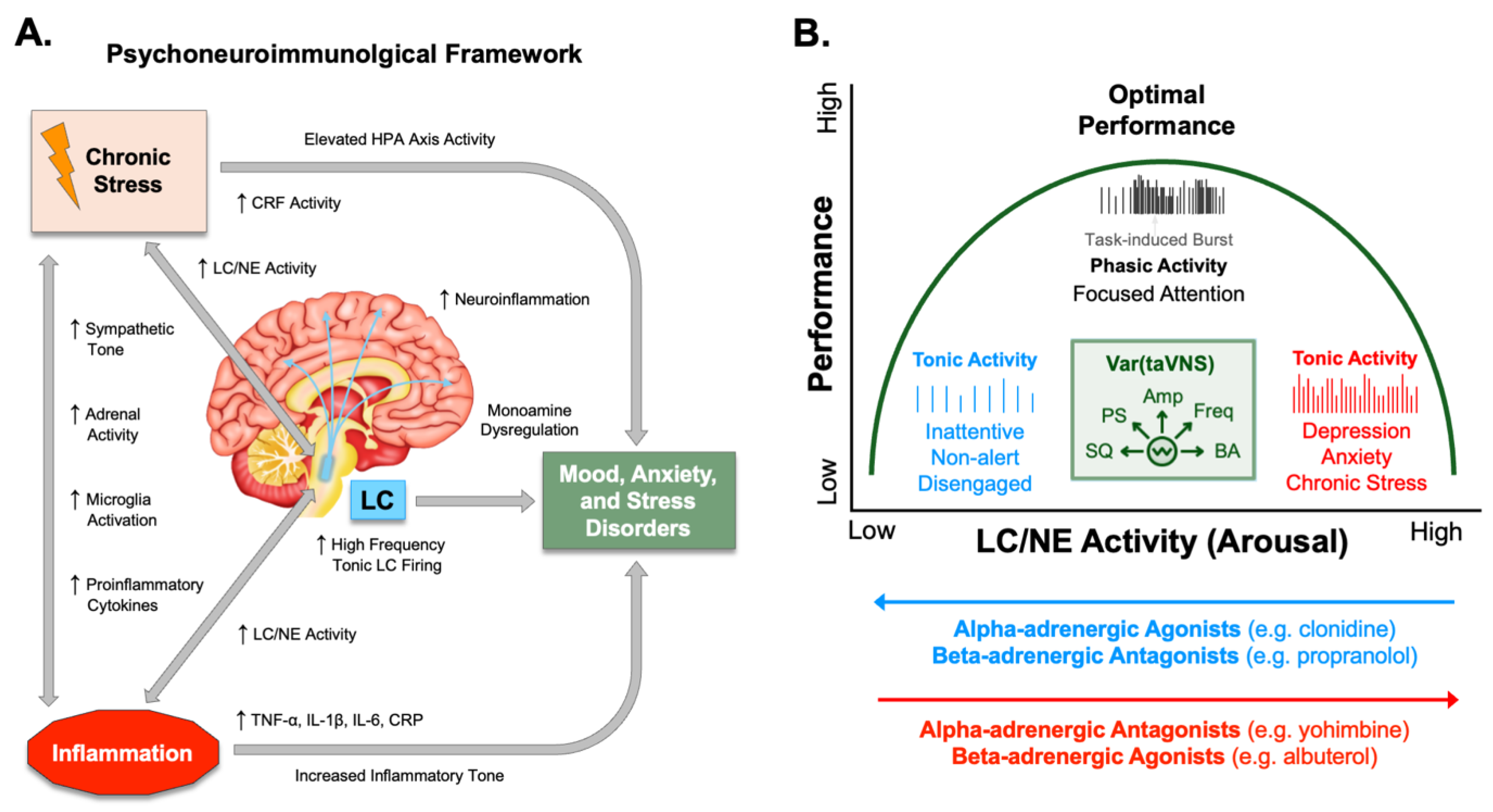
Disclaimer/Publisher’s Note: The statements, opinions and data contained in all publications are solely those of the individual author(s) and contributor(s) and not of MDPI and/or the editor(s). MDPI and/or the editor(s) disclaim responsibility for any injury to people or property resulting from any ideas, methods, instructions or products referred to in the content. |
© 2025 by the authors. Licensee MDPI, Basel, Switzerland. This article is an open access article distributed under the terms and conditions of the Creative Commons Attribution (CC BY) license (http://creativecommons.org/licenses/by/4.0/).
Copyright: This open access article is published under a Creative Commons CC BY 4.0 license, which permit the free download, distribution, and reuse, provided that the author and preprint are cited in any reuse.


