Submitted:
10 November 2025
Posted:
11 November 2025
You are already at the latest version
Abstract
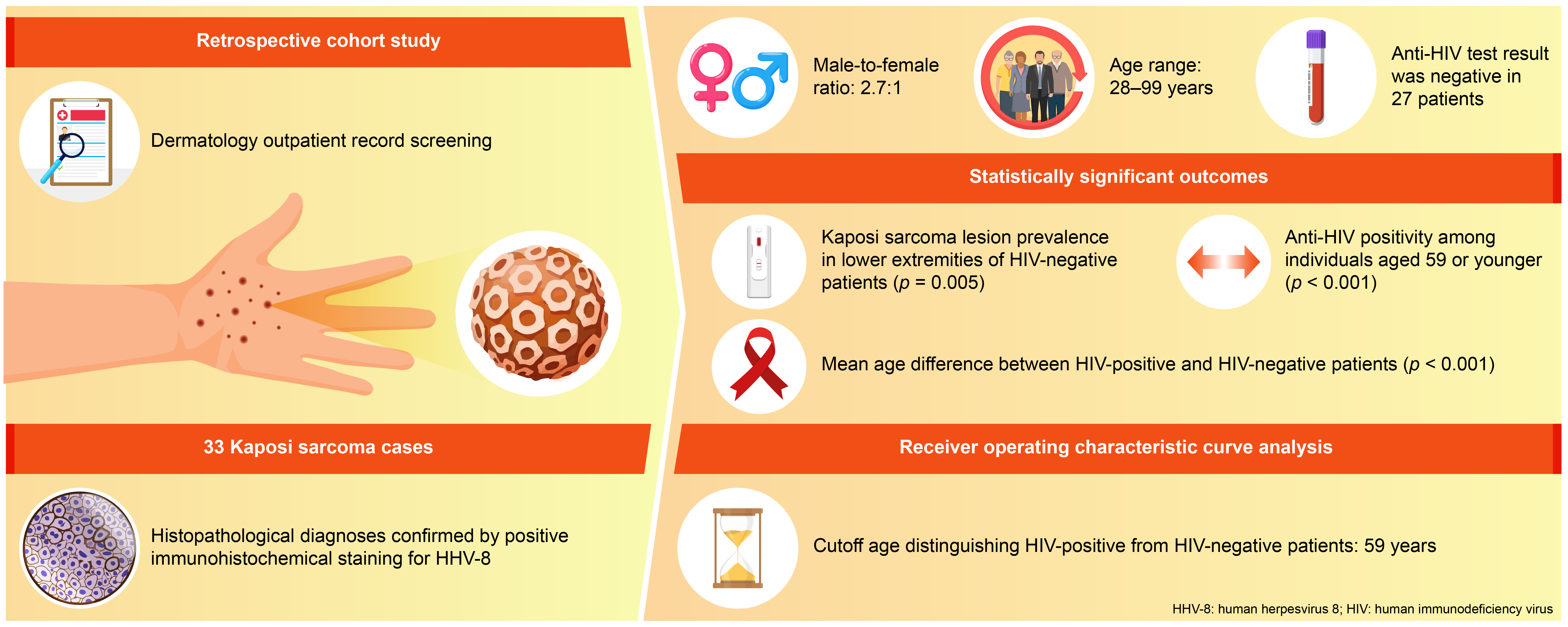
(1) Background: Studying the incidence of Kaposi sarcoma in relation to key variables can guide targeted research and subtype-specific clinical interventions. (2) Methods: We reviewed the records of all patients who visited our hospital’s dermatology outpatient clinic, and patients who were clinically and histopathologically diagnosed with Kaposi sarcoma were included in the study. The age, gender, lesion location, anti-HIV test results, and comorbidities of the patients were recorded. (3) Results: Thirty-three patients with Kaposi sarcoma were identified. Male-female ratio was 2.7:1. The Kaposi sarcoma lesions were statistically significantly more prevalent in the lower extremities of HIV-negative patients (p=0.005). Receiver Operating Characteristic (ROC) curve analysis identified 59 years as the optimal age cutoff for distinguishing between HIV-positive and HIV-negative patients. Anti-HIV positivity was significantly higher in individuals aged 59 and younger compared to those aged 60 and older (p < 0.001). (4) Conclusions: To the best of our knowledge, this is the first study to demonstrate a statistically significant higher prevalence of lower extremity lesions among HIV-negative patients and to identify 59 years as the optimal age cutoff for distinguishing between HIV-positive and HIV-negative Kaposi sarcoma patients using ROC curve analysis. The age-related patterns observed in this study warrant further investigation.
Keywords:
Kaposi sarcoma
; HIV
; age
; epidemiology
; comorbidity
1. Introduction
Kaposi sarcoma, first described by Hungarian dermatologist Moritz Kaposi in 1872, is a multifocal, endothelial proliferation of low-grade malignant potential caused by human herpesvirus 8 (HHV-8), most often with cutaneous involvement with or without visceral extension. There are five distinct subtypes: classic, endemic, iatrogenic, AIDS (acquired immunodeficiency syndrome) associated (epidemic) and the newest subtype arising in MSM (men who have sex with men) without HIV (human immunodeficiency virus) infection. Cutaneous lesions are typically dark blue or purple macules, papules or plaques [1]. Bullous Kaposi sarcoma has also been reported as an uncommon variant [2]. Lymph nodes, mucosae and viscera may be involved without skin involvement [1]. Kaposi sarcoma can also rarely involve the nail unit [3]. Although HHV-8 is considered the causative agent, multiple co-factors are required, the most powerful of which is HIV co-infection, which elevates the risk up to 20,000-fold. All forms of Kaposi sarcoma are associated with HHV-8 infection as the etiological agent. Discovered in 1994 and originally termed as Kaposi sarcoma herpesvirus, this gammaherpesvirus is easily transmitted through saliva and blood products [1]. Transmission of the virus is sexual in MSM. HHV-8 may also be transmitted by solid organ transplantation [4]. The genetic basis for the ethnogeographic predisposition of Kaposi sarcoma in the classic and endemic subtypes is unclear at present. The contribution of additional exposures has been suggested. These include exposure to quinine, nitrile inhalants, angiotensin-converting enzyme inhibitors and volcanic soil silicates [1]. SARS-CoV-2 (severe acute respiratory syndrome coronovirus 2) infection has also been reported as a possible etiological factor [5,6]. Management of Kaposi sarcoma depends on the clinical subtype. In localized cutaneous disease, excision and cryotherapy can be used. Kaposi sarcoma is highly radiosensitive with complete responses in up to 93% of patients. Liposomal doxorubicin and paclitaxel are approved by the United States Food and Drug Administration as first line and second line treatments, respectively, for advanced Kaposi sarcoma. There are also reports of responses to imatinib, sorafenib, bortezomib, nivolumab and pembrolizumab [1]. Intralesional cidofovir, intralesional vinblastine, intralesional vincristine, photodynamic therapy, Nd-YAG (neodymium-doped yttrium aluminium garnet) laser, topical sirolimus, topical imiquimod, topical alitretinoin, electrochemotherapy, topical and oral propranolol, acitretin and metformin have also been reported as successful treatments in the literature [7,8,9,10,11,12,13,14,15]. Interferon alfa, although effective in some patients, tends to be associated with lower response rates than paclitaxel and liposomal doxorubicin [16]. Investigating the incidence of Kaposi sarcoma according to various factors can contribute to more targeted research and intervention. This study investigates the epidemiological and clinical features of patients with Kaposi sarcoma at a major teaching hospital in Istanbul. The aim of of this study is to examine the incidence and clinical characteristics of different forms of Kaposi sarcoma, with particular emphasis on age patterns and HIV serostatus.
2. Materials and Methods
A retrospective cohort study design was used. The outpatient records of all patients who visited the dermatology outpatient clinic of our hospital between October 2016 and March 2025 were reviewed, and patients who were clinically and histopathologically diagnosed with Kaposi sarcoma were included in the study. Our hospital was a military hospital before October 2016, but it became a state hospital in October 2016 and currently serves as a tertiary referral center. For this reason, outpatient clinic records have been available for review since October 2016. Outpatient clinic records were screened for Kaposi sarcoma using the ICD (International Classification of Diseases) code C46. The age, gender, lesion location, anti-HIV test results, and comorbidities of patients diagnosed with Kaposi sarcoma were recorded. All of the patients in this study had been tested for HIV status using the ELISA (enzyme-linked immunosorbent assay) method, and those with positive ELISA results were confirmed by Western blot analysis. The data obtained from the study were analyzed using IBM SPSS Statistics for Windows, version 30.0 (IBM Corp., Armonk, NY, USA). Descriptive statistics for the sociodemographic and clinical characteristics of patients with Kaposi sarcoma were presented as counts (n), percentages (%), means, and standard deviations. Categorical variables were analyzed using Fisher’s exact test. The Shapiro–Wilk test was used to assess the normality of the age at diagnosis variable, and its association with anti-HIV positivity was evaluated using the Mann–Whitney U test. In addition, to identify the optimal age at diagnosis that could be a predictor for anti-HIV positivity, a receiver operating characteristic (ROC) curve analysis was performed, and the Youden index was used. A p-value of <0.05 was considered statistically significant.
3. Results
All patients who visited our outpatient clinic between October 2016 and March 2025 were screened, and 33 patients with Kaposi sarcoma were identified. These 33 patients underwent a punch biopsy and had been clinically and histopathologically diagnosed with Kaposi sarcoma. Their diagnoses had been confirmed by positive immunohistochemical staining for HHV-8. Patients were screened for metastasis in the medical oncology department. Only 1 patient (3%) had metastasis to gastrointestinal system. This patient was HIV-negative and had been receiving systemic treatment with 16 mg/day methylprednisolone for three months for anti-glomerular basement membrane (anti-GBM) disease of the kidney. The age, gender, anti-HIV test results, lesion locations, and comorbidities of the patients are summarized in Table 1. Twenty-four (72.7%) of the patients were male, and 9 (27.3%) were female. Male-female ratio was 2.7:1. The patients’ ages ranged from 28 to 99, with a median age of 72. The mean age and standard deviation of the patients was 67.61±15.99. The anti-HIV test result was negative in 81.8% of cases (n=27), while it was positive in 18.2% of cases (n=6). Lesion sites were grouped into upper extremities, lower extremities, head, trunk, diffuse skin involvement, and gastrointestinal involvement, and the relationship between anti-HIV test results and lesion sites was examined (Table 2). This examination revealed that lesions were statistically significantly more prevalent in the lower extremities of HIV-negative patients (p=0.005). Additionally, HIV-positive patients were statistically significantly more likely to have widespread skin involvement (p=0.004) and trunk involvement (p=0.028). No statistically significant difference was found between gender and lesion location (p=0.999). In 24.2% of cases (n=8), no comorbidities were present, while at least one comorbidity was present in 75.8% of cases (n=25). The comorbidities of the patients are summarized graphically in Figure 1. There was a statistically significant difference in the mean age between HIV-positive and HIV-negative patients (p<0.001) (Figure 2). Receiver operating characteristic (ROC) curve analysis shows that we can determine 59 years as the cutoff value for age between HIV-positive and HIV-negative patients (Figure 3). When we divided the groups according to the optimal cutoff value calculated from the ROC curve and analyzed them, anti-HIV positivity was found to be significantly higher among individuals aged 59 or younger than among those aged 60 or older (p<0.001) (Table 3).
4. Discussion
The current study is a retrospective analysis of 33 patients with Kaposi sarcoma at our institution between the years 2016 and 2025. Twenty-four (72.7%) of the patients were male, and 9 (27.3%) were female. The predominance of men in this cohort is in line with the literature. Kaposi sarcoma can occur at any age and in both sexes but is more common in adult men. According to Globocan 2022 data, the incidence of Kaposi sarcoma is 0.5 in men and 0.3 in women per 100,000 people [17]. In our study, male-female ratio is 2.7:1. The overall male-female ratio in patients with Kaposi sarcoma appears to range from 2.5:1 to 9:1 [16]. The prevalence of Kaposi sarcoma is higher in men than in women, likely due to various contributing factors like hormonal, viral, and genetic factors and high-risk behaviour [18]. The predominance of males may additionally be explained by the role of sex steroids on the immune system and that females have higher levels of circulating IgG, IgM, and CD4+ T cells than men [19]. In our study, all of the six HIV-positive patients were male which is likely due to high-risk behaviour in men. This finding is consistent with that reported in a study from Taiwan [20]. The mean age and standard deviation of the patients in our study was 67.61±15.99. This result is quite similar to that of another study conducted in our country, in which the mean age was found to be 68.42 ± 12.80 years [19]. The study conducted in Taiwan found a lower mean age of 58.7±21.2 [20]. The median age of our patients was 72. A study conducted in Finland found that the median age of patients was 44.5 years old [21]. In another study conducted in China, the median age was found to be 60 [22]. Both the mean age and median age of our patients were found to be higher than in other countries in different geographical regions. The reason for this is probably that, as our country is a Mediterranean country, classic Kaposi sarcoma, which is usually seen in older age groups, is more common in our country than other subtypes. Classic Kaposi sarcoma occurs predominantly in men of Eastern European and Mediterranean origin [15]. In our study, a statistically significant higher prevalence of lesions was observed in the lower extremities among HIV-negative patients (p=0.005). Previous retrospective studies on Kaposi sarcoma have reported the lower extremities as the most common site of lesions [19,21,23]. The study conducted in Taiwan found that HIV-positive patients had a lower incidence of lower extremity lesions compared to HIV-negative patients [20]. To the best of our knowledge, this is the first study to demonstrate a statistically significant higher prevalence of lesions in the lower extremities among HIV-negative patients. Why Kaposi sarcoma is common on the lower limbs is not clearly understood [23]. It has been suggested that lymphedema may contribute to tumorigenesis via local immunosuppression [19]. Some environmental factors such as exposure to volcanic soils have been hypothesized in the pathogenesis of Kaposi sarcoma. Chronic exposure of the skin to iron or alumino-silicate might induce localized immune dysfunction which might add additional explanation to the topography of the lesions at the extremities of the body [24]. In our study, a statistically significant association was observed between HIV-positive status and widespread skin lesion distribution in Kaposi sarcoma patients. This finding was also reported in the study conducted in China [22]. HIV-positive patients in our study were statistically significantly more likely to have trunk involvement which was also observed in the study conducted in Taiwan [20]. No statistically significant difference was found between gender and lesion location. The same finding was observed in a study conducted in Cameroon [25]. The histopathological diagnoses of the patients had been confirmed by positive immunohistochemical staining for HHV-8. Immunohistochemically, monoclonal antibodies against LANA (latency-associated nuclear antigen) are routinely used for specific HHV-8 detection. HHV-8 is transmitted primarily via saliva, but also sexually, vertically, and via blood products [15]. The role of HHV-8 in the pathogenesis of Kaposi sarcoma appears to be related to some of the proteins produced by the virus. HHV-8 gene encodes proteins that inhibit retinoblastoma and p53 tumor suppressor genes. HHV-8 viral interferon regulatory factor prevents interferon from respressing the c-myc oncogene. The cytokine, viral interleukin-6, produced by HHV-8 causes increased expression of vascular endothelial growth factor [26]. Thus, HHV-8 supports both angiogenesis and cell proliferation. Other factors affecting tumorigenesis include hypoxia, epigenetic modifications, immunosuppression, and hyperglycemia [22]. In HIV-infected cases, the Tat protein of HIV virus induces various cytokines that synergistically interact with the products of HHV-8 that may account for the development and progression of Kaposi sarcoma [27]. Kaposi sarcoma in people living with HIV is considered a sign that HIV has developed into AIDS [28]. But some HIV patients can develop Kaposi sarcoma while having a normal CD4-cell count [21]. HIV contributes to the pathogenesis of Kaposi sarcoma by inducing the immunosuppression necessary for the clinical expression of opportunistic disease [23]. In our study, no comorbidities were observed in 24.2% of our cases (n = 8), while 75.8% of our cases (n = 25) presented with at least one comorbidity. The most common comorbidity in our study was hypertension (n=8, 24.2%), followed by atherosclerosis (n=7, 21.2%). Hypertension is probably the most common comorbidity in our study because the mean age of our patients is over 65. A study found that blood pressure increases with age, with an average increase of 6.4 mmHg in systolic blood pressure per decade [29]. The second most common comorbidity in our study was atherosclerosis. Atherosclerosis may have contributed to the development of Kaposi sarcoma by affecting tumorigenesis through hypoxia. In six patients (18.2%), a second primary malignancy had been diagnosed before the onset of Kaposi sarcoma. Two patients had a history of prostate carcinoma. One patient had a history of both papillary thyroid carcinoma and glioblastoma. Another patient had a history of renal cell carcinoma, one had diffuse large B-cell lymphoma, and one had basal cell carcinoma. All six patients were male, HIV-negative, and aged 69 years or older. The prevalence of second primary malignancies in patients with Kaposi sarcoma ranges from 8.6% to 40% in the literature [21]. An 18.2% occurrence of second primary malignancies was observed in our cohort, consistent with existing literature. The literature highlights a notable association between Kaposi sarcoma and second primary malignancies, particularly those affecting the lymphoreticular system [30]. In our study, three patients had type 2 diabetes mellitus, and three had psoriasis. The relationship between Kaposi sarcoma and psoriasis is controversial. According to one study, the risk of Kaposi sarcoma among patients with psoriasis is comparable to that in the general population, with no apparent interaction between the two conditions [31]. But diabetes mellitus has been identified in the literature as an important predisposing factor for the development of Kaposi sarcoma, with the high-glucose microenvironment suggested as a contributing mechanism [32]. In our study, two patients had undergone kidney transplantation, two had chronic renal failure, two had a history of syphilis, and two had chronic hepatitis B. Single-occurrence comorbidities observed in the cohort included Crohn’s disease, glaucoma, allergic rhinitis, condyloma acuminatum, anogenital herpes, heart failure, rheumatoid arthritis, chronic peripheral venous insufficiency, Parkinson’s disease, and anti-glomerular basement membrane kidney disease. The patients diagnosed with syphilis, condyloma acuminatum, and anogenital herpes were concurrently HIV-positive. The literature describes a case of disseminated Kaposi sarcoma in an HIV-negative individual with chronic hepatitis B infection [20]. Reports in the literature have also documented cases of Kaposi sarcoma in patients undergoing hemodialysis or peritoneal dialysis for chronic renal failure [33,34]. However, our patients with chronic renal failure were not undergoing hemodialysis or peritoneal dialysis. No documented association exists between Parkinson’s disease, glaucoma, allergic rhinitis, and Kaposi sarcoma. Chronic peripheral venous insufficiency may have contributed to the development of Kaposi sarcoma through chronic lymphedema, whereas heart failure may have contributed through tissue hypoxia. Iatrogenic Kaposi sarcoma was first described in 1978 in a renal transplant recipient and was found to be related to reactivation of HHV-8 in immunosuppressed transplant patients [19,24]. Additionally, HHV-8 may be transmitted through donor organs [24]. Kaposi sarcoma has been reported to be 200 times more frequent in transplant recipients than in the general population. In the cases of non-transplant-related iatrogenic Kaposi sarcoma, the most common causative drugs are corticosteroids and cyclosporine [16]. Typically, steroid-associated immunosuppression has been considered when the dose exceeds 20 mg prednisone/day, particularly in chronic use [35]. In our study, all HIV-positive patients were aged 54 years or younger. Only three HIV-negative patients were under the age of 54, two of whom had undergone kidney transplantation, and one of whom had been receiving 16 mg/day of methylprednisolone which is equivalent to 20 mg/day of prednisone for three months to treat anti-GBM kidney disease. These three patients may be classified as having iatrogenic Kaposi sarcoma. The patient with Crohn’s disease was a 68-year-old HIV-negative man receiving treatment with mesalazine and azathioprine. The patient with rheumatoid arthritis was an 82-year-old HIV-negative woman receiving treatment with 12.5 mg/week methotrexate and 5 mg/day prednisolone. However, whether these two patients should be classified as having classic or iatrogenic Kaposi sarcoma remains a matter of debate. Therefore, further studies are needed to clearly identify which immunosuppressive drugs and dosages are associated with the development of non–transplant-related iatrogenic Kaposi sarcoma. In our study, a statistically significant difference in the mean age was observed between HIV-positive and HIV-negative patients (p < 0.001). A similar finding was reported by Chalya et al., who observed that patients with AIDS-associated Kaposi sarcoma were significantly younger than those with HIV-negative disease [23]. In the study by Hong and Lee, all HIV-positive patients were under 60 years of age, whereas the HIV-negative group included individuals aged 60 years and older; this difference was statistically significant [27]. All HIV-positive patients in our study were aged 54 years or younger; however, ROC curve analysis identified 59 years as the optimal age cutoff for distinguishing between HIV-positive and HIV-negative patients, demonstrating excellent discriminatory ability (AUC = 0.951), with a sensitivity of 88.9% and specificity of 100.0%. When patients were stratified based on the optimal age cutoff of 59 years identified by ROC curve analysis, anti-HIV positivity was significantly more prevalent in the group aged 59 years or younger compared to those aged 60 years or older (p < 0.001). To the best of our knowledge, this is the first study to identify 59 years as the optimal age cutoff for distinguishing between HIV-positive and HIV-negative patients using ROC curve analysis. This suggests that age may serve as a clinically useful indicator in differentiating between these patient populations. Kaposi sarcoma occurring in individuals aged 60 years and older may, in the future, be classified as a geriatric condition associated with immunosenescence. While the absence of HIV infection may be presumed in such cases, routine HIV testing should not be omitted without sufficient supporting evidence. Further research is needed to confirm this association. The observed age-related patterns warrant further investigation. The retrospective design and modest sample size represent limitations of the current study; nonetheless, the findings offer a foundation for future research in this area.
5. Conclusions
Kaposi sarcoma, first described by Hungarian dermatologist Moritz Kaposi in 1872, is a multifocal, endothelial proliferation of low-grade malignant potential caused by human herpesvirus 8 (HHV-8), most often with cutaneous involvement with or without visceral extension. All forms of Kaposi sarcoma are associated with HHV-8 infection as the etiological agent but not all forms of Kaposi sarcoma are associated with HIV infection. There are five distinct subtypes of Kaposi sarcoma: classic, endemic, iatrogenic, AIDS associated (epidemic) and the newest subtype arising in MSM without HIV infection. This research advances current knowledge by demonstrating that 59 years as the optimal age cutoff for distinguishing between HIV-positive and HIV-negative Kaposi sarcoma patients using ROC curve analysis. Anti-HIV positivity was significantly more prevalent in the group aged 59 years or younger compared to those aged 60 years or older (p < 0.001). The observed age-related patterns warrant further investigation.
Author Contributions
The research and writing presented in this work are entirely the author’s own, without contributions from other authors.
Funding
This research received no external funding.
Institutional Review Board Statement
The study was conducted in accordance with the Declaration of Helsinki, and the protocol was approved by the Ethics Committee of Istanbul Medipol University (E-10840098-202.3.02-3368) on 28 May 2025.
Informed Consent Statement
Informed consent was waived, because it is not required per local legislation as the study retrospectively used routine laboratory data in which patient identities were not disclosed.
Data Availability Statement
The original contributions presented in this study are included in the article in Table 1. Further inquiries can be directed to the corresponding author.
Acknowledgments
The author thanks biostatistician Alican Sarısaltık for conducting the statistical analyses for this study.
Conflicts of Interest
The author declares no conflicts of interest.
Abbreviations
The following abbreviations are used in this manuscript:
| HHV-8 | Human herpesvirus 8 |
| AIDS | Acquired immunodeficiency syndrome |
| MSM | Men who have sex with men |
| HIV | Human immunodeficiency virus |
| Sars-CoV-2 | Severe acute respiratory syndrome coronavirus 2 |
| Nd-YAG | Neodymium-doped yttrium aluminium garnet |
| ICD | International classification of diseases |
| ELISA | Enzyme-linked immunosorbent assay |
| ROC | Receiver operating characteristic |
| DM | Diabetes mellitus |
| AUC | Area under the curve |
| CI | Confidence interval |
| LANA | Latency-associated nuclear antigen |
| Anti-GBM | Anti-glomerular basement membrane |
References
- Tsai, K.Y. : West Sussex, Great Britain, 2024; Volume 4, pp. 138.1-138.6.Sarcoma. In Rook’s Textbook of Dermatology, 10thed.; Griffiths, C., Barker, J., Bleiker, T., Hussain, W., Simpson, R., Eds.; Wiley Blackwell: West Sussex, Great Britain, 2024; Volume 4, p. 138. [Google Scholar]
- Gherardi, E.; Tinunin, L.; Grassi, T.; Maio, V.; Grandi, V. Bullous Kaposi Sarcoma: An Uncommon Blistering Variant in an HIV-Negative Patient. Dermatol. Pract. Concept. 2024, 14, e2024113. [Google Scholar] [CrossRef]
- Krygier, J.; Sass, U.; Meiers, I.; Marneffe, A.; de Vicq de Cumptich, M.; Richert, B. Kaposi Sarcoma of the Nail Unit: A Case Report and Review of the Literature. Skin. Appendage. Disord. 2023, 9, 465–469. [Google Scholar] [CrossRef]
- Marcoval, J.; Bonfill-Ortí, M.; Martínez-Molina, L.; Valentí-Medina, F.; Penín, R.M.; Servitje, O. Evolution of Kaposi sarcoma in the past 30 years in a tertiary hospital of the European Mediterranean basin. Clin. Exp. Dermatol. 2019, 44, 32–39. [Google Scholar] [CrossRef]
- Magri, F.; Giordano, S.; Latini, A.; Muscianese, M. New-onset cutaneous kaposi’s sarcoma following SARS-CoV-2 infection. J. Cosmet. Dermatol. 2021, 20, 3747–3750. [Google Scholar] [CrossRef]
- Gardini, G.; Odolini, S.; Moioli, G.; Papalia, D.A.; Ferrari, V.; Matteelli, A.; Caligaris, S. Disseminated Kaposi sarcoma following COVID-19 in a 61-year-old Albanian immunocompetent man: a case report and review of the literature. Eur. J. Med. Res. 2021, 26, 152. [Google Scholar] [CrossRef]
- Fenner, B.; Burningham, K.; Alkul, M.; Tyring, S. Treatment of Kaposi sarcoma with intralesional cidofovir in an HIV-negative man. JAAD Case Rep. 2023, 42, 47–48. [Google Scholar] [CrossRef]
- Shiryaev, A.A.; Efendiev, K.T.; Kornev, D.O.; Samoylova, S.I.; Fatyanova, A.S.; Karpova, R.V.; Reshetov, I.V.; Loschenov, V.B. Photodynamic therapy of classic Kaposi’s sarcoma with video-fluorescence control. Photodiagnosis Photodyn. Ther. 2021, 35, 102378. [Google Scholar] [CrossRef]
- Nasca, M.R.; Luppino, I.; Spurio Catena, A.; Micali, G. Nodular Classic Kaposi’s Sarcoma Treated With Neodymium-Doped Yttrium Aluminum Garnet Laser Delivered Through a Tilted Angle: Outcome and 12-Month Follow Up. Lasers Surg. Med. 2020, 52, 979–983. [Google Scholar] [CrossRef] [PubMed]
- Tancredi, V.; Licata, G.; Buononato, D.; Boccellino, M.P.; Argenziano, G.; Giorgio, C.M. Topical Sirolimus 0.1% as Off-Label Treatment of Kaposi’s Sarcoma. Dermatol. Pract. Concept. 2024. [Google Scholar] [CrossRef]
- Giorgio, C.M.R.; Licata, G.; Briatico, G.; Babino, G.; Fulgione, E.; Gambardella, A.; Alfano, R.; Argenziano, G. Comparison between propranolol 2% cream versus timolol 0.5% gel for the treatment of Kaposi sarcoma. Int. J. Dermatol. [CrossRef]
- Genedy, R.M.; Owais, M.; El Sayed, N.M. Propranolol: A Promising Therapeutic Avenue for Classic Kaposi Sarcoma. Dermatol. Pract. Concept. 2025, 15, 4737. [Google Scholar] [CrossRef] [PubMed]
- Daadaa, N.; Souissi, A.; Chaabani, M.; Chelly, I.; Ben Salem, M.; Mokni, M. Involution of classic Kaposi sarcoma lesions under acitretin treatment Kaposi sarcoma treated with acitretin. Clin. Case Rep. 2020, 8, 3340–3343. [Google Scholar] [CrossRef] [PubMed]
- Su, T.; Yu, Y.; Su, Z.; Lu, Y. Rapid improvements in Kaposi sarcoma with metformin: a report of two cases. Br. J. Dermatol. 2025, 193, 175–176. [Google Scholar] [CrossRef]
- Esser, S.; Schöfer, H.; Hoffmann, C.; Claßen, J.; Kreuter, A.; Leiter, U.; Oette, M.; Becker, J.C.; Ziemer, M.; Mosthaf, F.; Sirokay, J.; Ugurel, S.; Potthoff, A.; Helbig, D.; Bierhoff, E.; Schulz, T.F.; Brockmeyer, N.H.; Grabbe, S. S1 Guidelines for the Kaposi Sarcoma. J. Dtsch. Dermatol. Ges. 2022, 20, 892–904. [Google Scholar] [CrossRef]
- Bettuzzi, T.; Lebbe, C.; Grolleau, C. Modern Approach to Manage Patients With Kaposi Sarcoma. J. Med. Virol. 2025, 97, e70294. [Google Scholar] [CrossRef]
- Kavak, E.E.; Ürün, Y. Classical Kaposi sarcoma: an insight into demographic characteristics and survival outcomes. BMC Cancer. 2025, 25, 690. [Google Scholar] [CrossRef]
- Balasubramanian, D.; Srinivasan, S.; Paul, P.M.; Ko, N.; Garlapati, S. A Retrospective Study on the Incidence of Kaposi Sarcoma in the United States From 1999 to 2020 Using the Centers for Disease Control and Prevention Wide-Ranging Online Data for Epidemiological Research (CDC WONDER) Database. Cureus. 2025, 17, e77213. [Google Scholar] [CrossRef] [PubMed]
- Yazici, S.; Zorlu, O.; Bulbul Baskan, E.; Balaban Adim, S.; Aydogan, K.; Saricaoglu, H. Retrospective Analysis of 91 Kaposi’s Sarcoma Cases: A Single-Center Experience and Review of the Literature. Dermatology. 2018, 234, 205–213. [Google Scholar] [CrossRef] [PubMed]
- Huang, A.Y.; Lin, C.L.; Chen, G.S.; Hu, S.C. Clinical features of Kaposi’s sarcoma: experience from a Taiwanese medical center. Int. J. Dermatol. 2019, 58, 1388–1397. [Google Scholar] [CrossRef]
- Kluger, N.; Blomqvist, C.; Kivelä, P. Kaposi sarcoma in Southern Finland (2006-2018). Int. J. Dermatol. 2019, 58, 1258–1263. [Google Scholar] [CrossRef]
- Yang, H.L.; He, F.; Jielili, A.; Zhang, Z.R.; Cui, Z.Y.; Wang, J.H.; Guo, H.T. A retrospective study of Kaposi’s sarcoma in Hotan region of Xinjiang, China. Medicine (Baltimore). 2023, 102, e35552. [Google Scholar] [CrossRef] [PubMed]
- Chalya, P.L.; Mbunda, F.; Rambau, P.F.; Jaka, H.; Masalu, N.; Mirambo, M.; Mushi, M.F.; Kalluvya, S.E. Kaposi’s sarcoma: a 10-year experience with 248 patients at a single tertiary care hospital in Tanzania. BMC Res. Notes. 2015, 8, 440. [Google Scholar] [CrossRef]
- Grabar, S.; Costagliola, D. Epidemiology of Kaposi’s Sarcoma. Cancers (Basel). 2021, 13, 5692. [Google Scholar] [CrossRef]
- Tounouga, D.N.; Kouotou, E.A.; Nansseu, J.R.; Zoung-Kanyi Bissek, A.C. Epidemiological and Clinical Patterns of Kaposi Sarcoma: A 16-Year Retrospective Cross-Sectional Study from Yaoundé, Cameroon. Dermatology. 2018, 234, 198–204. [Google Scholar] [CrossRef]
- Hong, A.; Lee, C.S. The emerging role of the human herpesvirus 8 (HHV8) in human neoplasia. Pathology. 2001, 33, 461–468. [Google Scholar] [CrossRef]
- Hong, A.; Lee, C.S. Kaposi’s sarcoma: clinico-pathological analysis of human immunodeficiency virus (HIV) and non-HIV associated cases. Pathol. Oncol. Res. 2002, 8, 31–35. [Google Scholar] [CrossRef]
- Fu, L.; Tian, T.; Wang, B.; Lu, Z.; Gao, Y.; Sun, Y.; Lin, Y.F.; Zhang, W.; Li, Y.; Zou, H. Global patterns and trends in Kaposi sarcoma incidence: a population-based study. Lancet Glob. Health. 2023, 11, e1566–e1575. [Google Scholar] [CrossRef]
- The China PEACE Collaborative Group. Association of age and blood pressure among 3.3 million adults: insights from China PEACE million persons project. J. Hypertens. 2021, 39, 1143–1154. [Google Scholar] [CrossRef] [PubMed]
- Safai, B.; Miké, V.; Giraldo, G.; Beth, E.; Good, R.A. Association of Kaposi’s sarcoma with second primary malignancies: possible etiopathogenic implications. Cancer. 1980, 45, 1472–1479. [Google Scholar] [CrossRef] [PubMed]
- Brambilla, L.; Genovese, G.; Tourlaki, A.; Della Bella, S. Coexistence of Kaposi’s sarcoma and psoriasis: is there a hidden relationship? Eur. J. Dermatol. 2018, 28, 320–325. [Google Scholar] [CrossRef]
- Chang, P.J.; Yang, Y.H.; Chen, P.C.; Chen, L.W.; Wang, S.S.; Shih, Y.J.; Chen, L. Y,.; Chen, C.J., Hung, C.H., Eds.; Lin, C.L. Diabetes and risk of Kaposi’s sarcoma: effects of high glucose on reactivation and infection of Kaposi’s sarcoma-associated herpesvirus. Oncotarget. 2017, 8, 80595-80611. [Google Scholar] [CrossRef]
- Tugcu, M.; Kasapoglu, U.; Ruhi, C.; Sayman, E. Kaposi Sarcoma in a Chronic Renal Failure Patient Treated by Hemodialysis. Turk. Neph. Dial. Transpl. 2016, 25, 136–138. [Google Scholar] [CrossRef]
- Lee, D.; Chun, J.S.; Hong, S.K.; Kang, M.S.; Seo, J.K.; Koh, J.K.; Sung, H.S. Kaposi sarcoma in a patient with chronic renal failure undergoing dialysis. Ann. Dermatol. 2013, 25, 475–478. [Google Scholar] [CrossRef] [PubMed]
- Cortés, J.A. Anergy and immunosuppression in patients with rheumatoid arthritis. Rev. Colomb. Reumatol. 2021, 28, 1–3. [Google Scholar] [CrossRef]
Figure 1.
Comorbidities observed in patients with Kaposi sarcoma.
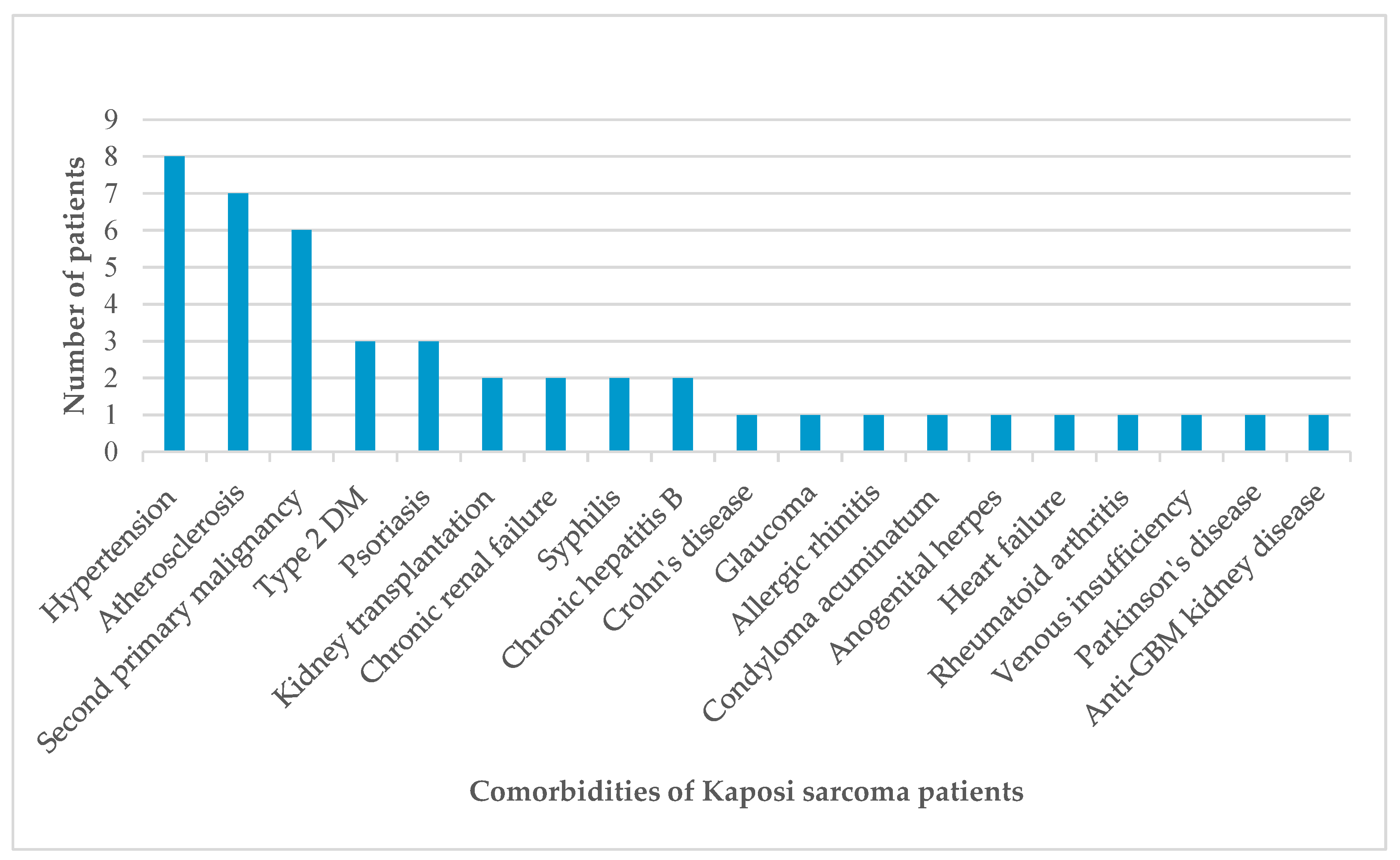
Figure 2.
Comparison of the mean age at diagnosis of Kaposi sarcoma by HIV serostatus.
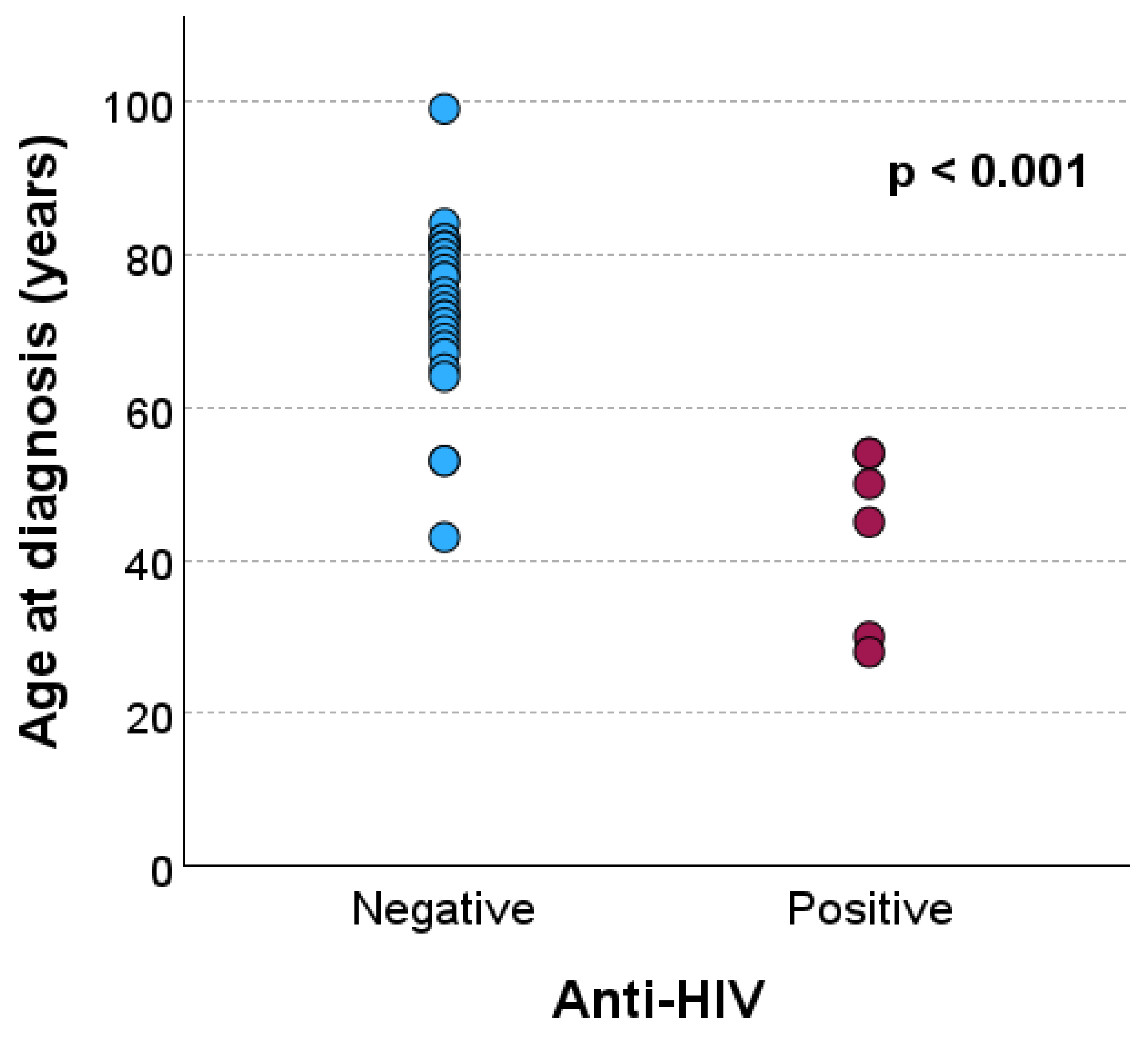
Figure 3.
ROC curve evaluating the performance of age at diagnosis in predicting HIV positivity among patients with Kaposi sarcoma. The area under the curve (AUC) was 0.951 (95% CI: 0.879–1.000), indicating excellent discriminatory ability. The optimal cutoff age, determined using the Youden index (0.889), was 59 years.
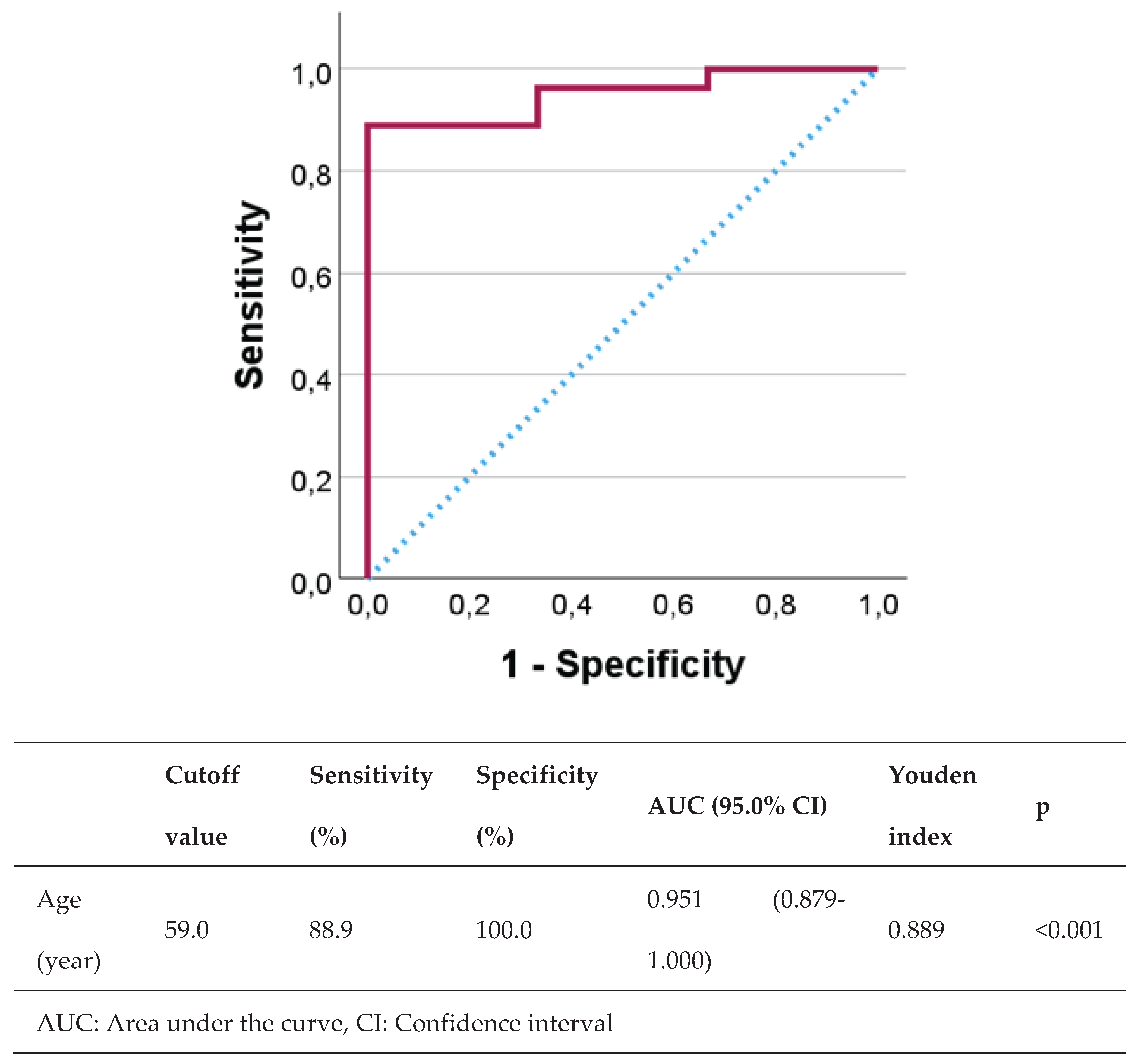

Table 1.
Demographic and clinical features of Kaposi sarcoma patients.
| Case | Age | Gender | Localization | Anti-HIV | Comorbidity |
| 1 | 53 | Male | Bilateral cruris and feet | Negative | Kidney transplant recipient |
| 2 | 82 | Male | Bilateral wrists and feet | Negative | Hypertension, atherosclerosis |
| 3 | 68 | Male | Left hand, right foot | Negative | Hypertension, chronic renal failure, Crohn’s disease |
| 4 | 67 | Female | Left arm, left hand, right foot | Negative | None |
| 5 | 81 | Female | Right ankle | Negative | Hypertension, glaucoma, chronic peripheral venous insufficiency |
| 6 | 65 | Female | Left foot | Negative | Allergic rhinitis |
| 7 | 50 | Male | Right axilla and chest | Positive | Type 2 diabetes mellitus (DM), condyloma acuminatum, anogenital herpes |
| 8 | 75 | Male | Right cruris | Negative | Type 2 diabetes mellitus, atherosclerosis, renal cell carcinoma |
| 9 | 84 | Male | Right foot | Negative | Hypertension, atherosclerosis |
| 10 | 30 | Male | Extensive skin lesions | Positive | None |
| 11 | 45 | Male | Extensive skin lesions | Positive | None |
| 12 | 78 | Male | Left ankle | Negative | Psoriasis |
| 13 | 54 | Male | Extensive skin lesions | Positive | Syphilis |
| 14 | 99 | Female | Bilateral cruris | Negative | Hypertension, atherosclerosis |
| 15 | 71 | Male | Left hand, left foot | Negative | Diffuse large B cell lymphoma, type 2 DM, atherosclerosis, chronic hepatitis B |
| 16 | 81 | Female | Right knee | Negative | Atherosclerosis |
| 17 | 72 | Male | Left elbow | Negative | Psoriasis |
| 18 | 69 | Male | Left arm, bilateral feet | Negative | Thyroid papillary carcinoma, glioblastoma |
| 19 | 70 | Male | Left knee, left labial commissure | Negative | None |
| 20 | 73 | Female | Right arm, right foot | Negative | Hypertension |
| 21 | 79 | Male | Bilateral cruris | Negative | Basal cell carcinoma |
| 22 | 74 | Male | Right thigh | Negative | Prostate carcinoma |
| 23 | 43 | Male | Bilateral cruris | Negative | Kidney transplant recipient |
| 24 | 81 | Male | Right ankle | Negative | Psoriasis |
| 25 | 28 | Male | Left arm, left cruris, trunk | Positive | None |
| 26 | 77 | Male | Bilateral arms and legs | Negative | Parkinson’s disease, hypertension, prostate carcinoma |
| 27 | 54 | Male | Left cruris, left ankle | Positive | Syphilis |
| 28 | 72 | Female | Right foot | Negative | None |
| 29 | 53 | Male | Bilateral feet, gastric antrum and corpus | Negative | Anti-glomerular basement membrane kidney disease |
| 30 | 82 | Female | Left cruris | Negative | Chronic hepatitis B, rheumatoid arthritis |
| 31 | 64 | Female | Left arm | Negative | None |
| 32 | 80 | Male | Right foot | Negative | None |
| 33 | 77 | Male | Right ankle | Negative | Hypertension, atherosclerosis, chronic renal failure, heart failure |
Table 2.
Anti-HIV test results and lesion locations.
| Anti-HIV | p | |||
| Negative | Positive | |||
| Age (year), mean ± standard deviation | 73.0 ± 11.2 | 43.5 ± 11.7 | <0.001a | |
| Age (year), n (%) | ≤59 | 3 (33.3) | 6 (66.7) | <0.001b |
| ≥60 | 24 (100.0) | 0 (0.0) | ||
| a Mann-Whitney U test; b Fisher’s exact test | ||||
Table 3.
The relationship between HIV positivity and age at diagnosis with Kaposi sarcoma.
| Anti-HIV | p* | ||
| Negative, n (%) | Positive, n (%) | ||
| Lower extremities | 25 (92.6) | 2 (33.3) | 0.005 |
| Upper extremities | 9 (33.3) | 1 (16.7) | 0.640 |
| Trunk | 0 (0.0) | 2 (33.3) | 0.028 |
| Head | 1 (3.7) | 0 (0.0) | 0.999 |
| Diffuse skin involvement | 0 (0.0) | 3 (50.0) | 0.004 |
| Gastrointestinal system | 1 (3.7) | 0 (0.0) | 0.999 |
| *Fisher’s exact test | |||
Disclaimer/Publisher’s Note: The statements, opinions and data contained in all publications are solely those of the individual author(s) and contributor(s) and not of MDPI and/or the editor(s). MDPI and/or the editor(s) disclaim responsibility for any injury to people or property resulting from any ideas, methods, instructions or products referred to in the content. |
© 2025 by the author. Licensee MDPI, Basel, Switzerland. This article is an open access article distributed under the terms and conditions of the Creative Commons Attribution (CC BY) license (https://creativecommons.org/licenses/by/4.0/).
Copyright: This open access article is published under a Creative Commons CC BY 4.0 license, which permit the free download, distribution, and reuse, provided that the author and preprint are cited in any reuse.


