Preprint
Review

This version is not peer-reviewed.

How to Read a Next-Generation Sequencing Report for AML and MDS? What Hematologists Need to Know

Submitted:

01 December 2025

Posted:

03 December 2025

You are already at the latest version

Abstract

Acute myeloid leukemia (AML) and myelodysplastic neoplasms (MDS) are clonal hematopoietic malignancies in which next-generation sequencing (NGS) has become integral for diagnosis, classification, risk stratification, and measurable residual disease (MRD) monitoring. Traditional cytogenetic and PCR-based assays remain useful, but targeted NGS panels now represent the standard of care, providing rapid and sensitive detection of recurrent gene mutations, structural variants, and gene fusions. Whole-genome, whole-exome, RNA sequencing, and long-read platforms expand the spectrum of detectable alterations, though targeted panels remain most practical for routine diagnostics. Bioinformatic pipelines and quality metrics-including read length, sequencing depth, and coverage-are critical for accurate variant calling, with validation often required for variants of uncertain significance or those near detection thresholds. NGS is now embedded in diagnostic frameworks, including the WHO 2022 and ICC classifications, which incorporate recurrently mutated genes such as TP53, ASXL1, RUNX1, and FLT3. These data inform prognostic models, with ELN-2022 defining adverse-risk AML subgroups for patients treated with intensive chemotherapy, ELN-2024 AML patients treated with less-intensive therapies, and the IPSS-M refining MDS risk categories by integrating mutational data. NGS also enables MRD monitoring, with gene panels and PCR-NGS hybrid approaches (e.g., for FLT3-ITD) showing increasing clinical utility, though standardization is still lacking. Furthermore, diagnostic NGS frequently uncovers germline predisposition syndromes (e.g., DDX41, GATA2), with significant implications for treatment decisions and donor selection in transplantation. In this manuscript, we review the advantages, limitations, and future perspectives of NGS in the clinical management of AML and MDS with a particular emphasis on the biological and technical principles underlying its use in these diseases. Furthermore, we discuss how NGS findings may influence diagnosis, prognostic classification and therapeutic decision-making within current clinical frameworks. Our aim is to provide a comprehensive overview of NGS fundamentals to support clinicians in navigating the increasing complexity of molecular data in daily practice.

Keywords: 
;  ;  ;  ;  ;  

1. Introduction

Acute myeloid leukemia (AML) and myelodysplastic neoplasms (abbreviated MDS) are clonal hematopoietic neoplasms, characterized by at least one somatic mutation in 96% of cases [1]. Although the WHO fifth edition still allows for the diagnosis of AML on the basis of differentiation in cases lacking defining genetic abnormalities, and dysplasia in MDS may still correspond to morphological criteria [2], in both diseases diagnosis increasingly relies on extensive laboratory workups aimed at identifying specific genetic features. In this contest, the classical PCR-based and cytogenetic techniques have been rapidly complemented by next-generation sequencing (NGS). It is now standard of care to perform NGS-based mutational profiling in all patients with AML, with direct implications for treatment selection and prognostication [3,4]. Indeed, NGS enables the simultaneous sequencing of millions of DNA fragments, providing comprehensive insights into the genome structure, genetic variations, gene expression profiles, and epigenetic alterations of neoplastic cells [5]. However, the broad availability and reduced cost of NGS in AML/MDS has also increased the uncertainty for both hematologists and patients who must interpret the expanding volume of molecular information from sequencing reports. Therefore, we conducted this review to support clinicians, similar to previously published guidance for oncologists like ESMO recommendations on the use of NGS in advanced cancers [6,7].

2. NGS Techniques

NGS is based on the principle of massively parallel sequencing, in which millions of DNA fragments are sequenced simultaneously [8]. Different NGS approaches can be applied depending on clinical and research needs, allowing the sequencing of the whole genome, exome, or transcriptome [9]. Whole genome sequencing (WGS) provides a complete picture of genetic variations; whole exome sequencing (WES) focuses on protein-coding regions; RNA-sequencing can be useful for identification of fusion genes, whereas targeted sequencing panels, the most widely used in clinical practice, concentrate on sets of genes recurrently mutated in MDS and AML [10].

2.1. NGS Workflow

The typical NGS workflow involves extraction of DNA or RNA from the sample, library preparation through fragmentation and adaptor ligation (as in hybridization capture-based assays) or target enrichment in amplification-based assays, followed by sequencing [9].
In hybridization capture, DNA is randomly fragmented and sequencing adapters are ligated to the ends. Biotinylated probes are then used to selectively isolate the regions of interest.
In contrast, amplicon sequencing relies on primers specifically designed to amplify specific targets by PCR after which. adapters are added to prepare the amplified products for sequencing.
The choice between these two approaches depends on several factors such as number of targets, workflow complexity and cost per sample.
In both methods, , patient-specific index sequences are incorporated in addition to adapters required for sequencing on the instrument. These indexes allow multiple libraries to be pooled in the same sequencing run, enabling simultaneous analysis of multiple samples, even from different patients [11].
Different platforms rely on distinct sequencing techniques, including sequencing by synthesis, sequencing by hybridization, sequencing by ligation, and electric impedance-based sequence detection [12]. The most widely used in clinical practice is sequencing by synthesis (SBS), employed by Illumina instruments, in which fluorescently labelled nucleotides are incorporated and detected in real time. A semiconductor-based variation of SBS, used by Ion Torrent systems, detects the release of H⁺ ions instead of fluorescence following nucleotide incorporation [13]. Both approaches require prior, amplification of DNA fragments: Illumina platforms use bridge amplification in which fragments are loaded onto a flow cell where they fold over to form bridges, whereas Ion Torrent amplifies fragments after binding them individually to magnetic beads [14].
More recently, long-read sequencing technologies have emerged. Single-molecule real-time sequencing (SMRT) by Pacific Biosciences produces highly accurate “HiFi” long reads, while nanopore sequencing (Oxford Nanopore Technologies) measures changes in ionic current as nucleic acids pass through protein pores, enabling ultra-long reads and real-time analysis [13]. While short-read SBS remains the gold standard for routine AML and MDS diagnostics, long-read platforms are increasingly being explored for the detection of structural variants, fusion genes, and transcript isoforms [9,15]. In these cases, amplification is not required, as nucleic acid fragments can be sequenced directly [11].
Bioinformatic pipelines then align reads to a reference genome, call and annotate variants, and generate interpretable data. Key biological metrics following alignment to a reference genome - excluding run quality-check - include read length, sequencing depth and coverage, duplication rate, limit of detection (LoD), CG-bias, asCG-rich fragments may show a lower coverage, and some other assay-specific parameters that will be shortly described below [9,11,13]. In the clinical setting, results often require validation, and variant interpretation must integrate allele frequency, functional annotation, and clinical relevance [6,9].
In NGS, a read represents the nucleotide sequence obtained from a single DNA or RNA fragment during sequencing. Read length varies by platform, but in clinical oncology and hematology, most targeted DNA and RNA panels produce reads of 50-300 bp (commonly 100-250 bp) sufficient to detect point mutations, small indels, and gene fusions. However, short reads can limit the precise detection of long insertions such as some FLT3 ITD variants, or long deletions like CALR type 1 mutations [12,16,17,18]. Whole-genome sequencing and long-read technologies can generate longer reads, typically in the range of 10–100 kilobases and sometimes several megabases, enabling the detection of structural variants, complex rearrangements, and full-length transcript isoforms [19,20,21]. Technically, reads may also fail to map uniquely, remain unmapped or, in RNA-seq, map to uninformative transcripts[22].
NGS platforms include built-in measures of read quality, ensuring high accuracy for variant detection[23].
The sequencing coverage indicates the proportion of a reference region (genome, exome, transcriptome, or targeted gene panel) that has been sequenced at least once. Insufficient coverage can lead to errors or uncertainty, making it difficult to distinguish between a true genetic finding and a technical artefact, and complicate downstream bioinformatical analysis [15]. Some fragments like CG-rich regions, polyploid genes or very rare variants may require increased sequencing depth or coverage [22].
Sequencing depth refers to the average number of times each nucleotide in a DNA or RNA sample is read during a NGS run, usually expressed as a multiple (e.g., 100x). Higher depth increases confidence in variant detection [22]. For RNA-seq and targeted RNA-sequencing, reporting the total number of reads per sample is essential, as it strongly impacts sensitivity for gene expression analysis, detection of fusion transcripts, and identification of low-abundance isoforms [24]. In human whole-transcriptome (mRNA) RNA-seq , 20–30 million reads per sample are generally sufficient, with 30–50 million reads recommended for higher sensitivity. Detection of low-expression transcripts or detailed splicing may require significantly deeper sequencing, while targeted RNA panels may need as few as 1-3 million reads per sample due to their focused design [24,25,26,27].
Different NGS types achieve different sequencing depths. Generally, WGS provides the sequencing depth that ranges at 30–60× and the exome-only coverage with WES reaches the depth range around 100-200× which may be insufficient for detecting low frequency variants. In contrast, targeted gene panels focus on a smaller genomic region and achieve a higher depth of sequencing (typically at 500–1000×) improving sensitivity for rare variants [28].
The sequencing depth required for reliable detection of low-frequency variants remains a critical issue in NGS diagnostics. The guidelines of the Association for Molecular Pathology and College of American Pathologists (AMP/CAP) recommend a minimum coverage of >250× per amplicon for routine somatic variant detection in targeted oncology panels typically enabling detection of variants with a 5% variant allele frequency (VAF). VAF represents the proportion of reads reporting a specific variant relative to the total number of reads covering this gene locus. However, this threshold does not ensure sensitivity for very low VAFs or applications such as minimal residual disease monitoring [9]. Indeed, several studies demonstrated that substantially higher coverage is needed to confidently detect variants with low VAF: for example, Petrackova et al. showed that detection of a 3% VAF with >99% confidence requires approximately 1,650× coverage,[29], and robust detection of a 2% VAF generally requires sequencing depth near or above 1,000× [28]. Targeted sequencing panels can achieve high sensitivity because selective amplification of specific genomic regions enriches variant-containing loci within the library allowing deeper coverage without excessive sequencing volume. [30].
In addition to sequencing depth and VAF, most NGS protocols require a minimum number of supporting reads—that is, individual sequencing containing the variant allele. This requirement is essential to distinguish true variants from sequencing artifacts, particularly at low VAF. In clinical and research practice, thresholds of around 10-30 supporting reads are commonly applied to ensure reliable variant calling and reduce the number of false positives [31,32]. For fusion detection, at least 3 supporting reads are generally required [31].

2.2. NGS Types in Clinical Practice

Whole genome sequencing (WGS) provides a comprehensive view of genetic variation, capturing both coding and non-coding regions as well as structural variants and copy number alterations, offering unparalleled breadth in genomic analysis. Whole exome sequencing (WES), in contrast, targets only the protein-coding exons—about 1–2.5% of the genome—providing a more cost-effective approach while still detecting a high proportion of clinically relevant variants[12,33,34].
RNA-sequencing (RNA-seq or transcriptome-wide sequencing) is particularly valuable for identifying fusion genes and for capturing gene expression profiles and transcriptomic alterations that may be missed by DNA-based assays [33]. RNA-seq can also be required to define the specific fusion partner of a biologically dominant gene such as in KM2TA rearrangements (e.g., KMT2A::MLLT3, KMT2A::MLLT1, KMT2A::ELL) which carry prognostic significance and cannot be fully resolved with cytogenetic assays such as FISH alone [2].
Finally, targeted sequencing panels—the most widely used in clinical diagnostics—focus on a selected set of genes recurrently mutated in a given disease (typically 20-50 genes for MDS and AML), providing higher coverage and analytic sensitivity at reduced cost and with faster turnaround time [10]. By selectively amplifying regions of interest, targeted panels increase the relative representation of relevant sequences within the library reducing the need for very high overall sequencing depth and allowing detection of variants with relatively low VAF [30,34]. Target panels can detect clinically relevant single nucleotide variants (SNV), small insertions and deletions (indels), copy number alterations, fusion genes, and structural variants supporting diagnosis, providing a more precise prognosis definition and guiding therapeutic choices [9]. Notably, targeted sequencing panel may have a limited sensitivity for detecting large insertions or deletions – such as FLT3 ITD, MLL PTD, and 52bp CALR deletions - which may require more sophisticated bioinformatical approaches or complementary molecular diagnostic assays. This limitation arises from the discrepancy between the large and/or variable size of these alterations and relatively small size of NGS reads (50-300 bp) [17,35,36,37]. However, recent clinical trials suggest that these challenges can be overcome with PCR-NGS-based methods even enabling patient-tailored measurement of FLT3-ITD MRD [38]. On the other hand, genomic regions of high GC content like CEBPA variants may be difficult to amplify during library preparation and may require higher increased sequencing depth to ensure reliable detection [17,39]. However, these problems could also addressed by using long-read NGS targeted panels designed for AML diagnostics [19]. Recent studies have demonstrated high diagnostic sensitivity and strong concordance with traditional analytical methods of targeted NGS panels generate target-enriched libraries with improved amplicon specificity making them suitable for accurate FLT3 ITD and CEBPA testing [9,18,37,40].
Most targeted panels have been modified with addition of new relevant genes according to the most recent WHO/ICC 2022 classifications [41]. The core genes commonly recommended for inclusion in myeloid malignancy panels typically include: NPM1, CEBPA, ASXL1, BCOR, EZH2, RUNX1, SF3B1, SRSF2, STAG2, U2AF1, ZRSR2, TP53, JAK2, CALR, MPL, CSF3R, KIT, FLT3, and IDH1/2 Many panels also frequently incorporate ETV6, KRAS, NRAS, and WT1 [2,18] (Table 1).
However, the numbers of genes (core and/or additional targets) and the specific gene fragments included in targeted panels – such as hotspot mutations, flanking regions, coding or non-coding sequences, or entire gene coverage - can differ substantially between laboratories, particularly in the absence of a standardized NGS approach for AML/MDS diagnosis [27,42,43].

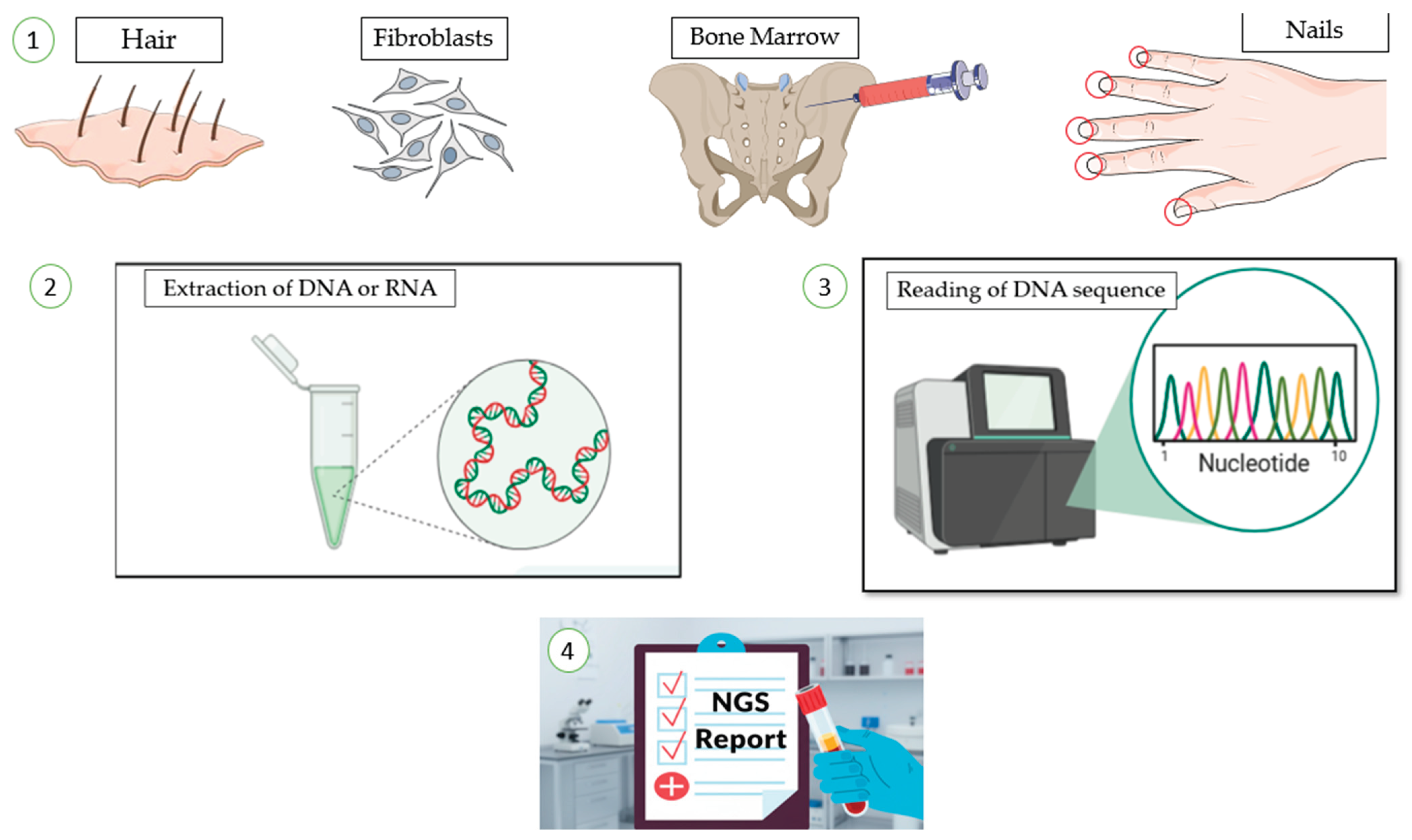
2.3. Sample Type and Quantity

In case of most hematological samples, including MDS or AML, tumor cells are obtained from bone marrow or peripheral blood (Figure 3). In myeloproliferative neoplasms, peripheral blood NGS generally shows high concordance with bone marrow results making it a reliable and less invasive diagnostic tool. However, yet bone marrow samples remain superior to peripheral blood for MRD measurement [44,45]. White blood cell counts or flow cytometry can be used to estimate the number of tumor cells in the sample. When bone marrow aspirate is not available, NGS can also be performed using formalin-fixed paraffin-embedded (FFPE) BM trephine biopsies. However, FFPE samples often yields lower sequencing quality than fresh samples because formaldehyde fixation induces cross-linking between cellular macromolecules and amino groups of DNA bases, reducing DNA sample quality [42]. The required sample amount varies between assays and studies. Illumina recommends around 80 ng of total DNA or RNA per sample, although good results can often be obtained with smaller inputs (40-60 ng) or with higher amounts (100-120 ng) when available[31,32,46]. One study comparing diagnostic panels for FLT3-ITD and CEBPA testing reported that 200 ng of DNA per sample were required for optimal library preparation [18]. Long-read and ultra-long-read NGS assays may need even larger amounts of DNA, ranging from several hundred nanograms to micrograms [19]. Table 2, summarizes the main NGS tests grouped by specific clinical question.

2.4. Results Validation

Due to the complexity of the NGS workflow and the risk of false positives from technical artifacts, bioinformatic errors, or very low-level variants close to the detection threshold, NGS results may require confirmation using other molecular methods like Sanger sequencing, droplet digital PCR, quantitative PCR or capillary electrophoresis. Current guidelines from the Association for Molecular Pathology (AMP) and the College of American Pathologists (CAP), recommend orthogonal confirmation in selected situations – for example, when results are ambiguous, close to the limit of detection, classified as variants of uncertain significance (VUS), or when they have immediate therapeutic impact [9,47]. For well-validated, high-performance targeted NGS panels, confirmation is often not required for recurrent hotspot mutations, given the high concordance with other molecular diagnostic methods [18,40]. Overall, validation strategies should be adapted to the assay design, the type of variant, and the clinical context, aiming to balance analytical reliability with turnaround time and cost [28].

2.5. Standardization

The integration of NGS into the diagnostic workflow for hematologic malignancies underscores the need for standardization across laboratories and platforms. Differences in sequencing chemistry, library preparation, bioinformatics pipelines, and reporting can affect variant detection and interpretation [18,41]. International guidelines from AMP and CAP recommend harmonizing procedures from sample collection and nucleic acid extraction to library preparation, sequencing performance metrics (read length, depth of coverage, uniformity, error rates, variant allele frequency) and post-analytical steps (variant annotation, classification, and reporting) [1,39]. Proficiency testing programs and inter-laboratory comparisons are also used to ensure reproducibility and clinical reliability [9,39]. Although ELN 2022 recommendations for AML, ESMO Clinical Practice Guidelines for MDS and the WHO 2022 and ICC 2022 diagnostic classifications highlight the integration of NGS into diagnostic and prognostic workflows of AML and MDS and define the minimal set of relevant genes, the insufficient standardization of diagnostic NGS limits its routine implementation in clinical practice [2,3,29,43,48]. Challenges remain in reaching consensus on minimal gene panels, defining limits of detection for clinically relevant variants, and standardizing the use of NGS for measurable residual disease (MRD) monitoring [49]. In addition, existing guidelines published several years ago should be updated to reflect recent technical advances that have since entered into clinical practice. Notably, Italy has recently issued specific SIES guidelines addressing the role of advanced molecular diagnostics, including NGS, in adult AML, and similar recommendations have been released in other countries [50,51].

2.6. Type of Alterations

NGS enables the detection of single nucleotide variants (SNVs), small insertions and deletions (indels), and, depending on the platform, copy number variations (CNVs) and structural rearrangements. RNA-based sequencing can also detect fusion transcripts, which are particularly relevant in AML.
It is important to use standardized and consistent nomenclature to ensure clear communication and effective data sharing. The Human Genome Variation Society (HGVS) provides widely accepted guidelines for variant naming, and their use is recommended in clinical reporting. Reports should specify the reference sequence used, along with the coding and protein nomenclature, to facilitate functional interpretation. For coding variants, the longest validated reference transcript for each gene should be used and clearly indicated.
Commonly used reference databases include RefSeq (https://www.ncbi.nlm.nih.gov/refseq), Ensembl (http://www.ensembl.org/index.html), and Locus Reference Genomic (LRG; https://www.lrg-sequence.org) [47].

2.7. Result Interpretation

Bioinformatic analysis can be divided into three main stages:
  • Primary Analysis:
  • Fluorescent signals generated during sequencing are converted into text files (FASTQ) representing the sequence of individual bases.
  • Secondary Analysis
  • Sequences from multiple samples run together are separated according to their indices in a process called demultiplexing. The reads are then aligned to a reference genome, and differences are detected in a step known as variant calling.
This process can identify thousands of variants, which are collected into a single VCF (“Variant Call Format”) file for each patient.
  • Tertiary Analysis
  • The final stage involves interpreting the identified variants in the clinical context, evaluating their potential relevance and pathogenicity, and determining possible causal relationship with the patient’s disease.
Variants can be classified according to their actionability or pathogenicity.
Currently, two main systems are used to classify the clinical significance of somatic variants: the AMP/ ASCO/CAP tier system and the ClinGen/CGC/VICC system.
In the first one, somatic variants are grouped into four tiers according to their clinical relevance for cancer diagnosis, prognosis, and treatment: Tier I includes variants with strong clinical significance; Tier II comprises variants with potential clinical significance; Tier III refers to variants of uncertain clinical significance; Tier IV includes variants that are benign or likely benign [47].
The second classification is based on a combined assessment of multiple criteria, including population frequency data, computational or predictive algorithm results, and whether the variant affects important functional domains of the protein. Importantly, no single criterion is sufficient to determine pathogenicity of a variant on its own; instead, pathogenicity must be supported by multiple lines of evidence. This integrated approach enables the classification of variants into five categories: 1-Benign; 2-Likely benign; 3- Variant of uncertain significance; 4-Likely pathogenic; 5-Pathogenic.
Based on the strength and extent of available scientific evidence, pathogenicity criteria are further ranked as oncogenic very strong (OVS1), strong (OS1-3), moderate (OM1-4), or supporting (OP1-4). Conversely, benignity criteria are classified as somatic benign very strong (SBVS1), strong (SBS1-2), or supporting (BP1-2) [52]. In Figure 1 we visualized a proposed practical algorithm on “How to read an NGS report”.

2.8. Useful Database

With the increasing number of large-scale genome sequencing projects across various cancer types, including hematological malignancies, a substantial amount of data has been generated and incorporated into multiple databases.
The clinical and biological relevance of a given variant is closely associated with its recurrence in cancer contexts. Therefore, it is essential to evaluate the incidence and prevalence of tumor variants identified in different tumor entities, using databases and published literature. For the annotation of somatic variants, several specialized cancer databases are available, including the Cancer Gene Census (http://cancer.sanger.ac.uk/cancergenome/projects/census/), the Catalogue of Somatic Mutations in Cancer (COSMIC; http://cancer.sanger.ac.uk/cancergenome/projects/cosmic/), and The Cancer Genome Atlas (TCGA; http://cancergenome.nih.gov/). These databases provide information on sequence variants in different cancer types and subtypes, cross-references to other genomic databases, as well as references to published literature, cellular pathways, targeted therapies, clinical trials, and outcome data [52].
Probable germline variants that may reside in genes associated with cancer predisposition syndromes can be detected during the sequencing of the tumor sample. Nevertheless, it is important to correlate this data with tumor cellularity [47].
In this regard, it may be useful to consult specialized databases, such as Online Mendelian Inheritance in Man (OMIM, http://www.ncbi.nlm.nih.gov/omim) and Human Gene Mutation Database (HGMD, http://www.hgmd.cf.ac.uk/ac/index.php) that provide information helpful for evaluating the potential nature of these variants. Among these, ClinVar (http://www.ncbi.nlm.nih.gov/clinvar) is widely used, cataloguing germline variants across various tissues and classifying them from pathogenic to benign. ClinVar also provides supporting clinical evidence, associated studies, and expert panel classifications, and occasionally reports somatic occurrences for certain variants [53].
When the germline nature of a variant is suspected, confirmation via a validated germline test is appropriate. This should be performed on a sample of tissue not affected by the disease for which the somatic analysis was initially requested following consulting with the physician and after obtaining a properly completed and signed informed consent from the patient.
Therefore, it is recommended that reports explicitly state that the test cannot reliably to distinguish somatic variants from germline or benign ones, in cases where a germline origin is suspected [47].
To evaluate the frequency of variants in large populations, population databases are used. These resources provide information on specific alleles at defined loci in large cohorts, often stratified by distinct subpopulations. When using these databases, it is necessary to consider whether the cohorts included healthy or affected individuals, whether multiple individuals from the same family were included, and the age range of the subjects. Population databases are frequently used to characterize benign variants, based on an arbitrary cut-off (Minor Allele Frequency, MAF). There is no standardized MAF threshold: a threshold of 1% is commonly recommended, potentially adjusted according to the patient’s specific ethnicity. Among the most widely used used population databases are: 1000 Genomes Project (http://browser.1000genomes.org), Exome Variant Server (http://evs.gs.washington.edu/EVS), dbSNP (http://www.ncbi.nlm.nih.gov/snp), dbVar (http://www.ncbi.nlm.nih.gov/dbvar), ExAC (http://exac.broadinstitute.org/ [47].
Finally, when evaluating a variant in a gene, in silico prediction algorithms are commonly employed to assess its potential impact on splicing, on thestructure and function of the encoded protein. These algorithms typically consider several key factors including the evolutionary conservation of the amino acid or nucleotide residue, the biochemical consequences of amino acid substitution based on physicochemical properties, and the position of the variant within the translated protein.
Some of the most used in silico tools are PolyPhen2 (http://genetics.bwh.harvard.edu/pph2), SIFT (http://sift.jcvi.org), MutationTaster (http://www.mutationtaster.org), and CADD (http://cadd.gs.washington.edu).
It is important to emphasize that these tools are predictive, and should be used with caution when interpreting sequencing variants as a sole evidence for clinical decision-making [47].

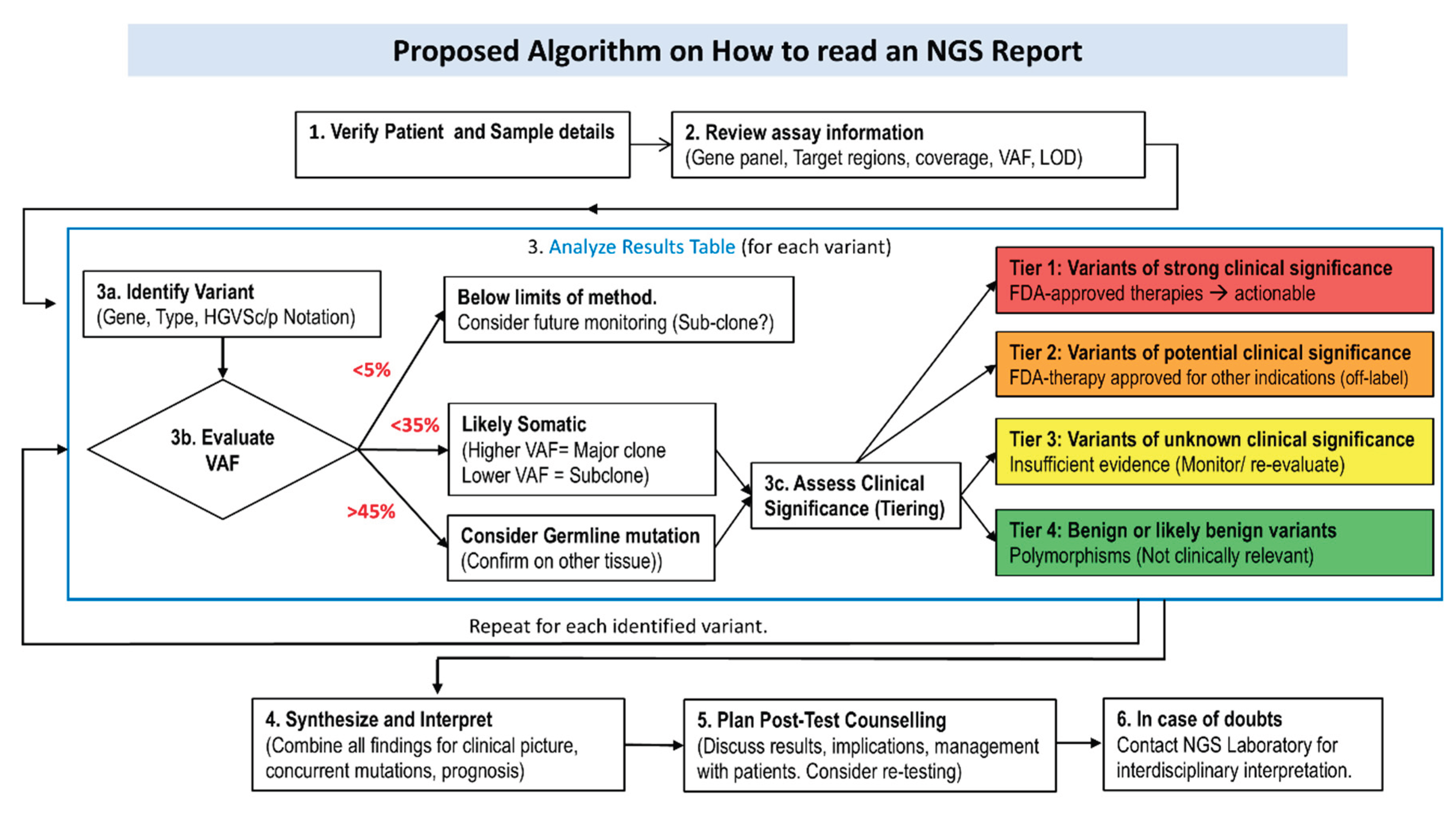
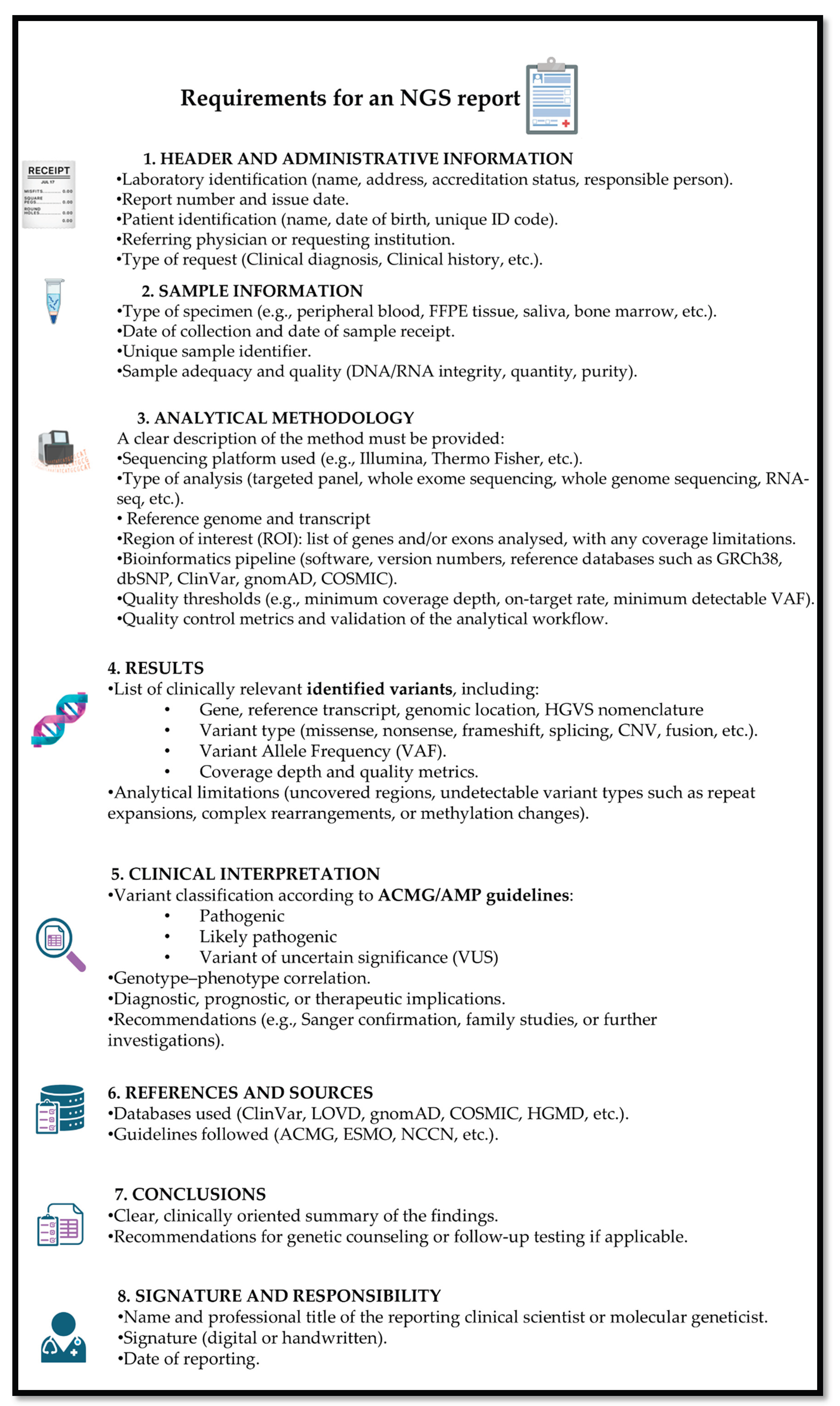
2.9. Report Structure

At the end of the NGS analysis process, it is essential to prepare a comprehensive report summarizing the results. This report should be thorough, providing the necessary technical details of the methods used, while remaining understandable to non-specialist healthcare professionals and patients. and concise enough, to avoid overwhelming the reader with excessive information. The report must include information about the laboratory that performed the analysis, the patient’s personal details, the sample’s traceability, the clinical question addressed, the variants identified along with their interpretation, and the methods employed for the analysis.
It is important that the report clearly displays the name of the laboratory with its certification (e.g., UNI EN ISO 9001) and accreditation (e.g., UNI EN ISO 15189, UNI CEI EN ISO/IEC 17025). Preferably, the report should include the name of the director, the laboratory’s contact information, as well as the name of the referring clinician and the department of origin, ensuring clarity and traceability of responsibility [23].
It is important that the report includes the patient’s personal information, such as surname, first name, date of birth, gender, and ethnicity.
The report must also provide details about the biological sample, including a unique identifier, date of collection, and sample type (e.g., peripheral blood, bone marrow) and, whenever possible, additional information such as an estimate of the percentage of blasts, the diagnosis, and/or the clinical indication for the test.
The report must include detailed information on the NGS assay used, specifying sequencing technology or platform, the panel name and version, as well as a list of the analyzed genes and the covered regions.
A concise description of the methodology and its limitations should also be provided and should include a list of all software tools used to generate the results with their version numbers, and the reference genome employed (GRCh37-hg19 or GRCh38) [54].
It is important to indicate the limitations of the method: specifying which types of variants the approach cannot detect, which alterations will not be reported, including their potential oncogenicity/benignity, or clinical actionability. The report may include the overall coverage achieved for each sample, measured as the average coverage over the entire sequenced genomic region. According to AMP/CAP guidelines, an average coverage of at least 250X is recommended to reliably detect somatic variants [9]. In this context, a VAF threshold of 5% is commonly used for variant calling and should be explicitly stated in the report [23].
The results section is the central component of the report. For clarity, it may be preferable to present the variants in order of clinical relevance, with the most pathogenic or actionable variants listed first.
All reported variants must be described using HGVS.c and HGVS.p notation, together with their VAF and classification. The identified variants should be summarized in a dedicated section, structured to ensure immediate understanding, and supplemented with references to relevant literature, when appropriate [47]. The report concludes with the date of reporting, the full name of the biologist who performed the analyses, and the signature of the laboratory manager. Each page of the report must be appropriately numbered [6] (Figure 2).

2.10. NGS vs. Quantitative-PCR vs. Digital-PCR

Today, laboratories have access to a variety of technologies that can be used individually or in combination to achieve their experimental objectives. These include long-established methods such as quantitative PCR (q-PCR) as well as more recent techniques such as digital PCR (dPCR) and NGS.
The qPCR allows real-time measurement of DNA amplification through fluorescence monitoring, providing either relative or absolute quantification.
In dPCR, the sample is divided into thousands of independent partitions: the ratio of positive partitions, those showing a fluorescent signal, to the total number of partitions is used to calculate the concentration of the target in the sample. This approach allows absolute quantification, enabling the detection of a very low number of target molecules without interference from PCR inhibitors and without the need for a standard curve, making it a sensitive and accurate technique [55,56]. Although qPCR has a more limited resolution, it remains a faster, and a more cost-effective method, suitable for the simultaneous analysis of multiple samples and targets. Both qPCR and dPCR require prior knowledge of the mutation of interest, which allows for longitudinal monitoring over time.
NGS, in contrast, supports large-scale quantitative and qualitative analyses, enabling simultaneous investigation of multiple mutations across numerous samples. This approach generates analysis with a larger and more complex datasets, with increased costs and reporting times, and typically it has lower sensitivity than dPCR [57,58].

3. NGS and Impact on Diagnosis and Classification

3.1. NGS Role in MDS: From Diagnosis to Classification

The transition from the previous WHO2016 to the updated ICC 2022 and WHO 5th editions was marked by the introduction of new genetically defined MDS categories [2,48,61,62]. In this context, the availability of NGS testing is crucial not only to correctly identify patient category but also to address patients to genetically-driven therapies. Among the major innovations of the WHO 5th is the definition of MDS with low blast count and SF3B1 mutations, which has been incorporated into the former category of MDS LB with ring sideroblasts (RS) [2]. This new category has also been adopted by the ICC classification, replacing the previous MDS with RS [48]. WHO-2022 has also introduced another genetically-driven category defined as MDS with bi-allelic TP53 mutations, independent of blast count [48]. Similarly, ICC introduced the diagnostic categories of MDS and MDS/AML with mutated TP53., which are distinguished on the basis of blast percentage. [2]. The new “MDS/AML” category represents an overlap group that includes cases with 10 to 19% blasts, replacing the former WHO category of MDS with excess-blast 2. This change better reflects the biological continuum between MDS and AML [61]. NGS is essential for defining MDS/AML, as several mutations must be either identified or excluded to ensure accurate classification. In particular, MDS/AML cases should not harbor AML-defining genetic abnormalities such as NPM1 or CEBPA. The ICC further subdivides MDS/AML into i) MDS/AML with mutated TP53, ii) MDS/AML with myelodysplasia-related gene mutations or cytogenetic abnormality es and iii) MDS/AML, NOS (not otherwise specified). Importantly, correct classification requires mutational assessment of several genes, includingTP53, ASXL1, BCOR, EZH2, SF3B1, SRSF2, STAG2, U2AF1, ZRSR2 and RUNX1 [48]. Finally, both WHO 2022 and the ICC classifications introduced relevant updates regarding clonal hematopoiesis, including the formal recognition of Clonal Hematopoiesis of Indeterminate Potential (CHIP) and clonal cytopenia of undetermined significance (CCUS) as part of the spectrum of myeloid neoplasms [2,48].

3.2. CHIP and CCUS, the Role of NGS

CHIP is currently defined by the presence of somatic mutations commonly seen in hematologic malignancies (e.g., DNMT3A, TET2, ASXL1) in individuals without cytopenia or dysplasia. This condition is increasingly detected through NGS in otherwise healthy individuals and is more common in older adults [63,64]. Several studies have demonstrated that individuals with CHIP have an increased risk of developing hematologic malignancies, especially MDS and AML, as well as a higher risk of atherosclerotic cardiovascular disease [65,66,67]. Variant allele frequency ≥2% is generally considered significant for CHIP. While CCUS is not recognized as a diagnostic category in WHO 2022 , the ICC classification defines it as a distinct diagnostic entity, requiring close clinical follow-up, particularly when high-risk mutations like TP53 are detected [2,48,68]. According to the ICC, CCUS is defined ad persistent unexplained cytopenia in at least one lineage, in the absence of morphologic dysplasia or increased blasts [48]. NGS is essential for this diagnosis, as it enables detection of at least one somatic mutation in genes commonly mutated in myeloid neoplasms (e.g., DNMT3A, TET2, ASXL1, SF3B1, SRSF2, etc.). The type of mutations and VAF are particularly relevant in these conditions. CHIP is typically asymptomatic and carries a 1% annual risk of progression to hematologic malignancy. However, multiple studies have identified several high-risk features in CHIP, including specific mutations (TP53, U2AF1, SRSF2, IDH1/2), the presence of multiple concomitant mutations, detection of larger clones (e.g. VAF 10-15%) or progressive clonal expansion over time [68,69]. In CCUS, mutations in spliceosome genes and epigenetic regulators are associated with a higher risk of evolution when compared to typical CHIP mutations. Additionally, the presence of ≥2 pathogenic mutations confers a ~50% risk of progression within 5 years [70].

3.3. Novel Genetically-Based Categories in MDS

3.3.1. SF3B1 in MDS

SF3B1 (splicing factor 3b subunit 1) is the largest subunit of the SF3B complex and serves as a core component of the U2 snRNP, where it plays a key role in branch site recognition in the early stages of spliceosome assembly. Mutations in SF3B1 were originally identified in 2011 and are observed across all spectrum of myeloid malignancies, including MDS, MDS/AML and AML [71]. However, they are particularly enriched in MDS and especially in MDS with ring sideroblasts, where up to 65% of patients carry a SF3B1 mutation [71,72]. Several studies have confirmed that SF3B1 mutations are detected in over 90% of MDS cases with ≥5% ring sideroblasts. Most SF3B1 mutations are heterozygous missense variants clustered in exons 12–15, with K700E representing approximately one-third of the variants. These mutations alter protein function but generally preserve its structural integrity [72]. According to the ICC classification, MDS with SF3B1 mutation is now recognized as a distinct diagnostic entity and no longer requires the presence of ring sideroblasts [48]. To meet diagnostic criteria, MDS patients with SF3B1 mutations must have <5% in the bone marrow and <2% in the peripheral blood and must not carry specific cytogenetic and molecular abnormalities, including isolated del(5q), −7/del(7q), abn3q26.2, or complex karyotype, TP53 multi-hit, or RUNX1 mutations [48]. The WHO 2022 is more conservative and still maintains the entity MDS with low blasts and ring sideroblasts in cases with wild-type SF3B1 and ≥15% ring sideroblasts [2]. This category represents a genetically homogeneous group and is associated with low progression rates and better overall survival (OS) when detected in patients with low blast counts and without additional genetic alterations [72].

3.3.2. TP53 in MDS

TP53 is a tumor suppressor gene located on chromosome 17p13.1 and is among the most frequently mutated genes in human cancers where its inactivation is typically associated with poor clinical outcomes. In MDS, pathogenic TP53 alterations are identified in 7-11% of cases. Importantly, TP53 inactivation in MDS is not limited to point mutations but may also involve additional mechanisms of loss of genetic material, including copy-neutral loss of heterozygosity and segmental deletions which further contribute to loss of tumor suppressor function [73,74]. Most TP53 mutations (-80%) are missense substitutions involving exons 5 to 8, resulting in amino-acid changes, while the remaining cases include truncating alterations. Physiologically, active p53 regulates the transcription of more than 150 genes implicated in DNA damage repair, apoptosis, senescence, and cell cycle arrest. Loss -of-function mutations lead to non-functional p53 protein, impair genomic stability, disrupt hematopoietic stem-cell self-, and promote uncontrolled proliferation. In the WHO 2022 classification, a distinct diagnostic category termed “MDS with biallelic TP53 inactivation (MDS-biTP53)” has been introduced, defined by the presence of multiple TP53 alterations leading to complete loss of p53 function [2]. Biallelic TP53 (biTP53) alterations may consist of multiple mutations, mutations with a VAF >50%, or mutations with concurrent deletion of the other allele (TP53 copy number loss or copy neutral loss of heterozygosity). The detection of biallelic inactivation of TP53 is complex and requires more than one technique. NGS analysis is essential to detect single mutation or multiple mutations in the gene, covering at least exons 4 to 11 [75]. The first-line method for detecting loss of genetic material at the TP53 site is fluorescence in situ hybridization with specific probe sets for the TP53 locus on 17p13.1. NGS, instead, can contribute to the identification of multi-hit TP53 alterations. In particular, the presence of more than one TP53 mutation typically suggests involvement of both alleles, indicating biallelic inactivation; at second, detection of TP53 mutations with VAF ≥ 50% can be used as a surrogate (although not certain) of loss of the second TP53allele [2]. Thus, NGS complements FISH by providing mutational data that supports the recognition of biallelic TP53 inactivation. At the same time, NGS panels may fail to detect 17p deletions present in low cell fractions, which are more easily identified by FISH, explaining the discrepancy between TP53 alteration detection by both methods and supporting their complementary use [76,77]. The ICC classification has introduced two distinct categories of MDs with TP53 mutations: MDS with mutated TP53 and MDS/AML with mutated TP53. The first includes MDS cases with blasts up to 9% with either multi-hit TP53 mutation or TP53 mutation (VAF > 10%) and complex karyotype often accompanied by loss of 17p [48]. Multi-hit TP53 mutations include the presence of either 2 distinct TP53 mutations (each VAF > 10%), a single TP53 mutation with 17p deletion or copy-neutral LOH at the TP53 locus or a TP53 mutant VAF of >50% [48]. MDS/AML with mutated TP53 is defined as cases with 10- 19% blasts and any TP53 mutation with VAF >10% [48]. Overall, these TP53-mutated MDS categories, particularly multi-hit TP53, are often associated with complex karyotype and poor prognosis [78].

3.4. Impact of NGS in the Definition of AML, Novel Genetic Classifications

Nowadays, AML can be classified according to two systems: the WHO fifth edition [2] or the International Consensus Classification (ICC) of Myeloid Neoplasms and Acute Leukemias [48]. Genetic features have now surpassed morphologic and cytometric features, firmly establishing NGS analysis as a cornerstone in AML diagnosis. The ICC classification has strengthened the role of genetics by lowering the blasts threshold to 10% for cases harboring recurrent genetic abnormalities (BCR::ABL1 excluded). In addition, the ICC has introduced the MDS/AML category in cases with 10 to 19% of blasts. The ICC approach can be viewed as hierarchical. In the first step, recurrent genetic abnormalities, NPM1 mutations and in-frame bZIP domain mutations of CEBPA should be investigated, and if none are present, the second step requires assessment for TP53 mutations (VAF > 10%) and myelodysplasia-related gene mutations or cytogenetic abnormalities. Therefore, the second step of the ICC relies almost entirely on NGS for both identification and quantification of TP53 mutations and for detecting mutations in ASXL1, BCOR, EZH2, SF3B1, SRSF2, STAG2, U2AF1, ZRSR2 and RUNX1, which are myelodysplasia-related genes [55].

3.4.1. TP53 Mutations in AML [56]

In de novo AML, TP53 mutations are detected in 5-10% of cases and are more frequent in elderly patients and in therapy-related AML. Similarly to MDS, TP53 mutations may be present in either monoallelic or biallelic form; in the latter condition, the inactivation of the second allele may occur through additional point mutations, copy neutral LOH (cnLOH) or loss of heterozygosity (LOH). TP53 mutations define an aggressive subset of AML, regardless of whether the disease is de novo, evolves from a preceding MDS, or is therapy-related, and they are frequently associated with a complex karyotype. Unlike in MDS, in the ICC classification, a single TP53 mutation with VAF>10% is sufficient to define AML with TP53 mutations (> 20% blasts) or MDS/AML with TP53 mutation (10-19%) [2,48].

3.4.2. MDS-Related Mutations

The ICC classification, as well as theELN2022 guidelines, has introduced a 9-gene signature (ASXL1, BCOR, EZH2, RUNX1, SF3B1, SRSF2, STAG2, U2AF1, and ZRSR2) to define the new category of AML (>20% blasts) or MDS/AML (10-19% blasts) with myelodysplasia-related mutations [2,48]. These mutations were classified as adverse risk in the ELN 2022 due to their strong association with secondary AML arising from prior MDS or chronic myelomonocytic leukemia (CMML) [79]. In the ICC classification, this category is incorporated alongside MDS-related cytogenetic abnormalities, thus expanding the former WHO 2016 category of AML with myelodysplasia related changes, which was based solely on morphologic assessment [48,61]. The WHO 5th edition also recognizes the category of AML, myelodysplasia-related (WHO-AML-MR) defined by specific molecular and cytogenetic abnormalities, while still including cases of AML arising from MDS or MDS/MPN [2]. Notably, the WHO 5th edition includes only 8 of the 9 genes used in the ICC classification, excluding RUNX1. These 8 genes have been demonstrated to maintain over 95% specificity for identifying secondary AML evolving from prior MDS, thus helping to distinguish s-AML from de novo AML [1]. More specifically, SRSF2, SF3B1, U2AF1, and ZRSR2 are mutated in nearly half of s-AML cases and encode spliceosome components critical for RNA processing, particularly for pre-mRNA splicing. Most SRSF2 mutations affect the P95 residue, whereas SF3B1 mutations predominantly occur at K700, with a smaller proportion involving K666. U2AF1 gene also shows well-defined mutational hotspots at S34F and Q157.In contrast, ZRZS2 mutations are scattered throughout the gene but occur most frequently within the Pre-ZF1 (49%), UHM (27%), and Post-ZF2 (13%) domains [80,81]. ASXL1, EZH2 and BCOR belong to the class of chromatin modifiers. ASXL1 regulates epigenetic programs and transcription through interaction with Polycomb complex proteins and various transcription activators and repressors. This gene is ubiquitously mutated in myeloid neoplasms, with most variants occurring in the final exon (exon 13). One of the most common alterations is the frameshift mutation G646Xfs*12 [80,82]. EZH2 is a lysine methyltransferase and represents the central core protein of the Polycomb Repressive Complex 2 (PRC2). Most mutations cluster in the SET ([Su(var)3-9, Enhancer-of-zeste and Trithorax]) or CXC (cysteine-rich region) domain at the C-terminus of the protein, regions that are essential for its methyltransferase activity. Approximately half of the variants are stop-gain or frameshift mutations, causing premature truncation of the protein [83]. BCOR is a transcription factor involved in embryogenesis, mesenchymal stem cell function, hematopoiesis, and lymphoid development. It acts as a tumor suppressor, and pathogenic variants may occur throughout the gene, including nonsense, frameshift, missense or splice-site mutations [84]. STAG2 is a core component of the cohesin complex essential for chromatin organization, transcriptional regulation, and DNA repair. Mutations are scattered throughout the gene, although most are located in the N-terminus and in the STAG domain, and are typically nonsense, frameshift or splice-site [85]. The identification of MDS-related mutations by NGS has relevant clinical implications since it can identify a sub-group of high-risk AML patients who may benefit from alternative frontline treatment (e.g., CPX351 [86]) and subsequent consolidation with allogeneic hematopoietic stem cell transplantation (allo-HSCT).

3.4.3. RUNX1

RUNX1 mutations are enriched in s-AML and the ICC classification included this gene among the defining features of AML with MDS-related mutations. RUNX1 encodes a transcription factor that is a key regulator of hematopoiesis involved in hematopoietic stem cell emergence and function. The mutational spectrum consists mainly of missense mutations in the N-terminal region, particularly within the RUNT domain, and truncating mutations in the C-terminal portion of the protein within the transactivation domain (TAD). RUNX1 mutations are consistently associated with poor outcomes in all myeloid malignancies, including AML [87].

3.4.4. AML-Defining Mutations

CEBPA. CEBPA gene, located on chromosome 19q13.11, encodes a transcription factor essential for myeloid differentiation. 5 to 15% of AML cases harbor CEBPA mutations that are more frequent in younger adults [88]. These mutations can affect both the N-terminal TAD and the C-terminal bZIP domain, and can be either monoallelic or biallelic [88]. Both the ICC and WHO 2022 classifications recognize CEBPA-mutated AML as a distinct entity, but differ in scope: the ICC includes only AML cases with in-frame bZIP domain mutations (allowing a lower blast count ≥10%) [2,48]. Conversely, WHO defines a broader category of AML with CEBPA mutations (including both biallelic and bZIP monoallelic mutations). CEBPA mutations can be detected by PCR, but NGS represents a useful tool which enables to identify both the number of mutations (mono vs biallelic) and their location across the N-terminal and the C-terminal regions [88]. This distinction is clinically relevant since most familiar CEBPA mutations are located in the N-terminal portion and are often accompanied by a “second-hit” in the C-terminal domain [89]. Detection of such variants with aVAF≥30%- 40%should prompt germline testing. Some studies have also highlighted challenges for NGS in detecting biallelic CEBPA mutations, partly due to the high GC content of the gene [57].
NPM1. The nucleophosmin (NPM1) gene encodes the most abundant nucleolar protein. Through its role as a histone chaperone and its ability to shuttle between the nucleus and cytoplasm, NPM1 participates in multiple cellular processes, including nucleolus formation via liquid-liquid phase separation, regulation of ribosome biogenesis and transport, control of DNA repair and centrosome duplication, and response to nucleolar stress. NPM1 is the most frequently mutated gene in AML occurring in about 30-35% of cases [90,91]. Due to its unique biological and clinical features, NPM1-mutated AML is recognized as a distinct entity in both the WHO 5th edition and ICC classifications of myeloid malignancies [2,48]. Mutations inNPM1 atypically affect the C-terminal region, leading to its abnormal localization in the cytoplasm of the leukemic cells [90]. A new class of drugs, the menin inhibitors,(e.g., revumenib), has shown activity in relapsed/refractory AML [92,93,94]. Several studies have compared detection of NPM1 by PCR and NGS [95,96,97] highlighting some differences. In practice, PCR offers faster turnaround times than NGS and high sensitivity suitable for MRD monitoring. NGS, on the other hand, allows detection of rare or atypical mutations beyond the common hotspot regions [59,98].

3.5. AML Mutations Driving Target Therapies (IDH1/2 and FLT3)

At diagnosis of AML, mutations in FLT3 and IDH1 or IDH2, represent actionable targets for patients eligible and ineligible for intensive chemotherapy (IC), respectively [99]. Detecting these mutations is therefore essential; PCR remains the gold standard (e.g., Abbott RealTime IDH1 [100]) due to its rapid turnaround time and lower costs. Clinically, we can add midostaurin to the standard 7+3 backbone in presence of FLT3-ITD or FLT3-TKD, while quizartinib is used specifically for FLT3-ITD [101,102]. Similarly, in patients ineligible for IC, IDH1 positive cases can benefit from the addition of ivosidenib to the azacitidine backbone [103]. NGS shows good concordance with PCR, although its sensitivity is slightly lower for FLT3-ITD [97].

4. NGS and Impact on Risk Stratification

When hematologists evaluate a new patient with AML or MDS, one of the first questions is “What is the prognosis of this neoplasia?”. Several prognostic tools have been developed to answer this key question, based on age, comorbidity, geriatric score, fitness of the immune system, genetic features of the disease, and effectiveness of available therapies play a critical role [104,105,106]. Earlier prognostic systems also relied on cytogenetical data (e.g., ELN2017) [107], but more recent risk stratification incorporates multiple genetic alterations, that require NGS for detection. Therefore, NGS analysis is increasingly considered essential at diagnosis of AML or MDS. Initial stratification also guides therapeutic decisions such as indication for allo-HSCT. In this section, we will focus on the impact of NGS results on risk stratification.

4.1. Myelodysplastic Syndromes: Prognostic Impact of Molecular Data

MDS represent both a diagnostic and classification challenge, mainly due to their genetic complexity. Somatic mutations are increasingly incorporated into modern WHO and ICC classifications, paralleling efforts to improve traditional risk stratification tools by including the mutational profile of the most frequently altered genes in MDS [108]. MDS is a heterogeneous disease and accurate survival prediction is essential for therapeutic decision-making. Traditionally, the IPSS-R has been the most widely used risk tool to predict outcomes, incorporating cytopenias, bone marrow blast percentage, and cytogenetic abnormalities [109]. IPSS-R has been widely validated [110], however disease evolution can vary considerably, depending on both disease- and patient-related factors. In recent years, our understanding of the genetic landscape of MDS has expanded and multiple studies have examined the impact of somatic mutations on patient outcomes. Based on data from 3711 patients with MDS profiled for mutations in 152 genes, the IPSS-M is a recently developed risk stratification score that integrates molecular information with traditional parameters, including cytopenias, bone marrow blast percentage, and cytogenetics. MDS patients are classified into six categories based on the mutational status of 31 genes commonly included in routine NGS myeloid panels, divided into 16 main effect genes and 15 residual genes. Multi-hit TP53, FLT3-ITD/TKD, and KMT2A-PTD are particularly influential in predicting adverse outcomes [111]. Interestingly, 46% of MDS patients were reclassified by IPSS-M compared with their original IPSS-R categories, with the majority being assigned to higher risk groups. IPSS-M has been validated in large patient cohorts for its prognostic accuracy; however, data on how this reclassification should guide therapeutic decisions is still limited [112,113,114].
In these cohorts, 20% of patients classified as low-risk by IPSS-R were upstaged to intermediate-risk by IPSS-M, and 25% of intermediate-risk IPSS-R patients were reclassified into higher-risk IPSS-M groups. Notably, a similar proportion of intermediate-risk IPSS-R patients were down staged to low-risk categories under IPSS-M. In clinical practice, however, re-stratification does not necessarily translate into major treatment changes for all affected MDS patients [112,113,114]. A recent analysis by Zamanillo et al. showed that IPSS-M reclassification would potentially influence treatment decisions in 22.2% of patients: 17% would become candidates for treatment intensification and 5% for de-escalation. Yet, only 6.6% of the overall cohort would actually qualify for higher-intensity therapy when age and fitness were considered [115]. In the context of HSCT, Tentori and colleagues reported that an IPSS-M–based strategy favored delayed transplantation for patients <70 years with low or moderate-low risk, whereas immediate transplantation improved life expectancy in those with moderately high, high, or very high risk [116]. Their findings indicated that 15% of patients originally considered for immediate HSCT by IPSS-R would benefit from delayed HSCT under IPSS-M, and 19% of those initially assigned to a delayed strategy by IPSS-R would instead benefit from immediate HSCT when reassessed by IPSS-M. Overall, an IPSS-M-based policy yielded a significant gain in life expectancy compared with IPSS-R (P = .001). To date, the lack of prospective clinical trials using IPSS-M limits its broad implementation for systematic MDS risk stratification. Nonetheless, the molecular information integrated into IPSS-M is likely to assist treatment decision-making, particularly for transplant-eligible younger patients (<70 years). Considering the entire MDS population, the proportion of patients who would undergo a meaningful therapeutic change based on IPSS-M remains relatively small, under 10% in older adults and under 19% in younger individuals. For these patients, molecular findings must be incorporated into a comprehensive clinical assessment that includes fitness, transplant eligibility, HLA matching, and donor-related factors.

4.2. Acute Myeloid Leukemia

ELN recommendations for AML risk stratification have evolved since the 2010 edition, progressively including the growing evidence on the prognostic impact of gene mutations. Compared with the 2010 and 2017 editions, the most recent 2022 update requires mutational analysis of 13 genes to accurately assign AML patients to favorable, intermediate or adverse risk categories. In particular, the adverse-risk group has been expanded to include 7 additional mutations: BCOR, EZH2, SF3B1, SRSF2, STAG2, U2AF1, ZRSR2 - together with ASXL1 and RUNX1 (already included in ELN 2017) forming the “myelodysplasia-related” (MR) gene group [6,7]. Therefore, the use of NGS has become essential for correctly accurate risk stratification in AML.
The presence of any of these mutations allocates patients in the adverse-risk category unless accompanied by favorable-risk alterations. TP53 mutations are particularly emphasized in ELN2022. Several studies have validated the ELN2022 guidelines and further explored the role of MR mutations. Most studies highlight significant heterogeneity in outcomes among patients with different MR mutations: for example RUNX1, ASXL1, U2AF1) are generally associated with worse outcomes, whereas EZH2, STAG2, and ZRSR2 tend to show intermediate-risk outcome [117]. The evolving AML treatment landscape has led to the development of low-intensity regimens for older or unfit AML patients [118]. Hypomethylating agents (HMA), azacitidine and decitabine, as well as HMA-based combination therapies with venetoclax or ivosidenib in IDH1-mutated AML, have improved outcomes in these patient categories [103]. However, previous risk stratifications, including ELN 2022, were based on clinical trials of intensively treated patients and the attempt to apply this classification to lower-intensity regimens has been shown to be unsatisfactory.

4.2.1. Stratification of AML for Patients Treated with Less-Intensive Therapy

In 2024, ELN proposed a genetic-based risk classification (2024 ELN Less-Intensive) for patients with newly diagnosed AML receiving lower-intensity regimens based on HMAs [119]. This classification was derived from large trial cohorts, including ASTRAL-1 randomizing guadecitabine or other therapies (guadecitabine vs azacitidine, decitabine, or low dose cytarabine (LDAC)) [120], VIALE-A trial (NCT02993523) randomizing the combination of azacitidine and venetoclax versus azacitidine and placebo, and phase 3 AGILE study (NCT03173248) adding IDH1-inhibitor ivosidenib to azacytidine, compared to azacitidine-placebo). Karyotype status together with a small set of genes including NPM1, IDH1/2, FLT3-ITD, DDX41, NRAS, KRAS and TP53, was sufficient to stratify patients into favorable, intermediate and adverse risk categories. The intermediate-risk group in ELN 2024 remains still quite broad, as many cytogenetic and molecular abnormalities lack solid prognostic data in the context of lower-intensity AML therapies. Furthermore, following the precedent of IDH1 mutations which are now considered favorable after the introduction of specific inhibitors, other gene mutations may be reclassified as new target therapies become available.

5. The Role of NGS in MRD Measurement and Monitoring

Measurable residual disease (MRD) has become increasingly important in AML patients for guiding therapy – such as selecting optimal consolidation strategies, maintenance therapy, follow-up, or donor lymphocyte infusions (DLI) - , for monitoring relapse, and as a prognostic biomarker [121,122].
The 2021 Update on MRD in AML, from the European LeukemiaNet MRD working party, recognizes NGS-based MRD monitoring as useful for refining prognosis in addition to multiparameter flow cytometry (MFC).However, current evidence is insufficient to recommend NGS-MRD as a stand-alone technique [123]. The main pitfall is the relatively low sensitivity of NGS methods for detecting very small MRD clones. At diagnosis, variants are considered significant if VAF >5%; whereas NGS-MRD positivity, measured on genomic DNA, is provisionally defined as ≥0.1% VAF. Although NGS-MRD test negativity is defined as VAF <0.1%, even results below this threshold may still be associated with adverse outcomes [123].
The advantage of NGS-MRD lies in its ability to detect clonal heterogeneity and track the gain or loss of mutations that may precede AML relapse. For example, NPM1 mutations, generally considered stable markers for qRT-PCR monitoring [124], can become undetectable at relapse in about 10% of patients [125,126]. Therefore, analyzing a broader panel of genes allows better monitoring of clonal evolution in AML and the identification of emerging mutations predicting imminent morphologic relapse. Nevertheless, NGS-MRD has limitations, including low sensitivity without error-correction methods [127,128], and the detection of mutations - particularly in DNMT3A, TET2, and ASXL1 (DTA genes) – that can occur in healthy individuals due to age-related clonal hematopoiesis. Germline mutations (typically showing VAF around 50%) are noninformative for MRD [123]. Additional limitations include genetic clonal heterogeneity and evolution over time [129,130,131], as well as the emergence or selection of subclones at relapse [132]. In a post hoc analysis of the MORPHO trial, MRD levels measured with NGS correlated remarkably with relapse-free survival (RFS) and the risk of relapse, moreover, MRD positivity at any level negatively affected RFS [133].
Despite the potential of NGS-based MRD assessment, it remains an investigational and non-standardized technique, and even the optimal time points for testing are still controversial and differ between laboratories [134]). However, two notable exceptions - FLT3 MRD and a 5 gene NGS panel - are approaching integration into clinical practice.
The gene encoding fms-like tyrosine kinase-3 (FLT3) isone of the most frequently mutated genes in AML, bein altered in approximately 25% of cases. FLT3 mutations most commonly consist of internal tandem duplications (ITDs), which can vary in length from as little as three base pairs (bp) to over 400 bp, while generally remaining in-frame and therefore resulting in a functional protein [135]. Each FLT3-mutated patient has a unique ITD sequence, making its detection and longitudinal monitoring with PCR-based MRD assays technically challenging. A noteworthy advanced laboratory technique has been developed by Invivoscribe® -and approved by the FDA. This method combines an initial PCR designed to amplify FLT3-ITD mutations, followed by NGS. Barcoded primers are used to target the area surrounding the juxtamembrane (JM) domain of the FLT3 gene where ITDs typically occur, to selectively amplify the sequences of interest.. The resulting PCR products are then purified and prepared for NGS analysis, enabling sensitive detection and quantification of ITD variants [136]. This assay is capable of detecting ITDs ranging from 9 bp to 252 bp and insertions from 3 bp up to the maximum detectable ITD size, with an analytical sensitivity of 5 x 10⁻⁵ mutant alleles per total alleles. A compelling demonstration of the clinical utility of this combined PCR/NGS method for the detection of FLT3 MRD assessment was provided by the MORPHO trial. In this study, patients received gilteritinib for 2 years following allo-HSCT. Approximately half of the participants had measurable MRD either before or after transplantation., In a prespecified subgroup analysis, gilteritinib significantly improved outcomes in MRD-positive patients (HR, 0.515 [95% CI, 0.316 to 0.838]; P = .0065), but not in patients with undetectable MRD [137]. It is anticipated that this NGS/PCR approach for FLT3 detection will likely be incorporated into future MRD testing guidelines [121].
The last example of NGS-based MRD is the pre-MEASURE study, which evaluated the prognostic value of a 5-gene panel in patients with AML. Targeted error–corrected DNA sequencing was performed on pretransplant blood samples to detect variants in FLT3, NPM1, IDH1, IDH2, and KIT . The persistence of FLT3-ITDor NPM1 variants in the blood at an allele fraction of 0.01% or higher was associated with increased relapse rates and worse survival compared with patients lacking these variants [138]. Moreover, in the ALFA0702 study (NCT00932412) an error-corrected-NGS-based MRD assessment demonstrated that the persistence of non-DTA mutations (HR = 2.23 for RFS and 2.26 for OS), and DTA mutations (HR = 2.16 for OS) was associated with poorer prognosis in multivariate analysis [139].

6. The Role of NGS in the Detection of Germline Predisposition [140]

When approaching a newly diagnosed AML patient, we now have an unprecedented opportunity to detect germline predisposition simply by performing a standard diagnostic NGS panel. In a Korean cohort of AML patients , the prevalence of germline mutations was 7.2% [141]. A mutation may be suspected to be of germline rather than acquired somatically, when the VAF is relatively high, typically ≥30%-40%. [142,143] Therefore, NGS has revolutioned the traditional clinical concept of “familial MDS/AML”, previously defined by the occurrence of acute leukemia, myeloid malignancies, or characteristic cytopenias in at least two or more first- and/ or second-degree relatives in the same family with at least one case being diagnosed as MDS or AML [144]. Conversely, when performing diagnostic NGS, clinicians should be aware, and ideally inform patients in advance, that testing at the time of diagnosis of AML/MDS diagnosis may uncover germline mutations, potentially reveal familial predisposition. Such findings may bring significant clinical, psychological, and social implications for patients and their relatives [144,145]. Moreover, the presence of germline mutations conferring familiar predisposition can have an impact on the choice of donor source when considering allo-HSCT from a relative. Although donor-derived leukemia is a rare event (about 1%), case reports of MDS arising from donor cells have been described [16,146,147,148]. The ELN-2022 guidelines outline clinical features that should prompt testing of germline predisposition, including at least 2 cancers, AML/MDS diagnosed at an unusually young age, a personal history of hematologic malignancy accompanied by a first- or second-degree relative within 2 generations with a hematologic cancer or a solid tumor before age 50 , or the presence of other hematopoietic abnormalities) [3]. Conversely, the Nordic MDS study group (NMDSG) proposed slightly different criteria for identifying patients, who should be evaluated for myeloid neoplasms with germline predisposition. They also recommend confirming any pathogenetic variants detected by diagnostic NGS on a different tissue such as fibroblasts or peripheral blood collected during remission [143,149].
The 5th edition of WHO classification of hematolymphoid tumors categorized myeloid neoplasms with germline predisposition into three subgroups: Myeloid neoplasms with germline predisposition without a preexisting platelet disorder or organ dysfunction (DDX41, TP53, CEBPA); Myeloid neoplasms with germline predisposition and pre-existing platelet disorder (RUNX1, ANKRD26, ETV6); and Myeloid neoplasms with germline predisposition and potential organ dysfunction (GATA2, Shwachman-Diamond syndrome (SDS) [150], Fanconi anemia (FA) [151], Down syndrome [152], etc.) [2,153].

6.1. DDX41

The most frequent genetic alteration associated with germline myeloid neoplasms is DDX41, accounting for about 80% of cases [154]. DEAD-box helicase 41 (DDX41), localized on chromosome 5q35, plays an important role in innate immune sensing and hematopoietic homeostasis. DDX41 recognizes foreign or self-derived nucleic acids generated during microbial infection, thereby initiating an anti-pathogen response [155]. In one of the largest international studies, involving346 patients with DDX41 among9082 patients with myeloid neoplasms (MN) , DDX41 accounted for 80% of germline predisposition to MN. Notably, in the Japanese male population, the incidence of DDX41 mutations was 10 times higher than in than the general population. Furthermore, individuals with mutated DDX41 have a lower risk of developing AML/MDS until around 40 years of age, but it increased to approximately 50% by 90 years of age [156]. Moreover, DDX41 mutations appear to be associated with favorable prognosis when AML is treated with HMA plus venetoclax according to ELN2024[119]; however, this observation is based on a limited number of patients [157,158] that make this findings preliminary. Similarly, in patients treated with intensive chemotherapy, the presence of mutated DDX41 in MDS/AML was reported to confer a favorable prognosis with longer OS compared to DDX41 wild-type AML and other favorable risk AML cases [159,160,161]. The protective effect of DDX41 appears to mitigate the negative impact of other adverse markers like TP53 [156]. Recommendations from the Nordic working group on germline predisposition for myeloid neoplasms include guidance for the surveillance of symptomatic carriers of germline DDX41 variants [154].

6.2. TP53

The TP53 gene encodes a protein considered one of the cell’s most crucial transcription factors, often referred to as the “guardian of the genome.” Due to its key functions, TP53 is the most frequently mutated tumor suppressor gene in human cancers. Li-Fraumeni syndrome is caused by a pathogenic germline mutation in TP53 and is linked to a markedly increased risk of various solid tumors—such as those of the breast, pancreas, central nervous system, and sarcomas—as well as blood malignancies like MDS, AML, and ALL. By the age of 70, the likelihood of developing cancer in affected individuals reaches nearly 100%. In these patients, myeloid hematologic malignancies often arise secondary to prior radiation or chemotherapy treatments administered for other cancers. Mutations in TP53 greatly increase the risk associated with these blood cancers and are consistently correlated with a poor prognosis [162].

6.3. GATA2

The GATA-binding protein 2 (GATA2) plays a central role in blood stem cell generation and maintenance and directly modulates p53-induced apoptosis via the regulation of the MDM2 modulator RASSF4, which may contribute to chemo-resistance [163]. The GATA2 syndrome was first described in 2011 in patients presenting with monocytopenia, B cells, B-cell precursors, and NK cells deficiency, and/or absence of plasmacytoid dendritic cells in blood; late onset infections (non-tuberculous mycobacterial, fungal, or human papillomavirus (HPV) infections); pulmonary alveolar proteinosis; increased risk of progress to MDS or AML, and an autosomal dominant inheritance pattern due to haplo-insufficient mutations in one allele of GATA2 [164]. Germline GATA2 mutations account for approximately 15% of advanced and 7% of all primary MDS cases in pediatric population, making GATA2 the most common pediatric germline mutation [165]. Although data on the efficacy of allo-HCST are limited to small cohorts of patients with somatic GATA2-mutated AML, transplantation appears beneficial, also reducing the risk of opportunistic infections [166]. Given that almost 200 likely unique pathogenic variants have been described, which can be classified into four groups: 1) truncating mutations (splice site, nonsense, frameshift, and whole-gene deletions) proximal to or within the ZF2 domain; 2) missense mutations within the ZF2 domain; 3) mutations resulting in aberrant mRNA splicing (e.g., synonymous changes); 4) other regulatory variants [167]; it is essential to perform an NGS study of the entire gene to detect all possible pathogenetic variants.

6.4. RUNX1

The RUNX1 gene encoding a key hematopoietic transcription factor was first identified in AML cases carrying translocation t(8,21). It was initially namedAML1. and later renamed RUNX1 due to its homology to the Drosophila transcription factor runt, forming the “Runx” family of transcription factors [168]. In 1999 RUNX1 germline mutations were linked to a familial platelet disorder with associated myeloid malignancy, a rare autosomal dominant disease characterized by thrombocytopenia and a predisposition to myeloid neoplasms. In a study of 27 families with RUNX1 variants, half of the affected individuals had CHIP, and sequential sequencing data from 19 patients demonstrated dynamic changes of somatic mutations over time [169]. AML patients with RUNX1 mutations have a dismal prognosis [170]. Since multiple alterations in RUNX1 have been described, including multiexon or whole gene deletions, frameshifts, missense and nonsense mutations, [171] NGS is essential for their comprehensive detection. Furthermore, differential diagnosis with other familial disorders associated with thrombocytopenia requires sequencing of ETV6 [172,173] and ANKRD26 [174,175,176].

6.5. ETV6

The ETV6 gene produces a transcriptional repressor belonging to the ETS family, playing a key role red blood cells and megakaryocytes maturation [173]. Germline ETV mutations are inherited in an autosomal dominant manner and can lead to thrombocytopenia type 5, with risk—approximately 30%—of developing blood cancers. These malignancies can be either lymphoid (such as acute lymphoblastic leukemia, ALL) or myeloid (including AML and). Similar to other hereditary blood disorders, the onset of cancer usually requires acquisition of at least one additional somatic mutation in genes commonly implicated in leukemia development.

7. The Impact of NGS in the Detection of Druggable Mutations at Relapse

In patients with AML relapsed after several lines of therapy or after allogeneic transplantation, the prognosis is extremely poor, making it necessary to activate early palliative care or to enroll patients in clinical trials whenever feasible [177,178,179]. However, in the near future, NGS may become a compass to guide targeting actionable genes detected in r/r AML patients. An Italian case report of two patients demonstrated clonal and sub-clonal evolution during disease progression highlighting how NGS can help to uncover new therapeutic targets [180]. In a more robust dataset of 1470 AML patients, NGS was used to analyze 17 potentially actionable genes (ALK, CSF1R, FGFR1/2/3, FLT3, IDH1/2, JAK2, KDR, KRAS/NRAS, NPM1, PDGFRA, PTPN11, RET and TP53), because these genes are directly or indirectly targetable with standard (with 6% of patients receiving off-label agents) or investigational agents (53% of these patients were enrolled in clinical trials) [181]. The main pitfall of this approach is that many mutations identified by NGS are either linked to drugs not yet approved, or cannot currently be targeted with available treatments (e.g., TP53). Nevertheless, NGS promises to improve survival and quality of life of older adults [182].
Figure 3. The workflow of NGS, from sample collection to result reporting. 1) Biological specimens, such as hair, fibroblasts, bone marrow, or nails, are first collected from the patient. 2) nucleic acids (DNA or RNA) are isolated from these samples in the laboratory. 3) The extracted genetic material is then processed and sequenced to determine the precise order of nucleotides. This process generates data that is analyzed by bioinformatics to interpret the genetic sequence. 4) The findings are compiled into a formal laboratory report for clinical assessment.
Preprints 187709 g003

8. Discussion

The advent of faster and less expensive NGS techniques promises to revolutionize the field of MN. The availability of several gene Panels (e.g., the Illumina TruSight Myeloid panel, the Archer VariantPlex Core Myeloid panel, the Human Myeloid Neoplasms QIASeq DNA Panel, and the AmpliSeq for Illumina Myeloid panel). The presence of several translocations can be detected using a single diagnostic test based on RNA-based NGS (RNA sequencing), reducing costs and preserving laboratory resources from extensive cytogenetic analyses, and multiple specific real-time quantitative PCR for selected transcripts [183]. With RNA sequencing of the known gene partner, finding other rare partner has become easier (e.g., KMT2A) [184]. Moreover, the possibility to sequence long genes, that present technical difficulty to sequence (e.g., CEBPA is a GC-rich gene which is particularly difficult to amplify by PCR or TP53), can help clinicians to assign a patient to a more appropriate risk group. Nevertheless, we still have a limited number of drugs in the first line setting, reducing the possibility to offer each patient a personalized approach and precision medicine. In a retrospective study, patients were reevaluated in light of newly available NGS and cytogenetic data,, however, no statistically significant difference in survival was found between the two groups [98].
For patients with MDS or AML, TP53 mutations probably represent one of the most negative prognostic markers, given their association with low sensitivity to chemotherapy (including venetoclax-based treatments) and substantially worse responses after HSCT [185,186,187,188]. Therefore, sequencing TP53 is of utmost importance, despite the technical challenges that remain, such as the presence of VUS not yet fully covered by current databases) [189]. Unfortunately, no p53-based therapeutics have been approved to date, probably because, as a nuclear transcription factor, p53 does not possess typical drug target features and has therefore long been considered undruggable [190]. Another hot topic is the use of NGS as an MRD marker, which can help to refine the selection of AML patients for transplant in first complete remission and to guide the decision on post-transplant maintenance therapy (e.g., gilteritinib in MRD-positive patients) [137]. Moreover, patients who relapse often exhibit clonal evolution and biological shifts that complicate treatment, but NGS offers an opportunity to study the specific mechanisms of resistance to targeted drugs such as FLT3-inhibitors [191,192]. In the future, personalized medicine could be applied to patients rapidly identified by NGS; for example, Menin inhibitors for KMT2A-rearranged AML [92].

9. Conclusions

Reading the final report of an NGS exam in a patient with AML or MDS is a challenging task. Clinicians should be aware of both the potential and the limitations of the techniques employed by their laboratory, including the platform used, sequencing depth, and the interpretation of identified variants. VAF is another critical parameter that should be considered especially in patient harboring mutations typical of clonal hemopoiesis (e.g., DNMT3A, SRSF2, TET2), as it reflects the dimension of the clone at diagnosis and during follow-up . Another important concept is that not all mutations in the same gene are equivalent, and some cases still have unknown clinical significance (VUS). These variants require consultation of specialized databases, which may provide limited information that can change with time since some VUS can be reclassified when new evidence becomes available [193]. We recognize that currently clinicians are navigating the growing complexity of molecular data in daily practice, therefore we encourage direct interaction with the biotechnologists and bioinformatics involved in NGS analysis and report preparation. Moreover, for patients with suspected germline predisposition to MN a genetic counseling and multigene panel testing are recommended [194]. In conclusion, the dialog between hematologists and biotechnologists about the NGS report should not be intended as the endpoint, but rather as the beginning of a multidisciplinary discussion, particularly in the most challenging cases.

10. Future Directions.

The field of NGS in AML and MDS is changing fast, with increasing availability of faster, simpler and more affordable sequencing platforms. Clinicians are gaining experience and confidence in interpreting NGS reports of their patients. We believe this will profoundly influence our clinical practice, because NGS will likely be performed for each patient, including elderly ones.. The complexity of the field, may appear overwhelming at first glance, including the need to consult website-based databases of pathologic variants. Nevertheless, the integration of algorithms of Artificial Intelligence, could simplify the task of hematologist and help avoiding the analysis paralysis [195,196].

Author Contributions

Conceptualization, S.R., S.P. and L.C.; writing—original draft preparation, N.Z, S.P., L.C.; writing—review and editing, all.; visualization, S.P.; supervision, C.T.; project. All authors have read and agreed to the published version of the manuscript.

Funding

This research received no external funding.

Institutional Review Board Statement

Not applicable.

Informed Consent Statement

Not applicable.

Data Availability Statement

Not applicable.

Acknowledgments

The authors have reviewed and edited the output and take full responsibility for the content of this publication.

Conflicts of Interest

The authors declare no conflicts of interest.

Abbreviations

The following abbreviations are used in this manuscript:
AML Acute myeloid leukemia
AMP Association for Molecular Pathology
CAP College of American Pathologists
CCUS Clonal cytopenia of undetermined significance
CHIP Clonal Hematopoiesis of Indeterminate Potential
cnLOH Copy neutral loss of heterozygosity
CMML Chronic myelomonocytic leukemia
HMA Hypomethylating agents
HR Hazard ratio
HSCT Hematopoietic stem cell transplantation
ICC International Consensus Conference
LOH Loss of heterozygosity
MDS Myelodysplastic neoplasms
MN Myeloid neoplasms
MRD Measurable residual disease
NGS Next generation sequencing
OS Overall survival
RFS Relapse free survival
SBS Sequencing by synthesis
VAF Variant allele frequencies
VUS Variants of uncertain significance
WES Whole exome sequencing
WGS Whole genome sequencing
WHO World Health Organization

References

  1. Papaemmanuil, E.; Gerstung, M.; Bullinger, L.; Gaidzik, V.I.; Paschka, P.; Roberts, N.D.; Potter, N.E.; Heuser, M.; Thol, F.; Bolli, N.; et al. Genomic Classification and Prognosis in Acute Myeloid Leukemia. New England Journal of Medicine 2016, 374, 2209–2221. [CrossRef]
  2. Khoury, J.D.; Solary, E.; Abla, O.; Akkari, Y.; Alaggio, R.; Apperley, J.F.; Bejar, R.; Berti, E.; Busque, L.; Chan, J.K.C.; et al. The 5th Edition of the World Health Organization Classification of Haematolymphoid Tumours: Myeloid and Histiocytic/Dendritic Neoplasms. Leukemia 2022, 36, 1703–1719. [CrossRef]
  3. Döhner, H.; Wei, A.H.; Appelbaum, F.R.; Craddock, C.; DiNardo, C.D.; Dombret, H.; Ebert, B.L.; Fenaux, P.; Godley, L.A.; Hasserjian, R.P.; et al. Diagnosis and Management of AML in Adults: 2022 Recommendations from an International Expert Panel on Behalf of the ELN. Blood 2022, 140, 1345–1377. [CrossRef]
  4. Gooptu, M.; Murdock, H.M.; Soiffer, R.J. How I Treat AML Relapse after Allogeneic HSCT. Blood 2025, 145, 2128–2137. [CrossRef]
  5. Satam, H.; Joshi, K.; Mangrolia, U.; Waghoo, S.; Zaidi, G.; Rawool, S.; Thakare, R.P.; Banday, S.; Mishra, A.K.; Das, G.; et al. Next-Generation Sequencing Technology: Current Trends and Advancements. Biology 2023, 12, 997. [CrossRef]
  6. Schmid, S.; Jochum, W.; Padberg, B.; Demmer, I.; Mertz, K.D.; Joerger, M.; Britschgi, C.; Matter, M.S.; Rothschild, S.I.; Omlin, A. How to Read a Next-Generation Sequencing Report—What Oncologists Need to Know. ESMO Open 2022, 7, 100570. [CrossRef]
  7. Mosele, M.F.; Westphalen, C.B.; Stenzinger, A.; Barlesi, F.; Bayle, A.; Bièche, I.; Bonastre, J.; Castro, E.; Dienstmann, R.; Krämer, A.; et al. Recommendations for the Use of Next-Generation Sequencing (NGS) for Patients with Advanced Cancer in 2024: A Report from the ESMO Precision Medicine Working Group. Annals of Oncology 2024, 35, 588–606. [CrossRef]
  8. Kim, Y.J.; Takahashi, K. Emerging Technologies of Single-Cell Multi-Omics. Haematologica 2025, 110, 1269–1277. [CrossRef]
  9. Jennings, L.J.; Arcila, M.E.; Corless, C.; Kamel-Reid, S.; Lubin, I.M.; Pfeifer, J.; Temple-Smolkin, R.L.; Voelkerding, K.V.; Nikiforova, M.N. Guidelines for Validation of Next-Generation Sequencing-Based Oncology Panels: A Joint Consensus Recommendation of the Association for Molecular Pathology and College of American Pathologists. J Mol Diagn 2017, 19, 341–365. [CrossRef]
  10. Duncavage, E.J.; Bagg, A.; Hasserjian, R.P.; DiNardo, C.D.; Godley, L.A.; Iacobucci, I.; Jaiswal, S.; Malcovati, L.; Vannucchi, A.M.; Patel, K.P.; et al. Genomic Profiling for Clinical Decision Making in Myeloid Neoplasms and Acute Leukemia. Blood 2022, 140, 2228–2247. [CrossRef]
  11. Head, S.R.; Komori, H.K.; LaMere, S.A.; Whisenant, T.; Van Nieuwerburgh, F.; Salomon, D.R.; Ordoukhanian, P. Library Construction for Next-Generation Sequencing: Overviews and Challenges. Biotechniques 2014, 56, 61–64, 66, 68, passim. [CrossRef]
  12. Satam, H.; Joshi, K.; Mangrolia, U.; Waghoo, S.; Zaidi, G.; Rawool, S.; Thakare, R.P.; Banday, S.; Mishra, A.K.; Das, G.; et al. Next-Generation Sequencing Technology: Current Trends and Advancements. Biology 2023, 12, 997. [CrossRef]
  13. Lee, J.Y. The Principles and Applications of High-Throughput Sequencing Technologies. Dev Reprod 2023, 27, 9–24. [CrossRef]
  14. Goodwin, S.; McPherson, J.D.; McCombie, W.R. Coming of Age: Ten Years of next-Generation Sequencing Technologies. Nat Rev Genet 2016, 17, 333–351. [CrossRef]
  15. Peroni, E.; Randi, M.L.; Rosato, A.; Cagnin, S. Acute Myeloid Leukemia: From NGS, through scRNA-Seq, to CAR-T. Dissect Cancer Heterogeneity and Tailor the Treatment. Journal of Experimental & Clinical Cancer Research 2023, 42, 259. [CrossRef]
  16. O’Connor, T.E.; Shaw, R.; Madero-Marroquin, R.; Roloff, G.W. Clinical Considerations at the Intersection of Hematopoietic Cell Transplantation and Hereditary Hematopoietic Malignancy. Front. Oncol. 2023, 13, 1180439. [CrossRef]
  17. Thomas, M.; Sukhai, M.A.; Zhang, T.; Dolatshahi, R.; Harbi, D.; Garg, S.; Misyura, M.; Pugh, T.; Stockley, T.L.; Kamel-Reid, S. Integration of Technical, Bioinformatic, and Variant Assessment Approaches in the Validation of a Targeted Next-Generation Sequencing Panel for Myeloid Malignancies. Arch Pathol Lab Med 2017, 141, 759–775. [CrossRef]
  18. Akabari, R.; Qin, D.; Hussaini, M. Technological Advances: CEBPA and FLT3 Internal Tandem Duplication Mutations Can Be Reliably Detected by Next Generation Sequencing. Genes (Basel) 2022, 13. [CrossRef]
  19. Cumbo, C.; Minervini, C.F.; Orsini, P.; Anelli, L.; Zagaria, A.; Minervini, A.; Coccaro, N.; Impera, L.; Tota, G.; Parciante, E.; et al. Nanopore Targeted Sequencing for Rapid Gene Mutations Detection in Acute Myeloid Leukemia. Genes (Basel) 2019, 10. [CrossRef]
  20. Warburton, P.E.; Sebra, R.P. Long-Read DNA Sequencing: Recent Advances and Remaining Challenges. Annu Rev Genomics Hum Genet 2023, 24, 109–132. [CrossRef]
  21. Moustakli, E.; Christopoulos, P.; Potiris, A.; Zikopoulos, A.; Mavrogianni, D.; Karampas, G.; Kathopoulis, N.; Anagnostaki, I.; Domali, E.; Tzallas, A.T.; et al. Long-Read Sequencing and Structural Variant Detection: Unlocking the Hidden Genome in Rare Genetic Disorders. Diagnostics (Basel) 2025, 15. [CrossRef]
  22. Sims, D.; Sudbery, I.; Ilott, N.E.; Heger, A.; Ponting, C.P. Sequencing Depth and Coverage: Key Considerations in Genomic Analyses. Nat Rev Genet 2014, 15, 121–132. [CrossRef]
  23. Anu, R.I.; Patel, A.; Pathak, N.; Mehta, P.; Chougule, A.; Sheth, H.; Veldore, V.; Suryavanshi, M.; Gupta, V.G.; Disel, U.; et al. Uniform Reporting of Next Generation Sequencing: Indian Society of Medical and Pediatric Oncology. Indian J Med Paediatr Oncol 2025, 46, 351–362. [CrossRef]
  24. Conesa, A.; Madrigal, P.; Tarazona, S.; Gomez-Cabrero, D.; Cervera, A.; McPherson, A.; Szczesniak, M.W.; Gaffney, D.J.; Elo, L.L.; Zhang, X.; et al. A Survey of Best Practices for RNA-Seq Data Analysis. Genome Biol 2016, 17, 13. [CrossRef]
  25. Dillon, L.W.; Hayati, S.; Roloff, G.W.; Tunc, I.; Pirooznia, M.; Mitrofanova, A.; Hourigan, C.S. Targeted RNA-Sequencing for the Quantification of Measurable Residual Disease in Acute Myeloid Leukemia. Haematologica 2019, 104, 297–304. [CrossRef]
  26. Sa, A.C.C.; Sadee, W.; Johnson, J.A. Whole Transcriptome Profiling: An RNA-Seq Primer and Implications for Pharmacogenomics Research. Clin Transl Sci 2018, 11, 153–161. [CrossRef]
  27. Baccarella, A.; Williams, C.R.; Parrish, J.Z.; Kim, C.C. Empirical Assessment of the Impact of Sample Number and Read Depth on RNA-Seq Analysis Workflow Performance. BMC Bioinformatics 2018, 19, 423. [CrossRef]
  28. Shin, H.T.; Choi, Y.L.; Yun, J.W.; Kim, N.K.D.; Kim, S.Y.; Jeon, H.J.; Nam, J.Y.; Lee, C.; Ryu, D.; Kim, S.C.; et al. Prevalence and Detection of Low-Allele-Fraction Variants in Clinical Cancer Samples. Nat Commun 2017, 8, 1377. [CrossRef]
  29. Petrackova, A.; Vasinek, M.; Sedlarikova, L.; Dyskova, T.; Schneiderova, P.; Novosad, T.; Papajik, T.; Kriegova, E. Standardization of Sequencing Coverage Depth in NGS: Recommendation for Detection of Clonal and Subclonal Mutations in Cancer Diagnostics. Front Oncol 2019, 9, 851. [CrossRef]
  30. Singh, R.R. Target Enrichment Approaches for Next-Generation Sequencing Applications in Oncology. Diagnostics (Basel) 2022, 12. [CrossRef]
  31. Al-Kateb, H.; Knight, S.M.; Sivasankaran, G.; Voss, J.S.; Pitel, B.A.; Blommel, J.H.; Jerde, C.R.; Rumilla, K.M.; Lee, J.L.; Mattson, N.R.; et al. Clinical Validation of the TruSight Oncology 500 Assay for the Detection and Reporting of Pan-Cancer Biomarkers. J Mol Diagn 2025, 27, 292–305. [CrossRef]
  32. Sahajpal, N.S.; Mondal, A.K.; Ananth, S.; Njau, A.; Ahluwalia, P.; Jones, K.; Ahluwalia, M.; Okechukwu, N.; Savage, N.M.; Kota, V.; et al. Clinical Performance and Utility of a Comprehensive Next-Generation Sequencing DNA Panel for the Simultaneous Analysis of Variants, TMB and MSI for Myeloid Neoplasms. PLoS One 2020, 15, e0240976. [CrossRef]
  33. Serrati, S.; De Summa, S.; Pilato, B.; Petriella, D.; Lacalamita, R.; Tommasi, S.; Pinto, R. Next-Generation Sequencing: Advances and Applications in Cancer Diagnosis. Onco Targets Ther 2016, 9, 7355–7365. [CrossRef]
  34. Koboldt, D.C. Best Practices for Variant Calling in Clinical Sequencing. Genome Med 2020, 12, 91. [CrossRef]
  35. Sun, Q.Y.; Ding, L.W.; Tan, K.T.; Chien, W.; Mayakonda, A.; Lin, D.C.; Loh, X.Y.; Xiao, J.F.; Meggendorfer, M.; Alpermann, T.; et al. Ordering of Mutations in Acute Myeloid Leukemia with Partial Tandem Duplication of MLL (MLL-PTD). Leukemia 2017, 31, 1–10. [CrossRef]
  36. Spencer, D.H.; Abel, H.J.; Lockwood, C.M.; Payton, J.E.; Szankasi, P.; Kelley, T.W.; Kulkarni, S.; Pfeifer, J.D.; Duncavage, E.J. Detection of FLT3 Internal Tandem Duplication in Targeted, Short-Read-Length, next-Generation Sequencing Data. J Mol Diagn 2013, 15, 81–93. [CrossRef]
  37. Aguilera-Diaz, A.; Vazquez, I.; Ariceta, B.; Manu, A.; Blasco-Iturri, Z.; Palomino-Echeverria, S.; Larrayoz, M.J.; Garcia-Sanz, R.; Prieto-Conde, M.I.; Del Carmen Chillon, M.; et al. Assessment of the Clinical Utility of Four NGS Panels in Myeloid Malignancies. Suggestions for NGS Panel Choice or Design. PLoS One 2020, 15, e0227986. [CrossRef]
  38. Levis, M.; Shi, W.; Chang, K.; Laing, C.; Pollner, R.; Gocke, C.; Adams, E.; Berisha, F.; Lameh, J.; Lesegretain, A. FLT3 Inhibitors Added to Induction Therapy Induce Deeper Remissions. Blood 2020, 135, 75–78. [CrossRef]
  39. Keegan, A.; Bridge, J.A.; Lindeman, N.I.; Long, T.A.; Merker, J.D.; Moncur, J.T.; Montgomery, N.D.; Nagarajan, R.; Rothberg, P.G.; Routbort, M.J.; et al. Proficiency Testing of Standardized Samples Shows High Interlaboratory Agreement for Clinical Next Generation Sequencing-Based Hematologic Malignancy Assays With Survey Material-Specific Differences in Variant Frequencies. Arch Pathol Lab Med 2020. [CrossRef]
  40. Kim, J.J.; Lee, K.S.; Lee, T.G.; Lee, S.; Shin, S.; Lee, S.T. A Comparative Study of Next-Generation Sequencing and Fragment Analysis for the Detection and Allelic Ratio Determination of FLT3 Internal Tandem Duplication. Diagn Pathol 2022, 17, 14. [CrossRef]
  41. Chaudhary, S.; Chaudhary, P.; Ahmad, F.; Arora, N. Acute Myeloid Leukemia and Next-Generation Sequencing Panels for Diagnosis: A Comprehensive Review. J Pediatr Hematol Oncol 2024, 46, 125–137. [CrossRef]
  42. Hobeck, A.D.; Wendt, S.; Krohn, S.; Knuebel, G.; Bartels, S.; Schipper, E.; Junghanss, C.; Murua Escobar, H. Comparative Analyses of Targeted Myeloid Cancer Next-Generation Sequencing Panel in Fresh Blood, Bone Marrow and FFPE Material. Int J Mol Sci 2024, 25. [CrossRef]
  43. Fenaux, P.; Haase, D.; Santini, V.; Sanz, G.F.; Platzbecker, U.; Mey, U.; Esmo Guidelines Committee. Electronic address: clinicalguidelines@esmo. org Myelodysplastic Syndromes: ESMO Clinical Practice Guidelines for Diagnosis, Treatment and Follow-up(Dagger☆). Ann Oncol 2021, 32, 142–156. [CrossRef]
  44. Jumniensuk, C.; Nobori, A.; Lee, T.; Senaratne, T.N.; Rao, D.; Pullarkat, S. Concordance of Peripheral Blood and Bone Marrow Next-Generation Sequencing in Hematologic Neoplasms. Adv Hematol 2022, 2022, 8091746. [CrossRef]
  45. Lucas, F.; Michaels, P.D.; Wang, D.; Kim, A.S. Mutational Analysis of Hematologic Neoplasms in 164 Paired Peripheral Blood and Bone Marrow Samples by Next-Generation Sequencing. Blood Adv 2020, 4, 4362–4365. [CrossRef]
  46. Ye, X.; Zheng, Z.; Wu, Y.; Zhang, Z.; Xu, Z.; Liu, Y.; Jiang, L.; Wu, J. NGS Panel Enhance Precise Diagnosis of Myeloid Neoplasms under WHO-HAEM5 and International Consensus Classification: An Observational Study. Medicine (Baltimore) 2024, 103, e38556. [CrossRef]
  47. Li, M.M.; Datto, M.; Duncavage, E.J.; Kulkarni, S.; Lindeman, N.I.; Roy, S.; Tsimberidou, A.M.; Vnencak-Jones, C.L.; Wolff, D.J.; Younes, A.; et al. Standards and Guidelines for the Interpretation and Reporting of Sequence Variants in Cancer: A Joint Consensus Recommendation of the Association for Molecular Pathology, American Society of Clinical Oncology, and College of American Pathologists. J Mol Diagn 2017, 19, 4–23. [CrossRef]
  48. Arber, D.A.; Orazi, A.; Hasserjian, R.P.; Borowitz, M.J.; Calvo, K.R.; Kvasnicka, H.-M.; Wang, S.A.; Bagg, A.; Barbui, T.; Branford, S.; et al. International Consensus Classification of Myeloid Neoplasms and Acute Leukemias: Integrating Morphologic, Clinical, and Genomic Data. Blood 2022, 140, 1200–1228. [CrossRef]
  49. Kwon, R.; Yeung, C.C.S. Advances in Next-Generation Sequencing and Emerging Technologies for Hematologic Malignancies. Haematologica 2024, 109, 379–387. [CrossRef]
  50. Horgan, D.; Curigliano, G.; Riess, O.; Hofman, P.; Buttner, R.; Conte, P.; Cufer, T.; Gallagher, W.M.; Georges, N.; Kerr, K.; et al. Identifying the Steps Required to Effectively Implement Next-Generation Sequencing in Oncology at a National Level in Europe. J Pers Med 2022, 12. [CrossRef]
  51. Darlington, M.; Sujobert, P.; Kosmider, O.; Luque Paz, D.; Kaltenbach, S.; Figeac, M.; Hayette, S.; Mezaour, N.; Coquerelle, S.; Alary, A.S.; et al. Targeted High-Throughput Sequencing for Hematological Malignancies: A GBMHM Survey of Practice and Cost Evaluation in France. Hemasphere 2023, 7, e943. [CrossRef]
  52. Horak, P.; Griffith, M.; Danos, A.M.; Pitel, B.A.; Madhavan, S.; Liu, X.; Chow, C.; Williams, H.; Carmody, L.; Barrow-Laing, L.; et al. Standards for the Classification of Pathogenicity of Somatic Variants in Cancer (Oncogenicity): Joint Recommendations of Clinical Genome Resource (ClinGen), Cancer Genomics Consortium (CGC), and Variant Interpretation for Cancer Consortium (VICC). Genetics in Medicine 2022, 24, 986–998. [CrossRef]
  53. Richards, S.; Aziz, N.; Bale, S.; Bick, D.; Das, S.; Gastier-Foster, J.; Grody, W.W.; Hegde, M.; Lyon, E.; Spector, E.; et al. Standards and Guidelines for the Interpretation of Sequence Variants: A Joint Consensus Recommendation of the American College of Medical Genetics and Genomics and the Association for Molecular Pathology. Genetics in Medicine 2015, 17, 405–424. [CrossRef]
  54. Christinat, Y.; Hamelin, B.; Alborelli, I.; Angelino, P.; Barbié, V.; Bisig, B.; Dawson, H.; Frattini, M.; Grob, T.; Jochum, W.; et al. Reporting of Somatic Variants in Clinical Cancer Care: Recommendations of the Swiss Society of Molecular Pathology. Virchows Arch 2024, 485, 1033–1039. [CrossRef]
  55. Galimberti, S.; Balducci, S.; Guerrini, F.; Del Re, M.; Cacciola, R. Digital Droplet PCR in Hematologic Malignancies: A New Useful Molecular Tool. Diagnostics 2022, 12, 1305. [CrossRef]
  56. Hindson, C.M.; Chevillet, J.R.; Briggs, H.A.; Gallichotte, E.N.; Ruf, I.K.; Hindson, B.J.; Vessella, R.L.; Tewari, M. Absolute Quantification by Droplet Digital PCR versus Analog Real-Time PCR. Nat Methods 2013, 10, 1003–1005. [CrossRef]
  57. Bacher, U.; Shumilov, E.; Flach, J.; Porret, N.; Joncourt, R.; Wiedemann, G.; Fiedler, M.; Novak, U.; Amstutz, U.; Pabst, T. Challenges in the Introduction of Next-Generation Sequencing (NGS) for Diagnostics of Myeloid Malignancies into Clinical Routine Use. Blood Cancer J 2018, 8, 113. [CrossRef]
  58. Panuzzo, C.; Jovanovski, A.; Ali, M.S.; Cilloni, D.; Pergolizzi, B. Revealing the Mysteries of Acute Myeloid Leukemia: From Quantitative PCR through Next-Generation Sequencing and Systemic Metabolomic Profiling. JCM 2022, 11, 483. [CrossRef]
  59. Wurm, S.; Waltersdorfer, M.; Loindl, S.; Moritz, J.M.; Herzog, S.A.; Bachmaier, G.; Berghold, A.; Kashofer, K.; Beham-Schmid, C.; Hoefler, G.; et al. Acute Myeloid Leukemia in the Next-Generation Sequencing Era: Real-World Data from an Austrian Tertiary Cancer Care Center. Wien Klin Wochenschr 2024. [CrossRef]
  60. da Rosa, S.E.A.; de Lima, L.B.; Silveira, C.N.; Cortes, L.G.F.; de Oliveira Filho, J.B.; de Souza Reis, R.; Cervato, M.C.; Rodrigues, P.H.S.; de Oliveira Pelegrino, K.; Petroni, R.C.; et al. Real-World Genomic Profiling of Acute Myeloid Leukemia and the Impact of European LeukemiaNet Risk Stratification 2022 Update. Clin Transl Oncol 2023, 25, 3431–3436. [CrossRef]
  61. Arber, D.A.; Orazi, A.; Hasserjian, R.; Thiele, J.; Borowitz, M.J.; Le Beau, M.M.; Bloomfield, C.D.; Cazzola, M.; Vardiman, J.W. The 2016 Revision to the World Health Organization Classification of Myeloid Neoplasms and Acute Leukemia. Blood 2016, 127, 2391–2405. [CrossRef]
  62. Zhang, Y.; Wu, J.; Qin, T.; Xu, Z.; Qu, S.; Pan, L.; Li, B.; Wang, H.; Zhang, P.; Yan, X.; et al. Comparison of the Revised 4th (2016) and 5th (2022) Editions of the World Health Organization Classification of Myelodysplastic Neoplasms. Leukemia 2022, 36, 2875–2882. [CrossRef]
  63. Jaiswal, S.; Fontanillas, P.; Flannick, J.; Manning, A.; Grauman, P.V.; Mar, B.G.; Lindsley, R.C.; Mermel, C.H.; Burtt, N.; Chavez, A.; et al. Age-Related Clonal Hematopoiesis Associated with Adverse Outcomes. N Engl J Med 2014, 371, 2488–2498. [CrossRef]
  64. Genovese, G.; Kähler, A.K.; Handsaker, R.E.; Lindberg, J.; Rose, S.A.; Bakhoum, S.F.; Chambert, K.; Mick, E.; Neale, B.M.; Fromer, M.; et al. Clonal Hematopoiesis and Blood-Cancer Risk Inferred from Blood DNA Sequence. N Engl J Med 2014, 371, 2477–2487. [CrossRef]
  65. DeZern, A.E.; Malcovati, L.; Ebert, B.L. CHIP, CCUS, and Other Acronyms: Definition, Implications, and Impact on Practice. American Society of Clinical Oncology Educational Book 2018. [CrossRef]
  66. Jaiswal, S.; Natarajan, P.; Silver, A.J.; Gibson, C.J.; Bick, A.G.; Shvartz, E.; McConkey, M.; Gupta, N.; Gabriel, S.; Ardissino, D.; et al. Clonal Hematopoiesis and Risk of Atherosclerotic Cardiovascular Disease. New England Journal of Medicine 2017, 377, 111–121. [CrossRef]
  67. Schuermans, A.; Honigberg, M.C. Clonal Haematopoiesis in Cardiovascular Disease: Prognostic Role and Novel Therapeutic Target. Nat Rev Cardiol 2025. [CrossRef]
  68. Xie, Z.; Komrokji, R.; Al Ali, N.; Regelson, A.; Geyer, S.; Patel, A.; Saygin, C.; Zeidan, A.M.; Bewersdorf, J.P.; Mendez, L.; et al. Risk Prediction for Clonal Cytopenia: Multicenter Real-World Evidence. Blood 2024, 144, 2033–2044. [CrossRef]
  69. Huber, S.; Baer, C.; Hutter, S.; Wossidlo, N.; Hoermann, G.; Pohlkamp, C.; Walter, W.; Meggendorfer, M.; Kern, W.; Haferlach, T.; et al. Genomic Landscape of CCUS Compared to MDS and Its Implications on Risk Prediction. Leukemia 2024, 38, 1634–1637. [CrossRef]
  70. Malcovati, L.; Gallì, A.; Travaglino, E.; Ambaglio, I.; Rizzo, E.; Molteni, E.; Elena, C.; Ferretti, V.V.; Catricalà, S.; Bono, E.; et al. Clinical Significance of Somatic Mutation in Unexplained Blood Cytopenia. Blood 2017, 129, 3371–3378. [CrossRef]
  71. Papaemmanuil, E.; Cazzola, M.; Boultwood, J.; Malcovati, L.; Vyas, P.; Bowen, D.; Pellagatti, A.; Wainscoat, J.S.; Hellstrom-Lindberg, E.; Gambacorti-Passerini, C.; et al. Somatic SF3B1 Mutation in Myelodysplasia with Ring Sideroblasts. N Engl J Med 2011, 365, 1384–1395. [CrossRef]
  72. Malcovati, L.; Papaemmanuil, E.; Bowen, D.T.; Boultwood, J.; Della Porta, M.G.; Pascutto, C.; Travaglino, E.; Groves, M.J.; Godfrey, A.L.; Ambaglio, I.; et al. Clinical Significance of SF3B1 Mutations in Myelodysplastic Syndromes and Myelodysplastic/Myeloproliferative Neoplasms. Blood 2011, 118, 6239–6246. [CrossRef]
  73. Haferlach, T.; Nagata, Y.; Grossmann, V.; Okuno, Y.; Bacher, U.; Nagae, G.; Schnittger, S.; Sanada, M.; Kon, A.; Alpermann, T.; et al. Landscape of Genetic Lesions in 944 Patients with Myelodysplastic Syndromes. Leukemia 2014, 28, 241–247. [CrossRef]
  74. Bernard, E.; Nannya, Y.; Hasserjian, R.P.; Devlin, S.M.; Tuechler, H.; Medina-Martinez, J.S.; Yoshizato, T.; Shiozawa, Y.; Saiki, R.; Malcovati, L.; et al. Implications of TP53 Allelic State for Genome Stability, Clinical Presentation and Outcomes in Myelodysplastic Syndromes. Nat Med 2020, 26, 1549–1556. [CrossRef]
  75. Hart, S.A.; Lee, L.A.; Seegmiller, A.C.; Mason, E.F. Diagnosis of TP53 -Mutated Myeloid Disease by the ICC and WHO Fifth Edition Classifications. Blood Advances 2025, 9, 445–454. [CrossRef]
  76. Malcikova, J.; Pavlova, S.; Baliakas, P.; Chatzikonstantinou, T.; Tausch, E.; Catherwood, M.; Rossi, D.; Soussi, T.; Tichy, B.; Kater, A.P.; et al. ERIC Recommendations for TP53 Mutation Analysis in Chronic Lymphocytic Leukemia—2024 Update. Leukemia 2024, 38, 1455–1468. [CrossRef]
  77. Aldoss, I.; Pham, A.; Li, S.M.; Gendzekhadze, K.; Afkhami, M.; Telatar, M.; Hong, H.; Padeganeh, A.; Bedell, V.; Cao, T.; et al. Favorable Impact of Allogeneic Stem Cell Transplantation in Patients with Therapy-Related Myelodysplasia Regardless of TP53 Mutational Status. Haematologica 2017, 102, 2030–2038. [CrossRef]
  78. Bejar, R.; Stevenson, K.; Abdel-Wahab, O.; Galili, N.; Nilsson, B.; Garcia-Manero, G.; Kantarjian, H.; Raza, A.; Levine, R.L.; Neuberg, D.; et al. Clinical Effect of Point Mutations in Myelodysplastic Syndromes. N Engl J Med 2011, 364, 2496–2506. [CrossRef]
  79. Lindsley, R.C.; Mar, B.G.; Mazzola, E.; Grauman, P.V.; Shareef, S.; Allen, S.L.; Pigneux, A.; Wetzler, M.; Stuart, R.K.; Erba, H.P.; et al. Acute Myeloid Leukemia Ontogeny Is Defined by Distinct Somatic Mutations. Blood 2015, 125, 1367–1376. [CrossRef]
  80. Makishima, H.; Visconte, V.; Sakaguchi, H.; Jankowska, A.M.; Abu Kar, S.; Jerez, A.; Przychodzen, B.; Bupathi, M.; Guinta, K.; Afable, M.G.; et al. Mutations in the Spliceosome Machinery, a Novel and Ubiquitous Pathway in Leukemogenesis. Blood 2012, 119, 3203–3210. [CrossRef]
  81. Visconte, V.; O. Nakashima, M.; J. Rogers, H. Mutations in Splicing Factor Genes in Myeloid Malignancies: Significance and Impact on Clinical Features. Cancers 2019, 11, 1844. [CrossRef]
  82. Paschka, P.; Schlenk, R.F.; Gaidzik, V.I.; Herzig, J.K.; Aulitzky, T.; Bullinger, L.; Spath, D.; Teleanu, V.; Kundgen, A.; Kohne, C.-H.; et al. ASXL1 Mutations in Younger Adult Patients with Acute Myeloid Leukemia: A Study by the German-Austrian Acute Myeloid Leukemia Study Group. Haematologica 2015, 100, 324–330. [CrossRef]
  83. Stasik, S.; Middeke, J.M.; Kramer, M.; Röllig, C.; Krämer, A.; Scholl, S.; Hochhaus, A.; Crysandt, M.; Brümmendorf, T.H.; Naumann, R.; et al. EZH2 Mutations and Impact on Clinical Outcome: An Analysis in 1,604 Patients with Newly Diagnosed Acute Myeloid Leukemia. Haematologica 2020, 105, e228–e231. [CrossRef]
  84. Ramil, G.; Pratcorona, M.; Nomdedéu, J.F. Be Aware of the X: BCOR Mutations in Myeloid Neoplasms. haematol 2024. [CrossRef]
  85. Eckardt, J.-N.; Stasik, S.; Röllig, C.; Sauer, T.; Scholl, S.; Hochhaus, A.; Crysandt, M.; Brümmendorf, T.H.; Naumann, R.; Steffen, B.; et al. Alterations of Cohesin Complex Genes in Acute Myeloid Leukemia: Differential Co-Mutations, Clinical Presentation and Impact on Outcome. Blood Cancer J. 2023, 13, 18. [CrossRef]
  86. Molica, M.; Perrone, S.; Mazzone, C.; Cesini, L.; Canichella, M.; de Fabritiis, P. CPX-351: An Old Scheme with a New Formulation in the Treatment of High-Risk AML. Cancers 2022, 14, 2843. [CrossRef]
  87. Gaidzik, V.I.; Bullinger, L.; Schlenk, R.F.; Zimmermann, A.S.; Röck, J.; Paschka, P.; Corbacioglu, A.; Krauter, J.; Schlegelberger, B.; Ganser, A.; et al. RUNX1 Mutations in Acute Myeloid Leukemia: Results From a Comprehensive Genetic and Clinical Analysis From the AML Study Group. JCO 2011, 29, 1364–1372. [CrossRef]
  88. Taube, F.; Georgi, J.A.; Kramer, M.; Stasik, S.; Middeke, J.M.; Röllig, C.; Krug, U.; Krämer, A.; Scholl, S.; Hochhaus, A.; et al. CEBPA Mutations in 4708 Patients with Acute Myeloid Leukemia: Differential Impact of bZIP and TAD Mutations on Outcome. Blood 2022, 139, 87–103. [CrossRef]
  89. Pan, L.; Li, Y.; Gao, H.; Cai, Y.; Lai, X.; Chen, Z.; Li, X.; Wang, S. Clinical Features and Monitoring of Germline CEBPA -Mutated Carriers. Blood 2023, 142, 1366–1366. [CrossRef]
  90. Falini, B.; Sorcini, D.; Perriello, V.M.; Sportoletti, P. Functions of the Native NPM1 Protein and Its Leukemic Mutant. Leukemia 2025, 39, 276–290. [CrossRef]
  91. Falini, B.; Brunetti, L.; Sportoletti, P.; Martelli, M.P. NPM1-Mutated Acute Myeloid Leukemia: From Bench to Bedside. Blood 2020, 136, 1707–1721. [CrossRef]
  92. Issa, G.C.; Aldoss, I.; Thirman, M.J.; DiPersio, J.; Arellano, M.; Blachly, J.S.; Mannis, G.N.; Perl, A.; Dickens, D.S.; McMahon, C.M.; et al. Menin Inhibition With Revumenib for KMT2A-Rearranged Relapsed or Refractory Acute Leukemia (AUGMENT-101). JCO 2025, 43, 75–84. [CrossRef]
  93. Dali, S.A.; Al-Mashdali, A.F.; Kalfah, A.; Mohamed, S.F. Menin Inhibitors in KMT2A-Rearranged and NPM1-Mutated Acute Leukemia: A Scoping Review of Safety and Efficacy. Critical Reviews in Oncology/Hematology 2025, 213, 104783. [CrossRef]
  94. Arellano, M.L.; Thirman, M.J.; DiPersio, J.F.; Heiblig, M.; Stein, E.M.; Schuh, A.C.; Žučenka, A.; de Botton, S.; Grove, C.S.; Mannis, G.N.; et al. Menin Inhibition with Revumenib for NPM1-Mutated Relapsed or Refractory Acute Myeloid Leukemia: The AUGMENT-101 Study. Blood 2025, 146, 1065–1077. [CrossRef]
  95. Severina, N.; Sidorova, Y.; Risinskaya, N.; Biderman, B.; Pshenychnyy, A.; Ryzhikova, N.; Lukianova, I.; Kashlakova, A.; Sudarikov, A. PB1792: NGS VS PCR FOR THE DETECTION OF NPM1 GENE MUTATIONS IN ACUTE MYELOID LEUKEMIA. Hemasphere 2022, 6, 1672–1673. [CrossRef]
  96. Vonk, C.M.; Grob, T.; Rijken, M.; Kavelaars, F.G.; Konijnenburg, J.M.L.; Ossenkoppele, G.J.; Manz, M.G.; Havelange, V.; Fløisand, Y.; Löwenberg, B.; et al. Advantages of a Genomic DNA-Based next-Generation Sequencing Assay for Detection of Mutant NPM1 Measurable Residual Disease in AML. Blood Adv 2025, 9, 1069–1077. [CrossRef]
  97. Boluda, B.; Rodriguez-Veiga, R.; Sargas, C.; Ayala, R.; Larráyoz, M.J.; Chillón, M.C.; Soria-Saldise, E.; Bilbao, C.; Prados De La Torre, E.P.; Navarro, I.; et al. Conventional PCR Versus Next Generation Sequencing for Diagnosis of FLT3, IDH and NPM1 Mutations in Acute Myeloid Leukemia: Results of the PETHEMA PCR-LMA Study. Cancers 2025, 17, 854. [CrossRef]
  98. Akabane, H. How Often Do Cytogenetics and NGS Information Impact Upfront Treatment in AML? Blood 2022, 140, 11607–11608. [CrossRef]
  99. Shimony, S.; Stahl, M.; Stone, R.M. Acute Myeloid Leukemia: 2025 Update on Diagnosis, Risk-Stratification, and Management. American Journal of Hematology 2025, 100, 860–891. [CrossRef]
  100. Research, C. for D.E. and FDA Approves Ivosidenib for Myelodysplastic Syndromes. FDA 2023.
  101. Stone, R.M.; Mandrekar, S.J.; Sanford, B.L.; Laumann, K.; Geyer, S.; Bloomfield, C.D.; Thiede, C.; Prior, T.W.; Döhner, K.; Marcucci, G.; et al. Midostaurin plus Chemotherapy for Acute Myeloid Leukemia with a FLT3 Mutation. N Engl J Med 2017, 377, 454–464. [CrossRef]
  102. Erba, H.P.; Montesinos, P.; Kim, H.-J.; Patkowska, E.; Vrhovac, R.; Žák, P.; Wang, P.-N.; Mitov, T.; Hanyok, J.; Kamel, Y.M.; et al. Quizartinib plus Chemotherapy in Newly Diagnosed Patients with FLT3-Internal-Tandem-Duplication-Positive Acute Myeloid Leukaemia (QuANTUM-First): A Randomised, Double-Blind, Placebo-Controlled, Phase 3 Trial. Lancet 2023, 401, 1571–1583. [CrossRef]
  103. Montesinos, P.; Recher, C.; Vives, S.; Zarzycka, E.; Wang, J.; Bertani, G.; Heuser, M.; Calado, R.T.; Schuh, A.C.; Yeh, S.-P.; et al. Ivosidenib and Azacitidine in IDH1-Mutated Acute Myeloid Leukemia. N Engl J Med 2022, 386, 1519–1531. [CrossRef]
  104. Petermichl, V.; Fuchs, S.; Weber, M.; Gobat, K.; Micheloud, C.; Graf, L.; Gerth, Y.; Goede, J.S.; Lehmann, T.; Driessen, C.; et al. Prognostic Impact of the AML60+ Score for Elderly Patients with Acute Myeloid Leukemia Treated with Hypomethylating Agents: A Retrospective Multicentric Analysis. Cancers (Basel) 2025, 17, 2658. [CrossRef]
  105. Bérard, E.; Röllig, C.; Bertoli, S.; Pigneux, A.; Tavitian, S.; Kramer, M.; Serve, H.; Bornhäuser, M.; Platzbecker, U.; Müller-Tidow, C.; et al. A Scoring System for AML Patients Aged 70 Years or Older, Eligible for Intensive Chemotherapy: A Study Based on a Large European Data Set Using the DATAML, SAL, and PETHEMA Registries. Blood Cancer J. 2022, 12, 107. [CrossRef]
  106. Clichet, V.; Boyer, T. Artificial Intelligence-Based Myelodysplastic Syndromes Score, 2022 Classifications, and the Molecular International Prognostic Scoring System: A Perfect Match. haematol 2024. [CrossRef]
  107. Döhner, H.; Estey, E.; Grimwade, D.; Amadori, S.; Appelbaum, F.R.; Büchner, T.; Dombret, H.; Ebert, B.L.; Fenaux, P.; Larson, R.A.; et al. Diagnosis and Management of AML in Adults: 2017 ELN Recommendations from an International Expert Panel. Blood 2017, 129, 424–447. [CrossRef]
  108. Garcia-Manero, G. Myelodysplastic Syndromes: 2023 Update on Diagnosis, Risk-Stratification, and Management. Am J Hematol 2023, 98, 1307–1325. [CrossRef]
  109. Greenberg, P.L.; Tuechler, H.; Schanz, J.; Sanz, G.; Garcia-Manero, G.; Solé, F.; Bennett, J.M.; Bowen, D.; Fenaux, P.; Dreyfus, F.; et al. Revised International Prognostic Scoring System for Myelodysplastic Syndromes. Blood 2012, 120, 2454–2465. [CrossRef]
  110. Voso, M.T.; Fenu, S.; Latagliata, R.; Buccisano, F.; Piciocchi, A.; Aloe-Spiriti, M.A.; Breccia, M.; Criscuolo, M.; Andriani, A.; Mancini, S.; et al. Revised International Prognostic Scoring System (IPSS) Predicts Survival and Leukemic Evolution of Myelodysplastic Syndromes Significantly Better Than IPSS and WHO Prognostic Scoring System: Validation by the Gruppo Romano Mielodisplasie Italian Regional Database. JCO 2013, 31, 2671–2677. [CrossRef]
  111. Bernard, E.; Tuechler, H.; Greenberg, P.L.; Hasserjian, R.P.; Arango Ossa, J.E.; Nannya, Y.; Devlin, S.M.; Creignou, M.; Pinel, P.; Monnier, L.; et al. Molecular International Prognostic Scoring System for Myelodysplastic Syndromes. NEJM Evidence 2022, 1, EVIDoa2200008. [CrossRef]
  112. Sauta, E.; Robin, M.; Bersanelli, M.; Travaglino, E.; Meggendorfer, M.; Zhao, L.-P.; Caballero Berrocal, J.C.; Sala, C.; Maggioni, G.; Bernardi, M.; et al. Real-World Validation of Molecular International Prognostic Scoring System for Myelodysplastic Syndromes. J Clin Oncol 2023, 41, 2827–2842. [CrossRef]
  113. Lee, W.-H.; Tsai, M.-T.; Tsai, C.-H.; Tien, F.-M.; Lo, M.-Y.; Tseng, M.-H.; Kuo, Y.-Y.; Liu, M.-C.; Yang, Y.-T.; Chen, J.-C.; et al. Validation of the Molecular International Prognostic Scoring System in Patients with Myelodysplastic Syndromes Defined by International Consensus Classification. Blood Cancer J. 2023, 13, 120. [CrossRef]
  114. Baer, C.; Huber, S.; Hutter, S.; Meggendorfer, M.; Nadarajah, N.; Walter, W.; Platzbecker, U.; Götze, K.S.; Kern, W.; Haferlach, T.; et al. Risk Prediction in MDS: Independent Validation of the IPSS-M—Ready for Routine? Leukemia 2023, 37, 938–941. [CrossRef]
  115. Zamanillo, I.; Poza, M.; Ayala, R.; Rapado, I.; Martinez-Lopez, J.; Cedena, M.T. Impact of IPSS-M Implementation in Real-Life Clinical Practice. Front. Oncol. 2023, 13. [CrossRef]
  116. Tentori, C.A.; Gregorio, C.; Robin, M.; Gagelmann, N.; Gurnari, C.; Ball, S.; Caballero Berrocal, J.C.; Lanino, L.; D’Amico, S.; Spreafico, M.; et al. Clinical and Genomic-Based Decision Support System to Define the Optimal Timing of Allogeneic Hematopoietic Stem-Cell Transplantation in Patients With Myelodysplastic Syndromes. JCO 2024, 42, 2873–2886. [CrossRef]
  117. Bill, M.; Eckardt, J.-N.; Döhner, K.; Röhnert, M.-A.; Rausch, C.; Metzeler, K.H.; Spiekermann, K.; Stasik, S.; Wurm, A.A.; Sauer, T.; et al. Differential Prognostic Impact of Myelodysplasia-Related Gene Mutations in a European Cohort of 4978 Intensively Treated AML Patients. Leukemia 2025. [CrossRef]
  118. DiNardo, C.D.; Jonas, B.A.; Pullarkat, V.; Thirman, M.J.; Garcia, J.S.; Wei, A.H.; Konopleva, M.; Döhner, H.; Letai, A.; Fenaux, P.; et al. Azacitidine and Venetoclax in Previously Untreated Acute Myeloid Leukemia. N Engl J Med 2020, 383, 617–629. [CrossRef]
  119. Kröger, N.; Bacigalupo, A.; Barbui, T.; Ditschkowski, M.; Gagelmann, N.; Griesshammer, M.; Gupta, V.; Hamad, N.; Harrison, C.; Hernandez-Boluda, J.C.; et al. Indication and Management of Allogeneic Haematopoietic Stem-Cell Transplantation in Myelofibrosis: Updated Recommendations by the EBMT/ELN International Working Group. The Lancet Haematology 2024, 11, e62–e74. [CrossRef]
  120. Fenaux, P.; Gobbi, M.; Kropf, P.L.; Issa, J.-P.J.; Roboz, G.J.; Mayer, J.; Krauter, J.; Robak, T.; Kantarjian, H.; Novak, J.; et al. Guadecitabine vs Treatment Choice in Newly Diagnosed Acute Myeloid Leukemia: A Global Phase 3 Randomized Study. Blood Adv 2023, 7, 5027–5037. [CrossRef]
  121. Blackmon, A.L.; Grunwald, M.R. Editorial: Molecular MRD Testing in Patients with Acute Myeloid Leukemia. Bone Marrow Transplant 2025, 60, 119–121. [CrossRef]
  122. Sahasrabudhe, K.D.; Mims, A.S. MRD in AML: Who, What, When, Where, and How? Blood 2024, 143, 296–298. [CrossRef]
  123. Heuser, M.; Freeman, S.D.; Ossenkoppele, G.J.; Buccisano, F.; Hourigan, C.S.; Ngai, L.L.; Tettero, J.M.; Bachas, C.; Baer, C.; Béné, M.-C.; et al. 2021 Update on MRD in Acute Myeloid Leukemia: A Consensus Document from the European LeukemiaNet MRD Working Party. Blood 2021, 138, 2753–2767. [CrossRef]
  124. Ivey, A.; Hills, R.K.; Simpson, M.A.; Jovanovic, J.V.; Gilkes, A.; Grech, A.; Patel, Y.; Bhudia, N.; Farah, H.; Mason, J.; et al. Assessment of Minimal Residual Disease in Standard-Risk AML. New England Journal of Medicine 2016, 374, 422–433. [CrossRef]
  125. Höllein, A.; Meggendorfer, M.; Dicker, F.; Jeromin, S.; Nadarajah, N.; Kern, W.; Haferlach, C.; Haferlach, T. NPM1 Mutated AML Can Relapse with Wild-Type NPM1: Persistent Clonal Hematopoiesis Can Drive Relapse. Blood Adv 2018, 2, 3118–3125. [CrossRef]
  126. Cocciardi, S.; Dolnik, A.; Kapp-Schwoerer, S.; Rücker, F.G.; Lux, S.; Blätte, T.J.; Skambraks, S.; Krönke, J.; Heidel, F.H.; Schnöder, T.M.; et al. Clonal Evolution Patterns in Acute Myeloid Leukemia with NPM1 Mutation. Nat Commun 2019, 10, 2031. [CrossRef]
  127. Marchetti, F.; Cardoso, R.; Chen, C.L.; Douglas, G.R.; Elloway, J.; Escobar, P.A.; Harper, T.; Heflich, R.H.; Kidd, D.; Lynch, A.M.; et al. Error-Corrected next Generation Sequencing – Promises and Challenges for Genotoxicity and Cancer Risk Assessment. Mutation Research - Reviews in Mutation Research 2023, 792, 108466. [CrossRef]
  128. Chen, H.; Yu, F.; Lu, D.; Huang, S.; Liu, S.; Zhang, B.; Shu, K.; Pu, D. Enhanced Error Suppression for Accurate Detection of Low-Frequency Variants. Electrophoresis 2025, 46, 65–75. [CrossRef]
  129. Romer-Seibert, J.S.; Meyer, S.E. Genetic Heterogeneity and Clonal Evolution in Acute Myeloid Leukemia. Curr Opin Hematol 2021, 28, 64–70. [CrossRef]
  130. Schuringa, J.J.; Bonifer, C. Dissecting Clonal Heterogeneity in AML. Cancer Cell 2020, 38, 782–784. [CrossRef]
  131. McMahon, C.M.; Ferng, T.; Canaani, J.; Wang, E.S.; Morrissette, J.J.D.; Eastburn, D.J.; Pellegrino, M.; Durruthy-Durruthy, R.; Watt, C.D.; Asthana, S.; et al. Clonal Selection with RAS Pathway Activation Mediates Secondary Clinical Resistance to Selective FLT3 Inhibition in Acute Myeloid Leukemia. Cancer Discovery 2019, 9, 1050–1063. [CrossRef]
  132. Walter, R.B. Perspective on Measurable Residual Disease Testing in Acute Myeloid Leukemia. Leukemia 2024, 38, 10–13. [CrossRef]
  133. Levis, M.J.; Hamadani, M.; Logan, B.R.; Jones, R.J.; Singh, A.K.; Litzow, M.R.; Wingard, J.R.; Papadopoulos, E.B.; Perl, A.E.; Soiffer, R.J.; et al. Measurable Residual Disease and Posttransplantation Gilteritinib Maintenance for Patients with FLT3-ITD-Mutated AML. Blood 2025, 145, 2138–2148. [CrossRef]
  134. Tsai, C.-H.; Tang, J.-L.; Tien, F.-M.; Kuo, Y.-Y.; Wu, D.-C.; Lin, C.-C.; Tseng, M.-H.; Peng, Y.-L.; Hou, M.-F.; Chuang, Y.-K.; et al. Clinical Implications of Sequential MRD Monitoring by NGS at 2 Time Points after Chemotherapy in Patients with AML. Blood Advances 2021, 5, 2456–2466. [CrossRef]
  135. Schnittger, S.; Schoch, C.; Dugas, M.; Kern, W.; Staib, P.; Wuchter, C.; Löffler, H.; Sauerland, C.M.; Serve, H.; Büchner, T.; et al. Analysis of FLT3 Length Mutations in 1003 Patients with Acute Myeloid Leukemia: Correlation to Cytogenetics, FAB Subtype, and Prognosis in the AMLCG Study and Usefulness as a Marker for the Detection of Minimal Residual Disease. Blood 2002, 100, 59–66. [CrossRef]
  136. Thol, F.; Kölking, B.; Damm, F.; Reinhardt, K.; Klusmann, J.-H.; Reinhardt, D.; von Neuhoff, N.; Brugman, M.H.; Schlegelberger, B.; Suerbaum, S.; et al. Next-Generation Sequencing for Minimal Residual Disease Monitoring in Acute Myeloid Leukemia Patients with FLT3-ITD or NPM1 Mutations. Genes, Chromosomes and Cancer 2012, 51, 689–695. [CrossRef]
  137. Levis, M.J.; Hamadani, M.; Logan, B.; Jones, R.J.; Singh, A.K.; Litzow, M.; Wingard, J.R.; Papadopoulos, E.B.; Perl, A.E.; Soiffer, R.J.; et al. Gilteritinib as Post-Transplant Maintenance for AML With Internal Tandem Duplication Mutation of FLT3. JCO 2024, 42, 1766–1775. [CrossRef]
  138. Dillon, L.W.; Gui, G.; Page, K.M.; Ravindra, N.; Wong, Z.C.; Andrew, G.; Mukherjee, D.; Zeger, S.L.; El Chaer, F.; Spellman, S.; et al. DNA Sequencing to Detect Residual Disease in Adults With Acute Myeloid Leukemia Prior to Hematopoietic Cell Transplant. JAMA 2023, 329, 745. [CrossRef]
  139. Hirsch, P.; Lambert, J.; Bucci, M.; Deswarte, C.; Boudry, A.; Lambert, J.; Fenwarth, L.; Micol, J.-B.; Terré, C.; Celli-Lebras, K.; et al. Multi-Target Measurable Residual Disease Assessed by Error-Corrected Sequencing in Patients with Acute Myeloid Leukemia: An ALFA Study. Blood Cancer J. 2024, 14, 97. [CrossRef]
  140. Godley, L.A.; Shimamura, A. Genetic Predisposition to Hematologic Malignancies: Management and Surveillance. Blood 2017, 130, 424–432. [CrossRef]
  141. Rio-Machin, A.; Vulliamy, T.; Hug, N.; Walne, A.; Tawana, K.; Cardoso, S.; Ellison, A.; Pontikos, N.; Wang, J.; Tummala, H.; et al. The Complex Genetic Landscape of Familial MDS and AML Reveals Pathogenic Germline Variants. Nat Commun 2020, 11, 1044. [CrossRef]
  142. Mestre, J.; Chaparro, L.; Manzanares, A.; Xicoy, B.; Zamora, L.; Sole, F.; Calvete, O. Beyond Myeloid Neoplasms Germline Guidelines: Validation of the Thresholds Criteria in the Search of Germline Predisposition Variants. eJHaem 2024, 5, 1021–1027. [CrossRef]
  143. Baliakas, P.; Tesi, B.; Wartiovaara-Kautto, U.; Stray-Pedersen, A.; Friis, L.S.; Dybedal, I.; Hovland, R.; Jahnukainen, K.; Raaschou-Jensen, K.; Ljungman, P.; et al. Nordic Guidelines for Germline Predisposition to Myeloid Neoplasms in Adults: Recommendations for Genetic Diagnosis, Clinical Management and Follow-Up. HemaSphere 2019, 3, e321. [CrossRef]
  144. Tawana, K.; Brown, A.L.; Churpek, J.E. Integrating Germline Variant Assessment into Routine Clinical Practice for Myelodysplastic Syndrome and Acute Myeloid Leukaemia: Current Strategies and Challenges. Br J Haematol 2022, 196, 1293–1310. [CrossRef]
  145. Godley, L.A.; DiNardo, C.D.; Bolton, K. Germline Predisposition in Hematologic Malignancies: Testing, Management, and Implications. Am Soc Clin Oncol Educ Book 2024, 44, e432218. [CrossRef]
  146. Farina, M.; Bernardi, S.; Gandolfi, L.; Zanaglio, C.; Morello, E.; Turra, A.; Zollner, T.; Gramegna, D.; Rambaldi, B.; Cattina, F.; et al. Case Report: Late Onset of Myelodysplastic Syndrome From Donor Progenitor Cells After Allogeneic Stem Cell Transplantation. Which Lessons Can We Draw From the Reported Case? Front. Oncol. 2020, 10, 564521. [CrossRef]
  147. Kato, M.; Yamashita, T.; Suzuki, R.; Matsumoto, K.; Nishimori, H.; Takahashi, S.; Iwato, K.; Nakaseko, C.; Kondo, T.; Imada, K.; et al. Donor Cell-Derived Hematological Malignancy: A Survey by the Japan Society for Hematopoietic Cell Transplantation. Leukemia 2016, 30, 1742–1745. [CrossRef]
  148. Gibson, C.J.; Kim, H.T.; Zhao, L.; Murdock, H.M.; Hambley, B.; Ogata, A.; Madero-Marroquin, R.; Wang, S.; Green, L.; Fleharty, M.; et al. Donor Clonal Hematopoiesis and Recipient Outcomes After Transplantation. JCO 2022, 40, 189–201. [CrossRef]
  149. Speight, B.; Hanson, H.; Turnbull, C.; Hardy, S.; Drummond, J.; Khorashad, J.; Wragg, C.; Page, P.; Parkin, N.W.; Rio-Machin, A.; et al. Germline Predisposition to Haematological Malignancies: Best Practice Consensus Guidelines from the UK Cancer Genetics Group ( UKCGG) , CanGene-CanVar and the NHS England Haematological Oncology Working Group. Br J Haematol 2023, 201, 25–34. [CrossRef]
  150. Reilly, C.R.; Shimamura, A. Predisposition to Myeloid Malignancies in Shwachman-Diamond Syndrome: Biological Insights and Clinical Advances. Blood 2023, 141, 1513–1523. [CrossRef]
  151. Olson, T.S. Management of Fanconi Anemia beyond Childhood. Hematology Am Soc Hematol Educ Program 2023, 2023, 556–562. [CrossRef]
  152. Mason, N.R.; Cahill, H.; Diamond, Y.; McCleary, K.; Kotecha, R.S.; Marshall, G.M.; Mateos, M.K. Down Syndrome-Associated Leukaemias: Current Evidence and Challenges. Ther Adv Hematol 2024, 15, 20406207241257901. [CrossRef]
  153. Molica, M.; Perrone, S. Molecular Targets for the Treatment of AML in the Forthcoming 5th World Health Organization Classification of Haematolymphoid Tumours. Expert Review of Hematology 2022, 15, 973–986. [CrossRef]
  154. Baliakas, P.; Tesi, B.; Cammenga, J.; Stray-Pedersen, A.; Jahnukainen, K.; Andersen, M.K.; Ågerstam, H.; Creignou, M.; Dybedal, I.; Raaschou-Jensen, K.; et al. How to Manage Patients with Germline DDX41 Variants: Recommendations from the Nordic Working Group on Germline Predisposition for Myeloid Neoplasms. HemaSphere 2024, 8, e145. [CrossRef]
  155. Ma, J.; Ross, S.R. Multifunctional Role of DEAD-Box Helicase 41 in Innate Immunity, Hematopoiesis and Disease. Front. Immunol. 2024, 15. [CrossRef]
  156. Makishima, H.; Saiki, R.; Nannya, Y.; Korotev, S.; Gurnari, C.; Takeda, J.; Momozawa, Y.; Best, S.; Krishnamurthy, P.; Yoshizato, T.; et al. Germ Line DDX41 Mutations Define a Unique Subtype of Myeloid Neoplasms. Blood 2023, 141, 534–549. [CrossRef]
  157. Wang, X.; Xiao, Z.; Qin, T.; Xu, Z.; Jia, Y.; Qu, S.; Li, B.; Pan, L.; Gao, Q.; Jiao, M.; et al. Combination Therapy with Venetoclax and Azacitidine for the Treatment of Myelodysplastic Syndromes with DDX41 Mutations. Hematology 2024, 29, 2338509. [CrossRef]
  158. Nanaa, A.; He, R.; Foran, J.M.; Badar, T.; Gangat, N.; Pardanani, A.; Hogan, W.J.; Litzow, M.R.; Patnaik, M.; Al-Kali, A.; et al. Venetoclax plus Hypomethylating Agents in DDX41-Mutated Acute Myeloid Leukaemia and Myelodysplastic Syndrome: Mayo Clinic Series on 12 Patients. British Journal of Haematology 2024, 204, 171–176. [CrossRef]
  159. Alkhateeb, H.B.; Nanaa, A.; Viswanatha, D.; Foran, J.M.; Badar, T.; Sproat, L.; He, R.; Nguyen, P.; Jevremovic, D.; Salama, M.E.; et al. Genetic Features and Clinical Outcomes of Patients with Isolated and Comutated DDX41-Mutated Myeloid Neoplasms. Blood Adv 2022, 6, 528–532. [CrossRef]
  160. Li, P.; White, T.; Xie, W.; Cui, W.; Peker, D.; Zeng, G.; Wang, H.-Y.; Vagher, J.; Brown, S.; Williams, M.; et al. AML with Germline DDX41 Variants Is a Clinicopathologically Distinct Entity with an Indolent Clinical Course and Favorable Outcome. Leukemia 2022, 36, 664–674. [CrossRef]
  161. Nanaa, A.; He, R.; Viswanatha, D.; Nguyen, P.; Jevremovic, D.; Foran, J.M.; Yi, C.A.; Greipp, P.T.; Gangat, N.; Patnaik, M.; et al. Comparison between GATA2 and DDX41-Mutated Myeloid Neoplasms. Leuk Res 2022, 121, 106931. [CrossRef]
  162. Dimopoulos, Y.P.; Wang, W.; Wang, S.A.; Loghavi, S.; DiNardo, C.D.; Gerstein, Y.; Hu, S.; Tang, Z.; Ilagan, C.J.L.; Thakral, B.; et al. The Spectrum of Hematologic Neoplasms in Patients with Li-Fraumeni Syndrome. American J Hematol 2024, 99, 2416–2419. [CrossRef]
  163. Alikarami, F.; Xie, H.M.; Riedel, S.S.; Goodrow, H.T.; Barrett, D.R.; Mahdavi, L.; Lenard, A.; Chen, C.; Yamauchi, T.; Danis, E.; et al. GATA2 Links Stemness to Chemotherapy Resistance in Acute Myeloid Leukemia. Blood 2025, 145, 2179–2195. [CrossRef]
  164. Calvo, K.R.; Hickstein, D.D. The Spectrum of GATA2 Deficiency Syndrome. Blood 2023, 141, 1524–1532. [CrossRef]
  165. Wlodarski, M.W.; Hirabayashi, S.; Pastor, V.; Starý, J.; Hasle, H.; Masetti, R.; Dworzak, M.; Schmugge, M.; van den Heuvel-Eibrink, M.; Ussowicz, M.; et al. Prevalence, Clinical Characteristics, and Prognosis of GATA2-Related Myelodysplastic Syndromes in Children and Adolescents. Blood 2016, 127, 1387–1397; quiz 1518. [CrossRef]
  166. Sanchez-Petitto, G.; El Boghdadly, Z.; Nicolet, D.; Cooper, J.; Eisfeld, A.-K.; Klein, V.; Walker, M.C.; Mrózek, K.; Bezerra, E.; Brammer, J.E.; et al. GATA2-Mutated AML: Clinical Outcomes and Spectrum of Infections in Patients Undergoing or Not Allogeneic Stem Cell Transplantation. Blood 2024, 144, 7309. [CrossRef]
  167. Santiago, M.; Liquori, A.; Such, E.; Zúñiga, Á.; Cervera, J. The Clinical Spectrum, Diagnosis, and Management of GATA2 Deficiency. Cancers 2023, 15, 1590. [CrossRef]
  168. de Bruijn, M.; Dzierzak, E. Runx Transcription Factors in the Development and Function of the Definitive Hematopoietic System. Blood 2017, 129, 2061–2069. [CrossRef]
  169. Yu, K.; Deuitch, N.; Merguerian, M.; Cunningham, L.; Davis, J.; Bresciani, E.; Diemer, J.; Andrews, E.; Young, A.; Donovan, F.; et al. Genomic Landscape of Patients with Germline RUNX1 Variants and Familial Platelet Disorder with Myeloid Malignancy. Blood Adv 2024, 8, 497–511. [CrossRef]
  170. Greif, P.A.; Konstandin, N.P.; Metzeler, K.H.; Herold, T.; Pasalic, Z.; Ksienzyk, B.; Dufour, A.; Schneider, F.; Schneider, S.; Kakadia, P.M.; et al. RUNX1 Mutations in Cytogenetically Normal Acute Myeloid Leukemia Are Associated with a Poor Prognosis and Up-Regulation of Lymphoid Genes. Haematologica 2012, 97, 1909–1915. [CrossRef]
  171. Homan, C.C.; Drazer, M.W.; Yu, K.; Lawrence, D.M.; Feng, J.; Arriola-Martinez, L.; Pozsgai, M.J.; McNeely, K.E.; Ha, T.; Venugopal, P.; et al. Somatic Mutational Landscape of Hereditary Hematopoietic Malignancies Caused by Germline Variants in RUNX1, GATA2, and DDX41. Blood Adv 2023, 7, 6092–6107. [CrossRef]
  172. Noetzli, L.; Lo, R.W.; Lee-Sherick, A.B.; Callaghan, M.; Noris, P.; Savoia, A.; Rajpurkar, M.; Jones, K.; Gowan, K.; Balduini, C.L.; et al. Germline Mutations in ETV6 Are Associated with Thrombocytopenia, Red Cell Macrocytosis and Predisposition to Lymphoblastic Leukemia. Nat Genet 2015, 47, 535–538. [CrossRef]
  173. Zhang, M.Y.; Churpek, J.E.; Keel, S.B.; Walsh, T.; Lee, M.K.; Loeb, K.R.; Gulsuner, S.; Pritchard, C.C.; Sanchez-Bonilla, M.; Delrow, J.J.; et al. Germline ETV6 Mutations in Familial Thrombocytopenia and Hematologic Malignancy. Nat Genet 2015, 47, 180–185. [CrossRef]
  174. Noris, P.; Favier, R.; Alessi, M.-C.; Geddis, A.E.; Kunishima, S.; Heller, P.G.; Giordano, P.; Niederhoffer, K.Y.; Bussel, J.B.; Podda, G.M.; et al. ANKRD26-Related Thrombocytopenia and Myeloid Malignancies. Blood 2013, 122, 1987–1989. [CrossRef]
  175. Pippucci, T.; Savoia, A.; Perrotta, S.; Pujol-Moix, N.; Noris, P.; Castegnaro, G.; Pecci, A.; Gnan, C.; Punzo, F.; Marconi, C.; et al. Mutations in the 5’ UTR of ANKRD26, the Ankirin Repeat Domain 26 Gene, Cause an Autosomal-Dominant Form of Inherited Thrombocytopenia, THC2. Am J Hum Genet 2011, 88, 115–120. [CrossRef]
  176. Noris, P.; Perrotta, S.; Seri, M.; Pecci, A.; Gnan, C.; Loffredo, G.; Pujol-Moix, N.; Zecca, M.; Scognamiglio, F.; De Rocco, D.; et al. Mutations in ANKRD26 Are Responsible for a Frequent Form of Inherited Thrombocytopenia: Analysis of 78 Patients from 21 Families. Blood 2011, 117, 6673–6680. [CrossRef]
  177. Potenza, L.; Borelli, E.; Bigi, S.; Giusti, D.; Longo, G.; Odejide, O.; Porro, C.A.; Zimmermann, C.; Efficace, F.; Bruera, E.; et al. Early Palliative Care in Acute Myeloid Leukemia. Cancers 2022, 14, 478. [CrossRef]
  178. Moore, C.G.; Stein, A.; Fathi, A.T.; Pullarkat, V. Treatment of Relapsed/Refractory AML—Novel Treatment Options Including Immunotherapy. American Journal of Hematology 2025, 100, 23–37. [CrossRef]
  179. Molica, M.; Perrone, S.; Andriola, C.; Rossi, M. Immunotherapy with Monoclonal Antibodies for Acute Myeloid Leukemia: A Work in Progress. Cancers 2023, 15, 5060. [CrossRef]
  180. Bruno, S.; Borsi, E.; Patuelli, A.; Bandini, L.; Mancini, M.; Forte, D.; Nanni, J.; Barone, M.; Grassi, A.; Cristiano, G.; et al. Tracking Response and Resistance in Acute Myeloid Leukemia through Single-Cell DNA Sequencing Helps Uncover New Therapeutic Targets. IJMS 2024, 25, 10002. [CrossRef]
  181. Assi, R.E.; Alfonso Pierola, A.; Kc, D.; Abaza, Y.M.; Abou Zahr, A.; Chamoun, K.; Montalban-Bravo, G.; Takahashi, K.; Jabbour, E.; Kadia, T.M.; et al. Impact of Next-Generation Sequencing (NGS) on Treatment Selection in Acute Myeloid Leukemia (AML). JCO 2018, 36, 103–103. [CrossRef]
  182. Upadhyay Banskota, S.; Khanal, N.; Bhatt, V.R. A Precision Medicine Approach to Management of Acute Myeloid Leukemia in Older Adults. Current Opinion in Oncology 2020, 32, 650. [CrossRef]
  183. Snaith, O.; Poveda-Rogers, C.; Laczko, D.; Yang, G.; Morrissette, J.J.D. Cytogenetics and Genomics of Acute Myeloid Leukemia. Best Pract Res Clin Haematol 2024, 37, 101533. [CrossRef]
  184. Dai, B.; Yu, H.; Ma, T.; Lei, Y.; Wang, J.; Zhang, Y.; Lu, J.; Yan, H.; Jiang, L.; Chen, B. The Application of Targeted RNA Sequencing for KMT2A-Partial Tandem Duplication Identification and Integrated Analysis of Molecular Characterization in Acute Myeloid Leukemia. J Mol Diagn 2021, 23, 1478–1490. [CrossRef]
  185. Nawas, M.T.; Kosuri, S. Utility or Futility? A Contemporary Approach to Allogeneic Hematopoietic Cell Transplantation for TP53-Mutated MDS/AML. Blood Advances 2024, 8, 553–561. [CrossRef]
  186. Kim, K.; Maiti, A.; Loghavi, S.; Pourebrahim, R.; Kadia, T.M.; Rausch, C.R.; Furudate, K.; Daver, N.G.; Alvarado, Y.; Ohanian, M.; et al. Outcomes of TP53-Mutant Acute Myeloid Leukemia with Decitabine and Venetoclax. Cancer 2021, 127, 3772–3781. [CrossRef]
  187. Shimony, S.O.; Murdock, H.; Keating, J.; Reilly, C.R.; Tsai, H.K.; Gibson, C.J.; Faderl, S.; Wagner, T.; Dronamraju, N.; Lin, T.L.; et al. AML-MR Mutations Drive the Benefit of CPX-351 over 7+3 in the Pivotal Phase 3 AML Trial. Blood 2024, 144, 60. [CrossRef]
  188. Badar, T.; Nanaa, A.; Atallah, E.; Shallis, R.M.; Guilherme, S. de C.C.; Goldberg, A.D.; Saliba, A.N.; Patel, A.; Bewersdorf, J.P.; DuVall, A.S.; et al. Comparing Venetoclax in Combination with Hypomethylating Agents to Hypomethylating Agent-Based Therapies for Treatment Naive TP53-Mutated Acute Myeloid Leukemia: Results from the Consortium on Myeloid Malignancies and Neoplastic Diseases (COMMAND). Blood Cancer J. 2024, 14, 32. [CrossRef]
  189. Sallman, D.A.; Stahl, M. TP53-Mutated Acute Myeloid Leukemia: How Can We Improve Outcomes? Blood 2025, 145, 2828–2833. [CrossRef]
  190. Hassin, O.; Oren, M. Drugging P53 in Cancer: One Protein, Many Targets. Nat Rev Drug Discov 2023, 22, 127–144. [CrossRef]
  191. Perrone, S.; Ottone, T.; Zhdanovskaya, N.; Molica, M. How Acute Myeloid Leukemia (AML) Escapes from FMS-Related Tyrosine Kinase 3 (FLT3) Inhibitors? Still an Overrated Complication? Cancer Drug Resist 2023, 6, 223–238. [CrossRef]
  192. Arora, S.; Loghavi, S.; Daver, N.; Ravandi, F.; DiNardo, C.D.; Kadia, T.M.; Borthakur, G.; Jabbour, E.; Yilmaz, M.; Issa, G.C.; et al. Cytomolecular Mechanisms of Relapse and Post-Relapse Outcomes After Frontline FLT3 Inhibitor-Based Therapy in FLT3-Mutated AML. American Journal of Hematology n/a. [CrossRef]
  193. Sasaki, K.; Takahashi, S.; Ouchi, K.; Otsuki, Y.; Wakayama, S.; Ishioka, C. Different Impacts of TP53 Mutations on Cell Cycle-Related Gene Expression among Cancer Types. Sci Rep 2023, 13, 4868. [CrossRef]
  194. Tung, N.; Ricker, C.; Messersmith, H.; Balmaña, J.; Domchek, S.; Stoffel, E.M.; Almhanna, K.; Arun, B.; Chavarri-Guerra, Y.; Cohen, S.A.; et al. Selection of Germline Genetic Testing Panels in Patients With Cancer: ASCO Guideline. JCO 2024, 42, 2599–2615. [CrossRef]
  195. Haferlach, T.; Eckardt, J.-N.; Walter, W.; Maschek, S.; Kather, J.N.; Pohlkamp, C.; Middeke, J.M. AML Diagnostics in the 21st Century: Use of AI. Seminars in Hematology 2025. [CrossRef]
  196. Qin, Y.; Pu, X.; Hu, D.; Yang, M. Machine Learning-Based Biomarker Screening for Acute Myeloid Leukemia Prognosis and Therapy from Diverse Cell-Death Patterns. Sci Rep 2024, 14, 17874. [CrossRef]
Figure 1. Proposed algorithm for the interpretation of Next-Generation Sequencing (NGS) reports in hematology. The flowchart outlines a stepwise approach, starting from the verification of pre-analytical variables and technical assay parameters (limit of detection, coverage). The core analysis focuses on variant evaluation based on Variant Allele Frequency (VAF) and potential germline origin. Variants are stratified into four tiers of clinical significance (Tier I–IV, AMP/ASCO/CAP Guidelines) to guide therapeutic decision-making and post-test counseling.
Preprints 187709 g001
Figure 2. This vignette shows the advised requirements for an effective NGS report.
Preprints 187709 g002
Table 1. This table shows the main genes mutated in AML or MDS, organized by functional category, with brief mechanisms of their alteration.
Functional Category Example Genes / Alterations Consequences of Genetic Alteration
Signal Transduction FLT3, NRAS, KRAS, c-KIT, PTPN11 Confers proliferative advantage through the hyperactivation of signaling pathways such as JAK/STAT, PI3K/AKT, and RAF/MEK/ERK.
Myeloid Transcription Factors RUNX1, CEBPA, or fusions like RUNX1::RUNX1T1, PML::RARA, CBFB::MYH11 Causes transcriptional deregulation, leading to impaired or blocked normal hematopoietic differentiation.
Tumor Suppressor Genes TP53, WT1, PHF6 Deregulates normal transcription and disrupts cell cycle checkpoints and responses to cellular stress, often resulting in increased proliferation and impaired differentiation.
Spliceosome Complex SRSF2, SF3B1, U2AF1, ZRSR2 Alters the proper maturation of mRNA, causing events such as intron retention or exon skipping, which can result in dysfunctional proteins.
Multifunctional Protein NPM1 Nucleophosmin mutations cause abnormal cytoplasmic localization of the protein, disrupting ribosome biogenesis and the stability of tumor suppressors such asp53.
Cohesin Complex SMC1A, SMC3, STAG2, RAD21 Affects chromosomal segregation and gene expression by altering chromatin accessibility, resulting in increased proliferation and impaired differentiation.
DNA Methylation DNMT3A, TET2, IDH1/2 Leads to global changes in the epigenetic landscape by altering DNA methylation patterns, thereby affecting gene expression.
Chromatin Modifiers ASXL1, EZH2, or fusions involving KMT2A Perturbs epigenetic homeostasis through aberrant histone modifications, resulting in widespread changes in gene transcription.
Table 2. Recommended NGS tests for specific clinical questions in the diagnosis, management and follow-up of MDS and AML.
Clinical question Recommended NGS test Rationale Main limitations
Initial diagnosis of MDS / AML Targeted DNA NGS panel (20-50 genes) Detect recurrent mutations relevant for WHO/ICC 2022 classification, ELN risk stratification, IPSS-M, therapeutic targets Does not detect unknown fusions; reduced sensitivity for long ITDs, large indels, GC-rich regions.
Suspected translocation or known gene fusion Targeted RNA-seq for fusion detection Identifies common/known fusions (KMT2A, RUNX1/RUNX1T1, CBFB/MYH11, NUP98, etc.) Does not detect rare/novel fusions; requires high-quality RNA.
Search for rare/ unexpected gene fusions Whole-transcriptome RNA-seq (WTS) Detects novel/atypical fusions; provides gene-expression and isoform profiling. Useful when cytogenetics is inconclusive. Expensive, long turn-around time, requires advanced bioinformatics.
Suspected germline predisposition WES (tumor ± germline) Broad analysis of coding genome; detects germline predisposition variants. Uneven coverage; limited SV detection; moderate depth
Suspected complex structural variant (e.g., long FLT3-ITD, MLL-PTD, large indels, GC-rich amplicons) Targeted long-read NGS Ideal for complex alterations such as variable FLT3-ITD, MLL-PTD, CALR type 1, GC-rich CEBPA; resolves complex rearrangements. Lower accuracy for SNVs; limited availability; higher costs.
MRD monitoring (known variant) Ultra-deep targeted NGS (DNA) or PCR-NGS Very high sensitivity. Ideal for FLT3, IDH1/2, etc. Requires an index variant; cannot identify new mutations
Confirmation and characterization of complex alterations Whole-genome sequencing (WGS) Covers the entire genome, including noncoding regions, SVs, CNVs, and cryptic translocations. High cost; relatively low depth (30–60×); less sensitive for low VAF variants.
Disclaimer/Publisher’s Note: The statements, opinions and data contained in all publications are solely those of the individual author(s) and contributor(s) and not of MDPI and/or the editor(s). MDPI and/or the editor(s) disclaim responsibility for any injury to people or property resulting from any ideas, methods, instructions or products referred to in the content.
Copyright: This open access article is published under a Creative Commons CC BY 4.0 license, which permit the free download, distribution, and reuse, provided that the author and preprint are cited in any reuse.
Prerpints.org logo

Preprints.org is a free preprint server supported by MDPI in Basel, Switzerland.

Subscribe

Disclaimer

Terms of Use

Privacy Policy

Privacy Settings

© 2025 MDPI (Basel, Switzerland) unless otherwise stated