Preprint
Review

This version is not peer-reviewed.

Targeting the Middle Meningeal Artery: Intra-Arterial Pharmacologic Strategies for Migraine Management

A peer-reviewed version of this preprint was published in:
Journal of Vascular Diseases 2026, 5(1), 9. https://doi.org/10.3390/jvd5010009

Submitted:

10 October 2025

Posted:

14 October 2025

Read the latest preprint version here

Abstract

Background: The middle meningeal artery (MMA) plays a central role in migraine pathophysiology as a vascular and neuroimmune interface driving the throbbing pain. Inhibition of this cascade has been explored as a therapeutic approach, yet fewer than a dozen centers worldwide have published procedural or mechanistic data. Given the nascency of this field and the need for standardization, this review synthesizes the mechanistic and clinical evidence supporting intra-arterial pharmacologic modulation of the MMA for migraine treatment. Methods: A focused narrative review was conducted using limited but high-impact studies from pioneering groups exploring intra-arterial approaches to the MMA. Literature was arranged thematically and organized by the sites of cascade interruption and associated outcomes. Results: Since 2009, the use of intra-arterial therapies for severe headache syndromes has evolved from nimodipine for vasospasm-related headaches to verapamil for reversible cerebral vasoconstriction and, more recently, lidocaine for refractory or status migrainosus cases, sometimes with MMA embolization. Current research reframes migraines as an immunologically mediated neurovascular process, rather than purely a vascular or neuronal phenomenon. Recent studies have identified interleukins such as IL-1β, TNF-α, and IL-6 as key amplifiers of trigeminovascular activation, while emerging evidence implicates purinergic (P2X3, P2Y13) and PACAP/VIP pathways in modulating MMA excitability and neuropeptide release. Novel CGRP receptor antagonists, including zavegepant further reinforce the artery’s role as a therapeutic target. Conclusion: Our findings highlight a transition toward immune-modulating intra-arterial strategies, suggesting that future migraine therapies may increasingly focus on cytokine and neuroimmune signaling within the MMA rather than traditional vasodilatory control.

Keywords: 
;  ;  ;  

1. Introduction

Migraine initiation often begins with abnormal vasomotion of the MMA. Pulsatile dilation and mechanical stress on the vessel wall activate endothelial cells, which release nitric oxide and prostaglandins, and upregulate adhesion molecules such as ICAM-1 and VCAM-1. These changes prime the environment for leukocyte adhesion, immune recruitment, and inflammation.
The perivascular sensory fibers of the ophthalmic division of the trigeminal nerve also lie tightly opposed to the MMA. So, when the vessel wall dilates or stiffens, mechanosensitive nociceptors are triggered [1]. These neurons release neuropeptides including calcitonin gene-related peptide (CGRP), substance P, and neurokinin A.
Neuropeptides, particularly substance P released from the vesicles of the neighboring trigeminal neurons will rapidly activate sentinel dural mast cells that line the MMA. Mast cells will then release histamine, tryptase, TNF-α, and prostaglandins, further amplifying pro-inflammatory mediators. Following mast cell activation, cytokines such as IL-1β, TNF-α, and IL-6 are released by immune and endothelial cells, while chemokines like CCL2 recruit monocytes, neutrophils, and T cells into the dura. Importantly, cytokines enhance the expression of TRPV1 and CGRP receptors on trigeminal neurons, making them more excitable and prone to firing.
The combined action of neuropeptides, histamine, and cytokines loosens endothelial junctions, allowing plasma proteins to leak into the dura. This produces tissue edema, which mechanically restimulates nociceptors, feeding back into the cycle of neuronal activation and immune recruitment.
As nociceptive signals continue, trigeminal neurons in the periphery upregulate ion channels and receptors, lowering their threshold for activation. In the brainstem, sustained input from the MMA drives central sensitization: microglia release IL-1β, TNF-α, and BDNF, while astrocytes release glutamate. This strengthens synaptic transmission in the trigeminal nucleus caudalis, producing allodynia and the sensory hypersensitivities that define migraine.
The result of this cascade is the throbbing headache of migraine, tightly linked to the pulsation of the MMA, along with associated features like nausea, photophobia, and phonophobia. In chronic migraine, this loop of vascular, neuronal, and immune activation becomes self-sustaining.

2. Materials and Methods

A comprehensive literature search was performed between January 2000 and September 2025 using the PubMed, Embase, Google Scholar, and Neuroscience Abstracts databases. Additional references were identified by reviewing the bibliographies of relevant articles.
Search terms included combinations of the following keywords: “middle meningeal artery,” “migraine,” “intra-arterial,” “nimodipine,” “verapamil,” “lidocaine,” “refractory headache,” “status migrainosus,” “reversible cerebral vasoconstriction syndrome (RCVS),” “MMA embolization,” “CGRP,” and “neuroinflammation.” Boolean operators (“AND,” “OR”) were used to refine results.
All peer-reviewed case reports, case series, reviews, and clinical studies that described intra-arterial administration of pharmacologic agents targeting the MMA or related cerebrovascular pathways were considered eligible. Studies focused solely on non-arterial routes of administration or unrelated headache etiologies were excluded.

3. Operative technique

The method for access and therapeutic modulation of the middle meningeal artery is institution and operator dependent, however it is conventionally performed under general anesthesia with bi-plane digital subtraction angiography via trans-radial or trans-femoral access, wherein after insertion of an introducer sheath, a catheter is navigated to the external carotid artery and a microcatheter navigated into the middle meningeal artery through standard endovascular technique. The therapeutic agent is tailored to the patient pathology—for example, in the case of chronic subdural hematoma, liquid embolic or particle embolization may be performed (e.g. via Onyx, Squid, cyanoacrylate glue, or polyvinyl alcohol) [2,3]. In the case of migraine headache, lidocaine, and so forth. Generally, control angiography of the internal carotid artery is performed to ensure no dangerous anastomoses are present (e.g. meningo-ophthalmic shunting), as well as confirmation via magnified super-selective runs of the vessel of interest.

4. Pharmacologic Interruption of the Cascade

With the cascade mapped, it becomes clear where intra-arterial medications act. Calcium-channel blockers (CCBs) act upstream. By inhibiting L-type calcium channels in medial meningeal artery (MMA) smooth muscle, they stabilize vascular tone and reduce the pulsatile stress that first activates nociceptors. Their effect on endothelial cells is largely indirect, mediated by smoothing hemodynamic forces.
Figure 1. Pathophysiology of migraines and pharmacological interruption of the process.
Preprints 180243 g001
Given their ability to decrease smooth muscle contraction, calcium channel blockers such as Nimodipine have been employed for the treatment of headaches. For a patient with a history of migraines, later diagnosed with Reversible Cerebral Vasoconstriction Syndrome, Elstner et al. utilized an intra-arterial infusion of nimodipine to the left internal carotid artery, right internal carotid and left vertebral artery to manage generalized vasospasm and ischemic injuries, reporting immediate resolution of the vasoconstriction and headaches on follow up [4]. Their case suggests the potential of intra-arterial calcium channel blockers for acute vasoconstriction management due to their hemodynamic role .
Verapamil, as a non-dihydropyridine CCB, shares these vascular effects but also exerts modest direct immunomodulation. It has been shown to add a layer of anti-inflammatory action on top of its vascular stabilizing properties.
Similar to nimopidine, it has been injected intra-arterially to the internal carotid and vertebral arteries for the management of RCVS. It was shown to be effective in causing dilation for the diagnosis of RCVS [5], in the treatment of severe RCVS with neurologic deficits [6], and in the treatment of 11 severe medication-refractory cases [7]. These studies found increased arterial calibers after injection and improved clinical symptoms, thereby reinforcing the potential of intra-arterial CCB injections as treatment strategies for vasoconstriction-driven headaches.
Lidocaine, in contrast, acts upstream in the neuronal and immune interface. By blocking voltage-gated sodium channels on trigeminal afferents, it silences action potentials and halts the release of CGRP and substance P. This directly prevents mast cell degranulation and neurogenic inflammation. Moreover, lidocaine stabilizes mast cells, inhibits NF-κB signaling in immune cells, reduces IL-1β and TNF-α release, and preserves endothelial junction integrity. The result is a comprehensive blockade of the neuro-immune synapse. Whereas calcium-channel blockers primarily prevent initiation, lidocaine excels in aborting an ongoing cascade.
Intra-Arterial Lidocaine
Several studies have investigated intra-arterial lidocaine infusion into the MMA for headache relief:
A case series of 45 patients with refractory headache found that intra-arterial lidocaine infusion provided immediate relief in 64.4% of patients, with 57% maintaining >50% reduction in headache days at 1 month, though the effect diminished over 3 months. The procedure was generally safe, with minor adverse events [8]
In patients with severe headaches related to subarachnoid hemorrhage, intra-arterial lidocaine led to immediate and sometimes complete headache resolution. The benefit may be linked to vasoconstriction of the MMA, reversing migraine-associated vasodilation [9,10]
In patients with intractable headache severity and status migrainosus, intra-arterial lidocaine injected to the MMA decreased headache intensity immediately and, in the latter group, it also significantly decreased migraine disability assessment (MIDAS) scores three months after the procedure. Benefits of treatment were tied to the blockage of sodium channels and desensitization of the central nervous system. Nonetheless, long-term action was considered insufficient and trials with other analgesics were recommended before the widespread application of this invasive procedure can be considered [11].
Recent reports have focused on employing a combination of intra-arterial lidocaine and posterior embolization, due to existing reports of decreased headache impact test (HIT) scores after MMA embolization for chronic subdural hematomas [12]. Intra-arterial lidocaine injection followed by bilateral embolization of the MMA has been used to treat 10 patients with refractory headaches, showing a significant improvement in the headache impact test that lasted 6 months. [13] Similarly, intra-arterial lidocaine and Onyx embolization of the MMA has also been attempted [14].
A review of the literature supports the anatomical and experimental basis for using the MMA for intra-arterial lidocaine in migraine, but highlights the need for further research to optimize protocols and clarify long-term efficacy [15].

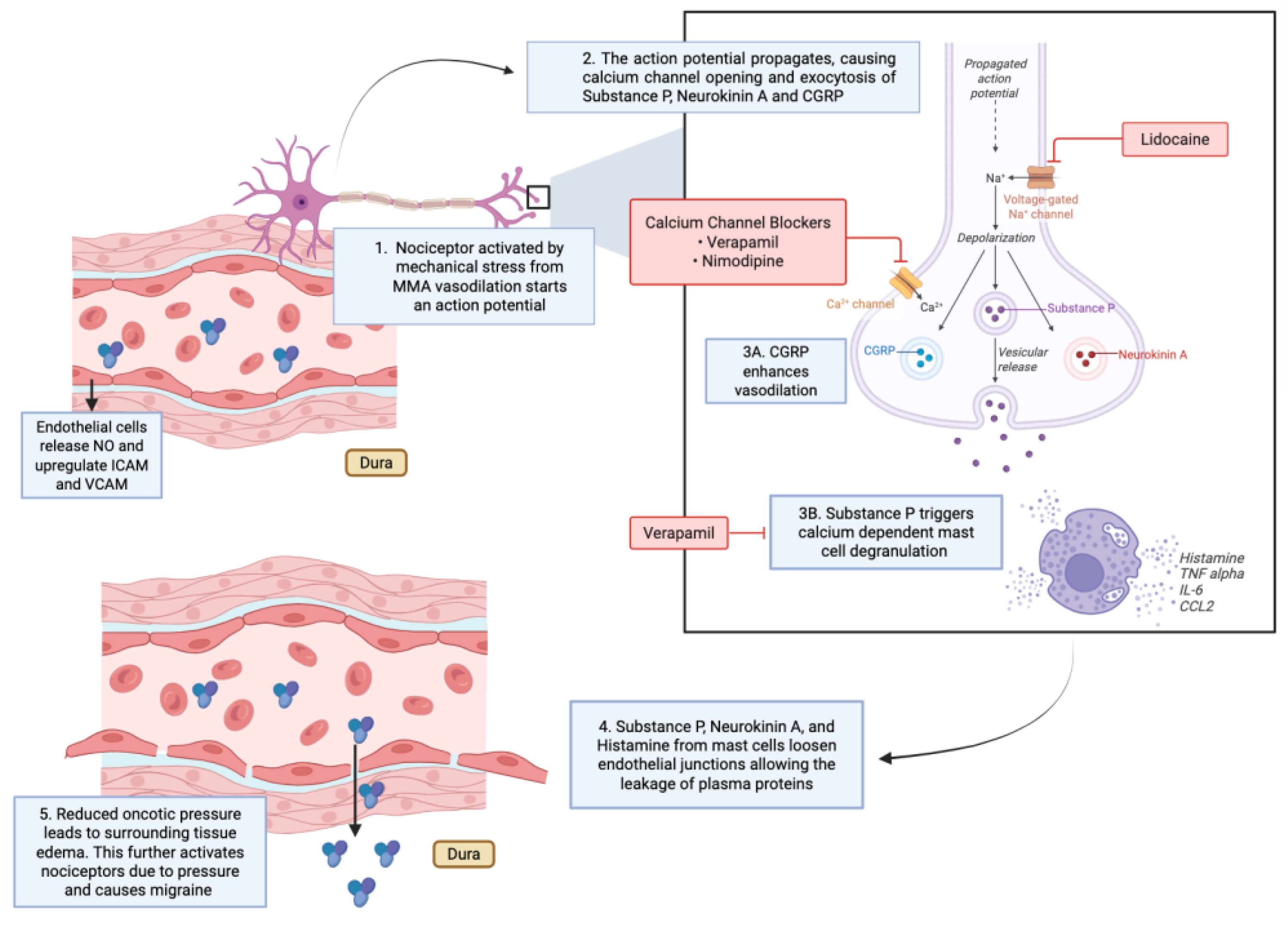
6. Other Pharmacological Targets

Preclinical and translational studies suggest that targeting purinergic receptors (P2Y13 agonists, P2 × 3 antagonists) and VPAC1 receptors in the MMA may offer future anti-migraine strategies, as these pathways influence MMA vasoreactivity and neuropeptide release [16,17,18]
CGRP receptor antagonists, including zavegepant and others, have shown efficacy in blocking CGRP-induced dilation in human MMAs, supporting the artery’s relevance in migraine therapy development [19].
Table 1. Summary of Indications Vasoactive Substances Targeting the MMA.
Vasoactive Substance Use Mechanism of Action (MOA) Citations
Nimodipine (CCB) Case use in RCVS-related vasospasm and migraine relief Inhibits L-type calcium channels in MMA smooth muscle → reduces vasomotion and pulsatile stress; stabilizes vascular tone Elstner et al., 2009
Verapamil (non-DHP CCB) RCVS treatment; diagnostic and therapeutic intra-arterial use for vasoconstriction-driven headaches Smooth muscle relaxation via calcium channel blockade; modest immunomodulatory effects (↓ inflammatory signaling) Farid et al., 2011
Ospel et al., 2020
Sequeiros et al., 2020
Lidocaine Refractory headaches, status migrainosus, SAH-related headache Blocks voltage-gated sodium channels on trigeminal afferents → silences action potentials; prevents CGRP/substance P release; stabilizes mast cells; ↓ NF-κB signaling & cytokines; preserves endothelial junctions. Qureshi et al., 2014; Khattar et al., 2025; Mancuso-Marcello et al., 2023;
Sirakov et al., 2025; Jaikumar et al., 2025
Lidocaine + MMA Embolization (Onyx, PVA, etc.) Refractory migraine and chronic headache (combined strategy) Combines sodium channel blockade with embolization-induced reduction of dural vascular contribution to pain Catapano et al., 2022; Vanzin & Manzato, 2025
Purinergic Receptor Modulators (P2Y13 agonists, P2 × 3 antagonists) Experimental/preclinical migraine targets Modulate MMA vasoreactivity and trigeminal neuropeptide release Haanes et al., 2019
VPAC1 Receptor Modulation (PACAP/VIP pathways) Experimental/preclinical PACAP and VIP cause vasodilation of MMA; receptor modulation may attenuate vasodilatory/mast cell-mediated pathways Boni et al., 2009; Bhatt et al., 2013
CGRP Receptor Antagonists (e.g., zavegepant) Translational/clinical migraine therapy Block CGRP-induced MMA dilation. De Vries et al., 2023

5. Discussion

The middle meningeal artery (MMA) has emerged as a critical locus in migraine biology. Intra-arterial pharmacological approaches directed at the MMA provide a means to intervene at distinct stages of the migraine cascade. Calcium channel blockers interrupt excessive vasomotion and vascular stress, while lidocaine arrests trigeminal neuronal activity and mast cell degranulation, thereby halting neurogenic inflammation. The future of this approach rests on therapies that exploit immunological cascades, and this direction reflects the broader movement in migraine research toward immune modulation, relying less heavily on older vascular stabilization techniques.
Initial clinical observations demonstrate both practicality and therapeutic promise. The pursuit of additional molecular targets, such as purinergic receptors and CGRP pathways, alongside findings from MMA embolization, underscores the artery’s central role in migraine pathophysiology.
Based on new clinical evidence and a better understanding of therapies targeting the immunological cascade, there is now a strong rationale for a controlled evaluation of intra-arterial MMA treatments. Such work may transform the artery from a mere conduit of blood flow into a precise therapeutic target and, in so doing, redefine the treatment paradigm for migraine, although evidence is still limited and further research is required to define its role in clinical practice.

Author Contributions

Conceptualization, J.S. and C.G.L.; methodology, J.S and C.G.L; formal analysis, J.; investigation, J.S. and C.G.L..; resources, J.S. and C.G.L; writing—original draft preparation, J.S, C.G.L, D.S.; writing—review and editing, J.S, C.G.L. All authors have read and agreed to the published version of the manuscript.

Funding

This research received no external funding.

Conflicts of Interest

The authors declare no conflicts of interest.

References

  1. Christensen, C.E.; Younis, S.; Lindberg, U.; et al. Intradural artery dilation during experimentally induced migraine attacks. Pain 2021, 162, 176–183. [Google Scholar] [CrossRef] [PubMed]
  2. Fiorella, D.; Monteith, S.J.; Hanel, R.; et al. Embolization of the middle meningeal artery for chronic subdural hematoma. N Engl J Med 2025, 392, 855–864. [Google Scholar] [CrossRef] [PubMed]
  3. Davies, J.M.; Knopman, J.; Mokin, M.; et al. Adjunctive middle meningeal artery embolization for subdural hematoma. N Engl J Med 2024, 391, 1890–1900. [Google Scholar] [CrossRef] [PubMed]
  4. Elstner, M.; Linn, J.; Müller-Schunk, S.; Straube, A. Reversible cerebral vasoconstriction syndrome: a complicated clinical course treated with intra-arterial application of nimodipine. Cephalalgia 2009, 29, 677–682. [Google Scholar] [CrossRef] [PubMed]
  5. Farid, H.; Tatum, J.K.; Wong, C.; et al. Reversible cerebral vasoconstriction syndrome: treatment with combined intra-arterial verapamil infusion and intracranial angioplasty. AJNR Am J Neuroradiol 2011, 32, E184–7. [Google Scholar] [CrossRef] [PubMed]
  6. Ospel, J.M.; Wright, C.H.; Jung, R.; et al. Intra-Arterial Verapamil Treatment in Oral Therapy-Refractory Reversible Cerebral Vasoconstriction Syndrome. AJNR Am J Neuroradiol 2020, 41, 293–299. [Google Scholar] [CrossRef] [PubMed]
  7. Sequeiros, J.M.; Roa, J.A.; Sabotin, R.P.; et al. Quantifying Intra-Arterial Verapamil Response as a Diagnostic Tool for Reversible Cerebral Vasoconstriction Syndrome. AJNR Am J Neuroradiol 2020, 41, 1869–1875. [Google Scholar] [CrossRef] [PubMed]
  8. Khattar, N.K.; Arthur, A.S.; Santori, D.; et al. Middle meningeal artery lidocaine infusion for refractory headache. J Neurointerv Surg 2025. [CrossRef] [PubMed]
  9. Qureshi, A.; Bains, N.; Bhatti, I.; et al. Abstract TMP6: Intra-arterial Lidocaine Administration of Lidocaine in Middle Meningeal Artery for Treatment of Subarachnoid Hemorrhage-Related Headaches. Stroke 2025;56.
  10. Sirakov, A.; Ninov, K.; Sirakova, K.; Sirakov, S.S. Blazing the trail! Interv Neuroradiol 2025.
  11. Qureshi, A.I.; Pfeiffer, K.; Babar, S.; et al. Intra-arterial injection of lidocaine into middle meningeal artery to treat intractable headaches and severe migraine. J Neuroimaging 2021, 31, 1126–1134. [Google Scholar] [CrossRef] [PubMed]
  12. Catapano, J.S.; Karahalios, K.; Srinivasan, V.M.; et al. Chronic headaches and middle meningeal artery embolization. J Neurointerv Surg 2022, 14, 301–303. [Google Scholar] [CrossRef] [PubMed]
  13. Vanzin, J.R.; Manzato, L.B. Chronic migraine and bilateral occlusion of the middle meningeal artery. Interv Neuroradiol 2025:15910199251337520.
  14. Jaikumar, V.; Moser, M.; Lim, J.; et al. Advanced Treatment Approach: Intra-arterial Lidocaine Injection and Middle Meningeal Artery Embolization With Onyx for Relief of Refractory Migraine: 2-Dimensional Operative Video. Oper Neurosurg (Hagerstown) 2025. [CrossRef] [PubMed]
  15. Mancuso-Marcello, M.; Qureshi, A.I.; Nikola, C.; et al. Intra-arterial lidocaine therapy via the middle meningeal artery for migraine headache: Theory, current practice and future directions. Interv Neuroradiol 2023:15910199231195470.
  16. Haanes, K.A.; Labastida-Ramírez, A.; Blixt, F.W.; et al. Exploration of purinergic receptors as potential anti-migraine targets using established pre-clinical migraine models. Cephalalgia 2019, 39, 1421–1434. [Google Scholar] [CrossRef] [PubMed]
  17. Boni, L.J.; Ploug, K.B.; Olesen, J.; et al. The in vivo effect of VIP, PACAP-38 and PACAP-27 and mRNA expression of their receptors in rat middle meningeal artery. Cephalalgia 2009, 29, 837–847. [Google Scholar] [CrossRef] [PubMed]
  18. Bhatt, D.K.; Gupta, S.; Olesen, J.; Jansen-Olesen, I. PACAP-38 infusion causes sustained vasodilation of the middle meningeal artery in the rat: possible involvement of mast cells. Cephalalgia 2014, 34, 877–886. [Google Scholar] [CrossRef] [PubMed]
  19. De Vries, T.; Boucherie, D.M.; van den Bogaerdt, A.; et al. Blocking the CGRP Receptor: Differences across Human Vascular Beds. Pharmaceuticals (Basel) 2023;16.
Disclaimer/Publisher’s Note: The statements, opinions and data contained in all publications are solely those of the individual author(s) and contributor(s) and not of MDPI and/or the editor(s). MDPI and/or the editor(s) disclaim responsibility for any injury to people or property resulting from any ideas, methods, instructions or products referred to in the content.
Copyright: This open access article is published under a Creative Commons CC BY 4.0 license, which permit the free download, distribution, and reuse, provided that the author and preprint are cited in any reuse.
Prerpints.org logo

Preprints.org is a free preprint server supported by MDPI in Basel, Switzerland.

Subscribe

Disclaimer

Terms of Use

Privacy Policy

Privacy Settings

© 2026 MDPI (Basel, Switzerland) unless otherwise stated