Preprint
Article

This version is not peer-reviewed.

Age-Dependent Time to Radiographic Union and Bone-Healing Dynamics in Distal Radius and Ulna Fractures in Small Dogs Managed with an Anatomically Molded Cast

Submitted:

16 August 2026

Posted:

18 August 2026

You are already at the latest version

Abstract

This study aimed to characterize the age dependence of time to union and healing morphology in longitudinally displaced distal radius and ulna fractures in small dogs. Medical records and serial radiographs of 107 small dogs treated with an anatomically molded cast (AMC) between 2019 and 2025 were retrospectively analyzed. Dogs were classified into four age groups (<6 months, 6 to <12 months, 1 to <2 years, and ≥2 years), and time to radiographic union, relative bone diameter expansion, and the interfragmentary distance reduction rate (IDR) were evaluated. Radiographic union was achieved in all cases. Median times to union were 40.0, 52.0, 64.0, and 81.0 days in ascending order of age, with a significant difference among age groups (P < 0.001). After adjustment for age group in multivariable analysis, each 1-kg decrease in body weight was associated with a 6.2% longer time to radiographic union (P = 0.014). Relative bone diameter expansion was greatest and IDR was lowest in dogs aged <6 months (both P < 0.001). Thus, both time to union and healing morphology under AMC treatment were strongly associated with developmental stage from growth to skeletal maturity. Healing morphology represented a continuous spectrum from envelopment-dominant healing, characterized by abundant callus formation during growth, to convergence-dominant healing, characterized by remodeling of the fracture ends and fragment convergence with skeletal maturation. These findings provide practical clinical guidance for age-specific prognostication, owner communication, and treatment planning.

Keywords: 
;  ;  ;  ;  

1. Introduction

1.1. Limitations of Conventional Cast Immobilization and Plate Fixation

Conventional cast immobilization for distal radius and ulna fractures in small dogs has been associated with difficulty maintaining reduction, delayed union, nonunion, and skin complications [1,2]. Consequently, surgical fixation, particularly plate fixation, has become a principal treatment option [3,4]. However, the distal radius in small dogs has a low intraosseous vascular density, an anatomical feature considered unfavorable for bone healing [5]. During plate fixation, surgical manipulation of the fracture site may disrupt the surrounding soft tissues and periosteum and may affect local osseous blood flow [6,7]. Contact between the implant and bone may further affect cortical blood flow [8,9]. To reduce such biological disruption, the concept of biological fixation has evolved, and minimally invasive techniques represented by minimally invasive plate osteosynthesis (MIPO) have been introduced [6,10,11]. More recently, treatment using non-rigid locking plate configurations that modify construct stiffness and promote indirect bone healing under relative stability has also been reported [12]. Nevertheless, these methods still rely on implant-mediated fixation and remain surgically invasive; they may also alter physiological load transfer through bone, potentially causing stress shielding and affecting bone remodeling [6,13].

1.2. Development and Clinical Application of the AMC

Focusing on the anatomical and mechanical characteristics of the distal radius and ulna in small dogs, the author developed and clinically implemented an anatomically molded cast (AMC) that integrates preservation of the biological healing environment, conformity to limb morphology, and the use of physiological loading during free ambulation.

1.3. Study Objective

This study aimed to determine the age dependence of time to radiographic union and healing morphology under AMC management in 107 small dogs with longitudinally displaced distal radius and ulna fractures. Serial radiographs were also used to characterize healing morphology and to analyze associations of age group with relative bone diameter expansion and the interfragmentary distance reduction rate (IDR), as well as the effect of body weight.

2. Materials and Methods

2.1. Study Design and Case Selection

This retrospective observational study used medical records and radiographs from cases treated at a single institution between 2019 and 2025. The same protocol for treatment eligibility, AMC fabrication, post-application management, and radiographic evaluation criteria was applied throughout the study period. The initial dataset comprised 337 treatment episodes in 308 dogs. Cases were excluded sequentially according to prespecified clinical eligibility and data-quality criteria to establish the final analytical cohort (Figure 1).
To ensure homogeneity of the study population, dogs with a history of surgical treatment for the index fracture, those presenting more than 7 days after injury, and those weighing more than 10 kg were excluded. The following fracture types were also excluded: incomplete fractures (greenstick, compression, or fissure fractures), comminuted fractures, isolated fractures of the radius or ulna, and fractures outside the distal one-third of the radius and ulna. Transverse fractures without longitudinal displacement at initial presentation and fractures that showed longitudinal displacement at presentation but no residual longitudinal displacement after manual reduction were excluded. In dogs that subsequently sustained a refracture, only the initial fracture-treatment episode was included; the repeat treatment episode after refracture was excluded. With respect to completeness of follow-up and radiographic evaluation, we excluded cases in which serial radiographic follow-up ended before the criteria for radiographic union were met, cases in which continued care was transferred for geographical reasons to another clinic whose staff had received AMC training at our institution, and cases in which inadequate radiographic positioning precluded quantitative measurement. Follow-up was discontinued primarily because of long travel distances or owner-related circumstances; no discontinuation attributable to treatment failure was identified. Finally, to avoid correlated observations within the same dog, when both fractured limbs were eligible, one limb was randomly selected using a computer-generated random number and the contralateral limb was excluded. The final analytical cohort comprised 107 limbs from 107 dogs. Dogs were classified by age at injury into four groups: <6 months, 6 to <12 months, 1 to <2 years, and ≥2 years. These age categories were selected based on age-dependent changes in periosteal reparative capacity and the timing of skeletal maturation in small dogs [14,15].

2.2. Ethical Considerations

This retrospective study used anonymized medical records and radiographs obtained during routine clinical care and involved no research-specific intervention; therefore, institutional ethical review was not applicable under the 2026 principles of the Japan Veterinary Medical Association. Written owner consent was obtained in all cases for the treatment and its associated risks and for the academic use of anonymized data and images.

2.3. AMC Treatment Protocol

AMC treatment followed a two-phase protocol based on the stage of healing. During the initial inflammatory phase after injury (Phase 1), strict local stability was provided with a splint; management was then transitioned to the AMC during the subsequent reparative phase (Phase 2).

2.3.1. Phase 1: Initial Reduction and Temporary Stabilization

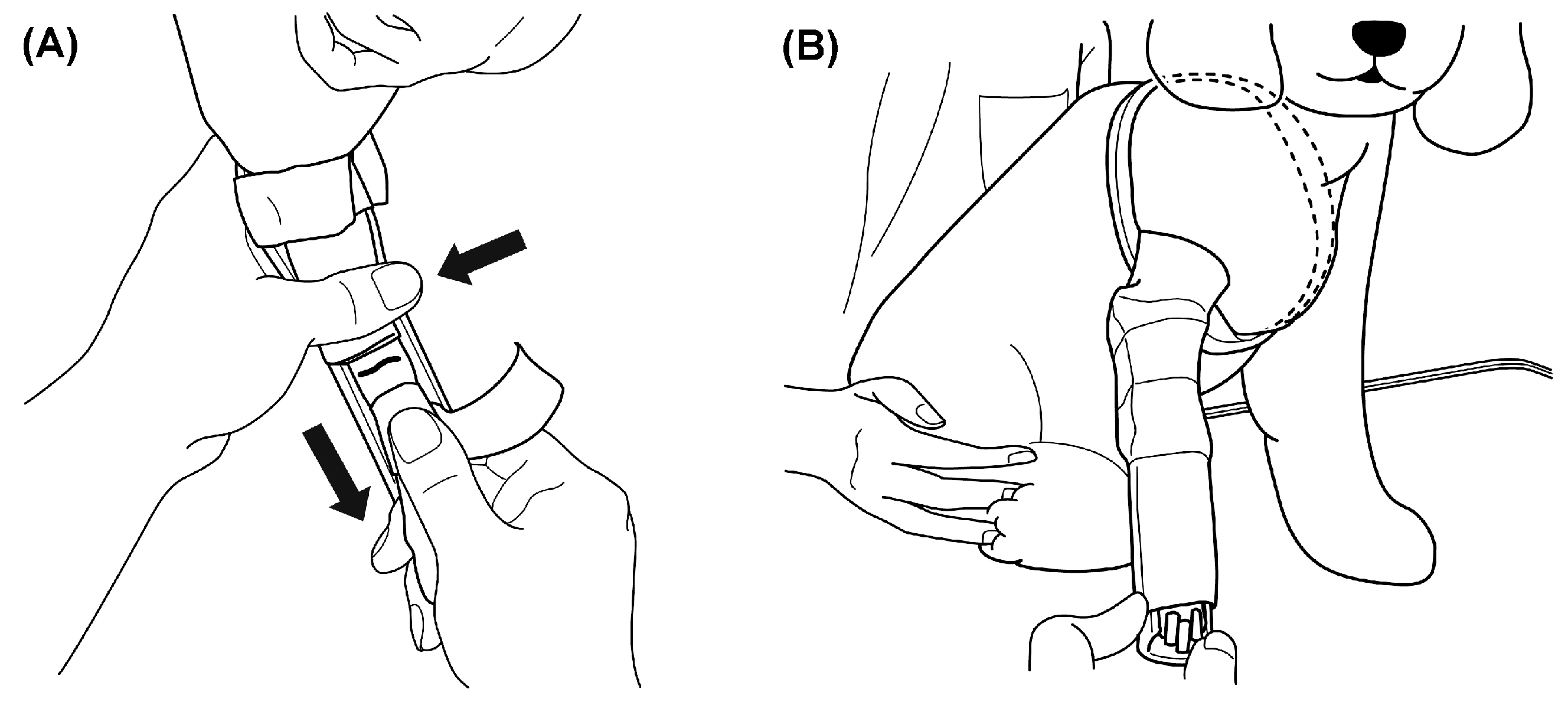
Angular displacement was corrected by manual reduction and longitudinal traction, and the reduction was maintained with a splint. The entire affected limb and splint were then secured together with an elastic bandage to achieve complete immobilization, which was maintained for approximately 1 week (Figure 2).
(A) Angular displacement was corrected manually. While longitudinal traction was maintained using anchor tape applied to the paw, the tape was secured to the distal end of the splint to preserve the reduction. Arrows indicate proximal stabilization and distal traction. (B) After reduction, the limb and splint were wrapped together with an elastic bandage to immobilize the entire affected limb.

2.3.2. Phase 2: Fabrication and Application of the AMC

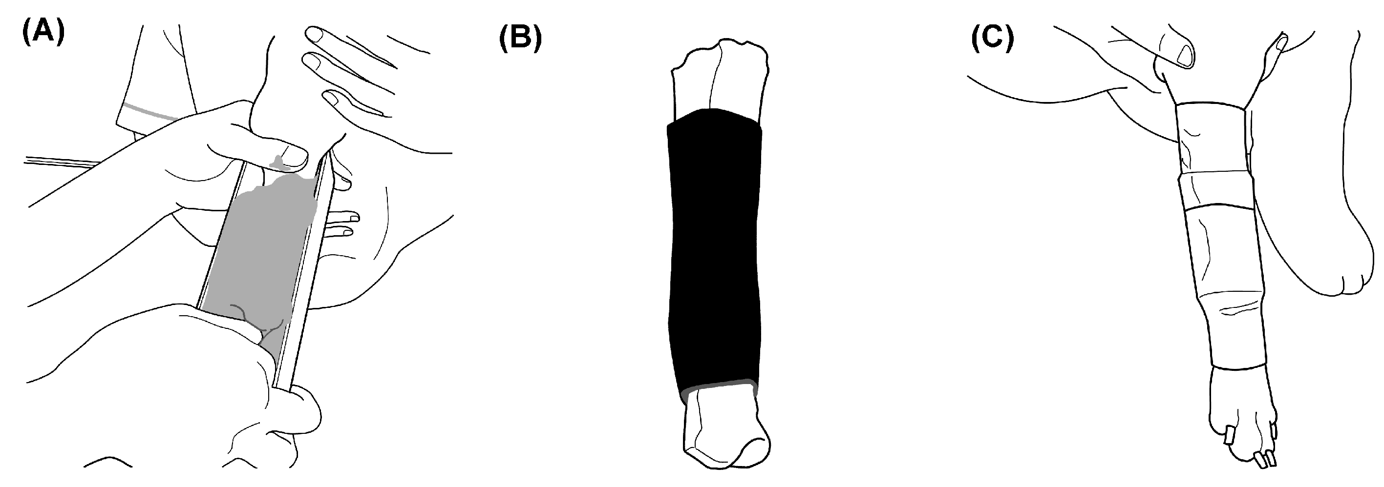
The AMC was fabricated and applied approximately 7 days after injury. A positive plaster model accurately reproducing the anatomical shape of the affected limb was used to fabricate a two-layer tubular AMC with an acrylic foam inner layer and a Scotchcast outer layer; the AMC was then applied to the limb (Figure 3).
(A) An impression of the affected limb was obtained, and a positive plaster model was produced from the resulting negative mold. (B) A two-layer tubular AMC consisting of an acrylic foam inner layer and a Scotchcast outer layer was fabricated on the positive plaster model. (C) The completed AMC was applied to the affected limb and secured with adhesive tape. Free ambulation during daily activities was permitted from the day of AMC application.
Materials used for the AMC are listed in Supplementary Table S1, and detailed fabrication and application procedures are presented in Supplementary Figures S1–S3. Representative serial radiographs obtained during AMC treatment are shown in Supplementary Figure S4.

2.4. Follow-Up Protocol

Because the acrylic foam inner layer of the AMC was in direct contact with the skin, regular skin care was provided throughout treatment. At weekly rechecks, the AMC was temporarily removed, the skin was inspected and washed with chlorhexidine-containing shampoo, and the AMC was reapplied. When dermatitis occurred, antimicrobial therapy was administered according to clinical severity. When distal edema was observed, a compression bandage was applied to the paw together with local cooling, and the tension of the fixation tape was adjusted as needed.

2.5. Radiographic Evaluation and Determination of Bone Union

Radiographic evaluations were performed at 2-week intervals using two orthogonal projections (craniocaudal and lateral). Radiographic union was defined as the first time continuous osseous bridging was identified across all four cortices and was determined without blinding by the first author, who was the attending veterinarian for all cases. The post-union course was evaluated from medical records for cases with in-person follow-up examinations after radiographic union.
Radiographs were acquired using a Toshiba VPX-100A X-ray unit (Toshiba, Tokyo, Japan). Computed radiography images were read using a REGIUS SIGMA A system (Konica Minolta, Tokyo, Japan), and image processing and digital measurements were performed using REGIUS Unitea (version 1.82R00B; Konica Minolta, Tokyo, Japan).

2.6. Radiographic Quantification of Relative Bone Diameter Expansion and the Interfragmentary Distance Reduction Rate (IDR)

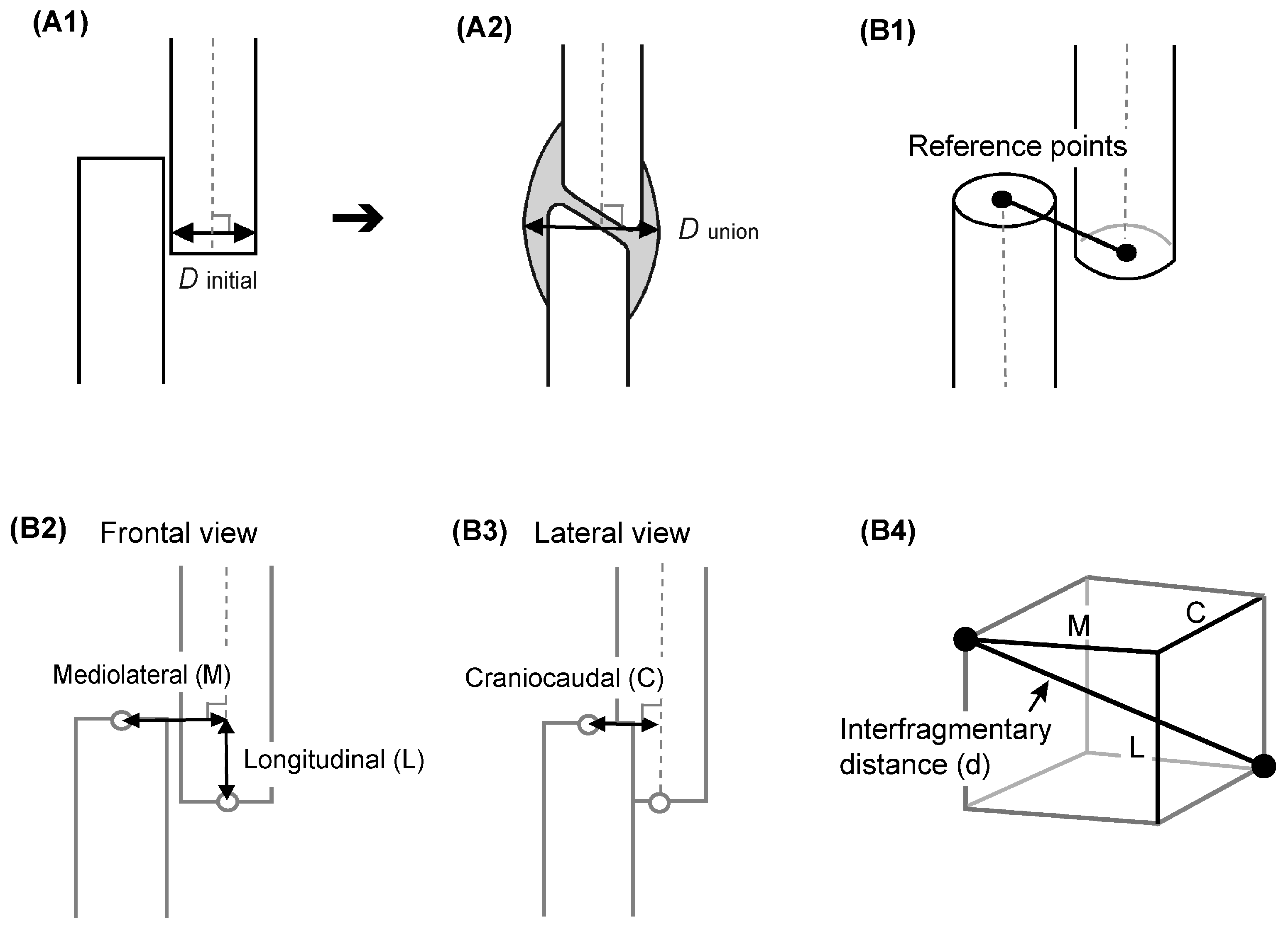
Two orthogonal radiographic projections were used to quantify changes in bone diameter and the distance between fragment reference points during AMC treatment. Relative bone diameter expansion represented the relative increase in the external diameter of the radius from initial presentation to radiographic union. IDR represented the relative decrease in the distance between the reference points on the proximal and distal fragments over the same period (Figure 4).
(A1) The initial bone diameter ( D i n i t i a l ) was measured immediately adjacent to the fracture end, perpendicular to the centerline of the proximal fragment.
(A2) The bone diameter at radiographic union ( D u n i o n ) was defined as the maximum external diameter, including callus (gray area), relative to the same reference axis. Both measurements were obtained in the mediolateral and craniocaudal directions.
(B1) The intersections of the centerlines of the proximal and distal fragments with their respective fracture ends were defined as the measurement reference points.
(B2) On the craniocaudal radiograph, the centerline of the proximal fragment was used as the longitudinal reference axis, and the relative position of the two reference points was measured as a longitudinal component parallel to this axis (L) and a mediolateral component perpendicular to it (M).
(B3) On the lateral radiograph, the craniocaudal component (C) between the two reference points was measured.
(B4) The interfragmentary distance ( d ) was defined by treating L, M, and C as the three mutually orthogonal edges of a rectangular cuboid and calculating its space diagonal as the interfragmentary distance ( d ) using d = L 2 + M 2 + C 2 . The right endpoint of C in the diagram represents the orthogonal projection of the distal reference point.

2.6.1. Calculation of Relative Bone Diameter Expansion

Relative bone diameter expansion in the mediolateral (ML) and craniocaudal (CrCa) directions was calculated using the following equation:
R e l a t i v e   b o n e   d i a m e t e r   e x p a n s i o n   ( % ) = D u n i o n D i n i t i a l D i n i t i a l × 100
A relative bone diameter expansion of 0% indicated no change from initial bone diameter, whereas 100% indicated that bone diameter at radiographic union was twice the initial diameter.

2.6.2. Calculation of Interfragmentary Distance and IDR

The directional components at initial presentation were designated L₀, M₀, and C₀, and those at radiographic union were designated L₁, M₁, and C₁. The interfragmentary distance at initial presentation ( d i n i t i a l ) and at radiographic union ( d u n i o n ) were calculated using the following equations.
d i n i t i a l = L 0 2 + M 0 2 + C 0 2
d u n i o n = L 1 2 + M 1 2 + C 1 2
The interfragmentary distance reduction rate (IDR) was calculated using the following equation.
I D R ( % ) = d i n i t i a l d u n i o n d i n i t i a l × 100
A positive IDR indicated a decrease in interfragmentary distance, whereas a negative IDR indicated an increase in interfragmentary distance.
To evaluate interobserver reliability, 30 cases were selected from the 107-case analytical cohort by stratified random sampling according to age group, and two observers independently performed the measurements. Each observer measured bone diameter in the mediolateral (ML) and craniocaudal (CrCa) directions and each component of interfragmentary distance (L, M, and C) at initial presentation and at radiographic union. Relative bone diameter expansion and IDR were then calculated from each observer's measurements using the same equations.

2.7. Statistical Analysis

All statistical analyses were performed using custom Python scripts in Python version 3.10.16 with SciPy version 1.12.0 and statsmodels version 0.14.0. Distributions of continuous variables were assessed using the Shapiro-Wilk test and are reported as medians and interquartile ranges (IQRs). Differences among age groups in time to radiographic union, relative bone diameter expansion in the ML and CrCa directions, and IDR were assessed using the Kruskal-Wallis test. When a significant difference was detected, post hoc pairwise comparisons were performed using Dunn's test with tie correction, with Bonferroni correction applied to the six pairwise comparisons. Paired ML and CrCa relative bone diameter expansion values within the same case were compared using the Wilcoxon signed-rank test. Homogeneity of variance in time to union was assessed using the Brown-Forsythe and Fligner-Killeen tests for the comparison between dogs aged <6 months and those aged ≥6 months and for the comparison among the three age groups aged ≥6 months.
Because time to union had a right-skewed distribution, the natural logarithm of time to union was used as the dependent variable in regression analyses. First, a univariable regression model was fitted with body weight as the sole explanatory variable. A multivariable linear regression model was then fitted with age group (categorical, with <6 months as the reference group) and body weight as explanatory variables to evaluate their independent associations with time to union. Regression coefficients and their 95% confidence intervals (CIs) were exponentiated and are presented as time ratios. To assess the potential effect of categorizing age, a sensitivity analysis was also performed using a multivariable linear regression model that included natural log-transformed age at injury as a continuous variable together with body weight.
Interobserver reliability was assessed using independent measurements obtained by two observers in 30 cases selected by age-stratified random sampling. Intraclass correlation coefficients [ICC(2,1): two-way random-effects model, absolute agreement, single measurement] and their 95% CIs were calculated. ICC values were interpreted as poor (<0.50), moderate (0.50 to <0.75), good (0.75 to 0.90), or excellent (>0.90) [16]. All tests were two-sided, and statistical significance was set at P < 0.05.

2.8. Use of Generative Artificial Intelligence

During preparation of this manuscript, the author used ChatGPT (OpenAI) to assist with English translation and with the development and execution of statistical analysis code. All generated code, analytical outputs, and text were reviewed and verified by the author, who assumes full responsibility for the content of the manuscript.

3. Results

3.1. Case Characteristics

The final analytical cohort comprised 107 cases (107 limbs). Median age at injury was 11 months (range, 2 months to 12 years 8 months), and median body weight was 2.3 kg (range, 1.0–7.7 kg).Radiographic union was confirmed in all 107 cases. Median time to radiographic union was 57 days (IQR, 45.0–74.5 days). In-person follow-up after radiographic union was available for 54 cases (50.5%), with a median interval of 96 days (range, 3–2,095 days) from the date of radiographic union to the final in-person examination.

3.2. Time to Radiographic Union According to Age

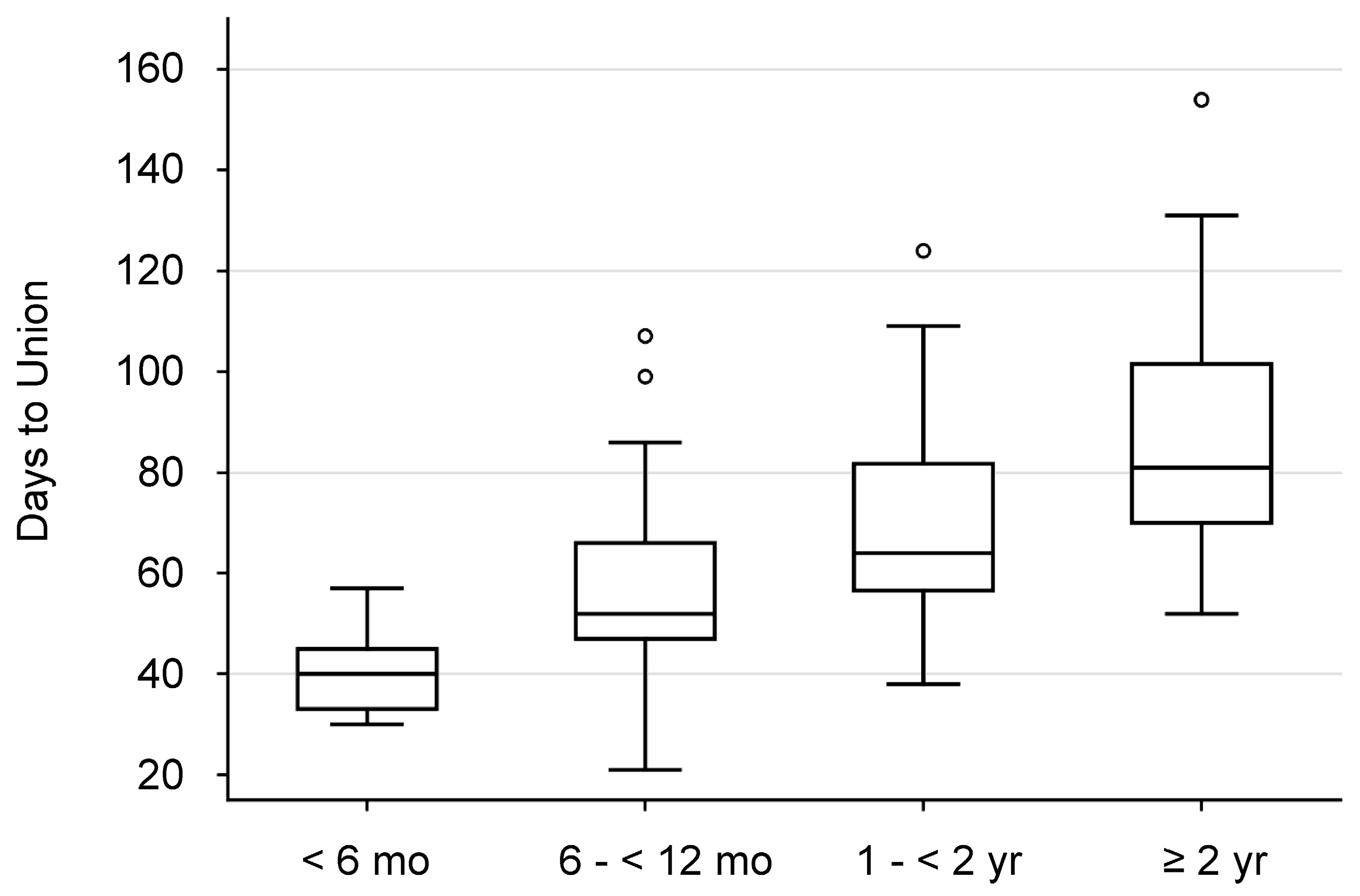
Time to radiographic union differed significantly among the four age groups (Kruskal-Wallis test, H = 49.83, P < 0.001). In Dunn's post hoc test with Bonferroni correction, time to union was significantly shorter in dogs aged <6 months than in every other age group (vs. 6 to <12 months, adjusted P = 0.002; vs. 1 to <2 years and ≥2 years, both adjusted P < 0.001). Time to union was also significantly shorter in dogs aged 6 to <12 months than in those aged ≥2 years (adjusted P < 0.001). No significant differences were observed between dogs aged 6 to <12 months and 1 to <2 years (adjusted P = 0.142) or between those aged 1 to <2 years and ≥2 years (adjusted P = 0.401) (Table 1).
Variability in time to union differed significantly between dogs aged <6 months and those aged ≥6 months and was lower in the <6-month group (Brown-Forsythe test, P = 0.0016; Fligner-Killeen test, P < 0.001). In contrast, no significant difference was detected among the three groups aged ≥6 months (Brown-Forsythe test, P = 0.211; Fligner-Killeen test, P = 0.279). These results indicate relatively low interindividual variability in time to union among dogs younger than 6 months (Figure 5).
Boxes represent the interquartile range (IQR), and horizontal lines within boxes indicate medians. Whiskers indicate the minimum and maximum values within 1.5 times the IQR; open circles indicate outliers. Variance (interindividual variability) in time to union was markedly lower in dogs aged <6 months than in the other age groups.

3.3. Age-Dependent Changes in Bone-Healing Morphology

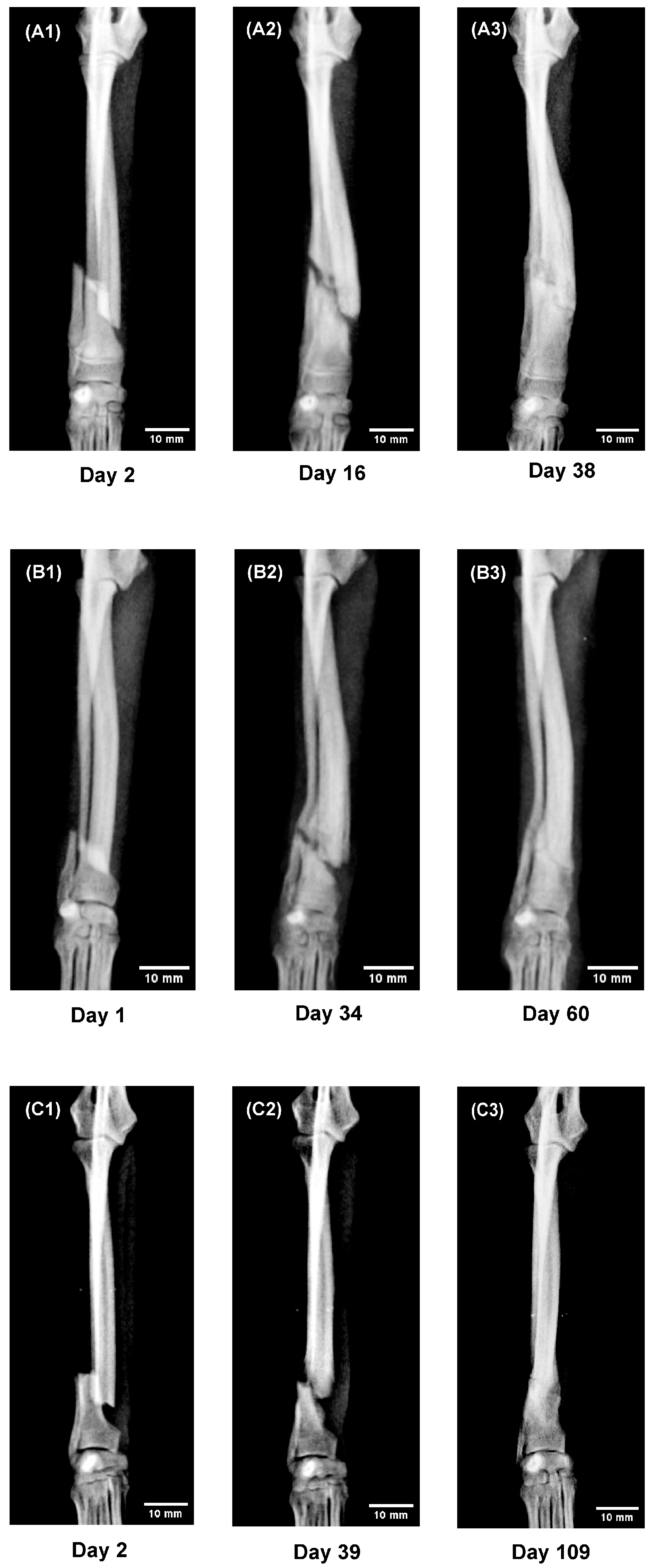
To evaluate differences in bone-healing morphology associated with growth and maturation, this section primarily uses craniocaudal radiographs to compare serial changes in the healing process among three representative cases from different age groups. As a supplementary qualitative assessment, lateral radiographs were used to examine how age and initial alignment affected the morphology and directionality of callus formation.

3.3.1. Serial Changes on Craniocaudal Radiographs

To illustrate age-related changes in bone-healing morphology, serial changes were compared among dogs aged 5, 11, and 19 months, representing different developmental stages (Figure 6).
A: A 5-month-old mixed-breed dog weighing 1.6 kg.
(A1) On day 2 after injury, clear longitudinal displacement was present. (A2) On day 16, extensive callus formation extending from the proximal and distal fragments toward the fracture gap was observed. (A3) On day 38, osseous bridging had formed as callus enveloped the fracture region, with little fragment convergence.
B: An 11-month-old Toy Poodle weighing 1.4 kg.
(B1) On day 1 after injury, longitudinal displacement was present. (B2) On day 34, formation of a contact surface between the fracture ends, shortening of the interfragmentary distance, and localized callus formation were observed; fragment convergence and callus formation progressed in parallel. (B3) On day 60, osseous bridging had formed.
C: A 19-month-old Toy Poodle weighing 1.8 kg.
(C1) On day 2 after injury, clear longitudinal displacement was present. (C2) On day 39, the contours of the opposing fracture ends had become rounded, with formation of a contact surface and localized callus formation adjacent to the fracture ends. (C3) On day 109, fragment convergence had progressed further, and osseous bridging had formed through localized callus formation. Scale bars = 10 mm.

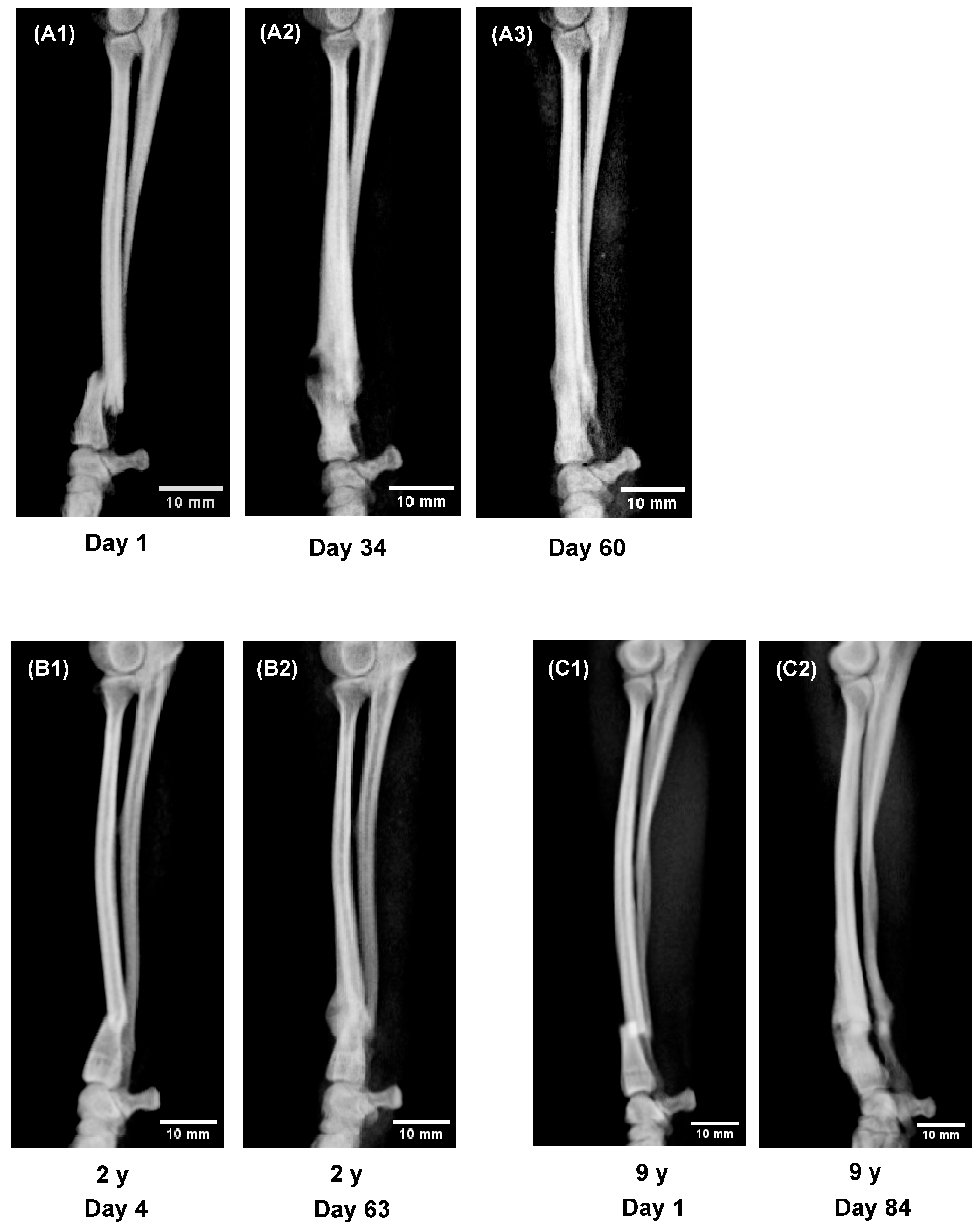
3.3.2. Directionality of Callus Formation on Lateral Radiographs

To illustrate the directionality of callus formation on lateral radiographs, serial images were compared among the 11-month-old dog (the same case as in Figure 6) and 2- and 9-year-old dogs with different initial alignments (Figure 7).
A: An 11-month-old Toy Poodle weighing 1.4 kg (the same case as in Figure 6B1–B3).
(A1) On day 1 after injury, the distal fragment was displaced cranially and longitudinally. (A2) On day 34, extensive callus formation was present along the cranial cortex of the radius, with spicule-like callus projections extending toward each other from the cranial cortices of the proximal and distal fragments. (A3) On day 60, the cranial callus had become continuous and formed osseous bridging. Callus formed predominantly on the cranial aspect of the radius, and the external contour of the fracture region was nearly linear.
B: A 2-year-old Toy Poodle weighing 2.1 kg.
(B1) On day 4 after injury, the distal fragment was angulated in the direction opposite to the physiological cranial curvature. (B2) On day 63, an arcuate callus had formed within the cranial concavity, with localized expansion of bone diameter at the fracture site.
C: A 9-year-old Pomeranian weighing 3.4 kg.
(C1) On day 1 after injury, the proximal and distal fragments largely maintained the physiological cranial curvature. (C2) On day 84, localized callus had formed while this curvature was maintained. Scale bars = 10 mm.

3.4. Age-Group Differences in Relative Bone Diameter Expansion and the Interfragmentary Distance Reduction Rate (IDR)

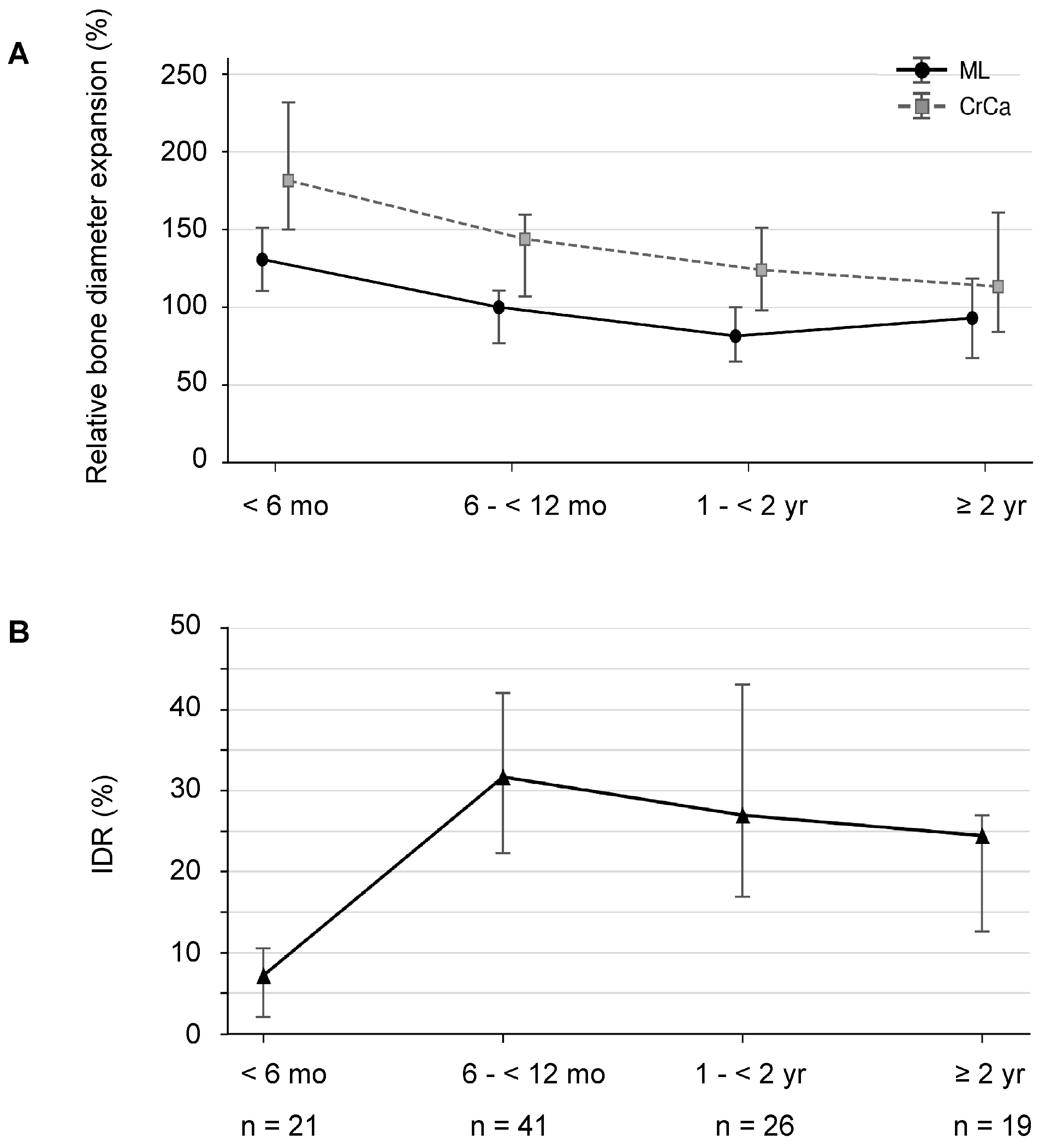
Relative bone diameter expansion in both the mediolateral (ML) and craniocaudal (CrCa) directions differed significantly among age groups (Kruskal-Wallis test: ML, H = 22.64, P < 0.001; CrCa, H = 17.50, P < 0.001). Post hoc testing (Dunn's test with Bonferroni correction) showed significantly greater expansion in dogs aged <6 months than in all three other groups in both directions (ML, all adjusted P ≤ 0.010; CrCa, all adjusted P ≤ 0.017). No significant differences were observed among the three groups aged ≥6 months. In paired comparisons across all 107 cases, CrCa relative bone diameter expansion was significantly greater than ML expansion (Wilcoxon signed-rank test, P < 0.001) (Figure 8A).
IDR also differed significantly among age groups (Kruskal-Wallis test, H = 37.08, P < 0.001; Figure 8B). In post hoc testing, IDR was significantly lower in dogs aged <6 months than in those aged 6 to <12 months and 1 to <2 years (both adjusted P < 0.001). IDR was also significantly higher in dogs aged 6 to <12 months than in those aged ≥2 years (adjusted P = 0.042). No significant differences were observed between dogs aged <6 months and ≥2 years (adjusted P = 0.072), between those aged 6 to <12 months and 1 to <2 years (adjusted P = 1.000), or between those aged 1 to <2 years and ≥2 years (adjusted P = 0.334) (Figure 8B). Median [IQR] values for relative bone diameter expansion and IDR in each age group are provided in Supplementary Table S2.
(A) Relative bone diameter expansion in the mediolateral (ML) and craniocaudal (CrCa) directions. (B) Interfragmentary distance reduction rate (IDR). Symbols indicate medians, and error bars indicate interquartile ranges (IQRs). ML = mediolateral; CrCa = craniocaudal; IDR = interfragmentary distance reduction rate.
Interobserver reliability was good to excellent for bone diameter measurements, moderate for ML relative bone diameter expansion, and good for CrCa relative bone diameter expansion and IDR (Supplementary Table S3).

3.5. Association Between Body Weight and Time to Radiographic Union

In univariable regression without adjustment for age, body weight was not significantly associated with time to radiographic union (time ratio per 1-kg decrease in body weight = 0.968; 95% CI, 0.914–1.024; P = 0.256). In contrast, in the multivariable log-linear regression analysis of all 107 cases adjusted for age group, lower body weight was significantly associated with a longer time to union (adjusted time ratio per 1-kg decrease in body weight = 1.062; 95% CI, 1.013–1.115; P = 0.014). Thus, after adjustment for age group, each 1-kg decrease in body weight was associated with an approximately 6.2% longer time to union (Table 2).
In a sensitivity analysis assessing the effect of age categorization, significant associations of age and body weight with time to union were retained in a model that included natural log-transformed age at injury as a continuous variable together with body weight (model R² = 0.512).

3.6. Refracture After Radiographic Union

Refracture at the same site as the initial fracture was identified in five cases after radiographic union (4.7% of the 107 analyzed cases). All five dogs were aged 1 to <2 years at the initial fracture, weighed 1.2–2.8 kg, and sustained refracture 22–231 days after radiographic union was determined (Supplementary Table S4).
On radiographs obtained at refracture, none of the five cases showed reconstruction of trabecular architecture across the initial fracture site. Compared with bone diameter at radiographic union, bone diameter at the fracture site had decreased in the ML direction in all five cases and in the CrCa direction in four; no change was observed in the remaining case (Supplementary Table S5).

4. Discussion

To our knowledge, this is the first study to evaluate time to union under AMC treatment in longitudinally displaced distal radius and ulna fractures in small dogs, quantify changes in healing morphology using relative bone diameter expansion and IDR, and demonstrate the age dependence of both outcomes. The following discussion considers healing morphology across developmental stages from growth to skeletal maturity, findings associated with refracture, and the clinical implications of AMC treatment in the context of the AMC design concept.

4.1. AMC Design Concept

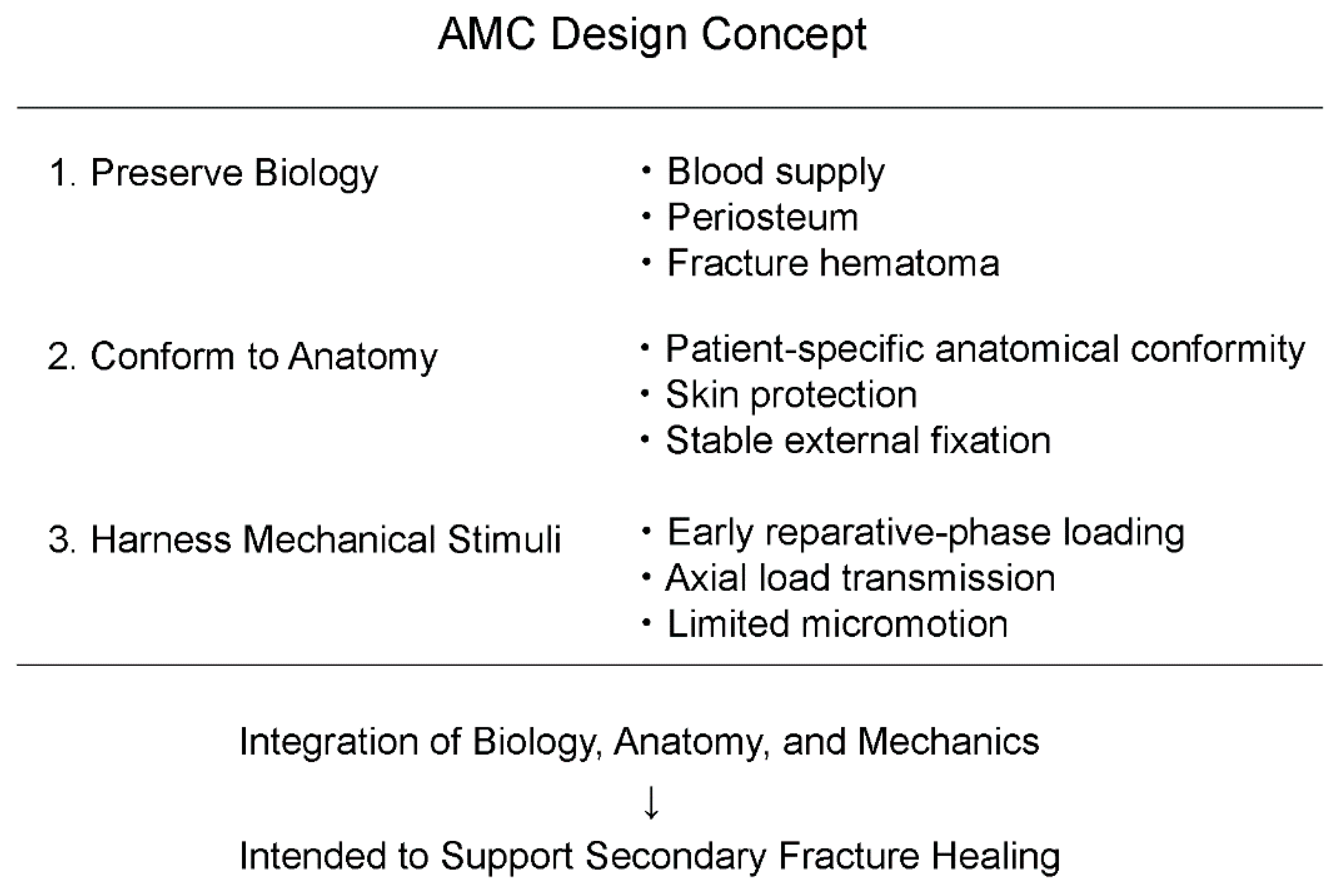
The AMC was designed according to the biological, anatomical, and mechanical characteristics of distal radius and ulna fractures in small dogs (Figure 9).
The AMC was designed around three integrated elements: preservation of blood supply, periosteum, and fracture hematoma; simultaneous skin protection and stable external fixation; and use of physiological mechanical stimulation during free ambulation. Local blood supply, the periosteum, and surrounding soft tissues are important for fracture healing in the distal radius and ulna of small dogs [5,6,17,18], and preservation of this biological healing environment was therefore a fundamental design principle. The AMC also uses a circumferential, two-layer tubular structure. The Scotchcast outer layer limits shear displacement and transverse motion at the fracture while retaining a degree of flexibility, whereas the acrylic foam inner layer improves conformity to the limb and reduces focal pressure on the skin. The tubular structure also helps prevent eccentric loading during ground contact. During canine forelimb locomotion, loads are transmitted primarily along the longitudinal axis [19], and axial loading and moderate micromotion under relative stability are known to promote secondary bone healing [20,21,22,23,24]. Accordingly, free ambulation was permitted with the AMC in place so that physiological loading could contribute to the healing process.

4.2. Mechanical Interpretation and Age-Dependent Healing Patterns During AMC Treatment

During free ambulation, repetitive loading in the stance phase could act to separate or displace the bone fragments. Nevertheless, IDR was positive in 101 of 107 cases (94.4%), indicating a reduction in interfragmentary distance from initial presentation to radiographic union. This finding is consistent with the possibility that the loading environment under AMC treatment contributed to fragment convergence.
Healing morphology also showed a clear age dependence. Extensive callus formation was characteristic in dogs aged <6 months, whereas remodeling of the fracture ends and formation of a contact surface were characteristic from 6 months onward.
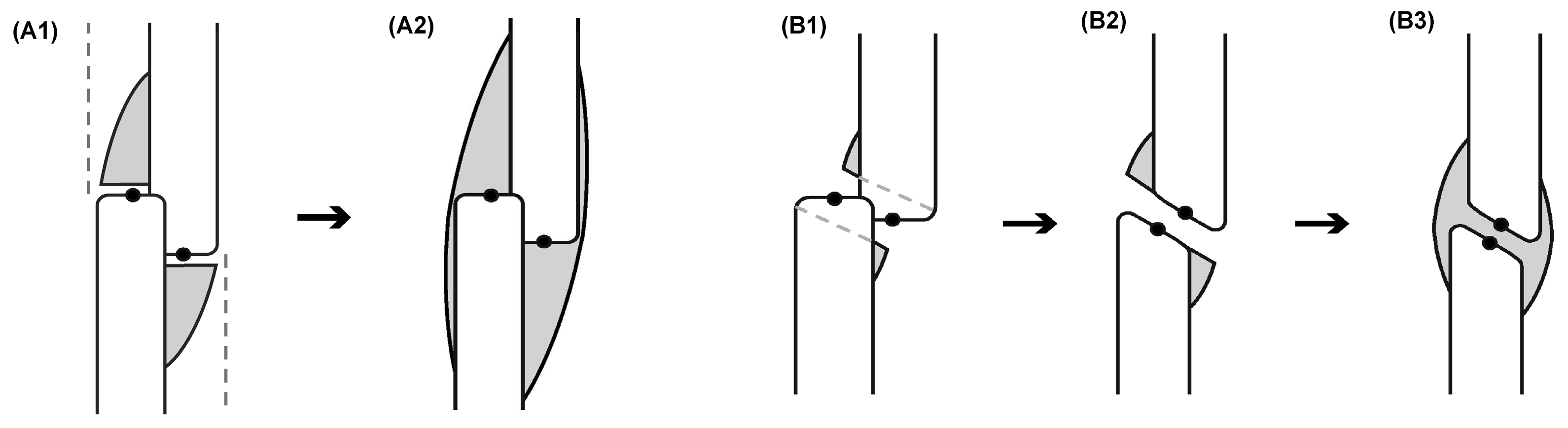
Based on these radiographic characteristics, we define the former as envelopment-dominant healing and the latter as convergence-dominant healing and propose them as two representative healing patterns observed during AMC treatment (Figure 10).
Gray areas indicate callus, and black dots indicate the proximal and distal fragment reference points used to calculate IDR. The dashed lines in A1 indicate the paths of callus extension from the proximal and distal fragments toward the cortex of the opposing fragment. The dashed outlines in B1 indicate the initial bone contours that disappear through bone resorption and remodeling of the fracture ends during healing.

4.2.1. Envelopment-Dominant Healing

Envelopment-dominant healing, observed predominantly in younger dogs, was characterized by high relative bone diameter expansion and low IDR. Callus extended from the cortex of each fragment toward the fracture end of the opposing fragment. It then expanded broadly around the fracture and formed an osseous bridge that enveloped both fragments (Figure 10A1,A2). The periosteum contains skeletal stem cells with high osteoregenerative potential [18]. In rabbits, the chondrogenic capacity of the periosteum and the thickness and cellularity of its cambium layer decrease with age [14], whereas in humans, periosteal bone formation contributes to increases in bone width during growth [25]. Such high periosteal regenerative capacity in young animals is consistent with the abundant callus formation and extensive osseous bridging observed in this study.

4.2.2. Convergence-Dominant Healing

In contrast, convergence-dominant healing, observed from 6 months of age onward, was characterized by relatively low bone diameter expansion and fragment convergence with high IDR. In this pattern, bone resorption and remodeling of the fracture ends created a new contact surface while the interfragmentary distance decreased; maturation of localized callus adjacent to the fracture ends then established osseous bridging (Figure 10B1–B3). This healing pattern is consistent with both the coordinated remodeling processes of bone resorption and formation and tissue adaptation to the local mechanical environment [19,21,26,27,28,29], as well as changes in periosteal function accompanying skeletal maturation [14,25,30]. The multiple morphological processes required to reach union may have contributed to the greater interindividual variability in time to union observed from 6 months of age onward.

4.2.3. Directionality of Callus Formation on Lateral Radiographs and Its Mechanical Basis

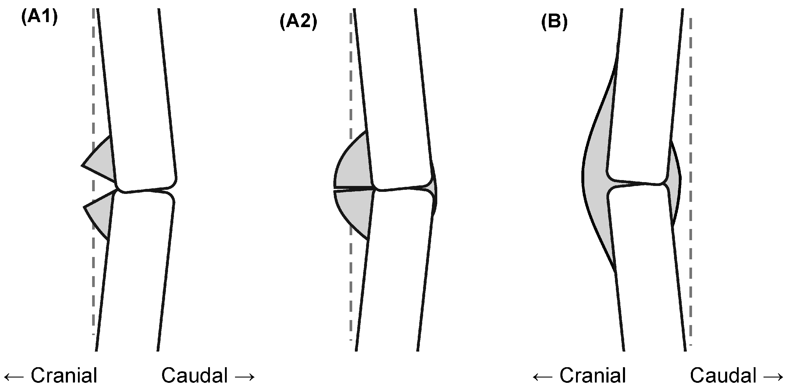
Although the directionality of callus formation on lateral radiographs was evaluated only qualitatively, a tendency toward predominantly cranial callus formation was observed in cases with different initial alignments (Figure 11).
In the case with an initially reverse-bowed configuration, callus formed in a manner that filled the cranial geometric concavity (Figures 7B and 11A1, A2). Predominantly cranial callus formation was also observed in the case in which the native cranial curvature of the radius was relatively well preserved (Figures 7C and 11B). This finding suggests that the directionality of callus formation may not be explained solely by geometric restoration of the bone contour. The canine radius reportedly experiences a three-dimensional strain distribution that varies spatially and temporally during walking and includes pronounced craniocaudal bending [31]. Collectively, these findings suggest that, in addition to geometry, the local mechanical environment during free ambulation may have influenced the craniocaudal morphology of callus formation [18,21,23,31].

4.3. Effect of Body Weight and Age Dependence of Time to Radiographic Union

In the large retrospective study by Marshall et al. [32], the risk of delayed union or nonunion increased with age, whereas toy-breed radioulnar fractures alone were not associated with increased risk. In the present study, body weight was not significantly associated with time to union in univariable analysis; however, a significant association with lower body weight emerged after adjustment for age group. In contrast, time to radiographic union showed a clear age dependence, with median values increasing stepwise across the age groups from youngest to oldest: 40, 52, 64, and 81 days.
These findings indicate that the association with time to union was explained primarily by age, whereas the contribution of body weight was secondary.

4.4. Morphological Compensation for the Growth-Related Decline in Callus Formation

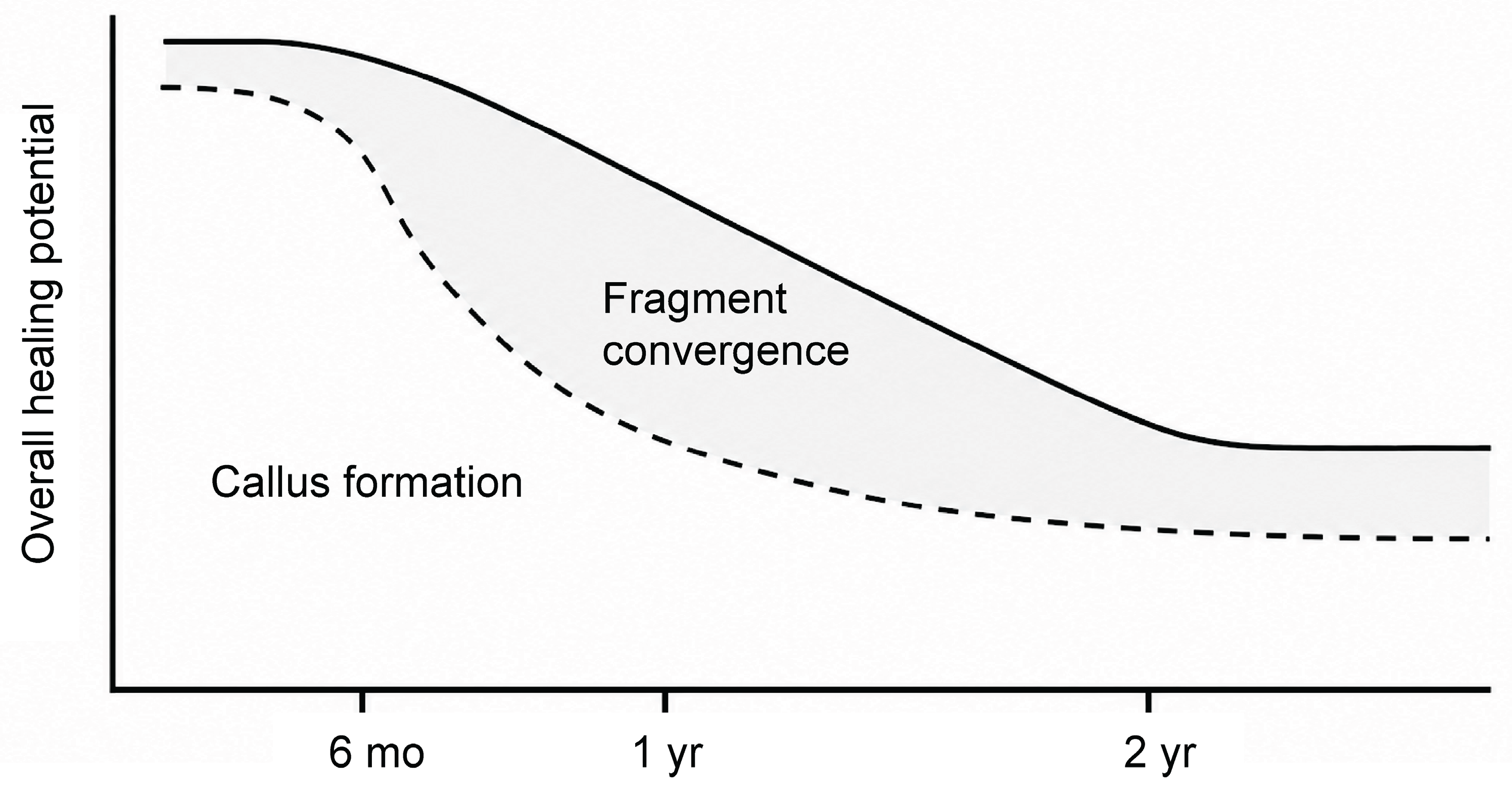
Time to union increased stepwise with developmental stage. In contrast, relative bone diameter expansion decreased markedly from 6 months of age onward, whereas IDR increased.The present results may be explained as a continuous healing spectrum in which the relative contributions of callus formation and fragment convergence change with developmental stage. Specifically, fragment convergence may morphologically compensate for the relative decline in callus formation that accompanies growth (Figure 12).
The solid line represents overall healing potential, expressed as being higher when time to radiographic union is shorter. The dashed line schematically represents the contribution of callus formation, and the gray area represents morphological compensation through fragment convergence.

4.5. Hypothesis of a Vulnerable Remodeling Period Based on Refracture Cases: The Window of Vulnerability

In all five dogs that sustained a refracture, more than 3 weeks had elapsed since radiographic union was determined and a reduction in bone diameter had begun in at least one projection, indicating that the fracture site was in a late remodeling phase. In addition, trabecular reconstruction remained incomplete radiographically, suggesting that reduction of the external diameter may have preceded maturation of the internal architecture. A recent study of dogs treated surgically with plate fixation also reported that reductions in radial thickness and bone-density indices were associated with refracture [33]. That report is consistent with our interpretation that bone mass and architecture after radiographic union may influence refracture risk. We propose the term 'window of vulnerability' for a period during which bone diameter decreases before trabecular reconstruction is complete, transiently increasing mechanical vulnerability. These findings indicate that fracture healing should not be regarded as complete at the moment cortical bridging is identified but should instead be evaluated as a continuous dynamic process that includes reconstruction of trabecular architecture after union.

4.6. Clinical Implications

The clinical implications of AMC treatment can be summarized in four points.
First, maintenance of activity during treatment: permitting free ambulation during daily activities from the early reparative phase with the AMC in place may offer an advantage for treatment management that supports animal welfare.
Second, visualization of the healing process: indirect bone healing, manifested as callus formation, morphological changes of the fracture ends, and fragment convergence during AMC treatment, can be followed serially within the same case.
Third, clarification of the expected treatment course: the age-group-specific times to radiographic union identified in this study provide practical clinical estimates of treatment duration and may assist owner communication and treatment planning.
Fourth, management of refracture risk: the refracture findings observed in this study suggest a risk period during recovery (the window of vulnerability) and highlight the importance of continued evaluation of trabecular restoration after radiographic union.

4.7. Study Limitations and Future Directions

This was a retrospective, single-center study, and radiographic union was determined by a single first author who treated all cases; consequently, observer bias may be present and the generalizability of the findings is limited. Because radiographic evaluations were performed at approximately 2-week intervals, the exact time of union could not be determined; time to union in this study was therefore based on the first imaging date on which osseous bridging of all four cortices was identified. Furthermore, interfragmentary distance was estimated from craniocaudal and lateral radiographs, and measurement error associated with limb positioning, projection angle, and identification of reference points could not be completely eliminated. Future work should include multicenter validation, three-dimensional computed tomography analysis, mechanical testing of AMC materials, gait analysis, and mechanical validation using finite element analysis.
Because oblique radiographs more clearly depicted interfragmentary separation, fragment convergence, and callus formation in some cases (Supplementary Figure S5), future studies should investigate more detailed healing assessment using multiple projections in addition to craniocaudal and lateral views.

5. Conclusions

In this study, radiographic union was achieved in all 107 analyzed cases of longitudinally displaced distal radius and ulna fractures in small dogs treated with an AMC. Both time to union and healing morphology under AMC treatment were strongly associated with age (developmental stage). Healing morphology represented a continuous spectrum from envelopment-dominant healing in immature dogs to convergence-dominant healing from late growth onward, and fragment convergence may morphologically compensate for the relative decline in callus formation that accompanies growth. In contrast, the contribution of body weight remained secondary to that of age. Findings from the refracture cases also led us to propose a 'window of vulnerability' during which structural vulnerability persists after radiographic union until trabecular continuity has been reconstructed. Collectively, these findings highlight the importance of viewing fracture healing as a continuous dynamic process that extends from developmentally determined healing responses through cortical bridging to reconstruction of trabecular architecture.

Supplementary Materials

The following supporting information can be downloaded at the website of this paper posted on Preprints.org. Supplementary Table S1: Materials used for AMC fabrication and application; Supplementary Figure S1: Initial reduction and temporary stabilization; Supplementary Figure S2: AMC fabrication process; Supplementary Figure S3: AMC application and maintenance; Supplementary Figure S4: Serial radiographs obtained during and after AMC treatment; Supplementary Table S2: Age-group-specific relative bone diameter expansion and interfragmentary distance reduction rate (IDR) (N = 107); Supplementary Table S3: Interobserver reliability of radiographic measurements and derived indices in a stratified random sample of 30 cases; Supplementary Table S4: Clinical and radiographic characteristics of dogs with refracture after radiographic union (n = 5); Supplementary Table S5: Bone diameter at the fracture site at initial presentation, radiographic union, and refracture in dogs with refracture (n = 5); Supplementary Figure S5: Oblique radiographic visualization of callus formation and fragment convergence. Supplementary Data S1: Anonymized dataset for the final quantitative analysis cohort (107 limbs from 107 dogs); Supplementary Data S2: Anonymized dataset used for the interobserver reliability analysis (30 cases); Supplementary Data S3: Anonymized dataset describing the five refracture cases. Definitions of variables, units, coding conventions, and missing-value handling are provided in the accompanying data dictionary

Author Contributions

Conceptualization, S.I.; methodology, S.I.; validation, S.I.; formal analysis, S.I.; investigation, S.I.; resources, S.I.; data curation, S.I.; writing—original draft preparation, S.I.; writing—review and editing, S.I.; visualization, S.I.; supervision, S.I.; project administration, S.I. The author has read and agreed to the published version of the manuscript.

Funding

This research received no external funding.

Institutional Review Board Statement

Ethical review and approval were not required for this retrospective study because it involved only medical records and radiographic images obtained during routine veterinary care and included no experimental intervention. All diagnostic and therapeutic procedures were performed as part of routine clinical care.

Data Availability Statement

The anonymized datasets supporting the findings of this study are provided as Supplementary Data S1 (final quantitative analysis cohort), Supplementary Data S2 (interobserver reliability analysis), and Supplementary Data S3 (refracture cases).

Acknowledgments

The author thanks Aki Sugiura and Haruka Tsukino for their assistance with radiographic measurements of bone diameter and interfragmentary distance, and the owners of the dogs included in this study for their cooperation with treatment and follow-up. ChatGPT (OpenAI) was used to assist with English translation and with the development and execution of the statistical analysis code. All AI-assisted outputs were reviewed and verified by the author, who assumes full responsibility for the content of the manuscript.

Conflicts of Interest

The author developed the AMC and provides AMC treatment at his veterinary practice. No external commercial funding or manufacturer support was received. The author declares no other conflicts of interest.

References

  1. Waters, D.J.; Breur, G.J.; Toombs, J.P. Treatment of common forelimb fractures in miniature- and toy-breed dogs. J. Am. Anim. Hosp. Assoc. 1993, 29, 442–448.
  2. Muir, P. Distal antebrachial fractures in toy-breed dogs. Compend. Contin. Educ. Pract. Vet. 1997, 19, 137–145.
  3. DeCamp, C.E.; Johnston, S.A.; Déjardin, L.M.; Schaefer, S.L. Brinker, Piermattei and Flo’s Handbook of Small Animal Orthopedics and Fracture Repair, 5th ed.; Elsevier: St. Louis, MO, USA, 2016.
  4. Larsen, L.J.; Roush, J.K.; McLaughlin, R.M. Bone plate fixation of distal radius and ulna fractures in small- and miniature-breed dogs. J. Am. Anim. Hosp. Assoc. 1999, 35, 243–250. [CrossRef]
  5. Welch, J.A.; Boudrieau, R.J.; DeJardin, L.M.; Spodnick, G.J. The intraosseous blood supply of the canine radius: Implications for healing of distal fractures in small dogs. Vet. Surg. 1997, 26, 57–61. [CrossRef]
  6. Perren, S.M. Evolution of the internal fixation of long bone fractures. The scientific basis of biological internal fixation: Choosing a new balance between stability and biology. J. Bone Joint Surg. Br. 2002, 84, 1093–1110. [CrossRef]
  7. Smith, S.R.; Bronk, J.T.; Kelly, P.J. Effect of fracture fixation on cortical bone blood flow. J. Orthop. Res. 1990, 8, 471–478. [CrossRef]
  8. Lippuner, K.; Vogel, R.; Tepic, S.; Rahn, B.A.; Cordey, J.; Perren, S.M. Effect of animal species and age on plate-induced vascular damage in cortical bone. Arch. Orthop. Trauma Surg. 1992, 111, 78–84. [CrossRef]
  9. Kregor, P.J.; Senft, D.; Parvin, D.; Campbell, C.; Toomey, S.; Parker, C.; Gillespy, T.; Swiontkowski, M.F. Cortical bone perfusion in plated fractured sheep tibiae. J. Orthop. Res. 1995, 13, 715–724. [CrossRef]
  10. Hudson, C.C.; Pozzi, A.; Lewis, D.D. Minimally invasive plate osteosynthesis: Applications and techniques in dogs and cats. Vet. Comp. Orthop. Traumatol. 2009, 22, 175–182. [CrossRef]
  11. Pozzi, A.; Hudson, C.C.; Gauthier, C.M.; Lewis, D.D. Retrospective comparison of minimally invasive plate osteosynthesis and open reduction and internal fixation of radius-ulna fractures in dogs. Vet. Surg. 2013, 42, 19–27. [CrossRef]
  12. Blanco, A.; San Román-Llorens, F.; San Román, F.; González, C.; Climent, A.; Fuertes, J.J.; Whyte, A. Treatment of Simple Fractures of Distal Aspect of Radius and Ulna in Miniature- and Toy-Breed Dogs with Locking Plate in a Non-Rigid Configuration: An Observational Study of 10 Cases. Animals 2026, 16, 2162. [CrossRef]
  13. Uhthoff, H.K.; Poitras, P.; Backman, D.S. Internal plate fixation of fractures: Short history and recent developments. J. Orthop. Sci. 2006, 11, 118–126. [CrossRef]
  14. O’Driscoll, S.W.M.; Saris, D.B.F.; Ito, Y.; Fitzimmons, J.S. The chondrogenic potential of periosteum decreases with age. J. Orthop. Res. 2001, 19, 95–103. [CrossRef]
  15. Hawthorne, A.J.; Booles, D.; Nugent, P.A.; Gettinby, G.; Wilkinson, J. Body-weight changes during growth in puppies of different breeds. J. Nutr. 2004, 134, 2027S–2030S. [CrossRef]
  16. Koo, T.K.; Li, M.Y. A guideline of selecting and reporting intraclass correlation coefficients for reliability research. J. Chiropr. Med. 2016, 15, 155–163. [CrossRef]
  17. Einhorn, T.A. The cell and molecular biology of fracture healing. Clin. Orthop. Relat. Res. 1998, 355 Suppl., S7–S21. [CrossRef]
  18. Duchamp de Lageneste, O.; Julien, A.; Abou-Khalil, R.; Frangi, G.; Carvalho, C.; Cagnard, N.; Cordier, C.; Conway, S.J.; Colnot, C. Periosteum contains skeletal stem cells with high bone regenerative potential controlled by Periostin. Nat. Commun. 2018, 9, 773. [CrossRef]
  19. Rubin, C.T.; Lanyon, L.E. Limb mechanics as a function of speed and gait: A study of functional strains in the radius and tibia of horse and dog. J. Exp. Biol. 1982, 101, 187–211. [CrossRef]
  20. Claes, L.E. Mechanobiologie der Frakturheilung Teil 1: Grundlagen. Unfallchirurg 2017, 120, 14–22. [CrossRef]
  21. Goodship, A.E.; Kenwright, J. The influence of induced micromovement upon the healing of experimental tibial fractures. J. Bone Joint Surg. Br. 1985, 67, 650–655. [CrossRef]
  22. Claes, L.; Heigele, C.A. Magnitudes of local stress and strain along bony surfaces predict the course and type of fracture healing. J. Biomech. 1999, 32, 255–266. [CrossRef]
  23. Carter, D.R.; Beaupré, G.S.; Giori, N.J.; Helms, J.A. Mechanobiology of skeletal regeneration. Clin. Orthop. Relat. Res. 1998, 355 Suppl., S41–S55. [CrossRef]
  24. Gläser, N.; Schröder, M.; Barcik, J.; Haffner-Luntzer, M.; Wehrle, E. Extended view on the mechanobiology of fracture healing: Interplay between mechanics and inflammation. Front. Bioeng. Biotechnol. 2025, 13, 1652897. [CrossRef]
  25. Rauch, F. Bone growth in length and width: The Yin and Yang of bone stability. J. Musculoskelet. Neuronal Interact. 2005, 5, 194–201.
  26. Schindeler, A.; McDonald, M.M.; Bokko, P.; Little, D.G. Bone remodeling during fracture repair: The cellular picture. Semin. Cell Dev. Biol. 2008, 19, 459–466. [CrossRef]
  27. Frost, H.M. Bone’s mechanostat: A 2003 update. Anat. Rec. A Discov. Mol. Cell. Evol. Biol. 2003, 275A, 1081–1101. [CrossRef]
  28. Wolff, J. The Law of Bone Remodelling; Maquet, P.; Furlong, R., Translators; Springer-Verlag: Berlin/Heidelberg, Germany, 1986. [CrossRef]
  29. Turner, C.H. Three rules for bone adaptation to mechanical stimuli. Bone 1998, 23, 399–407. [CrossRef]
  30. Shapiro, F. Bone development and its relation to fracture repair: The role of mesenchymal osteoblasts and surface osteoblasts. Eur. Cell. Mater. 2008, 15, 53–76. [CrossRef]
  31. Coleman, J.C.; Hart, R.T.; Owan, I.; Takano, Y.; Burr, D.B. Characterization of dynamic three-dimensional strain fields in the canine radius. J. Biomech. 2002, 35, 1677–1683. [CrossRef]
  32. Marshall, W.G.; Filliquist, B.; Tzimtzimis, E.; Fracka, A.; Miquel, J.; Garcia, J.; Dalla Fontana, M. Delayed union, non-union and mal-union in 442 dogs. Vet. Surg. 2022, 51, 1087–1095. [CrossRef]
  33. Muroi, N.; Kanno, N.; Harada, Y.; Hara, Y. A retrospective study of risk factors associated with refracture after repair of radial–ulnar fractures in small-breed dogs. Vet. Comp. Orthop. Traumatol. 2025, 38, 77–86. [CrossRef]
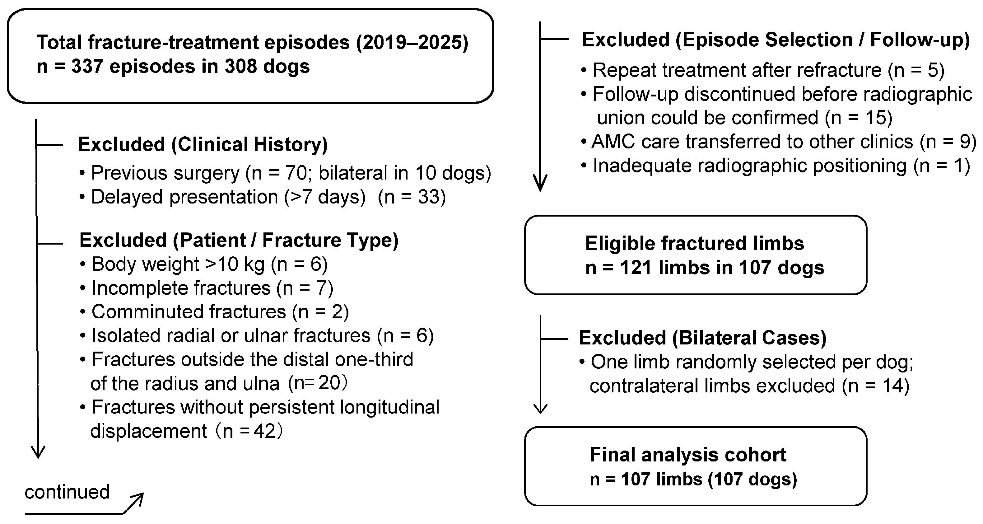
Figure 1. Selection process for the analytical cohort.
Preprints 228568 g001
Figure 2. Initial manual reduction and splint stabilization.
Preprints 228568 g002
Figure 3. Fabrication and application of the AMC.
Preprints 228568 g003
Figure 4. Schematic illustration of the measurement methods for relative bone diameter expansion and the interfragmentary distance reduction rate (IDR).
Preprints 228568 g004
Figure 5. Age-related differences in time to radiographic union.
Preprints 228568 g005
Figure 6. Age-related bone healing morphology under AMC treatment: serial changes in craniocaudal radiographs.
Preprints 228568 g006
Figure 7. Directional pattern of callus formation under AMC treatment: serial changes in lateral radiographs.
Preprints 228568 g007
Figure 8. Age-related changes in bone diameter expansion and interfragmentary distance reduction rate (IDR).
Preprints 228568 g008
Figure 9. Design concept of the anatomically molded cast (AMC).
Preprints 228568 g009
Figure 10. Schematic illustration of fracture-healing patterns during AMC treatment.
Preprints 228568 g010
Figure 11. Schematic representation of the directional pattern of callus formation in relation to initial alignment on lateral radiographs.
Preprints 228568 g011
Figure 12. Conceptual model of age-dependent fracture-healing patterns during AMC treatment.
Preprints 228568 g012
Table 1. Time to radiographic union according to age group in dogs treated with an anatomically molded cast (AMC) (N = 107).
Age Group n Median Days to Union [IQR]
<6 months 21 40.0 [33.0–45.0]
6 to <12 months 41 52.0 [47.0–66.0]
1 to <2 years 26 64.0 [56.5–81.8]
≥2 years 19 81.0 [70.0–101.5]
Table 2. Multivariable log-linear regression analysis of factors associated with time to radiographic union (N = 107).
Predictor β Adjusted time ratio 95% CI P value
Body weight (per 1-kg decrease) 0.061 1.062 1.013–1.115 0.014
Age 6 to <12 months vs. <6 months 0.341 1.407 1.213–1.632 < 0.001
Age 1 to <2 years vs. <6 months 0.553 1.738 1.478–2.045 < 0.001
Age ≥2 years vs. <6 months 0.860 2.363 1.947–2.868 < 0.001
Note: The outcome variable was the natural logarithm of time to radiographic union in days. Body weight was expressed per 1-kg decrease. Adjusted time ratios and their 95% CIs were calculated as exp(β). The age group <6 months was used as the reference category. Model R² = 0.468; adjusted R² = 0.448.
Disclaimer/Publisher’s Note: The statements, opinions and data contained in all publications are solely those of the individual author(s) and contributor(s) and not of MDPI and/or the editor(s). MDPI and/or the editor(s) disclaim responsibility for any injury to people or property resulting from any ideas, methods, instructions or products referred to in the content.
Copyright: This open access article is published under a Creative Commons CC BY 4.0 license, which permit the free download, distribution, and reuse, provided that the author and preprint are cited in any reuse.