
Submitted:
01 October 2025
Posted:
02 October 2025
You are already at the latest version
Abstract
Viruses account for the most abundant biological entities in the biosphere and can be either symbiotic or pathogenic. While pathogenic viruses have developed strategies to evade immunity, the host immune system evolved overlapping and redundant defenses to sense and fight viral infections. Nutrition and metabolic needs sculpt viral-host interactions and determine the course and outcomes of the infection. In this review, we focus on the hexosamine biosynthesis pathway (HBP), a nutrient sensing pathway that controls immune responses and host-viral interactions. The HBP converges on O-GlcNAcylation, a dynamic post-translational modification of cellular proteins, that emerged as a critical effector of immune cell development, differentiation and effector functions. We highlight recent discoveries identifying the impact of this metabolic modification on anti-viral immunity and viral restriction, and conversely on exacerbating viral-induced pathologic inflammation or viral oncogenesis. We discuss the clinical implications of these findings and perspectives of harnessing this pathway for therapeutic purposes.
Keywords:
virus
; infection
; innate immunity
; host-pathogen interaction
; inflammation
; pattern-recognition receptors
; immunometabolism
; nutrient sensing
; glycosylation
; post-translational modification
1. Introduction
1.1. Preface
Viruses account for the most abundant biological entities in the biosphere, with roughly 1031 particles estimated by viral metagenomics [1]. They have co-evolved with their hosts and can be either symbiotic or pathogenic. The human virome, which vastly outnumbers human and bacterial cells, is essential for human health but can contribute to disease in the event of dysbiosis [1,2]. Beyond endogenous viromes, pandemic viruses exert profound consequences on global health, society, and the economy. They often arise from zoonotic spillovers and are shaped by ecological, social, and evolutionary pressures. While each pandemic is unique, patterns of emergence, transmission, and immune responses have revealed recurring themes that have informed our understanding of host–pathogen interactions [3]. RNA viruses such as influenza viruses and coronaviruses have repeatedly driven pandemics due to their high mutation rates, recombination potential, antigenic shift and ability to cross species barriers. These viruses adopt myriads of evolutionary mechanisms that facilitate their survival and adaptation to environmental and host-related conditions. A range of anthropogenic, environmental, viral and host factors converge to determine the pandemic potential of RNA viruses. Anthropogenic factors such as deforestation, biodiversity loss, international travel and the trade of live animals increase interfaces for zoonotic spillover. At the ecological level, bats and birds serve as prolific reservoirs, with high viral diversity supported by their physiology and behavior. In addition, mutations in viral proteins enhance infectivity and interspecies adaptability. On the host side, disparities in disease severity and mortality are influenced by socioeconomic factors and biological differences, such as viral proteins’ receptor-binding compatibility, variations in immune-related genes and metabolic pre-existing illnesses or sequelae that considerably alter the course and outcome of the infection.
1.2. Viral-Immune Co-Evolution
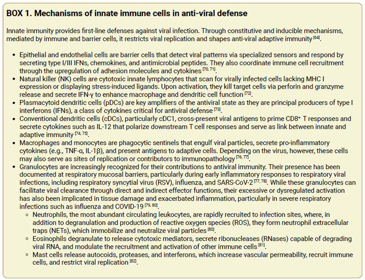
Co-evolutionary selective pressures have shaped both viral evasion and host defense mechanisms [4,5]. Viruses evade immune recognition through a plethora of sophisticated strategies, including inhibition of antigen presentation, disruption of innate sensing, subversion of apoptosis and autophagy, latency, rapid antigenic variation, and cytokine mimicry [6,7]. Several viruses evade detection by pattern-recognition receptors (PRRs), specialized sensors of the innate immune system. For example, members of the Bunyaviridae family encode phosphatases that remove the 5′-triphosphate group from their RNA, converting it to a 5′-monophosphate to avoid recognition by retinoic acid-inducible gene-I (RIG-I); hepatitis C virus (HCV) cleaves Mitochondrial Anti-Viral Signaling (MAVS) protein to disrupt RIG-I–mediated interferon signaling; poxviruses degrade cytosolic DNA to evade detection by the cyclic GMP-AMP synthase (cGAS) - stimulator of interferon genes (STING) pathway; and flaviviruses use non-structural proteins like NS5 to suppress interferon induction downstream of PRR signaling pathways (reviewed in [8]). The host immune system has also evolved overlapping and redundant defenses to fight viral infections. Cells of the innate immune system orchestrate immediate defenses through a plethora of constitutive and inducible mechanisms (Box 1). In addition, multiple innate sensors, such as Toll-like receptors, RIG-I-like receptors, and cytosolic DNA sensors, can detect the same viral molecular pattern through distinct pathways, providing backup if one is compromised (reviewed in [5]). Collectively, these pathways converge on the induction of type I, II, and III interferons, and of interferon-inducible genes, to induce an antiviral state [9,10] (Figure 2). The adaptive immune system bolsters the innate anti-viral response with antibodies that neutralize extracellular viruses and cytotoxic T cells that eliminate infected cells, to establish specific long-term immune memory [11]. This reciprocal adaptation plays out across scales, from molecular interactions between viral epitopes and host receptors to population-level phenomena like antigenic drift. Recent theoretical and computational models, such as those described by Marchi et al., illustrate how viral populations move through antigenic space in response to immune pressure [4]. As Mora and Walczak argue, virus–immune coevolution is not merely a conceptual framework but a quantifiable process with implications for understanding viral persistence, vaccine design, and the limits of immune memory [12].
1.3. Systemic and Cellular Metabolism in Viral Control
Nutrition and metabolic needs sculpt viral-host interactions and determine the course and outcomes of the infection [13,14]. Viruses, as obligate intracellular parasites, are entirely dependent on host-derived nutrients for energy and biosynthetic precursors, including nucleotides, amino acids, and lipids, to support replication, virion assembly, and spread. Similarly, the host immune system undergoes rapid and dynamic metabolic reprogramming during infection to support cellular proliferation, cytokine production, and effector functions. These opposing demands place cellular metabolism at the center of host–pathogen interactions [15]. In particular, nutrient sensing has emerged as a critical regulatory mechanism by which immune cells detect fluctuations in nutrient availability and coordinate metabolic responses to maintain homeostasis and achieve effective response to infection [16]. Nutritional status critically influences both susceptibility to viral infections and the capacity for immune protection. Undernutrition impairs innate and adaptive immunity, whereas overnutrition, particularly obesity, induces chronic low-grade inflammation and predisposes individuals to more severe viral disease outcomes [17,18]. The COVID-19 pandemic made this clinically apparent, with obesity and associated metabolic disorders such as type 2 diabetes emerging as strong predictors of severe disease and mortality. Mechanistically, this relationship is controlled by the overlap of nutrient and pathogen sensing pathways. Mammalian target of rapamycin (mTOR), AMP-activated protein kinase (AMPK), hexosamine biosynthesis pathway (HBP) and PRRs are not only responsive to nutrient fluxes and metabolic stress but also to infection and danger signals [19,20]. For instance, the cGAS-STING pathway, which is central to host antiviral defense, also senses self-DNA in the cytoplasm downstream of metabolic perturbations [21]. Metabolic demands also shape immune cell phenotypes and functions. Notably, inflammatory macrophages and effector T cells shift their cellular metabolism to aerobic glycolysis for enhanced production of building blocks needed for their proliferation and effector functions, whereas memory T cells and regulatory T cells rely on oxidative phosphorylation for bioenergetic needs [15,17,18].
2. The Hexosamine Biosynthesis Pathway and O-GlcNAcylation: From Nutrient Sensing to the Control of Immunity
The HBP has emerged as a unique pathway with capacity to intercept signals from both extracellular nutrients and intracellular metabolic intermediates to shape the immune response. Key substrates driving the HBP include glucose, glucosamine, amino acids, fatty acids, and nucleotides (Figure 1). The HBP consumes 2–3% of glucose from the glycolytic pathway in the form of fructose-6-phosphate (F6P). Similarly, it derives acetyl-CoA (AcCoA) from the oxidation of pyruvate, fatty acid and amino acid breakdown in the tricarboxylic acid (TCA) cycle and obtains uridine triphosphate (UTP) from the de novo pyrimidine biosynthesis pathway. Exogenous glucosamine or GlcNAc can also be salvaged into the pathway by conversion to GlcNAc-6-phosphate (GlcNAc-6-p), bypassing the HBP rate-limiting step at Glutamine-fructose-6-phosphate amidotransferase (GFAT/GFPT) [22]. HBP links nutrient sensing to a dynamic and reversible post-translation modification (PTM) termed O-GlcNAcylation. This PTM of cytoplasmic, mitochondrial and nuclear proteins regulates protein-protein interactions, protein stability and DNA-binding affinity, often in synergy or competition with other PTM such as phosphorylation and ubiquitination. It is catalyzed by one writer, O-GlcNAc transferase (OGT), that couples UDP-GlcNAc to Serine (S) or Threonine (T) residues in target substrates and is reversed by an eraser termed O-GlcNAcase (OGA) [22,23]. Emerging research demonstrates the indispensable role of this PTM in immunity, from controlling immune cell development and differentiation to fine-tuning inducible innate and adaptive immune responses, through direct modification of central effectors (Table 1). In T cells, O-GlcNAcylation peaks shortly after T cell receptor (TCR) engagement and both NFAT and NF-κB interact with OGT and are O-GlcNAcylated, which promotes IL-2 production and T cell activation [24,25,26]. The precise role of O-GlcNAcylation in T cell development was investigated by Swamy et al. who demonstrated that O-GlcNAcylation is central in driving T cell self-renewal and transformation [27]. Using different T cell-specific Ogt knockout mouse strains, they showed that Ogt is required for rapid self-renewal of T cell progenitors, malignant transformation of Pten-deficient thymocytes and T cell positive selection and maturation. They demonstrated that c-Myc is O-GlcNAcylated, which enhanced its protein stability, and that conversely c-Myc drives O-GlcNAcylation by upregulating nutrient uptake [27]. O-GlcNAcylation is equally essential for B cell development, survival and antibody responses. Wu et al. revealed a role of Lyn S19 O-GlcNAcylation in promoting Syk recruitment for efficient B cell receptor (BCR) signaling [28]. Lee et al. further highlighted the importance of c-Myc O-GlcNAcylation in pre-B cell proliferation [29]. More recently, Jeon et al. showed that O-GlcNAcylation regulates chromatin architecture at the immunoglobulin locus, impacting V(D)J recombination during early B-cell development [30]. Using genetic and pharmacological approaches, they showed that caloric restriction, ketogenic diets, or OGT knockdown or inhibition, led to a biased defect in V(D)J recombination and a selective reduction in distal VH gene usage. In contrast, Oga inhibition with Thiamet G, which boosts O-GlcNAcylation, rescued VH gene recombination. O-GlcNAcylation was shown to regulate the cohesin complex, essential for IgH loop extrusion, through direct modification of several of its components, including YY1, CTCF, SMC1 and SMC3. Notably, it promoted SMC1 and SMC3 interaction, as well as YY1 and CTCF DNA-binding to the IgH locus. Collectively, these results link nutritional status and O-GlcNAc metabolism to antibody repertoire diversity.
Besides lymphocyte development, O-GlcNacylation regulates T cell lineage commitment and effector functions. The lineage stability and suppressive program of regulatory T cells (Treg) is governed by Ogt that controls both FoxP3 stability, by preventing its K48 ubiquitin-mediated degradation, and STAT5 activation [31]. The central role of Ogt in this compartment was demonstrated by the severe scurfy-like autoimmune disease observed in mice harboring Treg-specific loss of Ogt [31]. In Th17 cells, O-GlcNAcylation supports the RORγt transcriptional program. This is mediated by acetyl CoA carboxylase 1 (ACC1) modification on S966 and S967 that boosts its enzymatic activity, which contributes to enhanced production of RORγt ligands [32]. It is thus posited that the chronic inflammation elicited in metabolic diseases might be driven by aberrant O-GlcNAcylation leading to heightened ACC1 activity and pro-inflammatory Th17 responses.
While the role of O-GlcNAcylation in myeloid cell development has not been elucidated, key effectors of macrophage metabolism and inflammatory signaling e.g., RIPK3 [33], S6K1 [34] and IRF5 [35] can be O-GlcNAcylated with context-dependent functional outcomes on macrophage inflammatory responses. For instance, while RIPK3 [33] and S6K1 [34] O-GlcNAcylation attenuates their activity in inflammatory signaling, that of IRF5 [35] exerts the opposite effect and leads to heightened inflammation, e.g., in viral infection, as we describe below.

Table 1.
Select O-GlcNAcylated substrates in immunity or viral restriction.
| Substrate | Identified Site | Effect on substrate | Cell type _ Functional Impact | Reference |
|---|---|---|---|---|
| RIPK3 | T467 | Inhibition of phosphorylation | Macrophages _ Reduced necroptosis & inflammation | [33] |
| S6K1 | S489 | Inhibition of phosphorylation | Macrophages _ Reduced inflammation | [34] |
| IRF5 | S430 | Activation through K63-Ub | Macrophages, PBMCs, epithelial cells _ Enhanced inflammation | [35] |
| MAVS | S366; domain 324-347 | Activation through K63-Ub | Macrophages _ Enhanced anti-viral response | [36,38] |
| S249, T250, T252, S253, S255, S256, S257 |
Inhibition | Epithelial cells and fibroblasts _ Reduced anti-viral response | [40] | |
| UBXN1 | S75, T95, S132 | Inhibition of UBXN1-MAVS interaction | Macrophages _ Enhanced MAVS anti-viral response | [41] |
| STING | T229 | Activation through K63-Ub | Fibroblasts _ Enhanced anti-viral response | [42] |
| STAT1 | T699 | Activation by inhibition of kbhb | Fibroblasts _ Enhanced anti-viral response | [43] |
| SAMHD1 | S93 | Stabilization | Macrophages, Hepatocytes _ Enhanced anti-viral response | [49] |
| YTHFD2 | S263 | Stabilization | Hepatocytes _ Enhanced proliferation | [57] |
| NFAT | n.d. | Activation | T cell activation | [24] |
| c-REL | S350 | Activation | T cell activation, FOXP3 suppression | [25,26] |
| CREB | S40 | Activation | HTLV-1 T cell _ Enhanced viral transcription | [56] |
| STAT5 | T38, S57, S58, S270, S273, |
Activation by phosphorylation | Treg _ Enhanced suppressive program | [31] |
| FOXP3 | T38, S57, S58, S270, S273 |
Stabilization | Treg _ Enhanced suppressive program | [31] |
| ACC1 | S966, S967 | Activation | Th17 _Enhanced RORγt transcriptional program | [32] |
| c-MYC | T58 | Stabilization | T cell, B cell _ Enhanced proliferation | [27,29] |
| LYN | S19 | Activation | B cell _ Enhanced BCR signaling | [28] |
| SMC1 | n.d. | Activation | B cell _ VH gene recombination | [30] |
| SMC3 | n.d. | Activation | B cell _ VH gene recombination | [30] |
| YY1 | T236 | Activation | B cell _ VH gene recombination | [30] |
| CTCF | T668 | Activation | B cell _ VH gene recombination | [30] |
n.d., not determined; kbhb, lysine β-hydroxybutyrylation; Ub, ubiquitin; S, serine; T, threonine.
4. O-GlcNAc Interference with the Viral Machinery
In addition to regulating antiviral innate immunity, O-GlcNAcylation can directly interfere with the viral machinery (Table 2). This has been demonstrated for human immunodeficiency virus (HIV-1), Kaposi’s sarcoma-associated herpesvirus (KSHV), hepatitis B virus (HBV), IAV and SARS-CoV-2. Jochmann et al. showed that glucosamine treatment or OGT overexpression inhibited HIV-1 transcription in infected primary CD4+ T cells. This was mediated by O-GlcNAcylation of the host transcription factor Sp1 and required functional Sp1-binding sites in the HIV-1 long terminal repeat (LTR) promoter [46]. While this evidence suggested a potential role of O-GlcNAcylation in restricting HIV-1 infection, the mechanism by which it occurred was not fully investigated, as the reported O-GlcNAcylation of Sp1 did not alter its nuclear levels or DNA-binding affinity to the LTR, suggesting that additional OGT substrates might be involved. In a different report, Jochmann et al. showed that OGT inhibits KSVH viral replication in a dose-dependent manner, potentially through direct modification of 18 viral proteins, demonstrated by mass spectrometry to be O-GlcNAcylated substrates [47] (Figure 3). Among these, ORF75 was confirmed to be O-GlcNAcylated in infected cells, and O-GlcNAc sites were identified in ORF3, ORF10, ORF8, ORF44, ORF21, and ORF29. Nearly half of the O-GlcNAcylated proteins are involved in DNA synthesis and replication, suggesting a central role for O-GlcNAc in regulating these processes, although this has not been formally demonstrated. Concordantly, Ko et al. showed that O-GlcNAcylation of KSVH replication and transcription activator (RTA [ORF50]) suppressed latent-lytic switch [48]. The HBP can additionally counteract viral replication through the O-GlcNAcylation of sterile alpha motif and histidine/aspartic acid domain-containing protein 1 (SAMHD1) [49], a nuclease and dNTP triphosphatase that restricts viral replication by cleaving viral genomes and depleting the dNTP pool. Hu et al. showed that inhibition or knockdown of GLUT1, GFPT1 or OGT increased HBV DNA replication. They identified that SAMHD1 is O-GlcNAcylated on S93, which protects it from K48 ubiquitin-mediated degradation and is required for its tetramerization and antiviral activity against HBV and HIV-1 [49] (Figure 3). In patients with chronic hepatitis B, UDP-GlcNAc levels, GLUT1 expression and total O-GlcNAcylation are higher than in healthy controls, and SAMHD1 is O-GlcNAcylated in liver tissues. It is argued that enhancing O-GlcNAcylation might suppress HBV replication. In a recent report, Dong et al. identified a novel role of OGT in restricting IAV infection. Besides its effect on promoting MAVS antiviral immunity through MAVS O-GlcNAcylation, OGT exerted an additional anti-viral function independently of its catalytic activity. The authors demonstrated that K908A catalytically-inactive OGT dampened IAV infection both in vitro and in vivo. Mechanistically, OGT bound to IAV genomic RNA and limited lipid droplet accumulation, which is required for efficient IAV replication [50]. This was achieved by promoting perilipin-2 (PLIN2) K48-ubiquitination and proteosomal degradation (Figure 3). These findings place OGT at the junction of sugar and lipid metabolism pathways collectively engaged in the fight against IAV infection.
5. Viral Hijacking of the O-GlcNAc Pathway to Dampen Anti-Viral Defenses, or Enhance Infectivity or Viral Oncogenic Transformation
Fricke et al. demonstrated that respiratory syncytial virus (RSV) counters anti-viral defenses by forming cytoplasmic inclusion bodies that sequester p38 and OGT, reducing stress-granule (SG) assembly and global O-GlcNAcylation that limit the infection [51]. Xu et al. described that O-GlcNAcylation of SARS-CoV-2 spike protein at S659 enhances infectivity by promoting more efficient pseudoparticle packaging [52] (Figure 3). They showed that OGT associates with spike protein and enhances its stability by antagonizing K48-ubiquitin tagging and proteosomal degradation. Using a four-plasmid system (pLenti6 luciferase, HIV Gag/pol, HIV Rev and GFP-S) to produce SARS-CoV-2 pseudoparticles (SARS-CoV-2pp) with either wild-type or S659A spike, they showed reduced packaging efficiency and infectivity in pseudoparticles containing the S659A mutant spike protein. Their results suggest that while S659 O-GlcNAcylation does not affect ACE2 receptor binding, it is important for efficient pseudoviral assembly or packaging. The O-GlcNAc pathway, which is elevated in several cancers [53], is also exploited by some oncoviruses to drive tumorigenesis. In the case of human papillomavirus (HPV), Zeng et al. observed that inhibition of O-GlcNAcylation blunted HPV-driven tumorigenesis in vivo. They demonstrated that HPV E6 and E7 oncoproteins stimulated OGT expression, leading to heightened global O-GlcNAcylation and specific c-Myc O-GlcNAcylation on T58, which promotes its stabilization and drives cellular transformation [54] (Figure 3). Kim et al. explored the mechanism of HPV cervical tumor metastasis to the lung. They showed that OGT expression correlates with that of two factors known to contribute to viral oncogenesis and metastasis, namely Host Cell Factor 1 (HCF1) and C-X-C chemokine receptor type 4 (CXCR-4) in HPV-infected cells, but the mechanism linking OGT to their regulation was not fully elucidated [55]. In Human T-lymphotropic virus 1 (HTLV-1), Groussaud et al. showed that the viral oncoprotein Tax hijacks the cellular O-GlcNAc machinery by inhibiting OGA activity, which elevates CREB O-GlcNAcylation promoting LTR activation [56] (Figure 3). This was validated by pharmacologic OGA inhibition or Tax overexpression that similarly increased CREB O-GlcNAcylation and LTR activity. CREB S40A mutation abolished Tax-induced O-GlcNAc modification of CREB and reduced Tax-mediated LTR activation, linking CREB O-GlcNAcylation to a proviral outcome. In hepatitis B virus (HBV)-associated hepatocellular carcinoma (HCC), Yang et al. showed that HBV upregulates the HBP leading to increased global O-GlcNAcylation and specific modification of the N6-methyladenosine (m6A) reader YTH N6-Methyladenosine RNA Binding Protein F2 (YTHFD2) on S263 that drove tumorigenesis [57] (Figure 3). YTHDF2 protein levels were higher in HBV-HCC than in matched non-tumoral tissue, as well as in HBV-transgenic mouse livers. HBV extended YTHDF2 protein half-life, while OGT knockdown or S263A mutation shortened it through K48-linked ubiquitination and proteosomal degradation. YTHDF2 knockdown significantly impaired cell growth, colony formation, and motility in HBV-infected hepatoma lines, functions that were restored by wild-type but not the S263A YTHDF2 mutant. Mechanistically, YTHDF2 O-GlcNAcylation enhanced its binding to m6A-modified transcripts, including those of ‘Minichromosome Maintenance Complex’ components MCM2 and MCM5, that are vital regulators of DNA replication. Thus, HBV engages the OGT pathway to promote HCC tumor formation through YTHDF2 modification and induction of central proliferation effectors.
6. Harnessing the HBP-O-GlcNAc Pathway for Clinical Applications
6.1. The HBP and O-GlcNAcylation as Potential Biomarkers of Disease Progression in Viral Infections
Severe influenza infection provides clinical evidence that O-GlcNAcylation correlates with disease severity [35]. Wang et al. noted that patients with acute influenza had significantly higher global O-GlcNAc levels and OGT expression in their PBMCs compared to healthy individuals. Heightened O-GlcNAc was linked to elevated serum cytokine levels in patients, linking O-GlcNAcylation to a cytokine storm phenotype in severe flu. Patients with chronic HBV infection also show higher UDP-GlcNAc levels and greater protein O-GlcNAcylation in their liver tissue compared to healthy controls [49]. In HBV-related HCC, the HBP-O-GlcNAc axis is often upregulated [57]. As mentioned before, YTHDF2 is heavily O-GlcNAcylated and its levels correlate with worse HBV-HCC patient survival, implicating O-GlcNAcylated YTHDF2 as a potential biomarker of aggressive disease. Similarly, elevated O-GlcNAcylation is a marker of HPV-driven oncogenesis e.g., in cervical cancer [54]. In clinical cervical tumor samples, HPV-positive lesions exhibit higher OGT and O-GlcNAc levels than HPV-negative tissue. The HPV16 E6–driven increase in total cellular O-GlcNAcylation is not only associated with tumorigenesis but also with disease severity, as higher OGT and O-GlcNAc levels correlate with advanced grade or malignant progression. Hence, O-GlcNAcylation can be measured as a clinically relevant biomarker of HPV-associated malignant transformation and progression.
6.2. HBP and O-GlcNAcylation as Therapeutic Targets in Viral Infections
Several studies have underscored immunometabolic modulation as a feasible adjunct treatment in counteracting virus-induced hyperinflammatory states. For instance, in a retrospective study of hospitalized COVID-19 patients, Sukkar et al. reported that a eucaloric ketogenic diet was associated with reduced mortality, fewer ICU admissions, and a favorable trend toward lower IL-6 levels [58]. N-acetyl glucosamine (GlcNAc) that drives the HBP has been tested as a low-cost anti-inflammatory immunomodulatory agent for inflammatory joint illnesses, inflammatory bowel disease and autoimmunity [59,60,61,62,63]. It has also been investigated as a potential anti-inflammatory treatment in respiratory viral infections. In a prospective observational study of COVID-19 patients, GlcNAc was associated with improved clinical outcome [64]. In addition, it has shown anti-viral activity in vitro [65]. Beyond metabolic modulation, direct pharmacological enhancement of the O-GlcNAc pathway is now being evaluated clinically using OGA inhibitors. Ceperognastat (LY3372689) is the most clinically advanced OGA inhibitor [66]. Phase I trials demonstrated oral bioavailability, robust target engagement across dose ranges, >95% OGA enzyme occupancy, and an acceptable safety profile, supporting its progression to Phase II trials. Other OGA inhibitors have also advanced into clinical evaluation. ASN90 (ASN120290) is an orally bioavailable compound in Phase II development with early human studies demonstrating dose-dependent increase in protein O-GlcNAcylation, reliable target engagement confirmed by positron emission tomography, and a favorable safety profile [67]. MK-8719 is another potent, selective OGA inhibitor that completed first-in-human testing [68]. Pharmacodynamic assays confirmed sustained elevation of O-GlcNAc in brain tissue, establishing feasibility of systemic OGA inhibition in humans. Although these compounds were initially developed for tauopathies such as Alzheimer’s disease, their pharmacological properties establish OGA inhibition as clinically viable and highlight the potential for its repurposing in viral infections, where O-GlcNAcylation shapes host-viral interactions.
References
- Koonin, E.V.; Krupovic, M.; Dolja, V.V. The global virome: How much diversity and how many independent origins? Environ Microbiol 2023, 25, 40–44. [Google Scholar] [CrossRef]
- Carroll, D.; Daszak, P.; Wolfe, N.D.; Gao, G.F.; Morel, C.M.; Morzaria, S.; Pablos-Mendez, A.; Tomori, O.; Mazet, J.A.K. The Global Virome Project. Science 2018, 359, 872–874. [Google Scholar] [CrossRef]
- Roychoudhury, S.; Das, A.; Sengupta, P.; Dutta, S.; Roychoudhury, S.; Choudhury, A.P.; Ahmed, A.B.F.; Bhattacharjee, S.; Slama, P. Viral Pandemics of the Last Four Decades: Pathophysiology, Health Impacts and Perspectives. Int J Environ Res Public Health 2020, 17. [Google Scholar] [CrossRef] [PubMed]
- Marchi, J.; Lassig, M.; Walczak, A.M.; Mora, T. Antigenic waves of virus-immune coevolution. Proc Natl Acad Sci U S A 2021, 118. [Google Scholar] [CrossRef]
- Muhammad, I.; Contes, K.; Bility, M.T.; Tang, Q. Chasing Virus Replication and Infection: PAMP-PRR Interaction Drives Type I Interferon Production, Which in Turn Activates ISG Expression and ISGylation. Viruses 2025, 17. [Google Scholar] [CrossRef]
- Beachboard, D.C.; Horner, S.M. Innate immune evasion strategies of DNA and RNA viruses. Curr Opin Microbiol 2016, 32, 113–119. [Google Scholar] [CrossRef]
- Tay, D.J.W.; Lew, Z.Z.R.; Chu, J.J.H.; Tan, K.S. Uncovering Novel Viral Innate Immune Evasion Strategies: What Has SARS-CoV-2 Taught Us? Front Microbiol 2022, 13, 844447. [Google Scholar] [CrossRef] [PubMed]
- Chan, Y.K.; Gack, M.U. Viral evasion of intracellular DNA and RNA sensing. Nat Rev Microbiol 2016, 14, 360–373. [Google Scholar] [CrossRef] [PubMed]
- Schneider, W.M.; Chevillotte, M.D.; Rice, C.M. Interferon-stimulated genes: a complex web of host defenses. Annu Rev Immunol 2014, 32, 513–545. [Google Scholar] [CrossRef]
- Acchioni, M.; Acchioni, C.; Hiscott, J.; Sgarbanti, M. Origin and function of anti-interferon type I viral proteins. Virology 2025, 605, 110456. [Google Scholar] [CrossRef]
- Chi, H.; Pepper, M.; Thomas, P.G. Principles and therapeutic applications of adaptive immunity. Cell 2024, 187, 2052–2078. [Google Scholar] [CrossRef]
- Mora, T.; Walczak, A.M. Quantitative Theory of Viral-Immune Coevolution May Be within Reach. PRX Life 2023, 1. [Google Scholar] [CrossRef]
- Palmer, C.S. Innate metabolic responses against viral infections. Nat Metab 2022, 4, 1245–1259. [Google Scholar] [CrossRef]
- Perakakis, N.; Harb, H.; Hale, B.G.; Varga, Z.; Steenblock, C.; Kanczkowski, W.; Alexaki, V.I.; Ludwig, B.; Mirtschink, P.; Solimena, M.; et al. Mechanisms and clinical relevance of the bidirectional relationship of viral infections with metabolic diseases. Lancet Diabetes Endocrinol 2023, 11, 675–693. [Google Scholar] [CrossRef] [PubMed]
- O’Neill, L.A.; Kishton, R.J.; Rathmell, J. A guide to immunometabolism for immunologists. Nat Rev Immunol 2016, 16, 553–565. [Google Scholar] [CrossRef] [PubMed]
- Baker, S.A.; Rutter, J. Metabolites as signalling molecules. Nat Rev Mol Cell Biol 2023, 24, 355–374. [Google Scholar] [CrossRef] [PubMed]
- Hu, T.; Liu, C.H.; Lei, M.; Zeng, Q.; Li, L.; Tang, H.; Zhang, N. Metabolic regulation of the immune system in health and diseases: mechanisms and interventions. Signal Transduct Target Ther 2024, 9, 268. [Google Scholar] [CrossRef]
- El Safadi, D.; Paulo-Ramos, A.; Hoareau, M.; Roche, M.; Krejbich-Trotot, P.; Viranaicken, W.; Lebeau, G. The Influence of Metabolism on Immune Response: A Journey to Understand Immunometabolism in the Context of Viral Infection. Viruses 2023, 15. [Google Scholar] [CrossRef]
- Kumar, V.; Stewart Iv, J.H. Pattern-Recognition Receptors and Immunometabolic Reprogramming: What We Know and What to Explore. J Innate Immun 2024, 16, 295–323. [Google Scholar] [CrossRef] [PubMed]
- Dsouza, L.; Pant, A.; Pope, B.; Yang, Z. Vaccinia growth factor-dependent modulation of the mTORC1-CAD axis upon nutrient restriction. J Virol 2025, 99, e0211024. [Google Scholar] [CrossRef]
- Gong, J.; Gao, X.; Ge, S.; Li, H.; Wang, R.; Zhao, L. The Role of cGAS-STING Signalling in Metabolic Diseases: from Signalling Networks to Targeted Intervention. Int J Biol Sci 2024, 20, 152–174. [Google Scholar] [CrossRef]
- Paneque, A.; Fortus, H.; Zheng, J.; Werlen, G.; Jacinto, E. The Hexosamine Biosynthesis Pathway: Regulation and Function. Genes (Basel) 2023, 14. [Google Scholar] [CrossRef]
- Issad, T.; Al-Mukh, H.; Bouaboud, A.; Pagesy, P. Protein O-GlcNAcylation and the regulation of energy homeostasis: lessons from knock-out mouse models. J Biomed Sci 2022, 29, 64. [Google Scholar] [CrossRef]
- Golks, A.; Tran, T.T.; Goetschy, J.F.; Guerini, D. Requirement for O-linked N-acetylglucosaminyltransferase in lymphocytes activation. EMBO J 2007, 26, 4368–4379. [Google Scholar] [CrossRef]
- Ramakrishnan, P.; Clark, P.M.; Mason, D.E.; Peters, E.C.; Hsieh-Wilson, L.C.; Baltimore, D. Activation of the transcriptional function of the NF-kappaB protein c-Rel by O-GlcNAc glycosylation. Sci Signal 2013, 6, ra75. [Google Scholar] [CrossRef]
- de Jesus, T.J.; Tomalka, J.A.; Centore, J.T.; Staback Rodriguez, F.D.; Agarwal, R.A.; Liu, A.R.; Kern, T.S.; Ramakrishnan, P. Negative regulation of FOXP3 expression by c-Rel O-GlcNAcylation. Glycobiology 2021, 31, 812–826. [Google Scholar] [CrossRef]
- Swamy, M.; Pathak, S.; Grzes, K.M.; Damerow, S.; Sinclair, L.V.; van Aalten, D.M.; Cantrell, D.A. Glucose and glutamine fuel protein O-GlcNAcylation to control T cell self-renewal and malignancy. Nat Immunol 2016, 17, 712–720. [Google Scholar] [CrossRef]
- Wu, J.L.; Chiang, M.F.; Hsu, P.H.; Tsai, D.Y.; Hung, K.H.; Wang, Y.H.; Angata, T.; Lin, K.I. O-GlcNAcylation is required for B cell homeostasis and antibody responses. Nat Commun 2017, 8, 1854. [Google Scholar] [CrossRef] [PubMed]
- Lee, D.H.; Kwon, N.E.; Lee, W.J.; Lee, M.S.; Kim, D.J.; Kim, J.H.; Park, S.K. Increased O-GlcNAcylation of c-Myc Promotes Pre-B Cell Proliferation. Cells 2020, 9. [Google Scholar] [CrossRef] [PubMed]
- Jeon, B.C.; Kim, Y.J.; Park, A.K.; Song, M.R.; Na, K.M.; Lee, J.; An, D.; Park, Y.; Hwang, H.; Kim, T.D.; et al. Dynamic O-GlcNAcylation governs long-range chromatin interactions in V(D)J recombination during early B-cell development. Cell Mol Immunol 2025, 22, 68–82. [Google Scholar] [CrossRef] [PubMed]
- Liu, B.; Salgado, O.C.; Singh, S.; Hippen, K.L.; Maynard, J.C.; Burlingame, A.L.; Ball, L.E.; Blazar, B.R.; Farrar, M.A.; Hogquist, K.A.; et al. The lineage stability and suppressive program of regulatory T cells require protein O-GlcNAcylation. Nat Commun 2019, 10, 354. [Google Scholar] [CrossRef]
- Machacek, M.; Saunders, H.; Zhang, Z.; Tan, E.P.; Li, J.; Li, T.; Villar, M.T.; Artigues, A.; Lydic, T.; Cork, G.; et al. Elevated O-GlcNAcylation enhances pro-inflammatory Th17 function by altering the intracellular lipid microenvironment. J Biol Chem 2019, 294, 8973–8990. [Google Scholar] [CrossRef]
- Li, X.; Gong, W.; Wang, H.; Li, T.; Attri, K.S.; Lewis, R.E.; Kalil, A.C.; Bhinderwala, F.; Powers, R.; Yin, G.; et al. O-GlcNAc Transferase Suppresses Inflammation and Necroptosis by Targeting Receptor-Interacting Serine/Threonine-Protein Kinase 3. Immunity 2019, 50, 576–590 e576. [Google Scholar] [CrossRef]
- Yang, Y.; Li, X.; Luan, H.H.; Zhang, B.; Zhang, K.; Nam, J.H.; Li, Z.; Fu, M.; Munk, A.; Zhang, D.; et al. OGT suppresses S6K1-mediated macrophage inflammation and metabolic disturbance. Proc Natl Acad Sci U S A 2020, 117, 16616–16625. [Google Scholar] [CrossRef]
- Wang, Q.; Fang, P.; He, R.; Li, M.; Yu, H.; Zhou, L.; Yi, Y.; Wang, F.; Rong, Y.; Zhang, Y.; et al. O-GlcNAc transferase promotes influenza A virus-induced cytokine storm by targeting interferon regulatory factor-5. Sci Adv 2020, 6, eaaz7086. [Google Scholar] [CrossRef] [PubMed]
- Li, T.; Li, X.; Attri, K.S.; Liu, C.; Li, L.; Herring, L.E.; Asara, J.M.; Lei, Y.L.; Singh, P.K.; Gao, C.; et al. O-GlcNAc Transferase Links Glucose Metabolism to MAVS-Mediated Antiviral Innate Immunity. Cell Host Microbe 2018, 24, 791–803 e796. [Google Scholar] [CrossRef]
- Liu, B.; Zhang, M.; Chu, H.; Zhang, H.; Wu, H.; Song, G.; Wang, P.; Zhao, K.; Hou, J.; Wang, X.; et al. The ubiquitin E3 ligase TRIM31 promotes aggregation and activation of the signaling adaptor MAVS through Lys63-linked polyubiquitination. Nat Immunol 2017, 18, 214–224. [Google Scholar] [CrossRef]
- Song, N.; Qi, Q.; Cao, R.; Qin, B.; Wang, B.; Wang, Y.; Zhao, L.; Li, W.; Du, X.; Liu, F.; et al. MAVS O-GlcNAcylation Is Essential for Host Antiviral Immunity against Lethal RNA Viruses. Cell Rep 2019, 28, 2386–2396 e2385. [Google Scholar] [CrossRef]
- Zhang, H.; Xue, K.; Li, W.; Yang, X.; Gou, Y.; Su, X.; Qian, F.; Sun, L. Cullin5 drives experimental asthma exacerbations by modulating alveolar macrophage antiviral immunity. Nat Commun 2024, 15, 252. [Google Scholar] [CrossRef] [PubMed]
- Seo, J.; Park, Y.S.; Kweon, T.H.; Kang, J.; Son, S.; Kim, H.B.; Seo, Y.R.; Kang, M.J.; Yi, E.C.; Lee, Y.H.; et al. O-Linked N-Acetylglucosamine Modification of Mitochondrial Antiviral Signaling Protein Regulates Antiviral Signaling by Modulating Its Activity. Front Immunol 2020, 11, 589259. [Google Scholar] [CrossRef] [PubMed]
- Xia, W.; Jiang, P. p53 promotes antiviral innate immunity by driving hexosamine metabolism. Cell Rep 2024, 43, 113724. [Google Scholar] [CrossRef]
- Li, Y.; An, W.; Lu, L.; Yuan, J.; Wu, D.; Yang, Q.; Guo, J.; Yang, J.; Liu, M.; He, K.; et al. O-GlcNAc of STING mediates antiviral innate immunity. Cell Commun Signal 2024, 22, 157. [Google Scholar] [CrossRef]
- Zuo, Y.; Wang, Q.; Tian, W.; Zheng, Z.; He, W.; Zhang, R.; Zhao, Q.; Miao, Y.; Yuan, Y.; Wang, J.; et al. beta-hydroxybutyrylation and O-GlcNAc modifications of STAT1 modulate antiviral defense in aging. Cell Mol Immunol 2025, 22, 403–417. [Google Scholar] [CrossRef] [PubMed]
- Williams, T.C.; Loo, S.L.; Nichol, K.S.; Reid, A.T.; Veerati, P.C.; Esneau, C.; Wark, P.A.B.; Grainge, C.L.; Knight, D.A.; Vincent, T.; et al. IL-25 blockade augments antiviral immunity during respiratory virus infection. Commun Biol 2022, 5, 415. [Google Scholar] [CrossRef] [PubMed]
- Liang, Y.; Ge, Y.; Sun, J. IL-33 in COVID-19: friend or foe? Cell Mol Immunol 2021, 18, 1602–1604. [Google Scholar] [CrossRef]
- Jochmann, R.; Thurau, M.; Jung, S.; Hofmann, C.; Naschberger, E.; Kremmer, E.; Harrer, T.; Miller, M.; Schaft, N.; Sturzl, M. O-linked N-acetylglucosaminylation of Sp1 inhibits the human immunodeficiency virus type 1 promoter. J Virol 2009, 83, 3704–3718. [Google Scholar] [CrossRef]
- Jochmann, R.; Pfannstiel, J.; Chudasama, P.; Kuhn, E.; Konrad, A.; Sturzl, M. O-GlcNAc transferase inhibits KSHV propagation and modifies replication relevant viral proteins as detected by systematic O-GlcNAcylation analysis. Glycobiology 2013, 23, 1114–1130. [Google Scholar] [CrossRef] [PubMed]
- Ko, Y.C.; Tsai, W.H.; Wang, P.W.; Wu, I.L.; Lin, S.Y.; Chen, Y.L.; Chen, J.Y.; Lin, S.F. Suppressive regulation of KSHV RTA with O-GlcNAcylation. J Biomed Sci 2012, 19, 12. [Google Scholar] [CrossRef]
- Hu, J.; Gao, Q.; Yang, Y.; Xia, J.; Zhang, W.; Chen, Y.; Zhou, Z.; Chang, L.; Hu, Y.; Zhou, H.; et al. Hexosamine biosynthetic pathway promotes the antiviral activity of SAMHD1 by enhancing O-GlcNAc transferase-mediated protein O-GlcNAcylation. Theranostics 2021, 11, 805–823. [Google Scholar] [CrossRef]
- Dong, H.; Liang, C.; Zhang, J.; Wu, W.; Kumar, N.; Liu, Z.; Sun, Y.; Liao, Z.; Cheng, X.; Yu, Y.; et al. O-GlcNAc transferase plays dual antiviral roles by integrating innate immunity and lipid metabolism. Nat Commun 2025, 16, 7721. [Google Scholar] [CrossRef]
- Fricke, J.; Koo, L.Y.; Brown, C.R.; Collins, P.L. p38 and OGT sequestration into viral inclusion bodies in cells infected with human respiratory syncytial virus suppresses MK2 activities and stress granule assembly. J Virol 2013, 87, 1333–1347. [Google Scholar] [CrossRef]
- Xu, T.; Li, J.; Lu, X.; Song, S.; Wang, S.; Li, J.; Zhang, L. O-GlcNAcylation at S659 enhances SARS-CoV-2 spike protein stability and pseudoparticle packaging efficiency. Microbiol Spectr 2025, 13, e0052725. [Google Scholar] [CrossRef] [PubMed]
- Ciraku, L.; Esquea, E.M.; Reginato, M.J. O-GlcNAcylation regulation of cellular signaling in cancer. Cell Signal 2022, 90, 110201. [Google Scholar] [CrossRef]
- Zeng, Q.; Zhao, R.X.; Chen, J.; Li, Y.; Li, X.D.; Liu, X.L.; Zhang, W.M.; Quan, C.S.; Wang, Y.S.; Zhai, Y.X.; et al. O-linked GlcNAcylation elevated by HPV E6 mediates viral oncogenesis. Proc Natl Acad Sci U S A 2016, 113, 9333–9338. [Google Scholar] [CrossRef] [PubMed]
- Kim, S.H.; Kim, Y.S.; Choi, M.Y.; Kim, M.; Yang, J.H.; Park, H.O.; Jang, I.S.; Moon, S.H.; Kim, H.O.; Song, D.H.; et al. O-linked-N-acetylglucosamine transferase is associated with metastatic spread of human papillomavirus E6 and E7 oncoproteins to the lungs of mice. Biochem Biophys Res Commun 2017, 483, 793–802. [Google Scholar] [CrossRef]
- Groussaud, D.; Khair, M.; Tollenaere, A.I.; Waast, L.; Kuo, M.S.; Mangeney, M.; Martella, C.; Fardini, Y.; Coste, S.; Souidi, M.; et al. Hijacking of the O-GlcNAcZYME complex by the HTLV-1 Tax oncoprotein facilitates viral transcription. PLoS Pathog 2017, 13, e1006518. [Google Scholar] [CrossRef]
- Yang, Y.; Yan, Y.; Yin, J.; Tang, N.; Wang, K.; Huang, L.; Hu, J.; Feng, Z.; Gao, Q.; Huang, A. O-GlcNAcylation of YTHDF2 promotes HBV-related hepatocellular carcinoma progression in an N(6)-methyladenosine-dependent manner. Signal Transduct Target Ther 2023, 8, 63. [Google Scholar] [CrossRef] [PubMed]
- Sukkar, S.G.; Cogorno, L.; Pisciotta, L.; Pasta, A.; Vena, A.; Gradaschi, R.; Dentone, C.; Guiddo, E.; Martino, E.; Beltramini, S.; et al. Clinical efficacy of eucaloric ketogenic nutrition in the COVID-19 cytokine storm: A retrospective analysis of mortality and intensive care unit admission. Nutrition 2021, 89, 111236. [Google Scholar] [CrossRef]
- Salvatore, S.; Heuschkel, R.; Tomlin, S.; Davies, S.E.; Edwards, S.; Walker-Smith, J.A.; French, I.; Murch, S.H. A pilot study of N-acetyl glucosamine, a nutritional substrate for glycosaminoglycan synthesis, in paediatric chronic inflammatory bowel disease. Aliment Pharmacol Ther 2000, 14, 1567–1579. [Google Scholar] [CrossRef]
- Grigorian, A.; Araujo, L.; Naidu, N.N.; Place, D.J.; Choudhury, B.; Demetriou, M. N-acetylglucosamine inhibits T-helper 1 (Th1)/T-helper 17 (Th17) cell responses and treats experimental autoimmune encephalomyelitis. J Biol Chem 2011, 286, 40133–40141. [Google Scholar] [CrossRef]
- Richter, J.; Capkova, K.; Hribalova, V.; Vannucci, L.; Danyi, I.; Maly, M.; Fiserova, A. Collagen-induced arthritis: severity and immune response attenuation using multivalent N-acetyl glucosamine. Clin Exp Immunol 2014, 177, 121–133. [Google Scholar] [CrossRef]
- Azuma, K.; Osaki, T.; Wakuda, T.; Tsuka, T.; Imagawa, T.; Okamoto, Y.; Minami, S. Suppressive effects of N-acetyl-D-glucosamine on rheumatoid arthritis mouse models. Inflammation 2012, 35, 1462–1465. [Google Scholar] [CrossRef]
- Lee, S.U.; Li, C.F.; Mortales, C.L.; Pawling, J.; Dennis, J.W.; Grigorian, A.; Demetriou, M. Increasing cell permeability of N-acetylglucosamine via 6-acetylation enhances capacity to suppress T-helper 1 (TH1)/TH17 responses and autoimmunity. PloS one 2019, 14, e0214253. [Google Scholar] [CrossRef] [PubMed]
- Hassan, A.E. An observational cohort study to assess N-acetylglucosamine for COVID-19 treatment in the inpatient setting. Ann Med Surg (Lond) 2021, 68, 102574. [Google Scholar] [CrossRef] [PubMed]
- Marchetti, M.; De Berardis, B.; Bigioni, I.; Mariano, A.; Superti, F.; Scotto d’Abusco, A. In Vitro Antiviral and Anti-Inflammatory Activities of N-Acetylglucosamine: Development of an Alternative and Safe Approach to Fight Viral Respiratory Infections. Int J Mol Sci 2023, 24. [Google Scholar] [CrossRef] [PubMed]
- Kielbasa, W.; Goldsmith, P.; Donnelly, K.B.; Nuthall, H.N.; Shcherbinin, S.; Fleisher, A.S.; Hendle, J.; DuBois, S.L.; Lowe, S.L.; Zhang, F.F.; et al. Discovery and clinical translation of ceperognastat, an O-GlcNAcase (OGA) inhibitor, for the treatment of Alzheimer’s disease. Alzheimers Dement (N Y) 2024, 10, e70020. [Google Scholar] [CrossRef] [PubMed]
- Permanne, B.; Sand, A.; Ousson, S.; Neny, M.; Hantson, J.; Schubert, R.; Wiessner, C.; Quattropani, A.; Beher, D. O-GlcNAcase Inhibitor ASN90 is a Multimodal Drug Candidate for Tau and alpha-Synuclein Proteinopathies. ACS Chem Neurosci 2022, 13, 1296–1314. [Google Scholar] [CrossRef]
- Selnick, H.G.; Hess, J.F.; Tang, C.; Liu, K.; Schachter, J.B.; Ballard, J.E.; Marcus, J.; Klein, D.J.; Wang, X.; Pearson, M.; et al. Discovery of MK-8719, a Potent O-GlcNAcase Inhibitor as a Potential Treatment for Tauopathies. J Med Chem 2019, 62, 10062–10097. [Google Scholar] [CrossRef]
- Pradeu, T.; Thomma, B.; Girardin, S.E.; Lemaitre, B. The conceptual foundations of innate immunity: Taking stock 30 years later. Immunity 2024, 57, 613–631. [Google Scholar] [CrossRef]
- Wells, A.I.; Coyne, C.B. Type III Interferons in Antiviral Defenses at Barrier Surfaces. Trends Immunol 2018, 39, 848–858. [Google Scholar] [CrossRef]
- Tomalka, J.A.; Suthar, M.S.; Diamond, M.S.; Sekaly, R.P. Innate antiviral immunity: how prior exposures can guide future responses. Trends Immunol 2022, 43, 696–705. [Google Scholar] [CrossRef]
- Vivier, E.; Rebuffet, L.; Narni-Mancinelli, E.; Cornen, S.; Igarashi, R.Y.; Fantin, V.R. Natural killer cell therapies. Nature 2024, 626, 727–736. [Google Scholar] [CrossRef]
- Ngo, C.; Garrec, C.; Tomasello, E.; Dalod, M. The role of plasmacytoid dendritic cells (pDCs) in immunity during viral infections and beyond. Cell Mol Immunol 2024, 21, 1008–1035. [Google Scholar] [CrossRef] [PubMed]
- Embgenbroich, M.; Burgdorf, S. Current Concepts of Antigen Cross-Presentation. Front Immunol 2018, 9, 1643. [Google Scholar] [CrossRef] [PubMed]
- Carroll, S.L.; Pasare, C.; Barton, G.M. Control of adaptive immunity by pattern recognition receptors. Immunity 2024, 57, 632–648. [Google Scholar] [CrossRef] [PubMed]
- Wang, X.L.; Wang, X.; Ho, W.Z. Roles of Macrophages in Viral Infections. Viruses 2024, 16. [Google Scholar] [CrossRef]
- Brancewicz, J.; Wojcik, N.; Sarnowska, Z.; Robak, J.; Krol, M. The Multifaceted Role of Macrophages in Biology and Diseases. Int J Mol Sci 2025, 26. [Google Scholar] [CrossRef]
- Rodrigo-Munoz, J.M.; Sastre, B.; Canas, J.A.; Gil-Martinez, M.; Redondo, N.; Del Pozo, V. Eosinophil Response Against Classical and Emerging Respiratory Viruses: COVID-19. J Investig Allergol Clin Immunol 2021, 31, 94–107. [Google Scholar] [CrossRef]
- Rupani, H.; Busse, W.W.; Howarth, P.H.; Bardin, P.G.; Adcock, I.M.; Konno, S.; Jackson, D.J. Therapeutic relevance of eosinophilic inflammation and airway viral interactions in severe asthma. Allergy 2024, 79, 2589–2604. [Google Scholar] [CrossRef]
- Ma, Y.; Zhang, Y.; Zhu, L. Role of neutrophils in acute viral infection. Immun Inflamm Dis 2021, 9, 1186–1196. [Google Scholar] [CrossRef]
- Sasaki, H.; Miyata, J.; Kawana, A.; Fukunaga, K. Antiviral roles of eosinophils in asthma and respiratory viral infection. Front Allergy 2025, 6, 1548338. [Google Scholar] [CrossRef] [PubMed]
- Gebremeskel, S.; Schanin, J.; Coyle, K.M.; Butuci, M.; Luu, T.; Brock, E.C.; Xu, A.; Wong, A.; Leung, J.; Korver, W.; et al. Mast Cell and Eosinophil Activation Are Associated With COVID-19 and TLR-Mediated Viral Inflammation: Implications for an Anti-Siglec-8 Antibody. Front Immunol 2021, 12, 650331. [Google Scholar] [CrossRef] [PubMed]
Figure 1.
The hexosamine biosynthesis pathway (HBP) links nutrient availability to dynamic protein O-GlcNAcylation. The HBP integrates inputs from multiple nutrient and metabolic sources to generate UDP-GlcNAc, the donor substrate for protein O-GlcNAcylation. A fraction of glucose entering glycolysis is diverted at fructose-6-phosphate (F6P), which together with glutamine fuels GFAT/GFPT, the rate-limiting step of the pathway. Additional inputs are provided by acetyl-CoA from the tricarboxylic acid (TCA) cycle, uridine triphosphate (UTP) from pyrimidine biosynthesis and nutrient-sensitive regulators (AMPK, mTOR, AKT, HIF1, MYC). The resulting UDP-GlcNAc is utilized by O-GlcNAc transferase (OGT) to modify serine/threonine residues on nuclear, cytoplasmic, and mitochondrial proteins, while O-GlcNAcase (OGA) removes the modification.
Figure 1.
The hexosamine biosynthesis pathway (HBP) links nutrient availability to dynamic protein O-GlcNAcylation. The HBP integrates inputs from multiple nutrient and metabolic sources to generate UDP-GlcNAc, the donor substrate for protein O-GlcNAcylation. A fraction of glucose entering glycolysis is diverted at fructose-6-phosphate (F6P), which together with glutamine fuels GFAT/GFPT, the rate-limiting step of the pathway. Additional inputs are provided by acetyl-CoA from the tricarboxylic acid (TCA) cycle, uridine triphosphate (UTP) from pyrimidine biosynthesis and nutrient-sensitive regulators (AMPK, mTOR, AKT, HIF1, MYC). The resulting UDP-GlcNAc is utilized by O-GlcNAc transferase (OGT) to modify serine/threonine residues on nuclear, cytoplasmic, and mitochondrial proteins, while O-GlcNAcase (OGA) removes the modification.
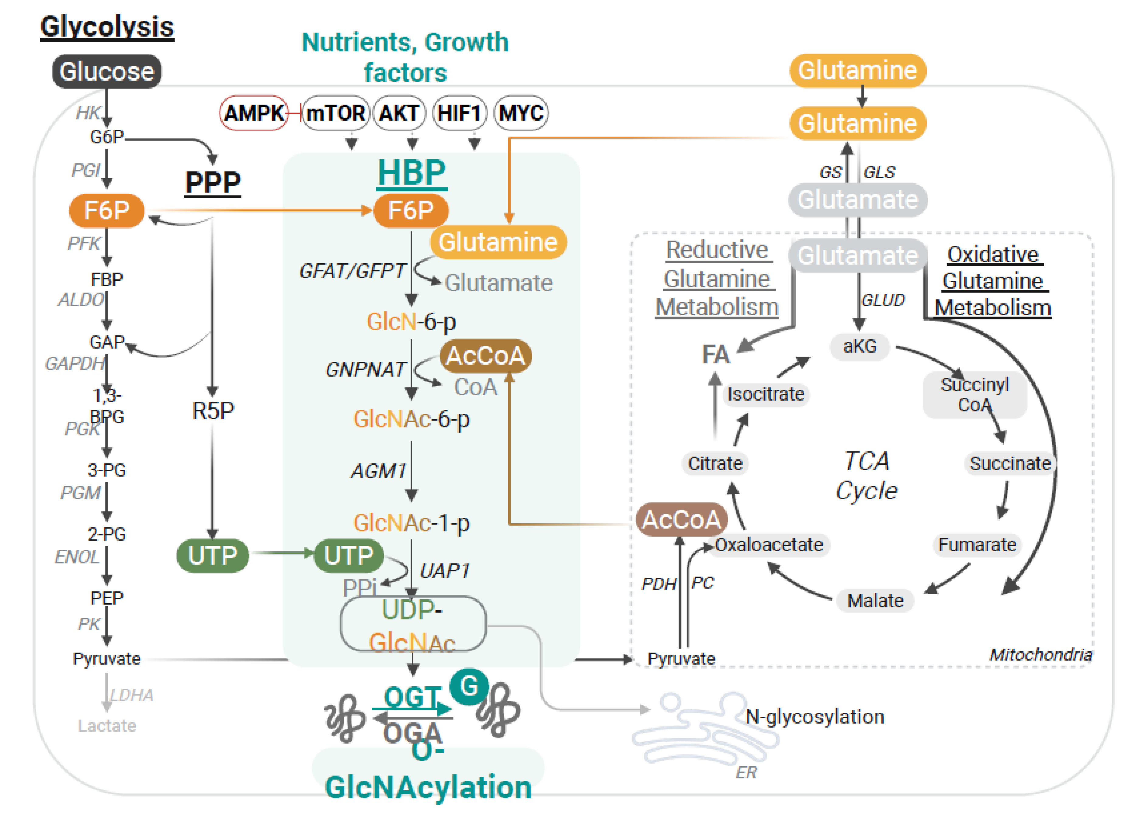
Figure 2.
O-GlcNAcylation in innate antiviral signaling. Pattern recognition receptors (PRRs) such as cGAS, RIG-I, MDA5, and TLRs detect viral nucleic acids and trigger downstream signaling through STING, MAVS, and adaptor complexes that converge on IRF3/5/7 and NF-κB to drive type I interferon and pro-inflammatory cytokine production. O-GlcNAc transferase (OGT) and the hexosamine biosynthesis pathway (HBP) integrate nutrient availability with these antiviral pathways by directly modifying central effectors. MAVS O-GlcNAcylation promotes K63-linked ubiquitination and antiviral signaling, while UBXN1 O-GlcNAcylation relieves its inhibitory effect on MAVS. STING requires O-GlcNAcylation for activation and IFN induction. IRF5 O-GlcNAcylation drives TRAF6-mediated ubiquitination and cytokine storm responses in severe influenza, whereas NF-κB and TAB1 O-GlcNAcylation promote inflammatory gene expression. STAT1 O-GlcNAcylation, that counteracts β-hydroxybutyrylation, is required for efficient IFNAR signaling. CUL5-mediated K48-linked ubiquitination mediates OGT degradation.
Figure 2.
O-GlcNAcylation in innate antiviral signaling. Pattern recognition receptors (PRRs) such as cGAS, RIG-I, MDA5, and TLRs detect viral nucleic acids and trigger downstream signaling through STING, MAVS, and adaptor complexes that converge on IRF3/5/7 and NF-κB to drive type I interferon and pro-inflammatory cytokine production. O-GlcNAc transferase (OGT) and the hexosamine biosynthesis pathway (HBP) integrate nutrient availability with these antiviral pathways by directly modifying central effectors. MAVS O-GlcNAcylation promotes K63-linked ubiquitination and antiviral signaling, while UBXN1 O-GlcNAcylation relieves its inhibitory effect on MAVS. STING requires O-GlcNAcylation for activation and IFN induction. IRF5 O-GlcNAcylation drives TRAF6-mediated ubiquitination and cytokine storm responses in severe influenza, whereas NF-κB and TAB1 O-GlcNAcylation promote inflammatory gene expression. STAT1 O-GlcNAcylation, that counteracts β-hydroxybutyrylation, is required for efficient IFNAR signaling. CUL5-mediated K48-linked ubiquitination mediates OGT degradation.
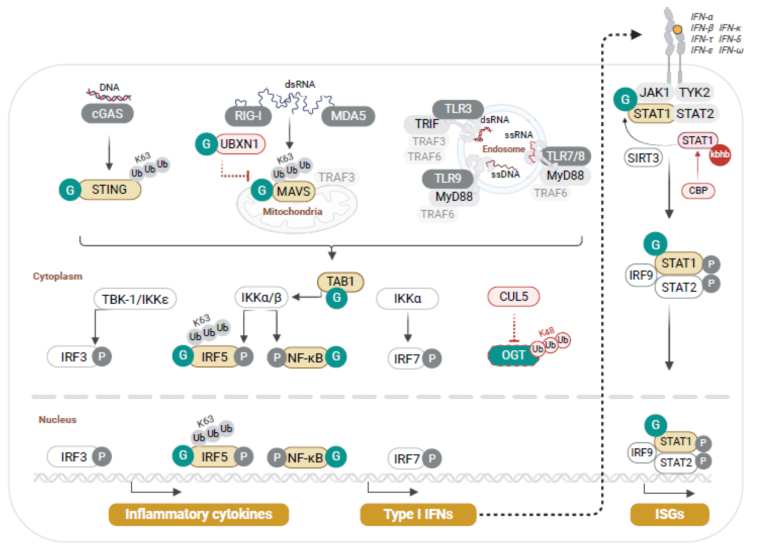
Figure 3.
O-GlcNAcylation modulates multiple steps of the viral life cycle and viral oncogenesis. O-GlcNAcylation stabilizes the antiviral factor SAMHD1, thereby enhancing restriction of HIV-1 and HBV. In HBV infection, O-GlcNAcylation of the m6A reader YTHDF2 promotes stabilization and binding to MCM2/5 transcripts, supporting viral oncogenesis. In influenza A virus (IAV), OGT restricts replication by targeting lipid droplet metabolism via PLIN2 degradation, while in SARS-CoV-2, O-GlcNAcylation of the spike protein enhances stability and particle packaging. In HTLV-1, the viral oncoprotein Tax inhibits OGA, elevating CREB O-GlcNAcylation and driving proviral transcription. HPV E6/E7 stimulate OGT expression, leading to c-MYC O-GlcNAcylation and stabilization, which sustains cellular transformation. In KSHV, O-GlcNAcylation of multiple proteins inhibits viral replication and the latent-lytic cycle.
Figure 3.
O-GlcNAcylation modulates multiple steps of the viral life cycle and viral oncogenesis. O-GlcNAcylation stabilizes the antiviral factor SAMHD1, thereby enhancing restriction of HIV-1 and HBV. In HBV infection, O-GlcNAcylation of the m6A reader YTHDF2 promotes stabilization and binding to MCM2/5 transcripts, supporting viral oncogenesis. In influenza A virus (IAV), OGT restricts replication by targeting lipid droplet metabolism via PLIN2 degradation, while in SARS-CoV-2, O-GlcNAcylation of the spike protein enhances stability and particle packaging. In HTLV-1, the viral oncoprotein Tax inhibits OGA, elevating CREB O-GlcNAcylation and driving proviral transcription. HPV E6/E7 stimulate OGT expression, leading to c-MYC O-GlcNAcylation and stabilization, which sustains cellular transformation. In KSHV, O-GlcNAcylation of multiple proteins inhibits viral replication and the latent-lytic cycle.
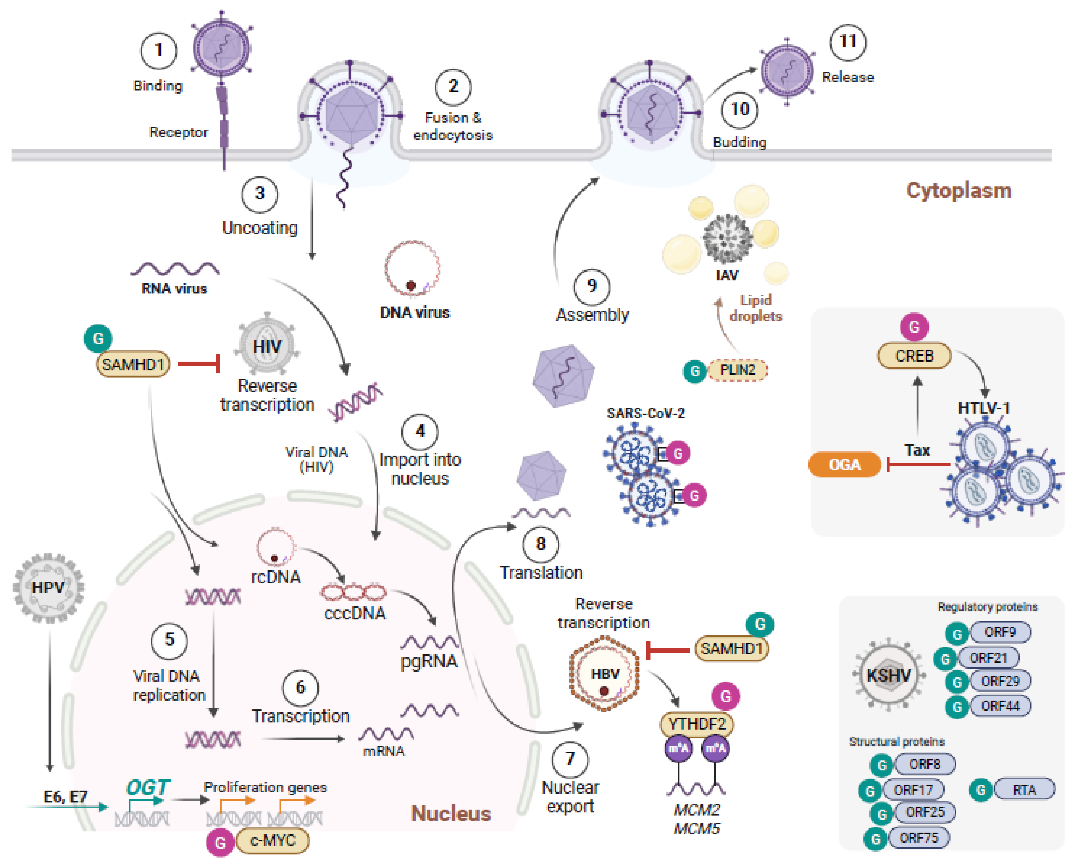
Table 2.
Select O-GlcNAcylated viral substrates.
| Virus | Substrate | Identified Site | Functional impact | Reference |
|---|---|---|---|---|
| KSHV | ORF3 | S278 | O-GlcNAc transferase inhibits KSHV propagation and modifies replication relevant viral proteins as detected by systematic O-GlcNAcylation analysis. | [47] |
| ORF10 | T225, T338, S594, T632, T709 | |||
| ORF8 | S92 | |||
| ORF44 | S727 | |||
| ORF21 | S62 | |||
| ORF29 | n.d. | |||
| ORF75 | n.d. | |||
| RTA (ORF50) | T366, T367 | O-GlcNAc suppresses transactivation & lytic reactivation | [48] | |
| SARS-CoV-2 | Spike | S659 | Spike stability and pseudo-particle packaging | [52] |
n.d., not determined; ORF, open reading frame; RTA, replication and transactivation; S, serine; T, threonine.
Disclaimer/Publisher’s Note: The statements, opinions and data contained in all publications are solely those of the individual author(s) and contributor(s) and not of MDPI and/or the editor(s). MDPI and/or the editor(s) disclaim responsibility for any injury to people or property resulting from any ideas, methods, instructions or products referred to in the content. |
© 2025 by the authors. Licensee MDPI, Basel, Switzerland. This article is an open access article distributed under the terms and conditions of the Creative Commons Attribution (CC BY) license (http://creativecommons.org/licenses/by/4.0/).
Copyright: This open access article is published under a Creative Commons CC BY 4.0 license, which permit the free download, distribution, and reuse, provided that the author and preprint are cited in any reuse.