Preprint
Article

This version is not peer-reviewed.

Development of Novel Anti-CDH1/E-Cadherin Monoclonal Antibodies for Flow Cytometry, Western Blotting, and Immunohistochemistry

Submitted:

28 September 2025

Posted:

29 September 2025

You are already at the latest version

Abstract

Cadherin (CDH)-mediated extracellular homophilic binding is crucial for maintaining tissue homeostasis. The epithelial cell-cell adhesion molecule cadherin 1 (CDH1/E-cadherin) forms the adherens junctions in epithelial cells, and the loss of CDH1 facilitates the migration and invasion of carcinoma cells. Although several anti-CDH1 monoclonal antibodies (mAbs) are available for western blotting and immunohistochemistry (IHC), a highly sensitive anti-CDH1 mAb suitable for flow cytometry has not been developed. We developed anti-CDH1 monoclonal antibodies through a flow cytometry-based high-throughput screening. Two anti-CDH1 mAb clones, Ca1Mab-3 (IgG1, κ) and Ca1Mab-5 (IgG1, κ), reacted with human CDH1-overexpressed Chinese hamster ovary-K1 (CHO/CDH1) cells in flow cytometry. Furthermore, Ca1Mab-3 and Ca1Mab-5 recognize endogenous CDH1-expressing human luminal-type breast cancer cells, such as MCF-7, but not triple-negative breast cancer cells, like MDA-MB-231. The dissociation constant values of Ca1Mab-3 and Ca1Mab-5 for CHO/CDH1 were determined as 4.8 × 10⁻¹⁰ M and 1.3 × 10⁻⁹ M, respectively. Ca1Mab-3 and Ca1Mab-5 can detect endogenous CDH1 in western blotting and IHC using a cell block. Furthermore, Ca1Mab-5 is available for IHC in formalin-fixed paraffin-embedded tumor tissues. These results indicate that Ca1Mab-3 and Ca1Mab-5, established by the CBIS method, are versatile for basic research and are expected to contribute to clinical applications, such as tumor diagnosis and therapy.

Keywords: 
;  ;  ;  ;  ;  

1. Introduction

Cadherins (CDHs) are single-pass transmembrane proteins composed of extracellular and cytoplasmic regions. The extracellular portion is organized into multiple tandemly repeated units, termed extracellular cadherin (EC) domains. CDHs on the cell surface engage in homophilic binding through their N-terminal EC domains, which mediates physical cell–cell adhesion [1]. In vertebrates, the CDH family consists of approximately 20 members, which are classified into type I and type II based on subtle differences in their amino acid sequences [2]. Expression of each type I CDH is essentially cell type–specific; for instance, CDH1/E-cadherin is predominantly found in epithelial cells, CDH2/N-cadherin in neural and mesenchymal cells, CDH3/P-cadherin in placental cells, CDH4/R-cadherin in retinal cells, and CDH15/M-cadherin in muscle cells [3]. The cytoplasmic domain of CDHs is highly conserved across subtypes. It associates directly with cytoplasmic proteins such as p120- and β-catenin. β-catenin, in turn, interacts with α-catenin, which subsequently binds to F-actin, thereby establishing the CDH–actin complex [4,5].
The epithelial–mesenchymal transition (EMT) is a fundamental biological process that governs body plan formation and the differentiation of diverse tissues and organs [6]. In addition to its physiological role in tissue repair, EMT can have pathological consequences, contributing to organ fibrosis and facilitating carcinoma progression [7]. During EMT, cells lose their epithelial characteristics, such as downregulation of CDH1, and acquire invasive capabilities, gain stem cell-like properties, evade apoptosis and senescence, and promote immunosuppressive environments [7]. Consequently, the mesenchymal state is linked to enhanced cellular plasticity, enabling dissemination to distant sites, maintenance of stemness, and subsequent differentiation into multiple lineages during both development and metastatic progression [8]. The invaded tumor cells enter the blood or lymphatic vessels, thereby gaining access to the systemic circulation [8]. Among the surviving cells, some can arrest within a distant capillary and extravasate into a secondary organ. Upon colonization of distant tissues, these mesenchymal-like cells may undergo a reverse process, the mesenchymal–epithelial transition (MET), which restores proliferative capacity and facilitates the formation of macroscopic secondary tumors [6,9].
Anti-CDH1 monoclonal antibodies (mAbs) have been developed for various applications. A clone ECCD-1 (E-cadherin Cell-Cell adhesion Disrupting-1) recognizes CDH1 ectodomain and could block CDH1-mediated cell aggregation [10]. Another clone, DECMA-1, recognizes the CDH1 ectodomain and can be used in various applications, including immunoprecipitation, western blotting, immunofluorescence, and biological functions (inhibition of cell aggregation) [11]. DECMA-1 is also used in studies targeting CDH1 in tumor xenograft models [12]. Several anti-CDH1 mAbs against the cytoplasmic domain exhibited a superior reactivity in immunohistochemistry (IHC) [13]. However, few mAbs have been developed for flow cytometry.
We previously established a series of mAbs targeting membrane proteins, including CDH15 [14], chemokine receptors [15], and receptor tyrosine kinases [16,17] using the Cell-Based Immunization and Screening (CBIS) method. The CBIS method involves immunization with antigen-overexpressed cells, followed by high-throughput flow cytometry–based screening. As a result, mAbs generated through the CBIS method typically recognize conformational epitopes and are particularly suitable for flow cytometric analysis. Additionally, several of these mAbs have been demonstrated to be suitable for Western blotting and IHC. In the present study, we employed the CBIS method to develop highly versatile anti-CDH1 mAbs.

2. Materials and Methods

2.1. Cell Lines

Chinese hamster ovary (CHO)-K1, mouse myeloma P3X63Ag8U.1 (P3U1), human glioblastoma LN229, and human breast cancer MDA-MB-231 cell lines were obtained from American Type Culture Collection (ATCC, Manassas, VA, USA). Human breast cancer MCF-7 was obtained from the Cell Resource Center for Biomedical Research Institute of Development, Aging, and Cancer at Tohoku University (Miyagi, Japan). Human embryonic kidney 293FT was purchased from Thermo Fisher Scientific, Inc. (Waltham, MA, USA).

2.2. Establishment of Stable Transfectants

We previously established the type I CDH-overexpressed stable transfectants: CHO/CDH1, CHO/PA16-CDH2 (CHO/CDH2), CHO/CDH3, CHO/PA16-CDH4 (CHO/CDH4), and CHO/PA16-CDH15 (CHO/CDH15) [14]. The N-terminal PA16-tag (GLEGGVAMPGAEDDVV) was recognized by an anti-PA16 tag mAb, NZ-1 [18].
The pCMV6neo-CDH1-myc-DDK (OriGene Technologies, Inc., Rockville, MD, USA) vector was transfected into LN229 using the Neon transfection system (Thermo Fisher Scientific, Inc.). The transfectants were sorted using an anti-CDH1 mAb (clone DECMA-1, BioLegend, San Diego, CA, USA) with an SH800 cell sorter (Sony Corporation, Tokyo, Japan). The CDH1-overexpressed LN229 (LN229/CDH1) was maintained in a medium containing 0.5 mg/mL of G418 (Nacalai Tesque, Inc.).

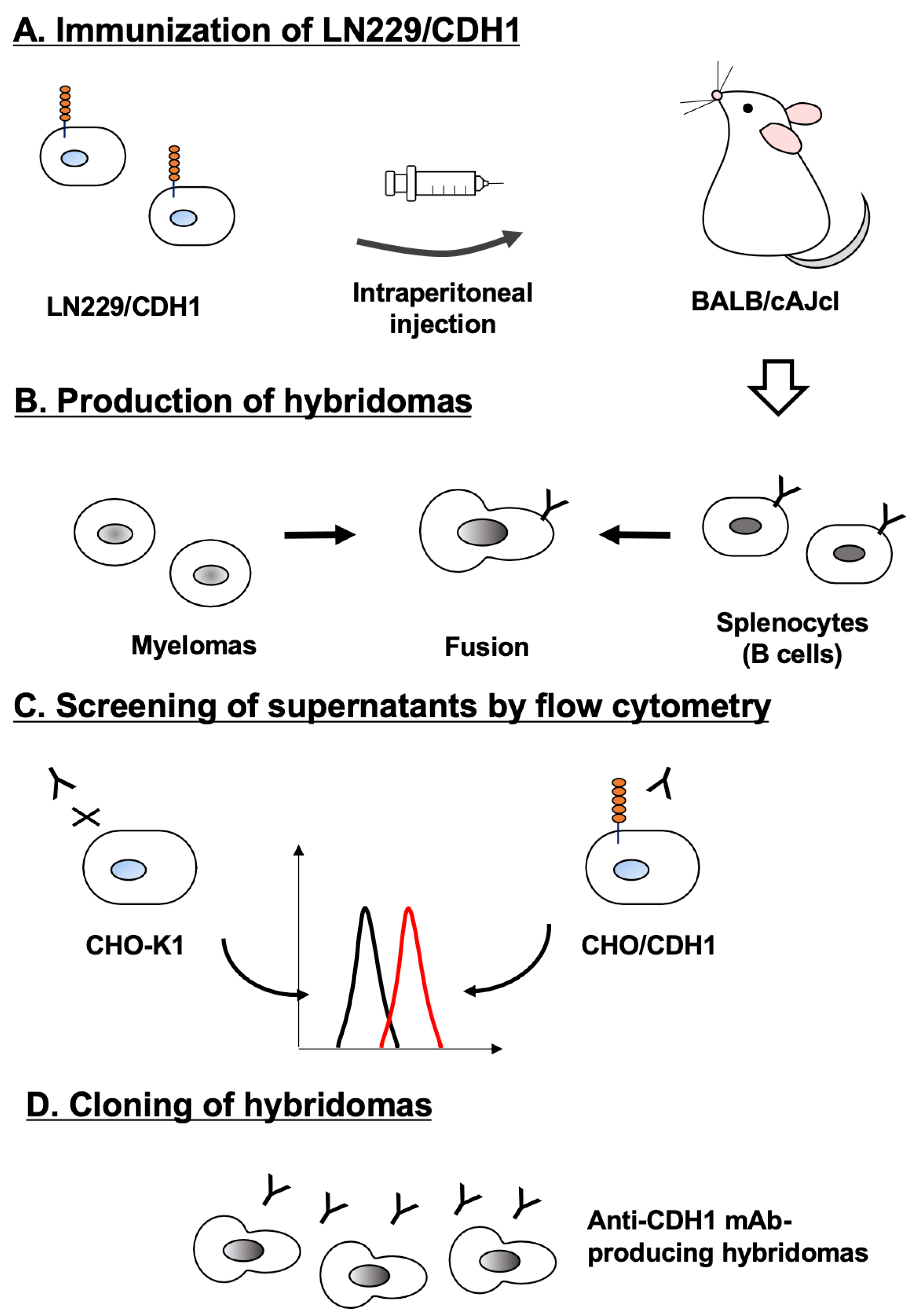
2.3. Development of Hybridomas

Female BALB/cAJcl mice were obtained from CLEA Japan (Tokyo, Japan). Mice were intraperitoneally immunized with LN229/CDH1 cells (1 × 10⁸ cells/injection) emulsified with 2% Alhydrogel adjuvant (InvivoGen). Following three additional weekly immunizations (1.0 or 1.5 × 10⁸ cells/injection), a booster dose (1 × 10⁸ cells/injection) was administered two days prior to spleen excision. Hybridomas were generated as previously described [31]. Supernatants positive for CHO/CDH1 and negative for CHO-K1 were screened using an SA3800 Cell Analyzer (Sony Corporation, Tokyo, Japan).

2.4. Flow Cytometry

Another commercially available anti-CDH1 mAb (clone 67A4) was purchased from BD Biosciences (Franklin Lakes, NJ, USA). The recommended applications are flow cytometry, western blot, and immunofluorescence. Cells were harvested with 1 mM ethylenediaminetetraacetic acid (EDTA; Nacalai Tesque, Inc.). A total of 1 × 10⁵ cells were washed with phosphate-buffered saline (PBS) containing 0.1% bovine serum albumin (BSA; blocking buffer) and incubated with mAbs for 30 min at 4 °C. Subsequently, the cells were stained with Alexa Fluor 488-conjugated anti-mouse IgG or anti-rat IgG (1:2000; Cell Signaling Technology, Danvers, MA, USA) for 30 min at 4 °C. Flow cytometric data were acquired on an SA3800 Cell Analyzer by collecting 5,000 events. Cells were gated based on forward scatter (FSC) and side scatter (SSC), and fluorescence intensity was analyzed using FlowJo software (BD Biosciences).

2.5. Determination of Dissociation Constant Values Using Flow Cytometry

MCF-7 and CHO/CDH1 were treated with serially diluted Ca1Mab-3 and Ca1Mab-5. Subsequently, the cells were treated with anti-mouse IgG conjugated with Alexa Fluor 488 (at a 200-fold dilution) for 30 minutes at 4 °C. The data (10,000 events) were collected using BD FACSLyric (BD Biosciences), and the geometric mean (GeoMean) was determined using FlowJo. The fitting binding isotherms (vertical axis, GeoMean; horizontal axis, mAb concentration) determined the dissociation constant (KD) values to built-in one-side binding models of GraphPad Prism 6 (GraphPad Software, Inc., La Jolla, CA, USA).

2.6. Western Blotting

Whole-cell lysates (10 μg of protein) were separated into polyacrylamide gels and transferred onto polyvinylidene difluoride membranes (Merck KGaA, Darmstadt, Germany). The membranes were blocked with 4% skim milk (Nacalai Tesque, Inc.) in PBS containing 0.05% Tween 20 and incubated with 5 μg/mL of Ca1Mab-3, 5 μg/mL of Ca1Mab-5, or 1 μg/mL of an anti-isocitrate dehydrogenase 1 (IDH1) mAb (clone RcMab-1, rat IgG2a). Then, the membranes were incubated with anti-mouse IgG (1,000-fold dilution; Agilent Technologies, Inc., Santa Clara, CA, USA) or anti-rat IgG conjugated with horseradish peroxidase (1:10,000-fold dilution; Sigma-Aldrich Corp., St. Louis, MO, USA). Chemiluminescence signals were developed and detected as described previously [19].

2.7. Immunohistochemistry (IHC) Using Cell Blocks

Cells were fixed with 4% paraformaldehyde, and the cell blocks were prepared using iPGell (Genostaff Co., Ltd., Tokyo, Japan) (FUJIFILM Wako Pure Chemical Corporation). The formalin-fixed paraffin-embedded (FFPE) cell sections were stained with Ca1Mab-3 (1 μg/mL) or Ca1Mab-5 (1 μg/mL) using BenchMark ULTRA PLUS with the OptiView DAB IHC Detection Kit (Roche Diagnostics, Indianapolis, IN, USA).

2.8. Immunohistochemistry (IHC) Using FFPE Tissues

The FFPE oral squamous cell carcinoma (OSCC) tissue array (OR601c) and breast invasive ductal carcinoma tissue array (T088b) were purchased from US Biomax Inc. (Rockville, MD, USA). The sections were stained with Ca1Mab-3 (10 μg/mL) or Ca1Mab-5 (10, or 25 μg/mL) as described in 2.7.

3. Results

3.1. Development of Anti-CDH1 mAbs

LN229/CDH1 was established as an antigen by sorting using an anti-CDH1 mAb, DECMA-1. Although the reactivity of DECMA-1 in flow cytometry was low (supplementary Figure 1A), LN229/CDH1 was immunized in two BALB/cAJcl mice for five times (Figure 1A). Then, hybridomas were generated (Figure 1B), and the supernatants were screened by a flow cytometry-based high-throughput screening to identify supernatants that were negative for CHO-K1 and positive for CHO/CDH1 (Figure 1C). As a result, 61 positive wells out of 956 wells (6.4%) were obtained. Subsequently, limiting dilution was performed and anti-CDH1 mAb-producing hybridoma clones were established (Figure 1D). Finally, clones Ca1Mab-1 (IgG2a, κ), Ca1Mab-2 (IgM, κ), Ca1Mab-3 (IgG1, κ), Ca1Mab-5 (IgG1, κ), Ca1Mab-6 (IgM, κ), Ca1Mab-7 (IgG1, κ), Ca1Mab-8 (IgM, κ), and Ca1Mab-9 (IgM, κ) were established.

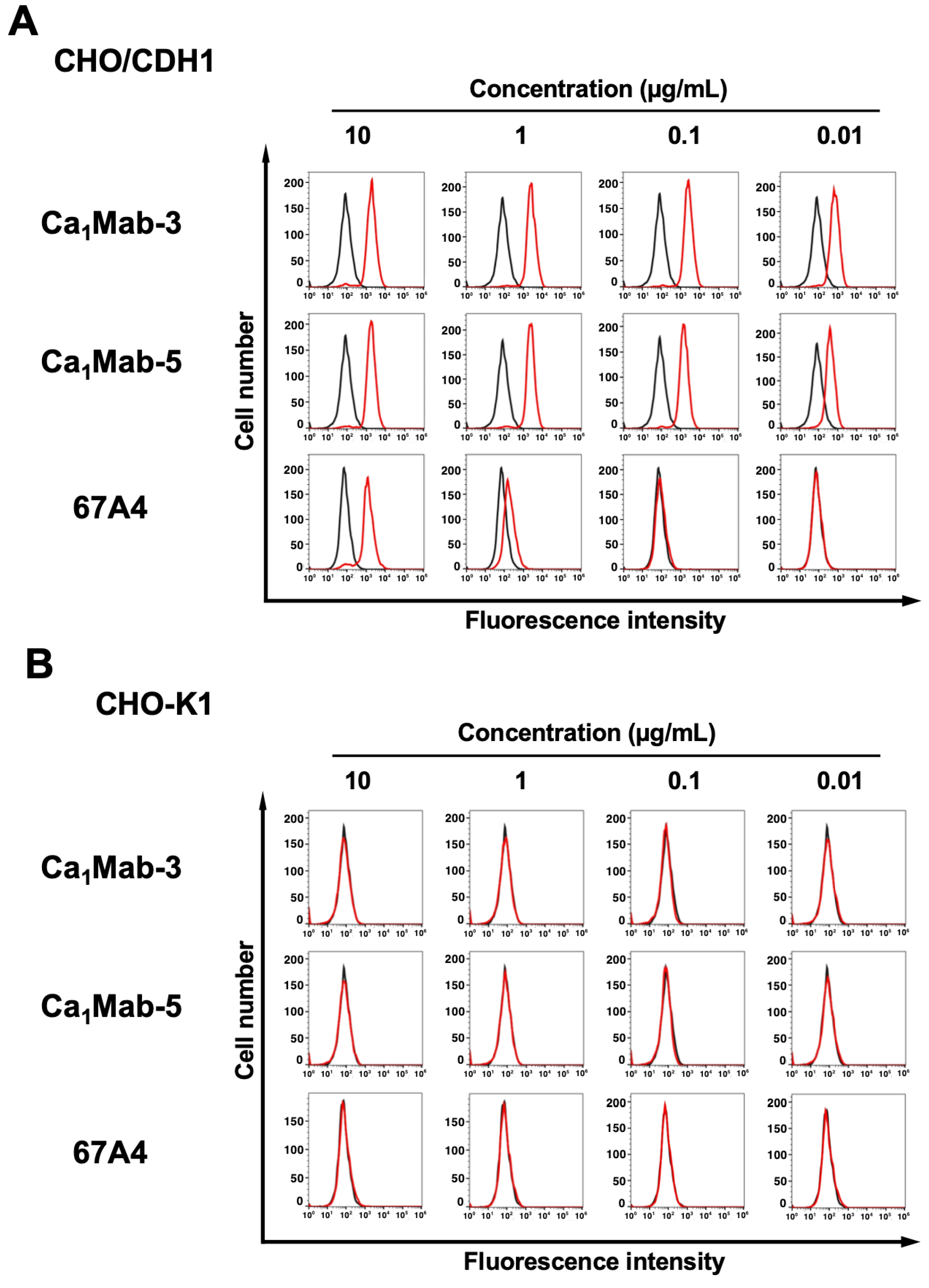
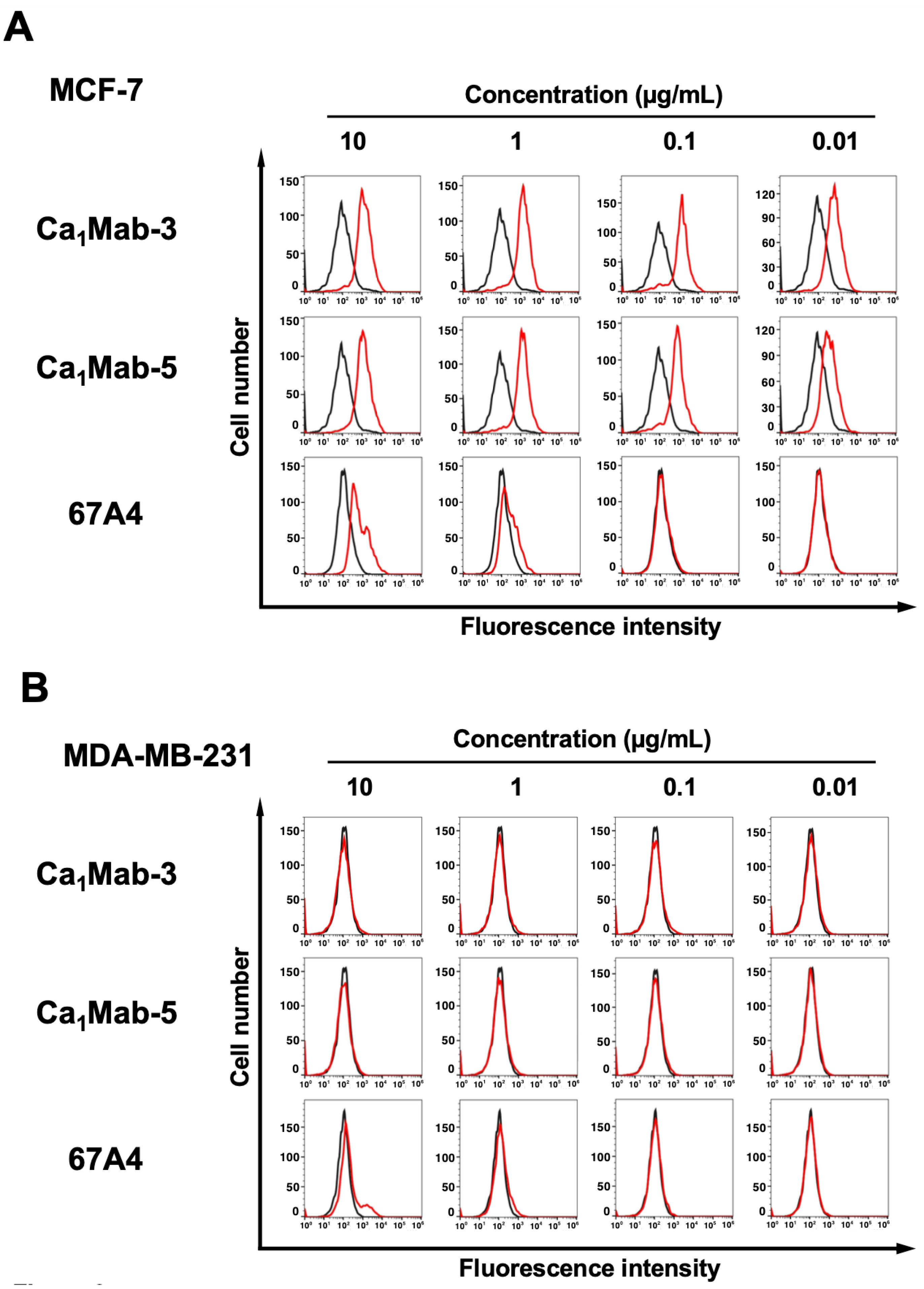
3.2. Flow Cytometry Using Ca1Mab-3 and Ca1Mab-5

Using the supernatants of clones, we next conducted the screening of applications, including flow cytometry, western blotting, and IHC using the CHO/CDH1 cell block (http://www.med-tohoku-antibody.com/topics/001_paper_antibody_PDIS.htm#CDH1+). Since Ca1Mab-3 and Ca1Mab-5 can be applied to the three applications, we further investigated the properties of these mAbs. Figure 2 shows flow cytometry analysis using the Ca1Mab-3 and Ca1Mab-5 against CHO/CDH1 and CHO-K1 cells. Ca1Mab-3 and Ca1Mab-5 recognized CHO/CDH1 in a dose-dependent manner (Figure 2A) from 10 to 0.01 μg/mL, but did not recognize CHO-K1 even at 10 μg/mL (Figure 2B). Since DECMA-1 also showed low reactivity to CHO/CDH1 (supplementary Figure 1B), another commercially available anti-CDH1 mAb, 67A4, was used. Compared to 67A4, Ca1Mab-3 and Ca1Mab-5 clearly showed a higher reactivity to CHO/CDH1 (Figure 2A).
Previous studies have showed that CDH1 expression is observed in luminal-type breast cancers, such as MCF-7, but not in basal B-type breast cancers, including MDA-MB-231 [20]. Ca1Mab-3 and Ca1Mab-5 recognized MCF-7 and showed a higher reactivity compared to 67A4 (Figure 3A). Ca1Mab-3 and Ca1Mab-5 did not recognize MDA-MB-231 even at 10 μg/mL, but weak reactivity of 67A4 to MDA-MB-231 was observed at 10 μg/mL (Figure 3B). We also found that Ca1Mab-3 and Ca1Mab-5 reacted to epithelial cell lines, including 293FT (supplementary Figure 2). These results indicate that Ca1Mab-3 and Ca1Mab-5 can be applied to flow cytometry for the detection of both exogenous and endogenous CDH1.

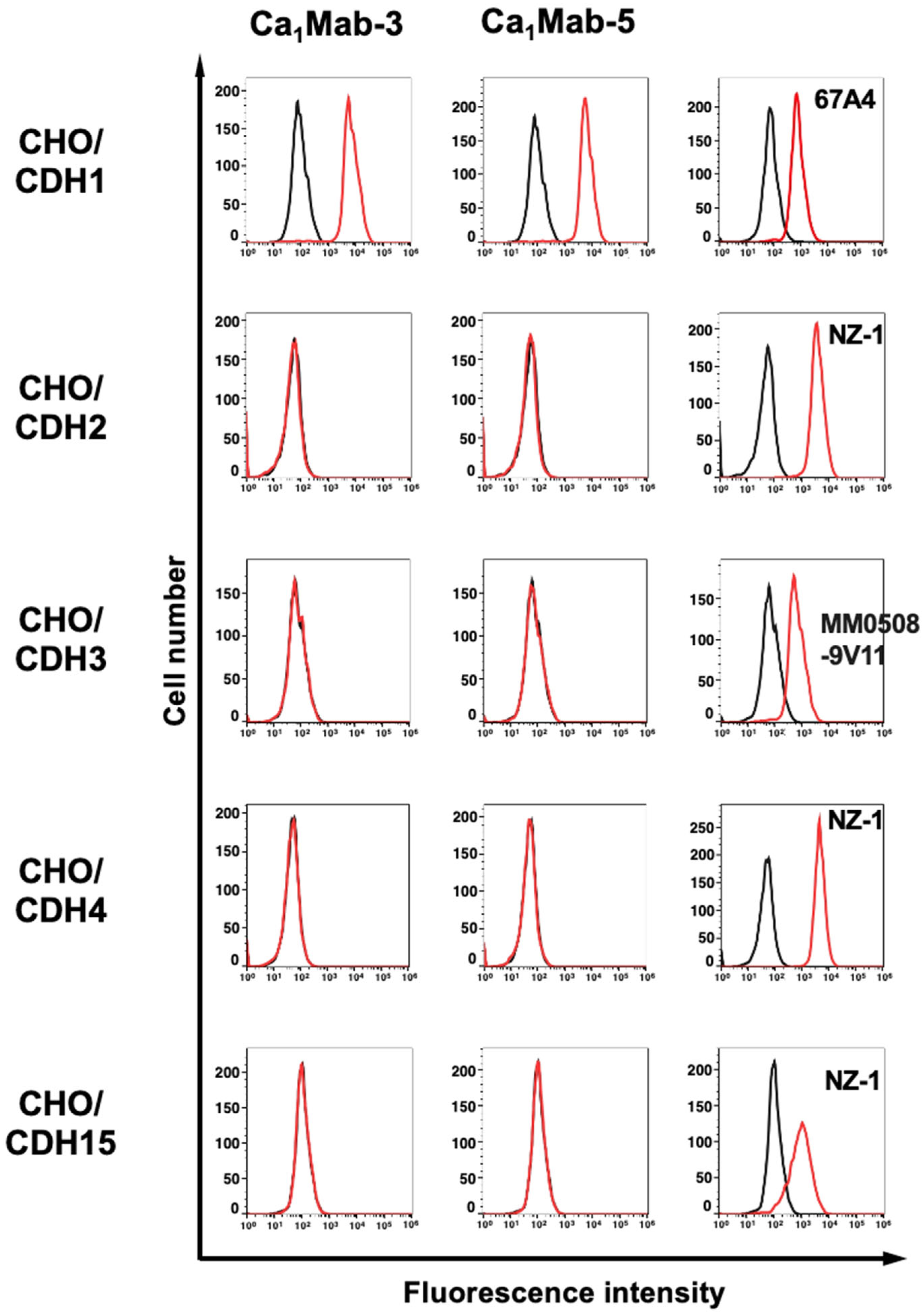
3.3. Specificity of Ca1Mab-3 and Ca1Mab-5 to Type I CDHs-Overexpressed CHO-K1

The type I CDH includes CDH1/E-cadherin, CDH2/N-cadherin, CDH3/P-cadherin, CDH4/R-cadherin, and CDH15/M-cadherin [3]. We previously established CHO-K1, which expresses each type I CDH [14]. Therefore, the specificity of Ca1Mab-3 and Ca1Mab-5 to type I CDHs was determined. As shown in Figure 4, Ca1Mab-3 and Ca1Mab-5 reacted with CHO/CDH1 but did not react with other type I CDHs-overexpressed in CHO-K1. 67A4 also exhibited the specificity to CHO/CDH1 (supplementary Figure 3). These results indicate that Ca1Mab-3 and Ca1Mab-5 are specific mAbs against CDH1 among type I CDH.

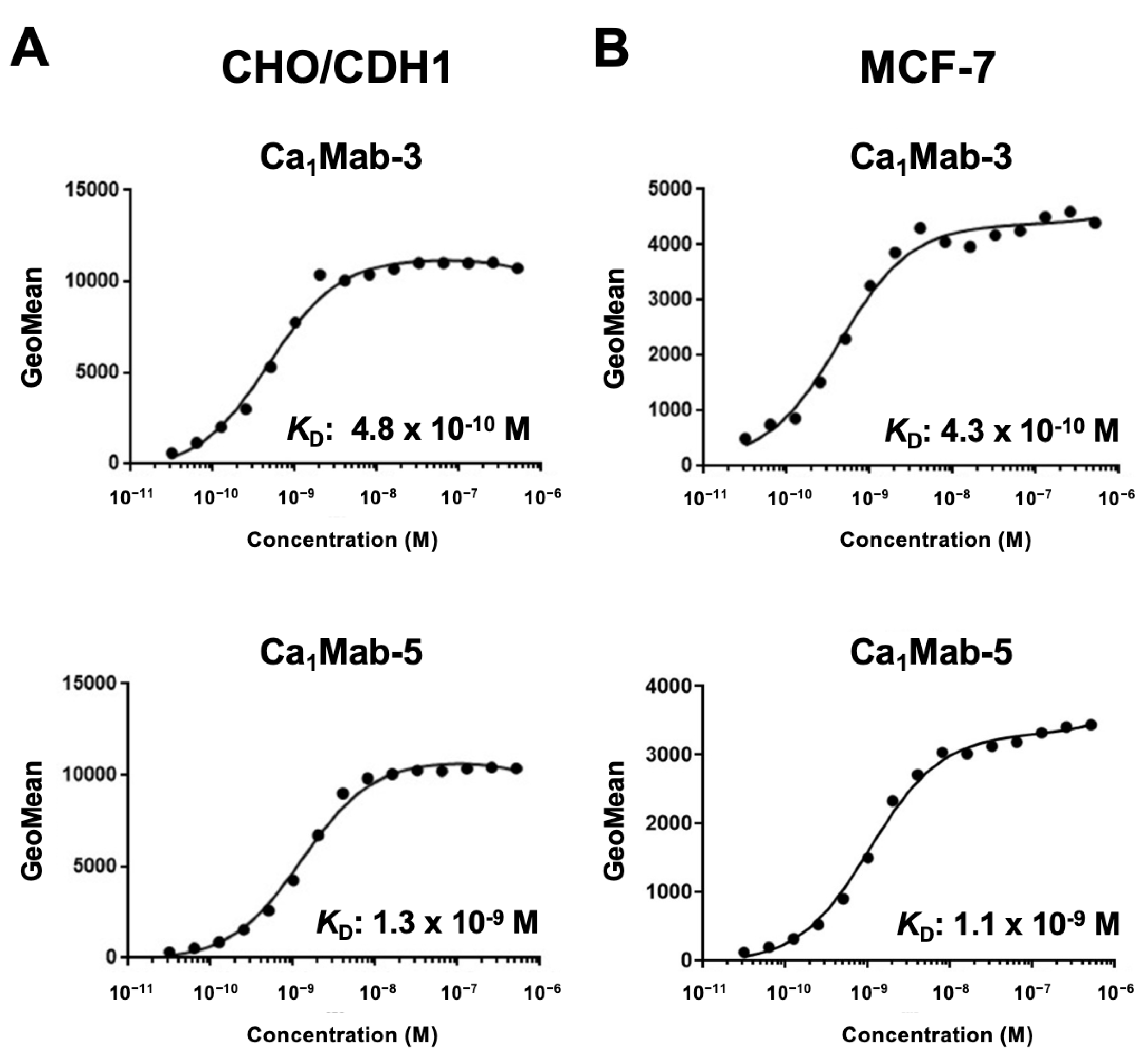
3.4. Determination of KD Values of Ca1Mab-3 and Ca1Mab-5 by Flow Cytometry

The binding affinity of Ca1Mab-3 and Ca1Mab-5 was measured using flow cytometry. The fitting binding isotherms of Ca1Mab-3 and Ca1Mab-5 to CHO/CDH1 and MCF-7 were shown in Figure 5. The KD values of Ca1Mab-3 for CHO/CDH1 and MCF-7 were 4.8 × 10⁻¹⁰ M and 4.3× 10⁻¹⁰ M, respectively. The KD values of Ca1Mab-5 for CHO/CDH1 and MCF-7 were 1.3 × 10⁻⁹ M and 1.1 × 10⁻⁹ M, respectively. These results showed that Ca1Mab-3 possesses superior binding affinity to CDH1-positive cells compared to Ca1Mab-5.

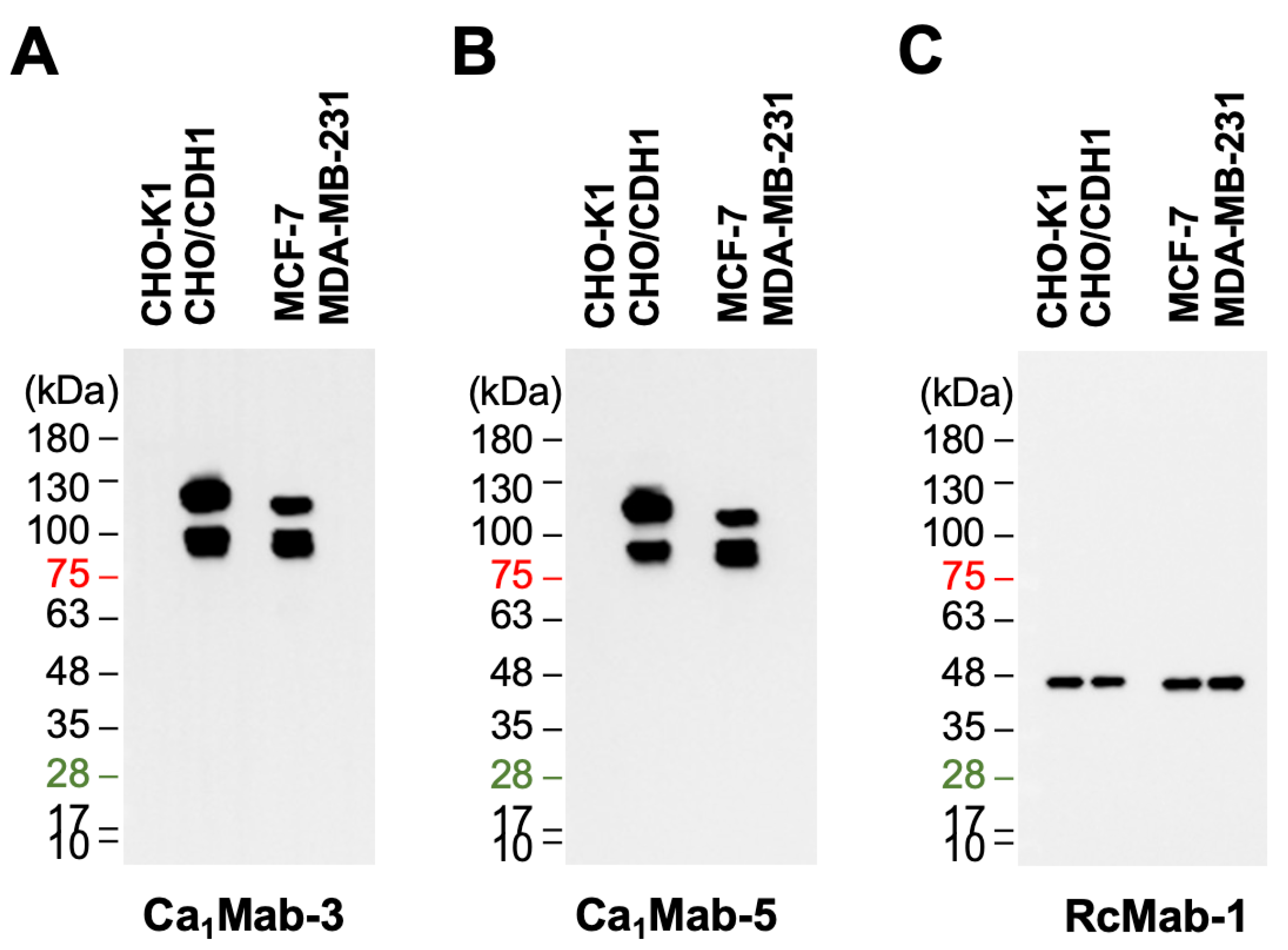
3.5. Western Blot Analysis Using Ca1Mab-3 and Ca1Mab-5

We next assessed whether Ca1Mab-3 and Ca1Mab-5 are suitable for western blotting. Whole-cell lysates of CHO-K1, CHO/CDH1, MCF-7, and MDA-MB-231 were analyzed. Ca1Mab-3 and Ca1Mab-5 exhibited clear bands from 100 to 130 kDa in CHO/CDH1 and MCF-7, but not in CHO-K1 and MDA-MB-231 (Figure 6A and B). An anti-IDH1 mAb (RcMab-1) served as an internal control (Figure 6C). These results indicate that Ca1Mab-3 and Ca1Mab-5 can detect exogenous and endogenous CDH1 in western blotting.

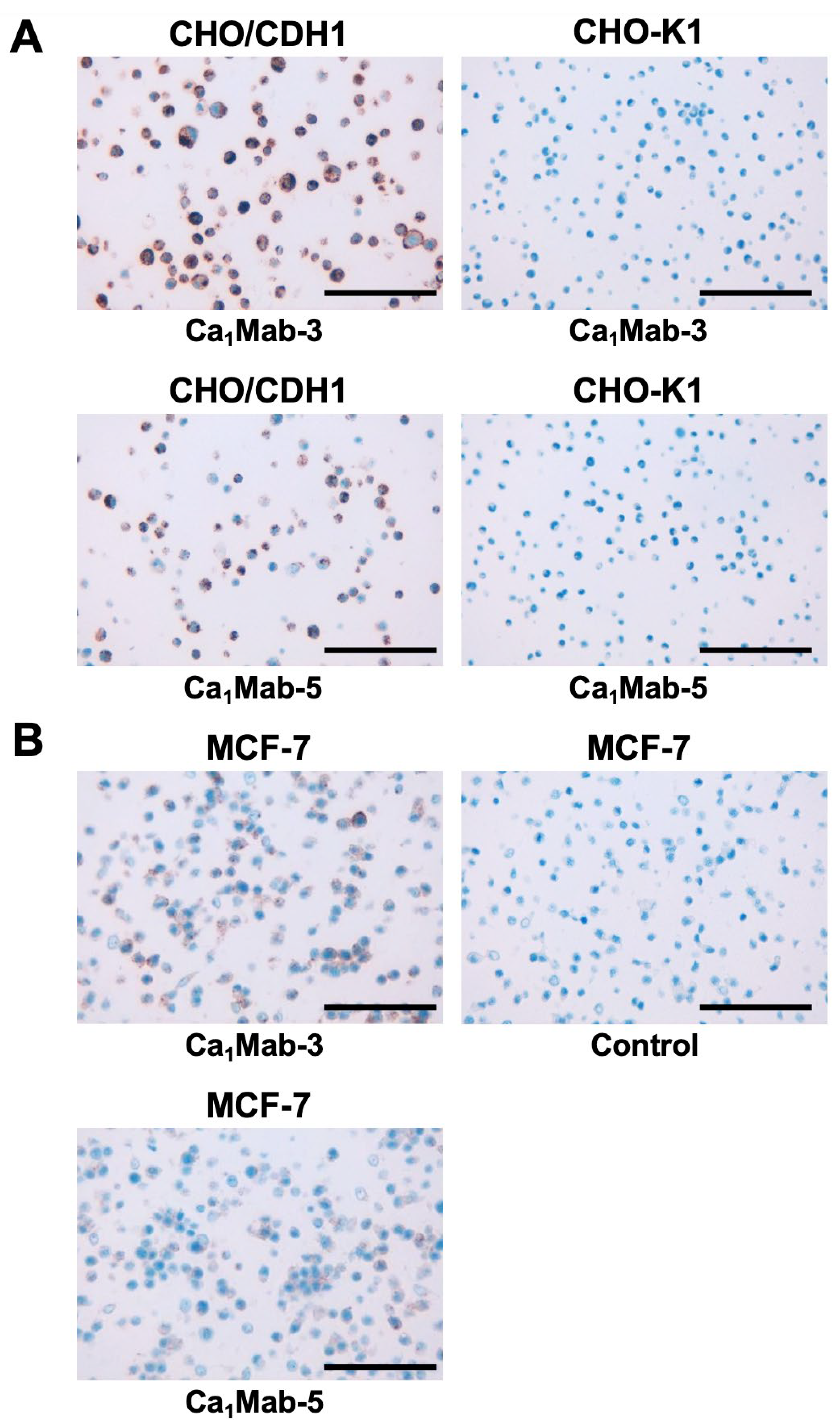
3.6. IHC using Ca1Mab-3 and Ca1Mab-5 in Formalin-Fixed Paraffin-Embedded Cell Blocks

We examined whether Ca1Mab-3 and Ca1Mab-5 are suitable for the IHC of FFPE sections of CHO-K1, CHO/CDH1, and MCF-7. Both intense cytoplasmic and membranous staining by Ca1Mab-3 were detected in CHO/CDH1 but not in CHO-K1 (Figure 7A). Using Ca1Mab-5, a weaker staining pattern was observed (Figure 7A). Furthermore, cytoplasmic and membranous staining by Ca1Mab-3 and Ca1Mab-5 were observed in MCF-7 (Figure 7B). These results indicate that Ca1Mab-3 and Ca1Mab-5 can detect exogenous and endogenous CDH1 in IHC of FFPE sections of cultured cells.

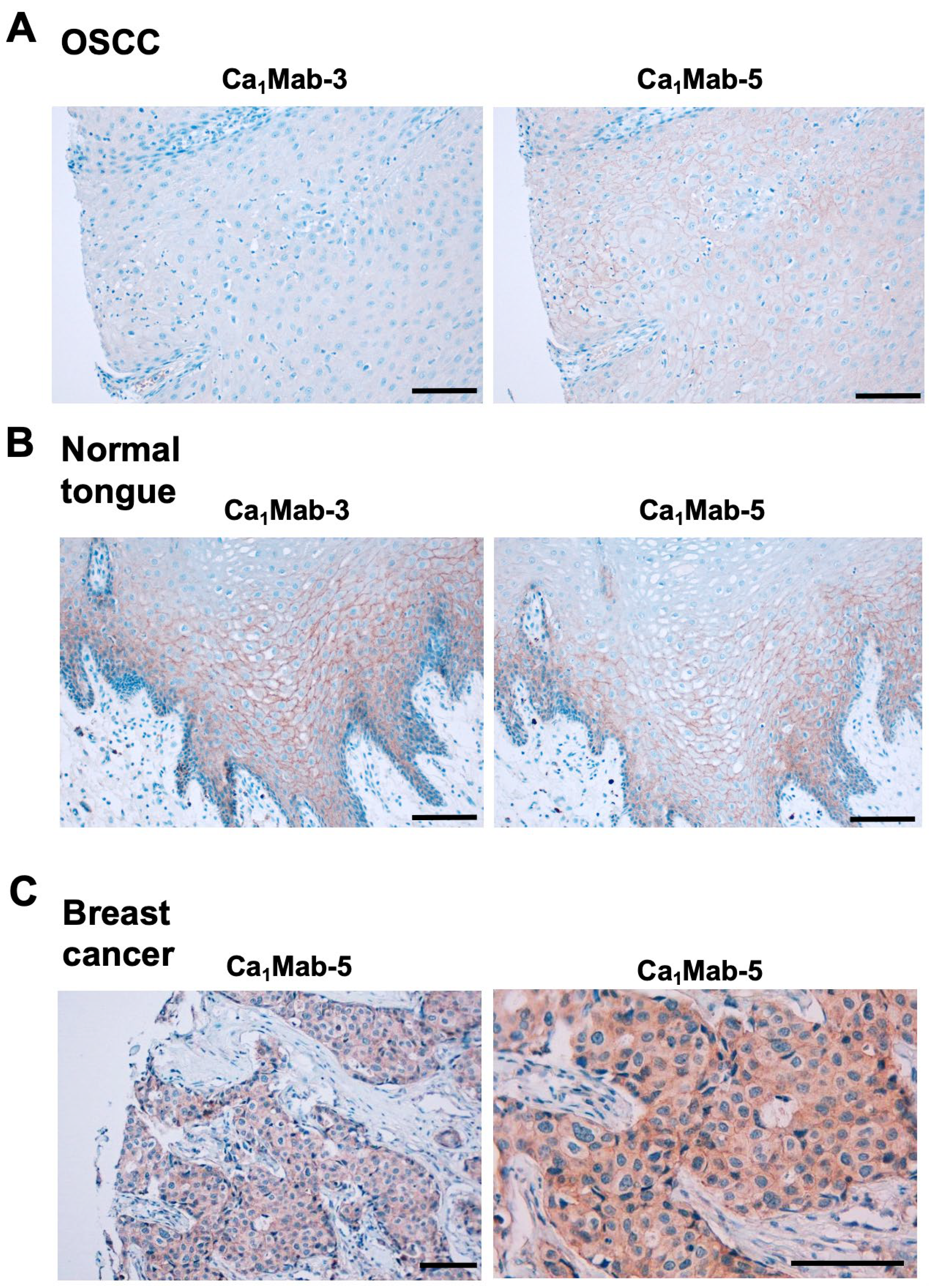
3.7. IHC Using Ca1Mab-3 and Ca1Mab-5 in Formalin-Fixed Paraffin-Embedded Tissues

The FFPE OSCC tissue array was stained with Ca1Mab-3 and Ca1Mab-5. Ca1Mab-5, but not Ca1Mab-3, exhibited a membranous staining in OSCC sections (Figure 8A). In contrast, both Ca1Mab-5 and Ca1Mab-3 showed a membranous staining in normal tongue squamous epithelium (Figure 8B). Furthermore, Figure 8C showed the cytoplasmic and membranous staining of a breast invasive ductal carcinoma tissue using Ca1Mab-5. These results indicated that Ca1Mab-5 is suitable for detecting CDH1 in FFPE tumor sections.

4. Discussion

This study reported novel anti-CDH1 mAbs for flow cytometry using the CBIS method (Figure 1). Two anti-CDH1 mAbs, Ca1Mab-3 and Ca1Mab-5, recognized both exogenous and endogenous CDH1 in flow cytometry with high reactivity compared to commercially available anti-CDH1 mAbs (DECMA-1 and 67A4) (Figure 2, Figure 3, and supplementary Figure 1) and a superior affinity (Figure 5). Ca1Mab-3 and Ca1Mab-5 showed the specificity among type I CDHs (Figure 4). Furthermore, Ca1Mab-3 and Ca1Mab-5 are suitable for western blotting (Figure 6) and IHC using cell block (Figure 7). Additionally, Ca1Mab-5 could stain the cell-cell junction in IHC using FFPE tumor tissues (Figure 8). Since IHC were performed using an automated slide staining system, BenchMark ULTRA PLUS, it is possible to standardize the staining conditions. Therefore, Ca1Mab-3 and Ca1Mab-5 are highly versatile for basic research and are expected to contribute to the understanding of epithelial and tumor biology.
Since Ca1Mab-3 can recognize a single cell by flow cytometry with high affinity (Figure 2, 3, and 5) or IHC using a cell block (Figure 7), Ca1Mab-3 is suitable for detecting or isolating individual epithelial or tumor cells. In the fluorescence-activated cell sorting system, Ca1Mab-3 will be useful for isolating CDH1-positive cells or performing negative selection. In IHC of tumor sections, high concentrations (10 or 25 µg/mL) of Ca1Mab-3 or Ca1Mab-5 were required for the detection (Figure 8). The structure of CDH1 is changed from closed monomer to strand-swap dimer when it forms adherens junction [21]. Therefore, Ca1Mab-3 and Ca1Mab-5 preferentially recognize the closed monomer conformation but have difficulty recognizing the strand-swap dimer conformation. Additionally, it is unclear why Ca1Mab-3 recognizes only normal squamous epithelial tissue but not tumor tissue (Figure 8). We will clarify this difference by determining the epitopes of Ca1Mab-3 and Ca1Mab-5.
Circulating tumor cells (CTCs) enter the peripheral circulation from primary or metastatic lesions either spontaneously or as a result of therapeutic manipulation [22]. Some CTCs may evade from immune systems, ultimately resulting in the establishment of microscopic cancer foci and metastasis [23]. CTCs have been identified across diverse cancer types using panels of molecular markers. Epithelial cell adhesion molecule (EpCAM) is the most extensively utilized marker [24]. The level of EpCAM expression varies among cancer types, and EpCAM-based CTC detection systems are particularly effective in malignancies with high EpCAM expression, such as prostate and breast cancers [25,26]. In addition, other epithelial-derived malignancies including colorectal [27], pancreatic [28], and hepatocellular carcinomas [29] also exhibit substantial frequencies of EpCAM-positive CTCs. The detection of EpCAM-positive CTCs is consistently associated with early distant metastasis and unfavorable patient survival outcomes [30,31]. Studies have demonstrated that E-cadherin is expressed at higher levels than EpCAM on clustered CTCs which exhibit potent metastatic potential and are associated with poor prognosis [32,33,34]. Ca1Mab-3 and Ca1Mab-5 might be able to capture CTCs or clustered CTCs efficiently.
CDH1 functions as a tumor suppressor, and the loss of CDH1 plays a central role in tumor metastasis [35]. The downregulation of CDH1 during EMT has traditionally been associated with an enhanced metastatic potential by enabling cancer cell dissociation and invasion [8,36]. Nevertheless, the notion that the loss of CDH1 universally drives metastasis is an oversimplification, as numerous metastatic lesions retain high CDH1 expression, and epithelial cancer cells can acquire invasive and metastatic capacities without a complete EMT [37,38]. Notably, among breast cancer subtypes, loss of CDH1 expression is characteristic only of lobular carcinoma, whereas the majority of ductal carcinomas and their metastases retain CDH1 expression [39,40]. Furthermore, clustered CTCs in mammary tumors demonstrate greater metastatic efficiency than single disseminated tumor cells, with CDH1-mediated collective migratory behaviors that promote invasion and dissemination [41,42]. More recently, a pro-metastatic role of CDH1 has also been proposed, attributed to its capacity to enhance cancer cell survival [43]. An anti-mouse CDH1 mAb reduced lung metastasis from genetically modified MMTV-PyMT mammary tumors or orthotopically grafted 4T1 tumors [44]. Therefore, it is worthwhile to evaluate the anti-metastatic ability of Ca1Mab-3 or Ca1Mab-5 in suppressing tumor metastasis in vivo. We converted the isotype of mAbs into mouse IgG2a to obtain antibody-dependent cellular cytotoxicity (ADCC). These mAbs were mainly used for evaluating antitumor activities in mouse xenograft models [45,46]. Since the subclass of Ca1Mab-3 or Ca1Mab-5 is mouse IgG1, it does not exert ADCC. A class-switched Ca1Mab-3 or Ca1Mab-5 will be helpful to investigate the effect of tumor metastasis in mouse models. Additionally, both Ca1Mab-3 and Ca1Mab-5 recognize the cell-cell junction in IHC in normal squamous epithelium (Figure 8C). Therefore, the side effects on normal epithelial cells should be investigated by Ca1Mab-3 and Ca1Mab-5. It is possible to evaluate whether the ADCC-mediated cell lysis is induced against normal epithelial cells by Ca1Mab-3 or Ca1Mab-5. If Ca1Mab-3 and Ca1Mab-5 can recognize mouse CDH1, the side effects will be evaluated in mice.

Supplementary Materials

The following supporting information can be downloaded at the website of this paper posted on Preprints.org.

Credit Authorship Contribution Statement

Rena Ubukata: Investigation Hiroyuki Suzuki: Writing – original draft Mika K. Kaneko: Conceptualization Yukinari Kato: Conceptualization, Funding acquisition, Project administration, Writing – review and editing All authors have read and agreed to the published version of the manuscript

Funding Information

This research was supported in part by Japan Agency for Medical Research and Development (AMED) under Grant Numbers: JP25am0521010 (to Y.K.), JP25ama121008 (to Y.K.), JP25ama221339 (to Y.K.), and JP25bm1123027 (to Y.K.), and by the Japan Society for the Promotion of Science (JSPS) Grants-in-Aid for Scientific Research (KAKENHI) grant no. 25K10553 (to Y.K.).

Institutional Review Board Statement

The animal study protocol was approved by the Animal Care and Use Committee of Tohoku University (Permit number: 2022MdA-001) for studies involving animals.

Informed Consent Statement

Not applicable.

Data Availability Statement

All related data and methods are presented in this paper. Additional inquiries should be addressed to the corresponding authors.

Conflicts of Interest

The authors declare no conflict of interest involving this article.

References

  1. Niessen, C.M.; Leckband, D.; Yap, A.S. Tissue organization by cadherin adhesion molecules: dynamic molecular and cellular mechanisms of morphogenetic regulation. Physiol Rev 2011;91(2): 691-731. [CrossRef]
  2. Nollet, F.; Kools, P.; van Roy, F. Phylogenetic analysis of the cadherin superfamily allows identification of six major subfamilies besides several solitary members11Edited by M. Yaniv. Journal of Molecular Biology 2000;299(3): 551-572.
  3. Oda, H.; Takeichi, M. Evolution: structural and functional diversity of cadherin at the adherens junction. J Cell Biol 2011;193(7): 1137-1146. [CrossRef]
  4. Lin, W.H.; Cooper, L.M.; Anastasiadis, P.Z. Cadherins and catenins in cancer: connecting cancer pathways and tumor microenvironment. Front Cell Dev Biol 2023;11: 1137013. [CrossRef]
  5. Yu, W.; Yang, L.; Li, T.; Zhang, Y. Cadherin Signaling in Cancer: Its Functions and Role as a Therapeutic Target. Front Oncol 2019;9: 989. [CrossRef]
  6. Nieto, M.A.; Huang, R.Y.; Jackson, R.A.; Thiery, J.P. EMT: 2016. Cell 2016;166(1): 21-45.
  7. Lambert, A.W.; Weinberg, R.A. Linking EMT programmes to normal and neoplastic epithelial stem cells. Nat Rev Cancer 2021;21(5): 325-338. [CrossRef]
  8. Valastyan, S.; Weinberg, R.A. Tumor metastasis: molecular insights and evolving paradigms. Cell 2011;147(2): 275-292. [CrossRef]
  9. Yang, J.; Antin, P.; Berx, G.; et al. Guidelines and definitions for research on epithelial-mesenchymal transition. Nat Rev Mol Cell Biol 2020;21(6): 341-352. [CrossRef]
  10. Ogou, S.I.; Yoshida-Noro, C.; Takeichi, M. Calcium-dependent cell-cell adhesion molecules common to hepatocytes and teratocarcinoma stem cells. J Cell Biol 1983;97(3): 944-948. [CrossRef]
  11. Vestweber, D.; Kemler, R. Identification of a putative cell adhesion domain of uvomorulin. Embo j 1985;4(13a): 3393-3398. [CrossRef]
  12. Brouxhon, S.M.; Kyrkanides, S.; Teng, X.; et al. Monoclonal antibody against the ectodomain of E-cadherin (DECMA-1) suppresses breast carcinogenesis: involvement of the HER/PI3K/Akt/mTOR and IAP pathways. Clin Cancer Res 2013;19(12): 3234-3246. [CrossRef]
  13. Hoang, L.L.; Tang, P.; Hicks, D.G.; et al. A new rabbit monoclonal E-cadherin antibody [EP700Y] shows higher sensitivity than mouse monoclonal E-cadherin [HECD-1] antibody in breast ductal carcinomas and does not stain breast lobular carcinomas. Appl Immunohistochem Mol Morphol 2014;22(8): 606-612. [CrossRef]
  14. Ubukata, R.; Suzuki, H.; Tanaka, T.; Kaneko, M.K.; Kato, Y. Development of an anti-CDH15/M-cadherin monoclonal antibody Ca(15)Mab-1 for flow cytometry, immunoblotting, and immunohistochemistry. Biochem Biophys Rep 2025;43: 102138. [CrossRef]
  15. Suzuki, H.; Tanaka, T.; Li, G.; et al. Development of a Sensitive Anti-Mouse CCR5 Monoclonal Antibody for Flow Cytometry. Monoclon Antib Immunodiagn Immunother 2024;43(4): 96-100. [CrossRef]
  16. Kaneko, M.K.; Suzuki, H.; Kato, Y. Establishment of a Novel Cancer-Specific Anti-HER2 Monoclonal Antibody H(2)Mab-250/H(2)CasMab-2 for Breast Cancers. Monoclon Antib Immunodiagn Immunother 2024;43(2): 35-43.
  17. Itai, S.; Yamada, S.; Kaneko, M.K.; et al. Establishment of EMab-134, a Sensitive and Specific Anti-Epidermal Growth Factor Receptor Monoclonal Antibody for Detecting Squamous Cell Carcinoma Cells of the Oral Cavity. Monoclon Antib Immunodiagn Immunother 2017;36(6): 272-281. [CrossRef]
  18. Fujii, Y.; Kaneko, M.; Neyazaki, M.; et al. PA tag: a versatile protein tagging system using a super high affinity antibody against a dodecapeptide derived from human podoplanin. Protein Expr Purif 2014;95: 240-247. [CrossRef]
  19. Nanamiya, R.; Suzuki, H.; Kaneko, M.K.; Kato, Y. Development of an Anti-EphB4 Monoclonal Antibody for Multiple Applications Against Breast Cancers. Monoclon Antib Immunodiagn Immunother 2023;42(5): 166-177. [CrossRef]
  20. Fukagawa, A.; Ishii, H.; Miyazawa, K.; Saitoh, M. δEF1 associates with DNMT1 and maintains DNA methylation of the E-cadherin promoter in breast cancer cells. Cancer Med 2015;4(1): 125-135. [CrossRef]
  21. Shapiro, L.; Weis, W.I. Structure and biochemistry of cadherins and catenins. Cold Spring Harb Perspect Biol 2009;1(3): a003053. [CrossRef]
  22. Gu, X.; Wei, S.; Lv, X. Circulating tumor cells: from new biological insights to clinical practice. Signal Transduct Target Ther 2024;9(1): 226w biological insights to clinical practice. [CrossRef]
  23. Deng, Z.; Wu, S.; Wang, Y.; Shi, D. Circulating tumor cell isolation for cancer diagnosis and prognosis. EBioMedicine 2022;83: 104237ncer diagnosis and prognosis. [CrossRef]
  24. Gires, O.; Pan, M.; Schinke, H.; Canis, M.; Baeuerle, P.A. Expression and function of epithelial cell adhesion molecule EpCAM: where are we after 40 years? Cancer Metastasis Rev 2020;39(3): 969-987. [CrossRef]
  25. Gorin, M.A.; Verdone, J.E.; van der Toom, E.; et al. Circulating tumour cells as biomarkers of prostate, bladder, and kidney cancer. Nat Rev Urol 2017;14(2): 90-97. [CrossRef]
  26. Criscitiello, C.; Sotiriou, C.; Ignatiadis, M. Circulating tumor cells and emerging blood biomarkers in breast cancer. Curr Opin Oncol 2010;22(6): 552-558. [CrossRef]
  27. Marcuello, M.; Vymetalkova, V.; Neves, R.P.L.; et al. Circulating biomarkers for early detection and clinical management of colorectal cancer. Mol Aspects Med 2019;69: 107-122. [CrossRef]
  28. Varillas, J.I.; Zhang, J.; Chen, K.; et al. Microfluidic Isolation of Circulating Tumor Cells and Cancer Stem-Like Cells from Patients with Pancreatic Ductal Adenocarcinoma. Theranostics 2019;9(5): 1417-1425. [CrossRef]
  29. Xia, W.; Li, H.; Li, Y.; et al. In Vivo Coinstantaneous Identification of Hepatocellular Carcinoma Circulating Tumor Cells by Dual-Targeting Magnetic-Fluorescent Nanobeads. Nano Lett 2021;21(1): 634-641. [CrossRef]
  30. Ye, Q.; Ling, S.; Zheng, S.; Xu, X. Liquid biopsy in hepatocellular carcinoma: circulating tumor cells and circulating tumor DNA. Mol Cancer 2019;18(1): 114. [CrossRef]
  31. Gall, T.M.H.; Belete, S.; Khanderia, E.; Frampton, A.E.; Jiao, L.R. Circulating Tumor Cells and Cell-Free DNA in Pancreatic Ductal Adenocarcinoma. Am J Pathol 2019;189(1): 71-81. [CrossRef]
  32. Zhang, Z.; Wuethrich, A.; Wang, J.; et al. Dynamic Monitoring of EMT in CTCs as an Indicator of Cancer Metastasis. Anal Chem 2021;93(50): 16787-16795. [CrossRef]
  33. Schuster, E.; Taftaf, R.; Reduzzi, C.; et al. Better together: circulating tumor cell clustering in metastatic cancer. Trends Cancer 2021;7(11): 1020-1032. [CrossRef]
  34. Hapach, L.A.; Carey, S.P.; Schwager, S.C.; et al. Phenotypic Heterogeneity and Metastasis of Breast Cancer Cells. Cancer Res 2021;81(13): 3649-3663. [CrossRef]
  35. van Roy, F. Beyond E-cadherin: roles of other cadherin superfamily members in cancer. Nat Rev Cancer 2014;14(2): 121-134.
  36. Hanahan, D.; Weinberg, R.A. Hallmarks of cancer: the next generation. Cell 2011;144(5): 646-674.
  37. Lüönd, F.; Sugiyama, N.; Bill, R.; et al. Distinct contributions of partial and full EMT to breast cancer malignancy. Dev Cell 2021;56(23): 3203-3221.e3211. [CrossRef]
  38. Hollestelle, A.; Peeters, J.K.; Smid, M.; et al. Loss of E-cadherin is not a necessity for epithelial to mesenchymal transition in human breast cancer. Breast Cancer Res Treat 2013;138(1): 47-57. [CrossRef]
  39. Horne, H.N.; Oh, H.; Sherman, M.E.; et al. E-cadherin breast tumor expression, risk factors and survival: Pooled analysis of 5,933 cases from 12 studies in the Breast Cancer Association Consortium. Sci Rep 2018;8(1): 6574. [CrossRef]
  40. Rodriguez, F.J.; Lewis-Tuffin, L.J.; Anastasiadis, P.Z. E-cadherin’s dark side: possible role in tumor progression. Biochim Biophys Acta 2012;1826(1): 23-31. [CrossRef]
  41. Cheung, K.J.; Ewald, A.J. A collective route to metastasis: Seeding by tumor cell clusters. Science 2016;352(6282): 167-169. [CrossRef]
  42. Cheung, K.J.; Gabrielson, E.; Werb, Z.; Ewald, A.J. Collective invasion in breast cancer requires a conserved basal epithelial program. Cell 2013;155(7): 1639-1651. [CrossRef]
  43. Padmanaban, V.; Krol, I.; Suhail, Y.; et al. E-cadherin is required for metastasis in multiple models of breast cancer. Nature 2019;573(7774): 439-444. [CrossRef]
  44. Na, T.Y.; Schecterson, L.; Mendonsa, A.M.; Gumbiner, B.M. The functional activity of E-cadherin controls tumor cell metastasis at multiple steps. Proc Natl Acad Sci U S A 2020;117(11): 5931-5937. [CrossRef]
  45. Tanaka, T.; Suzuki, H.; Ohishi, T.; Kaneko, M.K.; Kato, Y. A Cancer-Specific Anti-Podoplanin Monoclonal Antibody, PMab-117-mG(2a) Exerts Antitumor Activities in Human Tumor Xenograft Models. Cells 2024;13(22). [CrossRef]
  46. Kaneko, M.K.; Suzuki, H.; Ohishi, T.; et al. A Cancer-Specific Monoclonal Antibody against HER2 Exerts Antitumor Activities in Human Breast Cancer Xenograft Models. Int J Mol Sci 2024;25(3). [CrossRef]
Figure 1. Schematic representation of anti-CDH1 mAbs production. (A) LN229/CDH1 was injected intraperitoneally into BALB/cAJcl mice. (B) After five immunizations per week, spleen cells were fused with P3U1. (C) The supernatants from hybridomas were screened by flow cytometry using CHO/CDH1 and CHO-K1 cells. (D) Anti-CDH1 mAb-producing hybridoma clones (Ca1Mabs) were established through limiting dilution.
Preprints 178609 g001
Figure 2. Flow cytometry analysis of anti-CDH1 mAbs, Ca1Mab-3, Ca1Mab-5, and 67A4 against CHO/CDH1 and CHO-K1. CHO/CDH1 (A) and CHO-K1 (B) were treated with Ca1Mab-3, Ca1Mab-5, and 67A4 at the indicated concentrations (red) or with blocking buffer (black, negative control). The mAbs-treated cells were incubated with anti-mouse IgG conjugated with Alexa Fluor 488. The fluorescence data were collected using the SA3800 Cell Analyzer.
Preprints 178609 g002
Figure 3. Flow cytometry analysis of Ca1Mab-3, Ca1Mab-5, and 67A4 against MCF-7 and MDA-MB-231. MCF-7 (A) and MDA-MB-231 (B) were treated with Ca1Mab-3, Ca1Mab-5, and 67A4 at the indicated concentrations (red) or with blocking buffer (black, negative control). The mAbs-treated cells were incubated with anti-mouse IgG conjugated with Alexa Fluor 488. The fluorescence data were collected using the SA3800 Cell Analyzer.
Preprints 178609 g003
Figure 4. Flow cytometry analysis of Ca1Mab-3 and Ca1Mab-5 in type I CDHs-overexpressed CHO-K1. The type I CDHs (CDH1, CDH2, CDH3, CDH4, and CDH15)-expressed CHO-K1 cells were treated with 10 µg/mL of Ca1Mab-3 and Ca1Mab-5 (red) or with control blocking buffer (black, negative control), followed by treatment with anti-mouse IgG conjugated with Alexa Fluor 488. Each CDH expression was confirmed by 10 µg/mL of an anti-CDH1 mAb (clone 67A4), 1 µg/mL of an anti-CDH3 mAb (clone MM0508-9V11), and 1 µg/mL of an anti-PA16-tag mAb (clone NZ-1) to detect PA16-tagged CDH2, CDH4, and CDH15, followed by the treatment with anti-mouse IgG or anti-rat IgG conjugated with Alexa Fluor 488. The fluorescence data were collected using the SA3800 Cell Analyzer.
Preprints 178609 g004
Figure 5. Measurement of binding affinity of Ca1Mab-3 and Ca1Mab-5. CHO/CDH1 (A) and MCF-7 (B) were treated with serially diluted Ca1Mab-3 or Ca1Mab-5, followed by anti-mouse IgG conjugated with Alexa Fluor 488. The fluorescence data were analyzed using the BD FACSLyric. The KD values were determined using GraphPad PRISM 6.
Preprints 178609 g005
Figure 6. Western blotting using Ca1Mab-3 and Ca1Mab-5. The cell lysate (10 μg/lane) of CHO-K1, CHO/CDH1, MCF-7, and MDA-MB-231 were electrophoresed and transferred onto polyvinylidene difluoride membranes. The membranes were incubated with 5 μg/mL of Ca1Mab-3 (A), 5 μg/mL of Ca1Mab-5 (B), and 1 μg/mL of RcMab-1 (an anti-IDH1 mAb) (C), followed by the treatment with anti-mouse (Ca1Mab-3 and Ca1Mab-5) or anti-rat IgG (RcMab-1)-conjugated with horseradish peroxidase.
Preprints 178609 g006
Figure 7. Immunohistochemistry using Ca1Mab-3 and Ca1Mab-5 in formalin-fixed paraffin-embedded cell blocks. ((A) CHO/CDH1 and CHO-K1 sections were treated with 1 μg/mL of Ca1Mab-3 or Ca1Mab-5. (B) MCF-7 sections were treated with 1 μg/mL of Ca1Mab-3, 1 μg/mL of Ca1Mab-5, or control (without primary Ab). The staining was performed using BenchMark ULTRA PLUS with the OptiView DAB IHC Detection Kit. Scale bar = 100 μm.
Preprints 178609 g007
Figure 8. Immunohistochemistry using Ca1Mab-3 and Ca1Mab-5 in formalin-fixed paraffin-embedded tissues. (A) The sequential sections of a FFPE OSCC tissue were stained with 10 μg/mL of Ca1Mab-3 and Ca1Mab-5. (B) The sequential sections of a normal tongue squamous epithelium were stained with 10 μg/mL of Ca1Mab-3 and Ca1Mab-5. (C) A FFPE breast invasive ductal carcinoma tissue was stained with 25 μg/mL of Ca1Mab-5. The staining was performed using BenchMark ULTRA PLUS with the OptiView DAB IHC Detection Kit. Scale bar = 100 μm.
Preprints 178609 g008
Disclaimer/Publisher’s Note: The statements, opinions and data contained in all publications are solely those of the individual author(s) and contributor(s) and not of MDPI and/or the editor(s). MDPI and/or the editor(s) disclaim responsibility for any injury to people or property resulting from any ideas, methods, instructions or products referred to in the content.
Copyright: This open access article is published under a Creative Commons CC BY 4.0 license, which permit the free download, distribution, and reuse, provided that the author and preprint are cited in any reuse.
Prerpints.org logo

Preprints.org is a free preprint server supported by MDPI in Basel, Switzerland.

Subscribe

Disclaimer

Terms of Use

Privacy Policy

Privacy Settings

© 2025 MDPI (Basel, Switzerland) unless otherwise stated