
Submitted:
23 September 2025
Posted:
25 September 2025
You are already at the latest version
Abstract
Background and Objectives: Great efforts in orthopaedic surgery have been devoted to the ad-vancement in prosthetics, with a focus on various materials with enhanced mechanical, physical, and chemical properties, geometries of implants, and their surface morphology. A considerable in-terest relates to interfaces with the bone tissues, and their surroundings, e.g., body fluids, building interaction with the living body. Organ dysfunctions effect the quality of bone. The objective of the study is to find interconnections between the underlying conditions of patients, the quality of their bones, and their orthopaedic implants, on different time spans. We refer to femoral stems only. Materials and Methods: We report on three cases, two women (F1 aged over 35, and F2 aged over 80), and one man (M aged over 65), with diabetes, dyslipidaemia, and kidney dysfunction, under-going HA with uncemented Ti-alloy implants. The samples examined were: a segment of a broken Ti-alloy stem (7 years old), biological remnants on it (bone with a metallic layer, 1.75 cm2 and 2.53 g, and soft tissue, 1.50cm2 and 1.098 g), segments of two spare stems, and synthetic plasma enriched with glucose, urea, and cholesterol (according to the biochemistry tests of the respective patients). The stem pieces, 20 x 11 x 6.56 mm3, were immersed in the corresponding synthetic plasma at 38 C and ultrasonicated for 7h. Scanning electron microscopy and energy dispersive X-ray spectroscopy were employed to investigate the Ti-alloy stem samples, and electrochemistry - to adequately explore the plasma left after sonication (50µL/sample) of each sample. Histopatho-logical examination was performed on the soft tissue remnants on the broken stem. Results: EDX spectra have shown that all femoral stem samples are of Ti-6Al-4V, with various additions of C, Fe, Si, Zr, coated with hydroxyapatite. The electrochemistry study shows that the metallic layer on the bone fragment was caused by migration of vanadium during the 6 months since fracture to re-vision for M. That finding agrees with analysis of prosthetic vanadium concentration in se-rum/plasma after 6 months, as recently reported by other authors. Conclusions: The reactivity of the stems in altered synthetic plasma is given by the presence of glucose and urea, whereas choles-terol influences on bone quality only. The histopathological analysis shows severe panniculitis and myositis. Numerous neutrophils intermixed by macrophages, and a few lymphocytes, are present, supporting the abundance of small new capillaries (granulation tissue). Metal migration from the prostheses can occur through the chemical interactions between body fluids with abnor-mal biochemistry and the compounds building the orthopaedic prostheses, having however in mind that chemical corrosion, and wear resulting from friction during usual movements of the body, are competing mechanisms, especially through effects like surface cracks. Even though Ti-alloys are fully biocompatible, diabetes, dyslipidaemia, and renal dysfunction alter the bone-implant interface because of dramatically weakened and frail bones, no matter the age, and sex. Therefore, for those patients with complex conditions, it can be anticipated a shorter lifetime of implants till revision than in healthier ones. A brief review supports our experimental findings.
Keywords:
hip arthroplasty
; titanium alloy prostheses
; dyslipidaemia
; diabetes
; kidney dysfunction
; bone-Ti alloy interaction
; bone health
; post-traumatic panniculitis
; scanning electron microscopy
; electrochemistry
1. Introduction
Hip arthroplasty (HA) is a surgical reconstruction of the hip joint using artificial material, including, total hip arthroplasty (THA), when the femur’s head and the acetabulum are replaced. Orthopaedic surgery involves various kinds of implants, whose interfaces with the bone tissues, and their surroundings, are dramatically influenced by the characteristics of the implant, e.g., geometry, material, surface morphology, topology, mechanical, physical, chemical properties [1,2,3,4,5,6,7,8,9], and by interaction with the living body presenting sometimes dysfunctions of important internal organs, such as pancreas, kidneys, parathyroid, liver, heart [10,11,12,13,14,15]. Wear, corrosion, and bacteriogenesis are recognised among the chief mechanisms leading to revision at some instant. New material combinations have been developed to diminish friction and wear, along with improved simulations to reproduce clinical conditions, issued from patients’ activities, and mimic their related underlying conditions [12,13,14,15]. Biocompatibility is the primary request from implants and it includes the characteristics mentioned above. In addition, the interaction with plasma proteins, and cellular blood components, must occur at the equilibrium, such a way that the biological environment, e.g., bone, bone marrow, body fluids, would not alter the material properties
Titanium, and its alloys, such as Ti-6Al-4V, have gained the top of the preferred materials in orthopaedics prosthetics [2,9,15,16,17]. Tuning of implant surfaces through adjustment of porosity, and growth of oxide coatings, especially hydroxyapatite (HAp), has led to important improvements in terms of preventing migration of metal ions, and to enhancing osseointegration [5,6,9]. Even though titanium is known for its low toxicity, high biocompatibility, and exceptional mechanical properties (Young modulus, wear resistance, friction coefficient at bone/alloy interface), implants can be subject of corrosion when in contact with fluids in the human body. This can occur, for instance, when inflammatory states are present, or when HCl compounds are signalled [17,19]. In this work we bring to the readers’ attention three cases of patients suffering from diabetes, dyslipidaemia, and kidney dysfunction, undergoing HA with uncemented Ti-alloy, with an aim towards understanding better some interconnections between the underlying conditions of patients, the quality of their bones, and their orthopaedic implants, on different time spans. We refer to femoral stems only.
2. Materials and Methods
2.1. Three Case Report
The cases reported here were two women (F1 and F2) and one man (M), who were admitted hospital for HA. Their description is summarised in the Table 1.
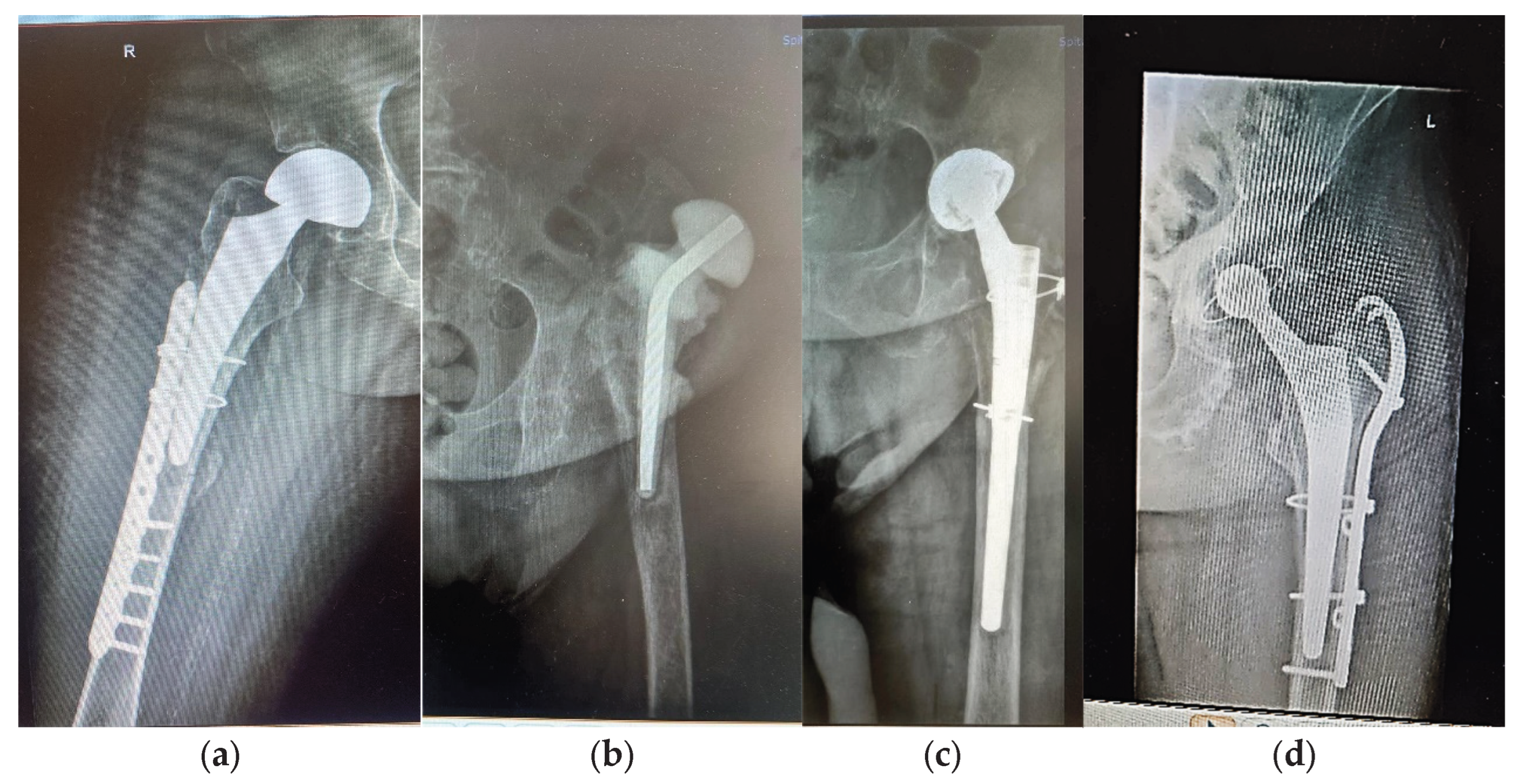
Case 1. A woman (F1) over 35 years old was admitted to hospital with a basicervical femoral neck fracture (BCFNF), right side, following an unspecified fall on a flat surface. Her underlying conditions were: Type 1 diabetes (insulin dependency); renal failure end stage (dialysis dependency); essential hypertension. BCFNFs are rare, about 1.8% of all proximal femoral fractures, as it results from the comprehensive critical review by Jun-Il Yoo et al., [20]. Though F1 is a young patient, and BCFNF are specific to the elderly, caused by low energy impact, her underlying conditions have contributed to bones weakness. In their review that consider 15 studies, Jun-Il Yoo et al. conclude that definitions of BCFNF vary largely, and in consequence, treatment methods and their results. The treatment option for F1 was hemiarthroplasty with a bipolar prosthesis. During surgery, the patient suffered a periprosthetic fracture (Figure 1 a) that needed cable fixation.
Case 2. A woman (F2) aged over 80 suffered an unspecified fall while running house duties at her home, resulting in a periprosthetic fracture of her left hip. The patient was admitted to our hospital for revision. F2 had had suffered a left side THA 15 years ago, at a different clinic, with a Zimmer implant. High blood pressure (AHT), dyslipidaemia, diabetes, hyper-uraemia, and hypothyroidism were her comorbidities. During the revision surgery, F2 was treated for prosthetic joint infection with antibiotic spacer for three months. Prosthetic joint infections are mainly treated through a two-stage process [21,22], where infected implants are removed and antibiotic spacers are placed to preserve the length. When the site is free of infection, revision is performed. The figures 1b and 1c show the anteroposterior radiographs of F2’s revision THA.
Case 3. A over 65 - year - old man (M) was admitted to our hospital for revision of a 7 years old implant (THA). The patient had fallen on a hard surface and suffered a periprosthetic fracture. The stem cracked. M presented himself to hospital 6 months after the fracture was produced. His underlying conditions were very complex: AHT third degree, congestive heart failure, old myocardial infarction, coronary artery disease (CAD), atrioventricular block (AV) first degree, right bundle branch block (RBBB), dyslipidaemia (mixt hyperlipidaemia), azotaemia. The cracked stem was extracted, and THA revision was performed using Zimmer Biomet Ti6Al4V stem (Figure 1d). Remnants of bone, and soft periprosthetic tissue, from the retrieved stem were collected for further investigations. The bone fragment exhibited a thin metal layer.

Table 1.
Summary of the characteristic of the three patients.
| Case No. | Sex | Age [Years] | HA/Cause |
Type of Prosthesis | Underlying Conditions | Remarks | |
|---|---|---|---|---|---|---|---|
| Primary | Revision Time [Years] | ||||||
| 1 -F1 | F | Over 35 | Yes; basicervical femoral neck fracture; unspecified fall; hemiarthroplasty; periprosthetic fracture |
- |
Bipolar, Zimmer Biomet Ti6Al4V stem and cable fixation |
Type 1 diabetes; renal failure end stage; essential hypertension | Insulin dependency, Dialysis dependency |
| 2 – F2 | F | Over 80 | Yes; 15; periprosthetic fracture; unspecified fall | Taper lock complete, Zimmer Biomet Ti6Al4V stem |
High blood pressure (AHT); Dyslipidaemia; hyperglycaemia; azotaemia; uraemia hypothyroidism | - | |
| 3 - M | M | Over 65 | Yes; 7; periprosthetic fracture; unspecified fall | Taper lock complete, Zimmer Biomet, Ti6Al4V stem |
AHT third degree, Congestive heart failure, old myocardial infarction, coronary artery disease (CAD), atrioventricular block (AV) first degree, right bundle branch block (RBBB), dyslipidaemia (mixt hyperlipidaemia), azotaemia, uraemia |
fractured stem left in place 6 months till revision; bone fragment with thin metal layer; periprosthetic tissue with cellulitis, panniculitis, myositis | |
Anterolateral radiographs of the three cases are shown in the Figure 1. F1 had a right sided fracture, with primary HA (hemiarthroplasty with bipolar prosthesis). During surgery a new fracture occurred, imposing supplementary fixation with cables. F2 and M underwent revisions on their left sides. All prosthetic elements were from Zimmer Biomet Romania SRL, Splaiul Independentei 319L, Paris Office Building, Corp A1, Ground Floor, Bucharest.
Figure 1.
Anterolateral radiographs of: (a)-patient F1 (primary HA; hemiarthroplasty with bipolar prosthesis); (b,c)-patient F2, AHT revision after 15 years, with antibiotic spacer; (d)-patient M, revision after 7 years, because of periprosthetic fracture and stem cracked 6 months before revision.
Figure 1.
Anterolateral radiographs of: (a)-patient F1 (primary HA; hemiarthroplasty with bipolar prosthesis); (b,c)-patient F2, AHT revision after 15 years, with antibiotic spacer; (d)-patient M, revision after 7 years, because of periprosthetic fracture and stem cracked 6 months before revision.
2.2. Disorders in General Functions of the Body and Bone Health
As observed from the case presentation, the three patients share several underlying conditions:
F1, F2, M: AHT, diabetes, renal dysfunction;
F2, M: AHT, diabetes, dyslipidaemia, renal disfunction;
all of them primarily influencing on the bone health.
A recent review, by Panagiotis Anagnostis et al. [13] underlines the role of dyslipidaemias in atherosclerotic cardiovascular diseases, as well as in bone, and vascular, mineralization, showing the strong link between dyslipidaemias and osteoporosis. The study covers population worldwide [23,24,25], focusing however on elderly, postmenopausal women, women in general. Nevertheless, disturbances in the metabolism of lypoproteins, oxidative stress, and increased parathyroid hormone, enhance processes of bone demineralization thus favouring fractures.
Diabetes mellitus is a major metabolic disorder, resulting from abnormal insulin production, and action, manifested through hyperglycaemia. The type 1 is an auto-immune disease, where the islet cells in the pancreas are attacked by the immune system of the body and insulin is not produced. In adults, this disease results in drastic modification of bone structure, mainly in the femur [26]. Type 2 diabetes mellitus is caused by insufficient use of insulin, leading to insulin resistance. One consequence is drastic decay of bone mineral density. Diabetes, type 1 and type 2, influence dramatically on bone health, lowering its mineral density, leading to degeneration of its structure, and hence to frailty. There is a great number of studies, meta-analyses, and reviews, dealing with characteristics, pathogenesis, of diabetes mellitus, their consequences on bone health, and various ways of treatment. A very well organised work is the narrative review by Bo Wu, et al. [27]. Diabetes, AHT and kidney dysfunction are linked. High levels of glucose in blood destroy the filtering units in kidneys, thus progressively decreasing their filtration function, leading eventually to end stage renal failure (see patient F1). AHT and cardiovascular disease (including dyslipidaemias) diminish blood flow to the kidneys; the blood vessels get narrow and the pressure increases [28]. Patients with chronic kidney disease are at higher risk of fractures, especially hip fractures, as compared to healthy ones because of changes in bone turnover, mineralization, and volume [29,30,31,32].
It is expected now from the three cases to provide very particular interactions between their fragile bones and the Ti-alloy prostheses.
2.3. Bone-Titanium Alloy Interaction-Study on Retrieved Prostheses, Ex Vivo Direct Bone Samples, and Simulated Body Fluids
Three stems made of Ti-alloys, and coated with HAp (Zimmer Biomet, Medical International, Romania), shown in the Figure 2, have been investigated. The broken stem had lasted for 7 years in the femur of patient M, till he fell on a hard surface 6 months before presentation to hospital with a diaphyseal femoral fracture, for THA revision. Fragments of soft tissue, and a small piece of bone with a metallic thin layer, were collected from the broken stem and investigated.
In the Table 2 the samples investigated in this work are concisely described.
2.3.1. Stems in Synthetic Plasma
Biocompatibility, low toxicity, good mechanical properties, and high resistance to chemical corrosion, render titanium and its alloys as most suitable for implants [18,19]. N. Eliaz [18] provides a complex and comprehensive review on corrosion of biomaterials, with historical background, evolution of materials, techniques for producing them, and fundamentals of corrosion and tribology. Studies on interaction between these materials and human body have used simulated body fluid at pH 7.4, in electrochemical corrosion experiments [16,19,35,36,37]. In the present work, the electrochemical measurements have been performed in a typical 3-electrode cell, having the calomel electrode as reference electrode, a Pt wire as auxiliary electrode, whereas the respective stem samples were employed as working electrode. The electrolyte was synthetic plasma that mimics the plasma of a healthy person (Table 3). The precursors were dissolved in distilled water by magnetic stirring for 8 hours and filtered to remove any undissolved particles or impurities. The pH immediately after synthesis was 9.2 and it was set at 7.4 by adding 1.6 mL HCl 37%.
Table 3.
Composition of the synthetic plasma corresponding to a healthy person.
| Compound | NaCl | CaCl2 | KCl | MgSO4 | NaHCO3 | Na2HPO4 | NaH2PO4 |
|---|---|---|---|---|---|---|---|
| Mass [g] | 6.80267 | 0.20370 | 0.40099 | 0.10153 | 2.20212 | 0.12780 | 0.02644 |
Instrumentation settings details: -1V→1V potential range, 100 mV/s scanning rate for 5 cycles and 1:8 sampling rate. The instrumentation used for the measurements was OrigaFlex potentiostat (Origalys, Rillieux-la-Pape, France). No smoothing was performed on the voltammograms. The peak intensity was determined by first manually drawing the baseline and then integrating each peak in OrigaMaster software (Origalys, Rillieux-la-Pape, France).
The next step was to alter the synthetic plasma with glucose and urea in agreement with the biochemistry values at the presentation of the respective patient, shown in the Table 4.
Since the present research considers three cases, diagnosed with AHT, cardiovascular disease (including dyslipidaemias), and chronic renal disease, we did not add cholesterol in the altered composition of the “healthy” synthetic plasma, but instead bore in mind that all their conditions generate inflammatory states.
In our simulation, the fragments of prostheses (dimensions: L=20 mm, l=11 mm and h=6.56 mm) were immersed in 2 mL altered synthetic plasma and ultrasonicated for 7 h at 36 kHz, at a temperature of 38oC. The electrochemical measurements were performed using the OrigaFlex potentiostat (Origalys, Rillieux-la-Pape, France) using DRP-150 microsensors from Dropsens (Metrohm DropSens, Vivero de Ciencias de la Salud, Spain). The DRP-150 microsensors are composed of: (1) a working electrode made of carbon; (2) a reference electrode made of silver; and (c) an auxiliary electrode made of platinum. Samples of 50 µL-100 µL were deposited on the microsensor and cyclic voltammetry was performed in the range [-1000; 1250] mV with different scanning rates (50 mV/s, 100 mV/s, and 150 mV/s), for 5 cycles, and 1:7 sampling rate. For the peak analysis, we chose the 3rd cycle, established the baseline, and determined the peak potential, peak intensity as current, using the dedicated software OrigaMaster 5 (Rillieux-la-Pape, France). Because of the small size of the samples, we switched from the typical 3-electrode assembly to a micro-electrode DRP-150 (Dropsens, Spain). The Origalys potentiostat (Metrohm, France) was replaced by the Dropsens bipotentiostat, adapted for microsensor electrochemisty. Briefly, the micro-electrode (composed of a carbon working electrode, a Pt auxiliary electrode and an Ag pseudo-reference electrode – all of them printed on a ceramic support) is inserted into a 3-channel slit connected to the bipotentiostat. A 50 µl sample was dropped and dispersed on the surface of the micro-electrode, to fully covering it. The potential was swept between -1 V and 1 V with 100 mV/s scan rate, 1:7 sampling rate, for 5 cycles.
2.3.2. Tissue in Synthetic Plasma
Five fragments (S1, …S4, soft tissue, and S5 bone with metal thin layer) remnant on the broken stem (Stem 1) have been immersed each in 10 mL synthetic plasma and ultrasonicated for 15 minutes at 25 kHz to extract the components. Three pH-values were used: 7.3 (S1 and S2), 6.9 (S3 and S4) and 8.5 (S5).
2.3.3. Surface Morphology and Element Analysis
The surface morphology is a key factor for osseointegration of implant, a mechanism occurring directly at the bone-metal/alloy interface. Osteoconductive properties of surfaces enhance osseointegration with host tissues, i.e., the growth of bone on the surface of an implant [38,39,40,41,42]. Therefore, characteristics of surfaces of implants are highly important. Surface morphology and the element compositions of the stem samples were examined by energy dispersive X-ray spectroscopy (EDX), using a Hitachi TM3030 Plus tabletop scanning electron microscope (SEM) equipped with a Brucker Quantax 7.0 energy dispersive X-ray spectrometer. The SEM enables effective image analysis by combining two signals into a single image: the secondary electron (SE) signal that provides surface-rich information, and the backscattered electron signal that supplies compositional information. The linear calibration of the EDX system was carried out using the Cu-Ka (8.037 keV) and Cu-La (0.926 keV) lines recorded from a Cu standard sample. The EDX spectra of the samples were recorded over the 0-15 keV energy range with an accumulation time of 1200 sec. In the Figure 3, the image of a cut and polished zone is displayed, showing the analysed zones.
2.3.4. Histopathological Analyses of Soft Tissue Remnant on Stem1
The slides were prepared following the usual flux: fixation in formalin 10%, trimming and transfer to labelled cassettes, dehydration, clearing, and embedding in paraffin (inclusion station Sakura Tissue-Tek), sectioning with a histoprocessor (Sakura-Japan), transferring to microscope slides, and staining with haematoxylin+eosin (HE). Further on, the slides were examined with an Olympus BH-2 BHS microscope, equipped with an Olympus SC50 camera and a Cell Sense dedicated software. All equipment is Made in Japan.
3. Results
Electrochemistry
3.1.1. Stems in Synthetic Plasma
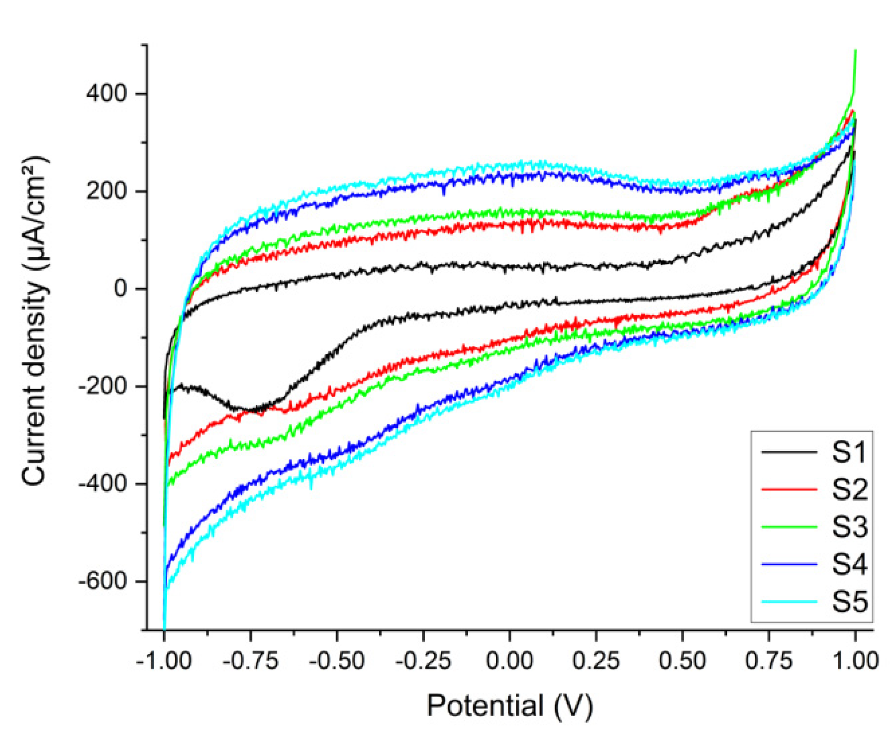
For the three stems, the reactions that correspond to the reduction peak could be attributed to either HPO(OH)2+2H++2e- → H2PO(OH)+H2O (standard potential is -0.5 V) present in the synthetic plasma or S+2e- → S2- (standard potential is -0.48 V), which could be deposited as residue on the prostheses. The main feature of the voltammograms is that the corrosion is not accelerated by the presence of the glucose or urea. In the voltammetry, the -515 mV potential is like the standard potential for the reduction of phosphide (H3PO3). In the Figure 4, a and b, are shown the voltammograms (a) and (b) the double-layer capacitance with the oxidation and reduction waves placed on the figure, as well as peak position (reduction potential) and the current density (Ipc).
The reactivity of the stems in altered synthetic plasma is given mainly by the presence of glucose and urea. Several studies refer to glucose, inflammatory synthetic environment [14,16,37,43,44,45], urea being mentioned rarely [14,45]. In our study, based on three real cases, the double-layer capacitance (Cdl) shows that the combination of glucose and urea mimicking real-life underlying conditions of the patient, have produced a decrease of the adsorption/desorption process that led to a reduction reaction evident in the CV at -500 mV. Except reference [44], a study on Gold 21k in artificial sweat (containing glucose and urea), the materials involved are Ti-based alloys. In our experiments, even though the intensities of the peaks are low, the occurrence of the reduction peaks shows that the presence of glucose and urea have a direct influence on the Ti-based prosthesis (Figure 5). Previous publications [13,14,28] suggest that changes in the biological environment would lead to changes in the surfaces of the implants immediately after implantation.
3.1.2. Tissue in Synthetic Plasma
From the cyclic voltammetry of the 5 tissue samples (S1…S5), in the first cycle, at 100 mV/s scanning rate, only S1 has a visible reduction peak at -0.752 V (Figure 4), corresponding to the reaction FeCO3(s)+2e- → Fe(s) + CO32- (standard potential at -0.756 V) [46]. Ferrous carbonate is a porous corrosion product that forms on the surface of the prosthesis through the reaction of the CO32- ions dissolved in the plasma with the Fe in the alloy (see EDX, and Figure 4 a, b). It acts as protection against further corrosion by installing a diffusion barrier and slowing down the leaching of the metal [17,18]. In comparison with S1, the other samples do not seem like leaching of the metals at this scanning rate. The reduction of the scanning rate down to 10 mV/s shows that for S1, the iron carbonate is consumed in the initial cycles, missing in the subsequent, low scanning rate, cycles. For the other samples, only S5 (sample with a bone inclusion and a fine metal like coating) demonstrates that a minute amount of vanadium has leached and is present in the extraction solution (V3++e- →V2+ standard potential at -0.255 V and the experimental potential of the reaction is -0.278 V for S5 [9,17]. For the rest of samples, the potential corresponds to the reduction of the dissolved oxygen to: O2+e- →O2- (standard potential is at -0.33 V whereas our experimental potential is determined to be -0.334 V).
Figure 5.
Voltammograms of the five tissue samples immersed in synthetic plasma. only S1 has a visible reduction peak at -0.752 V. S5 (sample with a bone inclusion and a fine metal like coating) demonstrates that a minute amount of vanadium has leached and is present in the extraction solution (standard potential at -0.255 V and the experimental potential of the reaction is -0.278 V).
Diabetic patients with Ti6Al4V prostheses, especially TH and femoral ones, can expect earlier loosening and failure than the non-diabetic ones because of an increased coefficient of friction [37]. This seems to be true for Case 2 -M, who fractured his femur and stem, and expected for F1 and F2.
3.1.3. Surface Morphology and Element Analysis
In the Figure 6 (a, b, c, d) are shown the SEM images of surfaces corresponding to: a- the broken, retrieved, Stem 1 (Figure 2, broken end), b-polished substrate of stem 2 (Figure 3, label 1), c-fine layer of stem 2 (Figure 3, label 2), d-rough layer of stem 2 (Figure 3, label 3). Stems 2 and 3 were identical. Therefore, we show here the results for stem2. From the Figure 6, it is remarked that the three layers of the Stem 2 have different structures, the roughest being the external one (Figure 3, label 3, and Figure 6d). The surface of Stem 1 corresponds to the rough layer of Stem 2. However, the morphology is most alike that of the fine layer (Figure 6 c and Figure 3-2). The diabetic and uremic environment of Stem 1 during 7 y 6m, accompanied by motion, led to wear by corrosion and friction. At the interface between the cancellous bone and the surface of implant, cells in the healing tissue can also produce corrosion [14]. Also, we bear in mind that Stem 1 and Stem 2 are different versions from the same producer, and we have no information of the initial image of the surface of Stem 1. However, the plasma spray technique to deposit Hap on Zimmer Biomet stems has been in use for several decades. EDX spectra show that the stems are basically built of Ti-6Al-4V, with a surface layer of HAp (Table 5 and Table 6), thus supporting the CV results (Figure 4). This spectral analysis is particularly useful to understand the probability of metal ion migration in the body. All the prostheses studied here include small amounts of iron and carbon.
From the analyses it is observed that the substrates of Stem 1 and Stem 2 are composed mainly of Ti, Al, and V, in similar proportions. The outer layers contain also elements of HAp. It is practically impossible to neatly separate the layers between each other, and from the substrates, because deposition techniques aim for adhesion, and interdiffusion at the interfaces [9,36,37,39]. An important remark is the small amount of phosphorus, especially in the layer of Stem 1. Calcium and phosphorous are rapidly absorbed in the healing process [14,38,41,48]. Also, there is a great difference between the vanadium concentration in the substrate of Stem 1 and its layer. We consider that vanadium leached indeed after the stem has broken in M’s femur, view the electrochemistry measurements, and supporting publications [5,17,35,37,45].
3.1.4. Histopathological Analyses of Soft Tissue Remnant on Stem1
The histopathology analysis (Figure 7 a, b, c) shows a pronounced effect of lipids in the sample extracted from the Stem 1, in agreement with the biochemistry blood tests of the patient. Severe subcutaneous reaction characterized by cellulitis, panniculitis, and myositis is present. There is no evidence of neoplastic aspect or other pathogens microscopically visible. However, loss of histological details of subcutaneous tissue indicates necrosis. Numerous neutrophils intermixed by macrophages, and a few lymphocytes, are present, supporting the abundance of small new capillaries (granulation tissue). The density of inflammatory cells, including neutrophils, macrophages, and fibroblasts, is approximately 80 cells per high-power field under high magnification. Direct corrosion by macrophages, monocytes and osteoclasts, has been reported in culture media [37].
4. Discussion
Understanding bones, their quality, damage, ways of recover, including prosthetics of all sorts, has demanded a great effort for ages. Orthopaedic surgery involves various kinds of implants, whose interfaces with the bone tissues, and their surroundings, are dramatically influenced by the characteristics of the implant, e.g., geometry, material, surface morphology, topology, mechanical, physical, chemical properties [1,2,3,4,5,6,7,8,9], and by interaction with the living body presenting dysfunctions of important internal organs, such as pancreas, kidneys, parathyroid, liver, heart [10,11,12,13,14,15]. The three cases we reported here are complex examples of diabetes, cardiovascular disease, kidney disease and dyslipidaemias, all requiring HA (either primary, o revision) with Ti6Al4V femoral stems. The enormous number of reviews on the material, processing of prostheses, and the interplay between those, and patients’ underlying conditions, gives important insights on the necessity of a more holistic diagnostic for orthopaedic patients. P. Anagnostis et. al. [13] touch a hot, although underrepresented subject, i.e. dyslipidaemias and osteoporosis. The review by M. Prestat and D. Thierry [37], claimed in 2021 as the first review “focused specifically on the topic of inflammation”, examines the research run over forty years on the in vitro studies on titanium corrosion under simulated inflammation conditions, involving materials science, corrosion, prosthetics. In the same trend, we have already mentioned the work by N. Eliaz [18]. This just strengthens the high importance of these studies, and reviews, for advanced knowledge in the long term. To summarise, the bone-Ti -alloy interaction follows two strands: 1) the bone health, and 2) the alloy characteristics, and the interface surfaces. Body fluids are permanent carriers of various chemical, and biochemical, compounds, released either through the metabolic functions of the patient, or from the interaction between those and the materials of prostheses, also accounting for the mechanical influence. Knowing from the start what conditions has a particular patient, e.g. from haematology and biochemistry tests, and the life of the prosthesis till revision (depending on case), makes it easier to understand the causes of the prosthesis failure and to eventually prescribe measures of prevention with respect to the bone quality preservation.
5. Conclusions
Three cases were reported, two women, F1 and F2, and one man, M, of different ages, undergoing HA (F1-primary, F2 and M-revision) with uncemented Ti-alloy (Ti6Al4V) and sharing cardiovascular disease, diabetes, and kidney disease. F2 and M shared dyslipidaemia too. Two stems (one broken in M’s femur, and one unused), and remnant tissue on the retrieved stem, were investigated by electrochemistry, showing that reactivity of the stems in altered synthetic plasma is given mainly by the presence of glucose and urea. One sample with a bone inclusion coated with a fine metal-like film demonstrates that a minute amount of vanadium has leached and is present in the extraction solution at the experimental potential of the reaction of -0.278 V. Leak of V is supported by EDX analysis, showing a remarkable difference between the V concentration (at. %) in the broken stem and the unused one. The high variation coefficients for the measurements of the biological samples (tissue in synthetic plasma) can be explained by the complex structures of the biological materials, the different material pre-treatment (synthetic plasma vs. natural blood plasma), different electrode preparation (screen printed electrodes vs. three electrode standard assembly) and the presence of corrosion products as impurities. Five repeated cyclic voltammetries were recorded for each measurement that indicates a variation coefficient of 5.88% for the stems. For the measurements of the tissue in synthetic plasma we calculated a variation coefficient of 21.21%. The diabetic and uremic environment of M’s broken stem, during 7 y 6m, accompanied by motion, led to wear by corrosion and friction. At the interface between the cancellous bone and the surface of implant, cells in the healing tissue can also produce corrosion. The histopathology analysis proves a density of inflammatory cells of, approximately, 80 cells per high-power field under high magnification. Those include neutrophils, macrophages, and fibroblasts that contribute to corrosion. Direct corrosion by macrophages, monocytes and osteoclasts, has been reported in culture media. The SEM-EDX analysis of the outer layers of the stems demonstrates that hydroxyapatite loses its compositions in time, owing to the high absorption of phosphorus and calcium during healing. Diabetic patients with Ti6Al4V prostheses, especially TH, and femoral ones, can expect earlier loosening and failure than the non-diabetic ones because of an increased coefficient of friction. This seems to be true for Case 2 -M, who fractured his femur and stem, and expected for F1 and F2. Patients with chronic kidney disease are at higher risk of fractures, especially hip fractures, as compared to healthy ones because of changes in bone turnover, mineralization, and volume. Dyslipidaemias, and cardiovascular disease, affect the bone, but do not produce corrosion at the bone – metal interface. The limitations of this experimental work consist in considering only femoral stems, Ti6Al4V alloy only, from Zimmer Biomet solely. The brief review ran along with description of the work.
Author Contributions
Funding
Institutional Review Board Statement
Informed Consent Statement
Data Availability Statement
Acknowledgments
Conflicts of Interest
References
- M. J. DeRogatis, E. Wintermeyer, T R. Sperring, P. S. Issack, Investigation performed at the Department of Orthopaedic Surgery, New York – Presbyterian Hospital, New York, NY, JBJS Am. 2019;101:745-54 d. [CrossRef]
- M. Kaur, K. Singh, Review on titanium and titanium - based alloys as biomaterials for orthopaedic applications. Mater Sci Eng C Mater Biol Appl. 2019, Vol.102, pp. 844–862. [CrossRef]
- M.P. Abdel, U. Cottino, D.R. Larson, A.D. Hanssen, D.G. Lewallen, D.J. Berry, Modular fluted tapered stems in aseptic revision total hip arthroplasty. J BJS Am., 2017 May 17;99(10):873-81. [CrossRef]
- M. B. Cross, W. G. Paprosky, Managing femoral bone loss in revision total hip replacement: fluted tapered modular stems, Bone Joint J 2013 ;95-B, Supple A:95–7. [CrossRef]
- N. M. Brown, M. Tetreault, C. A. Cipriano, C. J. Della Valle, W. Paprosky, S. Sporer, Modular Tapered Implants for Severe Femoral Bone Loss in THA: Reliable Osseointegration but Frequent Complications, Clin Orthop Relat Res 2015, 473:555–560. [CrossRef]
- D. Apostu, O. Lucaciu, C. Berce, D. Lucaciu, D. Cosma, Current methods of preventing aseptic loosening and improving osseointegration of titanium implants in cementless total hip arthroplasty: a review, J Int Med Res 2017, Nov 3;46(6):2104–2119. ,. [CrossRef]
- B. Ziaie, X. Velay, W. Saleem, Advanced porous hip implants: A comprehensive review, Heliyon, 2024, 10 :18, e37818. [CrossRef]
- K. Solou, A. Vasiliki Solou, I. Tatani, J. Lakoumentas, K. Tserpes, P. Megas, A Customized Distribution of the Coefficient of Friction of the Porous Coating in the Short Femoral Stem Reduces Stress Shielding, Prosthesis 2024, 6, 1310–1324. [CrossRef]
- P. Hameeda, V. Gopala, S. Bjorklund, A. Ganvir, D. Sen, N. Markocsan, G. Manivasagam, Axial Suspension Plasma Spraying: An ultimate technique to tailor Ti6Al4V surface with HAp for orthopaedic applications, Colloids Surf.B: Biointerfaces, 2018, 173 806-815. [CrossRef]
- C. C. Baciu, Energy, Waves, and Forces in Bilateral Fracture of the Femoral Necks: Two Case Presentations and Updated Critical Review, Diagnostics 2022, 12, 2592. [CrossRef]
- E. Onochie, B. Kayani, S. Dawson-Bowling, S. Millington, P. Achan, S. Hanna, Total hip arthroplasty in patients with chronic liver disease: A systematic review, SICOT-J 2019, 4;5:40. [CrossRef]
- S.-M. Lee, W. C. Shin, S. H. Woo, T. W. Kim, D. H. Kim, K. T. Suh, Hip arthroplasty for patients with chronic renal failure on dialysis, Sci. Rep. 2023 13:3311. [CrossRef]
- P. Anagnostis, M. Florentin, S. Livadas, I. Lambrinoudaki, D. G. Goulis, Bone Health in Patients with Dyslipidemias: An Underestimated Aspect, Int. J. Mol.Sci. 2022, 23, 1639. [CrossRef]
- A. Arteaga, J. Qu, S. Haynes, B. G. Webb, J. LaFontaine, D. C. Rodrigues, Diabetes as a Risk Factor for Orthopedic Implant Surface Performance: A Retrieval and In Vitro Study, Bio Tribocorros. 2021; 7(2). [CrossRef]
- N. R. Rundora, J. W. Van der Merwe, D. E. P. Klenam, M. O. Bodunrin, Enhanced corrosion performance of low-cost titanium alloys in a simulated diabetic environment, Mater.Corros. 2023. [CrossRef]
- Y. Si, M. Li, H. Liu, X. Jiang, H. Yu, D. Sun, Evaluation of tribocorrosion performance of Ti6Al4V alloy in simulated inflammatory and hyperglycemic microenvironments, Wear 2023, Volumes 532–533, 205077. [CrossRef]
- Q. Zhong, X. Pan, Y. Chen, Q. Lian, J. Gao, Y. Xu, J. Wang, Z. Shi, H. Cheng, Prosthetic Metals: Release, Metabolism and Toxicity, Int J Nanomedicine 2024; 19:5245-5267. [CrossRef]
- N. Eliaz, Corrosion of Metallic Biomaterials: A Review, Materials 2019 12(3), 407. [CrossRef]
- D.C. Rodrigues, R.M. Urban, J. J. Jacobs, J. L Gilbert, In vivo severe corrosion and hydrogen embrittlement of retrieved modular body titanium alloy hip-implants, J.Biomed. Mat. Res. Part B: Applied Biomaterials 2009 88(1), 206-219. [CrossRef]
- J.-I. Yoo, Y. Cha, J. Kwak, H.-Y. Kim, W.-S. Choy, Review on Basicervical Femoral Neck Fracture:.
- Definition, Treatments, and Failures, Hip Pelvis 2020 32(4): 170-181. [CrossRef]
- M. A. Maceroli, R. S. Yoon, F.A. Liporace, Use of Antibiotic Plates and Spacers for Fracture in the Setting of Periprosthetic Infection, J Orthop Trauma 2019; 33 Suppl 6:S21-S24. [CrossRef]
- S.M. Petis, M. P. Abdel, K. I. Perry, et al., Long-term results of a 2-stage exchange protocol for periprosthetic joint infection following total hip arthroplasty in 164 hips, J Bone Joint Surg Am. 2019; 101:74–84. [CrossRef]
- N. Panahi, A. Soltani, A. Ghasem-Zadeh, G. Shafiee, R. Heshmat, F. Razi, N. Mehrdad, I. Nabipour, B. Larijani, A. Ostovar, Associations between the lipid profile and the lumbar spine bone mineral density and trabecular bone score in elderly Iranian individuals participating in the Bushehr Elderly Health Program: A population-based study, Arch. Osteoporos. 2019, 14, 52. [CrossRef]
- K.Y. Chin, C.Y. Chan, S. Subramaniam, N. Muhammad, A. Fairus, P.Y. Ng, N.A. Jamil, N.A. Aziz, S. Ima-Nirwana, N. Mohamed, Positive association between metabolic syndrome and bone mineral density among Malaysians, Int. J. Med. Sci. 2020, 17, 2585–2593. [CrossRef]
- Q. Zhang, J. Zhou, Q. Wang, C. Lu, Y. Xu, H. Cao, X. Xie, X. Wu, J. Li, D. Chen, Association Between Bone Mineral Density and Lipid Profile in Chinese Women, Clin. Interv. Aging 2020, 15, 1649–1664. [CrossRef]
- V. N. Shah, K. K. Harrall, C. S. Shah, T. L. Gallo, P. Joshee, J. K. Snell-Bergeon & W. M. Kohrt, Bone mineral density at femoral neck and lumbar spine in adults with type 1 diabetes: a meta-analysis and review of the literature. Osteoporos Int 2017 28, 2601–2610. [CrossRef]
- B. Wu, Z. Fu, X. Wang , P. Zhou, Q. Yang, Y. Jiang, D. Zhu, A narrative review of diabetic bone disease: Characteristics, pathogenesis, and treatment, Front Endocrinol (Lausanne) 2022; 13:1052592. [CrossRef]
- R. Chermiti, S. Burtey, L. Dou, Role of Uremic Toxins in Vascular Inflammation Associated with Chronic Kidney Disease. J. Clin. Med, 2024, 13 (23), pp.7149. [CrossRef]
- A. G. Kattah, S. M. Titan, R. A. Wermers, The Challenge of Fractures in Patients With Chronic Kidney Disease, Endocr. Pract.2025, 31:4, 511-520. [CrossRef]
- A. Pimentel, P. Ureña-Torres, J. Bover, J. L. Fernandez-Martín, M. Cohen-Solal, Bone Fragility Fractures in CKD Patients. Calcif Tissue Int. 2021, 108(4):539-550. [CrossRef]
- Y. Kim, E. Lee, M.-J. Lee, B. Park, I. Park, Characteristics of fracture in patients who firstly starts kidney replacement therapy in Korea: a retrospective population-based study, Sci Rep 2022, 12 3107. [CrossRef]
- P. A. Ureña Torres, M. Cohen-Solal, Evaluation of fracture risk in chronic kidney disease, J Nephrol. 2017 30(5):653-661. [CrossRef]
- L. Benea, N. Simionescu-Bogatu, Reactivity and Corrosion Behaviors of Ti6Al4V Alloy Implant Biomaterial under Metabolic Perturbation Conditions in Physiological Solutions, Materials 2021; 14(23):7404. [CrossRef]
- R. A. Gittens, R. Olivares-Navarrete, R. Tannenbaum, B.D. Boyan, Z. Schwartz, Electrical Implications of Corrosion for Osseointegration of Titanium Implants, J Dent Res. 2011; 90(12):1389-97. [CrossRef]
- D. Prando, A. Brenna, M. V. Diamanti, S. Beretta, F. Bolzoni, M. Ormellese, and M.P. Pedeferri, Corrosion of Titanium: Part 1: Aggressive Environments and Main Forms of Degradation, J. Appl. Biomater. Functional Mater 2017, 15, 4, e291-e302. [CrossRef]
- J. van Drunen, B. Zhao, G. Jerkiewicz, Corrosion behavior of surface-modified titanium in a simulated body fluid, J Mater Sci. 2011, 46:5931–5939. [CrossRef]
- M. Prestat, D. Thierry, Corrosion of titanium under simulated inflammation conditions: clinical context and in vitro investigations, Acta Biomater 2021, 136, 72-87. [CrossRef]
- M. Darwish Elsayed, Biomechanical Factors That Influence the Bone-Implant-Interface, Res Rep Oral Maxillofsc Surg 2017, 3:023. [CrossRef]
- S.A. Naghavi, H. Wang, S.N. Varma, M. Tamaddon, A. Marghoub, R. Galbraith, J. Galbraith, M. Moazen, J. Hua, W. Xu, C. Liu, On the morphological deviation in additive manufacturing of porous Ti6Al4V scaffold: a design consideration, Materials 2022, 15. [CrossRef]
- N. Kohli, J. C. Stoddart, R. J. van Arkel, The limit of tolerable micromotion for implant osseointegration: a systematic review, Sci Rep 11, 2021 10797. [CrossRef]
- P. Kazimierczak, A. Przekora, Osteoconductive and Osteoinductive Surface Modifications of Biomaterials for Bone Regeneration: A Concise Review Coatings 2020, 10(10), 971. [CrossRef]
- W. Zuo, L. Yu, J. Lin, Y. Yang, Q. Fei, Properties improvement of titanium alloys scaffolds in bone tissue engineering: a literature review, Ann Transl Med. 2021, 9(15):1259. [CrossRef]
- V. Vasudha Nelluri, R. Kumar Gedela, M. Roseme Kandathilparambil, Influence of Surface Modification on Corrosion Behavior of the Implant Grade Titanium Alloy Ti-6Al-4V, in Simulated Body Fluid: An In Vitro Study. Int J Prosthodont Restor Dent 2020; 10(3):102–111. [CrossRef]
- T. Umamathi, R. Parimalam, V. Prathipa, A. Josephine Vanitha, K. Muneeswari, B. Mahalakshmi1, S. Rajendran, A. Nilavan, Influence of urea and glucose on corrosion resistance of Gold 21K alloy in the presence of artificial sweat, Zas Mat 2022 63:3, 341 – 352. [CrossRef]
- A. Ait Sidimou, D. M. El Marrakchia, E. Khoumri, Electrochemical corrosion behavior of a-titanium alloys in simulated biological environments (comparative study), RSC Adv., 2024, 14, 38110,. [CrossRef]
- A.J. Bard, R. Parsons, J. and Jordan, Standard Potentials in Aqueous Solution-1st Edition. IUPAC-Marcel Dekker Inc., New York ISBN 9780824772918, Published August 27, 1985 by CRC Press.
- S.A. H. Naghavi, S. N. Wang, M. Varma, A. Tamaddon, R. Marghoub, J. Galbraith, M. Galbraith, J. Moazen, J. Hua, W. Xu, C. Liu, On the morphological deviation in additive manufacturing of porous Ti6Al4V scaffold: a design consideration, Materials 2022, 15(14), 4729. [CrossRef]
- Alireza Rahimnia, Hamid Hesarikia, Amirhosein Rahimi, Shahryar Karami, and Kamran Kaviani, Evaluation and comparison of synthesised hydroxyapatite in bone regeneration: As an in vivo study, J Taibah Univ Med Sci. 2021 Jul 15;16(6):878-886. [CrossRef]
Figure 2.
Stem 1 was retrieved from the femur of patient M.
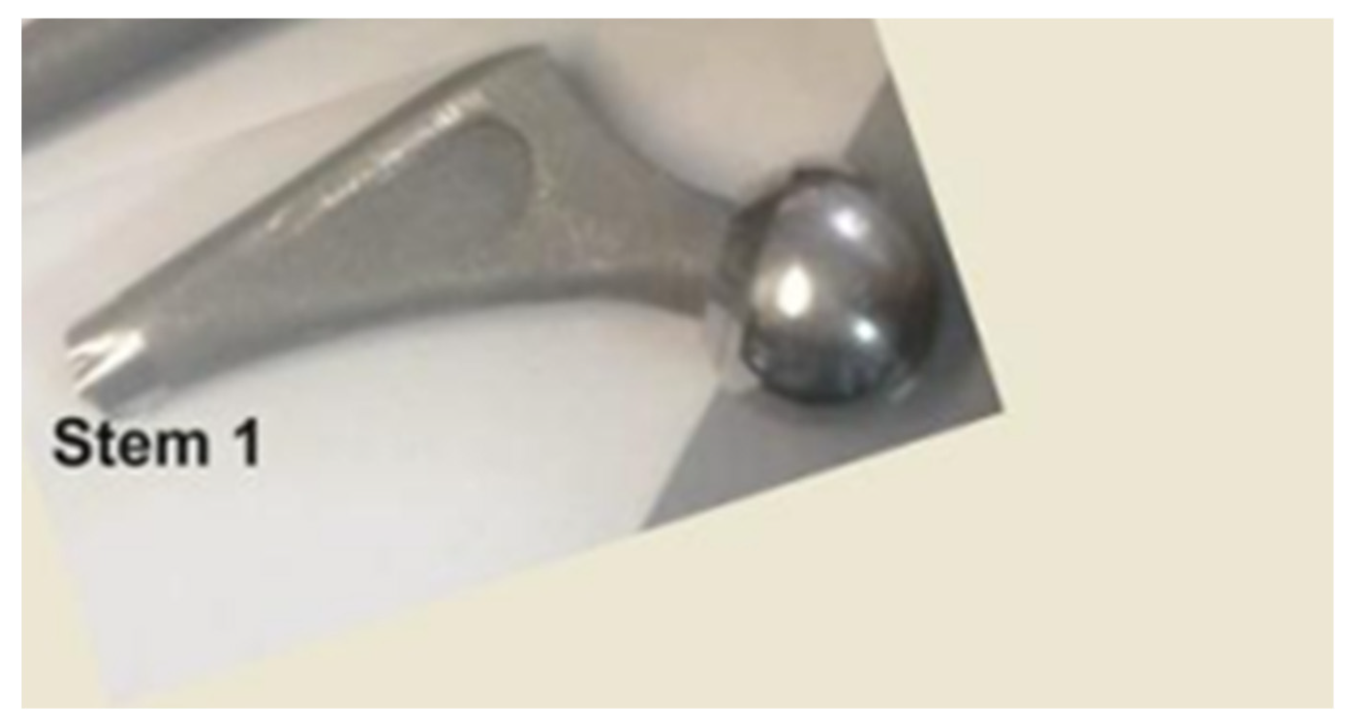
Figure 3.
Cut of the stem: 1-cross section, substrate polished; 2- fine layer; 3-. rough layer.
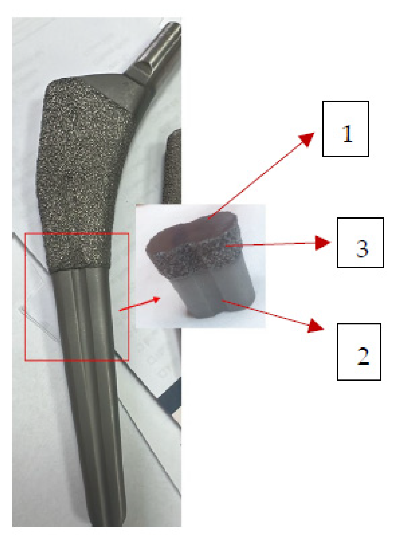
Figure 4.
(a)-The voltammograms, and (b)-the double-layer capacitance for cyclic voltammetry of samples.
Figure 4.
(a)-The voltammograms, and (b)-the double-layer capacitance for cyclic voltammetry of samples.
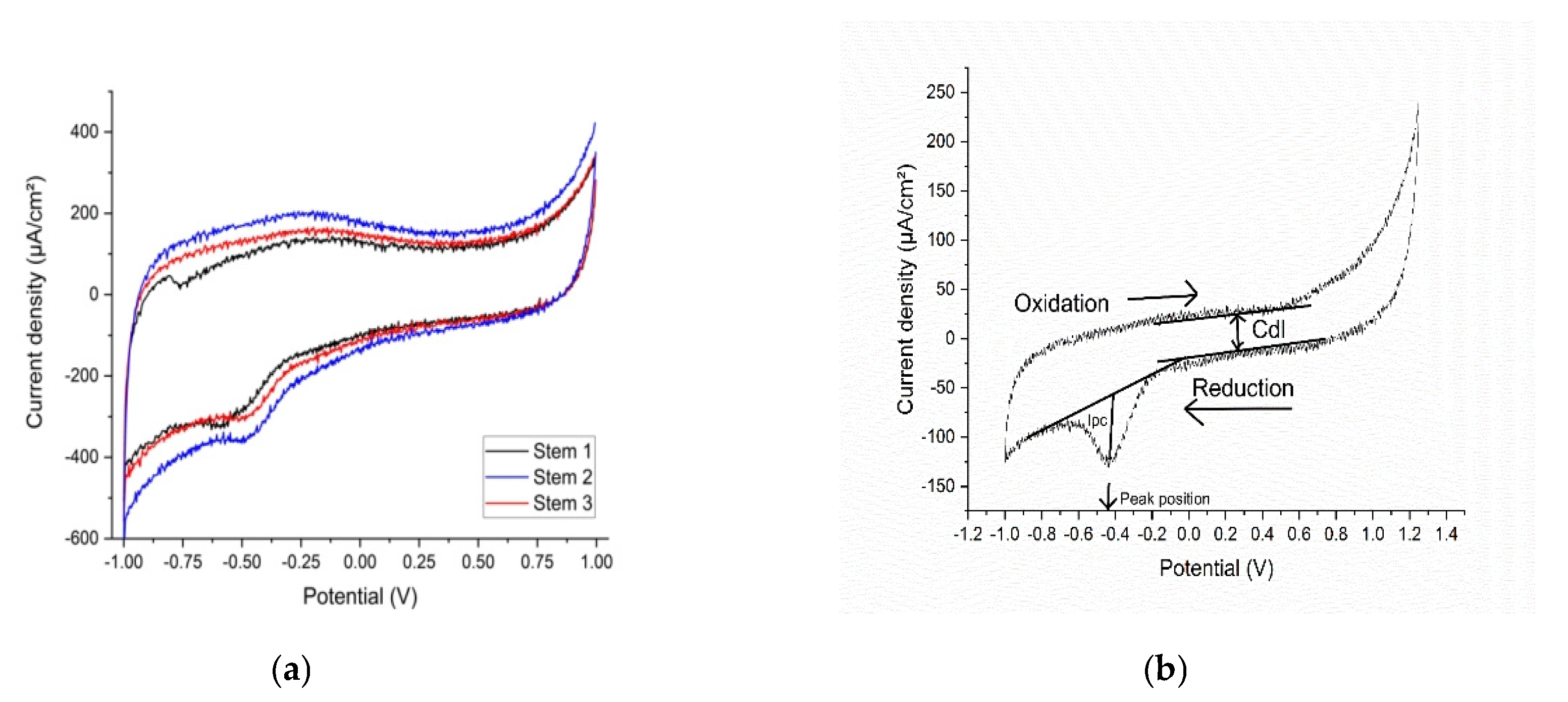
Figure 6.
SEM images of surfaces corresponding to: (a)- the broken, retrieved Stem 1 (Figure 2, broken end), (b)-polished substrate of stem 2 (Figure 3, label 1), (c)-fine layer of stem 2 (Figure 3, label 2), (d)-rough layer of stem 2 (Figure 3, label 3).
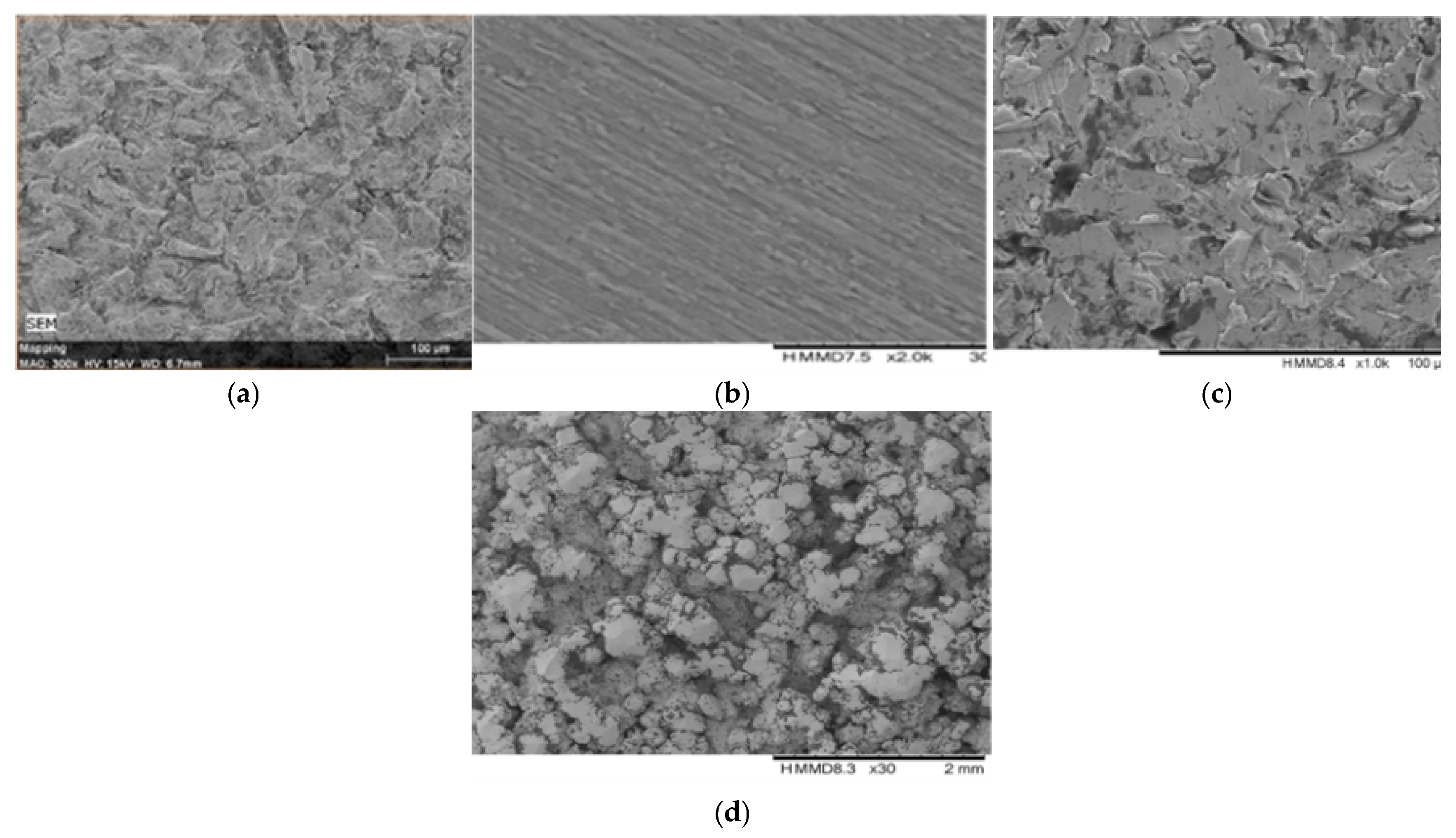
Figure 7.
The histopathology analysis (a, b, c) shows a pronounced effect of lipids in the sample extracted from Stem 1, in agreement with the biochemistry blood tests of the patient M: (a)- Loss of histological details of subcutaneous tissue (necrosis). Numerous neutrophils intermixed by macrophages and a few lymphocytes are present supporting the numerous small new capillaries (granulation tissue); (b)- additional numerous extravasated erythrocytes and several fragments of inflammatory cells (cellular dust) are very evident in the sections; (c)- Subcutaneous tissue detail: 80% intact, and degenerated neutrophils, intermixed with red blood cells (purulent inflammation and discrete haemorrhage are present.); red fibrillar to homogenized proteinaceous material is evident, closely related to inflammatory reaction described earlier; lymphocytes, and macrophages, with small capillaries, are completing the entire view of the lesion.
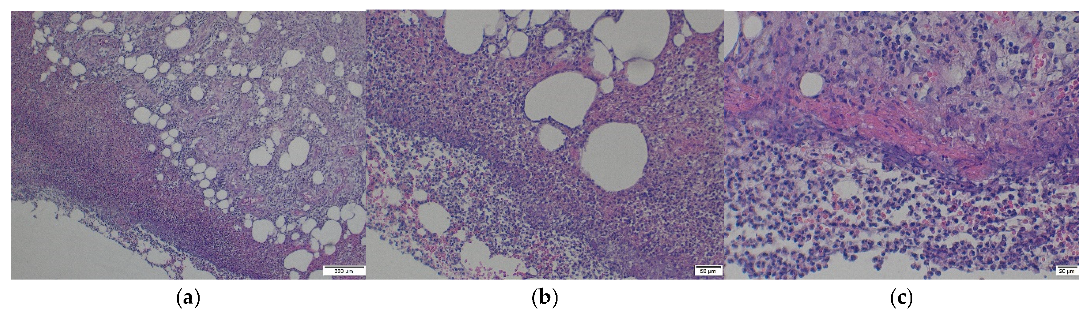
Table 2.
Characteristics of samples.
| Sample Name | Type | Condition | Source | Size |
|---|---|---|---|---|
| Stem 1 -fragment from the broken end | Solid, Ti-alloy +HAp, femoral stem | Broken approx. perpendicular to its longitudinal axis, 7 years used unbroken, 6 months used broken | Femur of patient M, aged over 65 years with cardiovascular disease, type 2 diabetes, dyslipidaemia, chronic renal disease, azotaemia, uraemia |
L=20 mm, l=11 mm and h=6.56 mm |
| Stem 2 -fragment from the distal end | Solid, Ti-alloy +HAp, femoral stem | Unutilized | Unutilized | L=20 mm, l=11 mm and h=6.56 mm |
| Stem 3- fragment from the distal end | Solid, Ti-alloy +HAP*, femoral stem | Unutilized | Unutilized | L=20 mm, l=11 mm and h=6.56 mm |
| Synthetic plasma (Table 3) | Liquid | Synthesized | 50 µL/sample | |
| 3 x soft tissue fragments | Solid | Ex vivo direct | Surface of Stem 1 | 1.5 cm2 1.098 g each |
| Bone tissue fragment with thin metal layer | Solid | Ex vivo direct | Surface of Stem 1 | 1.75 cm2 2.532 g |
Table 4.
Alteration of “healthy” synthetic plasma with glucose and urea in agreement with the biochemistry values at the presentation of the respective patient.
Table 4.
Alteration of “healthy” synthetic plasma with glucose and urea in agreement with the biochemistry values at the presentation of the respective patient.
| Compound Added to the “Healthy” Synthetic Plasma | Concentration in “Healthy Synthetic Plasma” [mg/dL] | Patient | |
|---|---|---|---|
| Glucose |
266 |
F1 |
|
| Urea | 119 |
||
| Glucose | 118 | F2 | |
| Urea | 85 |
||
| Glucose | 95 | M | |
| Urea | 50 | ||
Table 5.
Element analysis of Stem1, substrate, and external layer.
| Stem1, Layer | Stem1, Substrate | ||
|---|---|---|---|
| Element | At. % | Element | At. % |
| Titanium | 24.05 ± 1.73 | Titanium | 75.09 ± 2.91 |
| Aluminium | 10.53 ± 0.70 | Aluminium | 7.02 ± 0.27 |
| Vanadium | 0.82 ± 0.09 | Vanadium | 2.79 ± 0.14 |
| Oxygen | 55.22 ± 4.95 | Carbon | 8.50 ± 0.41 |
| Calcium | 0.81 ± 0.07 | Oxygen | 5.60 ± 0.42 |
| Phosphorus | 0.001 ± 0.00 | Nitrogen | 0.99 ± 0.10 |
| Carbon | 5.73 ± 0.48 | ||
| Silicon | 1.00 ± 0.09 | ||
| Sodium | 0.99 ± 0.10 | ||
| Potassium | 0.31 ± 0.04 | ||
| Iron | 0.30 ± 0.05 | ||
| Magnesium | 0.18 ± 0.04 | ||
| Molybdenum | 0.05 ± 0.03 | ||
| 99.991 ±8.37 | 99.99 ± 4.25 | ||
Table 6.
Element analysis of Stem 2.
| Stem 2, Substrate | Stem 2, Fine Layer | Stem 2 Rough, Layer | |||
|---|---|---|---|---|---|
| Element | At. % | Element | At. % | Element | At. % |
| Titanium | 72.52688066 | Titanium | 60.290 | Titanium | 37.180 |
| Aluminium | 7.863345632 | Aluminium | 3.536 | Aluminium | 3.411 |
| Vanadium | 2.347607468 | Vanadium | 1.902 | Vanadium | 1.300 |
| Silicon | 0.132498649 | Silicon | 3.725 | Silicon | 0.347 |
| Iron | 0.074318149 | Iron | 0.760 | Iron | 0.079 |
| Oxygen | 7.407082668 | Calcium | 0.609 | Calcium | 0.097 |
| Nitrogen | 0.908577893 | Phosphorus | 0.001 | Phosphorus | 0.043 |
| Carbon | 8.739688878 | Oxygen | 24.923 | Oxygen | 28.494 |
| Carbon | 3.590 | Carbon | 28.184 | ||
| Sodium | 0.512 | Sodium | 0.671 | ||
| Magnesium | 0.115 | Magnesium | 0.091 | ||
| Chlorine | 0.068 | ||||
| Potassium | 0.035 | ||||
| Sulphur | 0.001 | ||||
| 100.000 | 99.963 | 99.963 | 100.001 | ||
Disclaimer/Publisher’s Note: The statements, opinions and data contained in all publications are solely those of the individual author(s) and contributor(s) and not of MDPI and/or the editor(s). MDPI and/or the editor(s) disclaim responsibility for any injury to people or property resulting from any ideas, methods, instructions or products referred to in the content. |
© 2025 by the authors. Licensee MDPI, Basel, Switzerland. This article is an open access article distributed under the terms and conditions of the Creative Commons Attribution (CC BY) license (https://creativecommons.org/licenses/by/4.0/).
Copyright: This open access article is published under a Creative Commons CC BY 4.0 license, which permit the free download, distribution, and reuse, provided that the author and preprint are cited in any reuse.


