Submitted:
24 September 2025
Posted:
25 September 2025
You are already at the latest version
Abstract
Plasmonic Metal Nanoparticle (PMNPs) have emerged as transformative agent in the realm of spectroscopic biosensing, offering unparalleled sensitivity, tunable optical properties and surface enhanced signal amplification. This review explores recent innovations in label free detection strategies that harness the Localized Surface Plasmon Resonance (LSPR) and Surface-Enhanced Raman Scattering (SERS) phenomena intrinsic to (PNPs). Emphasis is placed on the design, functionalization and integration of gold, silver and hybrid nanostructures within spectroscopic platform for real-time, non-invasive biomolecular recognition. Advances in fabrication techniques, bio interface engineering and multiplexed detection are critically examined, alongside emerging applications in clinical diagnostic, environmental monitoring, and point of care technologies. The review also highlights challenges related to reproducibility, biocompatibility, and regulatory translation, offering insights into future detection for scalable, robust and miniaturized biosensor development. By bridging nanophotonics with analytical biochemistry, (PMNP-based spectroscopic biosensing is poised to redefine the landscape of rapid, label free diagnostics.
Keywords:
Metal nanoparticles
; spectroscopic
; biosensing
; label‐free
; diagnostics
1. Introduction
Plasmonic metal nanoparticles (PMNPs), particularly gold and silver, have become indispensable in spectroscopic biosensing due to their ability to enhance optical signals through Localized Surface Plasmon Resonance (LSPR) and Surface Enhanced Ramen Scattering (SERS) [1,2]. These phenomena enable both label-based and label-free detection of biomolecules, offering high sensitivity, specificity, and real time monitoring capabilities [3,4]. Label based spectroscopic biosensing traditionally relies on fluorescent dyes, Raman active tags, or enzymatic reporters conjugated to bio recognition elements [5]. These labels amplify detection signals and allow multiplexing, but they often introduce complexity, reduce stability and limit real-time analysis. For instance, fluorescent labels can suffer from photobleaching and spectral overlap, while enzymatic tags require additional substrates and reaction steps [6]. In contrast, label free strategies exploit intrinsic molecular properties, enable direct detection without external tags.
Among label-free approaches, LSP-based censors detect shifts in resonance wavelength caused by changes in the local reflective index upon analyte binding [7]. This method is quantitative and non-invasive, making it ideal for real-time monitoring of protein interactions. DNA hybridization, and pathogen reorganization. SERS, another powerful label-free technique, enhances Raman signals of molecules absorbed on PMNP surfaces by several orders of magnitude [8]. It provides molecular fingerprinting capabilities, allowing for the identification of specific biomolecules based on their vibrational spectra. Recent innovations have expanded label-free SERS detection through machine learning-assisted spectral analysis [9]. Barucci et al. demonstrated that principal component analysis (PCA) combined with multipeak fitting can discriminate protein species with closely overlapping spectral, profiles, offering a robust strategy to chem-structural classification in biomedical diagnostics [10]. This approach enhances the interpretability of complex SERS data and opens new avenues for high-throughput, level-free biosensing.
Electrochemical impedance spectroscopy (EIS) also plays a critical role in label-free detection [11,12,13,14]. By measuring resistive and capacitive changes at the sensing interface, EIS can detect biomolecular interactions without the need for labels. Garrote et al. emphasized that controlling the nanometer-scale properties of the interface such as charge transfer resistance and capacitance can significantly improve sensitivity [15]. EIS-based sensors are particularly suited for point-of-care diagnostics due to their portability and compatibility with miniaturized electronics [16]. Raman scattering, as a border spectroscopic technique, offers label-free chemical imaging by detecting inelastic scattering of light corresponding to molecular vibrations [17]. Schorr highlighted that advances in Raman microscopy have enabled specially resolved chemical characterization of biological samples, providing insights into molecular organization without the need for fluorescent or enzymatic labels [18].
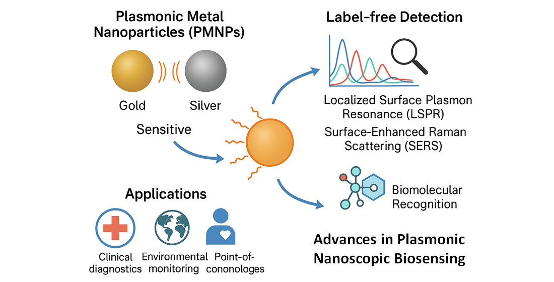
In summary, while label-based strategies remain valuable for multiplexing and signal amplification, label-free spectroscopic method specially lose leveraging LSPR, SERS, and EIS are gaining prominence for their simplicity, real-time capabilities, and translational potential. As illustrated in Scheme 1, this paradigm shift is driven by the convergence of advanced nanofabrication, surface engineering, and data-driven analytics. The integration of machine learning, nature-inspired nanostructure, and flexible substrates further enhancing the performance and applicability of plasmonic biosensor across the biomedical, environmental and food safety domains.
2. Fundamentals of Plasmonic Nanoparticles
Plasmonic metal nanoparticles (PMNPs), particularly those composed of gold (Au) silver (Ag), Copper (Cu), Aluminum (Al), Platinum (Pt), or their hybrids, exhibit unique optical properties arising from localized surface plasmon resonance (LSPR) [19]. LSPR occur when conduction electrons on the nanoparticle surface oscillate coherently in response to incident light at specific resonant frequencies. This resonance condition is highly sensitive to the nanoparticle’s size, shape, composition, and the dielectric environment surrounding it [20]. For instance, spherical nanoparticles typically exhibit a single LSPR peak, while anisotropic structures such as nano roads, nano stars, nano cubes, nano disks, or core- shell architecture can support multiple resonance across the visible and near infrared spectrum.Figure 1.
The electromagnetic field enhancement generated during LSPR is central to Surface Enhanced Raman Scattering (SERS) [2,21]. When molecules are adsorbed near the nanoparticle surface, the amplified local fields often concentrated in “hotspots” at junction, inter particle gaps, or sharp features can boost Raman signals by factor of 10 to 1012, enabling single molecule detection. This enhancement arises primarily from electromagnetic mechanism, although chemical interactions also contribute modestly to signal amplification [22]. In terms of material selection, silver nanoparticle offers superior plasmonic efficiency due to their sharper and more intense LSPR peaks. However, they are prone to oxidation and cytotoxicity, limiting their long-term stability and biocompatibility. Gold nanoparticles while slightly less efficient in field enhancement, provide excellent chemical stability, tunable surface chemistry, and proven biocompatibility, making them ideal for in vivo applications [23]. Copper nanoparticle presents a cost-effective alternative with strong plasmonic response, though they Suffer from rapid oxidation [24]. Aluminum nanostructures are particularly attractive for ultraviolet plasmonic, while platinum-based systems offer catalytic functionality alongside plasmonic behavior [25]. Hybrid nanostructures such as Au-Ag core-shell particles, alloyed systems, or emerging doped semiconductors (Indium tin oxide) and transition metal nitrates (TiN), aim to combine the best attributes of multiple materials enhanced sensitivity from silver or copper, stability from gold or platinum, and spectral tunability across broader ranges while also enabling multi-functional surface modification [26]. The interplay between nanoparticle geometry and plasmonic behavior is not merely academic it directly influences biosensor performance. Tailoring nanostructures to optimize LSPR and SERS responses allows for precise control over sensitivity, selectivity and spectral tunability, which are critical for real-time, label-free diagnostics.
3. Design Strategies for Biosensing Platforms
The efficacy of plasmonic biosensors is fundamentally governed by the strategic design of their interfaces, nanoparticle assemblies, and integrations modalities. These design choices dictate the sensor’s sensitivity, selectivity, and real-world applicability, especially in complex biological matrices. Recent advances have focused on optimizing surface functionalization, enhancing signal amplification through engineered nanostructures and, transitioning. Biosensors into miniaturized, Wearable formats for point-of-care diagnostics.
Surface functionalization is pivotal for imparting molecular specificity to plasmonic platforms. Techniques such as thiol gold chemistry, silanization and polymer grafting enable the immobilization of biorecognition elements-antibodies, aptamers, nucleic acid-onto nanoparticles surfaces or substrates [27]. These functional layers not only facilitate selective binding but also mitigate non-specific adsorption, which is critical for reproducibility. Kinza Ali et al. developed polymer monolith-MOF hybrid materials through post-synthesis surface functionalization, targeting enhanced performance in bioanalytical application [28]. Their study, demonstrated that layer-by- layer (LbL) functionalization of monoliths with MOF crystals significantly improved surface coverage and selectivity for nucleic acid capture in serum-based assays. Compared to direct MOF incorporation during polymerization which often leads to poor dispersion and compositional heterogeneity their approach provided better control over layer thickness and molecular accessibility. Despite challenges in scalability, the hybrid monoliths showed promise for biomolecule enrichment, pharmaceutical analysis, and environmental monitoring due to their hierarchical porosity and tunable surface chemistry.
Shangjie Zou et al. emphasizes the critical role of nano-coatings and 3D nanostructure in advancing multiplexed biosensing platforms [29]. Their review highlighted how hierarchical surface architecture such as micro-nano hybrid coatings, thin film nano-tags and spatially pattern nanostructures enhance target affinity while significantly reducing background noise. These functional layers enable the immobilization of multiple biorecognition elements in spatially resolved format, supporting simultaneous detection of diverse biomarkers. The author also discussed how surface modification techniques, including layer-by-layer deposition and plasma assisted grafting, influence probe orientation, accessibility, and signal uniformity. Their findings underscore that surface functionalization is not merely a preparatory step, but a performance defining parameter in biosensor engineering.
Zhen Zhou and Qinghong Shi introduce a novel class of zwitterionic polymer brushes based on trimethylamine N-oxide analogs, grafted onto quartz crystal microbalance QCM chips using dopamine-assisted surface-initiated ATRP [30]. These brushes formed a highly hydrated, charge-balanced layer that significantly suppressed protein absorption, specifically bovine serum albumin (BSA) by over 95%, as confirmed by frequency shift analysis (Figure 2). The antifouling effect was attributed to strong hydration and “anti-polyelectrolyte” behavior of the zwitterionic outer layer, which resisted ion and biomolecule addition even under low salt conditions. There were demonstrates the potential of zwitterionic N-oxide brushes for enhancing biosensor reliability in bio-fluid environments, particularly for implantable and point-of-care platforms.
Massia and Hubbell employed bioinspired peptide coatings that mimic extracellular matrix (EMC) motifs such as RG (Arg-Gly-Asp) and YIGSR (Tyr-Ile-Gly-Ser-Arg) to enhance biorecognition under dynamic flow conditions [31]. Their study focused on grafting these peptides onto plasmonic sensor surfaces using click chemistry and thiol-maleimide conjunction, ensuring stable anchoring and optimal orientation of recognition sites. Under microfluidic shear stress, the ECM-mimetic coating maintained high binding affinity for target proteins and cells, outperforming conventional antibody-surfaces in both selectivity and regeneration capacity. The dynamic adhesion behavior enabled reliable detection in serum and interstitial fluid, positioning these coatings as promising candidates for wearable biosensor and implantable diagnostics.
Signal enhancement in plasmonic biosensors particularly in surface-enhanced Raman scattering (SERS) and localized surface plasmon resonance (LSPR) platforms relies on the creation of electromagnetic “hot spots” through rational nanoparticle assembly. Seong Soo Yoo et al. developed a highly sensitive SERS platform by constructing a two-dimensional (2D) assembly of gold nanoparticles conformally blanketed with few-atom-thick MXene layers [32]. This hybrid architecture simultaneously intensified both localized surface plasmon resonance (LSPR) and short-range charge transfer (CT) effects. The MXene coating modulated the Fermi level of the substrate to align between the HOMO and LUMO levels of analytes, facilitating efficient CT enhancement. Additionally, the wrinkled MXene surface guided analytes toward plasmonic hotspots, boosting electromagnetic field concentration. Their design achieved an analytical enhancement factor of 1.6 x 10¹⁰, enabling trace detection of Cr (VI) down to 13 ng/L. This dual-mode amplification strategy offers a generalized framework for ultrasensitive detection of environmental and biomedical analytes.
Song Goa and Zhen Liu provided a comprehensive review of rational substrate engineering strategies for SERS biosensing, categorizing substrates into three major formats: colloidal suspensions, anisotropic nanoparticle clusters, and porous scaffolds [33]. Each format was tailored to specific bioassay requirements colloidal systems for rapid mixing and homogenous detection, anisotropic clusters for directional hotspot control, and porous scaffolds for high surface area and analyte accessibility. Among these, porous scaffolds such as metal-coated porous silicon and hierarchical oxide frameworks demonstrated superior hotspot density, enhanced analyte retention, and improved reproducibility across sample types. Their review emphasized that scaffold morphology, pore interconnectivity, and surface roughness are critical parameters for optimizing SERS enhancement and multiplexed detection in complex biological matrices.
Ghodeswar et al. developed a next generation plasmonic biosensing platform by integrating gap-tuned nanoplasmonic antennas with gradient-alloyed quantum dots (QDs) [34]. Their design confined electromagnetic fields within sub-10 nm gaps, amplifying QD photoluminescence by approximately 15×. This synergistic architecture achieved detection limits as low as 0.01nM, particularly for DNA and environmental pollutants. The platform also supported multiplex detection through angle-tuned metasurfaces, which phase-matched plasmonic resonances with QD emissions to resolve up to five analytes simultaneously. Additional enhancements included zwitterionic surface functionalization for antifouling (~95%) and second- harmonic generation (SHG) for nonlinear optical amplification. Collectively these, assemblies whether core-satellite, anisotropic, or hybrid enabled precise tuning of plasmonic coupling and spatial analyte localization, establishing a versatile framework for ultra-sensitive and high-specificity biosensing.
To transition from bench to bedside, plasmonic biosensors are increasingly integrated into miniaturized, flexible platforms that support real-time, non-invasive diagnostics. Xingfeng Ma et al. reviewed the convergence of artificial intelligence (AI) and biomedical microfluidics, highlighting how smart algorithms enable autonomous fluid routing, adaptive biomarker prioritization, and real-time decision making in wearable diagnostic platforms [35]. Their work emphasized that integrating microfluidics with AI=driven control systems such as machine learning classifiers and predictive flow models can optimize reagent usage, reduce detection latency and personalize assay protocols based on dynamic health data. The review also discussed the role of semiconductor microelectronics, wireless communication modules, and flexible substrates in transforming lab-on-chip devices into skin-mounted, portable biosensors. These innovations collectively advance the field toward intelligent, low-volume, and high-throughput diagnostics for personalized healthcare.
Taher Abbasiasl and Levent Beker developed a wearable paper-integrated microfluidic device designed for sequential sweat biomarker analysis using capillary action [36]. Their soft, skin-mounted platform leveraged the high capillary force of filter paper to route microliter volumes of eccrine sweat into discrete chambers without requiring external pumps or air vents. This design effectively eliminated evaporation artifacts, a common issue in absorbent pad-based systems, and enabled chrono-analysis of biomarkers such as glucose and pH over time. The device demonstrated reliable performance at flow rates up to 5 μL/min and supported colorimetric assays for real-time, non-invasive health monitoring. Its simplicity, low cost, and compatibility with flexible substrates make it a promising candidate for personalized diagnostics and field-deployable biosensing.
Simone highlighted the convergence of plasmonics, quantum sensing, and digital healthcare, emphasizing the transformative role of meta surface-enabled biosensors [37]. These ultrathin, nonstructured surfaces manipulate light at subwavelength scales, allowing for high-sensitivity, label-free detection in compact formats. The review discussed how mate surfaces integrated with smartphone imaging and cloud-based analytics enable decentralized diagnostics, particularly in resource-limited settings. Simone also explored the potential of quantum plasmonic platforms to overcome noise limitations and enhance signal fidelity, paving the way for real-time, multiplexed biosensing. The fusion of nanophotonics, microfluidics, and AI-driven data processing was identified as a key enabler of next-generation biosensors for personalized medicine and global health surveillance.
Liu and Zhang demonstrated the integration of patterned plasmonic nanostructured array with microfluidic chips, enabling high-throughput, label-free detection in compact lab-on-chip systems [38]. Their review highlighted how precise nanopatterning such as nanohole arrays, nanorod, and periodic gratings enhances plasmonic resonance sensitivity for SPR, LSPR, and SERS modalities. When coupled with microfluidics channels, these arrays support real-time multiplexed analysis of biochemical using minimal sample volumes (down to nanoliters). The authors emphasized that microfluidic integration not only automates sample handling and reaction kinetics but also improves reproducibility and scalability for point-of-care diagnostics. Their work underscores the synergy between nanophotonic design and fluidic control, advancing biosensor miniaturization and performance. Table 1. summarizes key innovations across six design domains in biosensor engineering, highlighting their impact on performance metrics such as selectivity, sensitivity, and portability. References indicate recent advances driving these enhancements, from MOF-polymer hybrids to quantum plasmonic interfaces.
4. Spectroscopic Techniques in Biosensing
Spectroscopic biosensing, particularly through surface -enhanced Raman scattering (SERS) and localized surface plasmon resources (LSPR), has emerged as a cornerstone of label-free diagnostics. These techniques harness the unique optical properties of plasmonic nanostructures to amplify molecular signals, enabling ultra-sensitive detection of biomolecules in real time and with high specificity.
4.1. Localize Surface Plasmon Resonance (LSPR) Biosensors
Localize surface plasmon resonance (LSPR) biosensors operate by detecting shifts in the resonance wavelength of metallic nanostructures upon analyte binding [2]. These shifts arise from changes in the local refractive index near the nanoparticle surface, enabling label-free, real-time detection with high sensitivity and spatial resolution. Compared to conventional SPR systems, LSPR platforms offer generate miniaturization, simple optics, and compatibility with portable formats. LSPR biosensors have been extensively applied to study protein-protein interactions, receptor-ligand binding, and antibody-antigen kinetics [39]. As reviewed by Singh et al, LSPR biosensors provide nanoscale resolution and enable real-time kinetic profiling without the need for molecular labels [40]. Unlike planar SPR systems, LSPR platforms utilize localized field confinement around nanostructures, enhancing sensitivity to surface-bound events. Gold nanorods and nanohole arrays are particularly effective, offering tunable plasmonic responses across visible an NIR spectra. These substrates require significantly smaller sample volumes, making them ideal for microfluidic integration. Their compact design also supports multiplexed detection and portable diagnostics. Overall, LSPR offers a more scalable and versatile alternative to conventional SPR.
Recent work by Hayashi and Latag highlighted the strategic use of self-assembled monolayers (SAMs) to tailor surface chemistry for enhance biomolecular recognition [41]. By optimizing SAM composition, the study achieved improved selectivity in complex biological matrices such as serum and plasma. Additionally, plasmon ruler’s nanoscale distance sensors base on LSPR shifts were employe to monitor binding events with high spatial precision. These rules enable dynamic tracking of molecular interactions without fluorescent labeling. The combination of SAMs and plasmon rulers significantly reduced nonspecific adsorption. This dual approach advances LSPR biosensors performance in clinically relevant environments.
DNA Hybridization and Nucleic Acid Detection Compact LSPR platforms have shown remarkable performance in DNA hybridization assays. Kaye et al. introduced a fiber-optic LSPR nanoprobe designed for ultra-sensitive detection of single-stranded DNA [42]. The device leveraged nanostructured gold arrays, precisely fabricated using electron beam lithography to ensure uniform plasmonic hotspots. This architecture enabled detection limits down to ~10 fM, demonstrating femtomolar sensitivity in aqueous environments. The fiber-optic format facilitated remote sensing and miniaturized integration for point-of-care diagnostics. Surface functionalization with DNA probes ensures high specificity and minimal background interference. Overall, the study showcased a robust platform for nucleic acid detection with potential for real-time clinical monitoring. Lu et al. demonstrated a significant advancement in LSPR biosensing by employing block copolymer-template AuNP monolayers [43]. This templating approach enabled uniform nanoparticle spacing, optimizing plasmonic coupling and signal amplification. The resulting platform achieved a detection limit of 67 pM, surpassing many conventional fluorescence-based assays. Its rapid response time and label-free operation reduced assay complexity and cost. Moreover, the monolayer design supports scalable fabrication and integration into portable diagnostic devices. These features make it highly suitable for field-deployable biosensing in resource-limited settings.
LSPR biosensors have gained traction in pathogen detection, particularly for rapid, field-deployable diagnostic. Alikhanian et al. engineered a label-free LSPR aptasensor targeting the SARS-CoV-2 spike protein using G12 aptamer-functionalized AuNPs [44]. The sensor exhibited a distinct colorimetric shift upon target binding, allowing rapid visual detection without instrumentation (Figure 3). It achieved a sensitivity of~10³ copies/mL, comparable to PCR-based assays, even in complex throat swab matrices. The aptamer’s high affinity and specificity minimized cross-reactivity with other respiratory pathogens. The assay’s simplicity and speed make it ideal for point-of-care diagnostics, especially in low-resource settings. This work underscored the potential of aptamer-LSPR platforms for pandemic-responsive biosensing. Petronella et al developed reusable Au nanorod arrays for sensitive and multifunctional detection of E. coli in water samples [45]. The LSPR-based platforms achieved a detection limit of 8.4 CFU/mL, outperforming many conventional microbial assays. These nanorods arrays were engineered for surface regeneration, allowing multiple detection cycles without loss of performance. Beyond sensing, the system enabled photothermal disinfection by converting incident light into localized heat, effectively neutralizing bacterial contaminants. The dual-functionality supports both monitoring and remediation in real time. The study highlights a promising route for sustainable water quality management using plasmonic nanomaterials.
Hoque et al provided a comprehensive review of on-chip LSPR devices tailored for biomarker detection, with a strong emphasis on lab-on-chip integration for infectious disease diagnostics [46]. These miniaturized platforms combine plasmonic nanostructures with microfluidic channels, enabling rapid, multiplexed analysis with minimal sample volumes. The review highlighted innovations in surface functionalization, signal readout, and device portability, making them suitable for point-of-care applications. Sayin et al further contributed by demonstrating scalable fabrication techniques and robust assay performance in clinical matrices [47]. Together, these works underscore the translation potential of chip-based LSPR biosensors in global health surveillance. These systems meet ASSURED criteria for point-of-care use, including affordability, sensitivity, and rapid response. Table 2 outlines cutting-edge biosensing strategies across key application domains, showcasing innovations from plasmon rulers to microfluidic nanohole arrays. Each approach delivers enhanced sensitivity, portability, or real-time detection, with references highlighting benchmark achievements in protein, DNA, and pathogen analysis.
4.2. Surface-Enhanced Raman Scattering (SERS) Biosensors
SERS biosensors utilize plasmonic metal nanoparticles (PMPSs) to amplify Raman signals through localized surface plasmon resonance (LSPR), enabling ultra-sensitive, label-free detection of biomolecules [48]. Recent advances have focused on optimizing substrate architecture to enhance electromagnetic field localization, reproducibility, and device integration. Core-shell Nanostructures: Au@Ag Architectures for Enhanced Sensitivity Core-shell designs, particularly Au@Ag nanoparticles, have emerged as high-performance SERS substrates due to their synergistic plasmonic properties. Wang et al. engineered Au@Ag core-shell nanoparticles as SERS-active substrates for acrylamide detection in food matrices [49].
The system exhibited a remarkably low detection limit of 1.27 × 10⁻⁹ mol/L, enabling trace-level quantification. Signal intensity showed strong linear correlation with acrylamide concentration, ensuring analytical reliability (Figure 4). The biosensor demonstrated excellent reproducibility across diverse food samples, validating its practical utility. Surface uniformity and hotspot density were optimized to enhance Raman signal amplification. This work highlights the translational potential of bimetallic nanostructures in food safety diagnostics.
Li et al utilized microfluidic synthesis to fabricate highly uniform Au@Ag core-shell nanoparticles for SERS application [50]. By embedding internal standards within the nanostructures, they enabled quantitative SERS with minimal signal fluctuation. The platform achieved an impressive limit of detection of 10 -10 M, suitable for ultra-trace biomolecular sensing. Microfluidic control ensured consistent particle morphology and reproducible hotspot distribution. Batch-to-batch variability was significantly reduced, enhancing analytical reliability for clinical and environmental assays. This approach sets a benchmark for scalable, precision-engineered SERS substrates with built-in calibration. Awiaz et al conducted a comprehensive review on the biological applications of Au@Ag core-shell SERS probes [51]. Their work highlights the use of these bimetallic nanostructures in detecting circulating tumor cells, miRNA, and protein biomarkers. Notably, they emphasized the detection of the receptor-binding domain (RBD) of SARS-CoV-2, showcasing clinical relevance. The review discusses how variations in core-shell thickness, morphology, and size influence SERS enhancement. Au@Ag probes demonstrated ultrahigh sensitivity and selectivity across various biological targets. This synthesis underscores the translational promise of Au@Ag SERS platforms in oncology, virology, and molecular diagnostics. Compared to earlier Au-only substrates, these bimetallic systems offer enhanced field confinement, broader spectral tunability, and improved biocompatibility, making them ideal for clinical diagnostics.
The creation of nanogaps between plasmonic structure is critical for generating electromagnetic “hotspots” that amplify Raman signals by up to 1010 -1012x. Xie et al provided a detailed review of hotspot engineering strategies for SERS enhancement, focusing on 3D nanostructures and tip-enhanced geometries [52]. They emphasized how multi-branched architectures and sub-10 nm gaps can dramatically increase local field density. Such designs eliminate the need for extremely narrow interparticle separations by maximizing near field confinement within individual nanostructures. Tip-enhanced Raman spectroscopy (TERS) was highlighted for its ability to achieve single molecule sensitivity through nanoscale precision. The review also discussed fabrication techniques for achieving reproducible hotspot distribution across large-area substrates. These insights pave the way for ultra-sensitive, spatially resolved SERS platforms in biomedical and environmental diagnostics.
Zhang et al introduce a DNA-mediated strategy for assembling SERS hotspots with precise spatial control and programmability. Using DNA nanostructures as linkers and templates [53]. They achieved tunable gap sizes and hotspot geometries tailored for biosensing and bioimaging. The review detailed static and dynamic regulation methods, including toehold-mediated strand displacement and enzyme driven reconfiguration. These approaches enabled the real-time modulation of hotspot intensity and location, enhancing sensitivity and multiplexing capabilities. Applications spanned from nucleic acid detection to cellular imaging, with emphasis on reproducibility and scalability. This work positions DNA nanotechnology as a versatile tool for next-generation SERS platforms in biomedical diagnostics. Maher et al. provided a foundational theoretical analysis of hotspot formation within interstitial crevices of metallic nanostructure [54]. These regions exhibit intense local field enhancement due to Localized Surface Plasmon Resonance (LSPR), crucial for SERS signal amplification. Hotspots formed in subwavelength crevices can boost Raman signal by up to 1015 X, enabling single molecule detection. The study emphasized that hotspot density and geometry directly influence the performance of SERS-active substances. Their work also linked hotspot behavior to tip-enhanced Raman spectroscopy (TERS), where nanoscale resolution is governed by field confinement.
The shift toward flexible, wearable SERS sensors has enabled real-time, non-invasive diagnostics on skin, saliva, and sweat. Liu et al reviewed the development of silk fibroin-based SERS substrate tailored for wearable sensing application [55]. Silk fibroin, derived from natural cocoons, offers excellent biocompatibility, air permeability and mechanical resilience, making it ideal for skin contact diagnostics. Their work emphasized the integration of Ag nanowires with silk film to form flexible, all fiber multifunctional sensors. These substrates demonstrated high sensitivity in pressure, temperature and humidity detection with stable performance under bending and strain. Applications included smart masks for respiratory monitoring and gloves for joint movement recognition, showcasing real world utility. This study positions silk fibroin as a promising scaffold for next generation wearable SERS performs in smart healthcare and IoT systems.
Chowdhury et al developed a flexible, stretchable SERS active sensors factor using PDMS as a substrate for variable biosensing applications [56]. The device incorporated plasmonic metasurfaces with the hotspot volume of 40 x 40 x 5 nm, enabling single molecule sensitivity. It maintained signal fidelity under bending angles up 100 degree and stretching strains up to 50%, demonstrating mechanical robustness. The sensor achieved an enhancement factor on the order of 10 11, with a high scattering-to-absorption ratio (2.5) PDMS provided transparency, biocompatibility and stability, making the sensor suitable for real time, non-invasive diagnostics. This work marks a significant advanced in wearable SERS platform for personalized medicine and remote patient monitoring. Xie et al emphasized the critical role of 3D periodic arrays in optimizing SERS substrates for point-of-care diagnostics [52]. These structures balance signal enhancement and spatial uniformity, overcoming limitations of random nano particle aggregates. Their design enables swap-sampling and in situ detection, making the platform suitable for rapid, non-invasive testing. The periodicity ensures consistent hotspot distribution, improving reproducibility across sensing surfaces. Applications include variable sensors for disease biomarkers and Covid-19 diagnostics, with integration into flexible electronics. This work advances the field towards scalable, high performance SERS platforms for real-world clinical deployment. Compared to rigid silicon-based platforms, these flexible systems offer conformal contact, multi model sensing, and integration with portable electronics, advancing personalized medicine. Table 3 summarizes key advances in SERS design, including core–shell nanostructures that enhance sensitivity and reproducibility, nanogap engineering for strong field localization and single-molecule detection, and flexible substrates that improve wearability and point-of-care applicability. Collectively, these strategies significantly improve sensitivity and practical utility.
5. Label-Free Detection Strategies
Label free biosensing eliminates the need for external tags or labels, relying instead on the intrinsic physicochemical properties of biomolecules such as mass, refractive index, and charge to generate detectable signals. This approach offers real time, quantitative, and non-invasive analysis, making it ideal for clinical diagnostics, environmental monitoring, and drug screening. Among label-free modalities, Localized Surface Plasmon Resonance (LSPR) sensing has emerged as a leading technique due to its sensitivity to local refractive index changes at the nanoparticle surface. LSPR-based biosensor detects analyte binding by monitoring shifts in the plasmon resonance wavelength, which occurs due to changes in the local dielectric environment surrounding plasmonic nanostructure.
Unser et al highlighted LSPR’s high molecular extension coefficients and intense near field confinement as key advantages for biosensing [57]. These properties enable sensitive detection of biomolecular interactions without the need for fluorescent or radioactive labels. Their work positioned LSPR as a powerful label-free technique for real-time, non-invasive diagnostic. Kabashin et al. reviewed cutting edge LSPR platforms that transcend conventional sensitivity limits through meta materials, phase-sensitive transducers, and bound states in continuum [58]. These innovations enable label-free biosensing with detection limits below 1 fg/mm2. Outer forming traditional labeled methods by several orders of magnitude. Their work establishes a new paradigm in optical biosensing, combining structural nanophotonics with singular phase optics for single-molecule resolution. Soler and Lechuga highlighted the integration of LSPR with silicon photonics and evanescent field sensing, enabling a new generation of miniaturized, Multiplexed, and portable label- free diagnostics [59]. Their review emphasized how combining plasmonic nanostructure with photonic waveguides enhances sensitivity while reducing device footprint, ideal for point-of-care application.
The integration of Localized Surface Plasmon Resonance (LSPR) with silicon photonics has emerged as a transformative approach in label-free biosensing. Soler and Lechuga underscored the synergy between plasmonic nanostructure and evanescent field sensing in photonic wave guides, enabling miniaturized, multiplexed and portable diagnostics [59]. This architecture leverages CMOS compatible fabrication allowing scalable production of photonic chip with embedded plasmonic elements for real time, high sensitivity detection. Building on this foundation, AI Qadasi et al introduce a segmented silicon photonic biosensor architecture that enables resonance tracking readout using fixed wavelength lasers eliminating the need for bulky tunable optics [60]. Their system demonstrated a limit of detection of 6.1 x 10⁵ RIU and successful immunoassay-based detection of SARS-CoV-2 spike protein, reinforcing the potential of LSPR-photonic platforms for decentralized, data-rich diagnostics. Complementarily, Puumala et al proposed an optimization framework for silicon photonic [61]. Evanescent field biosensor using Sub-Wavelength Greetings (SWGs). Their “Fishbone” SWG design enhanced robustness and sensitivity, achieving bulk sensitivities up to 438 nm/RIU and limits of detection as low as 5.1 x 10⁴ RIU. These innovation address long standing challenges in photonic biosensor fragility and signal fidelity, further solidifying silicon photonics as a viable platform for point-of-care diagnostics. In contrast, direct Raman fingerprinting via surface enhanced Raman scattering (SERS) offers unparalleled molecular specificity by amplifying vibrational signal of biomolecules adsorb on nanostructured substrates. This modality excels in applications requiring direct chemical identification, such as cancer biomarkers detection, food safety monitoring, and environmental toxic analysis. Recent advances have significantly expanded SERS capabilities. Dong et al developed a label-free SERS Artificial Intelligence Cancer Screening (SERS- AICS) platform using silver nanowires and machine learning to analyze serum samples [62]. Their system achieved over 95% accuracy in pan-cancer detection across large patient cohorts, Demonstrating the power of SERS when coupled with data-driven analytics for early-stage diagnostics (Figure 5).
Yuan et al reviewed nanomaterial enhanced SERS strategies for clinical diagnosis, emphasizing hybrid substrate that combine electromagnetic and chemical enhancement mechanisms [63]. These designs mitigate non-specific interactions and improve signal reliability in complex biological matrices, paving way for SERS-based platforms in pathogen detection and cancer screening. Wang et al further explored SERS applications in bacterial detection, integrating microfluidics and nano enhanced dielectric material to achieve rapid, high-sensitivity identification of pathogenic strains [64]. Their review highlights the growing role of SERS in infectious disease diagnostics, specially where conventional method falls short in speed and specificity.
6. Biomedical And Environmental Applications
Plasmonic biosensors have demonstrated exceptional versatility in detecting a wide spectrum of biological and chemical targets, ranging from disease biomarkers and pathogen to environmental toxins and pollutants. Their rapid, label-free, and high sensitivity detection capabilities have enabled transformative applications across clinical diagnostics, food safety, and water quality monitoring.
In biomedical diagnostics, plasmonic platforms have been employed for the detection of cancer biomarkers, viral antigens and microbial pathogens. Chenglong Lin et al developed multifunctional SERS substrates capable of detecting tumor markers and viral proteins with high specificity, integrating machine learning algorithms for spectral pattern recognition to enhance diagnostic accuracy [65]. Similarly, Yikai Xu et al emphasize the importance of quantitative calibration in SERS-based clinical assays, addressing challenges in reproducibility and signal normalization that are critical for regulatory approval and clinical translation [66]. Point-of-Care (POC) diagnostics have particularly benefited from plasmonic miniaturization. Jane Ru Choi et al reviewed emerging POC technologies for food safety analysis, highlighting paper based and chip integrated biosensors for detecting foodborne pathogen, allergens and chemical residues [67]. These devices offer rapid, cost-effective alternative to conventional methods like HPLC and ELISA, especially in resource-limited settings. Complimenting this, Praveen Kumar et al detailed smartphone-integrated biosensors that enable real-time contaminant tracking across the food supply chain, enhancing traceability, and decision making [68].
Environmental monitoring has also seen significant advances. Antia Fdez-Sanroman and colleagues reviewed biosensor technologies for water quality assessment, focusing on emerging contaminants such as pharmaceuticals, micro plastics, and antibiotic-resistant bacteria [69]. Their work emphasized the integration of molecularly imprinted polymers (MIPA), SELEX-derived aptamers, and Micro fluidic system to enhance selectivity and throughput. In parallel, Adams Ovie Iyiola et al explored the use of biomarkers ranging from plankton and benthic Organism to aquatic mammals for pollution detection in freshwater ecosystems [70]. Their chapter underscored the utility of histopathological and genotoxicity markers in assessing ecological stress and pollutant bioaccumulation. Case studies from industry further illustrate the translational relevance of plasmonic biosensors. For example, bioMérieux has deployed automated pathogen detection systems such as TEMPO® and GENE-UP® in food production environments, including dairy, juice, and salmon processing facilities. These platforms have enabled rapid detection of contaminants like Listeria monocytogenes, Salmonella, and Cronobacter sakazakii, improving product safety and reducing recall risks. In water monitoring, Caruncho-Pérez et al demonstrated electrochemical biosensors for detecting heavy metals and organic pollutants, integrating nanostructured electrodes and self-assembled monolayers for enhanced sensitivity [71].
Despite these advances, the path to widespread deployment is shaped by regulatory and ethical considerations. Mohammad Ershadul Karim outlined the legal frameworks governing biosensor development, emphasizing the need for validated protocols, public trust, and equitable access [72]. He noted that while biosensors align with global health and sustainability goals, regulatory instruments specific to biosensing technologies remain underdeveloped. International bodies such as ISO and WHO have begun to address these gaps, but harmonization across jurisdictions is still evolving. The convergence of plasmonic nanotechnology with biomedical and environmental diagnostics holds immense promise. However, realizing this potential requires not only technical innovation but also thoughtful engagement with regulatory, ethical, and societal dimensions. As biosensors move closer to clinical and field deployment, interdisciplinary collaboration will be essential to ensure safety, scalability, and impact.
7. Challenges and Future Directions
Plasmonic biosensors have demonstrated exceptional sensitivity and versatility across biomedical, environmental, and food safety domains. However, several critical challenges continue to impede their widespread clinical translation and commercial deployment. These include issues of reproducibility, long-term stability, and scalability each of which must be systematically addressed to realize the full potential of plasmonic platforms.
7.1. Reproducibility, Stability, and Scalability
The reproducibility of plasmonic biosensors is often compromised by batch-to-batch variability in nanoparticle synthesis, substrate fabrication, and surface functionalization. Signal fluctuations arising from minor deviations in particle morphology, interparticle spacing, or ligand density can significantly affect the reliability of LSPR and SERS-based measurements. Silver-based sensors, while offering superior plasmonic enhancement, are particularly prone to oxidation and aggregation, limiting their operational stability and shelf life in real-world conditions. Scalability remains a formidable barrier, especially for sensors requiring nanostructured substrates with precise geometries. Techniques such as electron-beam lithography and focused ion beam milling offer high resolution but are cost-intensive and unsuitable for mass production. Emerging approaches including roll-to-roll nanoimprinting, colloidal self-assembly, and laser interference lithography are being explored to bridge this gap, yet challenges persist in maintaining uniformity and performance across large areas.
6.2. Emerging Technological Trends
6.2.1. Machine Learning-Assisted Spectral Analysis
The integration of machine learning (ML) into plasmonic biosensing workflows is transforming data interpretation and sensor optimization. ML algorithms enable real-time spectral deconvolution, noise suppression, and pattern recognition, thereby enhancing analytical accuracy and enabling multiplexed detection. Recent studies have demonstrated the use of adversarial autoencoders and generative models to optimize metasurface geometries for targeted plasmonic responses, significantly reducing experimental overhead and design iteration cycles.
6.2.2. Quantum Plasmonics
Quantum plasmonics represents a frontier in biosensor development, leveraging quantum coherence, entanglement, and tunneling effects to surpass classical detection limits. Although still in its nascent stage, quantum-enhanced plasmonic platforms have shown promise for single-molecule detection, quantum dot-plasmon coupling, and ultra-sensitive fluorescence modulation. However, the requirement for cryogenic environments and complex instrumentation currently restricts their translational feasibility.
6.2.3. Bioinspired Architectures
Nature-inspired designs are increasingly being adopted to enhance plasmonic sensor performance. Hierarchical structures mimicking butterfly wings, diatom shells, and lotus leaves offer improved light trapping, analyte accessibility, and hotspot generation. Techniques such as cryosoret nano-assembly and ferroplasmon-on-mirror (FPoM) substrates have been employed to create plasmonic interfaces with enhanced emission coupling and signal amplification. These bioinspired platforms not only improve sensitivity but also offer new avenues for flexible and wearable sensor development.
6.3. Outlook on Clinical Translation and Commercialization
The clinical translation of plasmonic biosensors requires a multidisciplinary approach encompassing biomarker validation, device integration, regulatory alignment, and user-centric design. While academic innovation in plasmonic sensing is robust, only a limited number of platforms have progressed to market due to fragmented regulatory pathways and insufficient standardization. Successful models, such as glucose sensors, underscore the importance of simplicity, reliability, and regulatory clarity. To facilitate commercialization, plasmonic biosensors must be integrated with digital healthcare platforms, enabling real-time monitoring, remote diagnostics, and personalized medicine. Smartphone-based interfaces, cloud-connected analytics, and AI-driven decision support systems are being actively explored to enhance accessibility and clinical utility. Furthermore, scalable manufacturing techniques and modular sensor architectures will be essential to meet the demands of point-of-care diagnostics and decentralized healthcare systems.
6.4. Future Perspectives
Addressing reproducibility, stability, and scalability challenges will be pivotal for the next generation of plasmonic biosensors. The convergence of machine learning, quantum optics, and bioinspired engineering offers transformative potential, enabling intelligent design, enhanced sensitivity, and robust performance. By aligning sensor development with translational frameworks and regulatory standards, plasmonic biosensors are poised to become integral components of future diagnostic and monitoring platforms.
8. Conclusions
Plasmonic biosensors have emerged as transformative analytical platforms, offering unparalleled sensitivity, label-free detection, and real-time monitoring capabilities. Through the exploitation of surface plasmon resonance (SPR) and localized surface plasmon resonance (LSPR), these sensors have demonstrated robust performance across biomedical diagnostics, environmental monitoring, and food safety applications. Technological advancements in nanofabrication, surface functionalization, and hybrid material integration have significantly enhanced the specificity, stability, and scalability of plasmonic platforms. The development of tunable nanostructures, flexible substrates, and multiplexed detection formats marks a critical milestone in the evolution of these sensors from laboratory prototypes to deployable diagnostic tools. Emerging trends such as machine learning-assisted spectral analysis, quantum plasmonics, and bioinspired architectures are redefining the design and functionality of plasmonic biosensors. These innovations enable intelligent signal processing, ultra-sensitive detection, and adaptive sensor behavior, thereby expanding the scope of plasmonic technologies beyond conventional applications. Importantly, the integration of plasmonic biosensors into point-of-care (POC) systems and digital healthcare frameworks is accelerating their clinical translation. Portable, user-friendly devices capable of remote diagnostics and real-time data analytics are increasingly aligned with global healthcare needs, particularly in resource-limited settings. In conclusion, plasmonic biosensors represent a cornerstone of next-generation diagnostics. Their convergence with interdisciplinary technologies and alignment with translational goals positions them as key enablers of personalized medicine, rapid disease screening, and decentralized health monitoring. Continued efforts in standardization, regulatory alignment, and scalable manufacturing will be essential to fully realize their impact across clinical and commercial domains.
Reference
Author Contributions
C.R. conceived and designed the study. S.C. collected the literature and prepared the initial draft. C.R and S.C. contributed to data interpretation and critical revisions. All authors discussed the content, revised the manuscript, and approved the final version.
Acknowledgment
The author gratefully acknowledges the support and infrastructure provided by Meerut Institute of Engineering and Technology (MIET), Meerut, which facilitated the successful completion of this review. The academic environment and research resources at MIET have been instrumental in shaping the perspectives and depth of analysis presented in this work.
Conflicts of Interest
The authors declare no conflicts of interest.
References
- Hang Y, Wang A, Wu N. Plasmonic silver and gold nanoparticles: shape- and structure-modulated plasmonic functionality for point-of-caring sensing, bio-imaging and medical therapy. Chemical Society Reviews 2024;53:2932-71. [CrossRef]
- Nanda BP, Rani P, Paul P, Aman, Ganti SS, Bhatia R. Recent trends and impact of localized surface plasmon resonance (LSPR) and surface-enhanced Raman spectroscopy (SERS) in modern analysis. Journal of Pharmaceutical Analysis 2024;14:100959. [CrossRef]
- Samuel VR, Rao KJ. A review on label free biosensors. Biosensors and Bioelectronics: X 2022;11:100216. [CrossRef]
- Needham L-M, Saavedra C, Rasch JK, Sole-Barber D, Schweitzer BS, Fairhall AJ, et al. Label-free detection and profiling of individual solution-phase molecules. Nature 2024;629:1062-8. [CrossRef]
- Sharma A, Majdinasab M, Khan R, Li Z, Hayat A, Marty JL. Nanomaterials in fluorescence-based biosensors: Defining key roles. Nano-Structures & Nano-Objects 2021;27:100774. [CrossRef]
- Ciuffreda P, Xynomilakis O, Casati S, Ottria R. Fluorescence-Based Enzyme Activity Assay: Ascertaining the Activity and Inhibition of Endocannabinoid Hydrolytic Enzymes. International Journal of Molecular Sciences2024. [CrossRef]
- Rabbani A, Rudacille R, Hasegawa K. Local Refractive Index Sensitivity of Localized Surface Plasmon Resonance Biosensors. The Journal of Physical Chemistry C 2024;128:19210-21. [CrossRef]
- Cialla-May D, Bonifacio A, Markin A, Markina N, Fornasaro S, Dwivedi A, et al. Recent advances of surface enhanced Raman spectroscopy (SERS) in optical biosensing. TrAC Trends in Analytical Chemistry 2024;181:117990. [CrossRef]
- Khondakar KR, Mazumdar H, Das S, Kaushik A. Machine learning (ML)-assisted surface-enhanced raman spectroscopy (SERS) technologies for sustainable health. Advances in Colloid and Interface Science 2025;344:103594. [CrossRef]
- Barucci A, D’Andrea C, Farnesi E, Banchelli M, Amicucci C, de Angelis M, et al. Label-free SERS detection of proteins based on machine learning classification of chemo-structural determinants. Analyst 2021;146:674-82.
- Chomean S, Nakkam N, Tassaneeyakul W, Attapong J, Kaset C. Development of label-free electrochemical impedance spectroscopy for the detection of HLA-B*15:02 and HLA-B*15:21 for the prevention of carbamazepine-induced Stevens-Johnson syndrome. Analytical Biochemistry 2022;658:114931. [CrossRef]
- Rejeeth C, Almeer R, Sharma A, Varukattu NB. Label-free electrochemical assessment of human serum and cancer cells to determine the folate receptor cancer biomarker. Bioelectrochemistry 2025;163:108883. [CrossRef]
- Rejeeth C, Sharma A, Kannan S, Kumar RS, Almansour AI, Arumugam N, et al. Label-Free Electrochemical Detection of the Cancer Biomarker Platelet-Derived Growth Factor Receptor in Human Serum and Cancer Cells. ACS Biomaterials Science & Engineering 2022;8:826-33. [CrossRef]
- Zhang R, Rejeeth C, Xu W, Zhu C, Liu X, Wan J, et al. Label-Free Electrochemical Sensor for CD44 by Ligand-Protein Interaction. Analytical Chemistry 2019;91:7078-85.
- Garrote BL, Santos A, Bueno PR. Perspectives on and Precautions for the Uses of Electric Spectroscopic Methods in Label-free Biosensing Applications. ACS Sensors 2019;4:2216-27. [CrossRef]
- Zhang H, Sun Z, Sun K, Liu Q, Chu W, Fu L, et al. Electrochemical Impedance Spectroscopy-Based Biosensors for Label-Free Detection of Pathogens. Biosensors2025. [CrossRef]
- Chandra A, Kumar V, Garnaik UC, Dada R, Qamar I, Goel VK, et al. Unveiling the Molecular Secrets: A Comprehensive Review of Raman Spectroscopy in Biological Research. ACS Omega 2024;9:50049-63. [CrossRef]
- Schorr HC, Schultz ZD. Digital surface enhanced Raman spectroscopy for quantifiable single molecule detection in flow. Analyst 2024;149:3711-5. [CrossRef]
- Demishkevich E, Zyubin A, Seteikin A, Samusev I, Park I, Hwangbo CK, et al. Synthesis Methods and Optical Sensing Applications of Plasmonic Metal Nanoparticles Made from Rhodium, Platinum, Gold, or Silver. Materials2023. [CrossRef]
- McOyi MP, Mpofu KT, Sekhwama M, Mthunzi-Kufa P. Developments in Localized Surface Plasmon Resonance. Plasmonics 2025;20:5481-520.
- de Albuquerque CDL, Zoltowski CM, Scarpitti BT, Shoup DN, Schultz ZD. Spectrally Resolved Surface-Enhanced Raman Scattering Imaging Reveals Plasmon-Mediated Chemical Transformations. ACS Nanoscience Au 2021;1:38-46. [CrossRef]
- Li L, Zhang T, Zhang L, Wang G, Huang X, Li W, et al. Synergistic enhancement of chemical and electromagnetic effects in a Ti3C2Tx/AgNPs two-dimensional SERS substrate for ultra-sensitive detection. Analytica Chimica Acta 2024;1331:343330. [CrossRef]
- Almomani AS, Omar AF, Oglat AA, Al-Mafarjy SS, Dheyab MA, Khazaalah TH. Developed a unique technique for creating stable gold nanoparticles (AuNPs) to explore their potential against cancer. South African Journal of Chemical Engineering 2025;53:142-52. [CrossRef]
- Devaraji M, Thanikachalam PV, Elumalai K. The potential of copper oxide nanoparticles in nanomedicine: A comprehensive review. Biotechnology Notes 2024;5:80-99. [CrossRef]
- Shukla S, Arora P. Aluminum as a competitive plasmonic material for the entire electromagnetic spectrum: A review. Results in Optics 2025;18:100760. [CrossRef]
- Pisarek M, Krawczyk M, Hołdyński M, Ambroziak R, Bieńkowski K, Roguska A, et al. Hybrid Nanostructures Based on TiO2 Nanotubes with Ag, Au, or Bimetallic Au–Ag Deposits for Surface-Enhanced Raman Scattering (SERS) Applications. The Journal of Physical Chemistry C 2023;127:24200-10. [CrossRef]
- Oberhaus FV, Frense D, Beckmann D. Immobilization Techniques for Aptamers on Gold Electrodes for the Electrochemical Detection of Proteins: A Review. Biosensors2020. [CrossRef]
- Ali K, Munawar I, Manan S, Nawazish F, Fatima B, Jabeen F, et al. Development of polymer monolith-MOF hybrid via surface functionalization for bioanalytical sciences. Analytical and Bioanalytical Chemistry 2025;417:3503-11. [CrossRef]
- Zou S, Peng G, Ma Z. Surface-Functionalizing Strategies for Multiplexed Molecular Biosensing: Developments Powered by Advancements in Nanotechnologies. Nanomaterials2024. [CrossRef]
- Zhou Z, Shi Q. Bioinspired Dopamine and N-Oxide-Based Zwitterionic Polymer Brushes for Fouling Resistance Surfaces. Polymers2024. [CrossRef]
- Massia SP, Hubbell JA. Convalent surface immobilization of Arg-Gly-Asp- and Tyr-Ile-Gly-Ser-Arg-containing peptides to obtain well-defined cell-adhesive substrates. Analytical Biochemistry 1990;187:292-301. [CrossRef]
- Yoo SS, Ho J-W, Shin D-I, Kim M, Hong S, Lee JH, et al. Simultaneously intensified plasmonic and charge transfer effects in surface enhanced Raman scattering sensors using an MXene-blanketed Au nanoparticle assembly. Journal of Materials Chemistry A 2022;10:2945-56. [CrossRef]
- Gao S, Guo Z, Liu Z. Recent Advances in Rational Design and Engineering of Signal-Amplifying Substrates for Surface-Enhanced Raman Scattering-Based Bioassays. Chemosensors2023. [CrossRef]
- Ghodeswar US, Joshi KV, Waje MG, Patil TR, Upadhye S, Shelke N, et al. Sensitivity Improved Plasmonic Biosensors with Coupling Quantum-Dots for Optimization of Biosensing Process: Application in Biomedical Diagnostics and Environmental Monitoring Processes. Plasmonics 2025. [CrossRef]
- Ma X, Guo G, Wu X, Wu Q, Liu F, Zhang H, et al. Advances in Integration, Wearable Applications, and Artificial Intelligence of Biomedical Microfluidics Systems. Micromachines2023. [CrossRef]
- Abbasiasl T, Mirlou F, Istif E, Ceylan Koydemir H, Beker L. A wearable paper-integrated microfluidic device for sequential analysis of sweat based on capillary action††Electronic supplementary information (ESI) available: Fig. S1–S7.Sensors and Diagnostics 2022;1:775-86. [CrossRef]
- Simone, G. Trends of Biosensing: Plasmonics through Miniaturization and Quantum Sensing. Critical Reviews in Analytical Chemistry 2024;54:2183-208. [CrossRef]
- Liu Y, Zhang X. Microfluidics-Based Plasmonic Biosensing System Based on Patterned Plasmonic Nanostructure Arrays. Micromachines2021. [CrossRef]
- Austin Suthanthiraraj PP, Sen AK. Localized surface plasmon resonance (LSPR) biosensor based on thermally annealed silver nanostructures with on-chip blood-plasma separation for the detection of dengue non-structural protein NS1 antigen. Biosensors and Bioelectronics 2019;132:38-46.
- Singh S, Singh PK, Umar A, Lohia P, Albargi H, Castañeda L, et al. 2D Nanomaterial-Based Surface Plasmon Resonance Sensors for Biosensing Applications. Micromachines2020. [CrossRef]
- Hayashi T, Latag GV. Self-Assembled Monolayers as Platforms for Nanobiotechnology and Biointerface Research: Fabrication, Analysis, Mechanisms, and Design. ACS Applied Nano Materials 2025;8:8570-87. [CrossRef]
- Kaye S, Zeng Z, Sanders M, Chittur K, Koelle PM, Lindquist R, et al. Label-free detection of DNA hybridization with a compact LSPR-based fiber-optic sensor. Analyst 2017;142:1974-81. [CrossRef]
- Lu M, Zhu H, Lin M, Wang F, Hong L, Masson J-F, et al. Comparative study of block copolymer-templated localized surface plasmon resonance optical fiber biosensors: CTAB or citrate-stabilized gold nanorods. Sensors and Actuators B: Chemical 2021;329:129094. [CrossRef]
- Alikhanian A, Shadmehri N, Borghei Y-S, Arefian E, Habibi-Rezaei M. Rapid, Label-Free LSPR Aptasensor for Sensitive Detection of SARS-CoV-2 Spike Protein in Throat Swab Samples. Plasmonics 2025. [CrossRef]
- Petronella F, De Biase D, Zaccagnini F, Verrina V, Lim S-I, Jeong K-U, et al. Label-free and reusable antibody-functionalized gold nanorod arrays for the rapid detection of Escherichia coli cells in a water dispersion. Environmental Science: Nano 2022;9:3343-60. [CrossRef]
- Hoque SZ, Somasundaram L, Samy RA, Dawane A, Sen AK. Localized Surface Plasmon Resonance Sensors for Biomarker Detection with On-Chip Microfluidic Devices in Point-of-Care Diagnostics. In: Joshi SN, Chandra P, editors. Advanced Micro- and Nano-manufacturing Technologies: Applications in Biochemical and Biomedical Engineering. Singapore: Springer Singapore; 2022. p. 199-223.
- Sayin S, Zhou Y, Wang S, Acosta Rodriguez A, Zaghloul M. Development of Liquid-Phase Plasmonic Sensor Platforms for Prospective Biomedical Applications. Sensors2024. [CrossRef]
- Zhou Y, Lu Y, Liu Y, Hu X, Chen H. Current strategies of plasmonic nanoparticles assisted surface-enhanced Raman scattering toward biosensor studies. Biosensors and Bioelectronics 2023;228:115231. [CrossRef]
- Wang K, Yue Z, Fang X, Lin H, Wang L, Cao L, et al. SERS detection of thiram using polyacrylamide hydrogel-enclosed gold nanoparticle aggregates. Science of The Total Environment 2023;856:159108. [CrossRef]
- Li S, Chen J, Xu W, Sun B, Wu J, Chen Q, et al. Highly homogeneous bimetallic core–shell Au@Ag nanoparticles with embedded internal standard fabrication using a microreactor for reliable quantitative SERS detection. Materials Chemistry Frontiers 2023;7:1100-9.
- Awiaz G, Lin J, Wu A. Recent advances of Au@Ag core–shell SERS-based biosensors. Exploration 2023;3:20220072.
- Xie Y, Xu J, Shao D, Liu Y, Qu X, Hu S, et al. SERS-Based Local Field Enhancement in Biosensing Applications. Molecules2025. [CrossRef]
- Zhang J, Song C, He X, Liu J, Chao J, Wang L. DNA-mediated precise regulation of SERS hotspots for biosensing and bioimaging. Chemical Society Reviews 2025;54:5836-63.
- Maher, RC. SERS Hot Spots. In: Kumar CSSR, editor. Raman Spectroscopy for Nanomaterials Characterization. Berlin, Heidelberg: Springer Berlin Heidelberg; 2012. p. 215-60.
- Liu G, Mu Z, Guo J, Shan K, Shang X, Yu J, et al. Surface-enhanced Raman scattering as a potential strategy for wearable flexible sensing and point-of-care testing non-invasive medical diagnosis. Frontiers in Chemistry 2022;Volume 10 - 2022. [CrossRef]
- Haque Chowdhury MA, Tasnim N, Hossain M, Habib A. Flexible, stretchable, and single-molecule-sensitive SERS-active sensor for wearable biosensing applications. RSC Advances 2023;13:20787-98. [CrossRef]
- Unser S, Bruzas I, He J, Sagle L. Localized Surface Plasmon Resonance Biosensing: Current Challenges and Approaches. Sensors2015. p. 15684-716. [CrossRef]
- Kabashin AV, Kravets VG, Grigorenko AN. Label-free optical biosensing: going beyond the limits. Chemical Society Reviews 2023;52:6554-85. [CrossRef]
- Soler M, Lechuga LM. Principles, technologies, and applications of plasmonic biosensors. Journal of Applied Physics 2021;129:111102. [CrossRef]
- Al-Qadasi M, Grist S, Mitchell M, Newton K, Kioussis S, Chowdhury S, et al. An integrated evanescent-field biosensor in silicon2024.
- Puumala LS, Grist SM, Wickremasinghe K, Al-Qadasi MA, Chowdhury SJ, Liu Y, et al. An Optimization Framework for Silicon Photonic Evanescent-Field Biosensors Using Sub-Wavelength Gratings. Biosensors2022.
- Dong S, He D, Zhang Q, Huang C, Hu Z, Zhang C, et al. Early cancer detection by serum biomolecular fingerprinting spectroscopy with machine learning. eLight 2023;3:17. [CrossRef]
- Yuan K, Jurado-Sánchez B, Escarpa A. Nanomaterials meet surface-enhanced Raman scattering towards enhanced clinical diagnosis: a review. Journal of Nanobiotechnology 2022;20:537. [CrossRef]
- Wang W, Rahman A, Huang Q, Vikesland PJ. Surface-enhanced Raman spectroscopy enabled evaluation of bacterial inactivation. Water Research 2022;220:118668. [CrossRef]
- Lin C, Li Y, Peng Y, Zhao S, Xu M, Zhang L, et al. Recent development of surface-enhanced Raman scattering for biosensing. Journal of Nanobiotechnology 2023;21:149. [CrossRef]
- Xu Y, Aljuhani W, Zhang Y, Ye Z, Li C, Bell SEJ. A practical approach to quantitative analytical surface-enhanced Raman spectroscopy. Chemical Society Reviews 2025;54:62-84. [CrossRef]
- Choi JR, Yong KW, Choi JY, Cowie AC. Emerging Point-of-care Technologies for Food Safety Analysis. Sensors2019.
- Rayani PK, Changder S. Sensor-based continuous user authentication on smartphone through machine learning. Microprocessors and Microsystems 2023;96:104750. [CrossRef]
- Fdez-Sanromán A, Bernárdez-Rodas N, Rosales E, Pazos M, González-Romero E, Sanromán MÁ. Biosensor Technologies for Water Quality: Detection of Emerging Contaminants and Pathogens. Biosensors2025. [CrossRef]
- Iyiola A, Setufe S, Ofori-Boateng E, Jacob B, Ogwu M. Biomarkers for the Detection of Pollutants from the Water Environment. 2024. p. 569-602.
- Caruncho-Pérez S, Giráldez A, Terrón D, Pazos M, Sanromán MÁ, González-Romero E. Electrochemistry solving problems in water disinfection: Direct voltammetric monitoring of pyocyanin from Pseudomonas aeruginosa. Chemical Engineering Journal 2025;522:167828. [CrossRef]
- Karim, M. Biosensors: Ethical, Regulatory, and Legal Issues. 2021. p. 679-705.
Scheme 1.
Plasmonic spectroscopic-based biosensors integrating label-free techniques, nanofabrication’s and machine learning. Nature-inspired nanostructures and flexible substrates enhance real-time sensing capabilities. Applications span biomedical, environmental, and food safety domains.
Scheme 1.
Plasmonic spectroscopic-based biosensors integrating label-free techniques, nanofabrication’s and machine learning. Nature-inspired nanostructures and flexible substrates enhance real-time sensing capabilities. Applications span biomedical, environmental, and food safety domains.
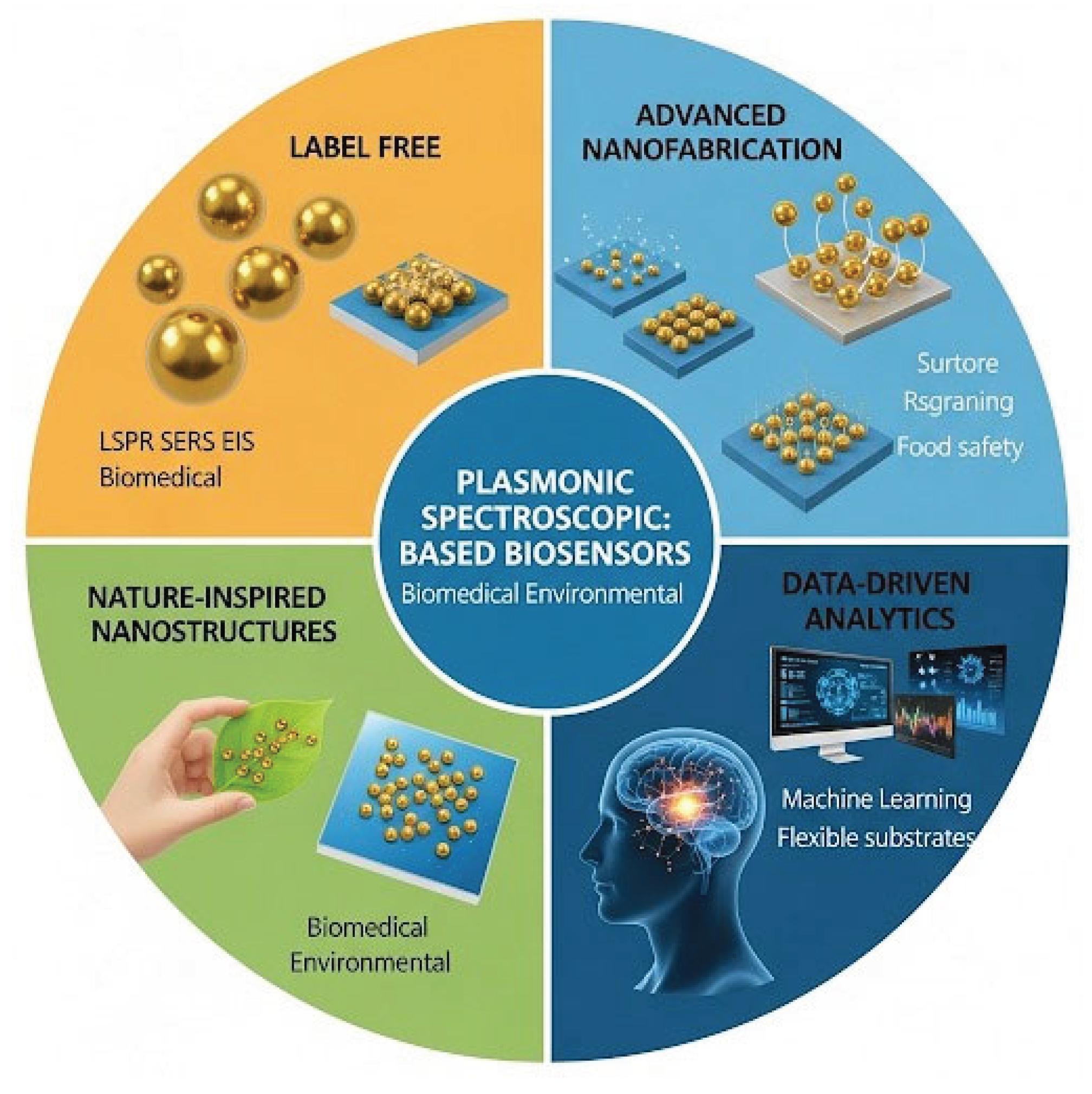
Figure 1.
plasmonic metal nanoparticles (PMNPs) exhibit LSPR, driven by coherent electron oscillations under incident light. Nanoparticle geometry and composition influence resonance peaks and electromagnetic field enhancement. These effects enable SERS-based single-molecule detection and tunable biosensor performance.
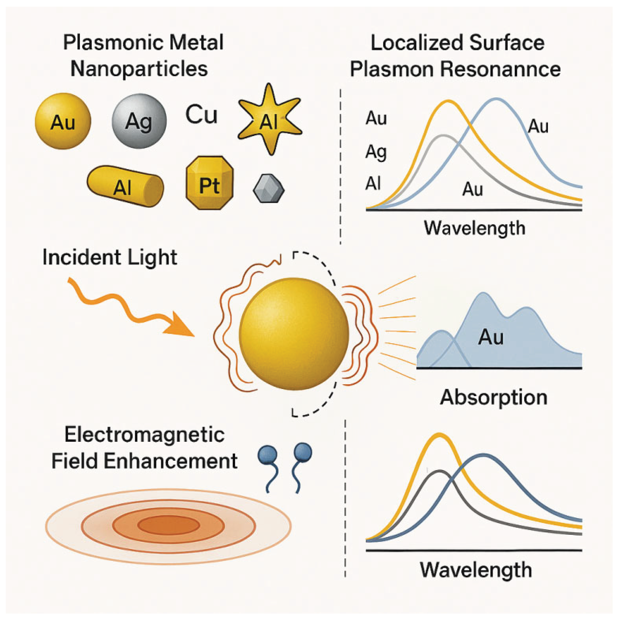
Figure 2.
(a) Schematic representation of dopamine (DA) oxidation and self-polymerization into polydopamine (pDA) in the presence of molecular oxygen and hydrogen peroxide. The resulting pDA contains catechol, amine, and imine functionalities, enabling strong surface adhesion and further chemical modification. (b) Stepwise surface functionalization of a gold substrate: initial deposition of a pDA layer via self-polymerization, followed by grafting of MADMPAO through Surface-Initiated Atom Transfer Radical Polymerization (SI-ATRP), forming a pDA-x-pMPAO hybrid coating. The final surface exhibits bio interactive properties suitable for protein immobilization and biosensing applications. Copyright permission in Ref. [27].
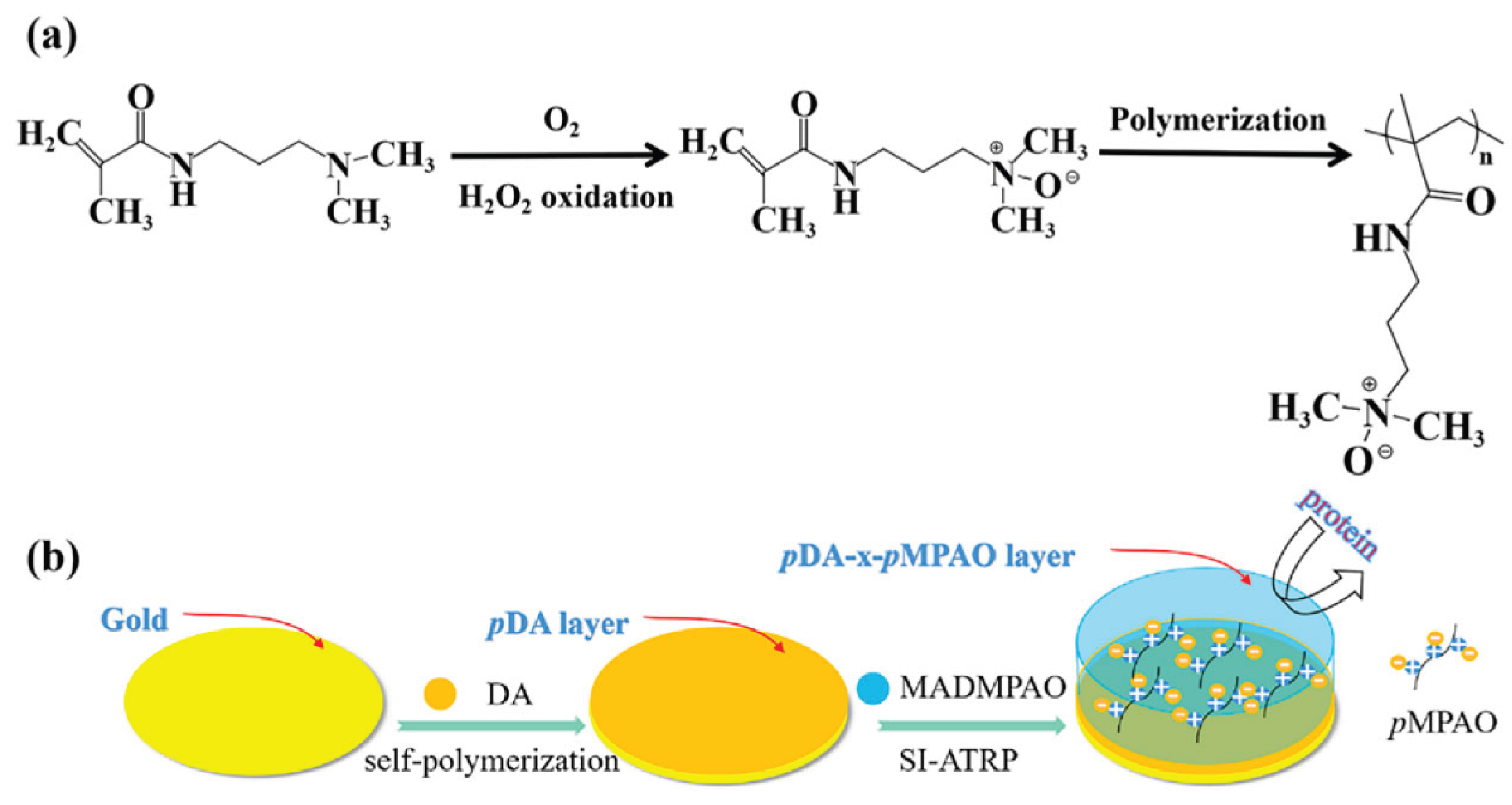
Figure 3.
Schematic illustration of aptamer-mediated gold nanoparticle (AuNP) synthesis for distinguishing between intact active virus and thermally inactivated virus. (i) AuCl in HEPES reacts with poly G aptamer within seconds to form AuNPs. (ii) In the presence of intact active virus, aptamers bind to viral surface proteins, limiting nanoparticle growth. (iii) In contrast, with thermally inactivated virus, structural alterations expose more binding sites, resulting in enhanced AuNP aggregation and distinct colorimetric changes. Copyright permission in Ref. [41].
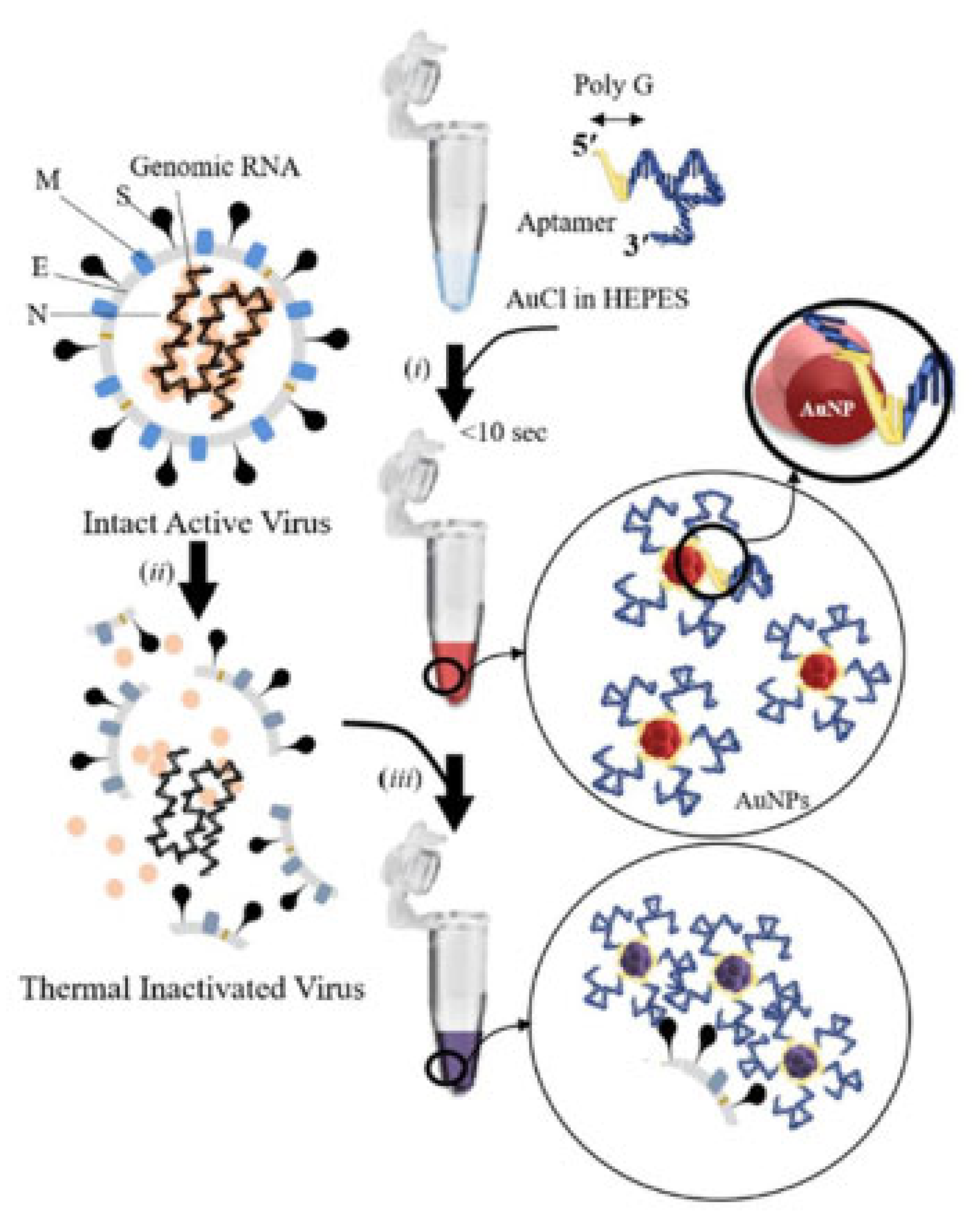
Figure 4.
Schematic illustration of surface-enhanced Raman scattering (SERS)-based detection of acrylamide using Au@Ag nanoparticles (NPs). Au NPs serve as the core, which is coated with Ag to form Au@Ag NPs. Upon interaction with acrylamide, the enhanced Raman signal is recorded under 785 nm laser excitation. The right panel shows the Raman spectra before and after the addition of acrylamide, confirming the SERS enhancement. Copyright permission in Ref. [46].
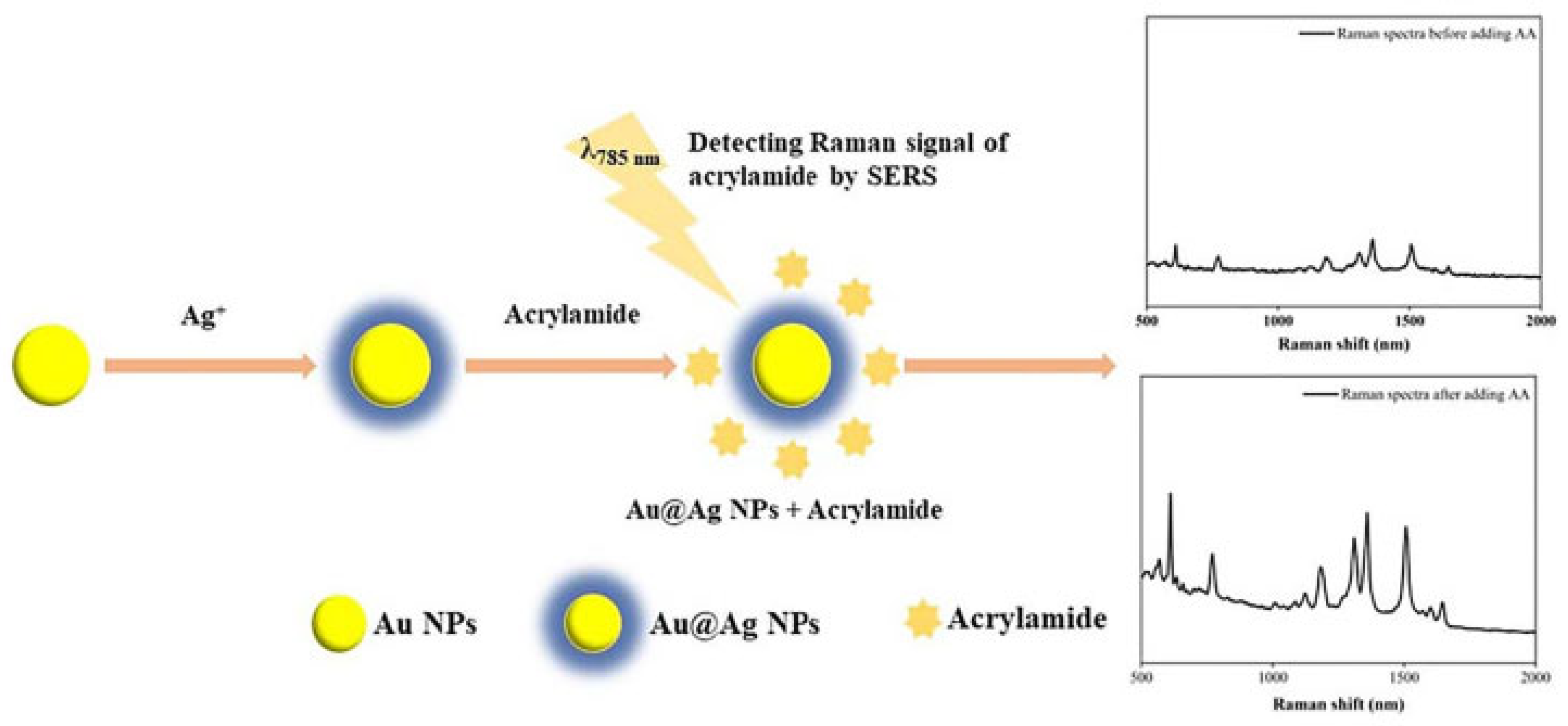
Figure 5.
Label-free SERS-AICS platform for pan-cancer detection using Ag nanowires and AI. Copyright permission in Ref. [59].
Figure 5.
Label-free SERS-AICS platform for pan-cancer detection using Ag nanowires and AI. Copyright permission in Ref. [59].
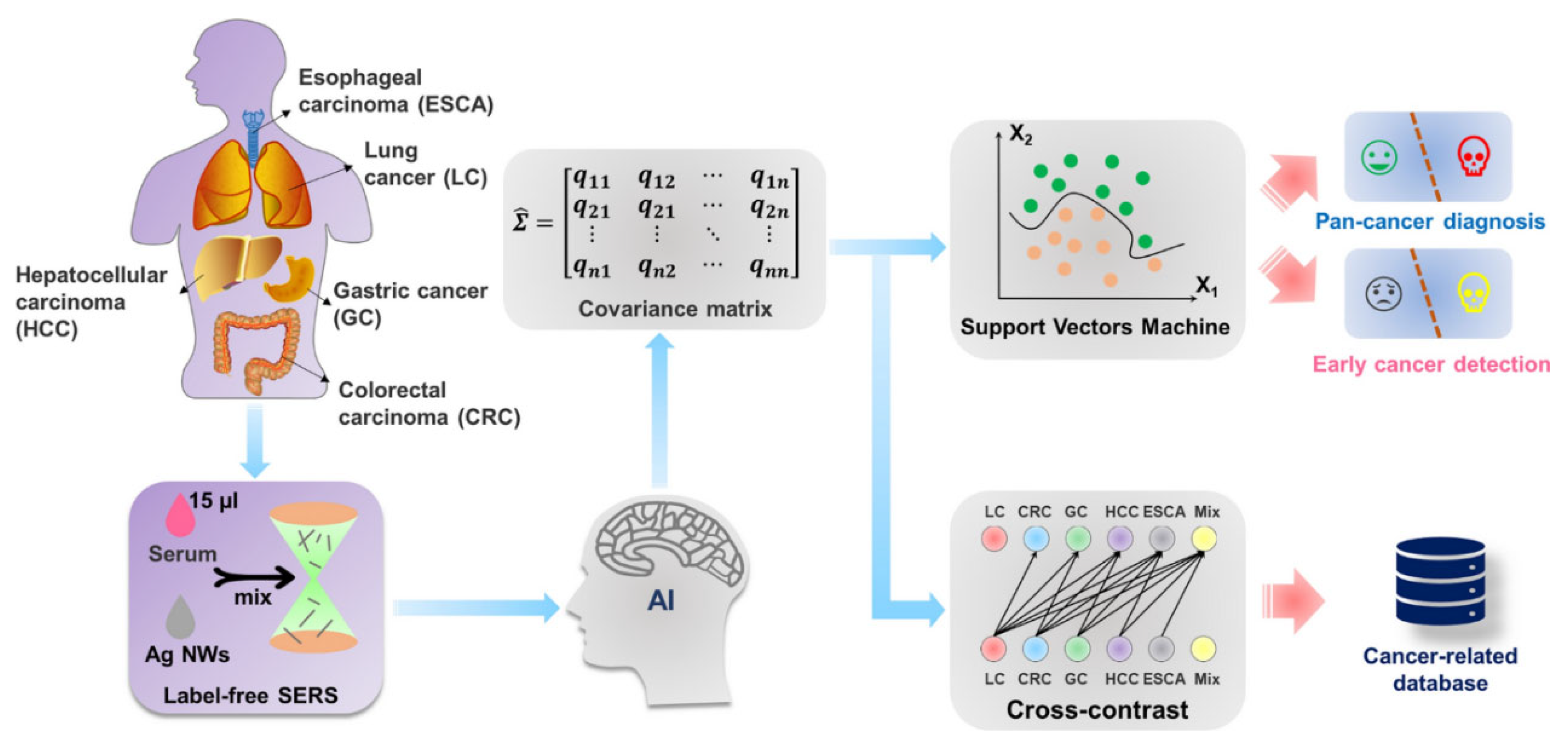

Table 1.
Key innovations in plasmonic biosensor design and their impact on performance across functional domains.
Table 1.
Key innovations in plasmonic biosensor design and their impact on performance across functional domains.
| Design Domain | Key Innovation | Performance Impact | Reference |
|---|---|---|---|
| Surface Functionalization | MOF–polymer hybrids, zwitterionic brushes | ↑ Selectivity, ↓ non-specific binding | [28,30] |
| Bioinspired Interfaces | Peptide motifs, ECM mimics | ↑ Biorecognition under flow | [31] |
| Signal Amplification | MXene–Au hybrids, QD-coupled metasurfaces | ↑ SERS EF, ↓ LOD, multiplexing | [32,34] |
| Substrate Engineering | Porous scaffolds, anisotropic clusters | ↑ Hotspot density, analyte accessibility | [33] |
| Miniaturization | Paper-based microfluidics, wearable patches | ↑ Portability, ↓ sample volume | [35,36] |
| Digital Integration | Smartphone interfaces, quantum plasmonics | ↑ Accessibility, real-time feedback | [37,38] |
Table 2.
Comparative Summary of LSPR Biosensor Applications.
| Application Domain | Key Innovation | Detection Limit / Benefit | Reference |
|---|---|---|---|
| Protein Interactions | Plasmon rulers, SAM-modified nanorods | High kinetic resolution, label-free | [40,41] |
| DNA Hybridization | Fiber-optic probes, polymer-templated AuNPs | 10 fM to 67 pM, portable formats | [42,43] |
| Pathogen Detection | Aptamer–AuNP sensors, reusable nanorod arrays | ~10³ copies/mL (SARS-CoV-2), 8.4 CFU/mL | [44,45] |
| Microfluidic Integration | PDMS channels, nanohole arrays | Real-time, low-volume, multiplexed assays | [46,47] |
Table 3.
Comparative Summary of SERS Biosensor Innovations.
| Design Feature | Key Advancement | Performance Impact | Reference |
|---|---|---|---|
| Core–Shell Nanostructures | Au@Ag NPs, microfluidic synthesis | ↑ SERS EF, ↓ LOD, improved reproducibility | [49,50,51] |
| Nanogap Engineering | DNA-mediated hotspots, 3D crevice structures | ↑ Field localization, single-molecule SERS | [52,53,54] |
| Flexible Substrates | PDMS, silk fibroin, 3D arrays | ↑ Wearability, ↓ sample volume, POCT-ready | [52,55,56] |
Disclaimer/Publisher’s Note: The statements, opinions and data contained in all publications are solely those of the individual author(s) and contributor(s) and not of MDPI and/or the editor(s). MDPI and/or the editor(s) disclaim responsibility for any injury to people or property resulting from any ideas, methods, instructions or products referred to in the content. |
© 2025 by the authors. Licensee MDPI, Basel, Switzerland. This article is an open access article distributed under the terms and conditions of the Creative Commons Attribution (CC BY) license (http://creativecommons.org/licenses/by/4.0/).
Copyright: This open access article is published under a Creative Commons CC BY 4.0 license, which permit the free download, distribution, and reuse, provided that the author and preprint are cited in any reuse.


