Submitted:
17 September 2025
Posted:
18 September 2025
You are already at the latest version
Abstract
Vitamin-conjugated metallic nanoparticles (VC-MNPs) have emerged as a transformative platform in nanomedicine, offering a dual advantage of targeted drug delivery and enhanced therapeutic efficacy. This review highlights synthesis, characterization and recent advancements of VC-MNPs for biomedical applications, with a focus on antimicrobial and anti-cancer therapies. Diverse synthesis methods including chemical reduction, co-precipitation, sol-gel, and green approaches are evaluated, along with the influence of synthesis parameters on nanoparticle performance. The mechanisms underlying enhanced antimicrobial and anti-cancer efficacy are discussed, high-lighting the contributions of vitamin functionalization to cellular uptake and biocompatibility. The review elucidates the mechanistic basis for improved antimicrobial and anticancer activity, particularly focusing on how vitamin functionalization enhances cellular internalization and bio-compatibility profiles. Critical challenges in clinical translation are systematically assessed, including nanoparticle stability under physiological conditions, potential toxicity concerns, regulatory approval pathways, and manufacturing scalability requirements. Finally, the paper considers future perspectives, focusing on synthesis innovations, novel therapeutic targets, interdisciplinary collaborations, and pathways for clinical translation. Therefore, this review of recent advances promote vitamin conjugated metallic nanoparticles as a potent platform for next-generation drug delivery systems in medicine.
Keywords:
targeted drug delivery
; antimicrobial therapy
; anticancer therapy
; surface functionalization
; cellular uptake
; drug resistance
1. Introduction
1.1. Overview of Nanotechnology and Its Significance in Drug Delivery
Recent advancements in nanotechnology have significantly influenced a broad spectrum of scientific disciplines encompassing physics, materials science, chemistry, biology, computer science, medicine, and engineering (Figure 1). Increasingly, research efforts are directed toward the precise control of atomic and molecular arrangements at the nanoscale to engineer structures, devices, and systems with novel functionalities and enhanced performance characteristics [1]. This broad impact is perhaps most evident in medicine, where nanotechnology has emerged as a transformative paradigm in drug delivery; revolutionizing how therapies are delivered at the cellular level [2,3]. Characterized by the manipulation of materials at the nanometer scale (1–100 nm), nanoparticles possess distinctive physicochemical attributes such as elevated surface area-to-volume ratios, customizable surface functionalities, and enhanced permeability that render them highly suitable for targeted therapeutic interventions [4]. Consequently, these unique features enable nanoparticles to circumvent many of the inherent limitations associated with conventional drug delivery systems, including poor solubility, limited bioavailability, and system toxicity [5].
Nanoparticles can be fabricated from a diverse array of materials, including organic polymers such as chitosan and PLGA, lipids (e.g. phospholipids, PEG, and nucleic acids), metals like gold, silver, iron, zinc, and copper, as well as silica. Each class of material offers distinct properties that are particularly tailored to specific therapeutic and diagnostic applications. For example, lipid-based nanoparticles are particularly effective at encapsulating hydrophobic drugs, enhancing their delivery and bioavailability. In contrast, metallic nanoparticles (MNPs) have gained significant attention in biomedical fields due to their exceptional physicochemical properties These MNPs are exemplified by metals such as gold and silver which are frequently utilized in imaging modalities and photothermal therapies [6]. This is largely attributed to their high surface area-to-volume ratios, which allow for greater interaction at the nanoscale, as well as their tunable surface chemistries that enable precise functionalization. Furthermore, MNPs possess unique optical and magnetic properties that make them highly versatile for a variety of biomedical applications [7]. Collectively, the choice of nanoparticle material depends on the intended clinical use, leveraging specific advantages to optimize therapeutic outcomes.
Effective drug delivery remains a cornerstone of modern therapeutics, ensuring optimal pharmacological outcomes while minimizing adverse effects [8]. In this regard, nanoparticle-based delivery systems have demonstrated considerable promise across various medical disciplines, including oncology, neurology, infectious diseases, and cardiovascular medicine. In cancer studies, nanoparticles facilitate the targeted delivery of chemotherapeutic agents directly to tumor sites, thereby reducing systemic toxicity and enhancing therapeutic efficacy [9]. For example, lipid and polymeric nanoparticles have been used to deliver agents such as doxorubicin and paclitaxel in breast and lung cancers, enhancing drug accumulation in tumors and minimizing side effects such as cardiotoxicity and system reactions [10,11]. Similarly, in neurodegenerative disorders such as Alzheimer’s and Parkinson’s diseases, nanoparticles facilitate crossing the blood-brain barrier allowing direct delivery of neurotherapeutic agents into the brain, a major obstacle for traditional treatments [12]. These advancements naturally extend to cardiovascular medicine, where nanoparticles have been developed to direct thrombolytic and anti-inflammatory agents to affected sites, decreasing the risk of systemic complications such as bleeding [13]. Additionally, emerging research has also explored the development of exosome-derived nanoparticles and environmentally benign synthesis methods to improve biocompatibility and reduce toxicity. These innovations are paving the way for more sustainable and patient-centric therapeutic strategies [14].
Building on these applications, nanoparticle systems significantly improve the bioavailability of therapeutic agents at pathological sites, by offering controlled release, reduced off-target effects, and enhanced solubility and stability for the target compounds [15]. These advantages stem from the intrinsic properties of nanocarriers, including their size, morphology, and material composition [16] which can be further optimized through surface functionalization with ligands, antibodies, or polymers to enhance their stability, biocompatibility, and targeting precision. Such functionalized nanoparticles enable receptor-mediated endocytosis, promote selective uptake by diseased cells, including malignant tissues. Moreover, these nanocarriers can be engineered to respond to specific physiological stimuli such as pH, temperature, or redox gradients which allows for controlled and site-specific drug release [17]. This technological sophistication contrasts with traditional drug delivery methods, which often suffer from stability issues, limited absorption, rapid clearance, and low specificity [18,19].
Targeted drug delivery refers to the strategic localization of therapeutic agents at the intended site of action, thereby enhancing local drug concentrations and reducing systemic exposure without compromising efficacy [20]. In line with this concept, one approach that has gained recent attention is the use of passive nanoparticle delivery systems for small molecules including vitamins and bioactive compounds, which traditionally face challenges related to poor absorption and degradation during transit. Among such strategies, vitamin-conjugated nanoparticles have emerged as a promising solution, wherein essential micronutrients serve dual roles: they act as therapeutic agents and simultaneously function as biological ligands to facilitate cellular uptake. Owing to this dual functionality, vitamin-conjugated nanoparticles have demonstrated improved bioavailability, more precise targeted delivery, and a sustained release profile for encapsulated compounds [21]. Given these advances, this review examines recent progress in the synthesis, characterization, and application of vitamin-conjugated metallic nanoparticles, while highlighting their potential to enhance therapeutic outcomes including mechanism of action.
1.2. Vitamin Conjugated Metallic Nanoparticles
Phytochemicals have emerged as key agents in the green synthesis of nanoparticles, owing biocompatibility and wide spectrum of therapeutic benefits including antimicrobial and anticancer activities [22]. Among these phytoconstituents, vitamins and essential micronutrients show defined physiological roles and have gained high attention for their multiple therapeutic domains such as antioxidant, anticancer, antimicrobial, and antidiabetic properties [23,24]. Despite these promising attributes, their conventional administration as clinical utility is hindered by inherent challenges, of chemical instability, rapid metabolic degradation, and limited bioavailability [25]. To address the limitations, nanotechnology has promoted the transformative strategies in which vitamins are conjugated with nanoparticles, to enhance pharmacokinetic profiles and enabling targeted delivery [26]. Both hydrophilic and lipophilic vitamins possess electron rich functional groups capable of coordinating with metal ions to facilitate the formation of stable metal-ligand complexes [27]. For instance, biotin’s ureido group facilitates carboxylation reactions and nanoparticle functionalization, its high affinity of conjugating with avidin has make it a better candidate for targeted cancer therapy [28]. Similarly, preliminary studies on thiamine (B1) demonstrated its ability to stabilize metal nanoparticles via heteroatom mediated coordination, whereas cobalamin (B12) exhibit structural features conducive to metal ion coordination and reduction, when it undergoes oxidation from cobalamin I to II it donates electrons to reduce metal ions and form nanoparticles [29]. Beyond metallic nanoparticles, liposomal, lipid-based, and polymeric nanocarriers functionalized with vitamins have been investigated and reports show their strong ability to enhance biocompatibility and therapeutic efficacy outperforms the traditional formulations [30,31,32,33].
2. Methods of Metal Nanoparticles Synthesis
Nanomaterial synthesis typically follows either a top-down approach: where bulk materials are reduced to nanoscale via techniques such as milling, sputtering, grinding, and thermal treatment or a bottom-up approach: which utilizes chemical and biological methods to assemble nanoparticles atom by atom. This review emphasizes various solution-based synthesis strategies which promotes functionalization and conjugation of biologically active molecules on to nanoparticles. These method is carried out under mild, biocompatible and controllable conditions aimed to enhance targeting and therapeutic efficacy of fabricated nanomaterials [34,35,36].
2.1. Chemical Approach
Chemical synthesis of metallic nanoparticles involves three primary components: metal salt precursors, reducing agents, and capping/stabilizing agents to coat the surface and prevent aggregation. Common chemical techniques include chemical reduction [37], co-precipitation [38], and sol-gel methods [39]. Chemical reduction methods typically use agents such as borohydrides, citric/oxalic acids, polyols, hydrogen peroxide, and sulfites to transfer electrons to metal ions, forming free atoms. Stabilizing agents like trisodium citrate dihydrate, sulfur ligands (thiolates), phosphorus ligands, polymers, and surfactants (e.g., cetyltrimethylammonium bromide, CTAB) are employed to prevent nanoparticle aggregation. In some cases, the same molecule serves as both reducing and stabilizing agent [40,41,42]. The co-precipitation method involves simultaneous precipitation of multiple metal ions using agents such as NaOH and NH4OH to produce uniformly sized nanoparticles with controlled composition and properties. Magnetite nanoparticles are commonly synthesized using this method. Aqueous solutions of Fe (II) and Fe (III) are mixed with a base at ambient or elevated temperatures, resulting in the formation of insoluble hydroxides that aggregate into nanoparticles. Surfactants such as cetyltrimethylammonium bromide (CTAB), cetyltrimethylammonium chloride (CTAC), sodium dodecyl sulfate (SDS), polyvinylpyrrolidone (PVP), or polyethylene glycol (PEG) are added dropwise to prevent agglomeration and oxidation [43,44,45]. The sol-gel process encompasses hydrolysis, polymerization/condensation of monomers, particle growth, and gel formation. It involves the phase transformation of a colloidal “sol” into a solid “gel.” This method enables the production of glass and ceramic materials at low temperatures, facilitating the incorporation of various organic and inorganic substances for applications such as drug delivery. It allows precise control over oxide stoichiometry and yields high-purity, uniform, and crystalline nanoparticles under mild conditions [46,47,48,49]. The chemical approach offers advantages such as enhanced reactivity, controlled nanoparticle shape and size, cost-effectiveness, and reproducibility [50]. However, byproducts from chemical reductants raise concerns regarding human health and environmental safety. Despite their benefits such as rapid reaction kinetics at low temperature and pressure and good solubility in aqueous and polar organic media; several drawbacks such as the impact of chemical reagents, high temperatures, and harsh reaction conditions to the environmental and human health have been reported. For example, the byproducts from NaBH4 and hydrazine are harmful to the respiratory system, and may cause neurotoxicity and encephalopathy [50,51,52,53].
2.2. Greener Approach
Recently, environmentally sustained synthesis of nanomaterials shows strong increased trend (Figure 2) and has gained significant attention due to its effectiveness, low cost, reduced failure rates, and ease of characterization, which makes it preferable over conventional methods [54]. This ecofriendly approach uses bioactive compounds from natural sources such as enzymes, vitamins, carbohydrates, bacteria, fungi, yeast, algae, and plant secondary metabolites which act simultaneously as capping and reducing agents. Such biologically synthesized nanoparticles exhibit superior biocompatibility, nano-dimensions, and pharmacological properties, making them suitable for medical applications such as drug delivery, cancer therapy, antimicrobial treatments, and tissue regeneration [55]. For example, vitamin conjugates including folic acid, riboflavin, thiamine, biotin, and vitamins C and D have been shown to improve nanoparticle stability and therapeutic efficacy which makes them the ideal candidates for therapeutic purpose including drug loading and delivery (Table 1). Several studies have highlighted the influence of vitamin conjugated nanoparticles on drug delivery, therapeutics and other applications. For instance, Singh et al. [56] prepared riboflavin and thiamine functionalized multi-walled carbon nanotubes for cancer therapy, demonstrating significant cytotoxicity against MCF-7 cancer cell lines. Subsequently, Chakraborty and Jana [57] synthesized vitamin C-conjugated gold nanoparticles with enhanced chemical stability under physiological conditions, according to their findings the conjugate has better reducing oxidative stress ability compared to vitamin C alone. Similarly, Kader et al. [58] fabricated vitamin C-adduct-conjugated zinc oxide nanoparticles for photocatalytic applications. Nah et al. [59] reported vitamin D-conjugated gold nanoparticles for osteogenic differentiation of human adipose-derived stem cells (hADSCs). In a recent study, Zamenraz et al. [60] confirmed that vitamin B5 (pantothenic acid) bonded to 2,4,6-trichloro-1,3,5-triazine conjugated copper has potentially enhanced visible-light photocatalytic activity of nanocrystalline TiO2. Furthermore, Anwar et al. [61] synthesized riboflavin-conjugated silver nanoparticles (VC-AgNPs) with selective antimicrobial activity against gram-positive bacteria. A common method for synthesizing vitamin-conjugated nanoparticles involves mixing a metal precursor solution with a vitamin, where the vitamin’s functional groups act as reducing and capping agents. Alternatively, nanoparticles are synthesized separately, dispersed in a solvent, and then mixed with a vitamin solution at room temperature for approximately 24 hours; optimization of nanoparticle size and morphology is achieved by adjusting temperature, pH, and reaction time [62,63]. Despite its advantages, green synthesis faces limitations including variability in source material availability due to geographic and seasonal factors, and challenges in scaling for industrial production [64]. Therefore, elucidating the molecular mechanisms of biosynthesis, and optimizing synthesis parameters to improve reproducibility will be crucial to fully harness the potential of green nanotechnology in medicine and beyond.
2.3. Influence of Synthesis Parameters
The physicochemical characteristics of materials, including their composition and morphology, are profoundly influenced by various synthesis parameters. Among these pH, reactant concentration, reaction duration, and temperature are the major factors, each of which can be strategically manipulated to tailor material properties [65,66]. Specifically, temperature and pH play pivotal roles in determining the size, shape, and formation rate of nanoparticles. Elevated pH levels have been shown to enhance the nucleation process, thereby accelerating the reduction of metal ions into metallic nanoparticles [67]. Building on this, Fernando and Zhou [68] evaluated the effect of pH on physicochemical behavior of AgNPs, focusing on their stability, dissolution behavior, and aggregation kinetics. Their findings revealed that pH significantly modulates the surface charge of AgNPs, which in turn governs their tendency to aggregate and undergo oxidative dissolution. Particularly, acidic and neutral conditions led to destabilization and increased aggregation, whereas alkaline environments, enriched with hydroxyl ions, promoted re-stabilization and the formation of more stable colloidal suspensions. This sensitivity to pH and temperature is not limited to synthesis but extends to biomedical applications, where these parameters critically affect drug loading and release in nanocarrier systems. Recently, pH-responsive nanodrug delivery platforms have garnered considerable interest for their ability to selectively deliver therapeutic and diagnostic agents to diseased tissues. The pH of the target environment influences both the binding efficiency of functional ligands on the nanoparticle surface and the release kinetics of the encapsulated drug. Improper pH control may lead to off-target effects and toxicity in healthy tissues [69,70].
Further, Awadh et al. [71] have demonstrated that altering pH and temperature during ZnO nanoparticle synthesis directly influence optical properties, with successful formation at pH 10 and 11 distinct absorption peaks at 366 and 364 nm. However, no such peaks were observed at pH 12 and 14, suggesting that excessively high pH levels accelerate nanoparticle aging. Similarly, temperature also played a crucial role: higher synthesis temperatures (80–90 °C) facilitate the formation of smaller, monodispersed nanoparticles with sharp surface plasmon resonance (SPR) features, whereas lower temperatures fail to produce well-defined nanoparticles. However, these trends are not universal [71,72]. For example, the stability of gold nanoparticles can decrease at elevated temperatures, highlighting thermal stability and morphological development vary with material type and synthesis pathway [73]. In addition to pH and temperature, both reactant concentration and reaction duration are crucial in controlling size and growth of nanoparticles during synthesis. Experimental studies show that increasing the concentration of reactants leads to a higher reaction speed, while lower concentrations slow down the process due to reduced collision frequency between reactant molecules [74]. Reaction time further influences nanoparticle properties as prolonged reaction times may result in excessive particle growth, aggregation, or even aging effects, thereby altering optical, structural, or functional properties [75]. For example, Dehsari et al. [76] evaluated the effect of precursor concentration on the size of iron oxide nanoparticles, showing that modifying reactant concentration can regulate particle size and distribution , however, the relationship is not always linear, as excessive concentration can also narrow particle size due to changes in aggregation and nucleation dynamics. Similarly, Yazdani et al. [77] reported that extended reaction times during the synthesis of gold nanoparticles enhanced aggregation and polydispersity, ultimately leading to undesirable size variations. Therefore, considering careful optimization of both reactant concentration and reaction time is essential to achieve desired material properties and reproducibility in synthesis protocols.
3. Characterization Techniques of Nanoparticles
Understanding nanoparticles (NPs) behavior in biomedical applications necessitates comprehensive evaluation of their fundamental properties. Critical determinants such as particle dimensionality, morphological architecture, surface electrostatics, porosity, polydispersity index, and specific surface area significantly influence their interactive dynamics and efficacy in targeted applications, encompassing drug delivery systems, catalytic processes, biomedical imaging, and environmental detoxification [78,79]. Comprehensive and precise characterization of these parameters is imperative for elucidating NPs behavior and refining synthesis methods to enhance application-specific performance [80]. To achieve this, a multifaceted suite of analytical methods is employed, each offering distinct and complementary insights into nanoparticle composition and functionality. These techniques encompass Transmission Electron Microscopy (TEM), Scanning Electron Microscopy (SEM), Ultraviolet-Visible (UV-Vis) Spectroscopy, Fourier Transform Infrared (FTIR) Spectroscopy, X-ray Diffraction (XRD), X-ray Photoelectron Spectroscopy (XPS), Dynamic Light Scattering (DLS), Atomic Force Microscopy (AFM), Nuclear Magnetic Resonance (NMR) Spectroscopy, and zeta potential analysis. Collectively, these tools facilitate a holistic understanding of NPs physicochemical profiles (Table 1), thereby informing their rational design and application [81].
Electron microscopy techniques, particularly TEM and SEM, serve as essential analytical tools for nanoparticle structural characterization. TEM facilitates ultra-high-resolution imaging, enabling the direct visualization of internal nanostructures and precise morphological delineation at the atomic scale, which is critical for examining spatial distribution and interfacial architecture. In parallel, SEM complements this analysis by generating three-dimensional surface topographies through secondary electron detection, thereby offering critical insights into surface morphology, texture, and particulate arrangement [81]. Beyond morphological characterization, UV-Vis spectroscopy introduces a rapid, non-invasive optical modality for monitoring nanoparticle synthesis and colloidal stability. Particularly efficacious for metallic NPs due to their surface plasmon resonance (SPR) phenomena, UV-Vis spectral profiles specifically the peak position and bandwidth serve as proxies for particle size, dispersion uniformity, and aggregation dynamics, linking structural and optical properties in real time [82]. To examine surface chemistry and functionalization, FTIR spectroscopy identifies functional groups, and elucidate interactions at the nanoparticle interface, which is particularly relevant in green synthesis approaches [83].
DLS serves as a pivotal analytical technique for characterizing colloidal systems by determining the hydrodynamic diameter and polydispersity index, thereby offering insights into particle size distribution and uniformity. When coupled with zeta potential analysis, DLS extends its utility to evaluating the electrostatic potential at the nanoparticle interface, a critical parameter for assessing colloidal stability. High absolute values of zeta potential regardless of polarity signify strong interparticle repulsion, which effectively minimizes aggregation and enhances dispersion stability under varying environmental conditions. This dual capability is instrumental in predicting nanoparticle behavior in both biological and industrial contexts, particularly with respect to long-term storage and functional performance. Moreover, DLS complements spectroscopic techniques such as FTIR by bridging surface chemistry with physicochemical stability, thus providing a comprehensive framework for nanoparticle evaluation [84,85].
XRD serves as a definitive tool for crystallographic analysis, offering quantitative data on lattice parameters, crystallite dimensions, and phase composition. It is instrumental in confirming the formation of specific crystalline phases and assessing structural integrity. When integrated with TEM and DLS data, XRD provides a holistic understanding of nanoparticle architecture from atomic arrangement to colloidal behavior [80,86].
AFM enriches surface characterization by delivering nanoscale three-dimensional imaging without the prerequisite of conductive coatings. Through precision scanning of a sharp probe across the sample surface, AFM captures topographical features such as surface roughness, particle height, and mechanical heterogeneity, thereby complementing SEM and TEM with tactile resolution [87].
XPS introduces a surface-sensitive analytical dimension, capable of resolving elemental composition and oxidation states within the top few nanometers of the nanoparticle surface. This technique is critical for validating surface modifications, dopant incorporation, and functional group presence, thereby linking surface chemistry to electronic structure and catalytic potential [88].
NMR spectroscopy culminates the analytical suite by offering molecular-level insights into the electronic environment, bonding configurations, and ligand-nanoparticle interactions. It is particularly efficient at elucidating surface chemistry, ligand density, and core composition, thereby providing a comprehensive understanding of nanoparticle functionality and reactivity [89].

Table 1.
Summary of metal nanoparticles with vitamins.
| S/N | Metal NP + Vitamin | Synthesis Method | Size & Morphology | Characterization Techniques | Applications | Key Findings | Ref. |
|---|---|---|---|---|---|---|---|
| 1 | Ag (Ag/Cu) + Ascorbic Acid | Chemical reduction | Ag: ~200–800 nm; Cu: ~160–630 nm spherical | UV–Vis, DLS, TEM | Antibacterial: Tested against Bacillus subtilis (Gram+) and E. coli (Gram–). | Strongest bactericidal effect (MIC ~0.05–0.08 mg/L). | [90] |
| 2 | Au + Vitamin C (with algal EPS) | Green biosynthesis | ~6–40 nm spherical AuNPs | UV–Vis, XRD, TEM, FTIR | Antibacterial: Multi-strain (E. coli, S. aureus, S. enterica, S. mutans, Candida spp.). Anticancer: Tested against MCF-7, A549, and CaCo-2 cells. | Most effective: >88% kill of E. coli and ~83% of S. aureus under light (via ROS generation), and ~70% growth inhibition of MCF-7 breast cancer cells. | [91] |
| 3 | CeO2 + Folic Acid | Green one-pot precipitation | ~21–28 nm polyhedral; DLS (hydrodynamic ~200 nm; 22 mV) | XRD, TEM/SEM | Antibacterial: Potent against MRSA. Antioxidant/Anti-inflammatory. Anticancer (MDA-MB-231). | Inhibit ~95.6% of MRSA growth, accelerated wound healing and selective toxicity toward bacteria and cancer cells. | [92] |
| 4 | Cu–MOF + Folic Acid (Cu-TCPP MOF/Pt-FA) | Chemical method | Nanosheets ~100–200 nm; Pt NPs ~2 nm | TEM/HRTEM, XRD, XPS, FTIR, Zeta potential | Anticancer (PDT & immunotherapy) | Greatly enhances PDT even in hypoxic conditions. | [93] |
| 5 | Cu2S + Vitamin C | Single-step aqueous synthesis (Chemical reduction) | CuS ~8–10 nm, quasi-spherical; agglomerates into 50–100 nm clusters | XRD, FTIR, TEM/SEM, EDS | Antibacterial (S. aureus, E. coli, K. pneumoniae), Antioxidant | Broad-spectrum bactericidal activity. MIC: ~2 mg/mL (E. coli) and 10 µg/mL (other strains). Also scavenged DPPH & NO radicals. | [94] |
| 6 | Fe3O4 + Riboflavin (B2) | Solvothermal method | ~200 nm spherical | XRD, XPS, Zeta potential, UV-Vis, Fluorescence | Antibacterial and Antioxidant | Kills >90% of S. aureus and ~88% of E. coli at 0.5 mg/mL. | [95] |
| 7 | Fe3O4 + Folic Acid (PLGA nanocarrier) | Double emulsion solvent evaporation | ~150–180 nm polymeric spheres (PLGA); Fe3O4 cores ~8 nm | TEM, DLS, FTIR, NMR, MRI | Anticancer | Induce ~90% cell death in ovarian cancer. | [96] |
| 8 | Fe3O4 + Folic/TNF/IFN/DOX | Surface functionalization & self-assembly | Fe3O4 core ~5 nm; clusters ~50 nm spherical | FTIR, DLS, UV–Vis | Anticancer (combined therapy) | Produced synergistic cancer cell killing with reduced systemic toxicity. | |
| 9 | Ag + Folic Acid (valve coating) | Biofunctional coating | ~10 nm AgNPs | SEM, EDS, FTIR | Antibacterial & anti-inflammatory implant | Reduced calcification and inflammation in vivo. | [97] |
| 10 | Ag/MOF + Folic Acid (nanocapsule) | Biopolymer-templated in situ MOF synthesis | ~320–350 nm mixture of rod-like and spherical particles | XRD, FTIR, SEM, TEM, BET | Antibacterial, antioxidant, targeted drug delivery | Single folate-targeted nanocapsule can deliver chemotherapeutics while preventing infection & oxidative damage. | [98] |
| 11 | Gd2O3 + Vitamin C | Biogenic precipitation | ~50 nm amorphous Gd2O3 particles | TEM, DLS, XPS, ICP | Antibacterial | Potent bactericidal effects against multiple pathogens. | [99] |
| 12 | Au + Riboflavin (B2) | Photochemical surface-modification | AuNP ~20 nm, spherical | UV–Vis | Photodynamic antimicrobial therapy (S. aureus, P. aeruginosa) | Vitamin B2 + AuNP create synergistic ROS + Au+ antibacterial effect. | [100] |
| 13 | Ag + α-Tocopherol Succinate (Vit E) | Surface functionalization | Ag core ~20 nm; hydrodynamic size ~25 nm (TOS coating) | UV–Vis, FTIR, DLS/Zeta potential | Anticancer (A549 lung carcinoma) | TOS coating enhanced cancer selectivity & therapeutic index of AgNPs. | [101] |
| 14 | Y2O3 + Folic Acid | Chemical synthesis/thermal decomposition | ~5–10 nm hexagonal phase; aggregates into ~100 nm clusters; folate-PEG ~120 nm | Photoluminescence, TEM, FTIR, DLS | Cancer imaging | Enabled precise NIR-triggered imaging & potential phototherapy. | [102] |
| 15 | Se + Vitamin C | Chemical reduction | ~50–60 nm spherical | UV–Vis, DLS, Zeta potential, XPS | Antibacterial | Strong activity against S. aureus; stabilized Se–VitC NPs retained activity 2–6 months. | [103] |
| 16 | Zn/Ag MOF + Vitamin C | Chemical synthesis with functionalization | ~100–200 nm polyhedral | XRD, TEM, SEM, FTIR, BET | Antibacterial | Strong activity against Gram+ and Gram– bacteria common in wound infections. | [104] |
| 17 | Fe/MOF + Riboflavin | Hydrothermal method | Uniform polyhedral morphology | TEM, DLS, Zeta potential, XRD, FTIR, SEM, EDS, UV-Vis, Thermal imaging | Treatment of bacterial keratitis (S. aureus, P. aeruginosa) | Rapid infection clearance with minimal collateral damage. | [105] |
| 18 | Ag NPs + Biotin, D-Pantothenic acid & Nicotinic acid | Chemical reduction with NaBH4 | ~10 nm spherical | UV-Vis, TEM, FTIR, DLS, TGA, FE-STEM | Antimicrobial | Effective at low concentrations (15.62–62.5 μg/mL) against planktonic cells & biofilms. | [106] |
4. Applications
4.1. Antimicrobial Applications
The rise of multidrug-resistant (MDR) pathogens has intensified the search for alternative antimicrobial strategies. Among metal nanoparticles such as silver, gold, zinc oxide, and copper have shown potent antimicrobial properties. Functionalizing these nanoparticles with vitamins (Table 1) further enhances their efficacy, selectivity, and safety profile and also improves their stability, solubility, and bioavailability [107]. These nanoparticles exhibit strong efficacy against both bacterial and fungal pathogens, often outperforming conventional antibiotics [108,109]. Importantly, vitamin conjugated metallic nanoparticles have been reported to improve antimicrobial activity when incorporated into drug delivery systems, resulting in enhanced pharmacokinetics, reduced dosage requirements, and minimized toxicity [110]. These properties position them as promising candidates for the treatment of persistent and resistant infections, with ongoing research demonstrating synergistic effects and broadened antimicrobial spectra when combined with commercial antimicrobial agents [111,112]. Additionally, these nanoparticles are increasingly incorporated into coatings for surgical instruments, wound dressings, and implantable devices to prevent microbial colonization and biofilm formation, thereby reducing infection rates in clinical settings [113].
4.2. Anti-Cancer Applications
In oncology, vitamin-conjugated metal nanoparticles have been studied both as single agents and in combination with traditional chemotherapeutics. Several case studies highlight that specific nanoparticle formulations, such as gold or platinum conjugated with vitamins, can enhance immunotherapeutic responses, promote targeted drug delivery, and induce tumor cell death through mechanisms like immunogenic cell death and photothermal/photodynamic therapy [114,115]. Combination therapies that integrate these nanoparticles with existing treatments have shown additive or even synergistic effects, often resulting in improved tumor suppression in preclinical models. Some have advanced to early-phase clinical evaluation, where the focus has been on enhancing efficacy while minimizing off-target toxicity [116,117,118]. For example, folate-conjugated gold nanoparticles have been explored for use in computed tomography (CT) imaging, targeted chemotherapy, radiation therapy, and hyperthermia, offering a multifunctional approach to cancer treatment and diagnosis [119]. Additionally, gold nanoparticles conjugated with vitamin B complexes have been proposed as multifunctional drug delivery systems, potentially exerting synergistic therapeutic effects and improving drug targeting [120].
4.3. Other Emerging Applications
Beyond direct therapeutic uses, vitamin-conjugated metal nanoparticles are gaining traction in diagnostic imaging and biosensor technologies. Their distinctive optical, electronic, and catalytic properties enable highly sensitive detection of biomolecules, facilitating early disease diagnosis via colorimetric changes or enhanced signal amplification [121,122]. In diagnostic imaging, vitamin conjugated metallic nanoparticles (VC-MNPs) enhance the specificity and sensitivity of imaging modalities such as MRI, CT, and PET by targeting overexpressed vitamin receptors on diseased cells [123]. For instance, biotinylated nanoparticles are being explored in biosensors for their ability to selectively bind to target biomolecules, enabling rapid and accurate detection of pathogens or disease biomarkers [124]. Furthermore, vitamin D-conjugated gold nanoparticles have been shown to enhance osteogenic differentiation in stem cells, indicating potential applications in bone regeneration and tissue engineering [59].
In gene delivery systems, VMNPs offer a promising non-viral alternative for transporting genetic material into cells. These platforms have demonstrated versatility in delivering a range of genetic payloads, such as siRNA for gene silencing, plasmids for gene expression, and CRISPR-Cas9 components for genome editing, thereby enabling precise genetic modulation for various therapeutic purposes [125,126]. As a result, their application extends to the treatment of inherited disorders, cancer, and other diseases where targeted gene regulation is crucial [127,128]. Clinical translation efforts are now focusing on the development and approval of non-viral gene therapy products, with several clinical trials underway for conditions such as genetic disorders and cancers, highlighting the growing recognition of VMNPs as safer and more versatile alternatives [129,130].
5. Mechanisms of Actions
Understanding the mechanisms through which pharmacological compounds exert their therapeutic effects is crucial for advancing drug development and optimizing therapeutic interventions. The mechanism of action refers to the specific biochemical interaction between a pharmacological compound and its molecular target, typically involving high binding affinity to structurally defined domains on proteins such as enzymes, ion channels, or membrane-bound receptors. This fundamental understanding enables the rational design of more effective and targeted therapies while providing insights into potential adverse effects and drug interactions [131,132]. Drug efficacy is frequently mediated through competitive inhibition, wherein the therapeutic agent competes with endogenous substrates or ligands to modulate enzymatic activity or receptor-mediated signaling pathways. Through this mechanism, drugs can either mimic the actions of natural ligands as agonists or block receptor activation as antagonists, ultimately determining the cellular response and therapeutic outcome [133]. In the context of oxidative stress, pharmacological agents may exert their therapeutic effects through multiple molecular strategies that collectively restore cellular redox homeostasis[134]. These include direct scavenging of reactive oxygen species (ROS) [135], modulation of redox-sensitive intracellular signaling cascades, and upregulation of endogenous antioxidant defense mechanisms such as superoxide dismutase, catalase, and glutathione peroxidase [136,137,138]. Under physiological conditions, oxidative free radicals generated by mitochondrial respiratory chains, endoplasmic reticulum, and NADPH oxidases are effectively neutralized by NRF2-mediated antioxidant responses that elevate the synthesis of key protective molecules. Disruption of this finely tuned equilibrium between oxidant generation and antioxidant capacity is closely linked to the pathogenesis of numerous diseases, making redox regulation an increasingly important therapeutic target [139,140]. Collectively, these interactions contribute to the downstream regulation of cellular homeostasis and are integral to the therapeutic efficacy of both natural and synthetic compounds [141]. The following sections discuss the effect of vitamin functionalization on mechanism of action of antimicrobial and anticancer agents.
5.1. Antimicrobial Mechanisms
5.1.1. Inhibition of Bacterial Growth
Understanding antimicrobial mechanisms at the molecular and genetic level provides critical insights into how vitamin-conjugated nanoparticles disrupt bacterial physiology and overcome resistance mechanisms [142]. Vitamin conjugated nanoparticles demonstrate potent antimicrobial activity through multiple interconnected mechanisms that synergistically inhibit bacterial growth [143]. The fundamental basis of this unique physicochemical properties of nanoparticles, including their high surface-to-volume ratio, enables enhanced interaction with bacterial cells [144,145]. The small size of these nanoparticles, typically ranging from 1-100 nm, facilitates their penetration through bacterial cell walls and membranes, allowing for more effective antimicrobial action (Fernando et al., 2018; More et al., 2023). At the molecular level, the antimicrobial effects involve comprehensive disruption of bacterial gene expression patterns and cellular processes. Transcriptomic analyses have revealed that nanoparticle treatment alters fundamental bacterial processes including protein translation, energy metabolism, and stress response pathways. These broad transcriptional changes occur before bacterial population density changes, indicating that nanoparticles directly interfere with essential gene regulatory networks rather than simply reducing bacterial numbers [147].The antimicrobial activity of vitamin conjugated nanoparticles operates through both contact killing and ion-mediated killing mechanisms. In contact killing, the nanoparticles directly interact with bacterial cell surfaces through electrostatic attraction, van der Waals forces, and hydrophobic interactions [148,149]. The negatively charged bacterial cell wall components, particularly phosphorous and sulfur-containing molecules, readily bind to positively charged nanoparticles, leading to membrane disruption [150]. Ion-mediated killing occurs through the sustained release of metal ions from the nanoparticle matrix. For instance, vitamin B12-conjugated nanoparticles containing silver demonstrate concentration-dependent bacterial growth inhibition, with minimum inhibitory concentrations (MIC) as low as 5 μM against Escherichia coli (Marcin., 2019). Similarly, selenium nanoparticles loaded with vitamin D3 showed remarkable inhibitory activity against both E. coli and Staphylococcus aureus, with inhibition zones ranging from 8-30 mm depending on concentration [151].
The mechanism of bacterial growth inhibition involves multiple cellular targets. Metal ions released from vitamin conjugated nanoparticles bind to sulfhydryl groups in essential enzymes and proteins, particularly those involved in transmembrane ATP generation and ion transport [152]. The metal ion binding forms stable complexes that result in protein deactivation and disruption of cellular metabolism. Additionally, these nanoparticles uncouple respiratory electron transport from oxidative phosphorylation, effectively limiting respiratory chain enzymes and obstructing membrane permeability to protons and phosphate [145]. Furthermore, reactive oxygen species (ROS) generation represents another critical mechanism of bacterial growth inhibition. Vitamin conjugated nanoparticles induce oxidative stress within bacterial cells through the production of hydroxyl radicals, superoxide anions, and hydrogen peroxide [153]. Such stress damages multiple cellular components simultaneously, including lipids in the cell membrane, proteins, and nucleic acids (Figure 3), leading to bacterial cell death [57,153]; The oxidative damage extends to bacterial DNA, causing mutations and interfering with essential genetic processes such as replication and transcription.[152].
5.1.2. Mechanisms of Resistance Overcoming
The emergence of antimicrobial resistance poses significant challenges to conventional antibiotic therapy, making the development of resistance-overcoming strategies crucial from both phenotypic and genetic perspectives. Vitamin conjugated nanoparticles offer several advantages in circumventing bacterial resistance mechanisms through their multi-target approach and synergistic effects [154]. Unlike conventional antibiotics that typically target a single cellular pathway, vitamin conjugated nanoparticles simultaneously attack multiple bacterial targets, making it extremely difficult for bacteria to develop comprehensive resistance [155]. The combination of vitamin-mediated targeting with nanoparticle-induced damage creates a multi-pronged assault on bacterial cells that overwhelms their adaptive mechanisms [150]. From a genetic standpoint, resistance mechanisms in bacteria involve specific genes that encode proteins responsible for antibiotic degradation, efflux pumps, and target modification. Key resistance genes such as trxA (thioredoxin A), trxB (thioredoxin reductase), dltA (D-alanine incorporation), and various efflux pump genes (mexA, mexB, mexC) are systematically disrupted by nanoparticle treatment. This genetic disruption occurs through multiple mechanisms including direct binding to DNA, interference with transcriptional machinery, and oxidative damage to genetic material [156,157].
Biofilm penetration represents a significant advantage of vitamin conjugated nanoparticles in overcoming resistance [158]. Many antibiotic-resistant bacteria exist within biofilms that provide protection against conventional treatments. The small size and surface properties of vitamin conjugated nanoparticles enable them to penetrate these biofilm matrices and reach embedded bacterial cells [145,159]. Studies have demonstrated that organic-inorganic hybrid nanoparticles show superior effectiveness against biofilm-associated bacteria compared to conventional treatments [159]. At the molecular level, biofilm-associated resistance involves complex gene regulatory networks including quorum sensing systems (luxS, lasI, rhlR), adhesion genes (fimH, bap), and extracellular polymeric substance production genes (psl, pel, icaA). Nanoparticles disrupt these genetic networks by interfering with quorum sensing signaling, preventing proper biofilm formation, and compromising the structural integrity of established biofilms [157,160].
Moreover, the vitamin targeting component provides specificity that helps overcome resistance by exploiting bacterial vitamin requirements. Bacteria require various vitamins for essential metabolic processes, and vitamin-conjugated nanoparticles can hijack these uptake pathways to deliver antimicrobial agents directly into resistant bacterial cells. This targeted approach bypasses many traditional resistance mechanisms that bacteria use to exclude antibiotics [151]. Synergistic combinations with conventional antibiotics (Figure 3) represent another resistance-overcoming strategy; occurs through simultaneous targeting of different genetic pathways, preventing bacteria from utilizing their resistance genes effectively while compromising their overall cellular integrity through multiple mechanisms of action [157]. Research has shown that combining vitamin conjugated nanoparticles with traditional antibiotics can restore sensitivity in resistant bacterial strains. For example, the combination of silver nanoparticles with vancomycin has demonstrated enhanced efficacy against vancomycin-resistant bacterial strains, suggesting that nanoparticle-antibiotic combinations can overcome established resistance mechanisms [154].
5.2. Anti-Cancer Mechanisms
5.2.1. Targeted Delivery to Tumors
Vitamin conjugated nanoparticles offer exceptional opportunities for targeted cancer therapy through exploitation of the enhanced vitamin requirements of rapidly dividing cancer cells. Malignant tumors exhibit increased demand for essential vitamins, resulting in overexpression of vitamin receptors on cancer cell surfaces compared to normal cells. This differential expression provides a molecular basis for selective targeting that can be exploited for therapeutic advantage. Folate receptor targeting represents one of the most extensively studied vitamin-mediated targeting approaches. Cancer cells, particularly those of epithelial origin, frequently overexpress folate receptors to meet their increased metabolic demands. Folate-conjugated nanoparticles can specifically bind to these overexpressed receptors, facilitating selective uptake by malignant cells while sparing normal tissues with lower receptor expression of Vitamin B12-mediated targeting exploits the high demand for cobalamin in rapidly dividing cells. Cancer cells upregulate trans-cobalamin receptors and other B12 transport mechanisms to support increased DNA synthesis and cellular proliferation. Vitamin B12-conjugated nanoparticles can exploit these transport pathways to achieve selective accumulation in tumor tissues [161].
Vitamin D3 nanoparticle platforms have demonstrated remarkable versatility in cancer targeting applications [162]. These biocompatible and biodegradable nanoparticles can be engineered to carry multiple therapeutic agents while maintaining tumor selectivity. The vitamin D3 component not only provides targeting specificity but also contributes intrinsic anti-cancer activity through modulation of calcium homeostasis and cellular differentiation pathways [163]. The enhanced permeability and retention effect works synergistically with vitamin-mediated targeting to improve tumor accumulation [164]. The abnormal vasculature of solid tumors, characterized by larger pore sizes and poor lymphatic drainage, allows preferential accumulation of nanoparticles within tumor tissues [165]. When combined with vitamin receptor targeting, this passive targeting mechanism significantly enhances the therapeutic index of vitamin conjugated nanoparticles pH-responsive drug release represents an additional targeting mechanism that exploits the acidic tumor microenvironment. Vitamin conjugated nanoparticles can be designed to remain stable in physiological pH conditions but rapidly release their therapeutic cargo in the acidic conditions (pH 5.5-6.5) found within tumor tissues and cellular compartments. This pH-triggered release ensures that therapeutic agents are preferentially activated within the target tissue [166,167].
5.2.2. Induction of Apoptosis in Cancer Cells
Vitamin conjugated nanoparticles induce cancer cell death through multiple apoptotic pathways, offering advantages over conventional chemotherapy approaches [168]. The multifaceted mechanism of action involves both the intrinsic properties of the vitamin components and the enhanced delivery of cytotoxic agents to intracellular targets, thereby amplifying their therapeutic potential. DNA damage-induced apoptosis represents a primary mechanism through which vitamin conjugated nanoparticles eliminate cancer cells [169]. Studies have demonstrated that dual drug-loaded vitamin D3 nanoparticles containing PI103 and cisplatin induce significant DNA damage in hepatocellular carcinoma cells, leading to apoptotic cell death within 24 hours. The nanoparticle delivery system ensures sustained release of DNA-damaging agents directly within cancer cells, maximizing therapeutic efficacy while minimizing systemic toxicity [166]. Multi-target apoptotic signaling is achieved through the simultaneous delivery of multiple therapeutic agents. Vitamin D3 nanoparticles have been successfully engineered to carry rational combinations of drugs, such as PI103 (a PI3 kinase inhibitor) with various cytotoxic agents including cisplatin, doxorubicin, and proflavine This multi-drug approach activates multiple apoptotic pathways simultaneously, overwhelming cancer cell survival mechanisms and inducing rapid cell death [170].
In addition to direct DNA-targeted mechanisms, mitochondrial pathway activation significantly contributes to the pro-apoptotic effects of vitamin-conjugated nanoparticles. For example, vitamin C-conjugated nanoparticles generate reactive oxygen species (ROS) and disrupt the redox balance of cancer cells. Interestingly, these effects are concentration-dependent, such that low concentrations exhibit protective antioxidant effects, while higher doses induce oxidative cell death. This selectivity is particularly effective in tumor cells, which are inherently more vulnerable to oxidative stress due to altered metabolic states [57]. Beyond mitochondrial disruption, lysosomal involvement has also been reported. Cellular uptake studies confirm that vitamin-conjugated nanoparticles enter cells via endocytosis and are localized within acidic lysosomal compartments, where the environment facilitates drug release. Lysosomal membrane permeabilization further amplifies apoptosis by releasing pro-apoptotic mediators into the cytoplasm, creating an additional layer of tumor-specific cytotoxicity [171].
5.2.3. Mechanisms of Drug Resistance Modulation
Cancer drug resistance remains a major obstacle to successful chemotherapy, but vitamin conjugated nanoparticles offer several mechanisms to overcome and modulate resistance in cancer cells. MDR circumvention is achieved through nanoparticle-mediated drug delivery that bypasses efflux pump mechanisms [172]. Traditional anticancer agents are rapidly expelled from tumor cells via ATP-binding cassette (ABC) transporters, which are upregulated in MDR phenotypes, leading to inadequate intracellular drug accumulation. In contrast, vitamin-modified nanoparticles can bypass these transporter-mediated efflux mechanisms by entering cells through receptor-mediated endocytosis, a route that avoids the classical efflux pumps [166]. Combination therapy approaches using vitamin conjugated nanoparticles have demonstrated superior efficacy against drug-resistant cancer cell lines such as HER2-positive breast cancer, non-small cell lung cancer, and ovarian cancer, where synergistic effects with chemotherapeutics like paclitaxel, gemcitabine, and doxorubicin have restored drug sensitivity and enhanced tumor suppression [173,174,175] Research has shown that vitamin D3 nanoparticles loaded with combinations of PI103 and cisplatin maintain effectiveness against cisplatin-resistant hepatocellular carcinoma cells (Hep3B-R) [166]. Furthermore, these dual drug-loaded nanoparticles showed improved efficacy against 5-fluorouracil resistant cells, with superior efficacy compared to 5-fluorouracil monotherapy [166]. Enhanced cellular uptake is a notable advantage: vitamin D3-PI103-proflavine nanoparticles are internalized much faster (within 3 minutes) in drug-resistant cancer cells than in drug-sensitive ones, likely due to altered membrane properties and increased metabolic demands in resistant cells [166,170]. The sustained and controlled release properties of vitamin D3-conjugated nanoparticles allow for prolonged therapeutic drug concentrations within cancer cells (up to 72 hours), which may overwhelm resistance mechanisms that depend on rapid drug elimination, ensuring that resistant cancer cells experience prolonged cytotoxic pressure beyond their adaptive capacity. Crucially, synergistic interactions arising within the nanoparticle platform enable multi pathway interference. Through concurrent delivery of vitamin-receptor–targeted ligands and multiple chemotherapeutics, these systems simultaneously inhibit survival pathways while enhancing drug accumulation, producing therapeutic outcomes greater than the sum of individual drugs. Such multipronged interference both reduces the likelihood of emergent resistance and enhances efficacy against already resistant populations. Finally, vitamin-conjugated nanoparticles contribute to overcoming drug resistance by modulating the tumor microenvironment, which plays a central role in resistance phenotypes. These nanoparticles can alter local oxygen gradients by improving drug penetration and disrupting hypoxic niches, buffer extracellular acidosis to normalize pH-dependent drug exclusion, and attenuate inflammation-associated signaling (e.g., NF-κB and cytokine pathways) that otherwise sustain resistant phenotypes [176,177]. Together, these microenvironment-level modifications not only sensitize resistant cells to therapy but also suppress protective signaling that underpins their drug-tolerant state [178].
5.3. Influence of Vitamin Functionalization
Functionalizing metallic nanoparticles (MNPs) with vitamins has emerged as a promising strategy to enhance performance in biomedical applications. This approach leverages the natural affinity of vitamins for specific cellular receptors, improving both targeting efficiency and safety profiles of nanoparticle-based systems. By integrating vitamins into nanocarrier platforms, researchers have achieved significant advances in controlled delivery, biocompatibility, and therapeutic performance [179,180].
5.3.1. Enhanced Cellular Uptake
Vitamin functionalization, particularly through nanoengineering and encapsulation strategies, has significantly enhanced the cellular uptake of vitamins. By modifying vitamins as part of nanocarrier systems such as nanoparticles, micelles, or nanoliposomes their stability and solubility are improved, which allows for more efficient transport across cell membranes and targeted delivery within biological systems [30,181]. For example, nanoparticles functionalized with vitamin B12 exploit the well-characterized B12 uptake pathway, resulting in energy-dependent, clathrin-mediated internalization and improved oral bioavailability, even bypassing efflux mechanisms that typically limit absorption [182]. Similarly, vitamin A-modified polymer micelles demonstrate targeted uptake by hepatic stellate cells, enhancing delivery specificity and cellular internalization in liver tissue [183]. Chitosan-coated nanoparticles encapsulating vitamin B2 also show significantly higher uptake in intestinal epithelial cells, indicating improved transport across biological barriers [184]. Additionally, the presence of lysophosphatidylcholine can further boost the cellular uptake of vitamin D variants by disrupting intercellular barriers, suggesting that co-formulation strategies can synergistically enhance vitamin delivery [185]. Techniques that incorporate vitamins into organic nanocarriers not only facilitate their entry into cells but also help achieve higher intracellular concentrations, leading to enhanced therapeutic results [30].
5.3.2. Improved Biocompatibility and Efficacy
Targeted delivery combined with reduced toxicity results in better therapeutic outcomes. Magnetic nanoparticles functionalized with vitamins have demonstrated significant potential across various biomedical fields, such as drug delivery, photothermal therapy, and gene transfection [186]. Their capability to accumulate selectively in diseased tissues enables higher concentrations of therapeutics locally while limiting systemic side effects [117,187]. In therapies like photothermal and photodynamic treatment, these vitamin-coated nanoparticles improve energy absorption and conversion efficiency, leading to more effective destruction of tumors [188,189]. Moreover, in gene therapy applications, vitamin-functionalized magnetic nanoparticles aid in efficient delivery of nucleic acids, minimizing immune system activation [190]. Vitamin functionalization greatly improves the biomedical applicability of metallic nanoparticles by enhancing cellular uptake, lowering toxicity, and boosting therapeutic effects. For example, Vitamin B12-functionalized nanoparticles exhibit high cytocompatibility, supporting cell proliferation and membrane integrity, which is crucial for clinical translation [191,192]. Similarly, vitamin A-modified micelles show excellent biocompatibility, eliciting no inflammatory response in vivo, and effectively inhibit fibrotic markers in liver disease models [172]. In addition to surface modification, encapsulating vitamins within biocompatible carriers, such as chitosan-coated nanoparticles, enhances stability, prolongs release, and protects against degradation, leading to more effective supplementation and therapeutic efficacy. These strategies collectively demonstrate that vitamin functionalization can optimize both the safety and performance of drug delivery systems, making them more suitable for clinical and nutraceutical applications [193]. Ongoing studies into vitamin-nanoparticle conjugates are expected to uncover innovative nanomedicine strategies with strong clinical potential [194].
6. Challenges and Limitations
Vitamin conjugated metal nanoparticles represent a promising approach for enhancing bioavailability and therapeutic efficacy, yet several significant challenges impede their clinical translation. These limitations span stability concerns, toxicity issues, regulatory hurdles, and manufacturing scalability, each requiring careful consideration for successful development and implementation. Among these challenges, stability is particularly critical, as the interaction of vitamin-functionalized metallic nanoparticles with complex biological environments introduces multiple barriers that can undermine their performance [195]. One of the primary concerns is the formation of a protein corona: a dynamic layer of biomolecules that adsorb onto the nanoparticle surface upon contact with biological fluids. This phenomenon can obscure vitamin moieties, thereby diminishing their targeting specificity and altering the nanoparticles’ pharmacokinetics [196]. Additionally, fluctuations in pH and ionic strength across different physiological compartments can induce nanoparticle aggregation or premature degradation, while enzymatic activity can cleave vitamin-nanoparticle linkages, leading to uncontrolled release or loss of functionality [109,197]. Taken together, these factors underscore the complexity of achieving stable, reliable, and clinically translatable vitamin-conjugated metal nanoparticle systems.
Despite their biomedical promise in improving site-specific delivery and therapeutic efficacy, vitamin-conjugated metallic nanoparticles (VC-MNPs) raise substantial concerns regarding biocompatibility and systemic safety. While surface functionalization with vitamins is intended to enhance cellular uptake and targeting, the intrinsic properties of certain metallic cores such as silver or cadmium pose risks due to ion leaching, which can trigger oxidative stress, mitochondrial dysfunction [198], and inflammatory responses in host tissues [199,200]. Furthermore, the long-term accumulation of non-biodegradable nanoparticles in clearance organs such as the liver, spleen, and kidneys exacerbate the potential for chronic toxicity, thereby undermining the advantages of targeted delivery [201,202,203]. Immunogenicity is another critical issue, as surface modifications may inadvertently activate immune pathways, leading to hypersensitivity or off-target effects [204].
The clinical translation of VC-MNPs is impeded by a lack of standardized protocols for synthesis, characterization, and quality control. Regulatory agencies such as the US Food and Drug Administration (FDA), and the European Medicine Agency (EMA) require comprehensive data on pharmacodynamics, toxicology, and long-term safety, which are often difficult to obtain due to the complex and heterogeneous nature of nanoparticle formulations. Furthermore, the ambiguous classification of VC-MNPs straddling the domains of pharmaceuticals, biologics, and medical devices complicates the regulatory approval process and necessitates multidisciplinary evaluation frameworks [205]. Generally, lack of standardized guidelines in the area of nanomedicine [206], requirement of extensive safety studies [207], and limited clinical translation success [116] are the significant regulatory barriers.
Scaling up the production of VC-MNPs from laboratory to industrial levels presents formidable technical and economic challenges. Reproducibility is a major concern, as maintaining uniformity in particle size, surface charge, and ligand density is difficult under large-scale synthesis conditions. While green and biologically inspired synthesis methods offer environmental advantages, they often lack the precision and throughput required for commercial manufacturing. Additionally, downstream purification processes to remove unbound ligands, reaction byproducts, and residual solvents are labor-intensive and may compromise nanoparticle stability or functionality [208,209].
7. Future Perspectives
The future trajectory of vitamin-conjugated nanoparticles is expected to be shaped by significant advancements in synthesis methodologies, particularly through the creation of innovative conjugates that utilize bio-responsive linkers and precision-controlled fabrication techniques. These developments are enhancing the structural uniformity and functional specificity of nanoparticles at the atomic scale [35]. Beyond structural considerations, deeper understanding of underlying mechanisms at molecular and genomic levels is becoming increasingly important. Studies focusing on nanoparticle-induced modulation of gene expression, signaling pathways, and protein interactions will provide critical insights into how vitamin conjugation can regulate cellular metabolism, oxidative stress responses, and immune system activation, thereby refining their therapeutic specificity and safety [210,211]. In addition to their well-established roles in antimicrobial and anticancer therapies, emerging research is expanding their application to include neurological disorders, metabolic diseases, and immunological conditions, thereby broadening their therapeutic landscape [212]. More recently, advanced applications in food safety have garnered attention, where vitamin-functionalized metallic nanoparticles show potential in pathogen detection, shelf-life extension, and real-time biosensing to ensure food quality and traceability. These strategies could significantly strengthen global food security by preventing microbial contamination and reducing foodborne illnesses. Despite these advances, several limitations remain to be addressed. Concerns regarding nanoparticle stability in complex biological and food matrices, potential off-target genetic or molecular effects, long-term toxicity, and environmental accumulation require careful evaluation. Furthermore, challenges in large-scale production, regulatory approval, and standardization of safety assessments continue to hinder their clinical and industrial translation [213]. Nevertheless, this progress is being accelerated by interdisciplinary collaborations that bridge chemistry, biology, and clinical sciences, fostering a systems-level approach to nanoparticle design and deployment [214]. As these technologies evolve, increasing attention is being directed toward their clinical translation, with a focus on scalable production, regulatory alignment, and validation through clinical trials-key steps toward achieving commercial viability and integration into routine medical practice [215,216].
8. Conclusions
Vitamin conjugated metallic nanoparticles have emerged as a powerful class of nanocarriers that integrate the therapeutic potential of vitamins with the structural and functional versatility of metal nanoparticles. They offer significant promise in overcoming limitations of current antimicrobial and anti-cancer therapies by enabling targeted, efficient, and biocompatible drug delivery. These hybrid systems provide enhanced cellular uptake, improved biocompatibility, and targeted delivery capabilities, making them particularly effective in combating microbial infections and cancer. Key insights from recent studies highlight their ability to overcome drug resistance, reduce systemic toxicity, and synergize with existing therapies. Despite considerable progress, challenges such as stability in biological environments, safety profiles, regulatory approval, and large-scale production must be addressed for successful clinical translation. Moreover, the lack of standardized synthesis protocols and limited scalability hinder their clinical translation. Future research should focus on developing robust, reproducible synthesis methods, exploring novel vitamin-metal combinations, and conducting comprehensive in vivo studies to evaluate safety and efficacy. Interdisciplinary collaboration will be essential to bridge the gap between laboratory innovation and clinical application. Ultimately, VC-MNPs hold transformative potential in precision medicine, offering a pathway toward more effective, personalized, and less invasive therapeutic strategies.
Author Contributions
Conceptualization, L.C., A.G., and M.G.; methodology, L.C., A.G., E.P., and M.G.; validation, L.C., A.G., and M.G., formal analysis L.C., A.G., and M.G.; investigation, L.C., A.G., and M.G.; data curation, L.C., A.G., and M.G.; writing—original draft preparation, L.C., A.G., E.P., and M.G.; writing—review and editing, L.C., A.G., and M.G., visualization, L.C., A.G., and M.G.; supervision, L.C., and A.G.; project administration, L.C., E.P., and A.G.; funding acquisition, L.C., and A.G. All authors have read and agreed to the published version of the manuscript.
Funding
United States Department of Agriculture National Institute of Food and Agriculture (USDA-NIFA) Evans-Allen Grant 180835-82601.
Institutional Review Board Statement
Not Applicable
Informed Consent Statement
Not Applicable
Data Availability Statement
The supporting data used in this manuscript has been obtained by Web of Science, Google Scholar, Pub med and Scopus.
Acknowledgments
Authors appreciate the Cooperative Agriculture Research Center at Prairie View A&M University
Conflicts of Interest
The authors declare no conflict of interest
References
- Bayda, S.; Adeel, M.; Tuccinardi, T.; Cordani, M.; Rizzolio, F. The History of Nanoscience and Nanotechnology: From Chemical–Physical Applications to Nanomedicine. Molecules 2019, 25, 112. [Google Scholar] [CrossRef] [PubMed]
- Maghsoudnia, N.; Eftekhari, R.B.; Sohi, A.N.; Zamzami, A.; Dorkoosh, F.A. Application of Nano-Based Systems for Drug Delivery and Targeting: A Review. J. Nanoparticle Res. 2020, 22, 245. [Google Scholar] [CrossRef]
- Mu, W.; Chu, Q.; Liu, Y.; Zhang, N. A Review on Nano-Based Drug Delivery System for Cancer Chemoimmunotherapy. Nano-Micro Lett. 2020, 12, 142. [Google Scholar] [CrossRef]
- Kulkarni, J.A.; Witzigmann, D.; Thomson, S.B.; Chen, S.; Leavitt, B.R.; Cullis, P.R.; Van Der Meel, R. The Current Landscape of Nucleic Acid Therapeutics. Nat. Nanotechnol. 2021, 16, 630–643. [Google Scholar] [CrossRef] [PubMed]
- Rahman, M.A.; Jalouli, M.; Yadab, M.K.; Al-Zharani, M. Progress in Drug Delivery Systems Based on Nanoparticles for Improved Glioblastoma Therapy: Addressing Challenges and Investigating Opportunities. Cancers 2025, 17, 701. [Google Scholar] [CrossRef] [PubMed]
- Ibrahim Khan, K.S.; Khan, I. Nanoparticles: Properties, Applications and Toxicities. Arab. J. Chem. 2019, 12, 908–931. [Google Scholar] [CrossRef]
- Wang, Y.; Zhang, L.; Wang, Q.; Zhang, D. Nanoparticle-Based Drug Delivery Systems: Strategies for Improving Oral Drug Bioavailability. J. Mater. Chem. B 2020, 8, 100–112. [Google Scholar]
- Meyers, J. The Importance of Drug Delivery: An Innovative Approach for Revolutionizing Healthcare. Drug Des. Open Access 2023, 12, 254. [Google Scholar]
- Karahmet Sher, E.; Alebić, M.; Marković Boras, M.; Boškailo, E.; Karahmet Farhat, E.; Karahmet, A.; Pavlović, B.; Sher, F.; Lekić, L. Nanotechnology in Medicine Revolutionizing Drug Delivery for Cancer and Viral Infection Treatments. Int. J. Pharm. 2024, 660, 124345. [Google Scholar] [CrossRef]
- Kassaee, S.N.; Richard, D.; Ayoko, G.A.; Islam, N. Lipid Polymer Hybrid Nanoparticles against Lung Cancer and Their Application as Inhalable Formulation. Nanomed. 2024, 19, 2113–2133. [Google Scholar] [CrossRef]
- Liu, S. Self-Assembled Lipid-Based Nanoparticles for Chemotherapy against Breast Cancer. Front. Bioeng. Biotechnol. 2024, 12, 1482637. [Google Scholar] [CrossRef] [PubMed]
- Mojarad-Jabali, S.; Roh, K.-H. Peptide-Based Inhibitors and Nanoparticles: Emerging Therapeutics for Alzheimer’s Disease. Int. J. Pharm. 2025, 669, 125055. [Google Scholar] [CrossRef] [PubMed]
- Lu, D.; Fan, X. Insights into the Prospects of Nanobiomaterials in the Treatment of Cardiac Arrhythmia. J. Nanobiotechnology 2024, 22, 523. [Google Scholar] [CrossRef] [PubMed]
- Wang, X.; Zhao, X.; Zhong, Y.; Shen, J.; An, W. Biomimetic Exosomes: A New Generation of Drug Delivery System. Front. Bioeng. Biotechnol. 2022, 10, 865682. [Google Scholar] [CrossRef]
- Hamimed, S.; Jabberi, M.; Chatti, A. Nanotechnology in Drug and Gene Delivery. Naunyn. Schmiedebergs Arch. Pharmacol. 2022, 395, 769–787. [Google Scholar] [CrossRef]
- Yetisgin, A.A.; Cetinel, S.; Zuvin, M.; Kosar, A.; Kutlu, O. Therapeutic Nanoparticles and Their Targeted Delivery Applications. Molecules 2020, 25, 2193. [Google Scholar] [CrossRef]
- Luo, S.; Lv, Z.; Yang, Q.; Chang, R.; Wu, J. Research Progress on Stimulus-Responsive Polymer Nanocarriers for Cancer Treatment. Pharmaceutics 2023, 15, 1928. [Google Scholar] [CrossRef]
- Cheng, X.; Xie, Q.; Sun, Y. Advances in Nanomaterial-Based Targeted Drug Delivery Systems. Front. Bioeng. Biotechnol. 2023, 11, 1177151. [Google Scholar] [CrossRef]
- Prabahar, K.; Alanazi, Z.; Qushawy, M. Targeted Drug Delivery System: Advantages, Carriers and Strategies. Indian J Pharm Educ 2021, 55, 346–353. [Google Scholar] [CrossRef]
- Ezike, T.C.; Okpala, U.S.; Onoja, U.L.; Nwike, C.P.; Ezeako, E.C.; Okpara, O.J.; Okoroafor, C.C.; Eze, S.C.; Kalu, O.L.; Odoh, E.C.; et al. Advances in Drug Delivery Systems, Challenges and Future Directions. Heliyon 2023, 9. [Google Scholar] [CrossRef]
- Azhar, S. Nano-Antioxidants: A New Frontier In Antioxidant Delivery Systems. 2025.
- Thatyana, M.; Dube, N.P.; Kemboi, D.; Manicum, A.-L.E.; Mokgalaka-Fleischmann, N.S.; Tembu, J.V. Advances in Phytonanotechnology: A Plant-Mediated Green Synthesis of Metal Nanoparticles Using Phyllanthus Plant Extracts and Their Antimicrobial and Anticancer Applications. Nanomaterials 2023, 13, 2616. [Google Scholar] [CrossRef] [PubMed]
- Barker, T. Vitamins and Human Health: Systematic Reviews and Original Research. Nutrients 2023, 15, 2888. [Google Scholar] [CrossRef] [PubMed]
- Fagbohun, O.F.; Gillies, C.R.; Murphy, K.P.J.; Rupasinghe, H.P.V. Role of Antioxidant Vitamins and Other Micronutrients on Regulations of Specific Genes and Signaling Pathways in the Prevention and Treatment of Cancer. Int. J. Mol. Sci. 2023, 24, 6092. [Google Scholar] [CrossRef] [PubMed]
- Sugandhi, V.V.; Pangeni, R.; Vora, L.K.; Poudel, S.; Nangare, S.; Jagwani, S.; Gadhave, D.; Qin, C.; Pandya, A.; Shah, P.; et al. Pharmacokinetics of Vitamin Dosage Forms: A Complete Overview. Food Sci. Nutr. 2024, 12, 48–83. [Google Scholar] [CrossRef]
- Bedhiafi, T.; Idoudi, S.; Fernandes, Q.; Al-Zaidan, L.; Uddin, S.; Dermime, S.; Billa, N.; Merhi, M. Nano-Vitamin C: A Promising Candidate for Therapeutic Applications. Biomed. Pharmacother. 2023, 158, 114093. [Google Scholar] [CrossRef]
- Crintea, A.; Dutu, A.G.; Sovrea, A.; Constantin, A.-M.; Samasca, G.; Masalar, A.L.; Ifju, B.; Linga, E.; Neamti, L.; Tranca, R.A.; et al. Nanocarriers for Drug Delivery: An Overview with Emphasis on Vitamin D and K Transportation. Nanomaterials 2022, 12, 1376. [Google Scholar] [CrossRef]
- Fathi-karkan, S.; Sargazi, S.; Shojaei, S.; Farasati Far, B.; Mirinejad, S.; Cordani, M.; Khosravi, A.; Zarrabi, A.; Ghavami, S. Biotin-Functionalized Nanoparticles: An Overview of Recent Trends in Cancer Detection. Nanoscale 2024, 16, 12750–12792. [Google Scholar] [CrossRef]
- Acharya, C.; Mishra, S.; Chaurasia, S.K.; Pandey, B.K.; Dhar, R.; Pandey, J.K. Synthesis of Metallic Nanoparticles Using Biometabolites: Mechanisms and Applications. BioMetals 2025, 38, 21–54. [Google Scholar] [CrossRef]
- Aggeletopoulou, I.; Kalafateli, M.; Geramoutsos, G.; Triantos, C. Recent Advances in the Use of Vitamin D Organic Nanocarriers for Drug Delivery. Biomolecules 2024, 14, 1090. [Google Scholar] [CrossRef]
- Hsu, C.-Y.; Wang, P.-W.; Alalaiwe, A.; Lin, Z.-C.; Fang, J.-Y. Use of Lipid Nanocarriers to Improve Oral Delivery of Vitamins. Nutrients 2019, 11, 68. [Google Scholar] [CrossRef]
- Karabasz, A.; Bzowska, M.; Szczepanowicz, K. Biomedical Applications of Multifunctional Polymeric Nanocarriers: A Review of Current Literature. Int. J. Nanomedicine 2020, 15, 8673–8696. [Google Scholar] [CrossRef]
- Rezigue, M. Lipid and Polymeric Nanoparticles: Drug Delivery Applications. In Integrative Nanomedicine for New Therapies; Krishnan, A., Chuturgoon, A., Eds.; Engineering Materials; Springer International Publishing: Cham, 2020; pp. 167–230 ISBN 978-3-030-36259-1.
- Altammar, K.A. A Review on Nanoparticles: Characteristics, Synthesis, Applications, and Challenges. Front. Microbiol. 2023, 14, 1155622. [Google Scholar] [CrossRef]
- Kulkarni, D.; Sherkar, R.; Shirsathe, C.; Sonwane, R.; Varpe, N.; Shelke, S.; More, M.P.; Pardeshi, S.R.; Dhaneshwar, G.; Junnuthula, V.; et al. Biofabrication of Nanoparticles: Sources, Synthesis, and Biomedical Applications. Front. Bioeng. Biotechnol. 2023, 11, 1159193. [Google Scholar] [CrossRef]
- Zhang, W.; Taheri-Ledari, R.; Ganjali, F.; Afruzi, F.H.; Hajizadeh, Z.; Saeidirad, M.; Qazi, F.S.; Kashtiaray, A.; Sehat, S.S.; Hamblin, M.R.; et al. Nanoscale Bioconjugates: A Review of the Structural Attributes of Drug-Loaded Nanocarrier Conjugates for Selective Cancer Therapy. Heliyon 2022, 8, e09577. [Google Scholar] [CrossRef]
- Khan, A.; Rashid, A.; Younas, R.; Chong, R. A Chemical Reduction Approach to the Synthesis of Copper Nanoparticles. Int. Nano Lett. 2016, 6, 21–26. [Google Scholar] [CrossRef]
- Gutierrez, F.V.; Lima, I.S.; De Falco, A.; Ereias, B.M.; Baffa, O.; Diego de Abreu Lima, C.; Morais Sinimbu, L.I.; de la Presa, P.; Luz-Lima, C.; Damasceno Felix Araujo, J.F. The Effect of Temperature on the Synthesis of Magnetite Nanoparticles by the Coprecipitation Method. Heliyon 2024, 10. [Google Scholar] [CrossRef]
- Yadav, A. A Review on Synthesis Methods of Materials Science and Nanotechnology. Adv. Mater. Lett. 2024, 15. [Google Scholar] [CrossRef]
- Daruich De Souza, C.; Ribeiro Nogueira, B.; Rostelato, M.E.C.M. Review of the Methodologies Used in the Synthesis Gold Nanoparticles by Chemical Reduction. J. Alloys Compd. 2019, 798, 714–740. [Google Scholar] [CrossRef]
- Iravani, S.; Korbekandi, H.; Mirmohammadi, S.V.; Zolfaghari, B. Synthesis of Silver Nanoparticles: Chemical, Physical and Biological Methods. Res. Pharm. Sci. 2014, 9, 385–406. [Google Scholar]
- Villaverde-Cantizano, G.; Laurenti, M.; Rubio-Retama, J.; Contreras-Cáceres, R. Reducing Agents in Colloidal Nanoparticle Synthesis—An Introduction. In Reducing Agents in Colloidal Nanoparticle Synthesis; Mourdikoudis, S., Ed.; The Royal Society of Chemistry, 2021; pp. 1–27 ISBN 978-1-83916-165-0.
- Antarnusa, G.; Jayanti, P.D.; Denny, Y.R.; Suherman, A. Utilization of Co-Precipitation Method on Synthesis of Fe3O4/PEG with Different Concentrations of PEG for Biosensor Applications. Materialia 2022, 25, 101525. [Google Scholar] [CrossRef]
- Kotresh, M.G.; Patil, M.K.; Inamdar, S.R. Reaction Temperature Based Synthesis of ZnO Nanoparticles Using Co-Precipitation Method: Detailed Structural and Optical Characterization. Optik 2021, 243, 167506. [Google Scholar] [CrossRef]
- Yazid, N.A.; Joon, Y.C. Co-Precipitation Synthesis of Magnetic Nanoparticles for Efficient Removal of Heavy Metal from Synthetic Wastewater.; Penang, Malaysia, 2019; p. 020019.
- Bokov, D.; Turki Jalil, A.; Chupradit, S.; Suksatan, W.; Javed Ansari, M.; Shewael, I.H.; Valiev, G.H.; Kianfar, E. Nanomaterial by Sol-Gel Method: Synthesis and Application. Adv. Mater. Sci. Eng. 2021, 2021, 5102014. [Google Scholar] [CrossRef]
- Danks, A.E.; Hall, S.R.; Schnepp, Z. The Evolution of ‘Sol–Gel’ Chemistry as a Technique for Materials Synthesis. Mater. Horiz. 2016, 3, 91–112. [Google Scholar] [CrossRef]
- Nasr Azadani, R.; Sabbagh, M.; Salehi, H.; Cheshmi, A.; Beena Kumari, A.R.; Erabi, G. Sol-Gel: Uncomplicated, Routine and Affordable Synthesis Procedure for Utilization of Composites in Drug Delivery: Review. J. Compos. Compd. 2021, 2, 57–70. [Google Scholar] [CrossRef]
- Waqar, M.A.; Mubarak, N.; Khan, A.M.; Khan, R.; Khan, I.N.; Riaz, M.; Ahsan, A.; Munir, M. Sol-Gel for Delivery of Molecules, Its Method of Preparation and Recent Applications in Medicine. Polym.-Plast. Technol. Mater. 2024, 63, 1564–1581. [Google Scholar] [CrossRef]
- Jain, K.; Takuli, A.; Gupta, T.K.; Gupta, D. Rethinking Nanoparticle Synthesis: A Sustainable Approach vs. Traditional Methods. Chem. Asian J. 2024, 19, e202400701. [Google Scholar] [CrossRef]
- Banjara, R.A.; Kumar, A.; Aneshwari, R.K.; Satnami, M.L.; Sinha, S.K. A Comparative Analysis of Chemical vs Green Synthesis of Nanoparticles and Their Various Applications. Environ. Nanotechnol. Monit. Manag. 2024, 22, 100988. [Google Scholar] [CrossRef]
- Gul, M.; Kashif, M.; Muhammad, S.; Azizi, S.; Sun, H. Various Methods of Synthesis and Applications of Gold-Based Nanomaterials: A Detailed Review. Cryst. Growth Des. 2025, 25, 2227–2266. [Google Scholar] [CrossRef]
- Reverberi, A.; Vocciante, M.; Lunghi, E.; Pietrelli, L.; Fabiano, B. New Trends in the Synthesis of Nanoparticles by Green Methods. Chem. Eng. Trans. 2017, 61, 667–672. [Google Scholar] [CrossRef]
- Gour, A.; Jain, N.K. Advances in Green Synthesis of Nanoparticles. Artif. Cells Nanomedicine Biotechnol. 2019, 47, 844–851. [Google Scholar] [CrossRef]
- Singh, H.; Desimone, M.F.; Pandya, S.; Jasani, S.; George, N.; Adnan, M.; Aldarhami, A.; Bazaid, A.S.; Alderhami, S.A. Revisiting the Green Synthesis of Nanoparticles: Uncovering Influences of Plant Extracts as Reducing Agents for Enhanced Synthesis Efficiency and Its Biomedical Applications. Int. J. Nanomedicine 2023, 18, 4727–4750. [Google Scholar] [CrossRef]
- Singh, S.; Mehra, N.K.; Jain, N.K. Development and Characterization of the Paclitaxel Loaded Riboflavin and Thiamine Conjugated Carbon Nanotubes for Cancer Treatment. Pharm. Res. 2016, 33, 1769–1781. [Google Scholar] [CrossRef]
- Chakraborty, A.; Jana, N.R. Vitamin C-Conjugated Nanoparticle Protects Cells from Oxidative Stress at Low Doses but Induces Oxidative Stress and Cell Death at High Doses. ACS Appl. Mater. Interfaces 2017, 9, 41807–41817. [Google Scholar] [CrossRef] [PubMed]
- Kader, D.A.; Rashid, S.O.; Omer, K.M. Green Nanocomposite: Fabrication, Characterization, and Photocatalytic Application of Vitamin C Adduct-Conjugated ZnO Nanoparticles. RSC Adv. 2023, 13, 9963–9977. [Google Scholar] [CrossRef]
- Nah, H.; Lee, D.; Heo, M.; Lee, J.S.; Lee, S.J.; Heo, D.N.; Seong, J.; Lim, H.-N.; Lee, Y.-H.; Moon, H.-J.; et al. Vitamin D-Conjugated Gold Nanoparticles as Functional Carriers to Enhancing Osteogenic Differentiation. Sci. Technol. Adv. Mater. 2019, 20, 826–836. [Google Scholar] [CrossRef]
- Zamenraz, S.; Jafarpour, M.; Eskandari, A.; Rezaeifard, A. Vitamin B5 Copper Conjugated Triazine Dendrimer Improved the Visible-Light Photocatalytic Activity of TiO2 Nanoparticles for Aerobic Homocoupling Reactions. Sci. Rep. 2024, 14, 2691. [Google Scholar] [CrossRef]
- Anwar, Z.; Ali, S.A.; Shah, M.R.; Ahmed, F.; Ahmed, A.; Ijaz, U.; Afzal, H.; Ahmed, S.; Sheraz, M.A.; Usmani, M. Photochemical Preparation, Characterization and Formation Kinetics of Riboflavin Conjugated Silver Nanoparticles. J. Mol. Struct. 2023, 1289, 135863. [Google Scholar] [CrossRef]
- Han, C.; Nagendra, V.; Baig, R.; Varma, R.; Nadagouda, M. Expeditious Synthesis of Noble Metal Nanoparticles Using Vitamin B12 under Microwave Irradiation. Appl. Sci. 2015, 5, 415–426. [Google Scholar] [CrossRef]
- Malassis, L.; Dreyfus, R.; Murphy, R.J.; Hough, L.A.; Donnio, B.; Murray, C.B. One-Step Green Synthesis of Gold and Silver Nanoparticles with Ascorbic Acid and Their Versatile Surface Post-Functionalization. RSC Adv. 2016, 6, 33092–33100. [Google Scholar] [CrossRef]
- Ying, S.; Guan, Z.; Ofoegbu, P.C.; Clubb, P.; Rico, C.; He, F.; Hong, J. Green Synthesis of Nanoparticles: Current Developments and Limitations. Environ. Technol. Innov. 2022, 26, 102336. [Google Scholar] [CrossRef]
- Maciejewska, K.; Marciniak, L. Influence of the Synthesis Conditions on the Morphology and Thermometric Properties of the Lifetime-Based Luminescent Thermometers in YPO4 :Yb3+,Nd3+ Nanocrystals. ACS Omega 2022, 7, 31466–31473. [Google Scholar] [CrossRef]
- Schiopu, A.-G.; Iordache, D.M.; Oproescu, M.; Cursaru, L.M.; Ioța, A.-M. Tailoring the Synthesis Method of Metal Oxide Nanoparticles for Desired Properties. Crystals 2024, 14, 899. [Google Scholar] [CrossRef]
- Vijayaraghavan, K.; Ashokkumar, T. Plant-Mediated Biosynthesis of Metallic Nanoparticles: A Review of Literature, Factors Affecting Synthesis, Characterization Techniques and Applications. J. Environ. Chem. Eng. 2017, 5, 4866–4883. [Google Scholar] [CrossRef]
- Fernando, I.; Zhou, Y. Impact of pH on the Stability, Dissolution and Aggregation Kinetics of Silver Nanoparticles. Chemosphere 2019, 216, 297–305. [Google Scholar] [CrossRef]
- Ding, H.; Tan, P.; Fu, S.; Tian, X.; Zhang, H.; Ma, X.; Gu, Z.; Luo, K. Preparation and Application of pH-Responsive Drug Delivery Systems. J. Controlled Release 2022, 348, 206–238. [Google Scholar] [CrossRef]
- Payanda Konuk, O.; Alsuhile, A.A.A.M.; Yousefzadeh, H.; Ulker, Z.; Bozbag, S.E.; García-González, C.A.; Smirnova, I.; Erkey, C. The Effect of Synthesis Conditions and Process Parameters on Aerogel Properties. Front. Chem. 2023, 11, 1294520. [Google Scholar] [CrossRef]
- Al Awadh, A.A.; Shet, A.R.; Patil, L.R.; Shaikh, I.A.; Alshahrani, M.M.; Nadaf, R.; Mahnashi, M.H.; Desai, S.V.; Muddapur, U.M.; Achappa, S. Sustainable Synthesis and Characterization of Zinc Oxide Nanoparticles Using Raphanus Sativus Extract and Its Biomedical Applications. Crystals 2022, 12, 1142. [Google Scholar] [CrossRef]
- Arya, S.; Mahajan, P.; Mahajan, S.; Khosla, A.; Datt, R.; Gupta, V.; Young, S.-J.; Oruganti, S.K. Review—Influence of Processing Parameters to Control Morphology and Optical Properties of Sol-Gel Synthesized ZnO Nanoparticles. ECS J. Solid State Sci. Technol. 2021, 10, 023002. [Google Scholar] [CrossRef]
- Islam, N.U.; Jalil, K.; Shahid, M.; Rauf, A.; Muhammad, N.; Khan, A.; Shah, M.R.; Khan, M.A. Green Synthesis and Biological Activities of Gold Nanoparticles Functionalized with Salix Alba. Arab. J. Chem. 2019, 12, 2914–2925. [Google Scholar] [CrossRef]
- Bano, A.; Dawood, A.; Rida; Saira, F.; Malik, A.; Alkholief, M.; Ahmad, H.; Khan, M.A.; Ahmad, Z.; Bazighifan, O. Enhancing Catalytic Activity of Gold Nanoparticles in a Standard Redox Reaction by Investigating the Impact of AuNPs Size, Temperature and Reductant Concentrations. Sci. Rep. 2023, 13, 12359. [Google Scholar] [CrossRef]
- Sánchez, M.J.F.; Sánchez, M.D.; Falcone, R.D.; Ritacco, H.A. Production of Pd Nanoparticles in Microemulsions. Effect of Reaction Rates on the Particle Size. Phys. Chem. Chem. Phys. 2022, 24, 1692–1701. [Google Scholar] [CrossRef]
- Sharifi Dehsari, H.; Halda Ribeiro, A.; Ersöz, B.; Tremel, W.; Jakob, G.; Asadi, K. Effect of Precursor Concentration on Size Evolution of Iron Oxide Nanoparticles. CrystEngComm 2017, 19, 6694–6702. [Google Scholar] [CrossRef]
- Yazdani, S.; Daneshkhah, A.; Diwate, A.; Patel, H.; Smith, J.; Reul, O.; Cheng, R.; Izadian, A.; Hajrasouliha, A.R. Model for Gold Nanoparticle Synthesis: Effect of pH and Reaction Time. ACS Omega 2021, 6, 16847–16853. [Google Scholar] [CrossRef]
- Carvalho, P.M.; Felício, M.R.; Santos, N.C.; Gonçalves, S.; Domingues, M.M. Application of Light Scattering Techniques to Nanoparticle Characterization and Development. Front. Chem. 2018, 6, 237. [Google Scholar] [CrossRef]
- Joudeh, N.; Linke, D. Nanoparticle Classification, Physicochemical Properties, Characterization, and Applications: A Comprehensive Review for Biologists. J. Nanobiotechnology 2022, 20, 262. [Google Scholar] [CrossRef]
- Joudeh, N.; Linke, D. Nanoparticle Classification, Physicochemical Properties, Characterization, and Applications: A Comprehensive Review for Biologists. J. Nanobiotechnology 2022, 20, 262. [Google Scholar] [CrossRef]
- Modena, M.M.; Rühle, B.; Burg, T.P.; Wuttke, S. Nanoparticle Characterization: What to Measure? Adv. Mater. 2019, 31, 1901556. [Google Scholar] [CrossRef]
- Ngumbi, P.K.; Mugo, S.W.; Ngaruiya, J.M. Determination of Gold Nanoparticles Sizes via Surface Plasmon Resonance. IOSR J Appl Chem 2018, 11, 25–29. [Google Scholar]
- Dawadi, S.; Katuwal, S.; Gupta, A.; Lamichhane, U.; Thapa, R.; Jaisi, S.; Lamichhane, G.; Bhattarai, D.P.; Parajuli, N. Current Research on Silver Nanoparticles: Synthesis, Characterization, and Applications. J. Nanomater. 2021, 2021, 6687290. [Google Scholar] [CrossRef]
- Alharbi, N.S.; Alsubhi, N.S.; Felimban, A.I. Green Synthesis of Silver Nanoparticles Using Medicinal Plants: Characterization and Application. J. Radiat. Res. Appl. Sci. 2022, 15, 109–124. [Google Scholar] [CrossRef]
- Serrano-Lotina, A.; Portela, R.; Baeza, P.; Alcolea-Rodríguez, V.; Villarroel, M.; Ávila, P. Zeta Potential as a Tool for Functional Materials Development. Catal. Today 2023, 423, 113862. [Google Scholar] [CrossRef]
- Stiufiuc, G.F.; Stiufiuc, R.I. Magnetic Nanoparticles: Synthesis, Characterization, and Their Use in Biomedical Field. Appl. Sci. 2024, 14, 1623. [Google Scholar] [CrossRef]
- Ma, C.; Arnold, W. Nanoscale Ultrasonic Subsurface Imaging with Atomic Force Microscopy. J. Appl. Phys. 2020, 128. [Google Scholar] [CrossRef]
- Zhou, X.-Q.; Hayat, Z.; Zhang, D.-D.; Li, M.-Y.; Hu, S.; Wu, Q.; Cao, Y.-F.; Yuan, Y. Zinc Oxide Nanoparticles: Synthesis, Characterization, Modification, and Applications in Food and Agriculture. Processes 2023, 11, 1193. [Google Scholar] [CrossRef]
- Prasad, R.D.; Prasad, R.S.; Prasad, R.B.; Prasad, S.R.; Singha, S.B.; Singha, D.; Prasad, R.J.; Sinha, P.; Saxena, S.; Vaidya, A.K. A Review on Modern Characterization Techniques for Analysis of Nanomaterials and Biomaterials. ES Energy Environ. 2024, 23, 1087. [Google Scholar] [CrossRef]
- Zain, N.M.; Stapley, A.G.F.; Shama, G. Green Synthesis of Silver and Copper Nanoparticles Using Ascorbic Acid and Chitosan for Antimicrobial Applications. Carbohydr. Polym. 2014, 112, 195–202. [Google Scholar] [CrossRef]
- El-Deeb, N.M.; Khattab, S.M.; Abu-Youssef, M.A.; Badr, A.M.A. Green Synthesis of Novel Stable Biogenic Gold Nanoparticles for Breast Cancer Therapeutics via the Induction of Extrinsic and Intrinsic Pathways. Sci. Rep. 2022, 12, 11518. [Google Scholar] [CrossRef]
- Boopathi, T.S.; Rajiv, A.; Patel, T.S.G.M.; Bareja, L.; Salmen, S.H.; Aljawdah, H.M.; Arulselvan, P.; Suriyaprakash, J.; Thangavelu, I. Efficient One-Pot Green Synthesis of Carboxymethyl Cellulose/Folic Acid Embedded Ultrafine CeO2 Nanocomposite and Its Superior Multi-Drug Resistant Antibacterial Activity and Anticancer Activity. Bioprocess Biosyst. Eng. 2025, 48, 121–131. [Google Scholar] [CrossRef]
- Chen, Z.; Wu, Y.; Yao, Z.; Su, J.; Wang, Z.; Xia, H.; Liu, S. 2D Copper(II) Metalated Metal–Organic Framework Nanocomplexes for Dual-Enhanced Photodynamic Therapy and Amplified Antitumor Immunity. ACS Appl. Mater. Interfaces 2022, 14, 44199–44210. [Google Scholar] [CrossRef]
- Haque, S.; Siddiqui, S.; Mashraqi, A.; Mandal, R.K.; Wahid, M.; Ahmad, F.; Fatima, B. Green Synthesis and Biomedical Potential of l -Ascorbic Acid-Stabilized Copper Sulfide Nanoparticles as Antibacterial and Antioxidant Agents. Drug Dev. Ind. Pharm. 2025, 51, 577–586. [Google Scholar] [CrossRef]
- Gu, Y.; Huang, Y.; Qiu, Z.; Xu, Z.; Li, D.; Chen, L.; Jiang, J.; Gao, L. Vitamin B2 Functionalized Iron Oxide Nanozymes for Mouth Ulcer Healing. Sci. China Life Sci. 2020, 63, 68–79. [Google Scholar] [CrossRef]
- Ghareghomi, S.; Ahmadian, S.; Zarghami, N.; Hemmati, S. hTERT-Molecular Targeted Therapy of Ovarian Cancer Cells via Folate-Functionalized PLGA Nanoparticles Co-Loaded with MNPs/siRNA/Wortmannin. Life Sci. 2021, 277, 119621. [Google Scholar] [CrossRef] [PubMed]
- Song, P.; Wu, Y.; Fan, M.; Chen, X.; Dong, M.; Qiao, W.; Dong, N.; Wang, Q. Folic Acid Modified Silver Nanoparticles Promote Endothelialization and Inhibit Calcification of Decellularized Heart Valves by Immunomodulation with Anti-Bacteria Property. Biomater. Adv. 2025, 166, 214069. [Google Scholar] [CrossRef] [PubMed]
- Safarpour, R.; Pooresmaeil, M.; Namazi, H. Folic Acid Functionalized Ag@MOF(Ag) Decorated Carboxymethyl Starch Nanoparticles as a New Doxorubicin Delivery System with Inherent Antibacterial Activity. Int. J. Biol. Macromol. 2024, 282, 137096. [Google Scholar] [CrossRef] [PubMed]
- Aashima; Pandey, S.K.; Singh, S.; Mehta, S.K. Biocompatible Gadolinium Oxide Nanoparticles as Efficient Agent against Pathogenic Bacteria. J. Colloid Interface Sci. 2018, 529, 496–504. [Google Scholar] [CrossRef]
- Rivas Aiello, M.B.; Ghilini, F.; Martínez Porcel, J.E.; Giovanetti, L.; Schilardi, P.L.; Mártire, D.O. Riboflavin-Mediated Photooxidation of Gold Nanoparticles and Its Effect on the Inactivation of Bacteria. Langmuir 2020, 36, 8272–8281. [Google Scholar] [CrossRef]
- Kang, X.-Q.; Qiao, Y.; Lu, X.-Y.; Jiang, S.-P.; Li, W.-S.; Wang, X.-J.; Xu, X.-L.; Qi, J.; Xiao, Y.-H.; Du, Y.-Z. Tocopherol Polyethylene Glycol Succinate-Modified Hollow Silver Nanoparticles for Combating Bacteria-Resistance. Biomater. Sci. 2019, 7, 2520–2532. [Google Scholar] [CrossRef]
- Kandhasamy, K.; Chinnaiyan, S.K.; Balakrishnan, K.; Baskaran, N.; Ali, S. Multifunctional Fucoidan and Folic Acid-Functionalized Yttrium Oxide Nanoparticles: A Novel Approach for Anti-Cancer, Antibacterial, Larvicidal and Environmental Remediation. Int. J. Biol. Macromol. 2025, 318, 144761. [Google Scholar] [CrossRef]
- Bužková, A.; Hochvaldová, L.; Večeřová, R.; Malina, T.; Petr, M.; Kašlík, J.; Kvítek, L.; Kolář, M.; Panáček, A.; Prucek, R. Selenium Nanoparticles: Influence of Reducing Agents on Particle Stability and Antibacterial Activity at Biogenic Concentrations. Nanoscale 2025, 17, 8170–8182. [Google Scholar] [CrossRef]
- Moaness, M.; Mabrouk, M.; Ahmed, M.M.; Das, D.B.; Beherei, H.H. Novel Zinc-Silver Nanocages for Drug Delivery and Wound Healing: Preparation, Characterization and Antimicrobial Activities. Int. J. Pharm. 2022, 616, 121559. [Google Scholar] [CrossRef]
- Zhou, J.; Zhang, L.; Wei, Y.; Wu, Q.; Mao, K.; Wang, X.; Cai, J.; Li, X.; Jiang, Y. Photothermal Iron-Based Riboflavin Microneedles for the Treatment of Bacterial Keratitis via Ion Therapy and Immunomodulation. Adv. Healthc. Mater. 2024, 2304448. [Google Scholar] [CrossRef]
- Tudose, M.; Culita, D.C.; Ionita, P.; Chifiriuc, M.C. Silver Nanoparticles Embedded into Silica Functionalized with Vitamins as Biological Active Materials. Ceram. Int. 2015, 41, 4460–4467. [Google Scholar] [CrossRef]
- Wahab, S.; Salman, A.; Khan, Z.; Khan, S.; Krishnaraj, C.; Yun, S.-I. Metallic Nanoparticles: A Promising Arsenal against Antimicrobial Resistance—Unraveling Mechanisms and Enhancing Medication Efficacy. Int. J. Mol. Sci. 2023, 24, 14897. [Google Scholar] [CrossRef]
- Kamer, A.M.A.; El Maghraby, G.M.; Shafik, M.M.; Al-Madboly, L.A. Silver Nanoparticle with Potential Antimicrobial and Antibiofilm Efficiency against Multiple Drug Resistant, Extensive Drug Resistant Pseudomonas Aeruginosa Clinical Isolates. BMC Microbiol. 2024, 24, 277. [Google Scholar] [CrossRef]
- Patel, J.; Kumar, G.S.; Roy, H.; Maddiboyina, B.; Leporatti, S.; Bohara, R.A. From Nature to Nanomedicine: Bioengineered Metallic Nanoparticles Bridge the Gap for Medical Applications. Discov. Nano 2024, 19, 85. [Google Scholar] [CrossRef]
- Shaikh, S.; Nazam, N.; Rizvi, S.M.D.; Ahmad, K.; Baig, M.H.; Lee, E.J.; Choi, I. Mechanistic Insights into the Antimicrobial Actions of Metallic Nanoparticles and Their Implications for Multidrug Resistance. Int. J. Mol. Sci. 2019, 20, 2468. [Google Scholar] [CrossRef] [PubMed]
- Agreles, M.A.A.; Cavalcanti, I.D.L.; Cavalcanti, I.M.F. Synergism between Metallic Nanoparticles and Antibiotics. Appl. Microbiol. Biotechnol. 2022, 106, 3973–3984. [Google Scholar] [CrossRef] [PubMed]
- Ribeiro, A.I.; Dias, A.M.; Zille, A. Synergistic Effects Between Metal Nanoparticles and Commercial Antimicrobial Agents: A Review. ACS Appl. Nano Mater. 2022, 5, 3030–3064. [Google Scholar] [CrossRef] [PubMed]
- Sánchez-López, E.; Gomes, D.; Esteruelas, G.; Bonilla, L.; Lopez-Machado, A.L.; Galindo, R.; Cano, A.; Espina, M.; Ettcheto, M.; Camins, A.; et al. Metal-Based Nanoparticles as Antimicrobial Agents: An Overview. Nanomaterials 2020, 10, 292. [Google Scholar] [CrossRef]
- Wang, L.; Lin, D.; Li, M.; Jiang, Y.; Yang, Y.; Wang, H.; Chu, H.; Ye, J.; Liu, Y. Bioactive Metallic Nanoparticles for Synergistic Cancer Immunotherapy. Acta Pharm. Sin. B 2025, 15, 1869–1911. [Google Scholar] [CrossRef]
- Xu, J.-J.; Zhang, W.-C.; Guo, Y.-W.; Chen, X.-Y.; Zhang, Y.-N. Metal Nanoparticles as a Promising Technology in Targeted Cancer Treatment. Drug Deliv. 2022, 29, 664–678. [Google Scholar] [CrossRef] [PubMed]
- Evans, E.R.; Bugga, P.; Asthana, V.; Drezek, R. Metallic Nanoparticles for Cancer Immunotherapy. Mater. Today 2018, 21, 673–685. [Google Scholar] [CrossRef] [PubMed]
- Tian, H.; Zhang, T.; Qin, S.; Huang, Z.; Zhou, L.; Shi, J.; Nice, E.C.; Xie, N.; Huang, C.; Shen, Z. Enhancing the Therapeutic Efficacy of Nanoparticles for Cancer Treatment Using Versatile Targeted Strategies. J. Hematol. Oncol.J Hematol Oncol 2022, 15, 132. [Google Scholar] [CrossRef] [PubMed]
- Zelechowska-Matysiak, K.; Salvanou, E.-A.; Bouziotis, P.; Budlewski, T.; Bilewicz, A.; Majkowska-Pilip, A. Improvement of the Effectiveness of HER2+ Cancer Therapy by Use of Doxorubicin and Trastuzumab Modified Radioactive Gold Nanoparticles. Mol. Pharm. 2023, 20, 4676–4686. [Google Scholar] [CrossRef]
- Beik, J.; Khademi, S.; Attaran, N.; Sarkar, S.; Shakeri-Zadeh, A.; Ghaznavi, H.; Ghadiri, H. A Nanotechnology-Based Strategy to Increase the Efficiency of Cancer Diagnosis and Therapy: Folate-Conjugated Gold Nanoparticles. Curr. Med. Chem. 2017, 24. [Google Scholar] [CrossRef]
- Samanta, S.; Arivarasan, V.K. A Multifunctional Drug Delivery Cargo: Folate-Driven Gold Nanoparticle-Vitamin B Complex for next-Generation Drug Delivery. Mater. Lett. 2024, 355, 135425. [Google Scholar] [CrossRef]
- Liu, B.; Duan, H.; Liu, Z.; Liu, Y.; Chu, H. DNA-Functionalized Metal or Metal-Containing Nanoparticles for Biological Applications. Dalton Trans. 2024, 53, 839–850. [Google Scholar] [CrossRef]
- Liu, G.; Swierczewska, M.; Lee, S.; Chen, X. Functional Nanoparticles for Molecular Imaging Guided Gene Delivery. Nano Today 2010, 5, 524–539. [Google Scholar] [CrossRef]
- Mirabello, V.; Calatayud, D.G.; Arrowsmith, R.L.; Ge, H.; Pascu, S.I. Metallic Nanoparticles as Synthetic Building Blocks for Cancer Diagnostics: From Materials Design to Molecular Imaging Applications. J. Mater. Chem. B 2015, 3, 5657–5672. [Google Scholar] [CrossRef]
- Karnwal, A.; Kumar Sachan, R.S.; Devgon, I.; Devgon, J.; Pant, G.; Panchpuri, M.; Ahmad, A.; Alshammari, M.B.; Hossain, K.; Kumar, G. Gold Nanoparticles in Nanobiotechnology: From Synthesis to Biosensing Applications. ACS Omega 2024, 9, 29966–29982. [Google Scholar] [CrossRef]
- Fathi-karkan, S.; Sargazi, S.; Shojaei, S.; Farasati Far, B.; Mirinejad, S.; Cordani, M.; Khosravi, A.; Zarrabi, A.; Ghavami, S. Biotin-Functionalized Nanoparticles: An Overview of Recent Trends in Cancer Detection. Nanoscale 2024, 16, 12750–12792. [Google Scholar] [CrossRef]
- Hamimed, S.; Jabberi, M.; Chatti, A. Nanotechnology in Drug and Gene Delivery. Naunyn. Schmiedebergs Arch. Pharmacol. 2022, 395, 769–787. [Google Scholar] [CrossRef]
- Pan, X.; Veroniaina, H.; Su, N.; Sha, K.; Jiang, F.; Wu, Z.; Qi, X. Applications and Developments of Gene Therapy Drug Delivery Systems for Genetic Diseases. Asian J. Pharm. Sci. 2021, 16, 687–703. [Google Scholar] [CrossRef]
- Sung, Y.; Kim, S. Recent Advances in the Development of Gene Delivery Systems. Biomater. Res. 2019, 23, 8. [Google Scholar] [CrossRef]
- Pan, X.; Veroniaina, H.; Su, N.; Sha, K.; Jiang, F.; Wu, Z.; Qi, X. Applications and Developments of Gene Therapy Drug Delivery Systems for Genetic Diseases. Asian J. Pharm. Sci. 2021, 16, 687–703. [Google Scholar] [CrossRef] [PubMed]
- Wang, C.; Pan, C.; Yong, H.; Wang, F.; Bo, T.; Zhao, Y.; Ma, B.; He, W.; Li, M. Emerging Non-Viral Vectors for Gene Delivery. J. Nanobiotechnology 2023, 21, 272. [Google Scholar] [CrossRef] [PubMed]
- Davis, R.L. Mechanism of Action and Target Identification: A Matter of Timing in Drug Discovery. iScience 2020, 23, 101487. [Google Scholar] [CrossRef] [PubMed]
- Huang, M.L.-H.; Chiang, S.; Kalinowski, D.S.; Bae, D.-H.; Sahni, S.; Richardson, D.R. The Role of the Antioxidant Response in Mitochondrial Dysfunction in Degenerative Diseases: Cross-talk between Antioxidant Defense, Autophagy, and Apoptosis. Oxid. Med. Cell. Longev. 2019, 2019, 6392763. [Google Scholar] [CrossRef]
- Attaallah, R.; Amine, A. The Kinetic and Analytical Aspects of Enzyme Competitive Inhibition: Sensing of Tyrosinase Inhibitors. Biosensors 2021, 11, 322. [Google Scholar] [CrossRef]
- Ray, P.D.; Huang, B.-W.; Tsuji, Y. Reactive Oxygen Species (ROS) Homeostasis and Redox Regulation in Cellular Signaling. Cell. Signal. 2012, 24, 981–990. [Google Scholar] [CrossRef]
- Kumar, S.; Saxena, J.; Srivastava, V.K.; Kaushik, S.; Singh, H.; Abo-EL-Sooud, K.; Abdel-Daim, M.M.; Jyoti, A.; Saluja, R. The Interplay of Oxidative Stress and ROS Scavenging: Antioxidants as a Therapeutic Potential in Sepsis. Vaccines 2022, 10, 1575. [Google Scholar] [CrossRef]
- Gusti, A.M.T.; Qusti, S.Y.; Alshammari, E.M.; Toraih, E.A.; Fawzy, M.S. Antioxidants-Related Superoxide Dismutase (SOD), Catalase (CAT), Glutathione Peroxidase (GPX), Glutathione-S-Transferase (GST), and Nitric Oxide Synthase (NOS) Gene Variants Analysis in an Obese Population: A Preliminary Case-Control Study. Antioxidants 2021, 10, 595. [Google Scholar] [CrossRef]
- Le Gal, K.; Schmidt, E.E.; Sayin, V.I. Cellular Redox Homeostasis. Antioxidants 2021, 10, 1377. [Google Scholar] [CrossRef]
- Li, B.; Ming, H.; Qin, S.; Nice, E.C.; Dong, J.; Du, Z.; Huang, C. Redox Regulation: Mechanisms, Biology and Therapeutic Targets in Diseases. Signal Transduct. Target. Ther. 2025, 10, 72. [Google Scholar] [CrossRef]
- Laurindo, F.R.M.; Araujo, T.L.S.; Abrahão, T.B. Nox NADPH Oxidases and the Endoplasmic Reticulum. Antioxid. Redox Signal. 2014, 20, 2755–2775. [Google Scholar] [CrossRef] [PubMed]
- Li, W.; Kong, A. Molecular Mechanisms of Nrf2-mediated Antioxidant Response. Mol. Carcinog. 2009, 48, 91–104. [Google Scholar] [CrossRef] [PubMed]
- Halliwell, B. Understanding Mechanisms of Antioxidant Action in Health and Disease. Nat. Rev. Mol. Cell Biol. 2024, 25, 13–33. [Google Scholar] [CrossRef] [PubMed]
- Modi, S.K.; Gaur, S.; Sengupta, M.; Singh, M.S. Mechanistic Insights into Nanoparticle Surface-Bacterial Membrane Interactions in Overcoming Antibiotic Resistance. Front. Microbiol. 2023, 14, 1135579. [Google Scholar] [CrossRef]
- Variyathody, H.; Arthanari, M.; Murugan, M.; Kuppannan, G. Evaluating the Antibacterial Profile of Copper Oxide Nanoparticles—Vitamin E (CuO NPs-Vit E) Complex against Multidrug-Resistant Escherichia Coli and Staphylococcus Aureus. J. Pure Appl. Microbiol. 2023, 17, 2080–2097. [Google Scholar] [CrossRef]
- More, P.R.; Pandit, S.; Filippis, A.D.; Franci, G.; Mijakovic, I.; Galdiero, M. Silver Nanoparticles: Bactericidal and Mechanistic Approach against Drug Resistant Pathogens. Microorganisms 2023, 11, 369. [Google Scholar] [CrossRef]
- Qing, Y.; Cheng, L.; Li, R.; Liu, G.; Zhang, Y.; Tang, X.; Wang, J.; Liu, H.; Qin, Y. Potential Antibacterial Mechanism of Silver Nanoparticles and the Optimization of Orthopedic Implants by Advanced Modification Technologies. Int. J. Nanomedicine 2018, 13, 3311–3327. [Google Scholar] [CrossRef] [PubMed]
- Fernando, S.S.N.; Gunasekara, T.; Holton, J. Antimicrobial Nanoparticles: Applications and Mechanisms of Action. 2018.
- Aunins, T.R.; Erickson, K.E.; Chatterjee, A. Transcriptome-Based Design of Antisense Inhibitors Potentiates Carbapenem Efficacy in CRE Escherichia Coli. Proc. Natl. Acad. Sci. 2020, 117, 30699–30709. [Google Scholar] [CrossRef] [PubMed]
- Afrasiabi, S.; Partoazar, A. Targeting Bacterial Biofilm-Related Genes with Nanoparticle-Based Strategies. Front. Microbiol. 2024, 15, 1387114. [Google Scholar] [CrossRef]
- More, P.R.; Pandit, S.; Filippis, A.D.; Franci, G.; Mijakovic, I.; Galdiero, M. Silver Nanoparticles: Bactericidal and Mechanistic Approach against Drug Resistant Pathogens. Microorganisms 2023, 11, 369. [Google Scholar] [CrossRef]
- Wang, L.; Hu, C.; Shao, L. The Antimicrobial Activity of Nanoparticles: Present Situation and Prospects for the Future. Int. J. Nanomedicine 2017, 1227–1249. [Google Scholar] [CrossRef]
- Imad Abd-AlAziz, S.; Thalij, K.M.; Zakari, M.G. Inhibitory Susceptibility of Synthetic Selenium Nanoparticles and Some Conjugate Nutritional Compounds in Inhibition of Some Bacterial Isolates Causing Food Poisoning. Tikrit J. Agric. Sci. 2023, 23, 26–34. [Google Scholar] [CrossRef]
- Qing, Y.; Cheng, L.; Li, R.; Liu, G.; Zhang, Y.; Tang, X.; Wang, J.; Liu, H.; Qin, Y. Potential Antibacterial Mechanism of Silver Nanoparticles and the Optimization of Orthopedic Implants by Advanced Modification Technologies. Int. J. Nanomedicine 2018, 13, 3311–3327. [Google Scholar] [CrossRef]
- Stevens, D.; Charlton-Sevcik, A.K.; Braswell, W.E.; Sayes, C.M. Evaluating the Antibacterial Potential of Distinct Size Populations of Stabilized Zinc Nanoparticles. ACS Appl. Mater. Interfaces 2025, 17, 322–332. [Google Scholar] [CrossRef]
- Khairnar, S.V.; Das, A.; Oupický, D.; Sadykov, M.; Romanova, S. Strategies to Overcome Antibiotic Resistance: Silver Nanoparticles and Vancomycin in Pathogen Eradication. RSC Pharm. 2025, 2, 455–479. [Google Scholar] [CrossRef]
- Mamun, M.M.; Sorinolu, A.J.; Munir, M.; Vejerano, E.P. Nanoantibiotics: Functions and Properties at the Nanoscale to Combat Antibiotic Resistance. Front. Chem. 2021, 9, 687660. [Google Scholar] [CrossRef] [PubMed]
- Dutt, Y.; Dhiman, R.; Singh, T.; Vibhuti, A.; Gupta, A.; Pandey, R.P.; Raj, V.S.; Chang, C.-M.; Priyadarshini, A. The Association between Biofilm Formation and Antimicrobial Resistance with Possible Ingenious Bio-Remedial Approaches. Antibiotics 2022, 11, 930. [Google Scholar] [CrossRef] [PubMed]
- Saffari Natanzi, A.; Poudineh, M.; Karimi, E.; Khaledi, A.; Haddad Kashani, H. Innovative Approaches to Combat Antibiotic Resistance: Integrating CRISPR/Cas9 and Nanoparticles against Biofilm-Driven Infections. BMC Med. 2025, 23, 486. [Google Scholar] [CrossRef] [PubMed]
- Ezeh, C.K.; Dibua, M.E.U. Anti-Biofilm, Drug Delivery and Cytotoxicity Properties of Dendrimers. ADMET DMPK 2024. [Google Scholar] [CrossRef] [PubMed]
- Aguilar, C.; Jiménez, A.; Silva, A.; Kaur, N.; Thangarasu, P.; Ramos, J.; Singh, N. Organic-Inorganic Hybrid Nanoparticles for Bacterial Inhibition: Synthesis and Characterization of Doped and Undoped ONPs with Ag/Au NPs. Molecules 2015, 20, 6002–6021. [Google Scholar] [CrossRef]
- Afrasiabi, S.; Partoazar, A. Targeting Bacterial Biofilm-Related Genes with Nanoparticle-Based Strategies. Front. Microbiol. 2024, 15, 1387114. [Google Scholar] [CrossRef]
- Bhole, R.P.; Jadhav, S.; Zambare, Y.B.; Chikhale, R.V.; Bonde, C.G. Vitamin-Anticancer Drug Conjugates: A New Era for Cancer Therapy. Istanb. J. Pharm. 2020, 50, 312–322. [Google Scholar] [CrossRef]
- Palvai, S.; Nagraj, J.; Mapara, N.; Chowdhury, R.; Basu, S. Dual Drug Loaded Vitamin D3 Nanoparticle to Target Drug Resistance in Cancer. RSC Adv 2014, 4, 57271–57281. [Google Scholar] [CrossRef]
- Patil, S.; Gawali, S.; Patil, S.; Basu, S. Synthesis, Characterization and in Vitro Evaluation of Novel Vitamin D3 Nanoparticles as a Versatile Platform for Drug Delivery in Cancer Therapy. J. Mater. Chem. B 2013, 1, 5742. [Google Scholar] [CrossRef]
- Seraphin, G.; Rieger, S.; Hewison, M.; Capobianco, E.; Lisse, T.S. The Impact of Vitamin D on Cancer: A Mini Review. J. Steroid Biochem. Mol. Biol. 2023, 231, 106308. [Google Scholar] [CrossRef]
- Sun, L.; Liu, H.; Ye, Y.; Lei, Y.; Islam, R.; Tan, S.; Tong, R.; Miao, Y.-B.; Cai, L. Smart Nanoparticles for Cancer Therapy. Signal Transduct. Target. Ther. 2023, 8, 418. [Google Scholar] [CrossRef]
- Palvai, S.; Nagraj, J.; Mapara, N.; Chowdhury, R.; Basu, S. Dual Drug Loaded Vitamin D3 Nanoparticle to Target Drug Resistance in Cancer. RSC Adv 2014, 4, 57271–57281. [Google Scholar] [CrossRef]
- Patil, S.; Gawali, S.; Patil, S.; Basu, S. Synthesis, Characterization and in Vitro Evaluation of Novel Vitamin D3 Nanoparticles as a Versatile Platform for Drug Delivery in Cancer Therapy. J. Mater. Chem. B 2013, 1, 5742. [Google Scholar] [CrossRef] [PubMed]
- Kim, J.; Shim, M.K.; Moon, Y.; Kim, J.; Cho, H.; Yun, W.S.; Shim, N.; Seong, J.-K.; Lee, Y.; Lim, D.-K.; et al. Cancer Cell-Specific and pro-Apoptotic SMAC Peptide-Doxorubicin Conjugated Prodrug Encapsulated Aposomes for Synergistic Cancer Immunotherapy. J. Nanobiotechnology 2024, 22, 109. [Google Scholar] [CrossRef] [PubMed]
- Zhao, Y.; Lei, Y.; Ning, H.; Zhang, Y.; Chen, G.; Wang, C.; Wan, Q.; Guo, S.; Liu, Q.; Xie, R.; et al. PGF2α Facilitates Pathological Retinal Angiogenesis by Modulating Endothelial FOS -driven ELR+ CXC Chemokine Expression. EMBO Mol. Med. 2023, 15, e16373. [Google Scholar] [CrossRef]
- Fageria, L.; Pareek, V.; Dilip, R.V.; Bhargava, A.; Pasha, S.S.; Laskar, I.R.; Saini, H.; Dash, S.; Chowdhury, R.; Panwar, J. Biosynthesized Protein-Capped Silver Nanoparticles Induce ROS-Dependent Proapoptotic Signals and Prosurvival Autophagy in Cancer Cells. ACS Omega 2017, 2, 1489–1504. [Google Scholar] [CrossRef]
- Zhu, L.; Hu, J.; Wu, X.; Zhang, J.; Xu, X.; Huang, X.; Tian, B.; Zhao, C.-X.; Du, Y.; Wu, L. Programmed Enhancement of Endogenous Iron-Mediated Lysosomal Membrane Permeabilization for Tumor Ferroptosis/Pyroptosis Dual-Induction. Nat. Commun. 2025, 16, 3017. [Google Scholar] [CrossRef]
- Liu, B.-Y.; Wu, C.; He, X.-Y.; Zhuo, R.-X.; Cheng, S.-X. Multi-Drug Loaded Vitamin E-TPGS Nanoparticles for Synergistic Drug Delivery to Overcome Drug Resistance in Tumor Treatment. Sci. Bull. 2016, 61, 552–560. [Google Scholar] [CrossRef]
- Choi, J.Y.; Thapa, R.K.; Yong, C.S.; Kim, J.O. Nanoparticle-Based Combination Drug Delivery Systems for Synergistic Cancer Treatment. J. Pharm. Investig. 2016, 46, 325–339. [Google Scholar] [CrossRef]
- Kydd, J.; Jadia, R.; Velpurisiva, P.; Gad, A.; Paliwal, S.; Rai, P. Targeting Strategies for the Combination Treatment of Cancer Using Drug Delivery Systems. Pharmaceutics 2017, 9, 46. [Google Scholar] [CrossRef]
- Shrestha, B.; Tang, L.; Romero, G. Nanoparticles-Mediated Combination Therapies for Cancer Treatment. Adv. Ther. 2019, 2, 1900076. [Google Scholar] [CrossRef]
- Huang, Z.; Zhang, Y.; Li, H.; Zhou, Y.; Zhang, Q.; Chen, R.; Jin, T.; Hu, K.; Li, S.; Wang, Y.; et al. Vitamin D Promotes the Cisplatin Sensitivity of Oral Squamous Cell Carcinoma by Inhibiting LCN2-Modulated NF-κB Pathway Activation through RPS3. Cell Death Dis. 2019, 10, 936. [Google Scholar] [CrossRef]
- Liu, J.; Huang, J.; Zhang, L.; Lei, J. Multifunctional Metal–Organic Framework Heterostructures for Enhanced Cancer Therapy. Chem. Soc. Rev. 2021, 50, 1188–1218. [Google Scholar] [CrossRef]
- Khamis, A.; Gül, D.; Wandrey, M.; Lu, Q.; Knauer, S.K.; Reinhardt, C.; Strieth, S.; Hagemann, J.; Stauber, R.H. The Vitamin D Receptor–BIM Axis Overcomes Cisplatin Resistance in Head and Neck Cancer. Cancers 2022, 14, 5131. [Google Scholar] [CrossRef] [PubMed]
- Khursheed, R.; Dua, K.; Vishwas, S.; Gulati, M.; Jha, N.K.; Aldhafeeri, G.M.; Alanazi, F.G.; Goh, B.H.; Gupta, G.; Paudel, K.R.; et al. Biomedical Applications of Metallic Nanoparticles in Cancer: Current Status and Future Perspectives. Biomed. Pharmacother. 2022, 150, 112951. [Google Scholar] [CrossRef] [PubMed]
- Truong, T.T.; Mondal, S.; Doan, V.H.M.; Tak, S.; Choi, J.; Oh, H.; Nguyen, T.D.; Misra, M.; Lee, B.; Oh, J. Precision-Engineered Metal and Metal-Oxide Nanoparticles for Biomedical Imaging and Healthcare Applications. Adv. Colloid Interface Sci. 2024, 332, 103263. [Google Scholar] [CrossRef] [PubMed]
- Miah, M.S.; Chy, M.W.R.; Ahmed, T.; Suchi, M.; Muhtady, M.A.; Ahmad, S.N.U.; Hossain, M.A. Emerging Trends in Nanotechnologies for Vitamin Delivery: Innovation and Future Prospects. Nano Trends 2025, 10, 100126. [Google Scholar] [CrossRef]
- Singh, A.; Yadagiri, G.; Javaid, A.; Sharma, K.K.; Verma, A.; Singh, O.P.; Sundar, S.; Mudavath, S.L. Hijacking the Intrinsic Vitamin B12 Pathway for the Oral Delivery of Nanoparticles, Resulting in Enhanced in Vivo Anti-Leishmanial Activity. Biomater. Sci. 2022, 10, 5669–5688. [Google Scholar] [CrossRef]
- Liu, X.Y.; Li, D.; Li, T.Y.; Wu, Y.-L.; Piao, J.S.; Piao, M.G. Vitamin A—Modified Betulin Polymer Micelles with Hepatic Targeting Capability for Hepatic Fibrosis Protection. Eur. J. Pharm. Sci. 2022, 174, 106189. [Google Scholar] [CrossRef]
- Sathiensathaporn, S.; Solé-Porta, A.; Baowan, D.; Pissuwan, D.; Wongtrakoongate, P.; Roig, A.; Katewongsa, K.P. Nanoencapsulation of Vitamin B2 Using Chitosan-modified Poly(Lactic- Co -glycolic Acid) Nanoparticles: Synthesis, Characterization, and in Vitro Studies on Simulated Gastrointestinal Stability and Delivery. J. Food Sci. 2025, 90, e17631. [Google Scholar] [CrossRef]
- Kotake-Nara, E.; Komba, S.; Hase, M. Uptake of Vitamins D2, D3, D4, D5, D6, and D7 Solubilized in Mixed Micelles by Human Intestinal Cells, Caco-2, an Enhancing Effect of Lysophosphatidylcholine on the Cellular Uptake, and Estimation of Vitamins D’ Biological Activities. Nutrients 2021, 13, 1126. [Google Scholar] [CrossRef]
- Albukhaty, S.; Sulaiman, G.M.; Al-Karagoly, H.; Mohammed, H.A.; Hassan, A.S.; Alshammari, A.A.A.; Ahmad, A.M.; Madhi, R.; Almalki, F.A.; Khashan, K.S.; et al. Iron Oxide Nanoparticles: The Versatility of the Magnetic and Functionalized Nanomaterials in Targeting Drugs, and Gene Deliveries with Effectual Magnetofection. J. Drug Deliv. Sci. Technol. 2024, 99, 105838. [Google Scholar] [CrossRef]
- Du, X.; Yang, X.; Zhang, Y.; Gao, S.; Liu, S.; Ji, J.; Zhai, G. Transdermal Delivery System Based on Heparin-Modified Graphene Oxide for Deep Transportation, Tumor Microenvironment Regulation, and Immune Activation. Nano Today 2022, 46, 101565. [Google Scholar] [CrossRef]
- Chen, G.; Zhao, Y.; Xu, Y.; Zhu, C.; Liu, T.; Wang, K. Chitosan Nanoparticles for Oral Photothermally Enhanced Photodynamic Therapy of Colon Cancer. Int. J. Pharm. 2020, 589, 119763. [Google Scholar] [CrossRef]
- Kim, H.S.; Lee, D.Y. Near-Infrared-Responsive Cancer Photothermal and Photodynamic Therapy Using Gold Nanoparticles. Polymers 2018, 10, 961. [Google Scholar] [CrossRef] [PubMed]
- Huang, R.-Y.; Liu, Z.-H.; Weng, W.-H.; Chang, C.-W. Magnetic Nanocomplexes for Gene Delivery Applications. J. Mater. Chem. B 2021, 9, 4267–4286. [Google Scholar] [CrossRef] [PubMed]
- Fidaleo, M.; Tacconi, S.; Sbarigia, C.; Passeri, D.; Rossi, M.; Tata, A.M.; Dini, L. Current Nanocarrier Strategies Improve Vitamin B12 Pharmacokinetics, Ameliorate Patients’ Lives, and Reduce Costs. Nanomaterials 2021, 11, 743. [Google Scholar] [CrossRef] [PubMed]
- Kuldyushev, N.A.; Simonenko, S.Y.; Goreninskii, S.I.; Pallaeva, T.N.; Zamyatnin, A.A.; Parodi, A. From Nutrient to Nanocarrier: The Multifaceted Role of Vitamin B12 in Drug Delivery. Int. J. Mol. Sci. 2025, 26, 5119. [Google Scholar] [CrossRef]
- Tian, H.; Zhang, T.; Qin, S.; Huang, Z.; Zhou, L.; Shi, J.; Nice, E.C.; Xie, N.; Huang, C.; Shen, Z. Enhancing the Therapeutic Efficacy of Nanoparticles for Cancer Treatment Using Versatile Targeted Strategies. J. Hematol. Oncol.J Hematol Oncol 2022, 15, 132. [Google Scholar] [CrossRef]
- Zeng, L.; Huang, L.; Han, G. Dye Doped Metal-Organic Frameworks for Enhanced Phototherapy. Adv. Drug Deliv. Rev. 2022, 189, 114479. [Google Scholar] [CrossRef]
- Wang, X.; Sun, T.; Zhu, H.; Han, T.; Wang, J.; Dai, H. Roles of pH, Cation Valence, and Ionic Strength in the Stability and Aggregation Behavior of Zinc Oxide Nanoparticles. J. Environ. Manage. 2020, 267, 110656. [Google Scholar] [CrossRef]
- Monopoli, M.P.; Åberg, C.; Salvati, A.; Dawson, K.A. Biomolecular Coronas Provide the Biological Identity of Nanosized Materials. Nat. Nanotechnol. 2012, 7, 779–786. [Google Scholar] [CrossRef] [PubMed]
- Lau, C.P.; Abdul-Wahab, M.F.; Jaafar, J.; Chan, G.F.; Rashid, N.A.A. Effect of pH and Biological Media on Polyvinylpyrrolidone-Capped Silver Nanoparticles.; Penang, Malaysia, 2016; p. 080002.
- Misra, S.K.; Rosenholm, J.M.; Pathak, K. Functionalized and Nonfunctionalized Nanosystems for Mitochondrial Drug Delivery with Metallic Nanoparticles. Molecules 2023, 28, 4701. [Google Scholar] [CrossRef] [PubMed]
- Lin, Z.; Monteiro-Riviere, N.A.; Riviere, J.E. Pharmacokinetics of Metallic Nanoparticles. WIREs Nanomedicine Nanobiotechnology 2015, 7, 189–217. [Google Scholar] [CrossRef] [PubMed]
- Nikolova, M.P.; Joshi, P.B.; Chavali, M.S. Updates on Biogenic Metallic and Metal Oxide Nanoparticles: Therapy, Drug Delivery and Cytotoxicity. Pharmaceutics 2023, 15, 1650. [Google Scholar] [CrossRef]
- Huang, X.; Ma, Y.; Li, Y.; Han, F.; Lin, W. Targeted Drug Delivery Systems for Kidney Diseases. Front. Bioeng. Biotechnol. 2021, 9, 683247. [Google Scholar] [CrossRef]
- Jakic, K.; Selc, M.; Razga, F.; Nemethova, V.; Mazancova, P.; Havel, F.; Sramek, M.; Zarska, M.; Proska, J.; Masanova, V.; et al. Long-Term Accumulation, Biological Effects and Toxicity of BSA-Coated Gold Nanoparticles in the Mouse Liver, Spleen, and Kidneys. Int. J. Nanomedicine 2024, 19, 4103–4120. [Google Scholar] [CrossRef]
- Shanahan, K.; Coen, D.; Nafo, W. Polymer-Based Nanoparticles for Cancer Theranostics: Advances, Challenges, and Future Perspectives. Explor. BioMat-X 2025, 2, 101342. [Google Scholar] [CrossRef]
- Devi, L.; Kushwaha, P.; Ansari, T.M.; Kumar, A.; Rao, A. Recent Trends in Biologically Synthesized Metal Nanoparticles and Their Biomedical Applications: A Review. Biol. Trace Elem. Res. 2024, 202, 3383–3399. [Google Scholar] [CrossRef]
- Geigert, J. The Challenge of CMC Regulatory Compliance for Biopharmaceuticals; Springer Nature Switzerland: Cham, 2023; ISBN 978-3-031-31908-2.
- Foulkes, R.; Man, E.; Thind, J.; Yeung, S.; Joy, A.; Hoskins, C. The Regulation of Nanomaterials and Nanomedicines for Clinical Application: Current and Future Perspectives. Biomater. Sci. 2020, 8, 4653–4664. [Google Scholar] [CrossRef]
- Skotland, T.; Iversen, T.-G.; Sandvig, K. Development of Nanoparticles for Clinical Use. Nanomed. 2014, 9, 1295–1299. [Google Scholar] [CrossRef]
- Chandrakala, V.; Aruna, V.; Angajala, G. Review on Metal Nanoparticles as Nanocarriers: Current Challenges and Perspectives in Drug Delivery Systems. Emergent Mater. 2022, 5, 1593–1615. [Google Scholar] [CrossRef] [PubMed]
- Feng, J.; Guo, X.; Ramlawi, N.; Pfeiffer, T.V.; Geutjens, R.; Basak, S.; Nirschl, H.; Biskos, G.; Zandbergen, H.W.; Schmidt-Ott, A. Green Manufacturing of Metallic Nanoparticles: A Facile and Universal Approach to Scaling Up. J. Mater. Chem. A 2016, 4, 11222–11227. [Google Scholar] [CrossRef]
- Cameron, S.J.; Sheng, J.; Hosseinian, F.; Willmore, W.G. Nanoparticle Effects on Stress Response Pathways and Nanoparticle–Protein Interactions. Int. J. Mol. Sci. 2022, 23, 7962. [Google Scholar] [CrossRef] [PubMed]
- Patel, T.A.; Kevadiya, B.D.; Bajwa, N.; Singh, P.A.; Zheng, H.; Kirabo, A.; Li, Y.-L.; Patel, K.P. Role of Nanoparticle-Conjugates and Nanotheranostics in Abrogating Oxidative Stress and Ameliorating Neuroinflammation. Antioxidants 2023, 12, 1877. [Google Scholar] [CrossRef]
- Chicea, D.; Nicolae-Maranciuc, A. Metal Nanocomposites as Biosensors for Biological Fluids Analysis. Materials 2025, 18, 1809. [Google Scholar] [CrossRef]
- Onyeaka, H.; Passaretti, P.; Miri, T.; Al-Sharify, Z.T. The Safety of Nanomaterials in Food Production and Packaging. Curr. Res. Food Sci. 2022, 5, 763–774. [Google Scholar] [CrossRef]
- Gawne, P.J.; Ferreira, M.; Papaluca, M.; Grimm, J.; Decuzzi, P. New Opportunities and Old Challenges in the Clinical Translation of Nanotheranostics. Nat. Rev. Mater. 2023, 8, 783–798. [Google Scholar] [CrossRef]
- Miah, M.S.; Chy, M.W.R.; Ahmed, T.; Suchi, M.; Muhtady, M.A.; Ahmad, S.N.U.; Hossain, M.A. Emerging Trends in Nanotechnologies for Vitamin Delivery: Innovation and Future Prospects. Nano Trends 2025, 10, 100126. [Google Scholar] [CrossRef]
- Peng, C. Editorial: Nanomedicine Development and Clinical Translation. Front. Chem. 2024, 12, 1458690. [Google Scholar] [CrossRef]
Figure 1.
Application of nanotechnology in different fields.
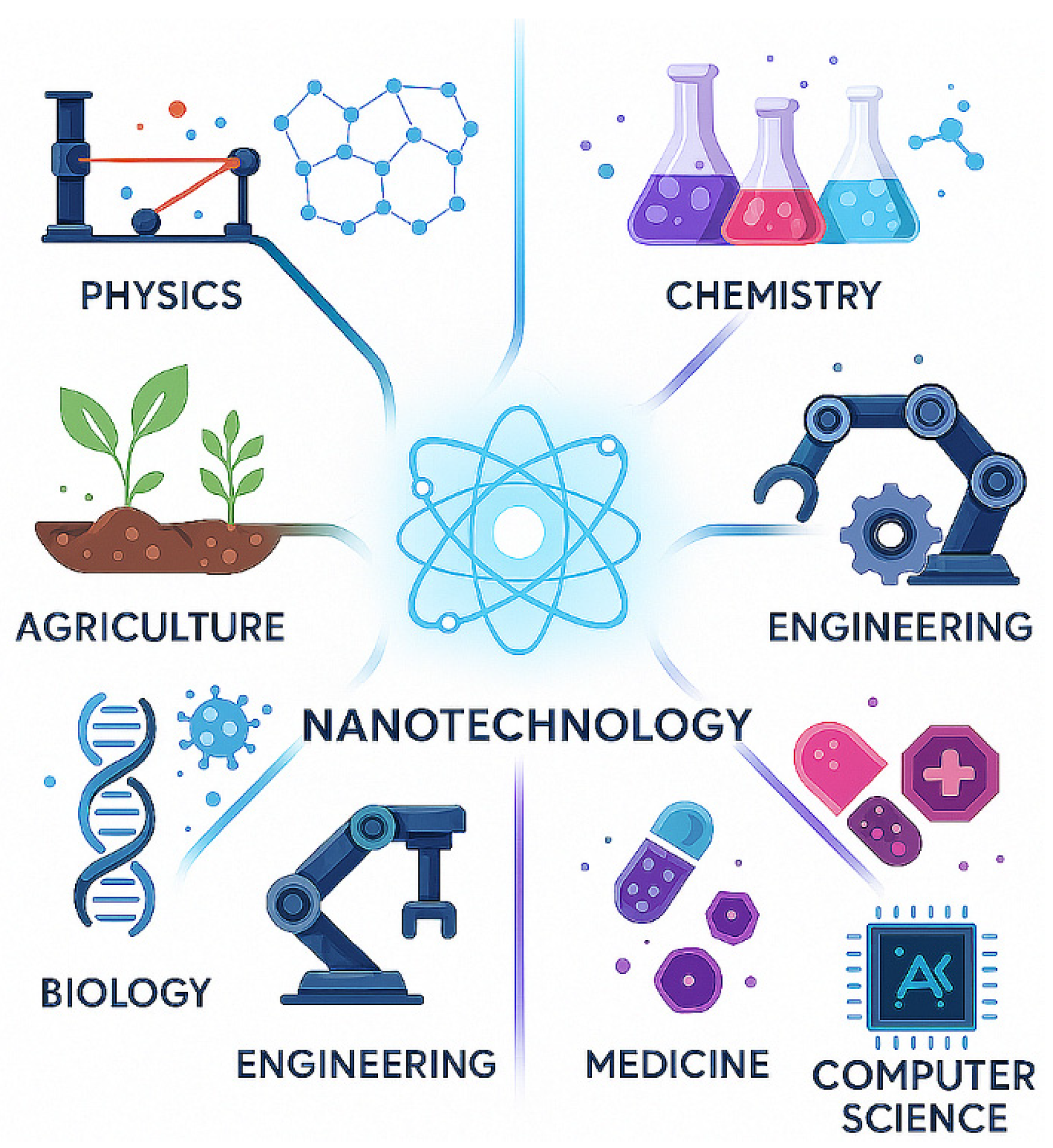
Figure 2.
Trend analysis of synthesis techniques used for metallic nanoparticles from 2000 to min 2025.
Figure 2.
Trend analysis of synthesis techniques used for metallic nanoparticles from 2000 to min 2025.
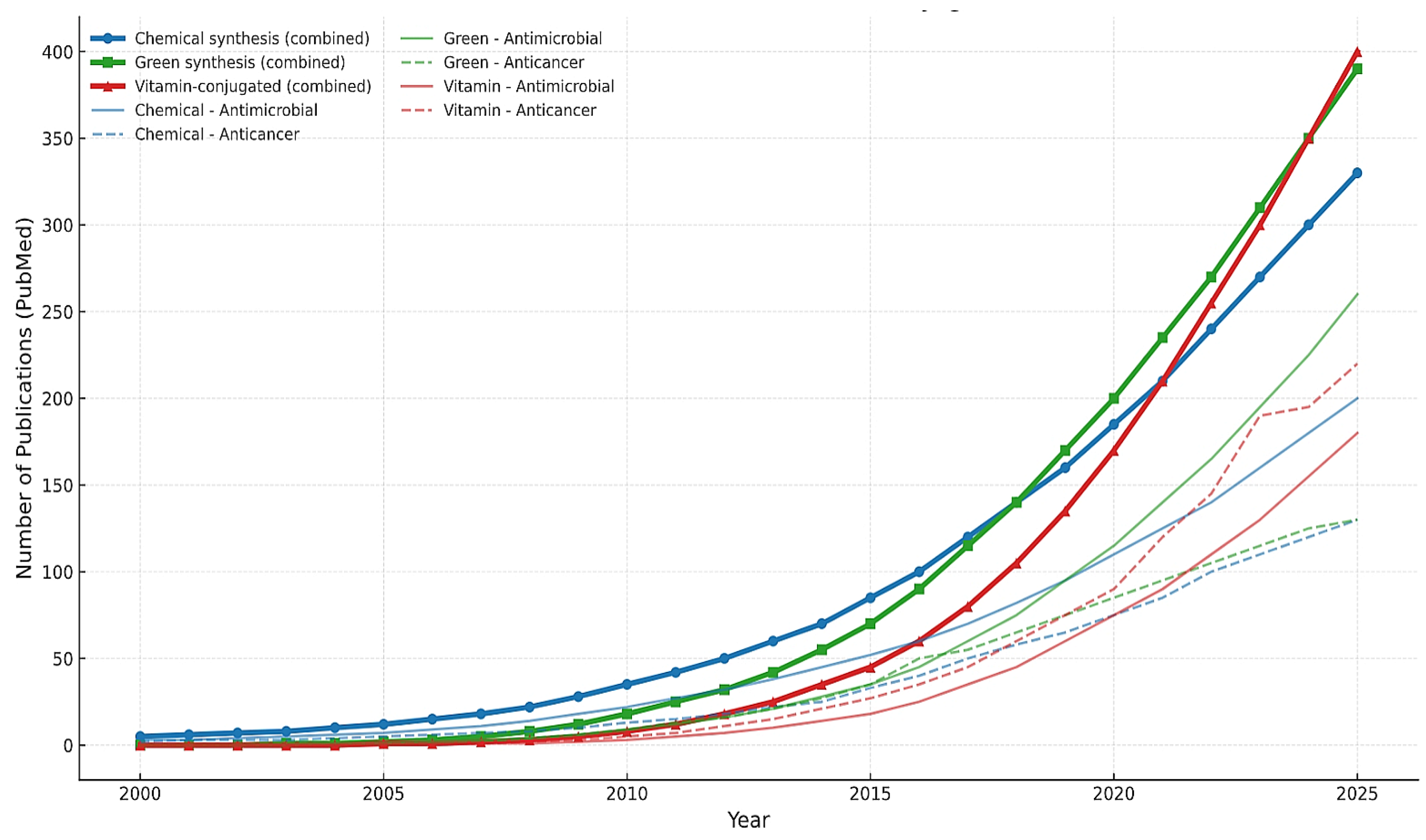
Figure 3.
A diagram showing antimicrobial mechanism of action.
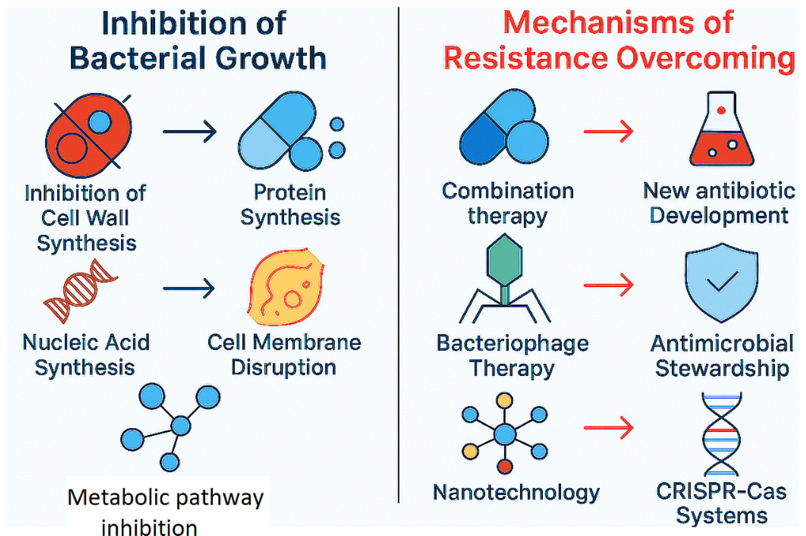
Disclaimer/Publisher’s Note: The statements, opinions and data contained in all publications are solely those of the individual author(s) and contributor(s) and not of MDPI and/or the editor(s). MDPI and/or the editor(s) disclaim responsibility for any injury to people or property resulting from any ideas, methods, instructions or products referred to in the content. |
© 2025 by the authors. Licensee MDPI, Basel, Switzerland. This article is an open access article distributed under the terms and conditions of the Creative Commons Attribution (CC BY) license (http://creativecommons.org/licenses/by/4.0/).
Copyright: This open access article is published under a Creative Commons CC BY 4.0 license, which permit the free download, distribution, and reuse, provided that the author and preprint are cited in any reuse.


