Preprint
Review

This version is not peer-reviewed.

The Causal Role of Bile Acids in Cancers of the Digestive System

A peer-reviewed article of this preprint also exists.

Submitted:

17 September 2025

Posted:

18 September 2025

You are already at the latest version

Abstract

Bile acids are widely distributed in the human gastrointestinal tract. A literature review indicates a role for bile acids in initiating cancers in every organ of the digestive system. The estimated number of new digestive system cancers world-wide in 2022 was about 5 million. For esophageal adenocarcinoma, stomach cancer and small intestine cancer, bile reflux plays a significant role in carcinogenesis. For colon cancer, secondary bile acids produced in response to a high fat diet disrupt colonic epithelial cell mitochondrial membranes. This disruption leads to release of oxidative free radicals that damage DNA and can cause carcinogenic mutations. Cancers of the pancreas, liver, and biliary tract can be caused by constriction of the common bile duct leading to reflux of bile acids back into these organs. Gastroesophageal reflux involving bile acids may also contribute to hypopharyngeal squamous cell carcinogenesis. Thus, bile acids are a likely major contributory cause of cancer throughout the digestive tract.

Keywords: 
;  ;  ;  

1. Introduction

Bile acids act as emulsifiers during digestion and absorption of dietary lipids and are also major signaling molecules [1]. However, high levels of bile acids cause the disruption of the mitochondrial outer cell membrane [2], causing generation of excessive reactive oxygen species, resulting in induction of DNA damage, mutations, and apoptosis [3]. As shown by O'Keefe et al. [4], high concentrations of bile acids are detected in the feces of volunteers consuming a high-fat diet.

2. Background

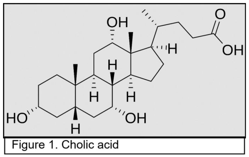
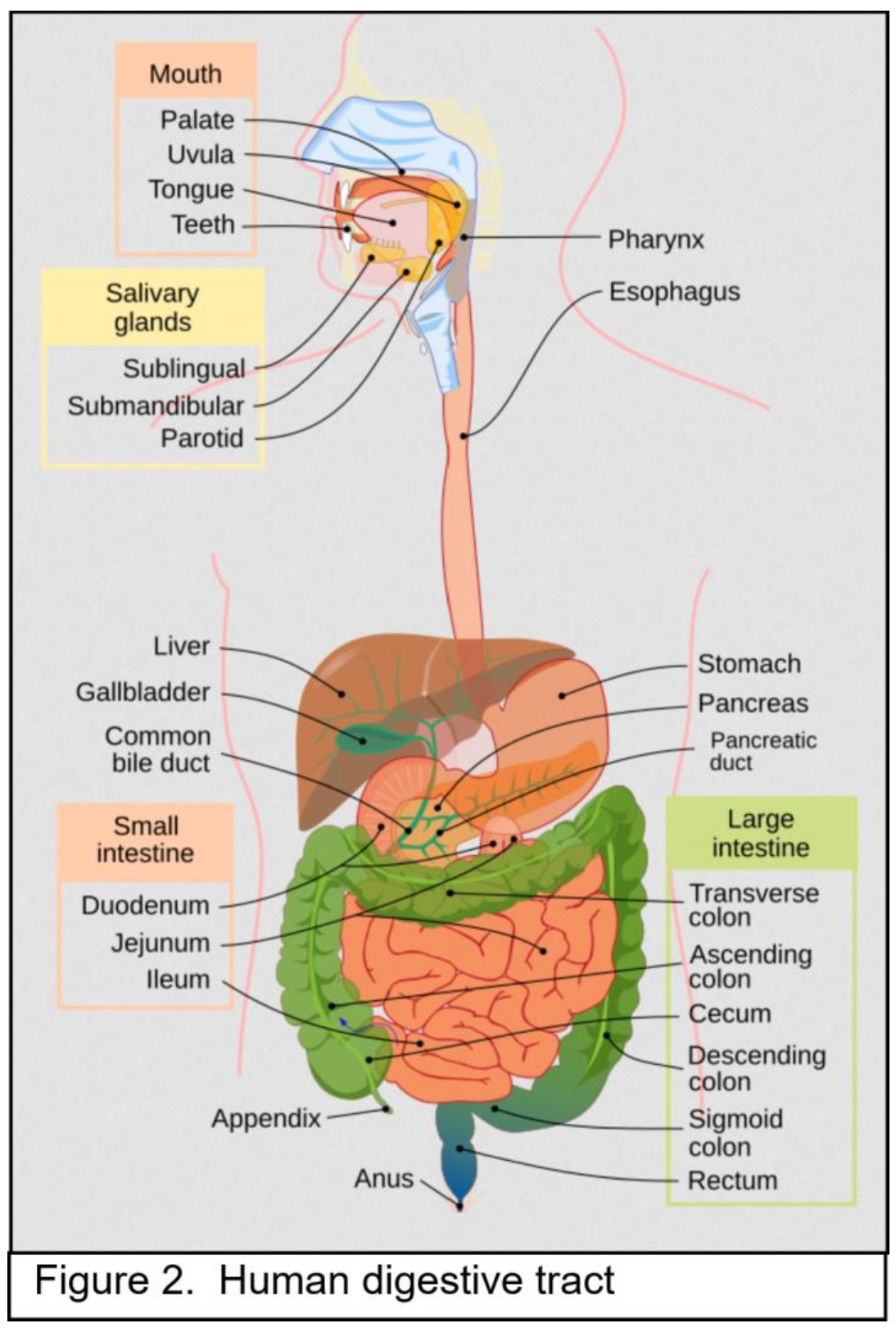
2.1. Bile Acids in the Gastrointestinal System

Bile acids are made by hepatocytes, which constitute 80% of the cells in the liver [5]. Hepatocytes act on cholesterol to create the primary bile acids, cholic acid and chenodeoxycholic acid [6]. Cholic acid and chenodeoxycholic acid are steroid molecules (steroids have three of six member rings and one five member ring [7]) (see Figure 1). The primary bile acids are conjugated with taurine or glycine which increases their solubility in an aqueous medium [8]. After conjugation, the primary bile acids have a hydrophilic side and a hydrophobic side, so they can adsorb to lipids on one side and use their other side to stay soluble in aqueous medium during digestion [8]. This promotes digestion of lipids.
In the hepatocyte, bile acids are at a concentration of 1-2 µM [8]. They are rapidly transported into the biliary canaliculus to a concentration of 1,000 µM (1 mM) and from there into the gall bladder where they become concentrated to 15.1–272.8 mM [9]. Bile acids are exported from the gall bladder into the duodenum of the small intestine (see Figure 2) in response to a fatty meal. The bile acids adsorb to the lipids in the food, allowing the lipids to be taken up by the small intestine, and then about 95% of the bile acids are absorbed in the ileum, the lower small intestine (see Figure 2). From the ileum, the reabsorbed bile acids are sent to the portal vein and passed back into the liver. When fasting, bile acids in the portal vein are at about 14 µM [10]. About 5% of the bile acids in the ileum are not absorbed and are passed through to the colon. The resulting level of bile acids in colonic contents is 0.6 mM (600 Mm)[11]. Bile acids in the colon undergo bacterial transformation, primarily through removal of the conjugated taurine or glycine (deconjugation) and dehydroxylation, leading to the formation of the secondary bile acids. Cholic acid is transformed into deoxycholic acid and chenodeoxycholic acid becomes lithocholic acid. Deoxycholic acid is reabsorbed in the colon and sent to the portal vein and from there into the liver, but lithocholic acid is largely removed through the feces. Because the bile acid pool is recirculated an average of six to eight times a day, and each time cholic acid is converted into deoxycholic acid, the final bile acid pool consists of about 40-50% cholic acid, 30-40% chenodeoxycholic acid and 20% deoxycholic acid [12,13].
Preprints 177176 i001
Preprints 177176 i002
The conjugated bile salts in the gall bladder are in micelles along with lecithin, and the micelles carry cholesterol with them. The amount of bile salts (in micelles) that are released into the small intestinal duodenum depends on the level of fat in a meal [14]. For locations of gall bladder, duodenum, etc., see Figure 2.
The “bile acid pool” is composed of all of the bile acids in circulation in the enterohepatic circulation. This includes the bile acids formed in the liver (<1%), those in the intestine (small intestine and colon) (∼85%–90%), and in the gallbladder (∼10%–15%) [12].
Bile acids are also spilled over from the portal vein to the systemic circulation [12]. Fasting serum bile acid levels in the systemic circulation are about 2.4 µM [10,15]. After a meal, within 15 – 60 minutes, serum bile acids increase in concentration to 5.2 µM. Similarly, after a meal, the concentration of bile acids in the portal vein (through which bile acids taken up from the ileum and colon are returned to the liver) increases from 14 µM to a median of 43 µM [10].

2.2. Elevated Bile Acids Cause Oxidative DNA Damages

The plasma membranes of cells are hardly affected by any of the bile acids, while the mitochondrial outer membrane structure is specifically modified by the cytotoxic secondary bile acid deoxycholic acid [2]. This apparently leads to release of reactive oxygen species [16]. At least 15 studies showed that bile acids caused DNA damage, likely through actions of reactive oxygen species [17].
Figure 3 panel B directly shows oxidative DNA damage (large presence of oxidized guanine: 8-OHdG) within the cellular nuclei of the colonic epithelium of a mouse fed for 6 months on a diet supplemented with deoxycholate (deoxycholate is the salt of deoxycholic acid). On a normal diet (panel A) there is little to no oxidized guanine in the nuclei of cells in the colonic epithelium. The mouse fed a diet supplemented with deoxycholate had colonic deoxycholate at a level similar to that of a human when the human is on a relatively high fat diet [18].
Preprints 177176 i003
In general, the carcinogenicity of bile acids appears to be related to their detergent action on mitochondrial membranes [2]. This detergent action leads to disruption of the electron transport system employed in oxidative phosphorylation in the mitochondria and the consequent release of highly reactive oxidative free radical intermediates [16]. These oxidative free radicals are extremely damaging to DNA. Thus cells that are excessively exposed to bile acids will experience elevated DNA damage, and this results in replication errors leading to mutations, some of which are carcinogenic.

2.3. Frequencies of Incidence and Mortality Due to Gastrointestinal System Cancers

In the US, the estimated number of new digestive system cancer cases in 2024 for both sexes was 353,820, and estimated deaths was 174,320 [19]. Digestive system cancers may occur in any part of the digestive tract including the esophagus, the stomach, the small intestine, the colon and rectum, the liver, the gallbladder, the biliary tract and the pancreas [20]. Cancers of the digestive system accounted for about 18% of all US cancer cases arising in 2024 and about 28% of all cancer deaths [19]. World-wide, the estimated number of new cases and deaths for cancers of the colorectum, stomach, liver, esophagus and pancreas in 2022 was 4,957,675 new cases and 3,223,794 deaths [21]. Table 1 shows the incidences per year of digestive tract cancers in the US and the world.

3. Roles of Elevated Bile Acids in Gastrointestinal System Cancers

In the following sections the evidence for a role of bile acids in cancers of each of the organs of the digestive system is described.

3.1. Esophageal Cancer

Barrett's esophagus is a precancerous condition in which the lining of the esophagus (see Figure 2) is damaged by acid reflux. This causes the esophageal cells to change from normal squamous epithelium (flat, scale-like cells) to columnar epithelium (tall, column-shaped cells) embedded with goblet cells (cup-shaped cells). Esophageal adencarcinoma arising from Barrett’s esophagus metaplasia is linked to reflux esophagitis (increased bile acids in the esophagus due to reflux) [22]. In 2024 in the US there were an estimated 22,370 new cases of cancer of the esophagus and 15,130 deaths [19]. World-wide in 2022, the estimated number of new cases and deaths for cancers of the esophagus was 510,716 new cases and 445,129 deaths [21].
About 10% of Barrett’s esophagus patients currently progress to cancer [22]. Barrett’s esophagus has been attributed mainly to gastroesophageal reflux disease leading to chronic inflammation of the esophagus [23].
Bile acids are present in gastroesophageal reflux into the esophagus and are implicated in neoplastic development in the esophagus [24]. Bile reflux contains unconjugated bile acids that include chenodeoxycholic acid and deoxycholic acid, likely contributing to development of esophageal adenocarcinoma by inducing oxidative stress and DNA damage [25]. The secondary bile acid deoxycholic acid at doses of 100 μM and higher induces DNA damage in esophageal cells by a mechanism involving reactive oxygen species [22]. Secondary bile acids are potent DNA damaging agents because they disrupt mitochondrial membranes leading to the release of DNA damaging reactive oxygen species into the cytosol [24].
In a transgenic mouse model, the progression of Barrett’s esophagus to esophageal adenocarcinoma was accelerated by exposure to bile acids [23].

3.2. Stomach Cancer

In 2024 in the US the incidence of stomach cancer was 26,890 and there were 19,880 deaths [19]. World-wide in 2022 the estimated number of new cases and deaths for cancers of the stomach was 968,50 new cases and 659,853 deaths [21].
In addition to Helicobacter pylori infection, bile reflux is a likely factor in causing gastric intestinal metaplasia, which is a precancerous lesion leading to gastric cancer [26,27]. Dysregulation of bile acids and gut bacteria, along with the resulting impairment of the gastric mucosa appear to be central factors in the pathogenesis of precancerous lesions [28]. Bile reflux alters the composition of the gastric bacteria [29]. Conjugated bile acids are increased in the stomach of people with bile reflux gastritis and gastric cancer [30]. Irritation of the gastric mucosa over a long period by bile reflux likely has a role in gastric carcinogenesis [31]. Bile acids, a component of bile reflux, appear to be a causal factor in gastric carcinogenesis [31]. Altered bile acid metabolism (particularly upregulation of the bile acid deoxycholic acid) when linked to iron deficiency promotes Helicobacter pylori-induced inflammation-driven gastric carcinogenesis [32,33]. In a National Retrospective Cohort Analysis, it was found that the use of bile acid sequestrant modification is protective against cardia (upper part of stomach) and non-cardia (main body of stomach) gastric cancer [34].
A carcinogenic interaction was demonstrated between bile acids and Helicobacter pylori in a mouse model of gastric cancer [32,33].

3.3. Small Intestine Cancer

The length of the small intestine is about 75 percent of the total length of the gastrointestinal tract [35]. About 50% of the adenomas occurring in the small intestine arise in the duodenum even though this location comprises only 4 percent of the length of the small intestine. Such adenomas arise mainly close to the ampulla of Vater. The ampulla of Vater is the outlet of the common bile duct where bile acids are released into the small intestine [36]. In 2024 in the US there were an estimated 12,440 new cases of cancer of the small intestine and 2,090 deaths [19].
A prospective study involving human subjects observed a markedly elevated risk for carcinoid tumors of the small intestine associated with dietary intake of saturated fat [37]. Such a diet is associated with increased bile acid exposure
Cholecystectomy, a surgical procedure to remove the gallbladder, alters the flow of bile to the small intestine and increases the risk of small intestinal adenocarcimoma [38]. This risk declines with increasing distance from the common bile duct.

3.4. Colon and Rectum Cancer

The International Agency for Research on Cancer (IARC) has classified processed meat as a group I carcinogen. This classification is based on the IARC finding of sufficient evidence that consumption of processed meat by humans causes colorectal cancer [39,40,41]). Processed meat is generally high in fat. In 2024 in the US there were an estimated 152,810 new cases of cancer of the colon and rectum and 53,010 deaths [19]. World-wide in 2022, the estimated number of new cases and deaths for cancers of the colorectum was 1,926,116 new cases and 903,859 deaths [21].
A high fat diet is associated with high levels of bile acids that appear to increase the risk of colorectal cancer [4,20,42]. In particular, the bile acid deoxycholic acid is increased in the colonic contents of humans on a high fat diet [20,42]. Also, fecal concentrations of bile acids, particularly deoxycholic acid, are higher in populations that have a high incidence of colorectal cancer [20,42]. Bacteria such as Fusobacterium nucleatum, Streptococcus bovis, Helicobacter, pylori, Bacteriodes fragilis and Clostridium septicum produce secondary bile acids from primary bile salts and these may cause inflammation and DNA damage leading to colorectal carcinogenesis [43].
A meta-analysis on the relationship of fecal bile acid concentrations to the development and progression of colorectal cancer showed that high fecal concentrations of the bile acids cholic acid and chenodeoxycholic acid are associated with an increased risk and increased incidence of colorectal cancer [44].
In one prospective study, baseline serum was collected 30 years prior to diagnoses of colorectal cancer [45]. It was found that, among women, there was a strong association between serum concentrations of bile acids (collected and analysed 30 years previously) and increased colorectal cancer risk [45]. In a separate study, fecal concentrations of deoxycholic acid were dramatically increased after cholic acid administration suggesting that the interaction between bile acids and the gut microbiota affect colon cancer progression [46]. In another prospective study it was found that prediagnostic plasma levels of certain conjugated primary and secondary bile acids (e.g. glycocholic acid, taurocholic acid, glycochenodeoxycholic acid, taurochenodeoxycholic acid, glycodeoxycholic acid, taurodeoxycholic acid) are positively associated with colon cancer risk [47].

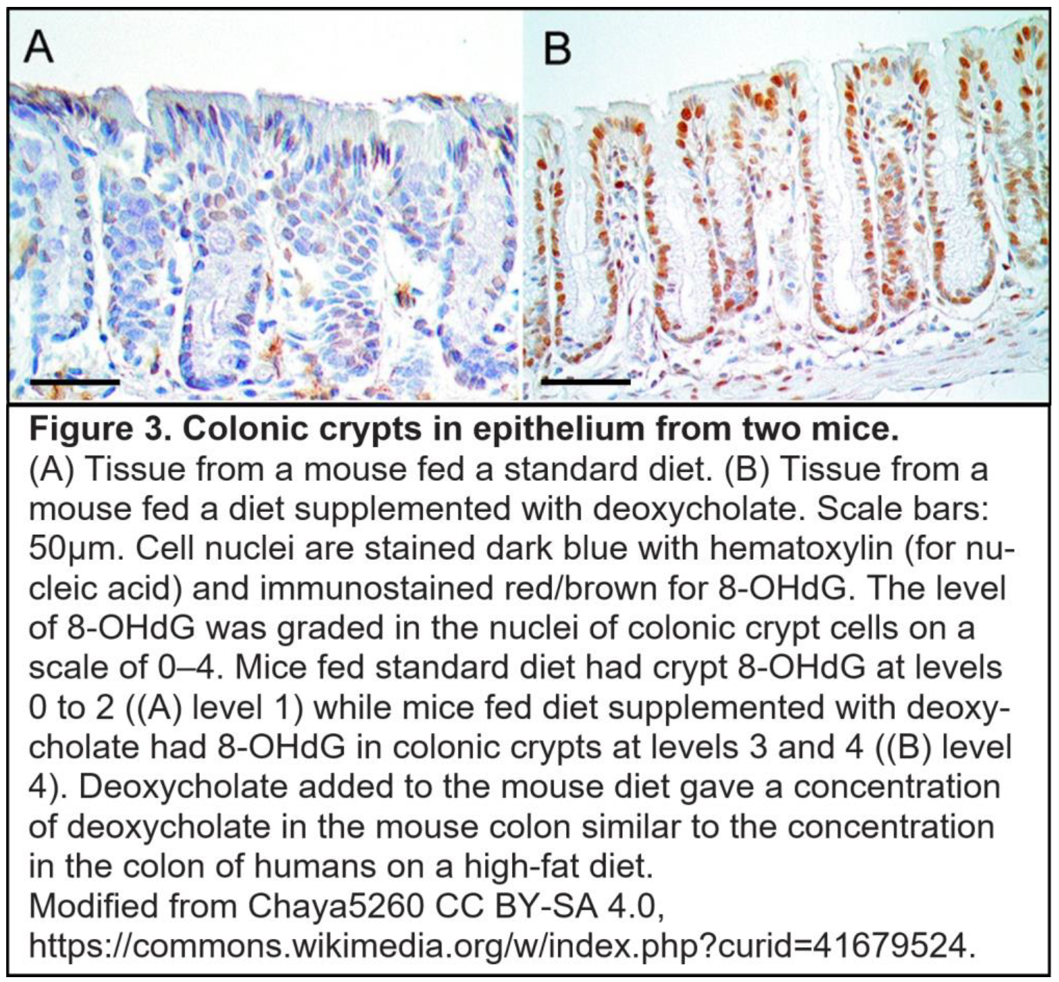
3.4.1. A Bile Acid Based Mouse Model of Colon Cancer

A diet related mouse model of colon cancer was devised that involved feeding wild-type mice a standard diet supplemented with deoxycholate. This produced a level of deoxycholate in the mouse colon comparable to that in the colon of humans eating a high fat diet [18,48](Bernstein et al., 2011; Prasad et al., 2014). Eight to ten months after initiating the diet, 45 to 56 percent of the mice developed colonic adenocarcinomas.
On the basis of histopathologic examination and the expression of specific markers, the colonic tumors in the mice were essentially identical to those in humans [18]. In humans, defective fields that surround colon cancers and the colon cancers themselves exhibit characteristic aberrant changes in molecular markers. The colonic tissues of the mice fed a diet containing deoxycholic acid showed similar changes in biomarkers. For instance, 8-OHdG in DNA was increased (see Figure 3, above), the DNA repair protein ERCC1 was decreased, the autophagy-related protein beclin-1 was increased, and, in the stem cell region at the base of crypts, there was a substantial nuclear localization of beta-catenin as well as an increase in cytoplasmic beta-catenin [18]. Aberrant activation of the Wnt/β-catenin signaling pathway leads to the accumulation of β-catenin in the nucleus, where it acts as an oncogenic transcription factor [49].

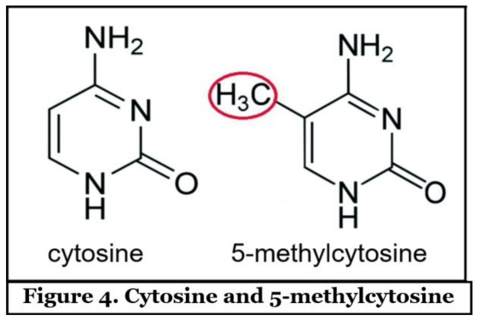
3.4.2. Epigenetic Alterations in Colon Cancer are More Frequent than Mutations

As described by Vogelstein et al., [50] an average colon cancer has 1 or 2 mutations in oncogenes and 1 to 5 mutations in tumor suppressor genes (together called "driver mutations"), and about 60 "passenger" mutations.
Epigenetic alterations, as distinct from mutations, change the protein expression of genes without changing the DNA sequence [51]. Epigenetic alterations are much more frequent in colon cancer than genetic (mutational) alterations.
In two studies, about 600 increased methylations of cytosines (see Figure 4) and about 300 decreased methylations of cytosines occurred in promoters of genes in colon cancers [52,53]. Decreased methylation of cytosine within the promoter of a gene is an epigenetic alteration that increases expression of the gene [54]. Methylation of cytosines in DNA can be reduced by increased presence of reactive oxygen species [55]. Bile acids at higher levels can cause increased release of reactive oxygen species from the mitochondria [16], thereby likely increasing epigenetic upregulated expression of some genes.
Preprints 177176 i004
A second type of epigenetic alteration is the increased/decreased production of particular microRNAs (miRNAs). Each miRNA typically regulates the expression of several hundred genes [56]. The miRNAs either cleave their target messenger RNAs (mRNAs) or they attach to their target mRNAs and prevent them from coding for proteins [57]. 164 miRNAs were found to be significantly altered in various colorectal cancers. About 2/3 of miRNAs were increased while about 1/3 were reduced [58]. Methylation alterations of cytosines within the promoters of genes with embedded miRNAs is one of the mechanisms for changing miRNA expression [59]. Oxidative damage to a guanine (caused by high levels of bile acids) can cause the demethylation of an adjacent cytosine 55], and this may be within promoters that control miRNAs, possibly causing the upregulation of the affected miRNA.

3.5. Liver Cancer (Hepatocellular Carcinoma)

Hepatocellular carcinoma is an aggressive and prevalent liver malignancy. In 2024 in the US there were an estimated 41,630 new cases of cancer of the liver and intrahepatic bile duct and 29,840 deaths [19]. World-wide in 2022, the estimated number of new cases and deaths for cancers of the liver was 865,269 new cases and 757,984 deaths [21].
Several recent studies have presented evidence that bile acids have an etiologic role in hepatocellular carcinoma [61,62,63]. Cholestasis is a condition where the flow of bile from the liver is reduced or blocked. A central factor in the pathogenesis of cholestasis-induced liver injury is hepatic accumulation of bile acids, and excessive cytotoxic bile acids in the liver may lead to liver fibrosis and cirrhosis, and then progression to liver cancer [60]. In a prospective study, it was found that risk of developing hepatocellular carcinoma is associated with higher levels of the major circulating bile acids that were measured several years prior to tumor diagnosis [64]. Another large prospective cohort study found that serum levels of total bile acids are associated with an increased risk of hepatocellular carcinoma in patients with cirrhosis [63]. Also evidence was reviewed indicating that alteration in bile acids by the human gut microbiome contribute to hepatocarcinogenesis [65]. The microbiota in the gut that contribute to hepatocellular carcinoma seem to be distinct from those that cause colorectal cancer [66]. The homeostasis of bile acids is especially disturbed in individuals with a history of alcohol intake [67]. Alcohol appears to trigger bile acid disequilibrium to initiate and promote hepatocellular carcinoma progression [67].
Using a mouse model, it was found that dysregulated hepatic bile acids collaboratively promote liver carcinogenesis [68].

3.6. Gallbladder Removal (Cholecystectomy) and Cancer

In vertebrates, the gallbladder is a small organ where bile is stored and concentrated prior to being released into the small intestine after a high fat meal. Bile is produced in the liver and transferred to the gallbladder via the common hepatic duct. The gall bladder can then release bile via the common bile duct (shown in Figure 2) into the duodenum, where it assists in the digestion of fats.
The major role of the gallbladder in humans is to protect cells of the gastrointestinal tract from the cytotoxic effects of some of the bile acids [69].
Thus, in a normal healthy individual, bile acids are primarily released from the gallbladder into the gastrointestinal tract in response to a fatty meal. The bile acids are passed down the gastrointestinal tract in a short period during digestion. The primary bile acids (cholic acid and chenodeoxycholic acid) and the secondary bile acids (deoxycholic acid and lithocholic acid) do not cause an increase in cell death if the exposure to each of the four bile acids is 2 hours and the level of exposure is less than 200 μM [70]. Bile acids may be at less than 200 μM when mixed with food during digestion.
However, if the gallbladder is removed (“cholecystectomy,” often due to gallstones), then the gastrointestinal tract is more continuously bathed in bile acids. Under continuous exposure, cholic acid is not cytotoxic, even at high levels of 500 μM. But, under continuous exposure, chenodeoxycholic acid, deoxycholic acid and lithocholic acid are cytotoxic at almost any level, with increasing cytoxicity at increasing levels of exposure [70]. Lithocholic acid, however, is not a major factor in toxicity since it is excreted in the feces. But if multiple fatty meals are eaten, then more bile acids are circulated through the colon, where non-toxic cholic acid is converted by bacterial action to cytotoxic deoxycholic acid [71]. Also, higher levels of circulation of the bile acid pool occurs after cholecystectomy [72]. Then, as seen in Table 2 (adapted from [13]), with cholecystectomy, cholic acid is reduced and replaced with the cytotoxic deoxycholic acid.
Cytotoxic deoxycholic acid concentration is doubled in the bile acid pool in patients with a cholecystectomy, compared to deoxycholic acid concentration in healthy subjects (Table 2) (Hepner et al., 1974).
Cholecystectomy is associated with increases in at least four different gastrointestinal cancers. (1) Cholecystectomy was significantly associated with increased right-sided colon cancer, especially in the cecum, the ascending colon and/or the hepatic flexure, but not in the transverse, descending, or sigmoid colon [73]. (2) Similarly, from five pooled studies, there is a 15% increased risk of gastric cancer after cholecystectomy [74]. (3) Again, in an examination of the records of 278,460 cholecystectomized patients in Sweden, cholecystectomy increased the risk of small intestinal cancer. The relative risk was 1.77 compared to individuals without cholecystectomy. The risk decreased with distance from the common bile duct [38]. (4) Among 42,098 patients in Denmark who had a cholecystectomy, there was a relative risk of pancreatic cancer of 1.42 [75].
As pointed out by [12], there is spillover of bile acids into the serum. With a cholecystectomy there would be relatively higher levels of the cytotoxic deoxycholic acid in the serum. The deoxycholic acid would be carried in the blood circulation to many organs of the body. Choi et al. [76] examined the data from 123,295 individuals who had a cholecystectomy and a matched set of 123,295 individuals who did not have a cholecystectomy in the database of the Korean National Health Insurance Service. They evaluated the records for occurrence of any of 23 cancers in a followup period of an average of 4.6 years. Those who had a cholecystectomy had an increased risk of a subsequent cancer in leukemia, colon, liver, pancreas, biliary tract, thyroid, pharynx and oral cavity.

3.7. Gallbladder and Biliary Tract Cancer

In 2024 in the US there were an estimated 12,350 new cases of cancer of the gallbladder and other biliary structures and 4,530 deaths [19].
Cholangiocarcinoma (bile duct cancer) is a type of cancer that is comprised of mutated epithelial cells that originate in the bile duct. The activities of gut microbiota and bile acids are linked and have crucial roles in the pathogenesis and progression of cholangiocarcinoma [77]. Primary sclerosing cholangitis is a chronic liver disease where bile ducts, both inside and outside the liver, become inflamed and scarred, leading to narrowing or blockage. Primary biliary cholangitis is a chronic autoimmune disease where the body's immune system mistakenly attacks and destroys the bile ducts in the liver. Progression to bile duct cancer is promoted both by primary sclerosing cholangitis and primary biliary cholangitis, both of which are associated with cholestasis where the flow of bile from the liver is reduced or blocked [77]. The tumors in about 25% of patients with biliary tract cancer have some form of deficiency in DNA damage repair [78].
Normal function of the gallbladder ordinarily protects against carcinogenesis. This protective function is indicated by observations that removal of the gallbladder (cholecystectomy) causes an increased subsequent risk of cancer. A systematic review and meta analysis of eighteen studies concluded that cholecystectomy had a harmful effect on the risk of right-sided colon cancer [73]. An additional recent study reported that subsequent to a cholecystectomy there is a significantly increased total cancer risk [76].
Bile salts from the cytoplasm of hepatocytes are transferred into the bile canaliculi by the bile salt export pump. When bile salt export is deficient due to a mutation in the gene ABCB11 (ATP-binding cassette, subfamily B member 11) that encodes the bile export pump, the result can be intrahepatic toxic accumulation of bile salts. Individuals who have such mutations experience an increased incidence of hepatocellular carcinoma or cholaniocarcinoma (bile duct cancer) [79].

3.8. Pancreatic Cancer

In 2024 in the US there were an estimated 66,440 new pancreas cancer cases and 51,750 deaths [19]. World-wide in 2022, the estimated number of new cases and deaths for cancers of the pancreas was 510,566 new cases and 467,005 deaths [21].The most common form of pancreatic cancer is pancreatic ductal adenocarcinoma [80]. Bile acids are strongly implicated in the growth and progression of pancreatic cancer [80].
Patients with pancreatic cancer often also have biliary obstruction and elevated bile acids caused by tumors [81]. Sixty percent of the cases arise in the pancreatic head, 15% in the body or tail, and 20% involve the gland diffusely [82]. Bile acid reflux caused by biliary obstruction appears to affect the epithelial cells or acinars from which pancreatic adenocarcinoma is derived [83]. When the composition of bile acids in bile (extracted from the common bile duct) of patients with adenocarcinoma of the pancreas was compared with the composition of bile acids from patients with benign disease, it was found that the concentration of the unconjugated bile acid cholic acid in the malignant group was significantly higher than in the benign group [84]. Dysregulation of the interaction of bile acids with gut microbiota appears to play a role in pancreatic cancer progression [85]. Bile acids appear to promote the carcinogenic process in pancreatic ductal adenocarcinoma cells by a process in which increased expression of the MUC4 protein plays an important role [86]. MUC4 protein is a major constituent of mucus, the viscous secretion that covers epithelial surfaces. The majority of the risk factors for developing pancreatic cancer including smoking, a high fat diet and alcohol intake leading to high bile acid secretion [83]
In hamsters, bile-reflux into the pancreatic ducts is observed to be associated with intraductal papillary carcinoma development [87].

3.9. Oral Cavity and Pharynx

Although often treated separately from the “digestive system”, the oral cavity and pharynx are also subject to bile acid induced cancer. In the US, the estimated number of new cancer cases in 2024 at sites in the oral cavity and pharynx for both sexes was 58,450 and estimated deaths was 12,230 [19]. Oral cavity & pharynx cancers may occur in the tongue, mouth and pharynx. In 2024 these cancers accounted for about 2.9% of all US cancer cases and about 2.1% of all cancer deaths [88].
Bile acids appear to be a significant causal factor in human hypopharyngeal squamous cell carcinoma [89]. In gastroesophageal refluxate, bile acids are often present and as a consequence can cause inflammatory and neoplastic alteration in the upper aerodigestive tract (Vageli et al.,2021). The STAT3 protein appears to have a significant role in bile reflux-induced molecular events linked to hypoparyngeal carcinogenesis [90]. STAT3 protein is a transcription factor that can bind to DNA and influence gene transcription and gene expression in such cellular processes as cell growth, differentiation and survival.
In a mouse study, it was found that acidic bile refluxate produces oxidative DNA damage, which can lead to progressive mutagenic effects and hypopharyngeal squamous cell carcinogenesis [89].

4. Bile Acids May Cause Field Defects Leading to Gastrointestinal Cancers

A field of defective tissue nearby a tumor may represent a pre-malignant stage in progression to the cancer. Field defects are commonly found in cancers of the gastrointestinal tract including cancers of the oropharynx, esophagus, stomach, bile duct, pancreas, small intestine and colon/recum [92]. Common characteristics of these field defects are reduced apoptosis competence, aberrant proliferation, and genome instability. Such characteristics are often associated with high bile acid exposure, and this exposure may explain the association of dietary-related factors with cancer progression [92](Bernstein et al., 2008).

5. Discussion

Although bile acids do not directly damage DNA, we indicated in the Introduction that bile acids are a likely major indirect source of DNA damage in the cell by disrupting mitochondrial membranes leading to the release of oxidative free radicals that are highly DNA damaging [2]. A comparative and comprehensive study was conducted on the impact of cytoprotective and cytotoxic bile acids on membrane structure of individual cellular compartments [2]. It was found that the outer membranes of mitochondria are the likely main target of the cytotoxic bile acid deoxycholic acid. The cell’s mitochondrial membranes were found to be more sensitive to deoxycholic acid induced structural changes than the cell’s plasma membrane. The disruption of mitochondrial outer membranes induced by bile acids causes the release of reactive oxygen species that may damage the cells DNA [93].
Bile acids serve an important nutritional function, namely in the utilization of dietary fat as a significant food source. However, the evidence reviewed above indicates that when exposure of the digestive tract to bile acids is excessive over extended periods it can cause DNA damage leading to cancer.
A basic idea in biology is that the level of expression of certain genetically determined capabilities that maintain vigor in youth may be insufficient to maintain vigor with aging. The term used to express this idea is antagonistic pleotropy [94]. One specific example of such a pleiotropic effect is that in humans the ability to carry out DNA repair is maintained at a level sufficient to promote healthy development in youth, but insufficient to maintain vigor in older individuals [95]. As another example, a high fat diet may be beneficial as a high calorie source of nutrition in youth, but may be disadvantageous in older individuals because of the cumulative DNA damage caused by the bile acids employed to digest the high fat diet [20].
For each of the organs of the digestive tract, we have summarized, above, evidence that elevated exposure to bile acids contributes to cancer development. In the particular case of colorectal cancer substantial evidence based on numerous studies supports a causal relation of bile acid activity to carcinogenesis. For the other organs of the digestive tract the evidence is also generally supportive of a significant role of bile acids in carcinogenesis. Thus we consider it reasonable to propose that bile acids, through their damaging effect on mitochondrial membranes resulting in the aberrant release of DNA damaging oxidative free radicals, is a major cause of cancer throughout the digestive system.

References

  1. Xie, C.; Huang, W.; Young, R.L.; Jones, K.L.; Horowitz, M.; Rayner, C.K.; Wu, T. Role of Bile Acids in the Regulation of Food Intake, and Their Dysregulation in Metabolic Disease. Nutrients 2021, 13, 1104. [Google Scholar] [CrossRef]
  2. Sousa, T.; Castro, R.E.; Pinto, S.N.; Coutinho, A.; Lucas, S.D.; Moreira, R.; Rodrigues, C.M.P.; Prieto, M.; Fernandes, F. Deoxycholic acid modulates cell death signaling through changes in mitochondrial membrane properties. J. Lipid Res. 2015, 56, 2158–2171. [Google Scholar] [CrossRef]
  3. Ajouz, H.; Mukherji, D.; Shamseddine, A. Secondary bile acids: an underrecognized cause of colon cancer. World J. Surg. Oncol. 2014, 12, 164–164. [Google Scholar] [CrossRef] [PubMed]
  4. O’Keefe, S.J.D.; Li, J.V.; Lahti, L.; Ou, J.; Carbonero, F.; Mohammed, K.; Posma, J.M.; Kinross, J.; Wahl, E.; Ruder, E.; et al. Fat, fibre and cancer risk in African Americans and rural Africans. Nat. Commun. 2015, 6, 6342. [Google Scholar] [CrossRef]
  5. Schulze, R.J.; Schott, M.B.; Casey, C.A.; Tuma, P.L.; McNiven, M.A. The cell biology of the hepatocyte: A membrane trafficking machine. J. Cell Biol. 2019, 218, 2096–2112. [Google Scholar] [CrossRef]
  6. Hundt M, Basit H, John S. Physiology, Bile Secretion. In: StatPearls. 2025. Available from: https://www.ncbi.nlm.nih. 13 September 4702.
  7. Chiang, J.Y. Regulation of bile acid synthesis: pathways, nuclear receptors, and mechanisms. J. Hepatol. 2004, 40, 539–551. [Google Scholar] [CrossRef]
  8. Hofmann, A.F. Bile Acids: The Good, the Bad, and the Ugly. Physiology 1999, 14, 24–29. [Google Scholar] [CrossRef] [PubMed]
  9. Shiffman ML, Sugerman HJ, Moore EW. Human gallbladder mucosal function. Effect of concentration and acidification of bile on cholesterol and calcium solubility. Gastroenterology. 1990;99(5):1452-9.
  10. Angelin B, Björkhem I, Einarsson K, Ewerth S. Hepatic uptake of bile acids in man. Fasting and postprandial concentrations of individual bile acids in portal venous and systemic blood serum. J Clin Invest. 1982;70(4):724-31.
  11. Hamilton, J.P.; Xie, G.; Raufman, J.-P.; Hogan, S.; Griffin, T.L.; Packard, C.A.; Chatfield, D.A.; Hagey, L.R.; Steinbach, J.H.; Hofmann, A.F. Human cecal bile acids: concentration and spectrum. Am. J. Physiol. Gastrointest. Liver Physiol. 2007, 293, G256–G263. [Google Scholar] [CrossRef] [PubMed]
  12. Chiang, J.Y.L.; Ferrell, J.M. Bile Acid Metabolism in Liver Pathobiology. Gene Expr. 2018, 18, 71–87. [Google Scholar] [CrossRef]
  13. Hepner GW, Hofmann AF, Malagelada JR, Szczepanik PA, Klein PD. Increased bacterial degradation of bile acids in cholecystectomized patients. Gastroenterology. 1974;66(4):556-64.
  14. Marciani, L.; Cox, E.F.; Hoad, C.L.; Totman, J.J.; Costigan, C.; Singh, G.; Shepherd, V.; Chalkley, L.; Robinson, M.; Ison, R.; et al. Effects of various food ingredients on gall bladder emptying. Eur. J. Clin. Nutr. 2013, 67, 1182–1187. [Google Scholar] [CrossRef]
  15. Patti, M.; Houten, S.M.; Bianco, A.C.; Bernier, R.; Larsen, P.R.; Holst, J.J.; Badman, M.K.; Maratos-Flier, E.; Mun, E.C.; Pihlajamaki, J.; et al. Serum Bile Acids Are Higher in Humans With Prior Gastric Bypass: Potential Contribution to Improved Glucose and Lipid Metabolism. Obesity 2009, 17, 1671–1677. [Google Scholar] [CrossRef]
  16. Payne, C.; Weber, C.; Crowley-Skillicorn, C.; Dvorak, K.; Bernstein, H.; Bernstein, C.; Holubec, H.; Dvorakova, B.; Garewal, H. Deoxycholate induces mitochondrial oxidative stress and activates NF- B through multiple mechanisms in HCT-116 colon epithelial cells. Carcinog. 2007, 28, 215–222. [Google Scholar] [CrossRef]
  17. Bernstein, H.; Bernstein, C.; Payne, C.M.; Dvorakova, K.; Garewal, H. Bile acids as carcinogens in human gastrointestinal cancers. Mutat. Res. Mutat. Res. 2005, 589, 47–65. [Google Scholar] [CrossRef]
  18. Prasad AR, Prasad S, Nguyen H, Facista A, Lewis C, Zaitlin B, et al. Novel diet-related mouse model of colon cancer parallels human colon cancer. World J Gastrointest Oncol. 2014;6(7):225-43.
  19. American Cancer Society. Cancer Facts & Figures 2024. Web site. https://www.cancer.org/research/cancer-facts-statistics/all-cancer-facts-figures/2024-cancer-facts-figures.html Accessed . 2025. 13 September.
  20. Bernstein, H.; Bernstein, C. Bile acids as carcinogens in the colon and at other sites in the gastrointestinal system. Exp. Biol. Med. 2022, 248, 79–89. [Google Scholar] [CrossRef]
  21. Bray, F.; Laversanne, M.; Sung, H.; Ferlay, J.; Siegel, R.L.; Soerjomataram, I.; Jemal, A. Global cancer statistics 2022: GLOBOCAN estimates of incidence and mortality worldwide for 36 cancers in 185 countries. CA: A Cancer J. Clin. 2024, 74, 229–263. [Google Scholar] [CrossRef] [PubMed]
  22. Jenkins, G.J.S.; Cronin, J.; Alhamdani, A.; Rawat, N.; D'SOuza, F.; Thomas, T.; Eltahir, Z.; Griffiths, A.P.; Baxter, J.N. The bile acid deoxycholic acid has a non-linear dose response for DNA damage and possibly NF- B activation in oesophageal cells, with a mechanism of action involving ROS. Mutagenesis 2008, 23, 399–405. [Google Scholar] [CrossRef]
  23. Quante, M.; Bhagat, G.; Abrams, J.A.; Marache, F.; Good, P.; Lee, M.D.; Lee, Y.; Friedman, R.; Asfaha, S.; Dubeykovskaya, Z.; et al. Bile Acid and Inflammation Activate Gastric Cardia Stem Cells in a Mouse Model of Barrett-Like Metaplasia. Cancer Cell 2012, 21, 36–51. [Google Scholar] [CrossRef]
  24. Cronin, J.; Williams, L.; McAdam, E.; Eltahir, Z.; Griffiths, P.; Baxter, J.; Jenkins, G. The role of secondary bile acids in neoplastic development in the oesophagus. Biochem. Soc. Trans. 2010, 38, 337–342. [Google Scholar] [CrossRef]
  25. Režen, T.; Rozman, D.; Kovács, T.; Kovács, P.; Sipos, A.; Bai, P.; Mikó, E. The role of bile acids in carcinogenesis. Cell. Mol. Life Sci. 2022, 79, 1–39. [Google Scholar] [CrossRef] [PubMed]
  26. Li, D.; Zhang, J.; Yao, W.Z.; Zhang, D.L.; Feng, C.C.; He, Q.; Lv, H.H.; Cao, Y.P.; Wang, J.; Qi, Y.; et al. The relationship between gastric cancer, its precancerous lesions and bile reflux: A retrospective study. J. Dig. Dis. 2020, 21, 222–229. [Google Scholar] [CrossRef] [PubMed]
  27. Qu, X.; Shi, Y. Bile reflux and bile acids in the progression of gastric intestinal metaplasia. Chin. Med J. 2022, 135, 1664–1672. [Google Scholar] [CrossRef]
  28. Zhang, M.; Zhong, J.; Shen, Y.; Song, Z. Crosstalk between bile acids and gut microbiota: a potential target for precancerous lesions of gastric cancer. Front. Pharmacol. 2025, 16, 1533141. [Google Scholar] [CrossRef]
  29. Huang, G.; Wang, S.; Wang, J.; Tian, L.; Yu, Y.; Zuo, X.; Li, Y. Bile reflux alters the profile of the gastric mucosa microbiota. Front. Cell. Infect. Microbiol. 2022, 12, 940687. [Google Scholar] [CrossRef]
  30. Wang, S.; Kuang, J.; Zhang, H.; Chen, W.; Zheng, X.; Wang, J.; Huang, F.; Ge, K.; Li, M.; Zhao, M.; et al. Bile Acid–Microbiome Interaction Promotes Gastric Carcinogenesis. Adv. Sci. 2022, 9, e2200263. [Google Scholar] [CrossRef] [PubMed]
  31. Lei, X.; Cui, Z.Y.; Huang, X.J. Exploration of gastric carcinogenesis from the relationship between bile acids and intestinal metaplasia and intragastric microorganisms (H. pylori and non-H. pylori). J. Cancer Res. Clin. Oncol. 2023, 149, 16947–16956. [Google Scholar] [CrossRef] [PubMed]
  32. Noto, J.M.; Piazuelo, M.B.; Shah, S.C.; Romero-Gallo, J.; Hart, J.L.; Di, C.; Carmichael, J.D.; Delgado, A.G.; Halvorson, A.E.; Greevy, R.A.; et al. Iron deficiency linked to altered bile acid metabolism promotes Helicobacter pylori–induced inflammation–driven gastric carcinogenesis. J. Clin. Investig. 2022, 132. [Google Scholar] [CrossRef] [PubMed]
  33. Alizadeh, M.; Raufman, J.-P. Gastrointestinal neoplasia: carcinogenic interaction between bile acids and Helicobacter pylori in the stomach. J. Clin. Investig. 2022, 132. [Google Scholar] [CrossRef]
  34. Canakis, A.; Lee, A.; Halvorson, A.E.; Noto, J.M.; Peek, R.M.; Wilson, O.; Hung, A.; Roumie, C.L.; Greevy, R.; Shah, S.C. Bile Acid Sequestrant Use and Gastric Cancer: A National Retrospective Cohort Analysis. Clin. Transl. Gastroenterol. 2023, 14, e00596. [Google Scholar] [CrossRef]
  35. Maguire, A.; Sheahan, K. Primary small bowel adenomas and adenocarcinomas-recent advances. Virchows Arch. 2018, 473, 265–273. [Google Scholar] [CrossRef]
  36. Ross, R.; Hartnett, N.; Bernstein, L.; Henderson, B. Epidemiology of adenocarcinomas of the small intestine: is bile a small bowel carcinogen? Br. J. Cancer 1991, 63, 143–145. [Google Scholar] [CrossRef]
  37. Cross, A.J.; Leitzmann, M.F.; Subar, A.F.; Thompson, F.E.; Hollenbeck, A.R.; Schatzkin, A. A Prospective Study of Meat and Fat Intake in Relation to Small Intestinal Cancer. Cancer Res. 2008, 68, 9274–9279. [Google Scholar] [CrossRef]
  38. Lagergren, J.; Ye, W.; Ekbom, A. Intestinal cancer after cholecystectomy: Is bile involved in carcinogenesis? Gastroenterology 2001, 121, 542–547. [Google Scholar] [CrossRef]
  39. IARC of World Health Organization. (2015, ). Cancer: Carcinogenicity of the consumption of red meat and processed meat (News release). https://www.who. 26 October 2025; 13.
  40. IARC Monographs evaluate consumption of red meat and processed meat (PDF). IARC. 26 October 2025.
  41. Chan, D.S.M.; Lau, R.; Aune, D.; Vieira, R.; Greenwood, D.C.; Kampman, E.; Norat, T. Red and Processed Meat and Colorectal Cancer Incidence: Meta-Analysis of Prospective Studies. PLoS ONE 2011, 6, e20456. [Google Scholar] [CrossRef]
  42. Fogelson, K.A.; Dorrestein, P.C.; Zarrinpar, A.; Knight, R. The Gut Microbial Bile Acid Modulation and Its Relevance to Digestive Health and Diseases. Gastroenterology 2023, 164, 1069–1085. [Google Scholar] [CrossRef]
  43. Dalal, N.; Jalandra, R.; Bayal, N.; Yadav, A.K.; Harshulika; Sharma, M. ; Makharia, G.K.; Kumar, P.; Singh, R.; Solanki, P.R.; et al. Gut microbiota-derived metabolites in CRC progression and causation. J. Cancer Res. Clin. Oncol. 2021, 147, 3141–3155. [Google Scholar] [CrossRef]
  44. Yang, S.; Wang, Y.; Sheng, L.; Cui, W.; Ma, C. The effect of fecal bile acids on the incidence and risk-stratification of colorectal cancer: an updated systematic review and meta-analysis. Sci. Rep. 2025, 15, 1–13. [Google Scholar] [CrossRef]
  45. Loftfield, E.; Falk, R.T.; Sampson, J.N.; Huang, W.-Y.; Hullings, A.; Murphy, G.; Weinstein, S.J.; Albanes, D.; Freedman, N.D.; Sinha, R. Prospective Associations of Circulating Bile Acids and Short-Chain Fatty Acids With Incident Colorectal Cancer. JNCI Cancer Spectr. 2022, 6. [Google Scholar] [CrossRef] [PubMed]
  46. Wang, S.; Dong, W.; Liu, L.; Xu, M.; Wang, Y.; Liu, T.; Zhang, Y.; Wang, B.; Cao, H. Interplay between bile acids and the gut microbiota promotes intestinal carcinogenesis. Mol. Carcinog. 2019, 58, 1155–1167. [Google Scholar] [CrossRef] [PubMed]
  47. Kühn, T.; Stepien, M.; López-Nogueroles, M.; Damms-Machado, A.; Sookthai, D.; Johnson, T.; Roca, M.; Hüsing, A.; Maldonado, S.G.; Cross, A.J.; et al. Prediagnostic Plasma Bile Acid Levels and Colon Cancer Risk: A Prospective Study. JNCI J. Natl. Cancer Inst. 2020, 112, 516–524. [Google Scholar] [CrossRef]
  48. Bernstein, C.; Holubec, H.; Bhattacharyya, A.K.; Nguyen, H.; Payne, C.M.; Zaitlin, B.; Bernstein, H. Carcinogenicity of deoxycholate, a secondary bile acid. Arch. Toxicol. 2011, 85, 863–871. [Google Scholar] [CrossRef] [PubMed]
  49. Liu, J.; Xiao, Q.; Xiao, J.; Niu, C.; Li, Y.; Zhang, X.; Zhou, Z.; Shu, G.; Yin, G. Wnt/β-catenin signalling: function, biological mechanisms, and therapeutic opportunities. Signal Transduct. Target. Ther. 2022, 7, 1–23. [Google Scholar] [CrossRef]
  50. Vogelstein, B.; Papadopoulos, N.; Velculescu, V.E.; Zhou, S.; Diaz, L.A., Jr.; Kinzler, K.W. Cancer Genome Landscapes. Science 2013, 339, 1546–1558. [Google Scholar] [CrossRef]
  51. Farsetti, A.; Illi, B.; Gaetano, C. How epigenetics impacts on human diseases. Eur. J. Intern. Med. 2023, 114, 15–22. [Google Scholar] [CrossRef]
  52. Beggs, A.D.; Jones, A.; El-Bahwary, M.; Abulafi, M.; Hodgson, S.V.; Tomlinson, I.P. Whole-genome methylation analysis of benign and malignant colorectal tumours. J. Pathol. 2012, 229, 697–704. [Google Scholar] [CrossRef] [PubMed]
  53. Wei, J.; Li, G.; Dang, S.; Zhou, Y.; Zeng, K.; Liu, M. Discovery and Validation of Hypermethylated Markers for Colorectal Cancer. Dis. Markers 2016, 2016, 1–7. [Google Scholar] [CrossRef] [PubMed]
  54. Mattei, A.L.; Bailly, N.; Meissner, A. DNA methylation: a historical perspective. Trends Genet. 2022, 38, 676–707. [Google Scholar] [CrossRef]
  55. Zhou, X.; Zhuang, Z.; Wang, W.; He, L.; Wu, H.; Cao, Y.; Pan, F.; Zhao, J.; Hu, Z.; Sekhar, C.; et al. OGG1 is essential in oxidative stress induced DNA demethylation. Cell. Signal. 2016, 28, 1163–1171. [Google Scholar] [CrossRef]
  56. Gurtan, A.M.; Sharp, P.A. The Role of miRNAs in Regulating Gene Expression Networks. J. Mol. Biol. 2013, 425, 3582–3600. [Google Scholar] [CrossRef]
  57. Price, C.; Chen, J. MicroRNAs in cancer biology and therapy: Current status and perspectives. Genes Dis. 2014, 1, 53–63. [Google Scholar] [CrossRef]
  58. Schetter AJ, Okayama H, Harris CC. The role of microRNAs in colorectal cancer. Cancer J. 2012;18(3):244-52.
  59. Gulyaeva, L.F.; Kushlinskiy, N.E. Regulatory mechanisms of microRNA expression. J. Transl. Med. 2016, 14, 1–10. [Google Scholar] [CrossRef] [PubMed]
  60. Luo, W.; Guo, S.; Zhou, Y.; Zhu, J.; Zhao, J.; Wang, M.; Sang, L.; Wang, B.; Chang, B. Hepatocellular carcinoma: Novel understandings and therapeutic strategies based on bile acids (Review). Int. J. Oncol. 2022, 61, 1–12. [Google Scholar] [CrossRef]
  61. Colosimo, S.; Tomlinson, J.W. Bile acids as drivers and biomarkers of hepatocellular carcinoma. World J. Hepatol. 2022, 14, 1730–1738. [Google Scholar] [CrossRef]
  62. Song, Y.; Lau, H.C.; Zhang, X.; Yu, J. Bile acids, gut microbiota, and therapeutic insights in hepatocellular carcinoma. Cancer Biol. Med. 2023, 21, 1–19. [Google Scholar] [CrossRef]
  63. El-Serag, H.B.; Thrift, A.P.; Duong, H.; Ning, J.; Khaderi, S.; Singal, A.G.; Asrani, S.K.; Marrero, J.A.; Powell, H.; Rizwan, K.; et al. Serum levels of total bile acids are associated with an increased risk of HCC in patients with cirrhosis. Hepatol. Commun. 2024, 8. [Google Scholar] [CrossRef] [PubMed]
  64. Stepien, M.; Lopez-Nogueroles, M.; Lahoz, A.; Kühn, T.; Perlemuter, G.; Voican, C.; Ciocan, D.; Boutron-Ruault, M.; Jansen, E.; Viallon, V.; et al. Prediagnostic alterations in circulating bile acid profiles in the development of hepatocellular carcinoma. Int. J. Cancer 2021, 150, 1255–1268. [Google Scholar] [CrossRef]
  65. Wu, L.; Feng, J.; Li, J.; Yu, Q.; Ji, J.; Wu, J.; Dai, W.; Guo, C. The gut microbiome-bile acid axis in hepatocarcinogenesis. Biomed. Pharmacother. 2021, 133, 111036. [Google Scholar] [CrossRef]
  66. Jia, W.; Rajani, C.; Xu, H.; Zheng, X. Gut microbiota alterations are distinct for primary colorectal cancer and hepatocellular carcinoma. Protein Cell 2020, 12, 374–393. [Google Scholar] [CrossRef] [PubMed]
  67. Chen, W.; Zhang, Q.; Ding, M.; Yao, J.; Guo, Y.; Yan, W.; Yu, S.; Shen, Q.; Huang, M.; Zheng, Y.; et al. Alcohol triggered bile acid disequilibrium by suppressing BSEP to sustain hepatocellular carcinoma progression. Chem. Interactions 2022, 356, 109847. [Google Scholar] [CrossRef]
  68. Xie, G.; Wang, X.; Huang, F.; Zhao, A.; Chen, W.; Yan, J.; Zhang, Y.; Lei, S.; Ge, K.; Zheng, X.; et al. Dysregulated hepatic bile acids collaboratively promote liver carcinogenesis. Int. J. Cancer 2016, 139, 1764–1775. [Google Scholar] [CrossRef] [PubMed]
  69. Turumin JL, Shanturov VA, Turumina HE. The role of the gallbladder in humans. Rev Gastroenterol Mex. 2013;78(3):177-87.
  70. Latta, R.; Fiander, H.; Ross, N.; Simpson, C.; Schneider, H. Toxicity of bile acids to colon cancer cell lines. Cancer Lett. 1993, 70, 167–173. [Google Scholar] [CrossRef]
  71. Wahlström, A.; Brumbaugh, A.; Sjöland, W.; Olsson, L.; Wu, H.; Henricsson, M.; Lundqvist, A.; Makki, K.; Hazen, S.L.; Bergström, G.; et al. Production of deoxycholic acid by low-abundant microbial species is associated with impaired glucose metabolism. Nat. Commun. 2024, 15, 1–11. [Google Scholar] [CrossRef]
  72. Roda, E.; Aldini, R.; Mazzella, G.; Roda, A.; Sama, C.; Festi, D.; Barbara, L. Enterohepatic circulation of bile acids after cholecystectomy. Gut 1978, 19, 640–649. [Google Scholar] [CrossRef]
  73. Mu, L.; Li, W.; Ren, W.; Hu, D.; Song, Y. The association between cholecystectomy and the risk of colorectal cancer: an updated systematic review and meta-analysis of cohort studies. Transl. Cancer Res. 2023, 12, 1452–1465. [Google Scholar] [CrossRef]
  74. Wang, Q.; Zhou, Y. Correlation Between Cholecystectomy and the Risk of Gastric Cancer. Am. J. Gastroenterol. 2024, 120, 1884–1884. [Google Scholar] [CrossRef]
  75. Johansen, C.; Chow, W.H.; Jorgensen, T.; Mellemkjaer, L.; Engholm, G.; Olsen, J.H. Risk of colorectal cancer and other cancers in patients with gall stones. Gut 1996, 39, 439–443. [Google Scholar] [CrossRef] [PubMed]
  76. Choi, Y.J.; Jin, E.H.; Lim, J.H.; Shin, C.M.; Kim, N.; Han, K.; Lee, D.H. Increased Risk of Cancer after Cholecystectomy: A Nationwide Cohort Study in Korea including 123,295 Patients. Gut Liver 2022, 16, 465–473. [Google Scholar] [CrossRef]
  77. Wu N, Bayatpour S, Hylemon PB, Aseem SO, Brindley PJ, Zhou H. Gut Microbiome and Bile Acid Interactions: Mechanistic Implications for Cholangiocarcinoma Development, Immune Resistance, and Therapy. Am J Pathol. 2025;195(3):397-408.
  78. Lamarca, A.; Barriuso, J.; McNamara, M.G.; Valle, J.W. Biliary Tract Cancer: State of the Art and potential role of DNA Damage Repair. Cancer Treat. Rev. 2018, 70, 168–177. [Google Scholar] [CrossRef] [PubMed]
  79. Strautnieks SS, Byrne JA, Pawlikowska L, Cebecauerová D, Rayner A, Dutton L, et al. Severe bile salt export pump deficiency: 82 different ABCB11 mutations in 109 families. Gastroenterology. 2008;134(4):1203-14.
  80. Sharma, B.; Twelker, K.; Nguyen, C.; Ellis, S.; Bhatia, N.D.; Kuschner, Z.; Agriantonis, A.; Agriantonis, G.; Arnold, M.; Dave, J.; et al. Bile Acids in Pancreatic Carcinogenesis. Metabolites 2024, 14, 348. [Google Scholar] [CrossRef] [PubMed]
  81. Wang, Y.; Xu, H.; Zhang, X.; Ma, J.; Xue, S.; Shentu, D.; Mao, T.; Li, S.; Yue, M.; Cui, J.; et al. The Role of Bile Acids in Pancreatic Cancer. Curr. Cancer Drug Targets 2024, 24, 1005–1014. [Google Scholar] [CrossRef]
  82. Winter, J.M.; Maitra, A.; Yeo, C.J. Genetics and pathology of pancreatic cancer. HPB 2006, 8, 324–336. [Google Scholar] [CrossRef]
  83. Feng, H.-Y.; Chen, Y.-C. Role of bile acids in carcinogenesis of pancreatic cancer: An old topic with new perspective. World J. Gastroenterol. 2016, 22, 7463–7477. [Google Scholar] [CrossRef]
  84. Rees, D.O.; Crick, P.J.; Jenkins, G.J.; Wang, Y.; Griffiths, W.J.; Brown, T.H.; Al-Sarireh, B. Comparison of the composition of bile acids in bile of patients with adenocarcinoma of the pancreas and benign disease. J. Steroid Biochem. Mol. Biol. 2017, 174, 290–295. [Google Scholar] [CrossRef]
  85. Malhotra, P.; Palanisamy, R.; Caparros-Martin, J.A.; Falasca, M. Bile Acids and Microbiota Interplay in Pancreatic Cancer. Cancers 2023, 15, 3573. [Google Scholar] [CrossRef] [PubMed]
  86. Gál, E.; Veréb, Z.; Kemény, L.; Rakk, D.; Szekeres, A.; Becskeházi, E.; Tiszlavicz, L.; Takács, T.; Czakó, L.; Hegyi, P.; et al. Bile accelerates carcinogenic processes in pancreatic ductal adenocarcinoma cells through the overexpression of MUC4. Sci. Rep. 2020, 10, 1–16. [Google Scholar] [CrossRef] [PubMed]
  87. Adachi, T.; Tajima, Y.; Kuroki, T.; Mishima, T.; Kitasato, A.; Fukuda, K.; Tsutsumi, R.; Kanematsu, T. Bile-Reflux into the Pancreatic Ducts is Associated with the Development of Intraductal Papillary Carcinoma in Hamsters. J. Surg. Res. 2006, 136, 106–111. [Google Scholar] [CrossRef] [PubMed]
  88. Available online:. Available online: https://seer.cancer.gov/statfacts/html/oralcav.html (accessed on 3 September 2021).
  89. Sasaki, C.T.; Doukas, S.G.; Costa, J.; Vageli, D.P. Biliary reflux as a causal factor in hypopharyngeal carcinoma: New clinical evidence and implications. Cancer 2019, 125, 3554–3565. [Google Scholar] [CrossRef]
  90. Vageli, D.P.; Doukas, S.G.; Doukas, P.G.; Judson, B.L. Bile reflux and hypopharyngeal cancer (Review). Oncol. Rep. 2021, 46, 1–14. [Google Scholar] [CrossRef]
  91. Vageli, D.P.; Doukas, P.G.; Siametis, A.; Judson, B.L. Targeting STAT3 prevents bile reflux-induced oncogenic molecular events linked to hypopharyngeal carcinogenesis. J. Cell. Mol. Med. 2021, 26, 75–87. [Google Scholar] [CrossRef]
  92. Bernstein, C.; Bernstein, H.; Payne, C.M.; Dvorak, K.; Garewal, H. Field defects in progression to gastrointestinal tract cancers. Cancer Lett. 2008, 260, 1–10. [Google Scholar] [CrossRef] [PubMed]
  93. Perez, M.J.; Briz, O. Bile-acid-induced cell injury and protection. World J. Gastroenterol. 2009, 15, 1677–1689. [Google Scholar] [CrossRef]
  94. Williams, GC. Pleotropy, natural selection, and the evolution of senesence. 3: Evolution 1957;11(4), 1957. [Google Scholar]
  95. Li, Z.; Zhang, W.; Chen, Y.; Guo, W.; Zhang, J.; Tang, H.; Xu, Z.; Zhang, H.; Tao, Y.; Wang, F.; et al. Impaired DNA double-strand break repair contributes to the age-associated rise of genomic instability in humans. Cell Death Differ. 2016, 23, 1765–1777. [Google Scholar] [CrossRef] [PubMed]
Table 1. Digestive tract cancer incidence per year. For global see [21], For the US [19].
Global United States
Whole digestive system 4,957,675 353,820
Colon and Rectum 1,926,118 152,810
Pancreas 510,566 66,440
Liver & intrahepatic bile duct 865,269 41,630
Stomach 968,350 26,890
Esophagus 510,716 22,370
Small intestine (duodenum) 12,440
Gall bladder and other biliary 122,462 12,350
Anus, anal canal & anorectum 54,194 10,540
Other digestive organs 8,350
Table 2. insert the caption.
Bile acid Pool size (μM per kilogram)
Cholecystectomy Healthy controls
Cholic 28.2 48.7
Chenodeoxycholic 33.0 31.1
Deoxycholic 42.5 21.4
Lithocholic 11.3 1.2
Disclaimer/Publisher’s Note: The statements, opinions and data contained in all publications are solely those of the individual author(s) and contributor(s) and not of MDPI and/or the editor(s). MDPI and/or the editor(s) disclaim responsibility for any injury to people or property resulting from any ideas, methods, instructions or products referred to in the content.
Copyright: This open access article is published under a Creative Commons CC BY 4.0 license, which permit the free download, distribution, and reuse, provided that the author and preprint are cited in any reuse.
Prerpints.org logo

Preprints.org is a free preprint server supported by MDPI in Basel, Switzerland.

Subscribe

Disclaimer

Terms of Use

Privacy Policy

Privacy Settings

© 2026 MDPI (Basel, Switzerland) unless otherwise stated