Submitted:
03 September 2025
Posted:
03 September 2025
You are already at the latest version
Abstract
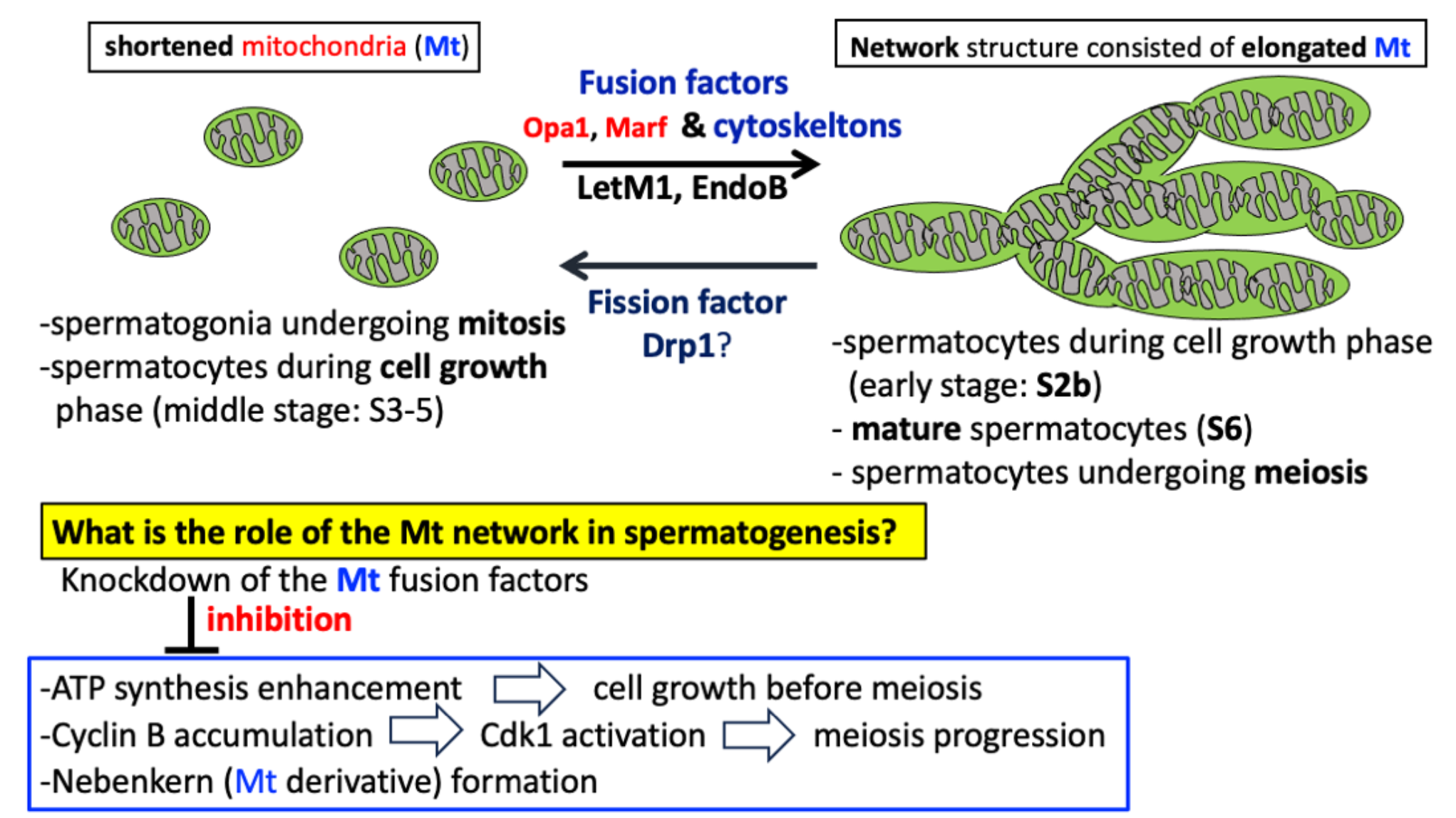
Mitochondria are dynamic organelles that undergo repeated fusion and fission. We studied how the distribution and shape of mitochondria change during Drosophila spermatogenesis and whether factors that regulate their dynamics are necessary for these changes. Unlike the shortened mitochondria seen in mitosis, an interconnected network of elongated mitochondria forms before meiosis and is maintained during meiotic divisions. Mitochondria are evenly divided into daughter cells, relying on microtubules and F-actin. To explore the role of mitochondrial network structure in cell growth and meiosis, we depleted mitochondrial fusion factors, Opa1 and Marf, as well as the morphology proteins Letm1 and EndoB, in spermatocytes. This knockdown led to inhibited cell growth and failed meiosis. As a result, the spermatocytes differentiated into spermatids without completing meiosis. The knockdown also inhibited the cytoplasmic and nuclear accumulation of Cyclin B before meiosis, and Cdk1 was not fully activated at the onset of meiosis. Notably, ectopic overexpression of Cyclin B partially rescued the failure of meiosis. Many spermatids from spermatocytes with the knockdowns contained multiple smaller nuclei and abnormally shaped Nebenkerns. These findings suggest that mitochondrial network structure, maintained by fusion and morphology factors, is essential for meiosis progression and Nebenkern formation in Drosophila spermatogenesis.
Keywords:
1. Introduction
2. Results
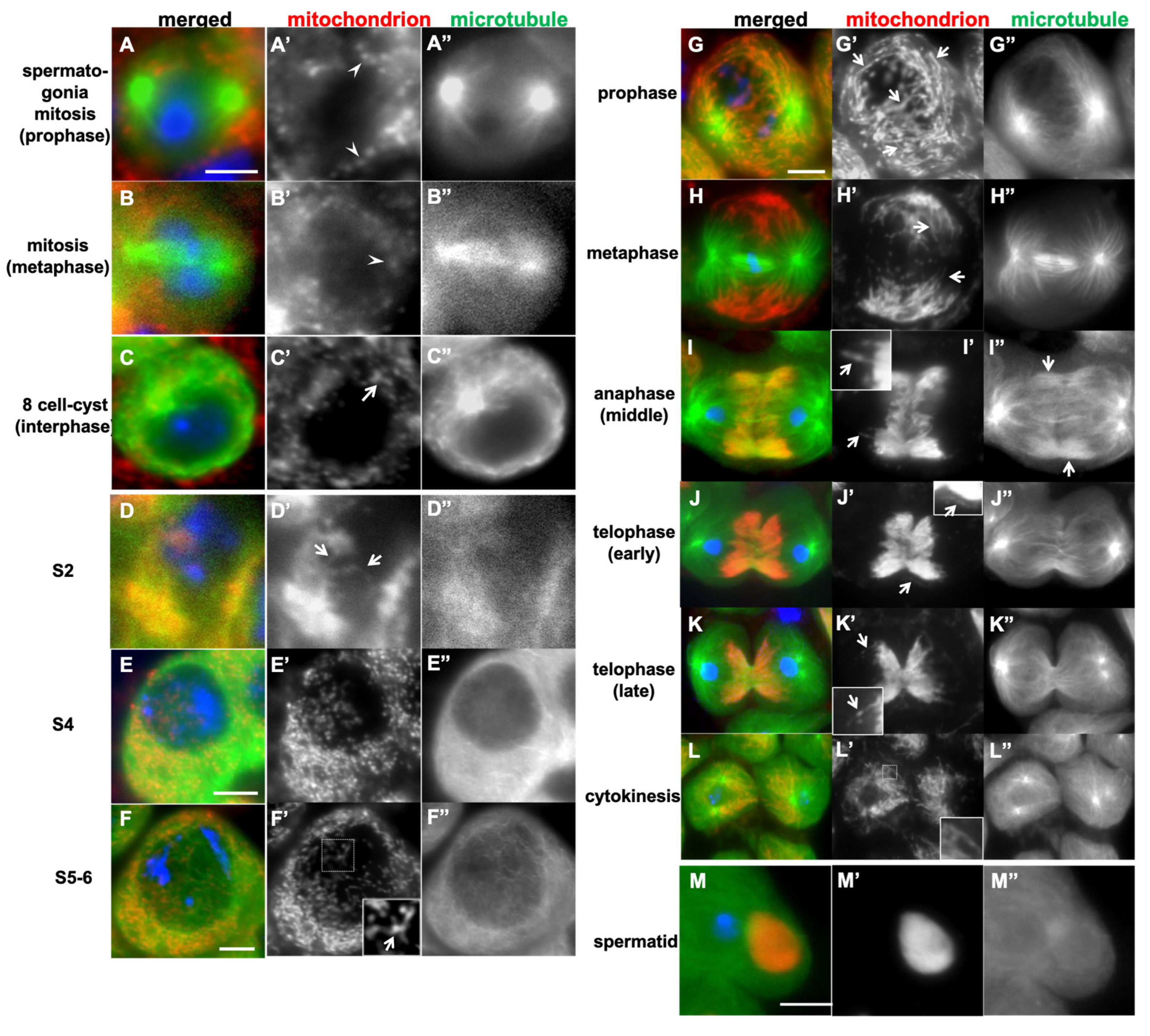
2.1. Differences in Mitochondrial Morphology and Distribution in Spermatogonia That Proliferate via Mitosis, Spermatocytes Before and During Male Meiosis in Drosophila
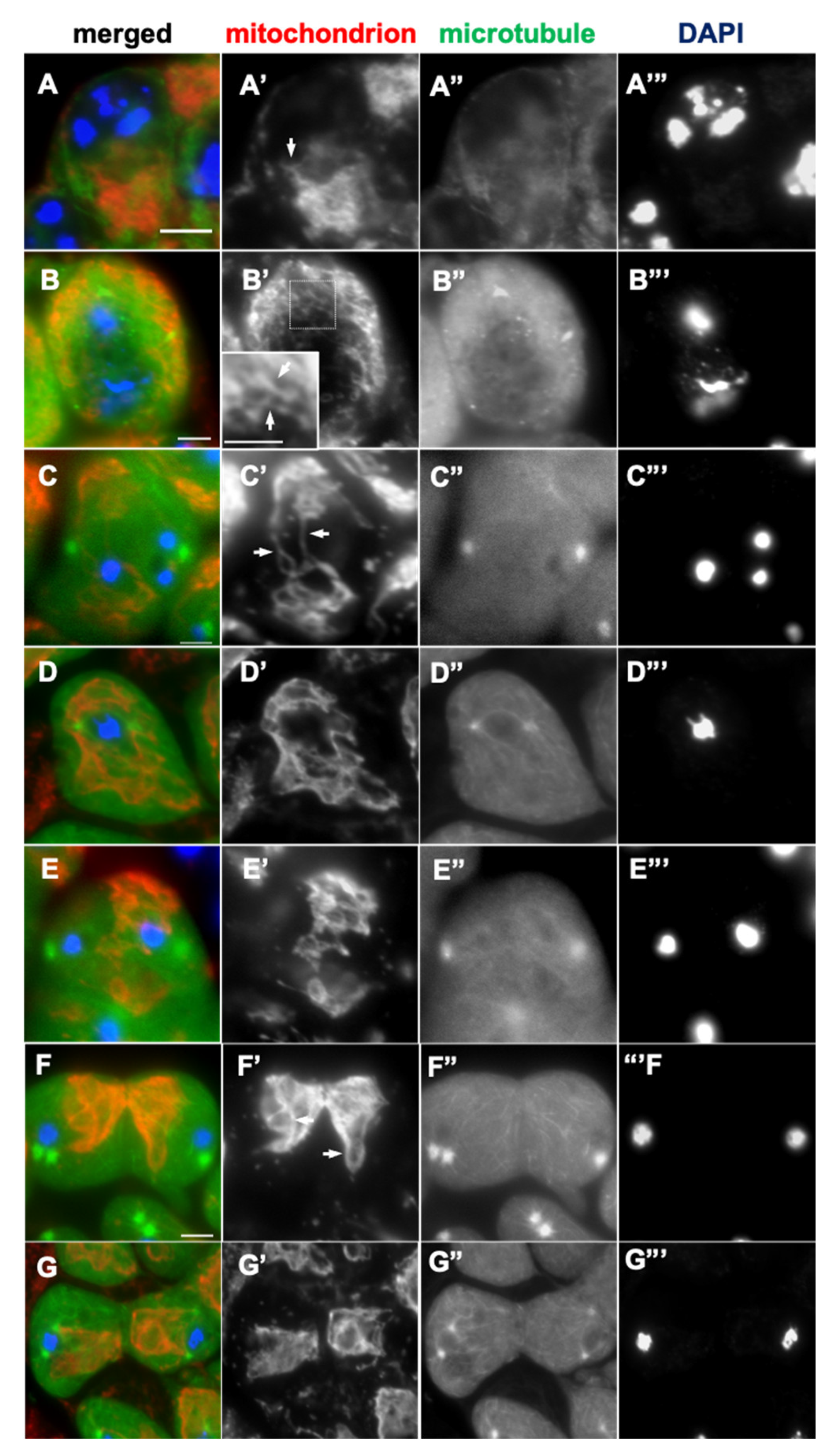
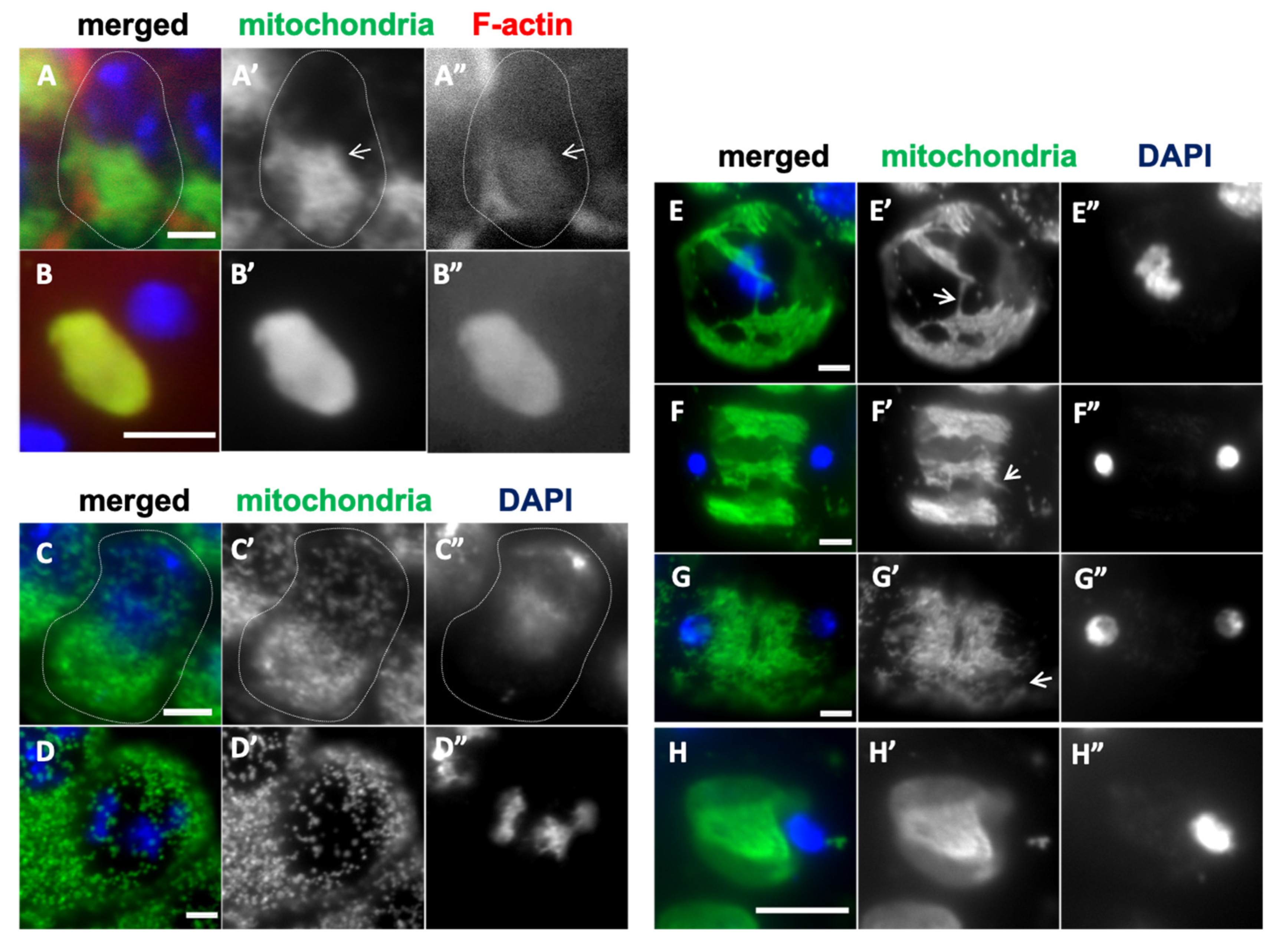
2.2. The Formation of Mitochondrial Network Structures and Their Subcellular Distribution Were Perturbed by the Inhibition of Microtubules and F-Actin
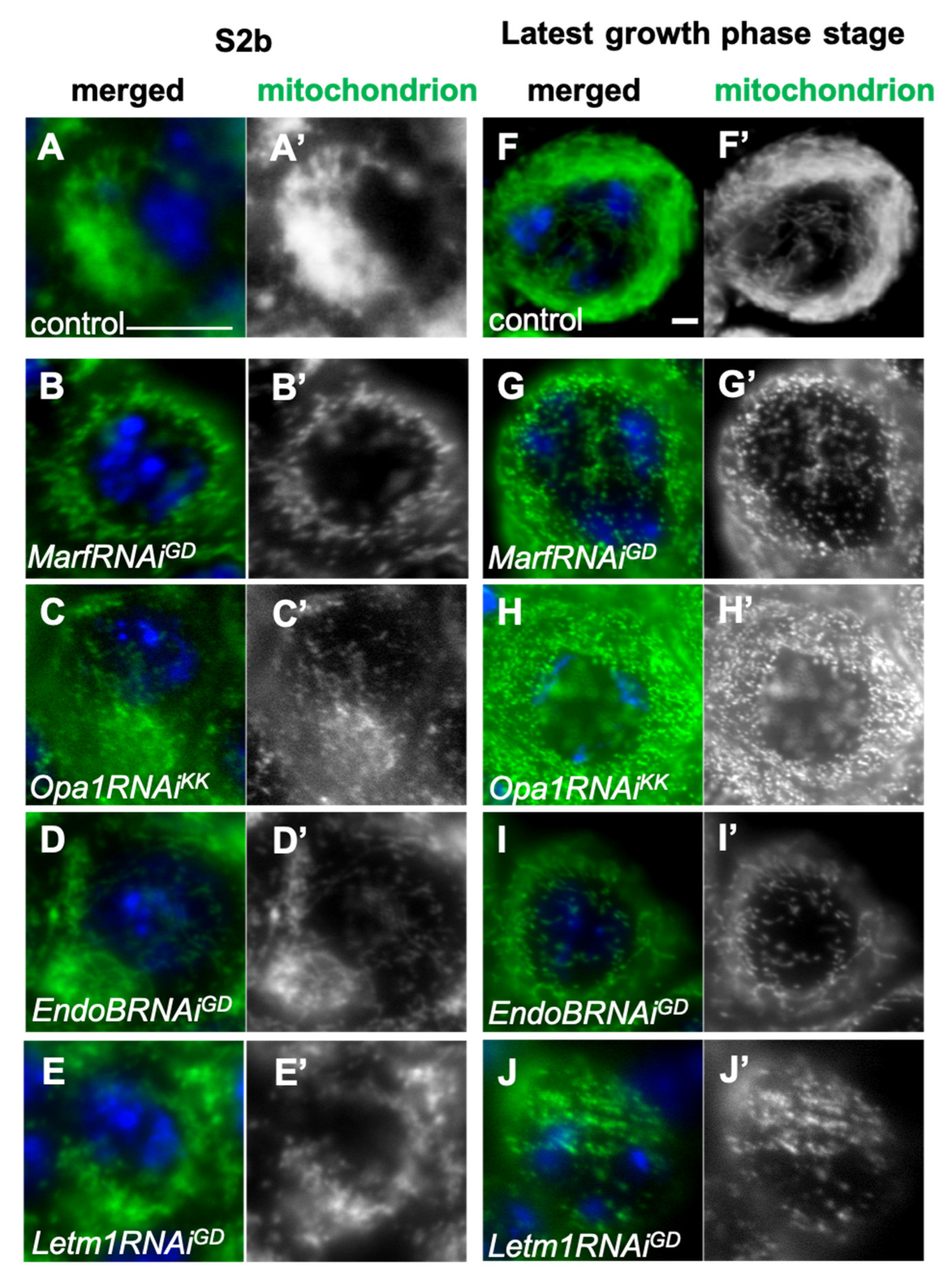
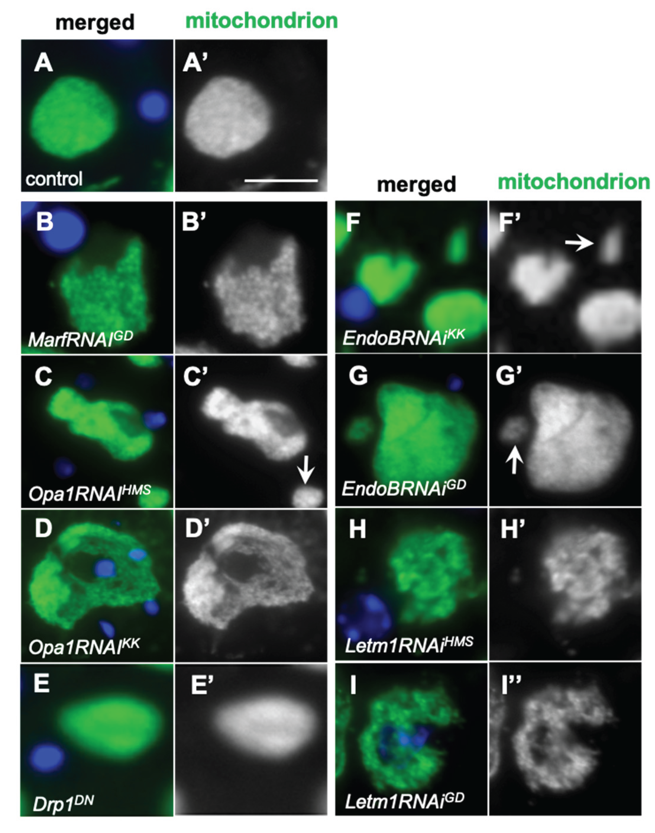
2.3. Formation of the Elongated Mitochondria Was Inhibited by the Knockdown of the Fusion Factors and the Mitochondrial Morphology Proteins
2.4. Knockdown of the Fusion Factors and the Morphology Proteins Inhibited ATP Synthesis and Cell Growth in the Spermatocytes Before Meiosis
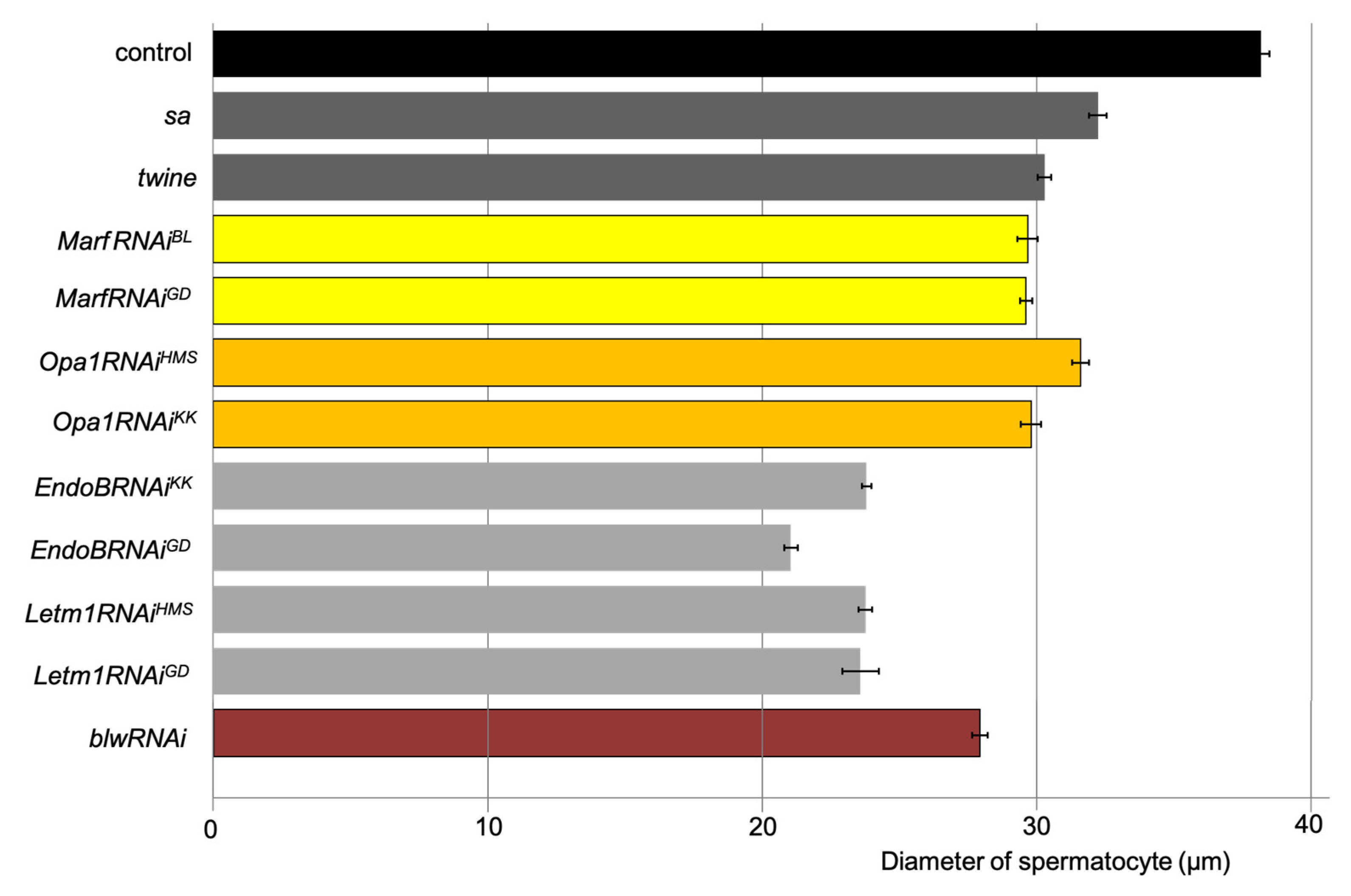
2.5. The Cell Growth Before Male Meiosis Was Affected in the Spermatocytes with the Knockdown of Mitochondrial Fusion Factors and the Morphology Proteins
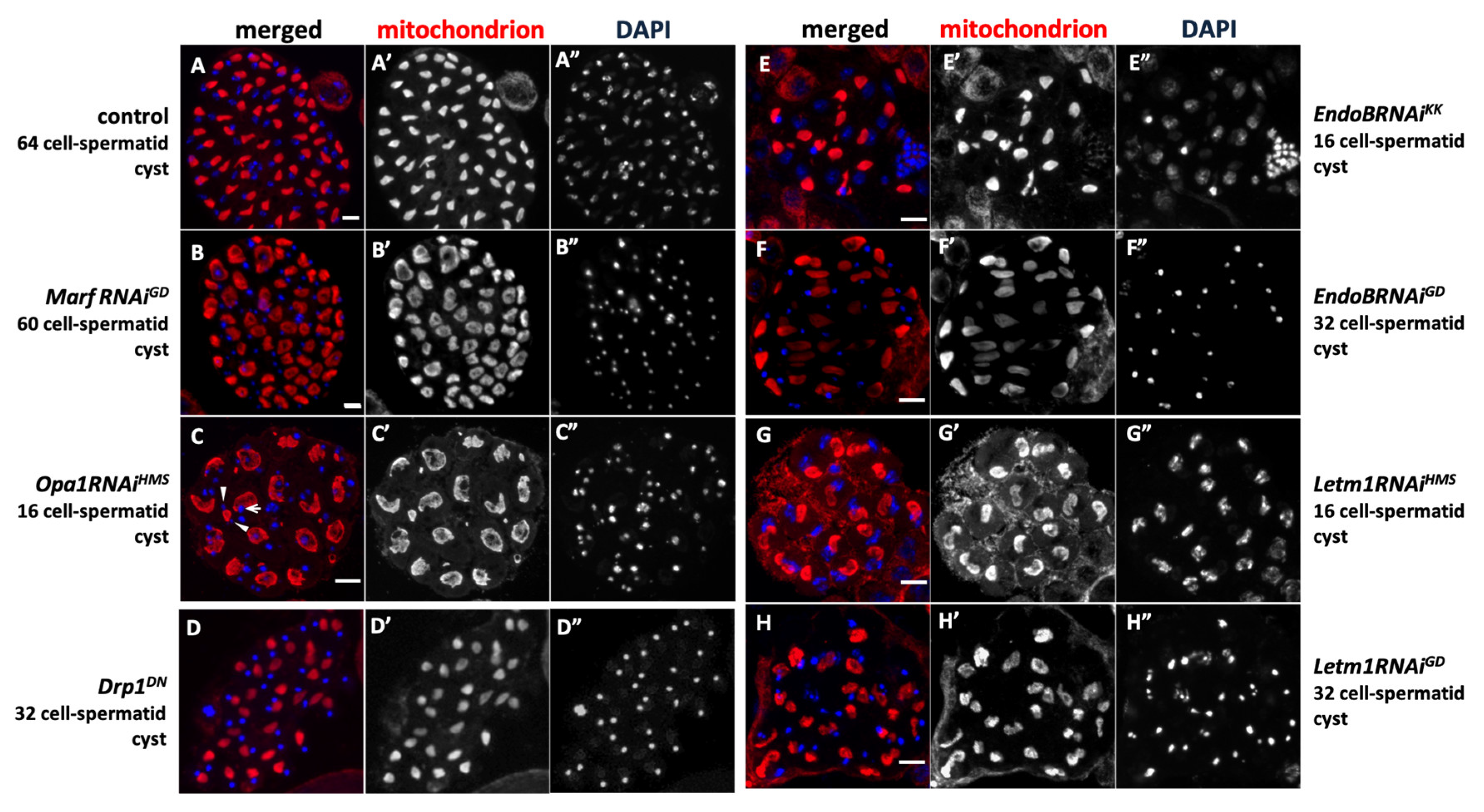
2.6. The Phenotype Observed in Spermatids when Chromosome Separation During Meiosis Is Abnormal Was Caused by Knockdown of Mitochondrial Fusion and Fission Factors, and Morphogenesis Proteins
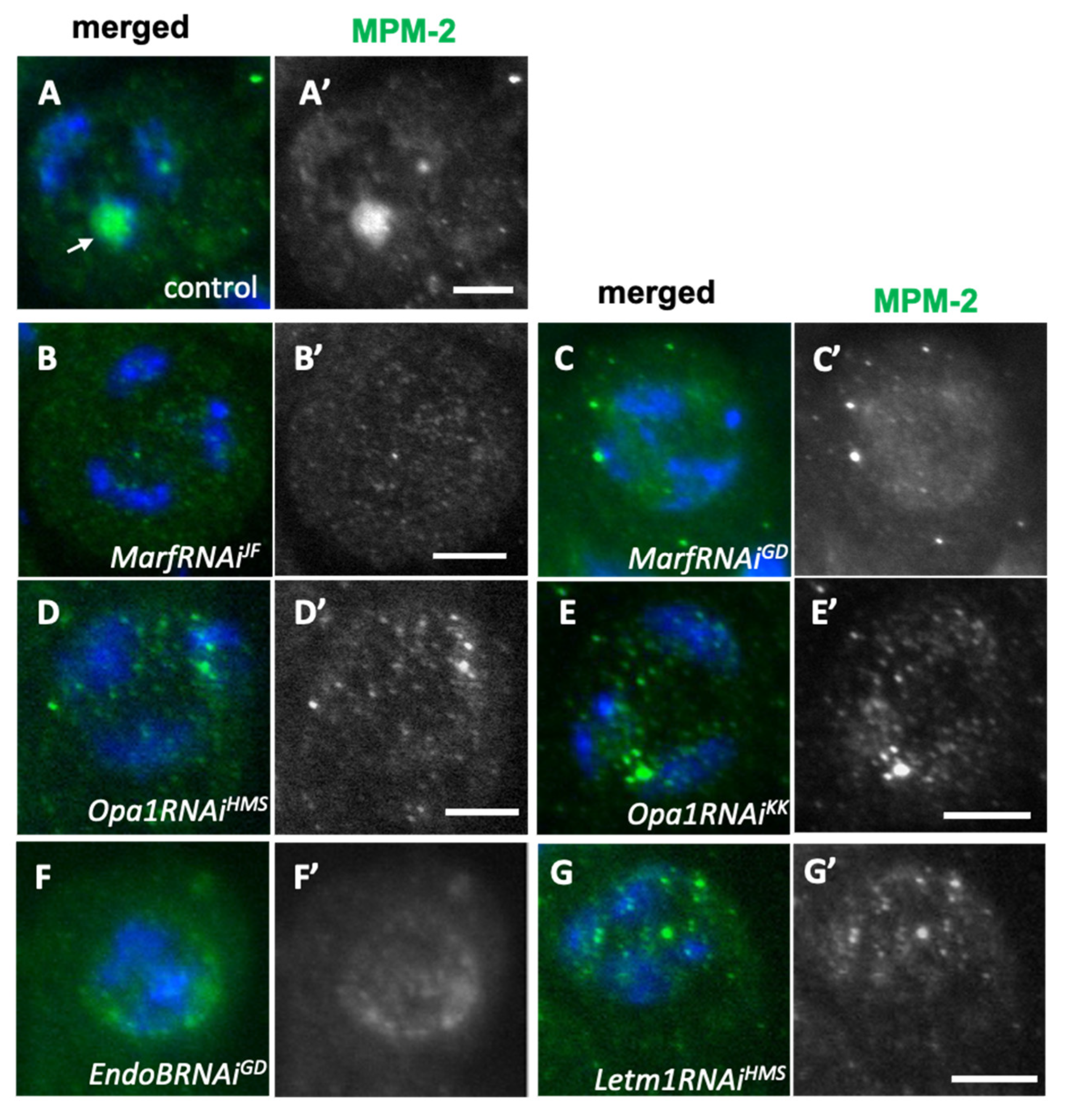
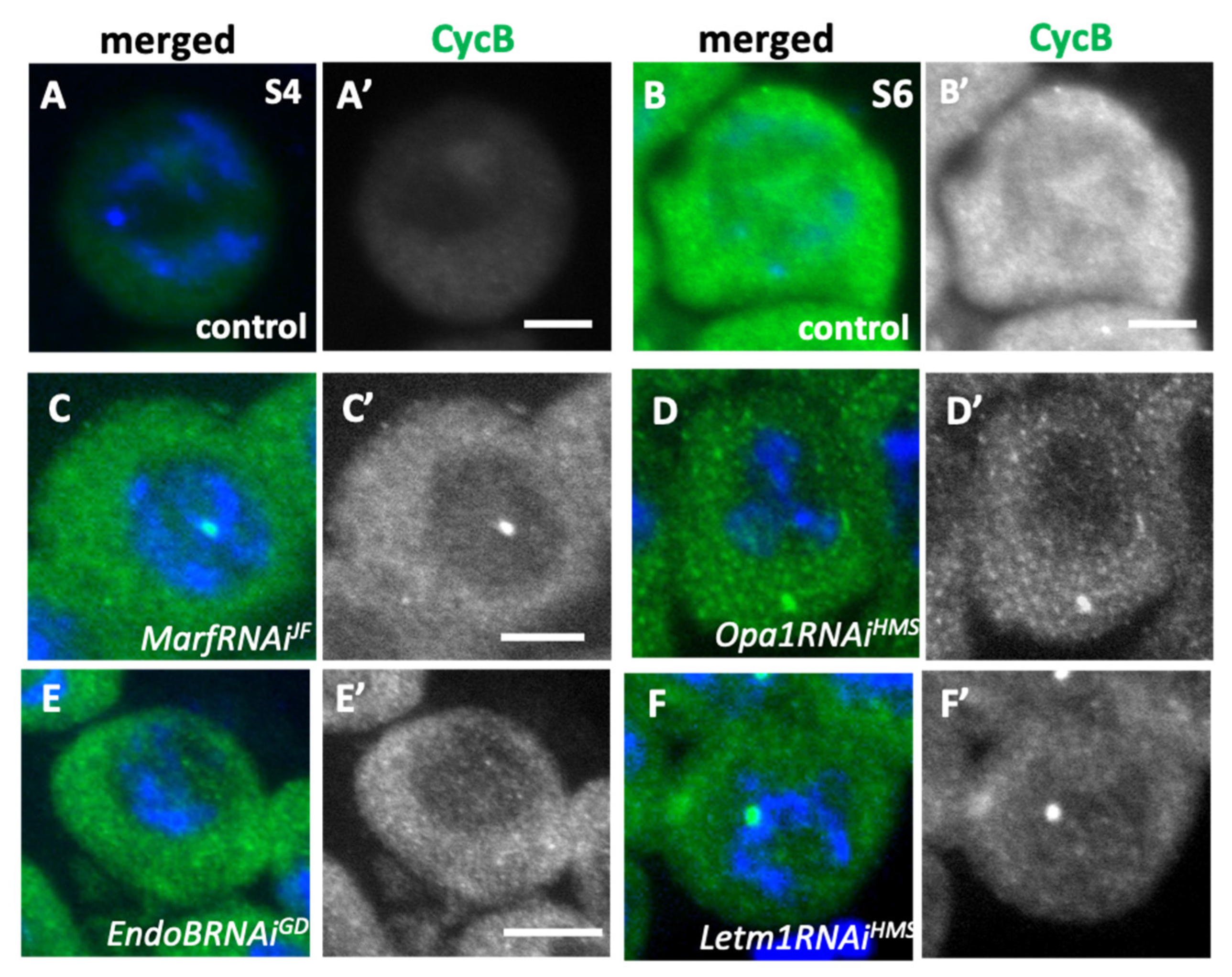
2.7. Knockdown of Mitochondrial Fusion Factors Inhibited Cdk1 Activation in the Primary Spermatocytes Before the Onset of Meiosis
2.8. Inhibition of Meiotic Initiation Caused by Knockdown of Mitochondrial Fusion Factors and the Morphology Proteins Was Partially Rescued by Ectopic Overexpression of Cyclin B
2.9. Knockdown of Mitochondrial Fusion and Fission Factors Caused Abnormalities in Nebenkern Formation in Early Spermatids After the Second Meiosis
3. Discussion
3.1. Mitochondria in Drosophila Spermatocytes Undergo Stage-Specific Changes Between a Shortened Form and an Interconnected Network Structure
3.2. Requirement of Fusion Factors for the Formation of the Mitochondrial Network, and Microtubules and F-Actin for Their Distribution Before and During Male Meiosis
3.3. Requirement of the Mitochondrial Network Formed via the Fusion Factors for the Cell Growth of Spermatocytes Before Meiosis
3.4. Elongated Mitochondria Networks Are Transferred to Daughter Cells While Maintaining the Structures, Depending on Microtubules and F-Actin in Male Meiosis
3.5. The Establishment of the Mitochondrial Network Structure, Formed via Fusion Factors and Morphology Proteins, Plays Important Roles in the Execution of Male Meiosis
4. Materials and Methods
4.1. Drosophila Stocks
4.2. Preparation of Post-Meiotic Spermatid Cysts
4.3. Drug Administration to the Testis Cells
4.4. Immunostaining of Testis Cells
4.5. ATP Assay
4.6. Transmission Electron Microscope Observation of Adult Testes
4.7. Statistical Analysis
Supplementary Materials
Author Contributions
Funding
Institutional Review Board Statement
Informed Consent Statement
Data Availability Statement
Acknowledgments
Conflicts of Interest
References
- Aldridge, A.C.; Benson, L.P.; Siegenthaler, M.M.; Whigham, B.T.; Stowers, R.S.; Hales, K.G. Roles for Drp1, a dynamin-related protein, and milton, a kinesin-associated protein, in mitochondrial segregation, unfurling and elongation during Drosophila spermatogenesis. Fly (Austin) 2007, 1, 38–46. [Google Scholar] [CrossRef]
- Azuma, M.; Ogata, T.; Yamazoe, K.; Tanaka, Y.; Inoue, Y.H. Heat shock cognate 70 genes contribute to Drosophila spermatocyte growth progression possibly through the insulin signaling pathway. Dev. Growth Differ. 2021, 63, 231–248. [Google Scholar] [CrossRef] [PubMed]
- Baker, C.C.; Gim, B.S.; Fuller, M.T. Cell type-specific translational repression of Cyclin B during meiosis in males. Development. 2015, 142, 3394–402. [Google Scholar] [CrossRef]
- Benard, G.; Bellance, N.; James, D.; Parrone, P.; Fernandez, H.; Letellier, T.; Rossignol, R. Mitochondrial bioenergetics and structural network organization. J. Cell. Sci. 2007, 120, 838–848. [Google Scholar] [CrossRef]
- Bereiter-Hahn, J.; Vöth, M. Dynamics of mitochondria in living cells: shape changes, dislocations, fusion, and fission of mitochondria. Microsc. Res. Tech. 1994, 27, 198–219. [Google Scholar] [CrossRef]
- Cenci, G.; Bonaccorsi, S.; Pisano, C.; Verni, F.; Gatti, M. Chromatin and microtubule organization during premeiotic, meiotic and early postmeiotic stages of Drosophila melanogaster spermatogenesis. J. Cell. Sci. 1994, 107, 3521–3534. [Google Scholar] [CrossRef]
- Chen, H.; Detmer, S.A.; Ewald, A.J.; Griffin, E.E.; Fraser, S.E.; Chan, D.C. Mitofusins Mfn1 and Mfn2 coordinately regulate mitochondrial fusion and are essential for embryonic development. J Cell Biol, 2003; 160, 189–200. [Google Scholar]
- Chen, H.; Vermulst, M.; Wang, Y.E.; Chomyn, A.; Prolla, T.A.; McCaffery, J.M.; Chan, D.C. Mitochondrial fusion is required for mtDNA stability in skeletal muscle and tolerance of mtDNA mutations. Cell 2010, 141, 280–289. [Google Scholar] [CrossRef]
- Cho, M.J.; Kim, Y.J.; Yu, W.D.; Kim, Y.S.; Lee, J.H. Microtubule Integrity Is Associated with the Functional Activity of Mitochondria in HEK293. Cells. 2021, 10, 3600. [Google Scholar] [CrossRef]
- Chung, J.Y.; Steen, J.A.; Schwarz, T.L. Phosphorylation-Induced Motor Shedding Is Required at Mitosis for Proper Distribution and Passive Inheritance of Mitochondria. Cell Rep. 2016, 16, 2142–2155. [Google Scholar] [CrossRef] [PubMed]
- Davies, V.J.; Hollins, A.J.; Piechota, M.J.; Yip, W.; Davies, J.R.; White, K.E.; Nicols, P.P.; Boulton, M.E.; Votruba, M. Opa1 deficiency in a mouse model of autosomal dominant optic atrophy impairs mitochondrial morphology, optic nerve structure and visual function. Hum. Mol. Genet. 2007, 16, 1307–1318. [Google Scholar] [CrossRef] [PubMed]
- Deng, H.; Dodson, M.W.; Huang, H.; Guo, M. The Parkinson’s disease genes pink1 and parkin promote mitochondrial fission and/or inhibit fusion in Drosophila. Proc. Natl. Acad. Sci. U.S.A. 2008, 105, 14503–14508. [Google Scholar] [CrossRef]
- Eura, Y.; Ishihara, N.; Yokota, S.; Mihara, K. Two Mitofusin proteins, mammalian homologues of FZO, with distinct functions are both required for mitochondrial fusion. J. Biochem. 2003, 134, 333–344. [Google Scholar] [CrossRef] [PubMed]
- Fuller, M.T. (1993). Spermatogenesis. In The development of Drosophila melanogaster. Bate M., Martinez Arias, A., Eds.; Cold Spring Harbor Laboratory Press, NewYork, USA, 1993, pp. 71–147.
- Hales, K.G.; Fuller, M.T. Developmentally regulated mitochondrial fusion mediated by a conserved, novel, predicted GTPase. Cell 1997, 90, 121–129. [Google Scholar] [CrossRef]
- Hayashi, D.; Tanabe, K.; Katsube, H.; Inoue, Y.H. B-type nuclear lamin and the nuclear pore complex Nup107-160 influences maintenance of the spindle envelope required for cytokinesis in Drosophila male meiosis. Biol. Open 2016, 5, 1011–1021. [Google Scholar] [CrossRef]
- Hinton, A. Jr.; Katti, P.; Christensen, T.A.; Mungai, M. , Shao, J.; Zhang, L.; Trushin, S.; Alghanem A, Jaspersen A, Geroux RE, Neikirk K, Biete M, Lopez EG, Shao B, Vue Z, Vang L, Beasley HK, Marshall AG, Stephens D, Damo S, Ponce J, Bleck CKE, Hicsasmaz I, Murray SA, Edmonds RAC, Dajles A, Koo YD, Bacevac S, Salisbury JL, Pereira, R.O.; Glancy, B.; Trushina, E.; Abel, E.D. A Comprehensive Approach to Sample Preparation for Electron Microscopy and the Assessment of Mitochondrial Morphology in Tissue and Cultured Cells. Adv. Biol. (Weinh), 2023; 7, e2200202. [Google Scholar]
- Hirusaki, K.; Yokoyama, K.; Cho, K.; Ohta, Y. Temporal depolarization of mitochondria during M phase. Sci. Rep. 2017, 7, 16044. [Google Scholar] [CrossRef]
- Ichihara, K.; Shimizu, H.; Taguchi, O.; Yamaguchi, M.; Inoue, Y.H. A Drosophila orthologue of Larp protein family is required for multiple processes in male meiosis. Cell Struct. Funct. 2007, 32, 89–100. [Google Scholar] [CrossRef] [PubMed]
- Inoue, Y.H.; Savoian, M.S.; Suzuki, T. : Máthé, E.; Yamamoto, M.T.; Glover, D.M. Mutations in orbit/mast reveal that the central spindle is comprised of two microtubule populations, those that initiate cleavage and those that propagate furrow ingression. J. Cell Biol. 2004; 166, 49–60. [Google Scholar]
- Inoue, Y. H.; Miyauchi, C.; Ogata, T.; Kitazawa, D. Dynamic alteration of cellular component of male meiosis in Drosophila. In Meiosis, A. Swan, Eds.; Intech Open, 2012; pp. 67–86.
- Inoki, K.; Zhu, T.; Guan, K.-L. TSC2 Mediates cellular energy response to control cell growth and survival. Cell 2003, 115, 577–590. [Google Scholar] [CrossRef] [PubMed]
- Ishihara, N.; Jofuku, A.; Eura, Y.; Mihara, K. Regulation of mitochondrial morphology by membrane potential, and DRP1-dependent division and FZO1-dependent fusion reaction in mammalian cells. Biochem. Biophys. Res. Commun. 2003, 301, 891–898. [Google Scholar] [CrossRef]
- Ishihara, N.; Nomura, M.; Jofuku, A.; Kato, H.; Suzuki, S.O.; Masuda, K.; Otera, H.; Nakanishi, Y.; Nonaka, I.; Goto, Y. Mitochondrial fission factor Drp1 is essential for embryonic development and synapse formation in mice. Nat. Cell Biol. 2009, 11, 958–966. [Google Scholar] [CrossRef]
- Jourdain, I.; Gachet, Y.; Hyams, J.S. The dynamin related protein Dnm1 fragments mitochondria in a microtubule-dependent manner during the fission yeast cell cycle. Cell Motil. Cytoskeleton. 2009, 66, 509–23. [Google Scholar] [CrossRef]
- Jones, M.D.; Naylor, K. Simple to Complex: The Role of Actin and Microtubules in Mitochondrial Dynamics in Amoeba, Yeast, and Mammalian Cells. Int. J. Mol. Sci. 2022, 23, 9402. [Google Scholar] [CrossRef] [PubMed]
- Karbowski, M.; Jeong, S.-Y.; Youle, R.J. Endophilin B1 is required for the maintenance of mitochondrial morphology. J. Cell Biol. 2004, 166, 1027–1039. [Google Scholar] [CrossRef]
- Kitazawa, D.; Yamaguchi, M.; Mori, H.; Inoue, Y.H. COPI-mediated membrane trafficking is required for cytokinesis in Drosophila male meiotic divisions. J Cell Sci. 2012, 125, 3649–60. [Google Scholar] [CrossRef]
- Labbé, K.; Murley, A.; Nunnari, J. Determinants and functions of mitochondrial behavior. Annu. Rev. Cell Dev. Biol. 2014, 30, 357–391. [Google Scholar] [CrossRef]
- Lawrence, E.J.; Boucher, E.; Mandato, C.A. Mitochondria-cytoskeleton associations in mammalian cytokinesis. Cell Div. 2016; 11, 3. [Google Scholar]
- Li, S.; Wu, Z.; Li, Y.; Tantray, I.; De Stefani, D.; Mattarei, A.; Krishnan, G.; Gao, F.B.; Vogel, H.; Lu, B. Altered MICOS Morphology and Mitochondrial Ion Homeostasis Contribute to Poly(GR) Toxicity Associated with C9-ALS/FTD. Cell Rep. 2020, 32, 107989. [Google Scholar] [CrossRef]
- Liesa, M.; Palacín, M.; Zorzano, A. Mitochondrial dynamics in mammalian health and disease. Physiol. Rev. 2009, 89, 799–845. [Google Scholar] [CrossRef] [PubMed]
- Lin, T.Y.; Viswanathan, S.; Wood, C.; Wilson, P.G.; Wolf, N.; Fuller, M.T. Coordinate developmental control of the meiotic cell cycle and spermatid differentiation in Drosophila males. Development 1996, 122, 1331–1341. [Google Scholar] [CrossRef] [PubMed]
- Lunova, M.; Jirsa, M.; Dejneka, A.; Sullivan, G.J.; Lunov, O. Mechanical regulation of mitochondrial morphodynamics in cancer cells by extracellular microenvironment. Biomater. Biosyst. 2024, 14, 100093. [Google Scholar] [CrossRef]
- Madan, S.; Uttekar, B.; Chowdhary, S.; Rikhy, R. Mitochondria Lead the Way: Mitochondrial Dynamics and Function in Cellular Movements in Development and Disease. Front. Cell Dev. Biol. 2022, 9, 781933. [Google Scholar] [CrossRef]
- Mattenberger, Y.; James, D.I.; Martinou, J.-C. Fusion of mitochondria in mammalian cells is dependent on the mitochondrial inner membrane potential and independent of microtubules or actin. FEBS. Letters 2003, 538, 53–59. [Google Scholar] [CrossRef]
- McQuibban, G.A.; Lee, J.R.; Zheng, L.; Juusola, M.; Freeman, M. Normal mitochondrial dynamics requires rhomboid-7 and affects Drosophila lifespan and neuronal function. Curr. Biol. 2006, 16, 982–989. [Google Scholar] [CrossRef]
- Mitra, K.; Rikhy, R.; Lilly, M.; Lippincott-Schwartz, J. DRP1-dependent mitochondrial fission initiates follicle cell differentiation during Drosophila oogenesis. J. Cell Biol. 2012, 197, 487–497. [Google Scholar] [CrossRef]
- Nakamura, S.; Matsui, A.; Akabane, S.; et al. The mitochondrial inner membrane protein LETM1 modulates cristae organization through its LETM domain. Commun. Biol. 2020, 3, 99. [Google Scholar] [CrossRef] [PubMed]
- Oka, S.; Hirai, J.; Yasukawa, T.; Nakahara, Y.; Inoue, Y.H. A correlation of reactive oxygen species accumulation by depletion of superoxide dismutases with age-dependent impairment in the nervous system and muscles of Drosophila adults. Biogerontol. 2015, 16, 485–501. [Google Scholar] [CrossRef] [PubMed]
- Okamoto, K.; Shaw, J.M. Mitochondrial morphology and dynamics in yeast and multicellular eukaryotes. Annu. Rev. Genet. 2005, 39, 503–536. [Google Scholar] [CrossRef] [PubMed]
- Okazaki, R.; Yamazoe, K.; Inoue, Y.H. Nuclear Export of Cyclin B Mediated by the Nup62 Complex Is Required for Meiotic Initiation in Drosophila Males. Cells 2020, 9, 270. [Google Scholar] [CrossRef]
- Ozaki, M.; Le, T.D.; Inoue, Y.H. Downregulating mitochondrial DNA polymerase γ in the muscle stimulated autophagy, apoptosis, and muscle aging-related phenotypes in Drosophila adults. Biomolecules 2022, 12, 1105. [Google Scholar] [CrossRef]
- Park, J.; Lee, G.; Chung, J. The PINK1-Parkin pathway is involved in the regulation of mitochondrial remodeling process. Biochem. Biophys. Res. Commun. 2009, 37, 518–523. [Google Scholar] [CrossRef]
- Parone, P.A.; Da Cruz, S.; Tondera, D.; Mattenberger, Y.; James, D.I.; Maechler, P.; Barja, F.; Martinou, J.-C. Preventing mitochondrial fission impairs mitochondrial function and leads to loss of mitochondrial DNA. PLoS ONE 2008, 3, e3257. [Google Scholar] [CrossRef]
- Poole, A.C.; Thomas, R.E.; Yu, S.; Vincow, E.S.; Pallanck, L. The mitochondrial fusion-promoting factor mitofusin is a substrate of the PINK1/parkin pathway. PLoS ONE 2010, 5, e10054. [Google Scholar] [CrossRef]
- Ratnaparkhi, A. Signaling by Folded gastrulation is modulated by mitochondrial fusion and fission. J. Cell Sci. 2013, 126, 5369–5376. [Google Scholar] [CrossRef] [PubMed]
- Sauvanet, C.; Duvezin-Caubet, S.; di Rago, J.-P.; Rojo, M. Energetic requirements and bioenergetic modulation of mitochondrial morphology and dynamics. Semin. Cell Dev. Biol. 2010, 21, 558–565. [Google Scholar] [CrossRef] [PubMed]
- Sawyer, E.M.; Brunner, E.C. , Hwang, Y.; Ivey, L,E,; Brown, O.; Bannon, M.; Akrobetu, D.; Sheaffer, K.E.; Morgan, O.; Field, C.O.; Suresh, N.; Gordon, M.G.; Gunnell, E.T.; Regruto, L.A.; Wood, C.G.; Fuller, M.T.; Hales, K.G. Testis-specific ATP synthase peripheral stalk subunits required for tissue-specific mitochondrial morphogenesis in Drosophila. BMC Cell Biol. 2017, 18, 6. [Google Scholar]
- Schnorrer, F.; Schönbauer, C.; Langer, C.C.; Dietzl, G.; Novatchkova, M.; Schernhuber, K.; Fellner, M.; Azaryan, A.; Radolf, M.; Stark, A.; Keleman, K.; Dickson, B.J. Systematic genetic analysis of muscle morphogenesis and function in Drosophila. Nature 2010, 464, 287–291. [Google Scholar] [CrossRef]
- Sênos Demarco, R.; Uyemura, B.S.; D’Alterio, C.; Jones, D.L. Mitochondrial fusion regulates lipid homeostasis and stem cell maintenance in the Drosophila testis. Nat. Cell Biol. 2019, 21, 710–720. [Google Scholar] [CrossRef] [PubMed]
- Stephan, T.; Ilgen, P.; Jakobs, S. Visualizing mitochondrial dynamics at the nanoscale. Light Sci. Appl. 2024, 13, 244. [Google Scholar] [CrossRef]
- Taguchi, N.; Ishihara, N.; Jofuku, A.; Oka, T.; Mihara, K. Mitotic phosphorylation of dynamin-related GTPase Drp1 participates in mitochondrial fission. J. Biol. Chem. 2007, 282, 11521–11529. [Google Scholar] [CrossRef]
- Tanabe, K.; Okazaki, R.; Kaizuka, K.; Inoue, Y.H. Time-lapse Observation of Chromosomes, Cytoskeletons and Cell Organelles during Male Meiotic Divisions in Drosophila. Bio Protoc. 2017, 7, e2225. [Google Scholar] [CrossRef]
- Tanabe, K.; Awane, R.; Shoda, T.; Yamazoe, K.; Inoue, Y.H. Mutations in mxc tumor-suppressor gene induce chromosome instability in Drosophila male meiosis. Cell Struct. Funct. 2019, 44, 121–135. [Google Scholar] [CrossRef]
- Trevisan, T.; Pendin, D.; Montagna, A.; Bova, S.; Ghelli, A.M.; Daga, A. Manipulation of Mitochondria Dynamics Reveals Separate Roles for Form and Function in Mitochondria Distribution. Cell Rep. 2018, 23, 1742–1753. [Google Scholar] [CrossRef]
- Ueishi, S.; Shimizu, H.; Inoue, Y.H. Male germline stem cell division and spermatocyte growth require insulin signaling in Drosophila. Cell Struct. Funct. 2009, 34, 61–69. [Google Scholar] [CrossRef]
- Udagawa, O.; Ishihara, T.; Maeda, M.; Matsunaga, Y.; Tsukamoto, S.; Kawano, N.; Miyado, K.; Shitara, H.; Yokota, S.; Nomura, M.; et al. Mitochondrial fission factor Drp1 maintains oocyte quality via dynamic rearrangement of multiple organelles. Curr. Biol. 2014, 24, 2451–2458. [Google Scholar] [CrossRef] [PubMed]
- Vedelek, V.; Jankovics, F.; Zádori, J.; Sinka, R. Mitochondrial Differentiation during Spermatogenesis: Lessons from Drosophila melanogaster. Int. J. Mol. Sci. 2024, 25, 3980. [Google Scholar] [CrossRef] [PubMed]
- Wakabayashi, J.; Zhang, Z.; Wakabayashi, N.; Tamura, Y.; Fukaya, M.; Kensler, T.W.; Iijima, M.; Sesaki, H. The dynamin-related GTPase Drp1 is required for embryonic and brain development in mice. J. Cell Biol. 2009, 186, 805–816. [Google Scholar] [CrossRef] [PubMed]
- Westermann, B. Molecular machinery of mitochondrial fusion and fission. J. Biol. Chem. 2008, 283, 13501–13505. [Google Scholar] [CrossRef]
- White-Cooper, H.; Alphey, L.; Glover, D.M. The cdc25 homologue twine is required for only some aspects of the entry into meiosis in Drosophila. J Cell Sci. 1993, 106, 1035–1044. [Google Scholar] [CrossRef]
- Wong, E.D.; Wagner, J.A.; Gorsich, S.W.; McCaffery, J.M.; Shaw, J.M.; Nunnari, J. The Dynamin-related GTPase, Mgm1p, is an intermembrane space protein required for maintenance of fusion competent mitochondria. J. Cell Biol. 2000, 151, 341–352. [Google Scholar] [CrossRef]
- Yamazoe, K.; Inoue, Y.H. Cyclin B Export to the Cytoplasm via the Nup62 Subcomplex and Subsequent Rapid Nuclear Import Are Required for the Initiation of Drosophila Male Meiosis. Cells. 2023, 12, 2611. [Google Scholar] [CrossRef]
- Yang, Y.; Hou, L.; Li, Y.; Ni, J.; Liu, L. Neuronal necrosis and spreading death in a Drosophila genetic model. Cell Death Dis. 2013, 4, e723. [Google Scholar] [CrossRef]
- Zhao, J. , Lendahl, U., and Nistér, M. Regulation of mitochondrial dynamics: convergences and divergences between yeast and vertebrates. Cell. Mol. Life Sci. 2013; 70, 951–976. [Google Scholar]
- Zhu, J.Y.; Vereshchagina, N.; Sreekumar, V.; Burbulla, L.F.; Costa, A.C.; Daub, K.J.; Woitalla, D.; Martins, L.M.; Krüger, R.; Rasse, T.M. Knockdown of Hsc70-5/mortalin Induces Loss of Synaptic Mitochondria in a Drosophila Parkinson’s Disease Model. PLoS ONE 2013, 8, e83714. [Google Scholar] [CrossRef]

| Knockdown & Dominant negative exp. | 16 cell- cysts* |
17-31 cell- cysts |
32 cell- cysts |
33-63 cell- cysts |
64 cell- cysts(normal) |
|---|---|---|---|---|---|
| control | 0(0) | 0(0) | 0(0) | 0(0) | 68(100) |
| MarfRNAiJF | 0(0) | 0(0) | 0(0) | 0(0) | 50(100) |
| MarfRNAiGD | 0(0) | 0(0) | 0(0) | 8(20.0) | 32(80.0) |
| Opa1RNAiHMS | 3(3.2) | 0(0) | 2(2.2) | 0(0) | 88(94.6) |
| Opa1RNAiKK | 97(84.3) | 16(15.7) | 0(0) | 0(0) | 0(0) |
| Drp1RNAiJF | 0(0) | 0(0) | 0(0) | 0(0) | 59(100) |
| Drp1DN | 0(0) | 0(0) | 0(0) | 30(58.8) | 21(41.2) |
| EndoBRNAiKK | 54(100) | 0(0) | 0(0) | 0(0) | 0(0) |
| EndoBRNAiGD | 42(82.4) | 0(0) | 9(17.6) | 0(0) | 0(0) |
| Letm1RNAiHMS | 40(100) | 0(0) | 0(0) | 0(0) | 0(0) |
| Letm1RNAiGD | 42(84.0) | 0(0) | 8(16.0) | 0(0) | 0(0) |
| Knockdown | n | nuclear numbers in a spermatid (%) | macro/ micro nuclei (% cells) |
||||||
|---|---|---|---|---|---|---|---|---|---|
| normal | abnormal | ||||||||
| 1 | 0 | 2 | 3 | 4 | 5> | Total (%) |
|||
| control | 1,290 | 99.5 | 0.4 | 0.1 | 0 | 0 | 0 | 0.5 | 0 |
| Opa1RNAiHMS | 1,713 | 27.1 | 7 | 24.3 | 15.8 | 12.3 | 13.4 | 73.9 | 23.5 |
| Opa1RNAiKK | 952 | 70 | 29.9 | 12.2 | 11.6 | 4.6 | 1.5 | 30 | 32.4 |
| Knockdown & ectopic expression | 16 cell- cysts (%)*1 |
17~31 cell -cysts |
32 cell -cysts*2 |
33-63 cell -cysts |
64 cell -cysts*3 |
Total cysts |
|---|---|---|---|---|---|---|
| Opa1RNAiKK, mCherry | 91(84.3) | 16(14.8) | 1(0.9) | 0(0) | 0(0) | 108 |
| Opa1RNAiKK, CycB | 67(51.9) | 7(5.4) | 33(25.6) | 4(3.1) | 18(14.0) | 129 |
| EendoBRNAiKK, mCherry | 67(62.6) | 26(24.3) | 3(2.8) | 11(10.3) | 0(0) | 107 |
| EndoBRNAiKK, CycB | 8(7.6) | 17(16.2) | 11(10.5) | 40(38.1) | 29(27.6) | 105 |
| CycB | 0(0) | 0(0) | 0(0) | 0(0) | 106(100) | 106 |
Disclaimer/Publisher’s Note: The statements, opinions and data contained in all publications are solely those of the individual author(s) and contributor(s) and not of MDPI and/or the editor(s). MDPI and/or the editor(s) disclaim responsibility for any injury to people or property resulting from any ideas, methods, instructions or products referred to in the content. |
© 2025 by the authors. Licensee MDPI, Basel, Switzerland. This article is an open access article distributed under the terms and conditions of the Creative Commons Attribution (CC BY) license (http://creativecommons.org/licenses/by/4.0/).


